Embed Size (px)
Citation preview
REVIEWARTICLE
Cytoplasmic vacuolization during exposure to drugsand other substances
Toshihiko Aki & Akina Nara & Koichi Uemura
Received: 8 October 2011 /Accepted: 19 January 2012 /Published online: 7 March 2012# Springer Science+Business Media B.V. 2012
Abstract Cytoplasmic vacuolization upon exposureto a variety of chemicals and bioactive substanceshas been extensively reported. Nearly 30 years havepassed since the description by Nobel Laureate Chris-tian de Duve of the mechanism underlying the lyso-somal accumulation of lipophilic weak bases referredto these substances as lysosomotropic agents. It hasnow been revealed, however, that vacuolization occursupon exposure to compounds other than lipophilicweak bases. Vacuolization of organelles/vesicles otherthan acidic compartments has also now been reported.In this mini-review, we provide an overview of theorigin, mechanism, and possible outcomes of cellularvacuolization during exposure to substances with lyso-somotropic as well as other properties.
Keywords Autophagy . Endocytosis . ER stress .
Lysosome . Lysosomotropicity . Vacuolization
Introduction
Cellular vacuolization is a frequently observed phe-nomenon upon exposure to pharmaceutical agents andother chemicals and has been extensively reported in
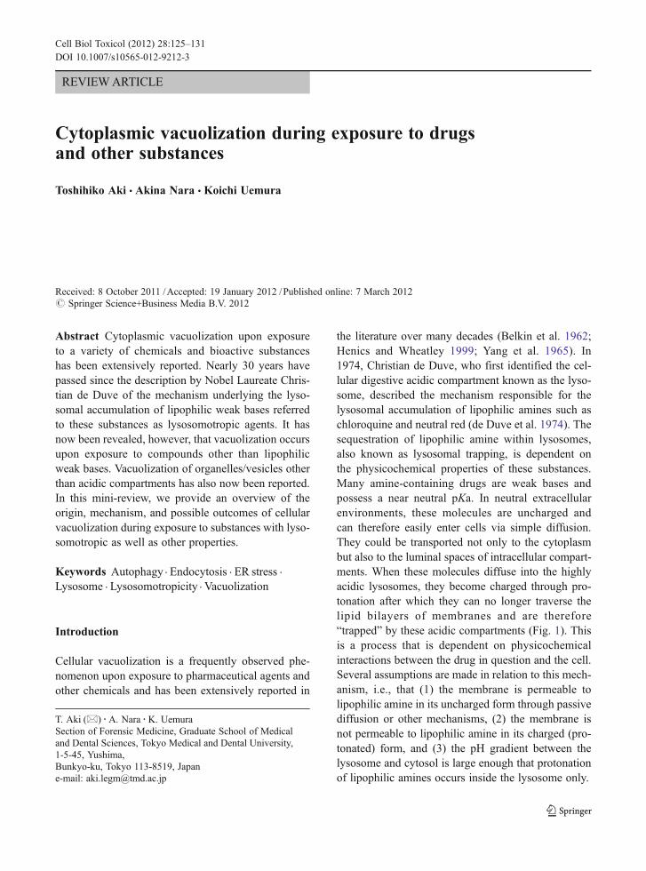
the literature over many decades (Belkin et al. 1962;Henics and Wheatley 1999; Yang et al. 1965). In1974, Christian de Duve, who first identified the cel-lular digestive acidic compartment known as the lyso-some, described the mechanism responsible for thelysosomal accumulation of lipophilic amines such aschloroquine and neutral red (de Duve et al. 1974). Thesequestration of lipophilic amine within lysosomes,also known as lysosomal trapping, is dependent onthe physicochemical properties of these substances.Many amine-containing drugs are weak bases andpossess a near neutral pKa. In neutral extracellularenvironments, these molecules are uncharged andcan therefore easily enter cells via simple diffusion.They could be transported not only to the cytoplasmbut also to the luminal spaces of intracellular compart-ments. When these molecules diffuse into the highlyacidic lysosomes, they become charged through pro-tonation after which they can no longer traverse thelipid bilayers of membranes and are therefore“trapped” by these acidic compartments (Fig. 1). Thisis a process that is dependent on physicochemicalinteractions between the drug in question and the cell.Several assumptions are made in relation to this mech-anism, i.e., that (1) the membrane is permeable tolipophilic amine in its uncharged form through passivediffusion or other mechanisms, (2) the membrane isnot permeable to lipophilic amine in its charged (pro-tonated) form, and (3) the pH gradient between thelysosome and cytosol is large enough that protonationof lipophilic amines occurs inside the lysosome only.
Cell Biol Toxicol (2012) 28:125–131DOI 10.1007/s10565-012-9212-3
T. Aki (*) :A. Nara :K. UemuraSection of Forensic Medicine, Graduate School of Medicaland Dental Sciences, Tokyo Medical and Dental University,1-5-45, Yushima,Bunkyo-ku, Tokyo 113-8519, Japane-mail: [email protected]
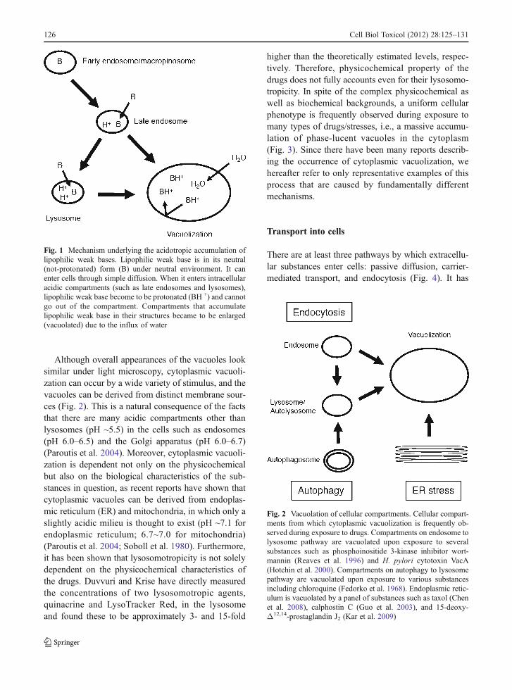
Although overall appearances of the vacuoles looksimilar under light microscopy, cytoplasmic vacuoli-zation can occur by a wide variety of stimulus, and thevacuoles can be derived from distinct membrane sour-ces (Fig. 2). This is a natural consequence of the factsthat there are many acidic compartments other thanlysosomes (pH ~5.5) in the cells such as endosomes(pH 6.0–6.5) and the Golgi apparatus (pH 6.0–6.7)(Paroutis et al. 2004). Moreover, cytoplasmic vacuoli-zation is dependent not only on the physicochemicalbut also on the biological characteristics of the sub-stances in question, as recent reports have shown thatcytoplasmic vacuoles can be derived from endoplas-mic reticulum (ER) and mitochondria, in which only aslightly acidic milieu is thought to exist (pH ~7.1 forendoplasmic reticulum; 6.7~7.0 for mitochondria)(Paroutis et al. 2004; Soboll et al. 1980). Furthermore,it has been shown that lysosomotropicity is not solelydependent on the physicochemical characteristics ofthe drugs. Duvvuri and Krise have directly measuredthe concentrations of two lysosomotropic agents,quinacrine and LysoTracker Red, in the lysosomeand found these to be approximately 3- and 15-fold
higher than the theoretically estimated levels, respec-tively. Therefore, physicochemical property of thedrugs does not fully accounts even for their lysosomo-tropicity. In spite of the complex physicochemical aswell as biochemical backgrounds, a uniform cellularphenotype is frequently observed during exposure tomany types of drugs/stresses, i.e., a massive accumu-lation of phase-lucent vacuoles in the cytoplasm(Fig. 3). Since there have been many reports describ-ing the occurrence of cytoplasmic vacuolization, wehereafter refer to only representative examples of thisprocess that are caused by fundamentally differentmechanisms.
Transport into cells
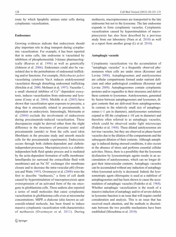
There are at least three pathways by which extracellu-lar substances enter cells: passive diffusion, carrier-mediated transport, and endocytosis (Fig. 4). It has
Fig. 1 Mechanism underlying the acidotropic accumulation oflipophilic weak bases. Lipophilic weak base is in its neutral(not-protonated) form (B) under neutral environment. It canenter cells through simple diffusion. When it enters intracellularacidic compartments (such as late endosomes and lysosomes),lipophilic weak base become to be protonated (BH +) and cannotgo out of the compartment. Compartments that accumulatelipophilic weak base in their structures became to be enlarged(vacuolated) due to the influx of water
Fig. 2 Vacuolation of cellular compartments. Cellular compart-ments from which cytoplasmic vacuolization is frequently ob-served during exposure to drugs. Compartments on endosome tolysosome pathway are vacuolated upon exposure to severalsubstances such as phosphoinositide 3-kinase inhibitor wort-mannin (Reaves et al. 1996) and H. pylori cytotoxin VacA(Hotchin et al. 2000). Compartments on autophagy to lysosomepathway are vacuolated upon exposure to various substancesincluding chloroquine (Fedorko et al. 1968). Endoplasmic retic-ulum is vacuolated by a panel of substances such as taxol (Chenet al. 2008), calphostin C (Guo et al. 2003), and 15-deoxy-Δ12,14-prostaglandin J2 (Kar et al. 2009)
126 Cell Biol Toxicol (2012) 28:125–131
been shown that organic cation transporter (OCT), amember of the SLC family, plays major role in thedelivery of organic amines into cells (Koepsell et al.2007). Carrier-mediated processes would be expectedto play important roles in the transport of organicamines into the cells when these agents are added atconcentrations around and below the Km value of thecarrier systems. However, this might not be case inmany of the examples of cytoplasmic vacuolization
induced by organic amines. Cytoplasmic vacuoliza-tion by these compounds generally occurs at severalhundred micromolar or even several millimolar doses(Ohkuma and Poole 1981). These concentrationswould typically be several orders of magnitude higherthan the Km of the carriers, suggesting a relativelylower contribution of carrier-mediated transport oforganic amines to cytoplasmic vacuolization. In addi-tion, cytoplasmic vacuolization by organic amines isgenerally abolished when the pH of the medium islowered (Ohkuma and Poole 1981). Hence, most or-ganic amines should be transported into cells in anuncharged form and not in a charged form, duringcytoplasmic vacuolization. For example, Morissetteet al. (2008, 2004) have extensively reported on cel-lular vacuolization processes upon exposure to a panelof basic organic compounds including procainamide.These authors have shown that cytoplasmic vacuoli-zation is activated upon exposure to millimolar con-centrations of procainamide in rabbit smooth musclecells. Moreover, this induction of vacuolization byprocainamide was not suppressed by any of the inhib-itors of membrane cation transporters tested, includingthat of OCT (Morissette et al. 2008). It was furthershown in this study that procainamide-induced vacuo-lization requires vacuolar proton ATPase, since aninhibitor of this enzyme bafilomycin A1 effectivelyinhibited this process. Procainamide is transported intocells through a “pseudo transport” mechanism, whichinvolves translocation of the drug in its unchargedform, followed by trapping within the cellular acidiccompartment on which vacuolar proton ATPaseresides (Morissette et al. 2008). Passive diffusionand/or endocytosis would be regarded as the major
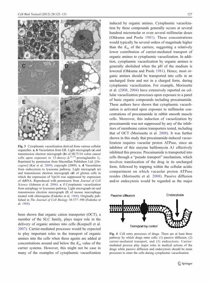
Fig. 3 Cytoplasmic vacuolization derived from various cellularorganelles. a, b Vacuolation from ER. Light micrograph (a) andtransmission electron micrograph (b) of HCT116 colon cancercells upon exposure to 15-deoxy-Δ12,14-prostaglandin J2.Reprinted by permission from Macmillan Publishers Ltd: [On-cogene] (Kar et al. 2009), copyright (2009). c, d Vacuolationfrom endocytosis to lysosome pathway. Light micrograph (c)and transmission electron micrograph (d) of glioma cells inwhich the expression of Vps34 was suppressed by expressionof shRNA. Reproduced with permission from Journal of CellScience (Johnson et al. 2006). e, f Cytoplasmic vacuolizationfrom autophagy to lysosome pathway. Light micrograph (e) andtransmission electron micrograph (f) of mouse macrophagetreated with chloroquine (Fedorko et al. 1968). Originally pub-lished in The Journal of Cell Biology 38:377–390 (Fedorko etal. 1968)
Fig. 4 Cell entry processes of drugs. There are at least threepathway by which drugs enter cells: (1) passive diffusion, (2)carrier-mediated transport, and (3) endocytosis. Carrier-mediated process play major roles in medical actions of thedrugs while passive diffusion and endocytosis should be mainprocesses to enter the cells during cytoplasmic vacuolization
Cell Biol Toxicol (2012) 28:125–131 127
route by which lipophilic amines enter cells duringcytoplasmic vacuolization.
Endosomes
Growing evidences indicate that endocytosis shouldplay important role in drug transport during cytoplas-mic vacuolization. For example, it has been reportedthat in some cells, late endosome is vacuolated byinhibition of phosphoinositide 3-kinase pharmacolog-ically (Reaves et al. 1996) as well as genetically(Johnson et al. 2006). Endosomes could also be vac-uolated due to the perturbations of endosomal traffick-ing and/or functions. For example, Helicobacter pylorivacuolating cytotoxin VacA induces endolysosomalvacuolation through disturbing endosomal trafficking(Hotchin et al. 2000; Molinari et al. 1997). Vacuolin-1,a small chemical inhibitor of Ca2+-dependent exocy-tosis, induces vacuolization from endosome and lyso-some (Cerny et al. 2004). Michlik et al. (2003) haveshown that vacuolization upon exposure to procaine, adrug that is structurally related to procainamide, isdependent on endocytosis. Interestingly, Morissette etal. (2004) exclude the involvement of endocytosisduring procainamide-induced vacuolization. Thesediscrepancies might be derived either from the slightdifference in the structures of procaine (ester) andprocainamide (amide) or from the cells used (skinfibroblasts in the procaine study and smooth musclecells for the procainamide experiments). Endocytosisoccurs through both clathrin-dependent and clathrin-independent processes. Macropinocytosis is a clathrin-independent bulk fluid uptake process and is mediatedby the actin-dependent formation of ruffle membrane/lamellipodia (to surround the extracellular fluid withmembrane) and an Na+/H+ exchanger (for membraneclosure and to decrease the intra-vesicular pH) (Swan-son and Watts 1995). Overmeyer et al. (2008) were thefirst to describe “methuosis,” a form of cell deathcaused by hyperstimulation of macropinocytosis uponoverexpression of an activated form of the ras onco-gene in glioblastoma cells. These authors also reporteda series of small molecules that cause cytoplasmicvacuolization in glioblastoma cells even at micromolarconcentrations. MIPP, a chalcone (also known as cal-conoid)-related molecule, has been found to inducemassive cytoplasmic vacuolization with characteristicsof methuosis (Overmeyer et al. 2011). During
methuosis, macropinosomes are transported to the lateendosome but not to the lysosome. The late endosomeexpands to form cytoplasmic vacuoles. Cytoplasmicvacuolization caused by hyperstimulation of macro-pinocytosis has also been described by a previousstudy from our laboratory (Nara et al. 2010) as wellas a report from another group (Li et al. 2010).
Autophagic vacuole
Cytoplasmic vacuolization via the accumulation of“autophagic vacuoles” is a frequently observed phe-nomenon when cells are under stress (Kroemer andLevine 2008). Autophagosomes and autolysosomesare cellular compartments formed under nutrient defi-cient and other pathological conditions (Kroemer andLevine 2008). Autophagosomes contain cytoplasmicproteins and/or organelles in their structures and deliverthese contents to lysosomes. Autolysosomes, generatedby fusion between autophagosomes and lysosomes, di-gest contents that are delivered from autophagosomes.In contrast to the relatively small size of autophago-somes (~1 μm in diameter), autolysosomes sometimesexpand to fill the cytoplasm (~10 μm in diameter) andtherefore often referred to as autophagic vacuoles,which could be observed under light microscopy(Fedorko et al. 1968). These dilated autolysosomes arenot true vacuoles, but they are observed as phase-lucentvacuoles due to the dilation of the compartments and thesubsequent dilution of their contents. Although autoph-agy is induced during stressed conditions, it also occursin the absence of stress and performs essential cellularactivities. Hence, there is a possibility that the lysosomedysfunction by lysosomotropic agents results in an ac-cumulation of autolysosomes, which can no longer di-gest their intravesicular contents. Autophagic vacuolescan be accumulated without any induction of autophagywhen lysosomal activity is decreased. Indeed, the lyso-somotropic agent chloroquine is used as a stabilizer ofautophagosomes and has been shown to cause the accu-mulation of autophagic vacuoles (Fedorko et al. 1968).Whether autophagic vacuolization is the result of amassive induction of autophagy and/or of severe defectsin lysosome function is an issue that will require carefulconsideration and analysis. This is an issue that hasreceived much attention, and the methods to discrimi-nate between the two possible mechanisms have beenestablished (Mizushima et al. 2010).
128 Cell Biol Toxicol (2012) 28:125–131
Lysosome
Lysosomes are the major targets of lipophilic bases,and a previous study from Ohkuma and Poole (1981)has reported that many drugs, including chloroquine,neutral red, propranolol, atropine, and lidocaine, showthe tendency to induce lysosome accumulation. Thesesame authors further showed in additional studies thatthese lysosomotropic agents promote a substantial in-crease in the intra-lysosomal pH and thereby a dys-functional lysosome (Ohkuma and Poole 1978; Pooleand Ohkuma 1981). Lysosomal dysfunction caused bylysosomotropic drugs is of importance in relation tothe cytotoxicity of these agents and is discussed in alater section.
Endoplasmic reticulum
The ER is involved in protein quality control through-out their life cycle from synthesis to degradation. Inaddition to autophagy, the ubiquitin–proteasome sys-tem is another major degradative process in the cellsthat is mainly carried out in the ER. ER stress, whichoften triggers apoptosis, sometimes causes cytoplas-mic vacuolization. The vacuolation of the ER pro-ceeds through a mechanism that is essentially distinctfrom that of the lysosome or endosome. Enlargement/dilation of the ER, which results in the occupation ofthe cytoplasm by massive vacuoles, is often observedfollowing treatments with proteasome inhibitors suchas lactacystin and MG132 (Ding et al. 2007; Ustundaget al. 2007; Wagenknecht et al. 2000). Mimnaugh et al.(2004, 2006) and Ustundag et al. (2007) have ob-served massive cytoplasmic vacuolization in theSV40-transformed monkey kidney cell line COS-7upon exposure of these cells to the anticancer drugsgeldanamycin and velcade (bortezomib). Geldanamy-cin is a well-known inhibitor of the chaperone proteinHSP90 (Taldone et al. 2008) while velcade is aninhibitor of the proteasome (Chauhan et al. 2005).When cells are exposed to these compounds, cytoplas-mic vacuolation occurs even at the concentration of50 nM geldanamycin plus 10 nM velcade (Mimnaughet al. 2006). It was further shown in this report thatthese cytoplasmic vacuoles are derived from the ER asthey incorporated red fluorescent protein harboring theKDEL ER localization sequence (Mimnaugh et al.2006). Recently, Kar et al. (2009) have observed that
a thiol-reactive cyclopentenone prostaglandin, 15-deoxy-Δ12,14-prostaglandin J2, causes ER-derived cy-toplasmic vacuolization in some cancer cells. All ofthe cytoplasmic vacuolization, ER stress, and celldeath events caused by this molecule were markedlyinhibited by a reactive oxygen species scavenger (Karet al. 2009). Taxol, an anticancer drug, also has beenshown to cause ER-derived vacuolization when addedat relatively high concentrations (e.g., 70 μM) com-pared with the dose used to induce mitotic arrest (lessthan 100 nM) (Chen et al. 2008). Interestingly, cal-phostin C, a well-known inhibitor for protein kinase C,induces ER-derived vacuolation and subsequent deathof the cells that are resistant to pacilitaxel (taxol) (Guoet al. 2003; Kaul and Maltese 2009), indicating itspossible usefulness for cancer therapy. Cytoplasmicvacuolization derived from enlarged/dilated ER uponexposure to these drugs could be rationally interpretedas a consequence of excessive ER stress. Althoughthese vacuolization processes are completely differentfrom that induced by lysosomotropic agents, theycould not be distinguished by light microscopic obser-vations. Electron microscopic observations, immuno-cytochemical analyses, and/or f luorescencemicroscopic observations with appropriate organellemarkers are required to elucidate the origin of thesevacuoles.
Cytotoxic implications of vacuolization
One of the most significant unsolved questions inrelation to cytoplasmic vacuolization is its role incytotoxicity, particularly cell death. There is limitedinformation on this issue at present. The origin, un-derlying mechanism, and consequences of cytoplas-mic vacuolization vary depending on the nature of thedrugs to which the cells have been exposed as well asthe cell types in which the vacuoles expand. Cytoplas-mic vacuolization may be a cell autonomous processthat is designed to protect the cell against toxins.Vacuoles are often cleared after the removal of drugsfrom the culture medium, suggesting that vacuoliza-tion is a reversible process and might be involved inthe isolation and buffering of toxins (Henics andWheatley 1999). However, prolonged exposure totoxic agents and the duration of the resultant cytoplas-mic vacuolization might cause irreversible cellularinjuries that ultimately lead to cell death (Henics and
Cell Biol Toxicol (2012) 28:125–131 129
Wheatley 1999). In support of this possibility,Ohkuma and Poole (1978) have reported that pro-longed vacuolation of the lysosome by organic basesresults in the release of these substances into cytosol intheir cationic forms, suggesting that a permeabilitytransition and/or rupture of the lysosomal membranehad occurred. During the last decade, the release oflysosomal enzymes to the cytoplasm due to an in-crease in the lysosomal membrane permeability hasbeen reported as one of the triggers of apoptotic celldeath (Kroemer and Jaattela 2005). In addition,methuosis, a form of cell death involving the accumu-lation of late endosomes in the cytoplasm, can beinduced by ectopic expression of an activated formof ras, with no exposure to chemicals (Overmeyer andMaltese 2011). During methuosis, cytoplasmic vacuo-lation persists for several days before the cells even-tually die (Overmeyer and Maltese 2011). This mightindicate that cytoplasmic vacuolation itself has toxiceffects in cells. Other than methuosis, cell deaths asso-ciated with cytoplasmic vacuolization are reported in-cluding oncosis (Trump et al. 1997), paraptosis(Sperandio et al. 2000), and necroptosis (Han et al.2007). During the execution of these processes, cyto-plasmic vacuolization from the compartments, such asER and Golgi, is observed along with the swelling ofmitochondria. However, there is a possibility that cyto-plasmic vacuolization is a process unrelated to cytotox-icity of the stimulants. This might be the case uponinfection of gastric epithelial cells with H. pylori. Al-though the H. pylori vacuolating cytotoxin VacA indu-ces both cytoplasmic vacuolization and mitochondrialapoptosis, recent reports indicate that the vacuolizationelicited by VacA is a process that is independent of itscytotoxicity (Willhite et al. 2003; Yamasaki et al. 2006).
Conclusions
A growing body of evidence now indicates that cyto-plasmic vacuolization is a phenomenon commonlyobserved during exposure to various clinically useddrugs as well as other substances. Vacuolation of thelysosome, endosome, autolysosome, and ER may sug-gest that almost all cellular compartments can be di-lated and vacuolated. Other than the dilation,cytoplasmic vacuolization could be derived from thehomotypic fusion of these compartments (Overmeyeret al. 2011). Although the origin and molecular
mechanisms underlying cytoplasmic vacuolizationhave been extensively studied to date, the pathophys-iological significance of cellular vacuolation has notbeen well characterized. The outcomes from cellularvacuolation are of some importance for a more com-prehensive understanding of the cytotoxicity of vari-ous pharmaceuticals that are both in current clinicaluse and still under development.
References
Belkin M, Hardy WG, Orr HC, Lachman AB. Induction in vitroby autonomic drugs of cytoplasmic vacuoles in ascitestumor cells. J Natl Cancer Inst. 1962;28:187–201.
Cerny J, Feng Y, Yu A, Miyake K, Borgonovo B, KlumpermanJ, et al. The small chemical vacuolin-1 inhibits Ca(2+)-dependent lysosomal exocytosis but not cell resealing.EMBO Rep. 2004;5:883–8.
Chauhan D, Hideshima T, Mitsiades C, Richardson P, AndersonKC. Proteasome inhibitor therapy in multiple myeloma.Mol Cancer Ther. 2005;4:686–92.
Chen TS, Wang XP, Sun L, Wang LX, Xing D, Mok M. Taxolinduces caspase-independent cytoplasmic vacuolizationand cell death through endoplasmic reticulum (ER) swell-ing in ASTC-a-1 cells. Cancer Lett. 2008;270:164–72.
de Duve C, de Barsy T, Poole B, Trouet A, Tulkens P, Van HoofF. Commentary. Lysosomotropic agents. Biochem Pharma-col. 1974;23:2495–531.
Ding WX, Ni HM, Yin XM. Absence of Bax switched MG132-induced apoptosis to non-apoptotic cell death that could besuppressed by transcriptional or translational inhibition.Apoptosis. 2007;12:2233–44.
Fedorko ME, Hirsch JG, Cohn ZA. Autophagic vacuoles pro-duced in vitro. I. Studies on cultured macrophages exposedto chloroquine. J Cell Biol. 1968;38:377–91.
Guo B, Hembruff SL, Villeneuve DJ, Kirwan AF, ParissentiAM. Potent killing of paclitaxel- and doxorubicin-resistant breast cancer cells by calphostin C accompaniedby cytoplasmic vacuolization. Breast Cancer Res Treat.2003;82:125–41.
Han W, Li L, Qiu S, Lu Q, Pan Q, Gu Y, et al. Shikonincircumvents cancer drug resistance by induction of a nec-roptotic death. Mol Cancer Ther. 2007;6:1641–9.
Henics T, Wheatley DN. Cytoplasmic vacuolation, adaptationand cell death: a view on new perspectives and features.Biol Cell. 1999;91:485–98.
Hotchin NA, Cover TL, Akhtar N. Cell vacuolation induced bythe VacA cytotoxin of Helicobacter pylori is regulated bythe Rac1 GTPase. J Biol Chem. 2000;275:14009–12.
Johnson EE, Overmeyer JH, Gunning WT, Maltese WA. Genesilencing reveals a specific function of hVps34 phosphati-dylinositol 3-kinase in late versus early endosomes. J CellSci. 2006;119:1219–32.
Kar R, Singha PK, Venkatachalam MA, Saikumar P. A novelrole for MAP1 LC3 in nonautophagic cytoplasmic vacuo-lation death of cancer cells. Oncogene. 2009;28:2556–68.
130 Cell Biol Toxicol (2012) 28:125–131
Kaul A, Maltese WA. Killing of cancer cells by the photoacti-vatable protein kinase C inhibitor, calphostin C, involvesinduction of endoplasmic reticulum stress. Neoplasia.2009;11:823–34.
Koepsell H, Lips K, Volk C. Polyspecific organic cation trans-porters: structure, function, physiological roles, and biophar-maceutical implications. Pharm Res. 2007;24:1227–51.
Kroemer G, Jaattela M. Lysosomes and autophagy in cell deathcontrol. Nat Rev Cancer. 2005;5:886–97.
Kroemer G, Levine B. Autophagic cell death: the story of amisnomer. Nat Rev Mol Cell Biol. 2008;9:1004–10.
Li C, Macdonald JI, Hryciw T, Meakin SO. Nerve growth factoractivation of the TrkA receptor induces cell death, bymacropinocytosis, in medulloblastoma Daoy cells. J Neu-rochem. 2010;112:882–99.
Michalik M, Pierzchalska M, Pabianczyk-Kulka A, KorohodaW. Procaine-induced enhancement of fluid-phase endocy-tosis and inhibition of exocytosis in human skin fibroblasts.Eur J Pharmacol. 2003;475:1–10.
Mimnaugh EG, Xu W, Vos M, Yuan X, Isaacs JS, Bisht KS, etal. Simultaneous inhibition of hsp 90 and the proteasomepromotes protein ubiquitination, causes endoplasmicreticulum-derived cytosolic vacuolization, and enhancesantitumor activity. Mol Cancer Ther. 2004;3:551–66.
Mimnaugh EG, Xu W, Vos M, Yuan X, Neckers L. Endoplasmicreticulum vacuolization and valosin-containing proteinrelocalization result from simultaneous hsp90 inhibitionby geldanamycin and proteasome inhibition by velcade.Mol Cancer Res. 2006;4:667–81.
Mizushima N, Yoshimori T, Levine B. Methods in mammalianautophagy research. Cell. 2010;140:313–26.
Molinari M, Galli C, Norais N, Telford JL, Rappuoli R, LuzioJP, et al. Vacuoles induced by Helicobacter pylori toxincontain both late endosomal and lysosomal markers. J BiolChem. 1997;272:25339–44.
Morissette G, Moreau E, Gaudreault RC, Marceau F. Massivecell vacuolization induced by organic amines such as pro-cainamide. J Pharmacol Exp Ther. 2004;310:395–406.
Morissette G, Lodge R, Marceau F. Intense pseudotransport of acationic drug mediated by vacuolar ATPase: procainamide-induced autophagic cell vacuolization. Toxicol Appl Phar-macol. 2008;228:364–77.
Nara A, Aki T, Funakoshi T, Uemura K. Methamphetamineinduces macropinocytosis in differentiated SH-SY5Y hu-man neuroblastoma cells. Brain Res. 2010;1352:1–10.
Ohkuma S, Poole B. Fluorescence probe measurement of the intra-lysosomal pH in living cells and the perturbation of pH byvarious agents. Proc Natl Acad Sci U S A. 1978;75:3327–31.
Ohkuma S, Poole B. Cytoplasmic vacuolation of mouse perito-neal macrophages and the uptake into lysosomes of weaklybasic substances. J Cell Biol. 1981;90:656–64.
Overmeyer JH, Maltese WA. Death pathways triggered by acti-vated Ras in cancer cells. Front Biosci. 2011;16:1693–713.
Overmeyer JH, Kaul A, Johnson EE, Maltese WA. Active rastriggers death in glioblastoma cells through hyperstimula-tion of macropinocytosis. Mol Cancer Res. 2008;6:965–77.
Overmeyer JH, Young AM, Bhanot H, Maltese WA. Achalcone-related small molecule that induces methuosis, anovel form of non-apoptotic cell death, in glioblastomacells. Mol Cancer. 2011;10:69.
Paroutis P, Touret N, Grinstein S. The pH of the secretorypathway: measurement, determinants, and regulation.Physiology (Bethesda). 2004;19:207–15.
Poole B, Ohkuma S. Effect of weak bases on the intralysosomalpH in mouse peritoneal macrophages. J Cell Biol.1981;90:665–9.
Reaves BJ, Bright NA, Mullock BM, Luzio JP. The effect ofwortmannin on the localisation of lysosomal type I integralmembrane glycoproteins suggests a role for phosphoinosi-tide 3-kinase activity in regulating membrane traffic late inthe endocytic pathway. J Cell Sci. 1996;109(Pt 4):749–62.
Soboll S, Elbers R, Scholz R, Heldt HW. Subcellular distribu-tion of di- and tricarboxylates and pH gradients in perfusedrat liver. Hoppe Seylers Z Physiol Chem. 1980;361:69–76.
Sperandio S, de Belle I, Bredesen DE. An alternative, non-apoptotic form of programmed cell death. Proc Natl AcadSci U S A. 2000;97:14376–81.
Swanson JA, Watts C. Macropinocytosis. Trends Cell Biol.1995;5:424–8.
Taldone T, Gozman A, Maharaj R, Chiosis G. Targeting Hsp90:small-molecule inhibitors and their clinical development.Curr Opin Pharmacol. 2008;8:370–4.
Trump BF, Berezesky IK, Chang SH, Phelps PC. The pathwaysof cell death: oncosis, apoptosis, and necrosis. ToxicolPathol. 1997;25:82–8.
Ustundag Y, Bronk SF, Gores GJ. Proteasome inhibition-induces endoplasmic reticulum dysfunction and cell deathof human cholangiocarcinoma cells. World J Gastroenterol.2007;13:851–7.
Wagenknecht B, Hermisson M, Groscurth P, Liston P, KrammerPH, Weller M. Proteasome inhibitor-induced apoptosis ofglioma cells involves the processing of multiple caspasesand cytochrome c release. J Neurochem. 2000;75:2288–97.
Willhite DC, Cover TL, Blanke SR. Cellular vacuolation andmitochondrial cytochrome c release are independent out-comes of Helicobacter pylori vacuolating cytotoxin activ-ity that are each dependent on membrane channelformation. J Biol Chem. 2003;278:48204–9.
Yamasaki E, Wada A, Kumatori A, Nakagawa I, Funao J,Nakayama M, et al.Helicobacter pylori vacuolating cytotox-in induces activation of the proapoptotic proteins Bax andBak, leading to cytochrome c release and cell death, inde-pendent of vacuolation. J Biol Chem. 2006;281:11250–9.
Yang WC, Strasser FF, Pomerat CM. Mechanism of drug-induced vacuolization in tissue culture. Exp Cell Res.1965;38:495–506.
Cell Biol Toxicol (2012) 28:125–131 131