Embed Size (px)
Citation preview
Vol. 14: 1-22. 1992 DISEASES OF AQUATIC ORGANISMS
Dis. aquat. Org. Published October 8
Cytopathology of spleen in eel Anguilla anguilla exposed to a chemical spill in the Rhine River
Eljalill Spazier, Volker Storch, Thomas Braunbeck*
Department of Zoology I, University of Heidelberg, Irn Neuenheimer Feld 230. W-6900 Heidelberg, Germany
ABSTRACT: Structural alterations in the spleen of eel Anguilla anguilla L., following exposure to a chemical spill in the Rhine h v e r in 1986, were investigated by means of light and electron microscopy. Relative abundance of reticulum and blood cells was quantified in comparison to controls. The data presented provide evidence of profound damage to the spleen and its cellular components. Histological and cytological modifications varied within a wide range from minor changes to cell death. Major cytological alterations of reticulum and blood cells included deviations in cytoplasmic density, loss of surface differentiations (cell junctions, pseudopodia), swelling of rnitochondria, formation of rnyelin-like membrane aggregations in the cytoplasm, mitochondria, lysosomes and nuclear membrane, as well as augmentation of lipid droplets. Severely damaged and necrotic cells were found either diffusely or focally distributed within the splenic reticulum. Melano-macrophages were strongly reduced in number and melano-macrophage centres were lacking. In contrast, considerable numbers of macrophages with intense lysosomal compartmentation, but free of pigments, were present. Spleens were further charac- terized by the absence of erythroblasts and plasma cells. Histological and cytological findings in the spleen of exposed eel indicate a severe impairment of the efficiency of the central splenic functions, in particular phagocytosis and cell-mediated immune response. Thus, chemical-induced patholopcal processes in the spleen have most likely been a contributing factor to the fatality of the chemical spill to the Rhine eel population. They are discussed in the context of earlier investigations on liver and intestine pathology of Rhine eel. Finally, cytological alterations of fish spleen are suggested as a potential biomarker for toxicant-induced damage in fish.
INTRODUCTION
Following a chemical accident at Basle in November 1986, large amounts of toxic chemicals including phos- phate esters, chlorinated and heterocyclic hydrocar- bons, urea derivatives, aromatic nitro and heavy metal compounds were spilled into the Rhine River causing a fish kill of hitherto unknown extent (Table 1). The selective death of certain fish species, especially eel, stimulated several investigations attempting to explain the magnitude and the underlying mechanisms of the fish kill (Kinzelbach & Friedrich 1990). Based on toxic- ant concentrations measured in the Rhine immediately after the spill, no acute toxic effects were expected (Deutsche Kommission zur Reinhaltung des Rheins 1986). However, no toxicological data were available on sublethal unique effects of most of the single com- pounds or, in particular, on synergistic or antagonistic effects of the chemical mixture.
Addressee for correspondence
One approach to elucidating the combined effects of the xenobiotics released was a thorough light and electron microscopical investigation of surviving eels. Severe injury of liver and intestine was revealed (Braunbeck et al. 1990a, Burkhardt-Holm et al. 1990). As a part of a complex syndrome of cytological altera- tions, tremendous invasion of macrophages and lym- phocytes was observed in both hepatic and intestinal tissues indicating inflammatory processes. According to Vogelbein et al. (1987), mononuclear phagocytes ultimately forming macrophage aggregates in the liver are recruited from the peripheral circulation. Primary source for monocytes is the head kidney, and lym- phocytes originate from head hdney and spleen as important lymphatic organs (Chiller et al. 1969, Pontius & Ambrosius 1972, Ellis & Sousa 1974, Ellis 1980).
Main functions of fish spleen comprise blood cell formation (Haider 1967a to d , Kreutzmann 1976a, b, 1977b, 1978, Lehmann & Stiirenberg 1976, Bielek 1978, 1980, Zapata 1980, D'Ippolito et al. 1985), blood cell storage and release (Miescher-Ruesch 1881, Stevens
O Inter-Research 1992
Dis. aquat. Org. 14: 1-22, 1992
Table 1. Pesticides released into the Rhine River during the chemical spill at Basle in November 1986. Source: Deutsche Kommission zur Reinhaltung des Rheins (1986)
Type of chemical Compound Maximal amount Maximum Maximum res~due released (kg) concentrations concentrations
in Rhine water in Rhine fish CL^ 1-7 ocl kg-']
Phosphate esters Dichlorvos 3 nm nm Disulfoton 8700 600 1900 Etrimfos 1800 50 180 Fenitrothion 288 c 1 0 160 Formothion 9 nm nm Parathion 690 200 34 Propetamphos 1500 100 58 Quinalphos 18 nm nm Thiometon 3600 500 390
Chlonnated hydrocarbons Captafol (phthalate derivative) 4.8 nm nm Endosulfan 60 0.34 bY Tetradifon 69 nm nrn
Heterocyclic hydrocarbons Atrazine (triazine) 12 2 nm Oxad~xyl 780 80 111il
Aromatic nitro compounds Dinitro-o-cresol 1620 nd nm Urea derivatives Metoxuron 321 0.2 nm
Heavy metal containing Ethoxyethyl mercury hydroxide 60 12 390 pesticides Zineb (thiocarbamate) 2 1 nm nm
Zinc phosphatide 0.39 nm nm
nd: not detected; nm: not measured
1968, Yamamoto et al. 1980), as well as destruction of effete blood cells and foreign agents by cells of the non- specific immune response (Zwillenberg 1964, Ellis et al. 1976, Agius 1979, Fulop & McMillan 1984, Fange & Nilsson 1985). Previous studies on structural and func- tional alterations of the haematological and immuno- logical properties in rainbow trout showed that head kidney and spleen react very sensitively towards stress (Peters & Schwarzer 1985, Peters et al. 1991). Under social stress, blood cells were damaged, haemopoiesis was disturbed, and the activity of phagocytes in spleen and head kidney was temporarily increased (Peters et al. 1991). Thermal stress may induce a higher fre- quency of macrophage aggregates (Blazer et al. 1987). Moreover, in different fish species, experimental condi- tions such as starvation and intoxication as well as certain infectious diseases and age were found to induce similar alterations in blood parameters and macrophage aggregates (Kreutzmann 1977a, Agius 1979, Poels e t al. 1980, Agius & Roberts 1981, Wasow 1984, Brown & George 1985, Wolke et al. 1985, Herraez & Zapata 1986, Blazer et al. 1987). These results sug- gest altered structure and function of fish spleen as a biomarker of general environmental degradation and possibly of the toxic effects of chemicals.
So far, toxicological studies on fish spleen have been limited to light microscopic and immunologic tech-
niques. There is infomation about toxic effects on general spleen histology (Poels et al. 1980, Moccia et al. 1984, Spitsbergen et al. 1988a, b), on components of the immune system and haematology (Kreutzmann 1977a, Wlasow 1984, Spitsbergen et al. 1986, Anderson et al. 1989, Lauren et al. 1989, Wlasow & Dabrowska 1989), and on the coincident occurrence of melano- macrophage centres (Wolke et al. 1985, Herraez & Zapata 1986, Kranz & Gercken 1987). However, there are only insufficient data about the bioconcentration of toxic substances in the spleen, which is generally re- garded negligible (hbeyre & Boudou 1984, Kleemann et al. 1986, Cossarini-Dunier et al. 1990).
Even under normal conditions, only few electron microscopical studies have been conducted on spleen cytology (Pulsford et al. 1982, Zapata 1982, Bodammer et al. 1990), phagocytosis (Fulop & McMillan 1984), pigment genesis (Agius & Agbede 1984), capillaries (Graf & Schliins 1979), and blood cells (Bielek 1978, 1980, Zapata 1980, Ishizeki et al. 1984). There is a complete lack of toxicological investigations on the ultrastructure of fish spleen, although light microscopic and related studies on this organ have already proven valuable in the detection of pathological effects. Since in other fish tissues, especially the liver, electron microscopy has proved highly suitable for the demon- stration of alterations induced not only by environ-
Spazier et al.. Cytopa .thology of eel spleen 3
mental factors such as temperature (Braunbeck et al. 1987), starvation and different diets (Segner & Braun- beck 1988), hibernation (Segner & Braunbeck 1990), but also by various toxicants (Braunbeck et al. 1989, 1990a to c, 1992), toxicological investigations in fish spleen including electron microscopy deserve particu- lar interest. Therefore, a detailed investigation of the spleen of Rhine eels exposed to the chemical spill in 1986 was initiated.
MATERIALS AND METHODS
Specimens. On 14 November 1986, 5 adult, sexually immature eels Anguilla anguilla L., which had survived the chemical spill at Basle (Rhine km 163) on 1 No- vember 1986, were collected with nets from a groyne on the right bank of the Rhine River at Ketsch near Heidelberg (Rhine km 406), where the toxic wave reached its maximum on 3 to 5 November. Individual length ranged from 35 to 40 cm, mean weight was 100 g; a distinction between sexes by macroscopic features was not feasible. Control specimens (length ca 40 cm; weight ca 100 g) were obtained from upstream of Basle and from an eel hatchery in Obervolkach, Germany. Controls were adapted to laboratory condi- tions for 4 wk without feeding in lots of 20 individuals in glass aquaria containing 80 1 of permanently aerated tap water (hardness 400 mg 1-' CaC03; ammonia < 0.01 mg 1 - l ; oxygen saturation between 90 and 95 %; 21 k 1 "C; pH 7.7 ? 0.1). Water was constantly replaced at a flow rate of 15 1 h-' equivalent to a 4-fold exchange per day. Subsequently, control fish were fed a mixture of ground trout pellets and cattle spleen at a rate of 1 to 2 % wet wt d-' for 4 wk. No mortality occurred throughout the experimental period.
Electron microscopy. To eliminate possible effects of diurnal variation, all fixation procedures were carried out in midmorning. For electron microscopy, 5 control fish were anaesthetized in a saturated solution of ben- zocaine (ethyl-4-aminobenzoate) and perfused in situ via the ventricle, first with 4 "C physiological saline containing 2 % polyvinylpyrrolidone (PVP, Merck, Darmstadt, Germany) and 0.5 % procainhydrochloride (Merck) for 2 min to remove blood cells, and then ice- cold 1.5 % glutardialdehyde and 1.5 % formaldehyde (freshly prepared from paraformaldehyde) in 0.1 M sodium phosphate buffer (pH 7.6) containing 2.5 O/O
PVP for 10 to 15 min. The spleen was excised immedi- ately after perfusion and immersed as small blocks of size 2 to 3 mm in perfusion fixative for at least 45 min at 4 "C.
The spleens of specimens collected from the Rhine River were fixed 1 h after capture by immersion of small blocks in ice-cold perfusion fixative for at least
l h. In order to evaluate differences between perfu- sion and immersion fixation, 2 additional control specimens were fixed by immersion, but no signifi- cant dilation of blood vessels in perfused spleens could be observed.
Subsequently, tissue blocks of controls and exposed specimens were cut into thin slices of 50 to 80 ym using an Oxford vibratome. Fixation was continued in 2.5 "L, glutardialdehyde in 0.1 M sodium cacodylate buffer (pH 7.6) containing 4 O/O PVP and 0.05 '10 calcium chloride a t 4 "C for 60 min. After rinsing in cacodylate buffer, tissue slices were post-fixed for 60 to 90 min a t 4 "C in osmium ferrocyanide (Karnovsky 1971).
Following several passages in 0.1 M cacodylate and 0.05 M maleate buffer (pH 5.2), the tissue was stained en bloc with 1 % uranyl acetate in maleate buffer for a t least 1 h at 4 "C. Specimens were dehydrated in a graded series of ethanol and embedded in Spurr's medium (Spurr 1969). Ultrathin sections were cut on a Reichert ultramicrotome OM U2 at a mean thickness of 100 nm, stained with alkaline lead citrate (Reynolds 1963) for 20 to 40 S and examined in a Zeiss EM 9 S-2 electron microscope.
Light microscopy. Semithin plastic sections of 0.5 pm were stained with methylene blue - Azur I1 (Richardson et al. 1960; modified) and used for orientation in the electron microscope. Sections were mounted in Entel- lan and examined in a Leitz Aristoplan photomicro- scope.
RESULTS
Histology of control eel spleen
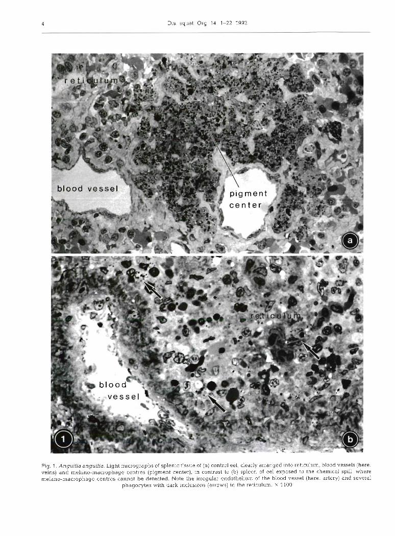
The spleen of eel is a discrete elongate organ with a massive connective tissue capsule, situated in the large curvature of the stomach. Its blood supply is provided by visceral arteries, and splenic veins join the hepatic portal system (Fange & Nilsson 1985). Eel spleen is traversed by many arteries and veins of different sizes, among them sheathed arterial capillaries (ellipsoids). Sinusoids without a continuous endothelium are not developed in eel. Following perfusion fixation, larger blood vessels were almost free of blood (Fig. l a ) , whereas a high blood cell content was characteristic of the remaining splenic reticular tissue. In eel, this reticulum is a non-septate cellular framework rich in intercellular substance, with extended spaces serving as pathways for migrating cells. Numerous large, dark melano-macrophage centres of slightly irregular shape and a diameter of up to 150 pm were distributed in the tissue, mainly close to large vessels (Fig. l a ) . Outside those melano-macrophage centres, neither vacuoles nor dark inclusions were apparent.
Tab
le 2
. A
ng
u~
lla a
ng
uil
la. C
ytol
ogic
al c
har
acte
riza
tio
n o
f re
ticu
lar
and
non
-ret
lcul
ar c
ell
typ
es I
n th
e sp
leen
of
cont
rol
eel
No
n-p
hag
ocy
tlc
Ph
ago
cyt~
c M
elan
o-m
acro
- M
acro
- re
ticu
lum
cel
ls
reti
culu
m c
ells
p
hag
es
ph
ages
-
-
Cel
l S
ize
ran
ge
of p
erik
aryo
n (k
m)
7-10
/2
61
0
8-12
([
len
gtw
wid
th] o
r d
iam
eter
) S
hap
e S
len
der
C
om
pac
t C
om
pac
t
Pla
smal
emm
a L
ong
bra
nch
ed
Lob
ular
S
moo
th
exte
nsi
on
s pr
otru
sion
s
- Nu
cleu
s S
ize
ran
ge
(pm
) 4-
6/1-
1.5
4-5/
2 3-
5 ([
len
gth
/wld
th]
or d
iam
eter
) S
hap
e C
lub
-sh
aped
, O
val
to
Co
mp
ress
ed t
o in
den
tati
on
s sl
ight
ly
vari
able
sh
ape
irre
gula
r
Het
ero
chro
mat
in d
istr
ibut
ion
Dis
pers
ed
Dis
pers
ed
Dis
pers
ed
Mit
och
ond
ria
No.
per
sec
tion
7
Siz
e ra
ng
e (k
m)
0.5
/0.3
([
len
gth
/wid
th] o
r d
iam
eter
) M
atri
x el
ectr
on
den
sity
M
ediu
m
Mem
bra
ne
who
rls
-pp-
En
dop
lasm
ic r
etic
ulu
m
Am
ou
nt
(ty
pe)
L
ittl
e (S
ER
+ R
ER
)
Arr
ang
emen
t of
cis
tern
ae
Elo
ng
ate,
ir
reg
ula
r
Fre
e ri
boso
mes
M
od
erat
e
Gol
gi f
ield
s N
o. p
er s
ecti
on
0- 1
N
o. o
f cl
ster
nae
per
fie
ld
4 E
xten
sion
of
c~
ste
rna
r (pm
) 0
.5
-
-
Pri
mar
y an
d se
con
dar
y ly
soso
mes
A
mou
nt
Few
S
ize
(urn
) 0.
4 ([
len
gth
/wld
th] o
r d
am
ete
r)
Sh
ape
Ov
al
6 M
ax.
1.5
/0 3
Med
ium
M
ediu
m
-
-
Mo
der
ate
Ver
y li
ttle
(S
ER
+ R
ER
) (m
ainl
y S
ER
)
SE
R: s
ho
rt
Sho
rt a
nd
tu
bu
lar;
RE
R:
vesl
cula
ted
elo
ng
ate,
ln
flat
ed
Mo
der
ate
Vrr
y f
ew
-
3 2
(ear
ly s
tag
e)
4 3
4
0.5-
1 1 -
--l .5
--p
Nu
mer
ou
s 0.
1-0.
5
Ova
l to
p
ean
ut-
shap
ed
Ery
thro
- E
ryth
ro-
Th
rom
bo
- L
ymph
o-
Pla
sma
Het
erop
hili
c bl
asts
cy
tes
cyte
s cy
tes
cell
s g
ran
ulo
cyte
s
Ov
al
Ell
ipti
cal
Sp
ind
le-
Sp
hen
cal
Co
mp
act
Co
mp
act
shap
ed
Co
rru
gat
ed
Sm
ooth
N
um
ero
us
Nu
mer
ou
s L
obul
ar
Lob
ular
, m
vag
l-
pse
ud
op
od
s co
rru
gat
ed
nat
ion
s
3/2-
2.5
Ova
l O
val
to
Elo
ng
ate,
S
ph
eric
al,
Ova
l O
val
to l
obul
ar
sbgh
tly
lon
g~
tud
. in
den
- ir
regu
lar
furr
ow
tah
on
s
Dis
pers
ed
Clo
tted
C
lott
ed
Dis
pers
ed
D~
sper
sed
D
isper
sed
or c
lott
ed
or c
lott
ed
5 2-
3 2
2-3
4 M
ax.
l/0
.4
Max
. 2
/0.3
0
8/0
.25
M
ax.
1/0
.3
1/0.
5
Me
d~
um
M
ediu
m
Med
ium
M
edlu
m
Med
ium
M
ediu
m
-
--
- P
Mo
der
ate
Ver
y li
ttle
h
ttle
L
ittl
e P
lent
y P
lent
y (
SW
(
SW
(
SW
(S
ER
) (S
ER
+ R
ER
) (m
ain
ly S
ER
) S
hort
an
d
Fra
g-
Elo
ng
ate
Elo
ng
ate
SE
R:
swol
- S
hort
tu
bu
lar
~rr
egu
larl
y
men
ted
, u
reg
ula
r le
n,
RE
R:
to v
esic
ular
tu
bu
lar
vesl
cula
ted
elo
ng
ate
and
par
alle
l
Nu
mer
ou
s F
ew
Nu
mer
ou
s N
um
ero
us
Mo
der
ate
-
Few
F
ew
Ver
y n
um
ero
us
-
0.1
4.2
0
.24
.4
0.6
4.8
/0.2
Sph
eric
al
Sp
he
r~c
al
Ell
ipti
cal
(Tab
le c
on
t~n
ue
d on
nex
t p
age)
6 Dis. aquat. Org. 14: 1-22, 1992
Histology of Rhine eel spleen
1 There were conspicuous histological alterations in the spleens of eel exposed to the chemical spill (Fig. l b ) . The tissue contained considerable amounts of transpa-
1 rent vacuoles and dark inclusions, both of varying sizes. By means of electron microscopy, most vacuoles were identified as swollen mitochondria or, to a smaller proportion, dilated endoplasmic reticulum (ER), while the dark inclusions represented residual bodies and myelinated structures (see Figs. 3c, d, f , g, 4b, 5d, e). Cytoplasmic density of spleen cells showed increased variability, as compared to controls. Dark and light cells were scattered randomly. As concluded from electron microscopy, the dark cells mostly represented reti- culum cells and blood cells, and a high percentage of the light cells could be identified as either dying cells or as (non-pigmented) macrophages of 8 to 12 pm in diameter, a cell type not detected in controls. In contrast to controls, clearly defined, large melano- macrophage centres were not present in the spleen of Rhine eel.
Ultrastructural organization of splenic reticulum
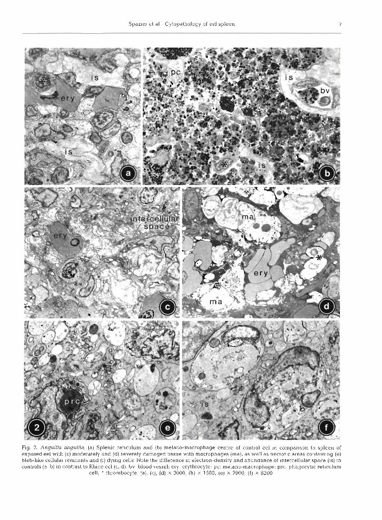
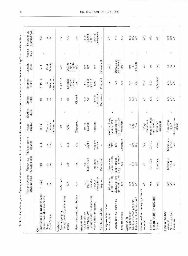
The different cell types characterized in detail in Tables 2 (controls) and 3 (Rhine eel) could be divided into 2 main categories: (1) reticulum cells (non- phagocytic and phagocytic) constituting the permanent reticular framework, and (2) blood cells found tem- poranly in vessels and intercellular spaces (Fig. 2a). Whereas for non-phagocytic reticulum cells no relation to any other cell type could be documented, phagocytic cells showed transitional stages to melano-macro- phages, which, therefore, might be regarded as a third (phagocytic) type of reticular cell. In controls, rnelano- macrophages were rarely found as single cells, but integrated in melano-macrophage centres (Fig. 2b). The cellular arrangement in controls appeared well- structured: cellular junctions between reticulum cells were regularly established, and amorphous intercellu- lar substance formed branching cords of 3 to 5 pm thickness lined by extensions of non-phagocytic reticulum cells (Fig. 2a, cf. Fig. 3a).
In exposed eel, the splenic tissue was less clearly structured (Fig. 2c-f). Intercellular substance appeared reduced and condensed; single cords were rarely thicker than 1.5 ,pm (Fig. 2c, d) and locally contained collagen. Cytological damage ranged from minor deviations (changes in cytoplasmic electron density, pseudopod retraction, swelling of mitochondria, forma- tion of myelinated structures and lipid droplets) to complete necrosis of single cells or cell groups distributed heterogeneously throughout the tissue.
Tab
le 3
. A
ngul
lla
angu
illa
. C
ytol
ogic
al a
lter
atio
ns o
f re
ticu
lar
and
non
-ret
icul
ar c
ell
type
s in
the
sp
leen
of
eel
exp
osc
d t
o th
e ch
emic
al s
p~
ll in t
he R
h~
ne
Riv
er
Non
-pha
gocy
tic
Pha
gocy
tic
Mel
ano-
mac
ro-
Mac
ro-
Ery
thro
- E
ryth
ro-
Thr
ombo
- L
ymph
o-
Pla
sma
Het
erop
hili
c re
ticu
lum
cel
ls
reti
culu
m c
ells
p
hag
es
ph
ages
bl
asts
cy
tes
cyte
s cy
tes
cell
s gr
anul
ocyt
es
Cel
l S
ize
ran
ge
of p
erik
aryo
n (p
m)
7-10
/3
(Ile
ng
th/w
idth
] or
dia
met
er)
Sh
ape
n/c
n/c
n/c
Com
pact
n/
c I~
/C
Ova
l n
/c
Pla
smal
emm
a n/
c n/
c n
/c
Lob
ular
n/
c F
ew i
n-
Sm
ooth
n/
c pr
otru
sion
s va
gina
tion
s --
Nu
cleu
s S
ize
ran
ge
(pm
) 4-
64.5
-2
n/c
n/c
5/2
n
/c
5-6/
2.5-
3 n/
c n/
c ([
len
gth
/wid
thl o
r d
iam
eter
) S
hap
e n/
c n/
c n/
c O
val
n/c
Elo
ngat
e,
Ova
l to
n
/c
furr
ow
slig
htly
re
du
ced
ir
regu
lar
Het
eroc
hrom
atin
dis
trib
utio
n n/
c n/
c n
/c
Dis
pers
ed
Clo
tted
n/
c n
/c
n/c
. -
Mil
och
on
dri
a N
o. p
er s
ecti
on
10
n/c
3
10-1
5 0-
2 2-
4 n
/c
4-5
Siz
e ra
ng
e (p
m)
0.5
-l/
Max
. 1
/0.3
0.
6/0.
3 1.
2/0.
25
n/c
0.
7/0.
2 n
/c
0.6/
0.3
([le
ng
th/w
idth
] or
dia
met
er)
0.25
-0.5
o
r 0.
5-1
or
0.7-
1 M
atri
x el
ectr
on d
ens~
ty
Low
to
tran
spar
ent
Med
ium
to
low
M
edlu
m
Med
ium
to
low
L
ow t
o 1
.0~
tr
ansp
arc?
nt
Low
to
tran
spar
ent
Mem
bra
ne
who
rls
Fre
qu
ent
Occ
asio
nal
-
- O
ccas
~o
rral
F
req
uen
t O
ccas
iona
l -
En
do
pla
smic
ret
icu
lum
A
mou
nt (
typ
e)
Mo
der
ate
Mo
der
ate
Litt
le
Mod
. to
plen
ty
n/c
n/c
n
/c
(SE
R +
RE
R)
(mai
nly
RE
R)
(SE
R)
(mai
nly
RE
R)
Arr
ang
emen
t of
cis
tern
ae
Elo
ngat
e, i
rre-
S
ho
rto
relo
n-
n/c
Sho
rt o
r elo
n-
n/c
Elo
ngat
e or
n/
c g
ula
r, in
flat
ed
gat
e +
narr
ow
gat
e +
narr
ow
vesi
cula
ted
Fre
e ri
boso
mes
n/
c n/
c M
od
erat
e M
od
erat
e n/
c n/
c n/
c
Go
lgi
fiel
ds
No.
per
sec
tion
n
/c
1 1
1-2
n/c
n/c
n/c
n/c
6-8
nf c
4
5
No.
of
cist
erna
e p
er f
ield
n/
c n
/c
n/c
n/c
Ext
ensi
on o
f ci
ster
nae
(pm
) n/
c n/
c 0.
6 1-
1.5
n/c
0
.5
0.5-
0.8
n/c
Pri
mar
y an
d s
eco
nd
ary
ly
soso
mes
A
mou
nt
n/c
n/c
F
ew
Ver
y n
/c
Few
n/
c n
/c
nu
mer
ou
s
S~
ze
(pm
) 0
.2
0.1-
0.25
0
.14
.2
Pri
m.
0.1
4.2
5
n/c
0.2
n/c
n/c
([le
ng
th/w
idth
] or
dia
met
er)
sec.
max
. 5
Sh
ape
n/c
Sph
eric
al
Ova
l O
val
and
n/
c S
pher
ical
n/
c n/
c ir
regu
lar
Res
idua
l b
od
ies
Am
ount
n/
c M
od
erat
e N
um
ero
us
Nu
mer
ou
s n/
c n/
c n/
c n
/c
Siz
e ra
ng
e (p
m)
n/c
0.5-
1 0.
7-3
0.5-
2 n/
c n/
c nf
c
n/c
Co
nte
nts
n
/c
n/c
n/c
Hom
ogen
eous
, nf
c
n/c
n/c
n/c
den
se

Spazier e t al.. Cytopathology of eel spleen
Extended areas without any change were rare. Within necrotic areas, normal reticular organization was replaced by a mixture of degenerating or necrotic blood and reticulum cells, bleb-like cell remnants con- taining residual bodies, and other cellular debris of unknown origin (Fig. 2e, f ) . Most intercellular junctions between reticulum cells were disrupted.
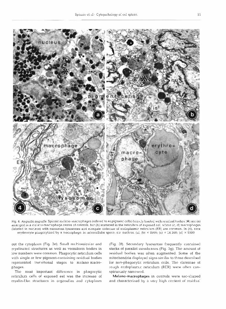
Semiquantitative evaluation revealed changes in the frequencies of different cell types (Table 4). Erythro- blasts and plasma cells could not be detected in exposed eel. Melano-macrophage centres were not present, and the total number of melano-macrophages dispersed throughout the reticulum was reduced. In contrast, hypertrophic macrophages without pigments could only be found in exposed fish (Fig. 2d), often accumulated close to necrotic areas.
Cytology of reticulum cells and macrophages
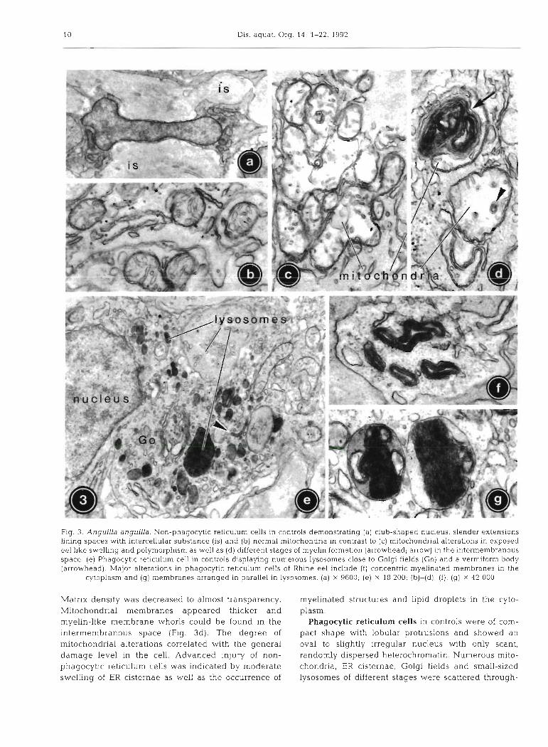
Non-phagocytic reticulum cells in controls could be easily distinguished by their slender shape with numerous long, branching extensions establishing cellular contacts to other reticulum cells and by a club- shaped nucleus with considerable amounts of ran- domly dispersed heterochromatin (Fig. 3a). The cyto- plasm contained few organelles in an irregular dis- tribution, mainly mitochondria (Fig. 3b), and short bundles of microfilaments. Lysosomes were very rare.
In Rhine eel, mitochondria were increased in fre- quency and showed polymorphism as well as progres- sive swelling of up to double the control size (Fig. 3c).
Table 4 . Anguilla anguilla. Semiquantitative evaluation of relative frequencies of reticular and non-reticular cell types in the spleens of control eel and eel exposed to the chemical spill
in the Rhine h v e r in November 1986
Control Rhine eel eel
Non-phagocytic reticulum cells Phagocytic reticulum cells Melano-macrophages
- reticulum - melano-macrophage centres
Macrophages Erythroblasts Erythrocytes Thrombocytes Lymphocytes Plasma cells
- reticulum - ellipsoids
Heterophilic granulocytes
- not detected; + occasionally present; + + common; +++ numerous; ++ ++ very numerous
Dis. aquat. Org. 14: 1-22, 1992
Fig. 3. Anguilla anguilla. Non-phagocytic reticulum cells in controls demonstrating (a) club-shaped nucleus, slender extensions linlng spaces w t h intercellular substance (IS) and ( b ) normal mitochondria in contrast to (c) mitochondrial alterations in exposed eel like swelling and polymorphism as well as (d) different stages of myelin formation (arrowhead; arrow) in the intermembranous space. (e) Phagocytic reticulum cell in controls displaying numerous lysosomes close to Golgi fields (Go) and a vermiform body (arrowhead). Major alterations in phagocytic reticulum cells of Rhine eel include ( f ) concentric myelinated membranes in the
cytoplasm and (g) membranes arranged in parallel in lysosomes. (a) X 9600; (e) X 18 200; (b)-(d), ( f ) , (g) x 42 000
Matrix density was decreased to almost transparency. myelinated structures and lipid droplets in the cyto- Mitochondrial membranes appeared thicker and plasm. myelin-like membrane whorls could be found in the Phagocytic reticulum cells in controls were of com- intermembranous space (Fig. 3d). The degree of pact shape with lobular protrusions and showed an mitochondrial alterations correlated with the general oval to slightly irregular nucleus with only scant, damage level in the cell. Advanced injury of non- randomly dispersed heterochromatin. Numerous mito- phagocytic reticulum cells was indicated by moderate chondria, ER cisternae, Golgi fields and small-sized swelling of ER cisternae as well as the occurrence of lysosomes of different stages were scattered through-
Spazier et al .- Cytopathology of eel spleen 11
Fig. 4. Anguilla anguilla. Splenic melano-macrophages (refered to as pigment cells) heavily loaded with residual bodies (*) are (a) arranged in a melano-macrophage centre of controls, but (b) scattered in the reticulum of exposed eel, where (c, d) macrophages (labeled in nucleus) with numerous lysosomes and elongate cisternae of endoplasmic reticulum (ER) are common In (d), note
erythrocyte phagocytlzed by a macrophage. is: intercellular space; nu: nucleus. (a), (b) x 8500; (c) x 14 300; (d) x 9900
out the cytoplasm (Fig. 3e). Small multivesicular and (Fig. 3f). Secondary lysosornes frequently contained myelinated structures as well as vermiform bodies in stacks of parallel membranes (Fig. 3g). The amount of low numbers were common. Phagocytic reticulum cells residual bodies was often augmented. Some of the with single or few pigment-containing residual bodies mitochondria displayed signs similar to those described represented transitional stages to melano-macro- for non-phagocytic reticulum cells. The cisternae of phages. rough endoplasmic reticulum (RER) were often con-
The most important difference in phagocytic spicuously narrowed. reticulum cells of exposed eel was the increase of Melano-macrophages in controls were sac-shaped rnyelin-like structures in organelles and cytoplasm and characterized by a very high content of residual
Spazier e t al.: Cytopa thology of eel spleen 13
bodies with a diameter of 1 to 4 pm in contrast to the sparsity of other organelles, in particular primary lysosomes (Fig. 4a). Due to dense package of residual bodies, several nuclei were compressed. Residual bodies contained granular pigments of various size and density, embedded in a moderately electron-lucent matrix (Fig. 4a). Three types of pigments could be identified (terminology of Agius & Agbede 1984): melanin (discrete oval granules, 0.3 to 0.5 pm, uniformly very dense), lipofuscin (irregular shape, up to 2.5 pm, varying size and electron density) and haemosiderin (finely granular, clustered, electron- dense). Melanin usually predominated. Occasionally, multivesicular bodies and lipid droplets could be dis- cerned. Some (premature?) melano-macrophages were of smaller size (6 to 8 pm) and contained fewer and smaller residual bodies than typical melano-macro- phages, but a higher number of other organelles.
Whereas fully formed melano-macrophages were less abundant in Rhine eel, smaller melano-macro- phages with a reduced number of residual bodies, but poorly developed ER and Golgi fields, were increased. Melanin content was generally reduced, whereas fine lamellar and granular materials with lipofuscin and haemosiderin portions predominated (Fig. 4b). In melano-macrophages, mitochondria did not show any swelling.
Macrophages were only observed in Rhine eel (Fig. 4c, d). They could be distinguished from similar phagocytic reticulum cells by their marked hypertro- phy, being larger in size and showing more protrusions. Their oval nucleus was poor in heterochromatin and organelles were numerous in the electron-lucent cyto- plasm. Compared to phagocytic reticulum cells in Rhine eel, mitochondria were twice as frequent, nar- rowed RER cisternae appeared increased, Golgi fields were extended, and lysosomes were more numerous and frequently contained undigested material such as erythrocytes (Fig. 4d). Moderate amounts of myeli- nated structures, lipid droplets, multivesicular and ver- miform bodies could be observed. Some macrophages showed symptoms of cellular-injury-like rupture of cell membranes and release of cytoplasmic contents.
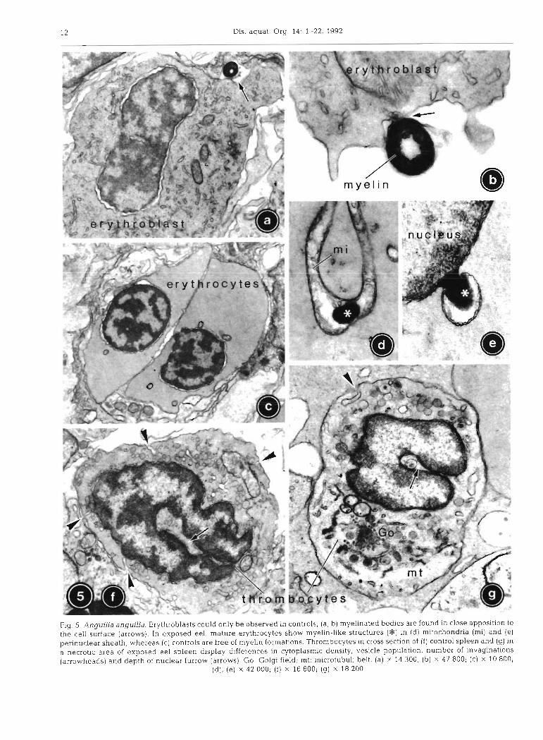
Cytology of blood cells
Erythroblasts could in most cases be classified as late basophilic stages according to Bielek (1978). The cyto- plasm of the oval cells already contained haemoglobin, but was still rich in organelles, mainly mitochondria and irregular tubular smooth endoplasmic reticulum (SER), as well as some Golgi fields, multivesicular bodies and numerous free ribosomes (Fig. 5a). Occa- sionally, erythroblasts exhibited ring-shaped myeli-
nated bodies close to the cell surface (Fig. 5a, b) measuring 0.35 to 0.4 pm in diameter and consisting of about 10 to 15 concentric membrane layers. In the spleen of eel exposed to the chemical spill, erythro- blasts were not detectable.
Erythrocytes. In the elliptical erythrocytes of con- trols, the oval nucleus displayed wide nuclear pores and strongly condensed heterochromatin (Fig. 5c), the amount of which clearly exceeded that of erythroblasts. Except for few mitochondria and cisternae of SER, the cytoplasm was extremely poor in organelles, but rich in fine, homogeneously distributed haemoglobin. The marginal microtubuli belt of the erythrocytes was about 0.2 pm thick and consisted of 8 to 10 microtubuli.
In erythrocytes of exposed eel, the nucleolus appeared more prominent than in control fish (Fig. 6b). The cytoplasm was of slightly increased density, but almost or entirely free of organelles except for few mitochondria. Some of these were swollen and showed reduced cristae in an electron-lucent to transparent matrix. Occasionally, myelin-like membrane whorls could be found in the intermembranous space of mitochondria (Fig. 5d) , as well as in the locally dilated perinuclear sheath (Fig. 5e). In no case, however, were these myelin-like membrane whorls found attached to the cell surface as described for control erythroblasts.
Thrombocytes were spindle-shaped blood cells with an elongate nucleus displaying a characteristic lon- gitudinal furrow and abundant, condensed hetero- chromatin (Fig. 5f). In controls, about 10 plasmalemmal invaginations per section with a diameter of about 80 nm could be counted. The peripheral cytoplasm contained 30 to 50 vesicles per section with a constant diameter of 0.15 pm and a homogeneous matrix. Nor- mally, the 0.5 to 1 pm thick belt of microtubuli sur- rounding the cell along its longitudinal axis was only difficult to discern.
In exposed fish, both cell and nuclear length were reduced in thrombocytes. The nuclear furrow was fre- quently less prominent (Fig. 5g). Plasmalemmal invagi- nations were reduced in number by about 50 %, and their diameter was diminished to 50 nm. Each thrombo- cyte displayed 10 to 50 vesicles per section, with a diameter increased by 30 O/O to 0.2 pm and a highly variable matrix. Part of the mitochondria appeared swollen with low matrix density and membranous whorls in the intermembranous space. Similarly to ery- throcytes, myelinated structures were occasionally found in the perinuclear sheath. Golgi fields were diminished, and some lysosomes and multivesicular bodies were present. Due to a decrease in cytoplasmic density of severely damaged thrombocytes, the lon- gitudinal microtubuli belt was more evident (Fig. 5g) .
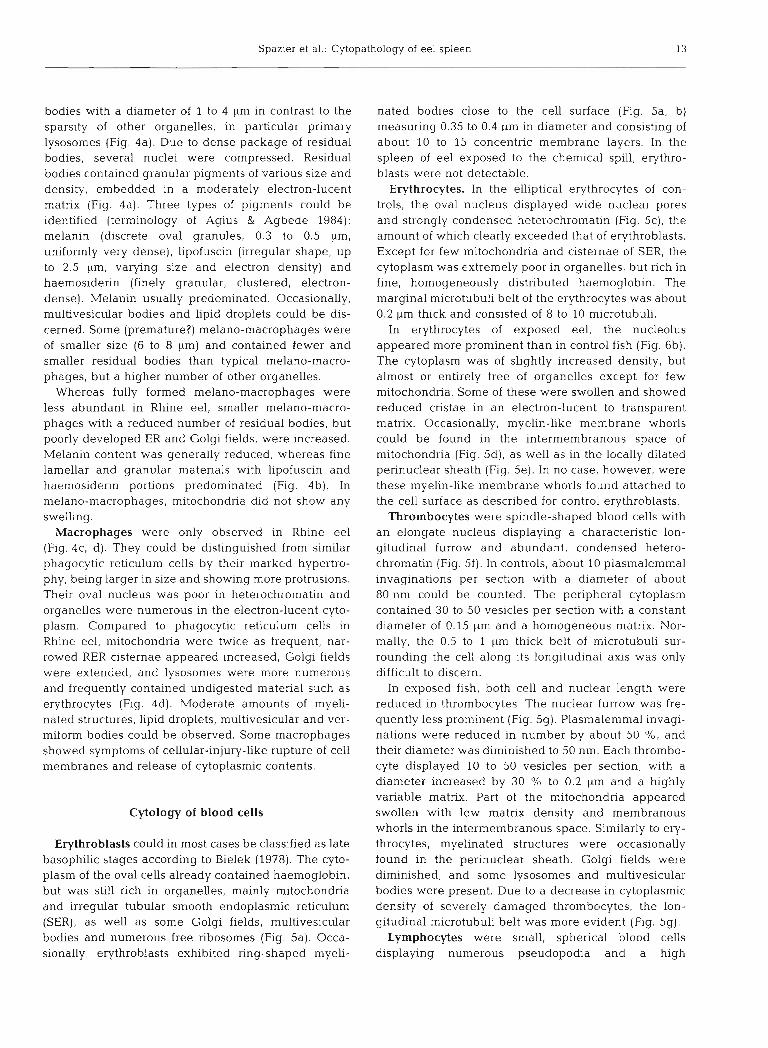
Lymphocytes were small, spherical blood cells displaying numerous pseudopodia and a high
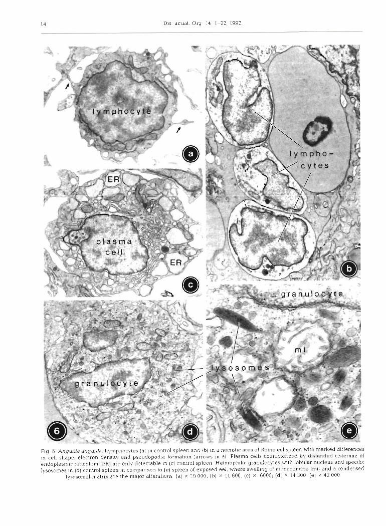
Spazier et a1 - Cytopathology of eel spleen 15
nuclear : cytoplasmic ratio (Fig. 6a). Except for high amounts of evenly distributed free ribosomes, the cyto- plasm only contained some mitochondria, elongated cisternae of SER and few small lysosomes.
In exposed fish, cell shape and nuclei of most lym- phocytes were no longer spherical. Even cells with- out other modif~cations displayed a reduction of pseudopodia, whereas the plasmalemma of severely damaged lymphocytes appeared completely smooth (Fig. 6b). In addition, the cytoplasmic density was either drastically decreased or, more rarely, increased. Further alterations included small membranous whorls within mitochondria, partial vesiculation of SER and increased density of lysosomes.
Plasma cells in control eel were more commonly found in the wall of ellipsoids than in the reticulum (Fig. 6c). Both elongate RER lamellae in parallel array and highly swollen SER cisternae could be found in the same cell. Free ribosomes were numerous. Several large Golgi fields per section were arranged in groups mainly in a perinuclear position. Golgi cisternae were inflated and intensively fenestrated. Only few lyso- somes were present. In the spleens of exposed fish, plasma cells could not be detected.
Heterophilic granulocytes in controls contained an oval to lobular nucleus and numerous small organelles including mitochondria, short tubular to vesicular cis- ternae of SER, and primary lysosomes (Fig. 6d). These lysosomes equivalent to the specific granules in light microscopy had a characteristic elliptic shape and a crystalline matrix. Abundant p-glycogen particles were dispersed throughout the cytoplasm.
In Rhine eel, mitochondria of heterophilic granulo- cytes were reduced in number, some of them appeared
heavily swollen, with reduced cristae in an almost transparent matrix (Fig. 6e). The electron density of the crystalline lysosomal matrix was increased, and lipid droplets of 0.5 pm in diameter occurred in the cyto- plasm. Heterophilic granulocytes were the only blood cell type in exposed eel that were free of myelinated structures.
DISCUSSION
The present study was designed to serve several purposes: (1) to give a comprehensive account of the histology and cytology of resident and free cells in the spleen of eel, (2) to investigate the cytopathological effects of the chemical spill into the Rhine h v e r at Basle in November 1986 on eel spleen, (3) to assess potential functional implications of splenic cytopathol- ogy in context with earlier findings on cytological alter- ations in liver and intestine (Braunbeck et al. 1990a, Burkhardt-Holm et al. 1990), and, thus, (4 ) to evaluate the utility of splenic alterations at the ultrastructural level as a biomarker for toxicant-induced damage of fish.
To date, rainbow trout Oncorhynchus mykiss is the most-investigated species with regard to studies on fish spleen. For many other species, including eel, data are fragmentary. Except for melano-macrophage centres, which have been described by Agius (1979, 1980), Agius & Roberts (1981), Agius & Agbede (1984) and Roberts (1975), histology and cytology of eel spleen has not previously been investigated. A general outline of histological and cytological investigations on fish spleen is given in Tables 5 & 6.
Table 5. Reviews and systematical investigations on histology and cytology of fish spleen
Species Comment Method Reference
(Review) Immune response in fish (Review) Corbel (1975)
(Review) Comparative study of lympho-myeloid tissue (Review) Fange (1982) in fish
(Review) Structure and function of fish spleen (Review) Fange & Nilsson (1985)
Teleosts (14 spp.; e.g. eel, Role of melano-macrophage centres in iron HC, LM Agius (1979) plaice, rainbow trout) storage of normal and diseased fish
Agnatha, chondrichthyes and Phylogenetic development of melano-macro- LM Agius (1980) osteichthyes (72 spp.) phage centres
Dogfish, eel, plaice, ralnbow Pigment genesis in haemopoietic tissues EM Agius & Agbede (1984) trout, tilapia
Dogfish, eel, plaice, rainbow Effects of starvation on melano-macrophage LM, h 4 0 Agius & Roberts (1981) trout, sword tail, tilapia centres
Carp, crucian carp, golden ide, General anatomy and histology LM Haider (1966) perch, rainbow trout, tench
EM: electron microscopy: HC: histochemistry; LM: light microscopy; MO: morphometry
Tab
le 6
. H
isto
logi
cal
and
cyt
olog
ical
inv
esti
gati
ons
on
sp
leen
of
sin
gle
fis
h sp
ecie
s
Sp
ecie
s C
om
men
t M
etho
d R
efer
ence
Dog
fish
S
cyli
orhi
nus
cani
cula
Dog
fish
S
cyli
orhi
n us
ste
llar
is
Ray
R
aja
clav
ata,
Tor
pedo
mar
mor
ata
Ch
imae
ra
Ch
imae
ra r
nons
lros
a
Cyt
olog
y of
spl
enic
cel
l ty
pes
E
M
Pul
sfor
d et
al.
(198
2)
Blo
od p
recu
rsor
cel
ls in
hae
mop
oiet
ic s
ites
C
C, L
M
D'I
ppoI
ito
et a
l. (
1985
)
Ery
thro
poie
sis a
nd
thro
mbo
poie
sis
Zap
ata
(198
0)
Mor
phol
ogy
and
hae
mop
oies
is o
f di
ffer
ent l
ymph
omye
loid
tis
sues
F
ang
e &
Su
nd
ell (
1969
)
Eu
rop
ean
eel
A
ngui
lla
angu
llla
S
tres
s di
agno
sis
by h
aem
atol
ogic
al a
nd
mor
phol
ogic
al m
eth
od
s E
fTec
ts o
f 2
anti
biot
ics
on h
aem
opoi
esis
P
hysi
olog
y an
d m
orph
olog
y of
blo
od u
nd
er s
tres
s
Ter
min
al c
apil
lari
es (
elli
pso
~d
s)
Su
ble
thal
eff
ects
of
amm
onia
on
haem
opoi
etic
tis
sue
Eff
ects
of
ph
eny
lhy
dra
zin
e o
n m
elan
o-m
acro
ph
age
cen
tres
H+
S,L
M
CC
. LM
H
+ S
, LM
HC
, EM
H
+ S
, LM
MO
an
ge
r (1
983)
K
reut
zman
n (1
977a
) P
eter
s e
t al.
(19
80)
Gra
f &
Sch
liin
s (1
979)
W
aso
w &
Dab
row
ska
(198
9)
Her
raez
& Z
apat
a (1
986)
Car
p
Cy
pri
nu
s ca
rpio
Gol
dfis
h C
aras
siu
s au
ralu
s
Roa
ch a
nd
go
by
R
util
us ru
tilu
s, G
obio
go
bio
Loa
ch
Msg
urn
us
ang
uil
lica
ud
atu
s
Cat
fish
H
eter
op
neu
sles
fos
sjli
s
Rai
nbow
trou
t O
nco
rhy
nch
us
myk
iss
Str
uctu
re a
nd
ult
rast
ruct
ure
of l
ymph
oid
tiss
ue
LM
, EM
Z
apat
a (1
982)
Hae
mop
oiet
ic s
ites
of e
osin
ophi
lic
gran
uloc
ytez
L
M, E
M
Ishi
zeki
et a
l. (1
984)
Ban
o &
Has
an (
1990
) F
list
opat
holo
gica
l le
sion
s by
mer
cury
Sup
pres
sion
of
anti
bo
dy
-pro
du
cin
g ce
lls
expo
sed
to c
oppe
r D
emon
stra
tion
of
imm
un
oco
mp
eten
t cel
ls
Eff
ects
of
an a
ntib
ioti
c (f
urna
gill
in) o
n h
aem
opoi
etic
tis
sue
Blo
od c
ells
an
d p
recu
rsor
s in
hae
mop
oiet
ic s
ites
E
ffec
ts o
f ox
idiz
ed f
ish
oil o
n b
lood
an
d ti
ssue
A
lter
atio
ns in
hae
mop
oiet
ic t
issu
e u
nd
er s
tres
s S
truc
tura
l an
d f
unct
iona
l al
tera
tion
s of
ph
ago
cyte
s u
nd
er s
tres
s L
ong-
term
eff
ects
of
poll
uted
wat
er o
n b
lood
an
d t
issu
e E
ffec
ts o
f di
oxin
on
lym
phom
yelo
id ti
ssue
an
d h
aem
opoi
esis
E
ffec
t of
exe
rcis
e on
blo
od s
tora
ge
and
rel
ease
O
nto
gen
etic
dev
elo
pm
ent o
f re
ticu
lo-e
ndot
heli
al s
yste
m
Su
bac
ute
eff
ects
of p
hen
ol o
n e
ryth
rocy
te s
yste
m
Ana
tom
y, h
isto
logy
an
d p
hysi
olog
y
Eff
ects
of
adre
ner
gic
an
d c
holi
nerg
ic d
rug
s A
dre
ner
gic
an
d c
holi
nerg
ic in
nerv
atio
n (E
xtra
-)ne
uron
al u
pta
ke
of c
atec
ho
lam
ines
IM, T
C, L
M
IM
LM
, EM
H
+ S,
LM
C
C, L
M
H+
S,L
M
H +
S, L
M, E
M
H +
S, L
M, E
M
H+
S,L
M
H +
E, L
M
H+
S,W
L
M
H+
S,L
M
LM
, EM
And
erso
n et
al.
(19
89)
Chi
ller
et a
l. (
1969
) L
aure
n et
al.
(19
89)
Leh
man
n &
Sti
iren
berg
(19
76)
Moc
cia
et a
l. (
1984
) P
eter
s &
Sch
war
zer
(198
5)
Pet
ers
et a
l. (1
991)
P
oels
et a
l. (1
980)
S
pit
sber
gen
et a
l. (
1988
a)
Ste
ven
s (1
968)
T
atn
er &
Man
ning
(19
85)
Wla
sow
(19
84)
Zw
ille
nber
g (1
964)
Atl
anti
c co
d G
adu
s rn
orhu
a T
C
RI,,
IM
HC
R
L
Hol
mgr
en &
Nil
sson
(19
75)
Nil
sson
& G
rove
(19
74)
Ung
ell
& N
ilss
on (
1984
)
Pon
tius
& A
mbr
osiu
s (1
972)
IM
, LM
P
erch
P
erca
flu
via
l~li
s P
roli
fera
tion
an
d d
iffe
rent
iati
on o
f an
tibo
dy-p
rodu
cing
cel
ls
Yel
low
per
ch
Per
ca I
lave
scen
s R
uffe
C
yrn
no
cep
hal
us c
ern
ua
Sun
fish
L
epom
is g
ibb
osu
s X
L.
cy
anel
lus
Ag
e-d
epen
den
t acc
umul
atio
n of
mel
ano
-mac
rop
hag
e ag
gre
gat
es
Eff
ects
of
diox
in o
n ly
rnph
omye
loid
tis
sue
Mel
ano-
rnac
roph
age
cen
tres
un
der
(p
re-)
tum
oro
us c
ondi
tion
s
LM
LM
LM
, MO
Bro
wn
& G
eorg
e (1
985)
S
pit
sber
gen
et a
l. (1
9881
3)
Kra
nz &
Pet
ers
(198
4)
Pha
gocy
tosi
s an
d fi
lter
ing
func
tion
s L
M, S
EM
, EM
F
ulop
& M
cMil
lan
(198
4)
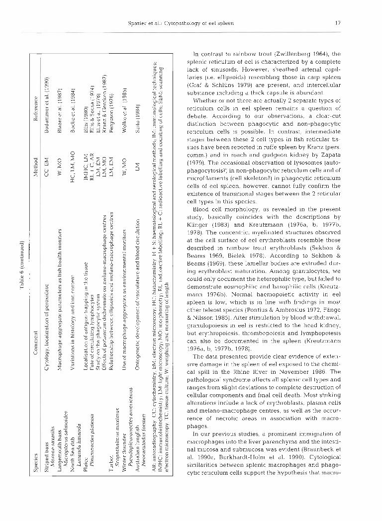
Spaz~e r et al.: Cytopathology of eel spleen 17
C C .- 0 ji
E zo m 2 g f .-
L oz Q m C > 0 s .g X f _a d .- m u g : V) L
" L r l n W
In contrast to rainbow trout (Zwillenberg 1964), the splenic reticulum of eel is characterized by a complete lack of sinusoids. However, sheathed arterial capil- laries (i.e. ellipsoids) resembling those in carp spleen (Graf & Schluns 1979) are present, and intercellular substance including a thick capsule is abundant.
Whether or not there are actually 2 separate types of reticulum cells in eel spleen remains a question of debate. According to our observations, a clear-cut distinction between phagocytic and non-phagocytic reticulum cells 1s possible. In contrast, intermediate stages between these 2 cell types in flsh reticular tis- sues have been reported in ruffe spleen by Kranz (pers comm.) and in roach and gudgeon kidney by Zapata (1979). The occasional observation of lysosomes (auto- phagocytosis?) in non-phagocytic reticulum cells and of microfilaments (cell skeleton?) in phagocytic reticulum cells of eel spleen, however, cannot fully confirm the existence of transitional stages between the 2 reticular cell types in this species.
Blood cell morphology, as revealed in the present study, basically coincides with the descriptions by Klinger (1983) and Kreutzmann (1976a, b, 1977b, 1978). The concentric, myelinated structures observed at the cell surface of eel erythroblasts resemble those described in rainbow trout erythroblasts (Sekhon & Beams 1969, Bielek 1978). According to Sekhon &
Beams (1969), these lamellar bodles are extruded dur- ing erythroblast maturation. Among granulocytes, we could only document the heterophilic type, but failed to demonstrate eosinophilic and basophilic cells (Kreutz- mann 197613). Normal haemopoietic activity in eel spleen is low, which is in line with findings in most other teleost species (Pontius & Ambrosius 1972, Fange & Nilsson 1985). After stimulation by blood withdrawal, granulopoiesis in eel is restricted to the head kidney, but erythropoiesis, thrombopoiesis and lymphopoiesis can also be documented in the spleen (Kreutzmann 1976a, b, 197713, 1978).
The data presented provide clear evidence of exten- sive damage in the spleen of eel exposed to the chemi- cal spill in the Rhine River in November 1986. The pathological syndrome affects all splenic cell types and ranges from slight deviations to complete destruction of cellular components and final cell death. Most striking alterations include a lack of erythi-oblasts, plasma cells and melano-macrophage centres, as well as the occur- rence of necrotic areas in association with macro- phages.
In our previous studies, a prominent immigration of macrophages into the liver parenchyma and the intesti- nal mucosa and submucosa was evident (Braunbeck et al. 1990a, Burkhardt-Holm et al. 1990). Cytological similarities between splenic macrophages and phago- cytic reticulum cells support the hypothesis that macro-
18 Dis. aquat. Org. 1 4 : 1-22, 1992
phages in the spleen, and possibly other organs, might arise from phagocytic reticulum cells representing a reservoir of fixed macrophages, as already suggested for fish kidney and spleen (Bertin 1958, Zapata 1979, Peters & Schwarzer 1985). Moreover, macrophages (i.e. histiocytes) in fish tissues might additionally originate from monocytes imported via blood circulation, as documented for mammals (Sutton & Weisser 1966) and fish (Vogelbein et al. 1987).
Lkewise, the relationship of melano-macrophages to resident phagocytic reticulum cells or to mobile macro- phages is subject to discussion. Since, from a cytologi- cal point of view, premature stages of melano-macro- phages can be recognized as transitions between phagocytic reticulum cells and mature melano-macro- phages, a derivation from reticulum cells appears plausible. On the other hand, melano-macrophages are capable of active migration allowing them to (tem- porarily) sett!e in me!ano-~acrophage centres, and. thus, share important characters with common macro- phages (Ellis et al. 1976, Ferguson 1976, Kranz & Gercken 1987).
The lack of melano-macrophage centres in the spleen of exposed Rhine eel is a striking phenomenon hitherto not reported. Due to the important function of melano-macrophage centres as repositories for meta- bolically inert or recycled materials (Roberts 1975) and as units competent in immune defense against micro- bial attack in fish ('primitive germinal centres'; Roberts 1975, Ellis et al. 1976, Ferguson 1976), their absence implicates significant negative consequences for splenic metabolism and immune defence. It is well documented that number and size of melano-macro- phage centres in fish are highly variable in adaptation to actual requirements. Various adverse conditions associated with extended tissue breakdown and haemolysis cause enlargement of melano-macrophage centres in eel and other species (Agius 1979, Agius &
Roberts 1981, Wolke et al. 1985, Herraez & Zapata 1986), whereas exposure of plaice Pleuronectes pla- tessa to potassium dichromate results in fragmentation of splenic melano-macrophage centres along with increased numbers of melano-macrophages in splenic blood vessels (Kranz & Gercken 1987). These authors conclude a decreased ability of melano-macrophages to accumulate, possibly due to a toxicant-induced reduction of chemotactic activity (Weeks et al. 1986). Similar processes may have been active in intoxicated Rhine eel. However, since dissipation of melano- macrophage centres should give rise to elevated num- bers of scattered melano-macrophages, this hypothesis fails to explain the apparent reduction of the number of splenic melano-macrophages in total, unless emigra- tion from the spleen is taken into account.
Even the damage of non-phagocytic reticulum cells
has serious consequences for splenic functions. In ne- crotic areas, the reticular architecture with its rich inter- cellular space is broken down, and, thereby, pathways for migrating cells and blood filtration are obstructed. High amounts of tissue fragments result in a n addi- tional congestion and obstruction of the remaining intercellular space. In turn, damage of splenic phago- cytes leads to a reduction of the phagocytotic capacity of the spleen.
Likewise, for some of the observations in blood cells of exposed Rhine eel, possible functional implications may be speculated. The failure to detect erythroblasts might indicate disturbance of erythropoiesis, at least in the spleen as a secondary erythropoietic organ, or reduced import of erythroblasts from the peripheral circulation. Together with an increased number of ery- throcytes ingested by macrophages, this implies a sig- nificant reduction of erythrocytes in the spleen and, possibly, other organs. Oxyqen depletion might result and account for part of the damage of mitochondria observed in liver (Braunbeck et al. 1990a), intestine (Burkhardt-Holm et al. 1990) and spleen.
Morphological alterations in cellular and nuclear shape of lymphocytes, lower electron density of their cytoplasm and reduction of pseudopodia in combina- tion with the absence of fully differentiated plasma cells indicate a n impairment of specific immune de- fense.
The augmentation of macrophages in the spleen and the induction of their phagocytic activity as indicated by an elevated number of lysosomal elements are clearly of adaptive nature. This stimulation may have been a direct effect of the chemicals, or, more likely, a consequence of the high degree of toxicant-induced necrosis resulting in debris that has to be removed. Normally, the phagocytotic capacity of the spleen in eel and other species is mainly associated with number and extent of melano-macrophage centres (Agius & Roberts 1981, Wolke et al. 1985). In Rhine eel, however, increased numbers of macrophages may even have taken over part of the phagocytic function of disinte- grated melano-macrophage centres.
In contrast to adaptive phenomena, many cyto- pathological reactions in the spleen of exposed fish must be interpreted as degenerative. Deviations from normal cytoplasmic density are regarded typical of dying cells (Wyllie 1981), reflecting changes in the cytoplasmic osmolality caused by disturbed plasma- lemma1 permeability. The early loss of surface differ- entiations such as pseudopodia or cell junctions, as observed in lymphocytes and non-phagocytic reti- culum cells, is another common feature of cell death (Wyllie 1981). Possibly due to impeded oxygen supply, mitochondria display degenerative symptoms such as swelling, destruction of cristae, formation of membrane
Spazier et al.: Cytopat hology of eel spleen 19
whorls in the intermembranous space and reduced matrix electron density. In necrotic cells, similar changes were discussed by Wyllie (1981) in connection with altered ATP-levels.
The ER is known to be particularly sensitive to toxic impact (Wyllie 1981, Braunbeck et al. 1989, 1990a-c). A dilat~on or vesiculation of ER cisternae as revealed in exposed eel is regarded a non-specific reaction that can be observed together with numerous functional dis- turbances (Wyllie 1981). Myelin formation as a promi- nent feature in many splenic cells may be the mor- phological equivalent of toxicant-induced disturbance in biomembrane synthesis, e.g., synthesis of non-func- tioning membranes that only consist of phospholipids (Rez 1986, Phillips et al. 1987). Such membranes fold up to form myelin-like structures and are rapidly sub- jected to lysoson~al destruction, a s frequently observed in phagocytic reticulum cells.
As indicated by the impairment of vital functions of the spleen in Rhine eel such as erythropoiesis, phagocytosis including blood clearance, concentrated deposition of non-degradable materials, as well as slight decline and damage of cells involved in the specific immune response, degenerative mechanisms apparently dominated over adaptive ones. Adaptive processes may be active in the early phases of intoxica- tion or during exposure to very low concentrations of toxicants. Such processes have also been initiated in liver and intestinal tract of Rhine eel (Braunbeck et al. 1990a, Burkhardt-Holm et a1.1990), thus demonstrating that biochemical and morphological defense mechan- isms were induced prior to death of the eels. However, as with any detoxification system, sequestration of for- eign compounds, proliferation of phagocytic cells as well as lysosomal compartmentation and accumulation of contaminants can only be effective as long as the capacity of the organelles is not overloaded (Hutterer et al. 1968, Ghadially 1982, Moore 1985, Braunbeck & Volkl 1991), or the respective organelles are not irreversibly damaged by the contaminants. Apparently these defense systems were insufficient to meet the challenge in exposed Rhine eel.
From a comparison with findings in Liver and gut of the same eels revealing high similarities in degree and nature of the cytopathological syndrome, it can be concluded that most of the splenic reactions towards the toxicants are not specific for spleen. The results of the alterations in all 3 organs of the Rhine eel taken together, however, give a highly complex pathological syndrome which may be specific of the accident in November 1986.
The non-specificity of splenic reaction may also be concluded from a comparison of toxicant-induced changes in Rhine eel spleen with alterations in blood, spleen and head kidney of rainbow trout under stress
conditions. Peters & Schwarzer (1985) and Peters et al. (1991) revealed severe damage to blood cells and dis- turbances of blood cell formation, as well as initially increased phagocytosis in combination with long-term immunosuppression. The functional alterations could be correlated to morphological deviations in rainbow trout spleen and kidney, closely resembling many of those observed in the spleen of intoxicated Rhine eel. As an interesting similarity, necrosis of macrophages is also induced in the head kidney of stressed rainbow trout (Peters e t al. 1991).
Ultrastructural studies in spleen provide important information about mechanisms underlying a pathologi- cal syndrome. However, purely qualitative observa- tions have proved not sufficient to fully appreciate the processes active during intoxication. Since major environment-induced alterations in spleen occur in size and composition of different cell populations, quantita- tive evaluation appears necessary, e.g. estimation of relative cell numbers or morphometric measurements of cell size. In addition, a determination of splenic phagocyte index (relative numbers of phagocytic cells per tissue area) and phagocytosis rate (as a measure for phagocytotic activity; Peters et al. 1991) might help to evaluate the extent of the toxic impact and its signifi- cance for the organism.
With regard to the chemical spill at Basle in November 1986, the data presented document that not only changes in liver and gut, but also cytopathological alterations in the spleen, may have made a significant contribution to the fatal consequences for the eel popu- lation in the Rhine. Most important, disturbances in central splenic functions may have resulted in a long- term deterioration of the immunological status of the fish giving rise to secondary infection and increased susceptibility to disease.
The present study provides a detailed inventory of possible morphological modifications resultant from exposure to a complex mixture of xenobiotic com- pounds in the spleen of eel. With respect to the intri- cacy of possible interactions between the chemicals and the sparsity of investigations on cytological effects even of single toxic conlpounds, it appears too specula- tive to attribute particular alterations to particular com- pounds at the present state. Laboratory studies on structural and functional effects of selected toxic chem- icals involved in the chemical spill in various organs of eel have been initiated.
Acknowledgements. This study was supported by a grant from the Environmental Protection Agency of the state of Baden-W~irttemberg (Federal Republ~c of Germany) by con- tract no. 121180.11.24. The authors are grateful to Dr Patricia Burkhardt-Holm for excellent cooperation and Mrs G. Adam for techilical assistance.
20 Dis. aquat. Org. 14: 1-22, 1992
LITERATURE CITED
Agius, C. (1979). The role of melano-macrophage centres in iron storage in normal and diseased fish. J. Fish Dis. 2: 337-343
Agius, C. (1980). Phylogenetic development of melano-macro- phage centres in fish. J. Zool., Lond. 191: 11-31
Agius, C., Agbede, S. A. (1984). An electron microscopical study of the genesis of lipofuscin, melanin and hemoside- rln in the haemopoietic tissues of fish. J Fish Biol 24. 471-488
Agius. C., Roberts, R. (1981). Effects of starvation on the melano-macrophage centres of fish. J. Fish Biol. 19: 161-169
Anderson, D. P., Dixon, 0. W. , Bodammer, J. E., Lizzio, E. F. (1989). Suppression of antibody-producing cells in rain- bow trout spleen sections exposed to copper in vitro. J. aquat. Anim. Health 1. 57-61
Bano, Y . , Hasan, M. (1990) Histopathological lesions in the body organs of cat-fish (Heteropneustes fossilis) following mercury intoxication. J. environ. Sci. Health 25: 67-85
Bertin, L. (1958). Appareil circulatoire. In: Grasse, P. P (ed . ) - ~ r a i t e d e zooiogie, Tume Xiii. Ayrlatl~es et poissons, 2&me fascicule, p. 1399. Masson e t Cie., Paris
Bielek, E. (1978). Elektronenmikroskopische Untersuchungen der Blutzellen der Teleosteer: Teil I. Erythrocyten. Zool. Jb . Anat. 100: 579-591
Bielek, E. (2980). Elektronenmikroskopische Untersuchungen der Blutzellen der Teleosteer: Teil 111. Granulocyten. Zool. Jb . Anat. 103: 105-121
Blazer, V. S . , Wolke, R. E., Brown, J . , Powell, C. A. (1987). Plscine macrophage aggregate parameters a s health monitors - effect of age , sex, relative weight, season and site quality in largemouth bass (~Ll icropter~~s salmoides). Aquat. Toxicol. 10: 199-215
Bodammer, J. E., Anderson, D. P . , Dixon, 0 . M. (1990). Ultra- structure of the spleen and head kidney of striped bass. J. aquat. Anim. Health 2: 182-193
Braunbeck, T., Gorgas, K. , Storch, V., Volkl, A. (1987). Ultra- structure of hepatocytes in golden ide (Leuciscus idus melanotus L., Cyprinidae: Teleostei) during thermal adap- tion. Anat. Embryol. 175: 303-313
Braunbeck, T , Storch, V. , Nagel, R. (1989). Sex-speci.fi.c reac- tion of liver ultrastructure in zebra fish (Rrachydanio rerio) after prolonged sublethal exposure to 4-nitrophenol. Aquat. Toxicol. 14: 185-202
Braunbeck, T., Burkhardt-Holm, P.. Storch, V (1990a). Liver pathol.ogy In eels (Anguilla anguilla L.) from the Rhine nver exposed to the chemical s p ~ l l a t Basle in November 1986. Limnologie aktuell 1: 371-392
Braunbeck, T. , Gorge, G. , Storch, V , Nagel, R. (1990b). Hepa- tic s tea tos~s In zebra fish (Brachydanio reno) induced by long-term exposure to gamma-hexachlorocyclohexane. Ecotoxicol. Environ. Safety 19: 355-374
Braunbeck, T , Storch, V., Bresch, H. ( 1 9 9 0 ~ ) . Species-specific reaction of liver ultrastructure in zebrafish (Brachydanio rerio) and trout (Salmo gajrdneri) after prolonged expo- sure to 4-chloroaniline. Arch. environ. Contam. Toxicol. 19: 405-418
Braunbeck, T., Volkl, A (1991). Induction of biotransformation in the liver of eel (Anguilla angullla L.) by sublethal expo- sure to d~nitro-o-cresol: a n ultrastructural and blochemica1 study. Ecotoxicol. Environ. Safety 21: 109-127
Braunbeck, T., Burkhardt-Holm, P , Gorge, G., Nayel, R., Neqele, R. D., Storch, V (1992) Regenbogenforrlle und Zebrabarbling. zwei Modelle fiir verlangerte Toxizitats- tests: relative Empfindlichkeit, Art- und Organspezifitat in
der cytopathologischen Reaktion von Leber und Darm auf Atrazin. V ~ r h . Ver. Wasser Boden Lufthyg (In press1
Brown, C. L., George, C. J. (1985). Age-dependent accumula- tion of macrophage aggregates in the vellow perch. Perca flavescens (Mitchill) J . Fish Dis. 8 . 135-138
Bucke, D., Waterm.ann, B., Feist, S (1984). H~stoloyical varla- tions of hepato-splen~c organs from the North Sea dab, Limanda limanda (L.). J. Fish Dis. 7. 255-268
Burkhardt-Holm, P,, Braunbeck, T , Storch, \J' (1990). Auswir- kung der beim Sandoz-Unfall im November 1986 in den Rhein gelangten Chemikalien auf die Ultrastruktur des Darmes von Aalen. Limnologie aktuell 1 393-404
Chiller, M. J., Hodgins. H. O., Chambers, V. C., Weiser. R. S. (1969). Antibody response in rainbow trout (Salmo g a ~ r d - neri) . 1 lmmunocompetent cells in spleen and anterior kidney. J. Immun. 102: 1193-1201
Corbel, M. J. (1975). The immune response in fish: a review. J. Fish Biol. 7 . 539-563
Cossarini-Dunler, M. , Demael, A . , Siwicki, A. K. (1990). In vivo effect of the organophosphorus insect~cide trichlorphon on the immune response of carp (Cyprinus carpio): effect of contamination on antibody production in relation to residue level in oigans. Ec~:oxicol. Env:rgr.. Safety 19: 93-98
D'lppolito, S., Gr imald~, M.C., Pica, A., Della Corte, F. (1985). The blood cells and their precursors in the haemopoietic organs of the dogfish (Scyliorhinus stellaris L.) Arch. ital. Anat. Embriol. 90: 31-46
Deutsche Kommission zur Reinhaltung des Rheins (1986). Deutscher Bericht zum Sandoz-Unfall mit MeBprogramm. ArbeitsausscbuB MeBmethoden der Deutschen Kommis- sion zur Reinhaltung des Rheins. Deutsche Kommission zur Reinhaltung des Rheins, 103 pp.
Ellis, A. E. (1980). Antigen-trapping in the spleen and kidney of the plaice. J. Fish Dis. 3: 4 2 3 4 2 6
Ellis, A. E. , McMunroe, A. L. S., Roberts, R. J (1976). Defence mechanism in fish. I A study on the phagocytic system and the fate of intraperitoneally injected particulate material in the plaice (Pleuronectesplatessa). J . Flsh Biol. 8 : 67-78
Ell~s, A. E , Sousa, M (1974). The phylogeny of the lymphoid system. I A study of the fate of circulating lymphocytes in plaice (Peuronectes platessa). Eur. J. Immunol. 4: 338-343
Fange, R. (1982). A comparative study of lymphomyeloid tissue in fish. Dev. Comp. Immunol. 6 (suppl. 2) : 23-33
Fange, R., Nllsson, S. (1985). The fish spleen: structure and function. Experientia 41, 2: 152-158
Fange, R.. Sundell, G. (1969). Lymphomyeloid tissues, blood cells and plasma proteins in Chimaera Ei~ismonstrosa (Pis- ces, Holocephali). Acta Zool. 50 155-168
Ferguson, H. W (1976). The relationship between ell~psoids and melano-macrophage centres in the spleen of turbot (Scophthalmus maximus). J. Comp. Pathol 86: 377-380
Fulop, G M I . , McMillan, D. B. (1984). Phagocytosls in the spleen of the sunfish (Lepomis spp.). J. Morph. 179: 175-195
Ghadially, F. N. (1982). Ultrastructural pathology of the cell and matrix. Butterworths, London
Graf, R., Schluns, J. (1979). Ultrastructural and histochemical investigation of the terminal capillaries in the spleen of the carp (C'yprinus carpio). Cell Tiss. Res. 296: 289-306
Haider, G , (1966). Beitrag zur Kenntnis der mikroskopischen Anatom~e der Mllz einiger Teleosteer Zool. Anz 177. 348-376
Haider. G. (1967a). Vergleichende Untersuchungen zur Blut- morphologie und Hamatopoese e in~ge r Te leos t~ r r . Tell I . Beobachtungen a n Zellen der roten R e ~ h e 2001. Anz 179: 355-384
Haider, G. (196713). Vergleichende Untersuchungen zur Blut-
Spazier et al.: Cytop athology of eel spleen 2 1
morphologie und Hamatopoese einiger Teleosteer: Teil 11. Beobachtungen an Spindelzellen. 2001. Anz. 179: 384-409
Haider, G. (1967~) . Vergleichende Untersuchungen zur Blut- morphologie und Hamatopoese einiger Teleosteer. Teil 111. Beobachtungen an Lymphocyten und Plasmazellen. Zool. Anz. 180: 110-130
Haider, G. (1967d). Vergleichende Untersuchungen zur Blut- morphologie und Hamatopoese einiger Teleosteer. Teil Vl. Blutbildungsstatten und Blutbildung. 2001. Anz. 181: 203-226
Herraez, M. P., Zapata, A. G. (1986). Structure and function of the melano-macrophage centres in the goldfish (Carassius auratus). Vet. Immunol. Immunopathol. 12: 117-126
Holmgren, S. , Nilsson, S. (1975). Effect of some adrenergic and cholinergic drugs on isolated spleen-stnps from the cod, Gadus rnorhua. Eur. J. Pharm. 32: 163-169
Hutterer, F., Schaffner, F., Klion, F. M,, Popper, H. (1968). Hypertrophic, hypoactive smooth endoplasmic reticulum: a sensitive indicator of hepatotoxicity exemplified by diel- drin. Science 161: 1017-1019
Ishizeki. K., Nawa, T., Tachibana, T., Sakakura, Y., Iida, S. (1984). Haernopoietic sites and development of eosinophil granulocytes in the loach, Misgurnus anguillicaudatus. Cell Tiss. Res. 235: 419-426
Karnowsky, M. J. (1971). Use of ferrocyanide-reduced osmium tetroxide in electron microscopy. J. Cell Biol. 52: 284
Kinzelbach, R., Fnedrich, G. (1990). Biologie des Rheins. Lim- nologie aktuell 1. G. Fischer Verlag, Stuttgart. New York, 489 pp.
Kleeman, J. M., Olson, J. R., Chen, S. M.. Peterson, R. E. (1986). Metabolism and disposition of 2,3,7,8-tetra- chlorodibenzo-p-dioxin in rainbow trout. Toxicol. appl. Pharmacol. 83 391-401
Klinger, H. (1983). Grundlagen und Anwendung hamato- logischer und morphologischer Methoden zur Diagnose von Stress in der F~schhaltung unter besonderer Beriick- sichtigung des Aals. Ph.D. thesis, Univ. of Hamburg
Kranz. H., Gercken. J. (1987). Effects of sublethal concen- trations of potassium dichromate on the occurrence of splenic melano-macrophage centres in juvenile plaice. J . Fish Biol. 31 A: 75-80
Kranz, H., Peters, G. (1984) Melano-macrophage centres in liver and spleen of ruffe (Gyrnnocephalus cernua) from the Elbe estuary. Helgol. Meeresunters. 37: 415424
Kreutzmann, H.-L. (1976a). Untersuchungen zur Morphologie des Blutes vom europaischen Aal (Anguilla anguilla): Teil I. Die Erythrocyten und ihre Entwicklungsstadien. Folia Haematol. Leipzig 103: 226-235
Kreutzmann, H.-L. (1976b). Untersuchungen zur Morphologie des Blutes vom europaischen Aal (Anguilla anguilla): Teil 11. Untersuchungen zur Granulopoese. Folia Haematol. Leipzig 103: 686-700
Kreutzmann, H.-L. (1977a). The effects of chloramphenicol and oxytetracycline on haematopoiesis in the European eel (Anguilla anguilla). Aquaculture 10: 323-334
Kreutzmann, H.-L. (1977b). Untersuchungen zur Morphologie des Blutes vom europaischen Aal (Anguilla anguilla): Teil 111. Beobachtungen an monozytaren und lymphatischen Zellen. Folia Haematol. Leipzig 104: 538-557
Kreutzmann, H.-L. (1978). Untersuchungen zur Morphologie des Blutes vom europaischen Aal (Angullla anguilla): Teil IV. Die Thrombozyten und ihre Entwicklungsreihe. Folia Haematol. Leipzig 105: 239-247
Lauren, D. J., Wishkovsky, A., Groff, J . M.. Hedrick, R. P.. Hinton, D. E. (1989). Toxicity and pharmacokinetics of the antibiotic fumagillin in yearling rainbow trout (Salmo gairdneri). Toxicol. appl. Pharmacol. 98: 4 4 4 4 5 3
Lehmann, J., Stiirenberg. F. J. (1976). Hamatologische und serologische Substratuntersuchungen an der Regen- bogenforelle: Teil 11. Beschreibung und Darstellung der mchtigsten Zellen in der Blutbildungsstatte und im peripheren BlutgefaRsystem. Gewasser und Abwasser 55/56: 5-123
Miescher-Ruesch, F. (1881). Uber das Leben des Rhein-Lach- ses in SiiOwasser. I. Abhandlung: Die Milz des Rhein- Lachses und ihre Veranderungen. Arch. Anat. Physiol. (Anat. Abt.): 193-218
Moccia. R. D., Hung, S. S. 0.. Slinger, S. J., Ferguson, H. W (1984). Effect of oxidized fish oil, vit. E and ethoxyquin on the histopathology and haematology of ralnbow trout (Salmo gairdneri Rich.). J. Fish Dis. 7: 269-282
Moore, M. N. (1985). Cellular responses to pollutants. Mar Pollut. Bull. 16: 134-139
Nilsson, S., Grove, D. J . (1974). Adrenergic and cholinergic innervation of the spleen of the cod, Gadus rnorhua. Eur. J . Pharmacol. 28: 135-143
Peters, G., Delventhal, H., Klinger, H. (1980). Physiological and morphological effects of social stress in the eel, Anguilla angudla. Arch. Flsch. Wiss. 30: 157-180
Peters, G . , NiiOgen, A., Raabe, A. , Mock, A. (1991). Social stress induces structural and functional alterations of phagocytes in rainbow trout (Oncorhynchus mykiss). Fish and Shellfish Immun. 1: 17-31
Peters. G.. Schwarzer, R. (1985). Changes in haemopoietic tissue of rainbow trout under influence of stress. Dis. aquat. Org. 1: 1-10
Phillips, M. J., Pucell, S. J., Patterson, J., Valenc~a, P. (1987). The liver: an atlas and text of ultrastructural pathology. Raven Press, New York, p. 159-238
Poels, C. L. M., van der Graag, M. A., van de Kerkhoff, J. F. J. (1980). An investigation into the long-term effects of Rhine water on rainbow trout. Water Res. 14: 1029-1035
Pontius, H., Ambrosius, H. (1972). Beitrage zur Im- munbiologie poikilothermer Wirbeltiere. IX. Unter- suchungen zur zellularen Grundlage humoraler Immunreaktion der Knochenfische am Beispiel des FluDbarsches (Perca fluviatilis). Acta biol med. Ger 29: 319-339
Pulsford, A. , Fange, R., Morrow, W. J . W (1982) Cell types and interactions in the spleen of the dogfish. Scyliorhinus canicula. A n electron microscopic study. J. Fish Biol. 21: 649-626
Reynolds, E. S. (1963). The use of lead citrate at high pH as an electron-opaque stain in electron microscopy. J. Cell Biol. 17: 208-212
Rez, G. (1986). Electron microscopic approaches to environ- mental toxicity. Acta Biol. Hung. 37: 3 1 4 5
Ribeyre, F . , Boudou. A. (1984). Bioaccumulation et repartition tissulaire du nlercure - HgClz et CH3HgC1 - chez Salrno gairdneri apres contamination par voie directe. Water Air Soil Pollut. 23: 169-186
Richardson, K. C., Jarett, L., Finke, E. H. (1960). Embedding in epoxy resins for ultrathin sectioning in electron micro- scopy. Stain. Technol. 35: 313-325
Roberts, R. (1975). Melanin-containing cells of teleost fish and their relation to disease. In: hbe l in , W. E. , Migaki, G (eds.) The pathology of fishes. University of Wisconsin Press, Madison, p. 399-428
Saito, H. (1984). The development of the spleen in the austra- lian lungfish (Neoceratodus forsteri Krefft), with special reference to its relationship to the 'gastro'-enteric vascula- ture. Am. J . Anat. 169: 337-360
Segner, H., Braunbeck, T. (1988). Hepatocellular adaptation to extreme nutritional conditions in ide, Leuciscus idus
22 Dis. aquat. Org. 14: 1-22, 1992
melanotus L. (Cyprinidae). A morphofunctional analysis. Fish Physiol. Biochem. 5: 79-97
Segner, H., Braunbeck, T (1990). Adaptive changes of liver composition and structure in golden ide during winter acclimatization. J. exp. 2001. 255: 171-185
Sekhon, S. S., Beams, H. W (1969). Fine structure of the developing trout erythrocytes and thrombocytes with spe- cial reference to the marginal band and the cytoplasmic organelles. Am. J. Anat. 125: 353-374
Spitsbergen, J. M. , Kleemann, J. M,, Peterson, R. E. (1988a). Morphologic lesions and acute toxicity in rainbow trout (Salmo gairdneri) treated with 2,3,7,8-tetrachiordbenzo- p-dioxin (TCDD). J. Toxicol. Environ. Health 23: 333-358
Spitsbergen, J. M., Kleemann, J. M., Peterson, R. E. (1988b). 2,3,7,8-tetrachlordibenzo-p-dioxin (TCDD) toxicity in yel- low perch (Perca flavescens). J. Toxicol. Environ. Health 23: 359-383
Spitsbergen, J. M. , Schat, K A., Kleemann, J M,, Peterson, R. E. (1986). Interactions of 2,3,7,8-tetrachlorodbenzo-p- dioxin (TCDD) w ~ t h immune responses oi rainbow trout. Vet. Immunol. Immunopathol. 12: 263-268
Spurr, A. R . (1969). A low viscosity embedding medium for electror? rr.!croscopy. J. Ultrastruct. Res 26. 31-43
Stevens, E. D (1968). The effect of exercise on the distribution of blood to various organs in ra~nbow trout. Comp. Biochem. Physiol. 25: 615-625
Sutton, J. S., Weisser, L. V. (1966). Transformation of mono- cytes in tissue culture into macrophages, epithelioid cells and multinuclear giant cells. An electron microscope study. J. Cell Biol. 28. 303-332
Tatner, M. F., Manning, M. J. (1985). The ontogenet~c development of the reticulo-endothelial system in the rain- bow trout. J. Fish Dis. 8: 35-41
Ungell, A. L., Nilsson, S. (1984). Neuronal and extraneuronal uptake of L-3H-adrenaline and L-14C-noradrenaline in the fish spleen. Acta Physiol. Scand. 122. 455-460
Responsible Subject Editor: 0. Kinne, Oldendorf/Luhe, Germany
Vogelbe~n, W. K., Fournle, J. W., Overstreet, R. M. (1987). Development and morphology of experimentally induced hepatic melano-macrophage centers in Rivulus mar- moratus. J. Fish Biol. 31. Suppl. A. 145-153
Weeks, B. A. , Warinner, J . E., Mason, P. L., McGinnis, D. S. (1986). Influence of toxic chemicals on the chemotactic response of fish macrophages. J. Fish Blol. 28: 653-658
Wiasow, T (1984). Erythrocyte system of rainbow trout, Salmo gairdnen Rich., affected by prolonged subacute phenol intoxication. Acta Ichthyol. Pisc. 14: 105-118
Wasow. T., Dabrowska. H. (1989). Cellular changes in the blood and haemopoietic tissues of common carp exposed to sublethal concentration of ammonia. Ressour. Vivantes Aquat. 2: 169-174
Wolke. R. E., Murchelano, R. A., Dickstein, C. D., George, C. J. (1985). Preliminary evaluation of the use of macrophage aggregates (MA) as fish health monitors. Bull. environ. Contam. Toxicol. 35: 222-227
Wyllie. A. X. j1981j. Ceii death: a new c1assi:icaiion separat- ing apoptosis from necrosis. In: Brown, D., Lockshin, R. A. (eds.) Cell death in biology and pathology. Chapman and Hall. London. New York, p. 9-34
Yamamoto, K., Itazawa, Y., Kobayashl, H. (1980). Supply of erythrocytes into the circulating blood from the spleens of exercised fish. Comp. Biochem. Physiol. 65A: 5-11
Zapata. A. (1979). Ultrastructural study of the teleost fish kidney. Dev. Comp. Immun. 3: 55-65
Zapata, A. (1980). Splenic erythropoiesls and thrombopoiesis in elasmobranchs. An ultrastructural study. Acta 2001. 61: 59-64
Zapata, A. (1982). Lymphoid organs of teleost fish. 111. Splenic lymphoid tissue of Rutilus and Gobio. Dev. comp. Immunol. 6: 87-94
Zwillenberg, H. H. L. (1964). Bau und Funktion der Forellen- milz Academic Proefschrift, Verlag Hans Huber, Bern
Manuscript first received: November 27, 1991 Revised version accepted: June 26, 1992