Embed Size (px)
Citation preview
W.E. Khalbuss, M.D., Ph.D.Director of Cytology & Cytopathology Fellowship
University of Pittsburgh Medical Center
3rd KAMC CYTOPATHOLOGY SYMPOSIUM
JANUARY 23-25, 2011; MARRIOTT HOTEL, RIYADH,
SAUDI ARABIA
““Cytopathology of Soft Tissue and Bone Lesions:Cytopathology of Soft Tissue and Bone Lesions:
Outlines•
The advantage & Limitations
• Accuracy of FNA
• Practical approach
• Illustrations
• Conclusions
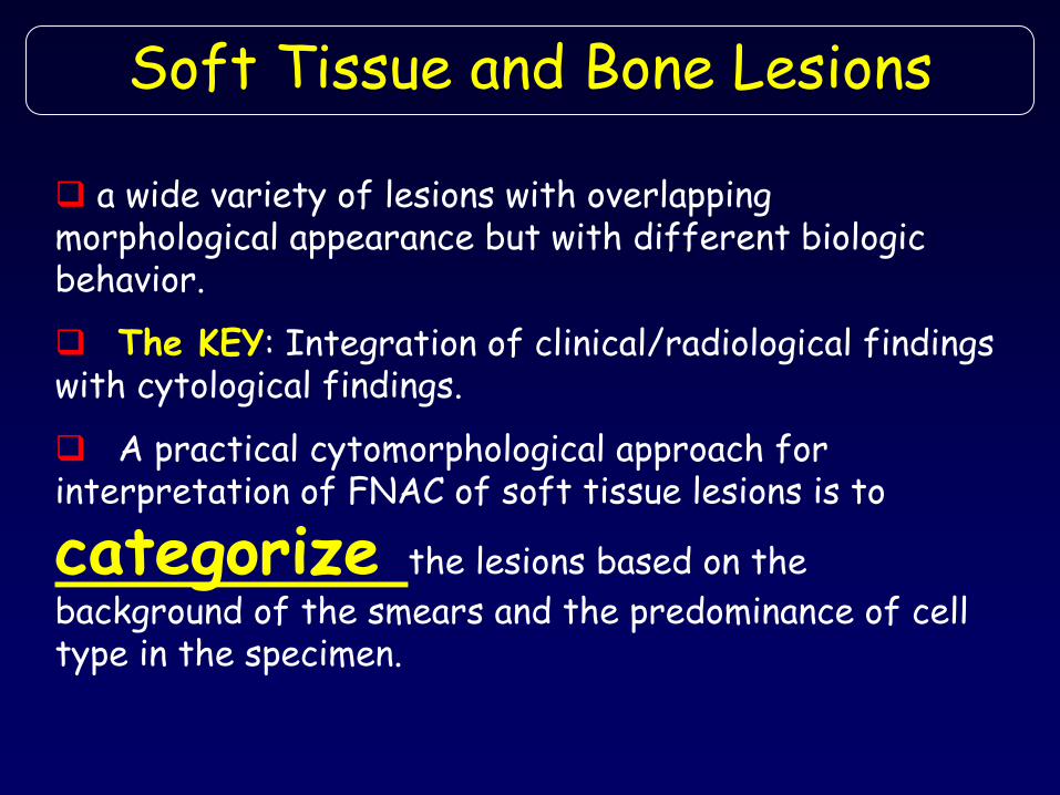
a wide variety of lesions with overlapping
morphological appearance but with different biologic behavior.
The KEY: Integration of clinical/radiological findings
with cytological findings.
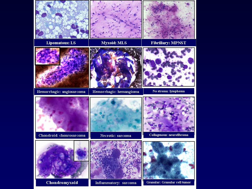
A practical cytomorphological approach for
interpretation of FNAC of soft tissue lesions is to
categorize the lesions based on the background of the smears and the predominance of cell type in the specimen.
Soft Tissue and Bone Lesions
64/F came to the ER with deterioration for her conscience. She has Hx
of breast cancer and
found to have a 2-cm occipital mass
Case 1
11/M came to FNA Clinic for left palm mass that growing slowly.
Case 2
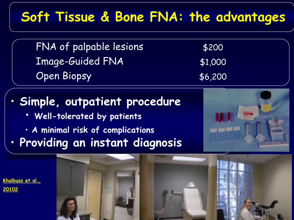
FNA of palpable lesions $200 Image-Guided FNA $1,000 Open Biopsy $6,200
• Simple, outpatient procedure• Well-tolerated by patients• A minimal risk of complications
• Providing an instant diagnosis
Soft Tissue & Bone FNA: the advantagesSoft Tissue & Bone FNA: the advantages
Khalbuss et al.,
20102
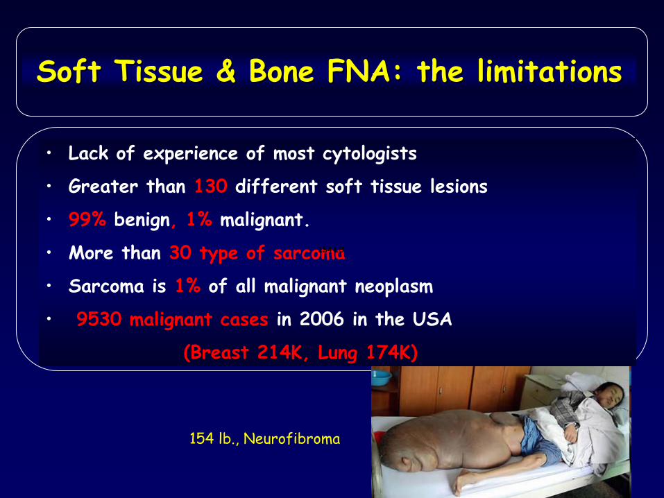
Soft Tissue & Bone FNA: the limitationsSoft Tissue & Bone FNA: the limitations
• Lack of experience of most cytologists
• Greater than 130 different soft tissue lesions
• 99%
benign, 1%
malignant.
• More than 30 type of sarcoma
• Sarcoma is 1%
of all malignant neoplasm
• 9530 malignant cases
in 2006 in the USA
(Breast 214K, Lung 174K)
154 LB 154 LB
154 lb., Neurofibroma
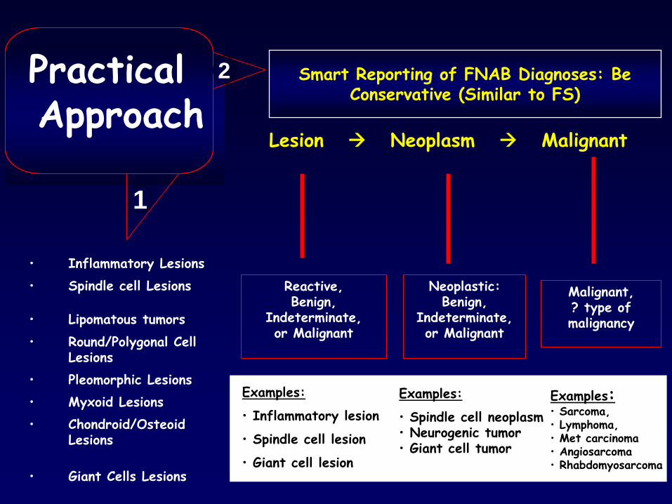
Lesion
Neoplasm
Malignant
Reactive,Benign,
Indeterminate,or Malignant
Neoplastic: Benign,
Indeterminate,or Malignant
Examples:
• Inflammatory lesion
• Spindle cell lesion
• Giant cell lesion
Examples:
• Spindle cell neoplasm• Neurogenic tumor• Giant cell tumor
Examples:• Sarcoma, • Lymphoma,• Met carcinoma• Angiosarcoma• Rhabdomyosarcoma
Smart Reporting of FNAB Diagnoses: Be Conservative (Similar to FS)
Malignant, ? type of malignancy
•
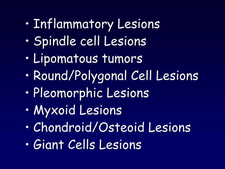
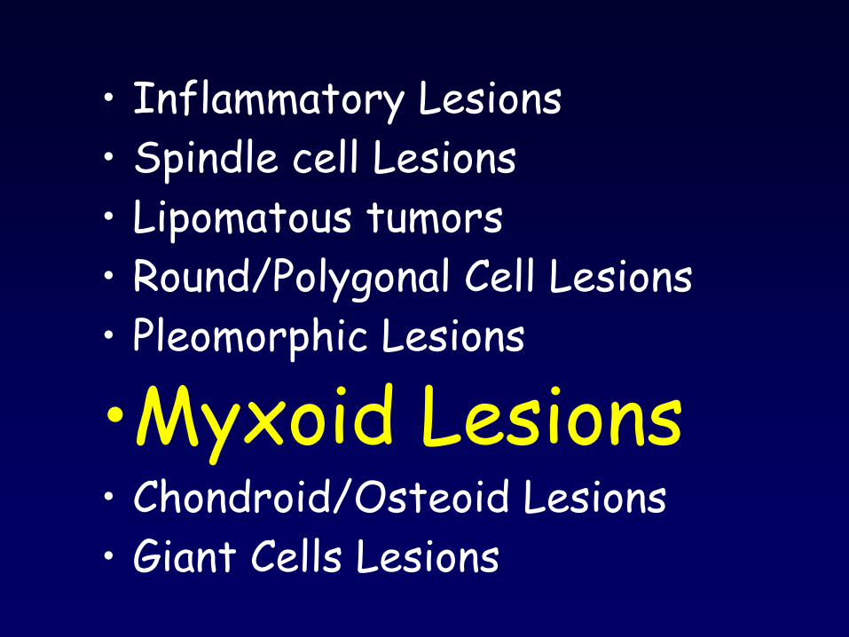
Inflammatory Lesions•
Spindle cell Lesions
•
Lipomatous tumors•
Round/Polygonal Cell Lesions
•
Pleomorphic Lesions•
Myxoid Lesions•
Chondroid/Osteoid Lesions
•
Giant Cells Lesions
Practical Approach
1
2
•
Inflammatory Lesions•
Spindle cell Lesions
•
Lipomatous tumors•
Round/Polygonal Cell Lesions
•
Pleomorphic Lesions•
Myxoid Lesions
•
Chondroid/Osteoid Lesions•
Giant Cells Lesions
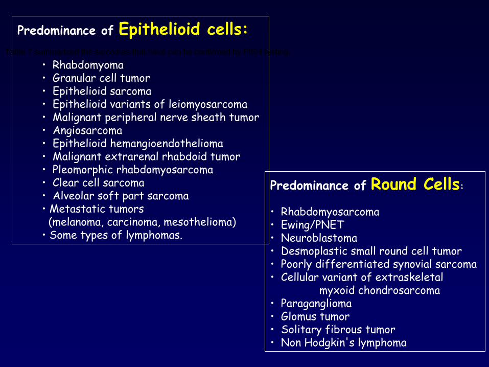
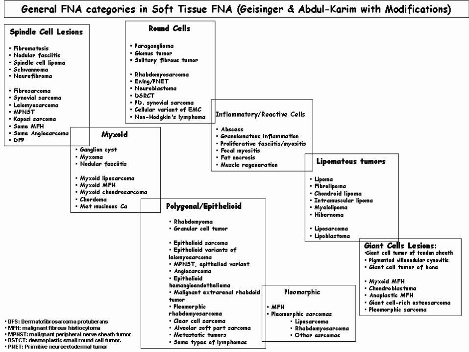
Predominance of
Epithelioid cells:
• Rhabdomyoma• Granular cell tumor• Epithelioid sarcoma• Epithelioid variants of leiomyosarcoma• Malignant peripheral nerve sheath tumor• Angiosarcoma• Epithelioid hemangioendothelioma• Malignant extrarenal rhabdoid tumor• Pleomorphic rhabdomyosarcoma• Clear cell sarcoma• Alveolar soft part sarcoma• Metastatic tumors (melanoma, carcinoma, mesothelioma)
• Some types of lymphomas.
Predominance of
Round Cells:
• Rhabdomyosarcoma• Ewing/PNET• Neuroblastoma• Desmoplastic small round cell tumor• Poorly differentiated synovial sarcoma•
Cellular variant of extraskeletal myxoid chondrosarcoma
• Paraganglioma• Glomus tumor• Solitary fibrous tumor• Non Hodgkin's lymphoma
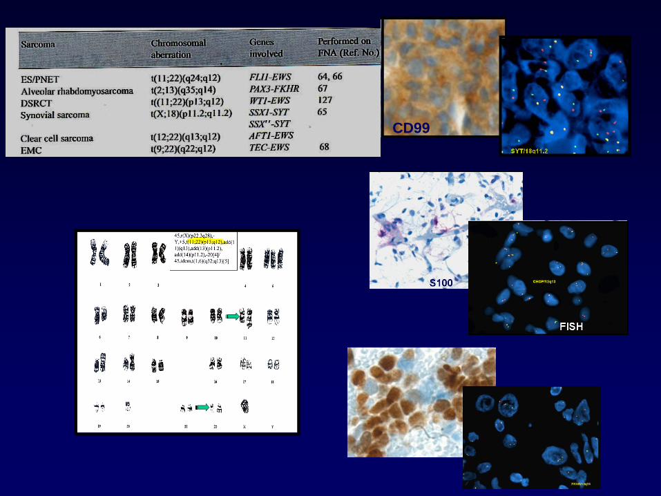
Table 7 summarized the sarcomas that have can be confirmed by FISH testing.
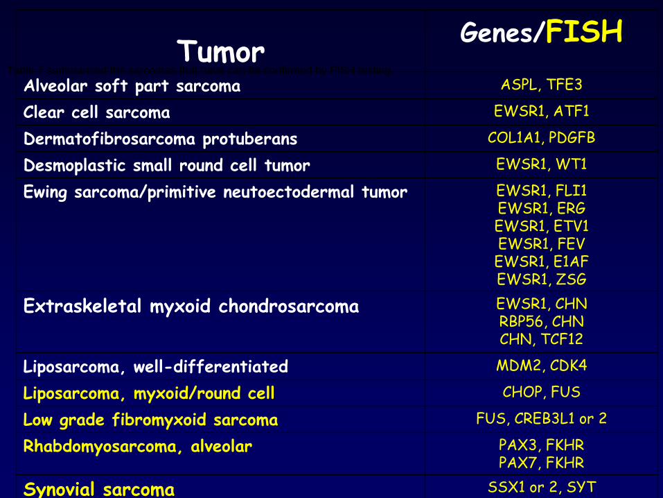
Table 7 summarized the sarcomas that have can be confirmed by FISH testing.Tumor Genes/FISH
Alveolar soft part sarcoma ASPL, TFE3
Clear cell sarcoma EWSR1, ATF1
Dermatofibrosarcoma protuberans COL1A1, PDGFB
Desmoplastic small round cell tumor EWSR1, WT1
Ewing sarcoma/primitive neutoectodermal tumor EWSR1, FLI1EWSR1, ERGEWSR1, ETV1EWSR1, FEVEWSR1, E1AFEWSR1, ZSG
Extraskeletal myxoid chondrosarcoma EWSR1, CHNRBP56, CHNCHN, TCF12
Liposarcoma, well-differentiated MDM2, CDK4
Liposarcoma, myxoid/round cell CHOP, FUS
Low grade fibromyxoid sarcoma FUS, CREB3L1 or 2
Rhabdomyosarcoma, alveolar PAX3, FKHRPAX7, FKHR
Synovial sarcoma SSX1 or 2, SYT
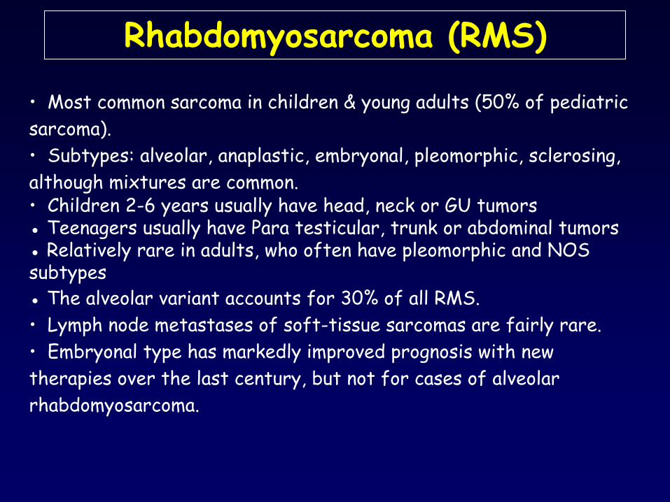
•
Most common sarcoma in children & young adults (50% of pediatric sarcoma). •
Subtypes: alveolar, anaplastic, embryonal, pleomorphic, sclerosing, although mixtures are common.• Children 2-6 years usually have head, neck or GU tumors●
Teenagers usually have Para testicular, trunk or abdominal tumors●
Relatively rare in adults, who often have pleomorphic and NOS subtypes●
The alveolar variant accounts for 30% of all RMS. • Lymph node metastases of soft-tissue sarcomas are fairly rare. •
Embryonal type has markedly improved prognosis with new therapies over the last century, but not for cases of alveolar rhabdomyosarcoma.
Rhabdomyosarcoma (RMS)
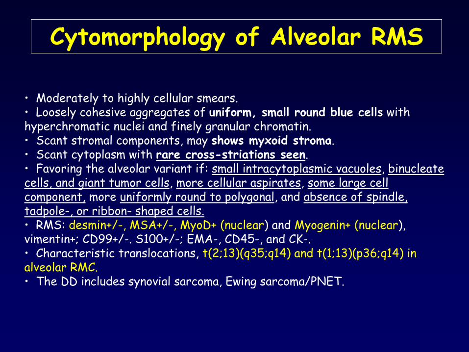
• Moderately to highly cellular smears.•
Loosely cohesive aggregates of uniform, small round blue cells
with hyperchromatic nuclei and finely granular chromatin. • Scant stromal components, may shows myxoid stroma. • Scant cytoplasm with rare cross-striations seen. •
Favoring the alveolar variant if: small intracytoplasmic vacuoles, binucleate cells, and giant tumor cells, more cellular aspirates, some large cell component,
more uniformly round to polygonal, and absence of spindle, tadpole-, or ribbon-
shaped cells. •
RMS: desmin+/-,
MSA+/-,
MyoD+ (nuclear) and Myogenin+ (nuclear), vimentin+; CD99+/-. S100+/-; EMA-, CD45-, and CK-. •
Characteristic translocations, t(2;13)(q35;q14) and t(1;13)(p36;q14) in alveolar RMC.• The DD includes synovial sarcoma, Ewing sarcoma/PNET.
Cytomorphology of Alveolar RMS
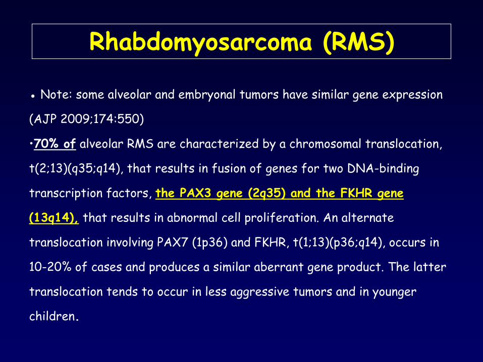
●
Note: some alveolar and embryonal tumors have similar gene expression
(AJP 2009;174:550)
•70% of
alveolar RMS are characterized by a chromosomal translocation,
t(2;13)(q35;q14), that results in fusion of genes for two DNA-binding
transcription factors, the PAX3 gene (2q35) and the FKHR gene
(13q14),
that results in abnormal cell proliferation. An alternate
translocation involving PAX7 (1p36) and FKHR, t(1;13)(p36;q14), occurs in
10-20% of cases and produces a similar aberrant gene product. The latter
translocation tends to occur in less aggressive tumors and in younger
children.
Rhabdomyosarcoma (RMS)
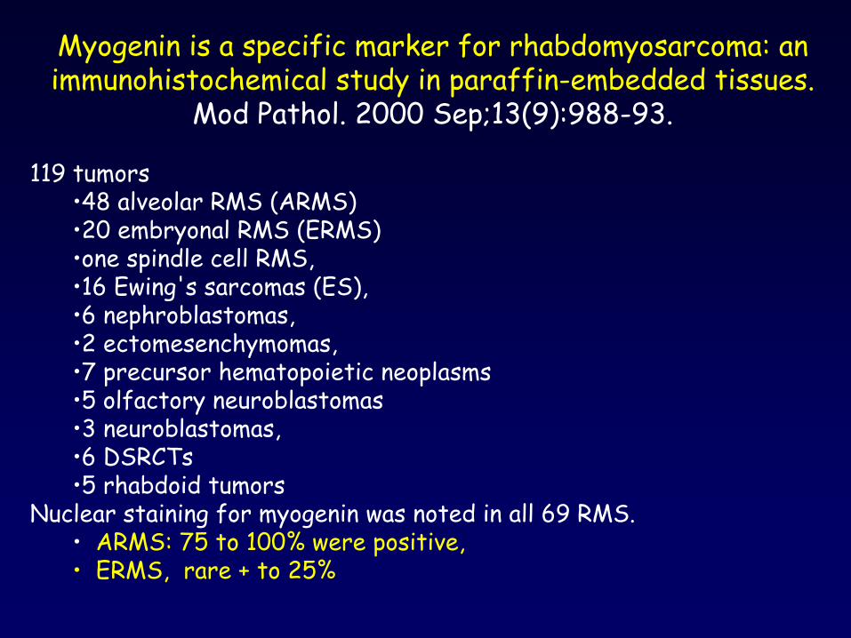
Myogenin
is a specific marker for rhabdomyosarcoma: an immunohistochemical study in paraffin-embedded tissues.
Mod Pathol.
2000 Sep;13(9):988-93.
119 tumors •48 alveolar RMS (ARMS)•20 embryonal RMS (ERMS)•one spindle cell RMS, •16 Ewing's sarcomas (ES), •6 nephroblastomas, •2 ectomesenchymomas, •7 precursor hematopoietic neoplasms•5 olfactory neuroblastomas•3 neuroblastomas, •6 DSRCTs•5 rhabdoid tumors
Nuclear staining for myogenin
was noted in all 69 RMS. • ARMS: 75 to 100% were positive, • ERMS, rare + to 25%
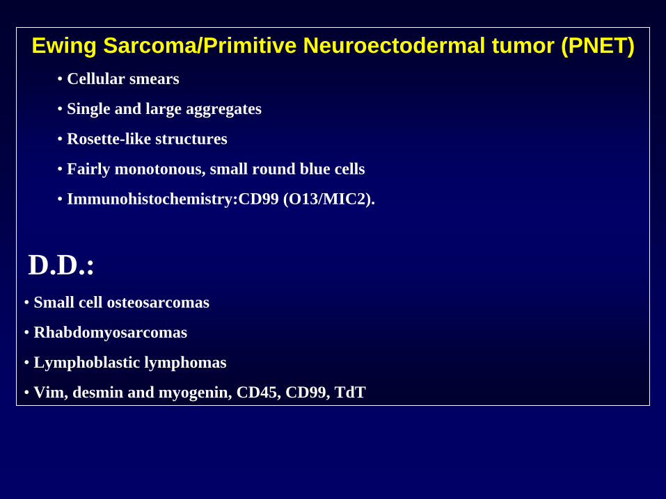
Ewing Sarcoma/Primitive Neuroectodermal tumor (PNET) • Cellular smears
• Single and large aggregates
• Rosette-like structures
• Fairly monotonous, small round blue cells
• Immunohistochemistry:CD99 (O13/MIC2).
D.D.:• Small cell osteosarcomas
• Rhabdomyosarcomas
• Lymphoblastic lymphomas
• Vim, desmin and myogenin, CD45, CD99, TdT
CD99
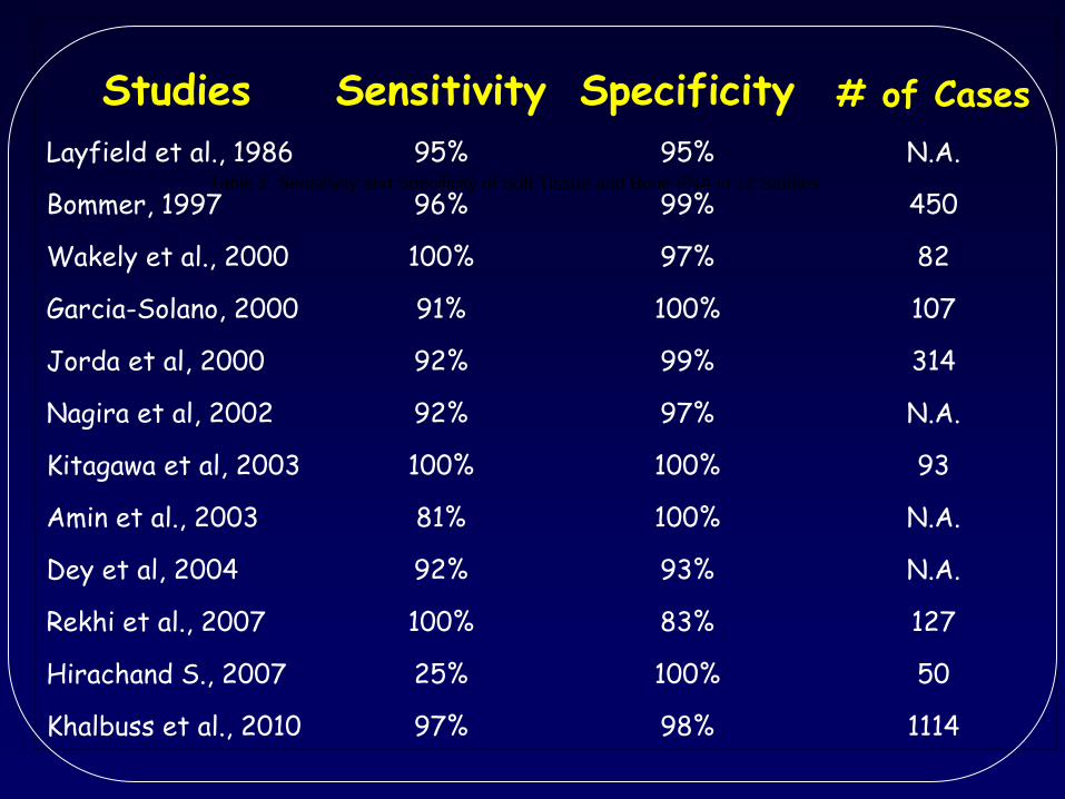
Table 1. Sensitivity and Specificity of Soft Tissue and Bone FNA in 12 Studies
Studies Sensitivity Specificity # of CasesLayfield
et al., 1986 95% 95% N.A.
Bommer, 1997 96% 99% 450
Wakely
et al., 2000 100% 97% 82
Garcia-Solano, 2000 91% 100% 107
Jorda
et al, 2000 92% 99% 314
Nagira
et al, 2002 92% 97% N.A.
Kitagawa et al, 2003 100% 100% 93
Amin et al., 2003 81% 100% N.A.
Dey
et al, 2004 92% 93% N.A.
Rekhi
et al., 2007 100% 83% 127
Hirachand
S., 2007 25% 100% 50
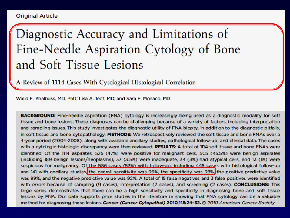
Khalbuss et al., 2010 97% 98% 1114

Predominant cell type: Spindle Cells Large Round/Polygonal cells Pleomorphic Cells, Small/Round Cell Inflammatory-Type Cells, Giant-Cell Containing.
Multidisciplinary Approach Clinical presentation Radiological findings Cytomorphological features Ancillary studies: Immuno/FISH
The background: Clean Inflammatory Myxoid/Mucinous Collagenous/Fibrotic Cartilaginous/Osseous Hemorrhagic Necrotic background
Management

Accuracy of Soft Accuracy of Soft Tissue & Bone FNATissue & Bone FNA
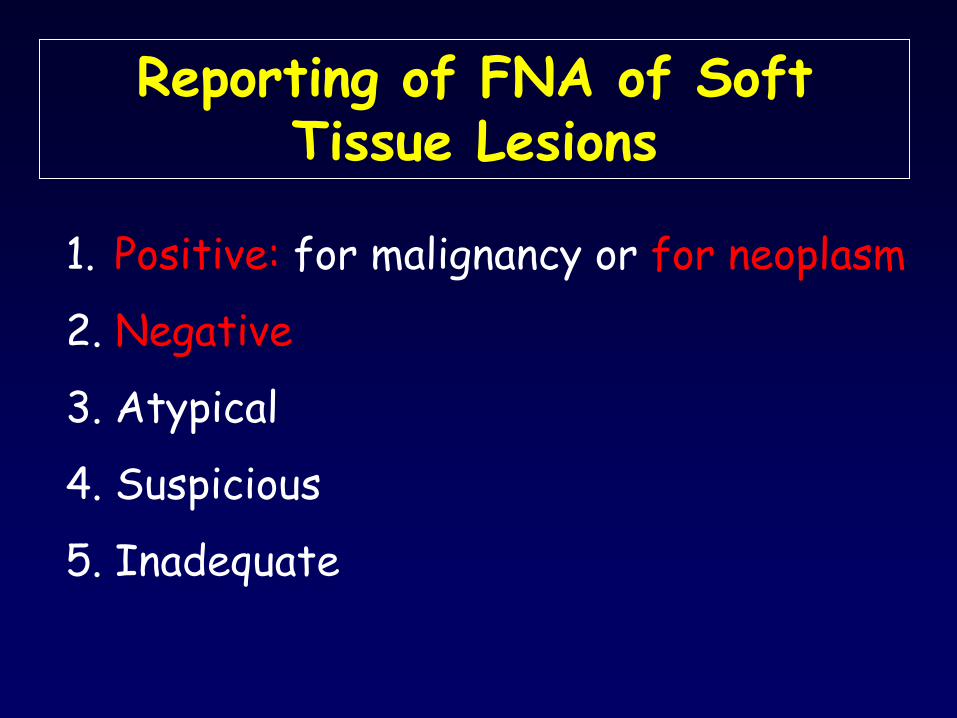
Reporting of FNA of Soft Tissue Lesions
1.
Positive:
for malignancy or for neoplasm
2. Negative
3. Atypical
4. Suspicious
5. Inadequate
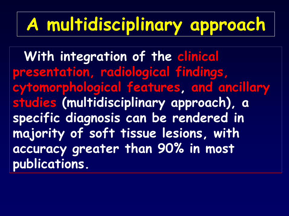
With integration of the clinical presentation, radiological findings, cytomorphological features, and ancillary studies
(multidisciplinary approach), a
specific diagnosis can be rendered in majority of soft tissue lesions, with accuracy greater than 90% in most publications.
A multidisciplinary approach
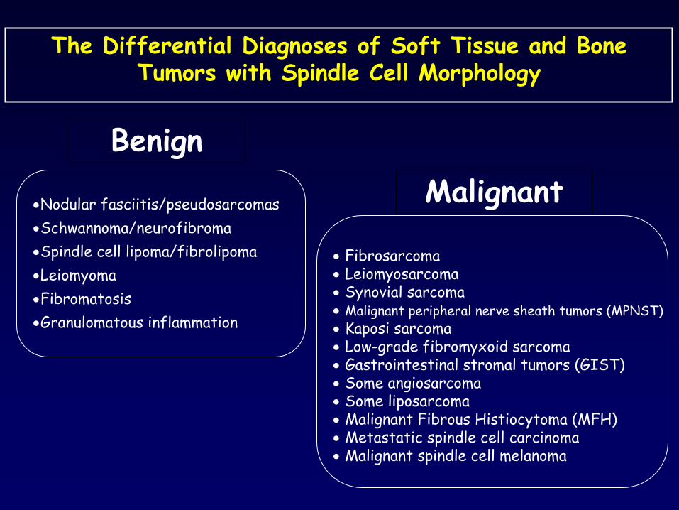
Nodular fasciitis/pseudosarcomasSchwannoma/neurofibromaSpindle cell lipoma/fibrolipomaLeiomyomaFibromatosisGranulomatous inflammation
Fibrosarcoma Leiomyosarcoma Synovial sarcoma Malignant peripheral nerve sheath tumors (MPNST) Kaposi sarcoma Low-grade fibromyxoid sarcoma Gastrointestinal stromal tumors (GIST) Some angiosarcoma Some liposarcoma Malignant Fibrous Histiocytoma (MFH) Metastatic spindle cell carcinoma Malignant spindle cell melanoma
The Differential Diagnoses of Soft Tissue and Bone Tumors with Spindle Cell Morphology
BenignMalignant
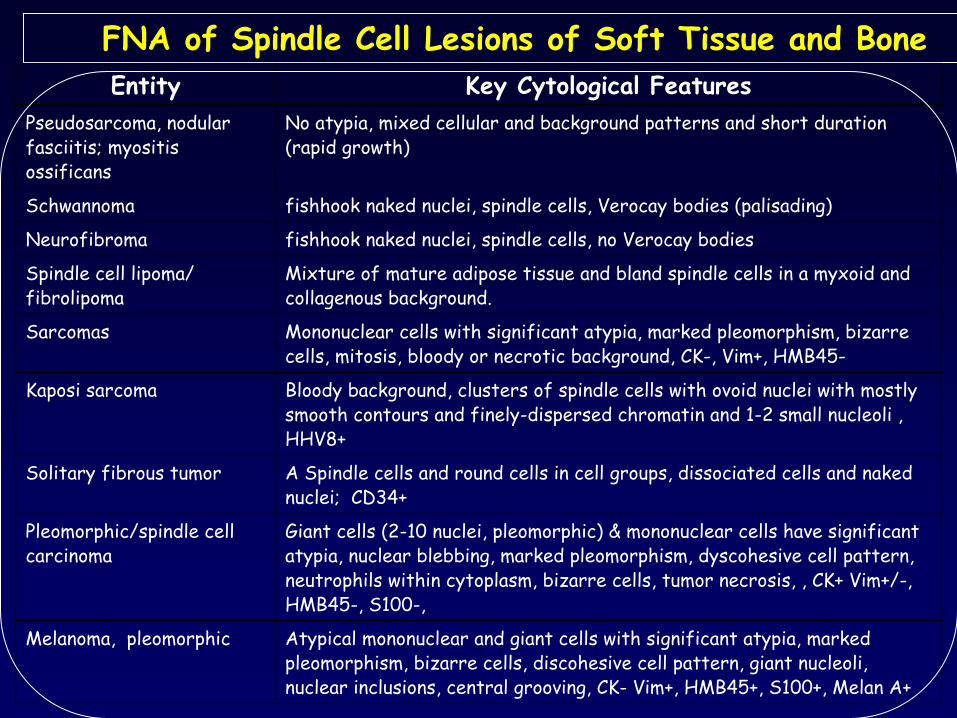
FNA of Spindle Cell Lesions of Soft Tissue and Bone Entity Key Cytological Features
Pseudosarcoma, nodular fasciitis; myositis ossificans
No atypia, mixed cellular and background patterns and short duration (rapid growth)
Schwannoma fishhook naked nuclei, spindle cells, Verocay bodies (palisading)
Neurofibroma fishhook naked nuclei, spindle cells, no Verocay bodies
Spindle cell lipoma/ fibrolipoma
Mixture of mature adipose tissue and bland spindle cells in a myxoid and collagenous background.
Sarcomas Mononuclear cells with significant atypia, marked pleomorphism, bizarre cells, mitosis, bloody or necrotic background, CK-, Vim+, HMB45-
Kaposi sarcoma Bloody background, clusters of spindle cells with ovoid nuclei with mostly smooth contours and finely-dispersed chromatin and 1-2 small nucleoli , HHV8+
Solitary fibrous tumor A Spindle cells and round cells in cell groups, dissociated cells and naked nuclei; CD34+
Pleomorphic/spindle cell carcinoma
Giant cells (2-10 nuclei, pleomorphic) & mononuclear cells have significant atypia, nuclear blebbing, marked pleomorphism, dyscohesive
cell pattern, neutrophils within cytoplasm, bizarre cells, tumor necrosis, , CK+ Vim+/-, HMB45-, S100-,
Melanoma, pleomorphic Atypical mononuclear and giant cells with significant atypia, marked pleomorphism, bizarre cells, discohesive
cell pattern, giant nucleoli, nuclear inclusions, central grooving, CK-
Vim+, HMB45+, S100+, Melan
A+
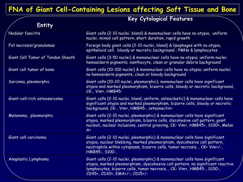
FNA of Giant Cell-Containing Lesions affecting Soft Tissue and Bone
EntityKey Cytological Features
Nodular fasciitis Giant cells (2-10 nuclei, bland) & mononuclear cells have no atypia, uniform nuclei, mixed cell pattern, short duration, rapid growth
Fat necrosis/granulomas Foreign body giant cells (2-10 nuclei, bland) & lipophages with no atypia, epithelioid cell, bloody or necrotic background ; PMNs
& lymphocytes
Giant Cell Tumor of Tendon Sheath Giant cells (3-50 nuclei) & mononuclear cells have no atypia; uniform nuclei; hemosiderin pigments; xanthocyte, clean or granular debris background
Giant cell tumor of bone Giant cells (10-100 nuclei) & mononuclear cells have no atypia; uniform nuclei; no hemosiderin pigments, clean or bloody background
Sarcoma, pleomorphic Giant cells (10-20 nuclei, pleomorphic); mononuclear cells have significant atypia and marked pleomorphism, bizarre cells, bloody or necrotic background, CK-, Vim+, HMB45-
Giant-cell-rich osteosarcoma Giant cells (2-10 nuclei, bland, uniform, osteoclastic) & mononuclear cells have significant atypia and marked pleomorphism, bizarre cells, bloody or necrotic background, CK-, Vim+, HMB45-, osteonectin+
Melanoma, pleomorphic Giant cells (2-10 nuclei, pleomorphic) & mononuclear cells have significant atypia, marked pleomorphism, bizarre cells, discohesive
cell pattern, giant nucleoli, nuclear inclusions, central grooving, CK-
Vim+, HMB45+, S100+, Melan
A+
Giant cell carcinoma Giant cells (2-10 nuclei, pleomorphic) & mononuclear cells have significant atypia, nuclear blebbing, marked pleomorphism, dyscohesive
cell pattern, neutrophils within cytoplasm, bizarre cells, tumor necrosis, , CK+ Vim+/-, HMB45-, S100-,
Anaplastic Lymphoma Giant cells (2-10 nuclei, pleomorphic) & mononuclear cells have significant atypia, marked pleomorphism, dyscohesive
cell pattern, no significant reactive lymphocytes, bizarre cells, tumor necrosis, , CK-
Vim+, HMB45-, S100-, CD45+, CD30+, EMA+/-; CD15+/-
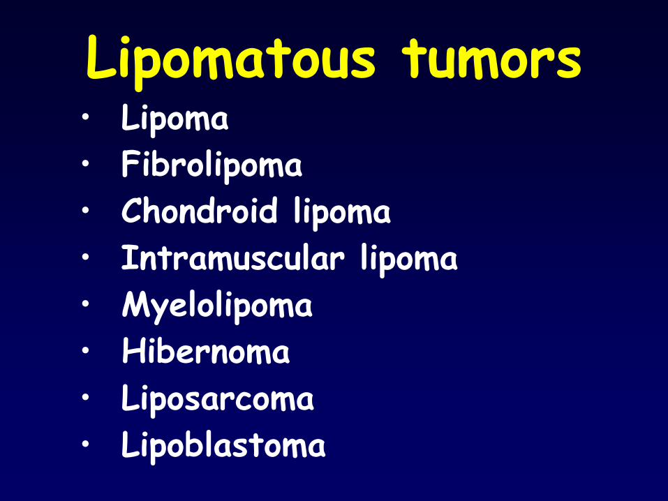
Lipomatous tumors• Lipoma• Fibrolipoma• Chondroid lipoma• Intramuscular lipoma• Myelolipoma• Hibernoma• Liposarcoma• Lipoblastoma
•
Inflammatory Lesions•
Spindle cell Lesions
•
Lipomatous tumors•
Round/Polygonal Cell Lesions
•
Pleomorphic Lesions
•Myxoid Lesions•
Chondroid/Osteoid Lesions
•
Giant Cells Lesions
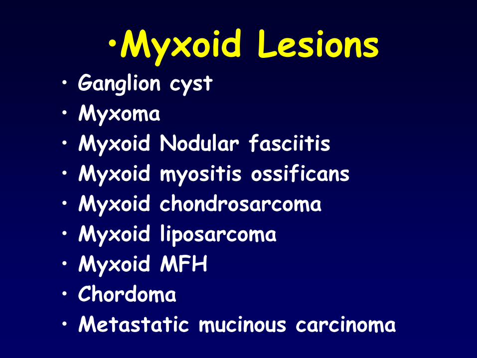
•Myxoid Lesions• Ganglion cyst• Myxoma• Myxoid Nodular fasciitis• Myxoid myositis ossificans• Myxoid chondrosarcoma• Myxoid liposarcoma• Myxoid MFH• Chordoma• Metastatic mucinous carcinoma
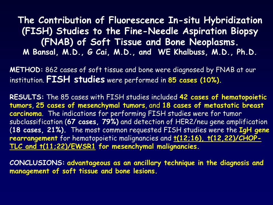
The Contribution of Fluorescence In-situ Hybridization (FISH) Studies to the Fine-Needle Aspiration Biopsy
(FNAB) of Soft Tissue and Bone Neoplasms.M Bansal, M.D., G Cai, M.D., and WE Khalbuss, M.D., Ph.D.
METHOD:
862 cases of soft tissue and bone were diagnosed by FNAB at our
institution. FISH studies
were performed in 85 cases (10%).
RESULTS:
The 85 cases with FISH studies included 42 cases of hematopoietic tumors, 25 cases of mesenchymal tumors, and 18 cases of metastatic breast carcinoma. The indications for performing FISH studies were for tumor subclassification (67 cases, 79%)
and detection of HER2/neu gene amplification (18 cases, 21%).
The most common requested FISH studies were the IgH gene rearrangement
for hematopoietic malignancies and t(12;16), t(12,22)/CHOP-
TLC and t(11;22)/EWSR1
for mesenchymal malignancies.
CONCLUSIONS:
advantageous as an ancillary technique in the diagnosis and management of soft tissue and bone lesions.
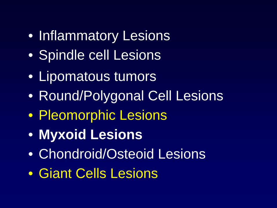
• Inflammatory Lesions• Spindle cell Lesions• Lipomatous tumors• Round/Polygonal Cell Lesions • Pleomorphic Lesions• Myxoid Lesions• Chondroid/Osteoid Lesions• Giant Cells Lesions
CONCLUSIONS• FNA of soft tissue lesions can be challenging
•
Soft tissue lesions can usually be classified into general categories based on the predominant cell type and background
and often
a specific diagnosis can be made or suggested based on:
-
Cytomorphologic characteristics,
-
Clinicoradiographic features, and
- Ancillary studies.