Embed Size (px)
Citation preview
CyTOF analyses in rheumatoid arthritis
Deepak Rao, MD PhD
Rheumatology, Immunology, Allergy, BWH
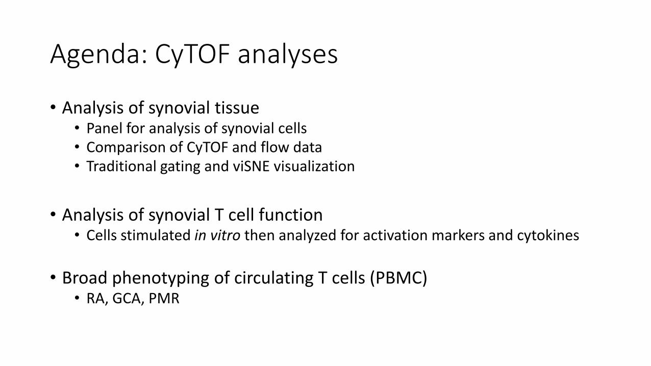
Agenda: CyTOF analyses
• Analysis of synovial tissue • Panel for analysis of synovial cells • Comparison of CyTOF and flow data • Traditional gating and viSNE visualization
• Analysis of synovial T cell function • Cells stimulated in vitro then analyzed for activation markers and cytokines
• Broad phenotyping of circulating T cells (PBMC) • RA, GCA, PMR
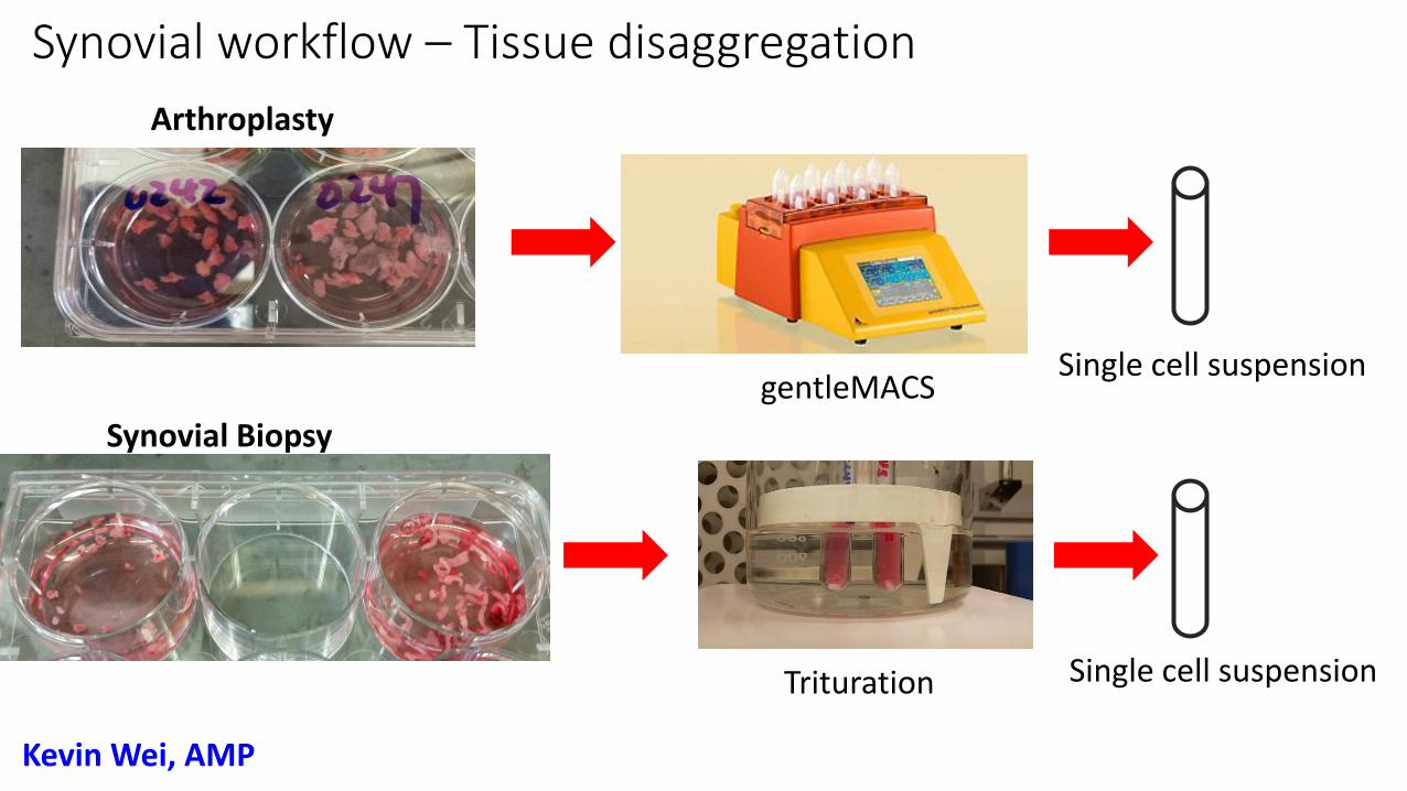
Synovial Biopsy
Arthroplasty
Single cell suspension
Single cell suspension
gentleMACS
Trituration
Synovial workflow – Tissue disaggregation
Kevin Wei, AMP
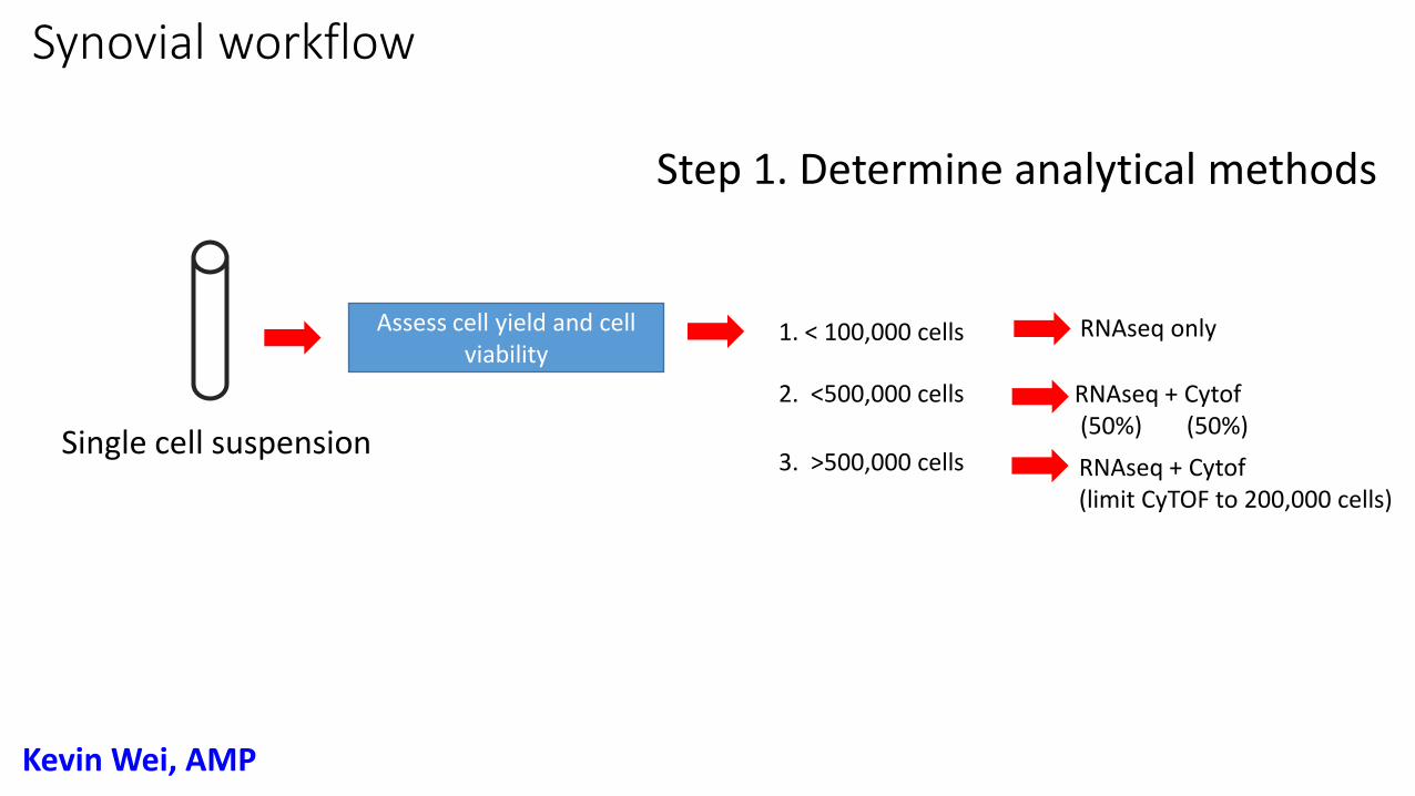
Synovial workflow
Assess cell yield and cell viability
Step 1. Determine analytical methods
1. < 100,000 cells
2. <500,000 cells
3. >500,000 cells
RNAseq only
RNAseq + Cytof (50%) (50%)
RNAseq + Cytof (limit CyTOF to 200,000 cells)
Single cell suspension
Kevin Wei, AMP
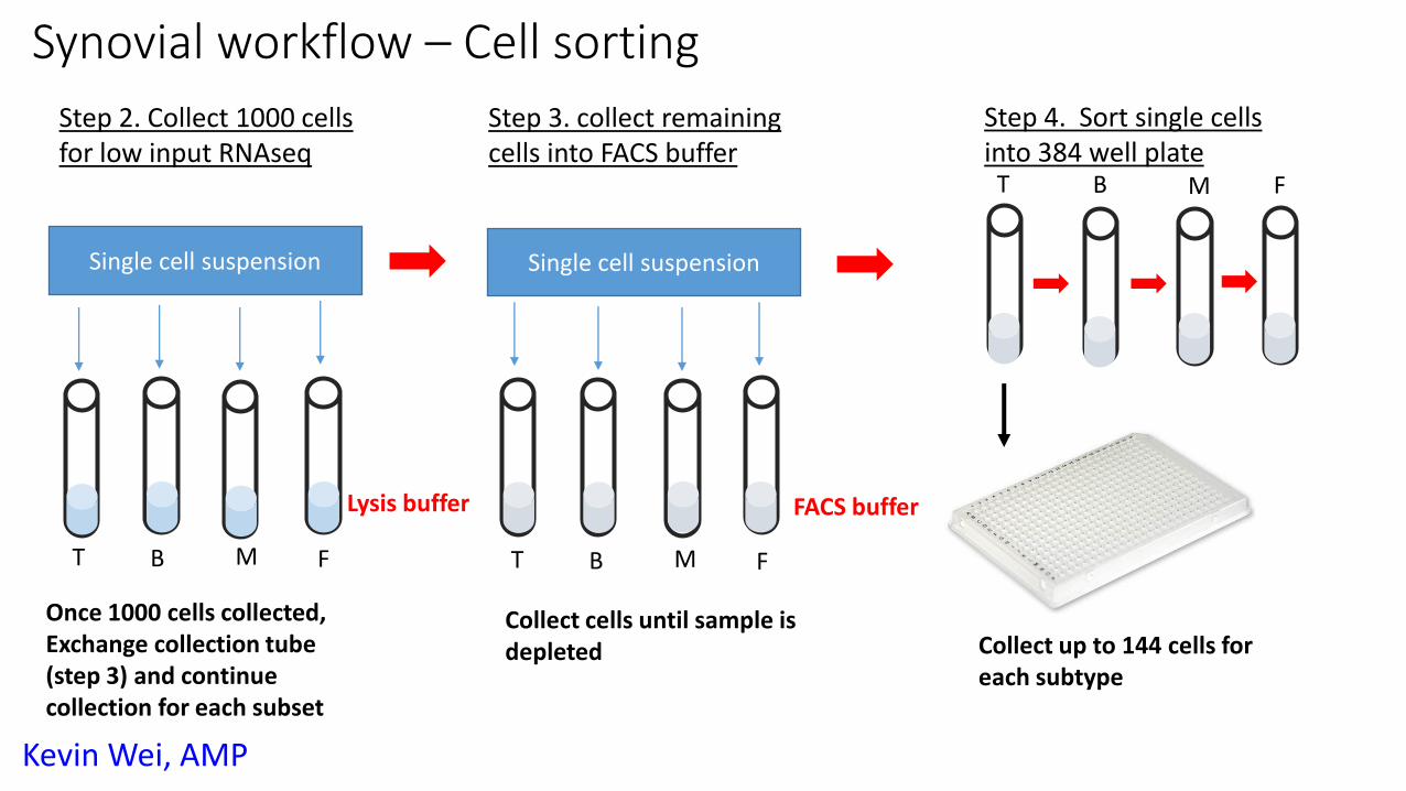
Step 2. Collect 1000 cells for low input RNAseq
Single cell suspension
Once 1000 cells collected, Exchange collection tube (step 3) and continue collection for each subset
Step 3. collect remaining cells into FACS buffer
Single cell suspension
Lysis buffer
Step 4. Sort single cells into 384 well plate
T B M F
T B M F
FACS buffer
T B M F
Collect cells until sample is depleted Collect up to 144 cells for
each subtype
Synovial workflow – Cell sorting
Kevin Wei, AMP
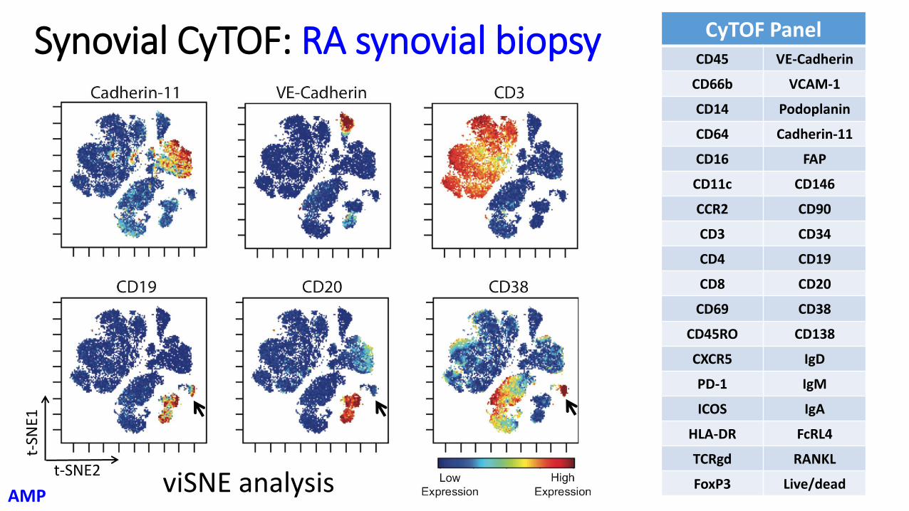
Synovial CyTOF: RA synovial biopsy CyTOF Panel CD45 VE-Cadherin
CD66b VCAM-1
CD14 Podoplanin
CD64 Cadherin-11
CD16 FAP
CD11c CD146
CCR2 CD90
CD3 CD34
CD4 CD19
CD8 CD20
CD69 CD38
CD45RO CD138
CXCR5 IgD
PD-1 IgM
ICOS IgA
HLA-DR FcRL4
TCRgd RANKL
FoxP3 Live/dead
t-SN
E1
t-SNE2 viSNE analysis
AMP
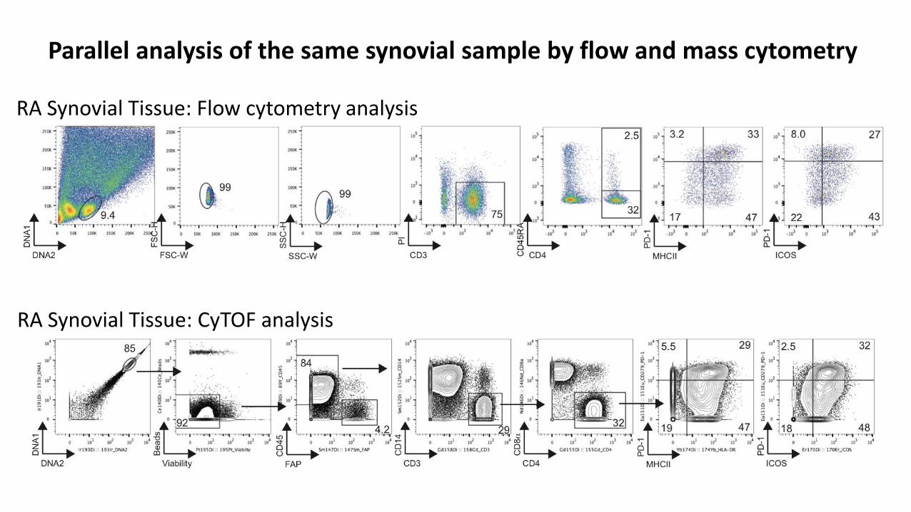
RA Synovial Tissue: Flow cytometry analysis
RA Synovial Tissue: CyTOF analysis
Parallel analysis of the same synovial sample by flow and mass cytometry
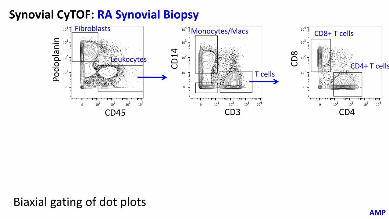
Synovial CyTOF: RA Synovial Biopsy
CD4 CD3 CD45
CD
8
CD
14
Pod
op
lan
in Monocytes/Macs
T cells
CD8+ T cells
CD4+ T cells Leukocytes
Fibroblasts
Biaxial gating of dot plots AMP
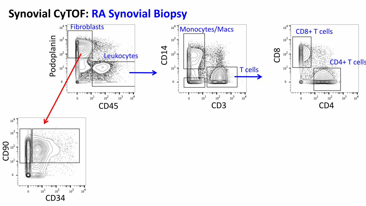
CD34
CD4 CD3 CD45
CD
8
CD
14
Pod
op
lan
in
CD
90
Monocytes/Macs
T cells
CD8+ T cells
CD4+ T cells Leukocytes
Fibroblasts
Synovial CyTOF: RA Synovial Biopsy
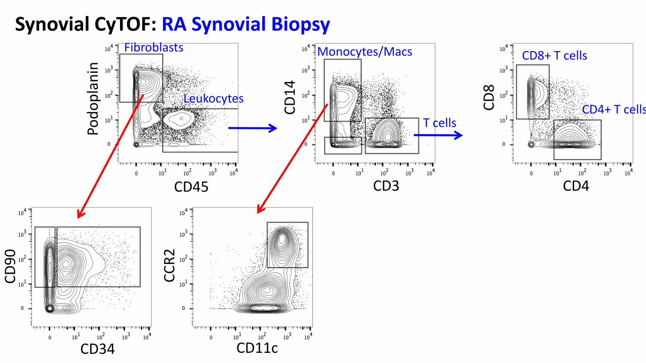
CD11c CD34
CD4 CD3 CD45
CD
8
CD
14
Pod
op
lan
in
CC
R2
CD
90
Monocytes/Macs
T cells
CD8+ T cells
CD4+ T cells Leukocytes
Fibroblasts
Synovial CyTOF: RA Synovial Biopsy
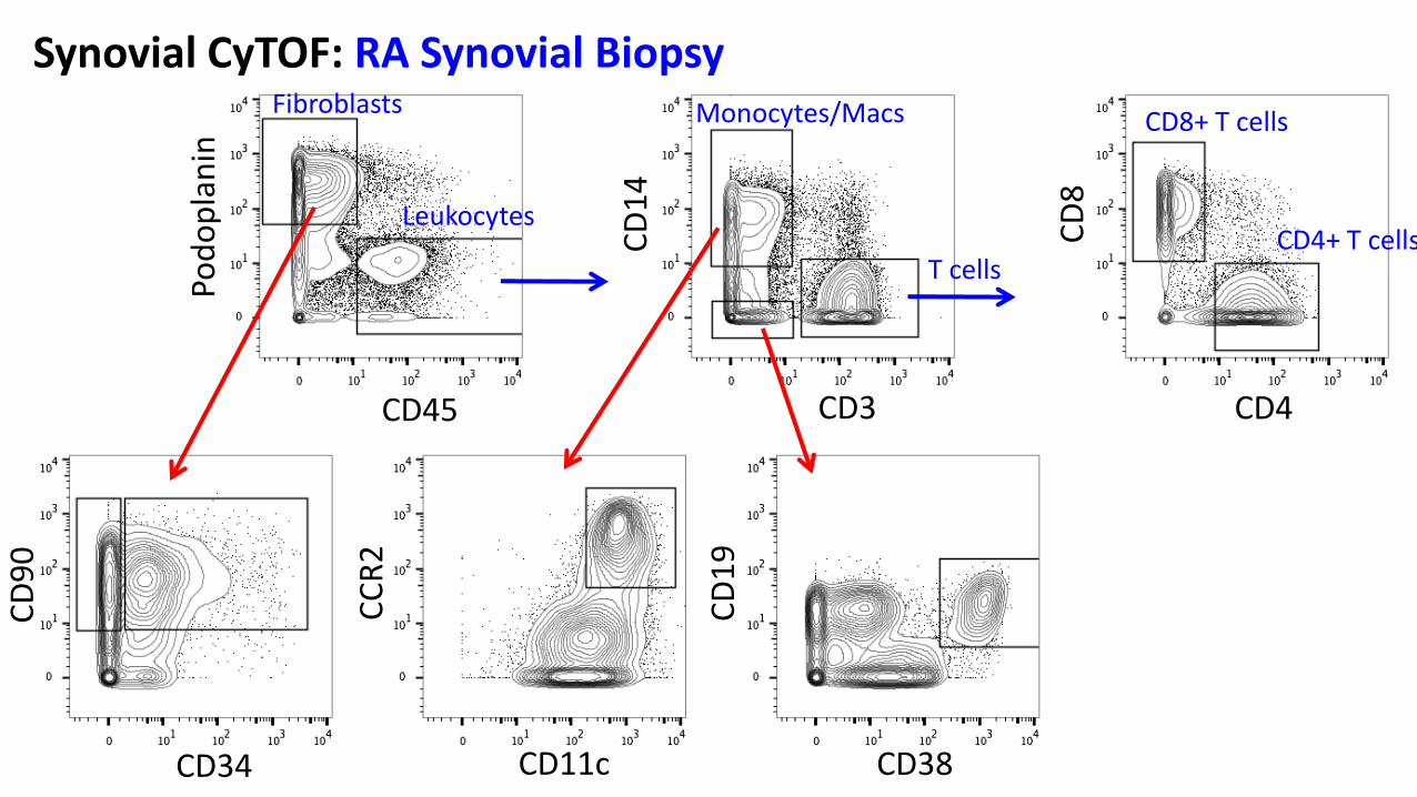
CD38 CD11c CD34
CD4 CD3 CD45
CD
8
CD
14
Pod
op
lan
in
CD
19
CC
R2
CD
90
Monocytes/Macs
T cells
CD8+ T cells
CD4+ T cells Leukocytes
Fibroblasts
Synovial CyTOF: RA Synovial Biopsy
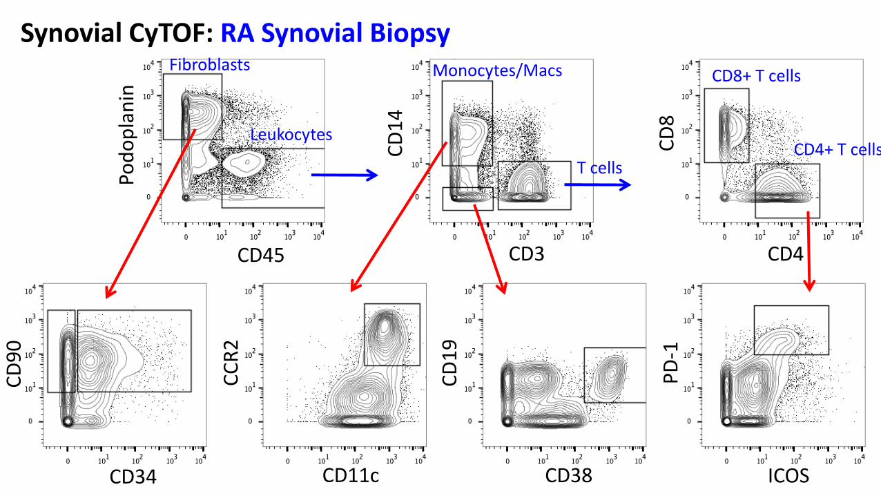
ICOS CD38 CD11c CD34
CD4 CD3 CD45
CD
8
CD
14
Pod
op
lan
in
CD
19
CC
R2
CD
90
PD
-1
Monocytes/Macs
T cells
CD8+ T cells
CD4+ T cells Leukocytes
Fibroblasts
Synovial CyTOF: RA Synovial Biopsy

Synovial CyTOF: RA synovial biopsy CyTOF Panel CD45 VE-Cadherin
CD66b VCAM-1
CD14 Podoplanin
CD64 Cadherin-11
CD16 FAP
CD11c CD146
CCR2 CD90
CD3 CD34
CD4 CD19
CD8 CD20
CD69 CD38
CD45RO CD138
CXCR5 IgD
PD-1 IgM
ICOS IgA
HLA-DR FcRL4
TCRgd RANKL
FoxP3 Live/dead
t-SN
E1
t-SNE2 viSNE analysis
AMP
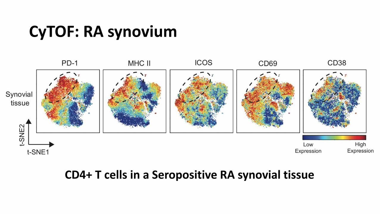
CyTOF: RA synovium
CD4+ T cells in a Seropositive RA synovial tissue
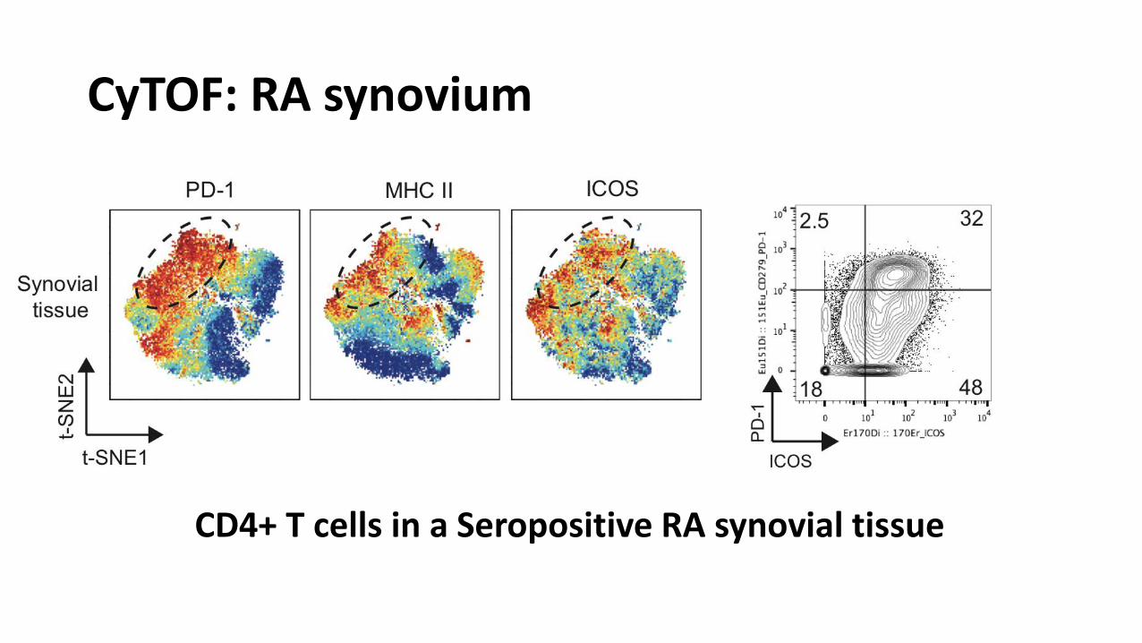
CyTOF: RA synovium
CD4+ T cells in a Seropositive RA synovial tissue
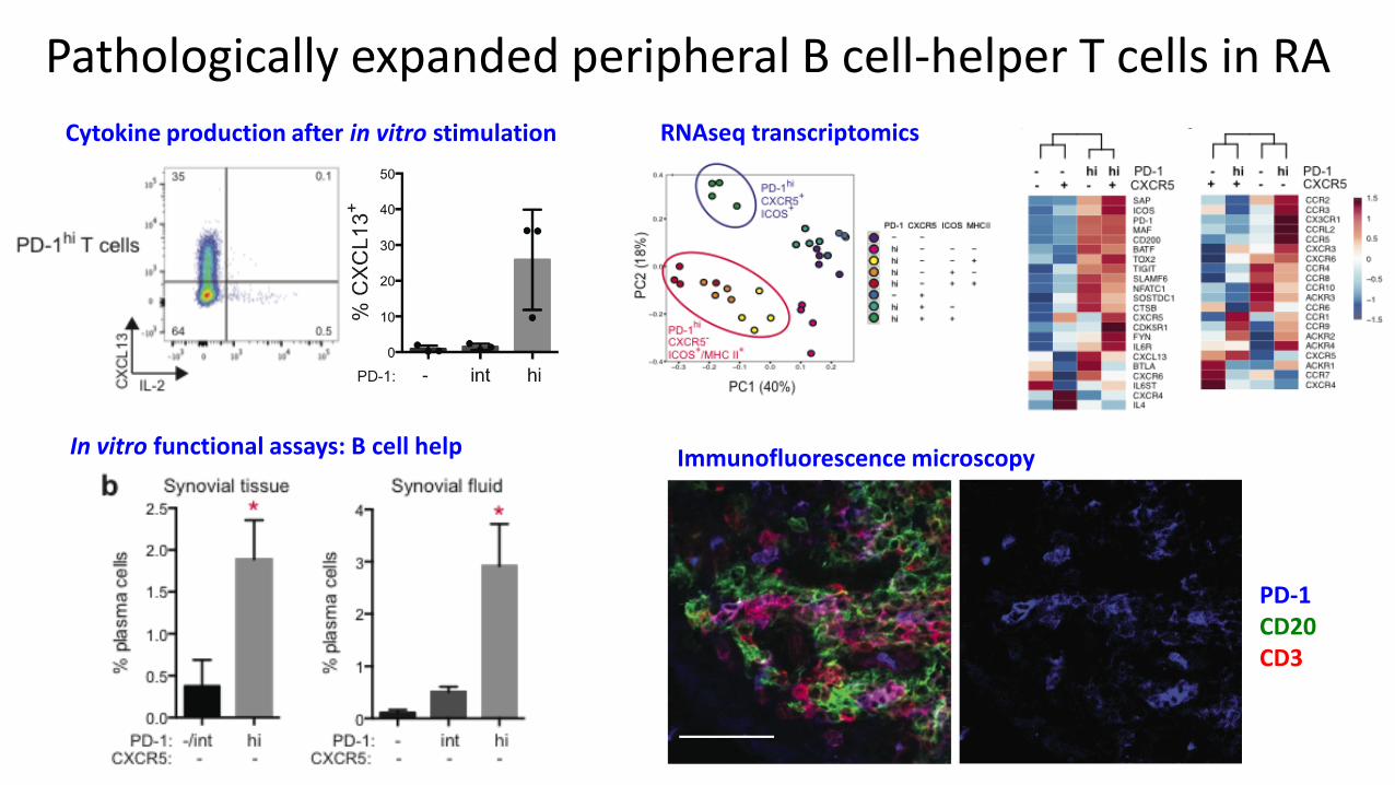
Pathologically expanded peripheral B cell-helper T cells in RA
Cytokine production after in vitro stimulation RNAseq transcriptomics
In vitro functional assays: B cell help Immunofluorescence microscopy
PD-1 CD20 CD3
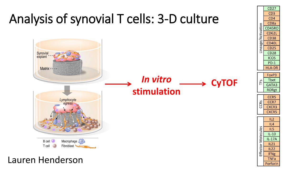
Analysis of synovial T cells: 3-D culture
Lauren Henderson
In vitro stimulation
CyTOF
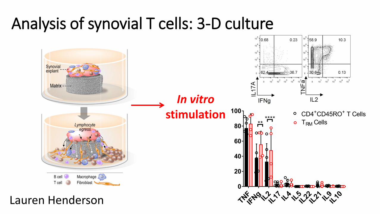
Analysis of synovial T cells: 3-D culture
Lauren Henderson
In vitro stimulation
Immunophenotyping of PBMCs by CyTOF
• Identify immune cell populations with altered frequencies in specific autoimmune diseases • Analytical methods in the Raychaudhuri lab – Chamith Fonseka
• Determine expression of many different proteins on specific immune cell populations of interest
• Data generated so far: • RA vs controls (n=7 each)
• RA vs controls w/ and w/o stimulation (Raychaudhuri lab, n=~30 each)
• RA, OA, SLE, controls (AMP, Lederer lab, in progress)
• GCA, PMR, seronegative RA, controls (n=4 each)
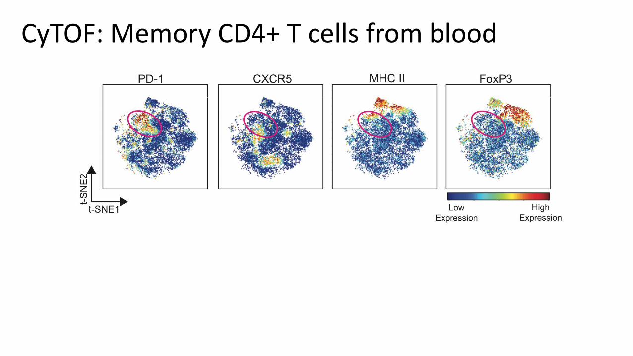
CyTOF: Memory CD4+ T cells from blood
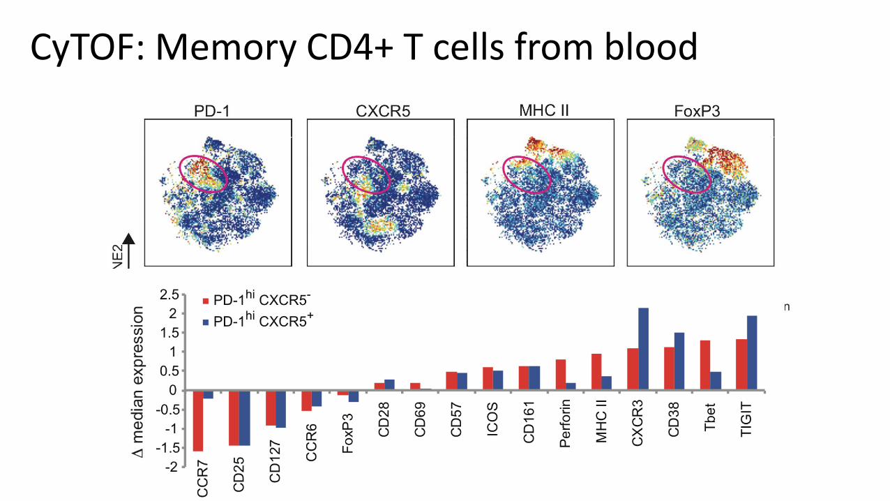
CyTOF: Memory CD4+ T cells from blood
CyTOF for analysis of rheumatic disease samples
• Powerful, accessible discovery tool to measure 35+ markers on cell samples • Staining protocols have undergone substantial maturation and now regularly utilized
(Raychaudhuri, Lederer, Brenner labs)
• Panels developed for broad analysis of synovial cells
• Panels developed to phenotype T cells, B cells, myeloid cells in detail
• Simple data analysis strategies that we can teach you (e.g. viSNE)
• New, powerful analysis methods under development in the Raychaudhuri lab