Embed Size (px)
Citation preview
140International Journal of Scientifi c Study | September 2015 | Vol 3 | Issue 6
Cyto-histological Correlative Study of Thyroid Neoplasms by Imprint MethodH B Shashidhar1, D Vani2, N S Ashwini2,3, M Sandhya4, M Bharathi5
1Associate Professor, Department of Pathology, Mysore Medical College & Research Institute, Mysore, Karnataka, India, 2Associate Professor, Department of Pathology, Mysore Medical College & Research Institute, Mysore, Karnataka, India, 3Post-graduate, Student, Department of Pathology, Mysore Medical College & Research Institute, Mysore, Karnataka, India, 4Post-graduate, Student, Department of Pathology, Mysore Medical College & Research Institute, Mysore, Karnataka, India, 5Head of Department, Department of Pathology, Mysore Medical College & Research Institute, Mysore, Karnataka, India
and the operating surgeon. Thyroid nodules are common with an estimated prevalence ranging from 4% by palpation to 67% by ultrasonography.1 Most of the thyroid neoplasms are clinically palpable and after a provisional clinical diagnosis, fi ne-needle aspiration plays a major role in determining nature of the tumor. It is during the operative procedure, that the role of pathologist comes into force, through the intra-operative diagnosis.
Imprint cytology (IC) is an intra-operative diagnostic technique introduced by Dudgeon and Patrick in the year 1927. After pioneering work, this technique remained in oblivion for years. Later, the immense potential of intra-operative imprint cytodiagnosis was recognized and it came
INTRODUCTION
Disorders involving thyroid have been unique in both presentation and symptoms. Due to this, diagnosis of thyroid lesions and most importantly, thyroid neoplasms, forms an important aspect involving both the pathologist
Original Article
Abstract
Background: Imprint cytology (IC) has become an indispensable component in the intra-operative rapid cytodiagnosis of tumor types. Its rise as a worthy alternative to frozen sections in places where facilities for the same are not available is commendable as it is inexpensive procedure which yields smears, easy to interpret and is worthy of consideration to be included as a routine intra-operative diagnostic procedure in the evaluation of thyroid neoplasms.
Objectives: The objective of this study was to adopt intra-operative thyroid neoplasms in order to facilitate decision regarding extent of surgery to be performed, expedite the planning for therapy and to assess the effi cacy of intra-operative IC by correlation with histopathological diagnosis.
Methods: A total of 96 patients with clinically diagnosed thyroid swellings from K. R. Hospital referred to the Department of Pathology and subsequently confi rmed as thyroid neoplasms by fi ne-needle aspiration cytology and posted for surgery formed the basis for this study. Immediately following the surgical removal of the neoplasm, imprint smears were taken from the tumor area. The air dried or wet-fi xed smears were stained routinely by rapid hematoxylin and eosin and by modifi ed rapid papanicolaou method.
Results: Out of 96 cases of thyroid neoplasms, 58 cases were diagnosed as benign and 38 cases as malignant. Follicular neoplasms were 54 forming the majority among the benign tumors with 4 cases of Hurthle cell adenomas. Amongst the malignant tumors, 30 papillary carcinomas, 4 follicular carcinomas, and 2 cases each of non-Hodgkin lymphoma maltoma and anaplastic carcinoma were diagnosed.
Conclusion: The overall sensitivity was 95.45%, the specifi city, and positive predictive value was 100% each and negative predictive value of 90.6% was seen in the present study. The overall accuracy encountered was 95.52%.
Key words: Adenomas, Benign, Neoplasm, Thyroid
Access this article online
www.ijss-sn.com
Month of Submission : 07-2015Month of Peer Review : 08-2015Month of Acceptance : 09-2015Month of Publishing : 09-2015
Corresponding Author: Dr. NS Ashwini, Department of Pathology, Mysore Medical College & Research Institute, Mysore, Karnataka, India.Phone: +91-09886021425. E-mail: [email protected]
DOI: 10.17354/ijss/2015/410
Shashidhar, et al.: Imprint cytology study of thyroid neoplasms
141 International Journal of Scientifi c Study | September 2015 | Vol 3 | Issue 6
to be utilized in various diagnostic works.2 Since freezing artifacts are avoided, the cytological details provided by the intra-operative cytological imprint specimens are superior to that of frozen sections (FS). Imprints cover a wide area of tissue, and therefore, differences of histological structures are more likely to be represented adequately and clearly than in conventional paraffi n embedded sections taken from a small area. IC is simple, inexpensive, rapidly diagnostic, and easy to perform procedure especially in thyroid neoplasms. It has gained immense popularity globally and has been accepted as a routine diagnostic procedure in most of the hospitals worldwide. The present study was carried out to evaluate the diagnostic value of IC intraoperatively along with histopathological correlation.
METHODS
The study comprises of 96 cases of IC of thyroid neoplasms. The lesions were confirmed as thyroid neoplasms by fi ne-needle aspiration cytology (FNAC) and posted for surgery. The imprint procedure was done intraoperatively at K. R. Hospital attached to Mysore Medical College, Mysore. The results were subsequently correlated with the histopathological diagnosis.
IC was done intraoperatively under the guidance of the operating surgeon after obtaining informed consent from the patient. The procedure was done under absolute aseptic precautions by the pathologist.
The imprint smears were prepared from the cut surface of freshly dissected thyroid specimen by touching the glass slides on surface, focusing on suspicious areas. The smears were immediately wet-fi xed in 95% alcohol fi xative and stained by ultrafast Papanicolaou technique. Carnoy’s fi xative was used when smears were hemorrhagic and in few cases, smears were air dried and stained with May-Grünwald-Giemsa stain. The resected specimen was sent for histopathological examination for correlative study.
RESULTS
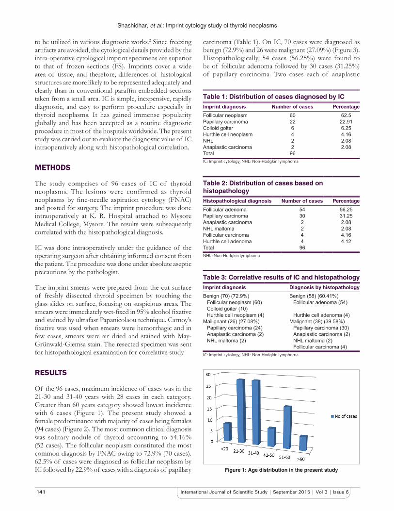
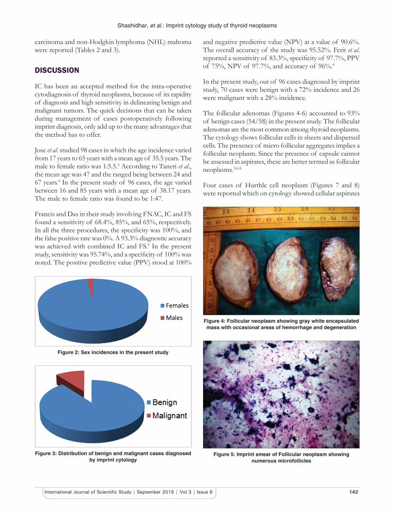
Of the 96 cases, maximum incidence of cases was in the 21-30 and 31-40 years with 28 cases in each category. Greater than 60 years category showed lowest incidence with 6 cases (Figure 1). The present study showed a female predominance with majority of cases being females (94 cases) (Figure 2). The most common clinical diagnosis was solitary nodule of thyroid accounting to 54.16% (52 cases). The follicular neoplasm constituted the most common diagnosis by FNAC owing to 72.9% (70 cases). 62.5% of cases were diagnosed as follicular neoplasm by IC followed by 22.9% of cases with a diagnosis of papillary
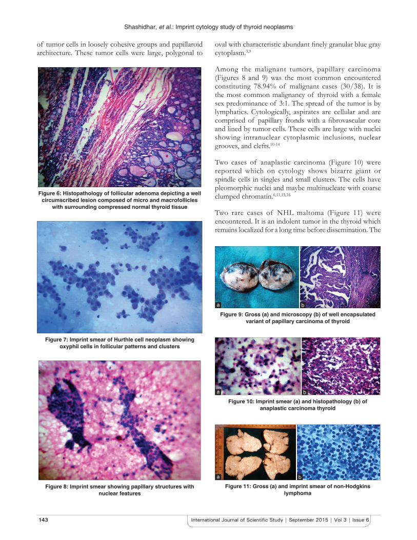
carcinoma (Table 1). On IC, 70 cases were diagnosed as benign (72.9%) and 26 were malignant (27.09%) (Figure 3). Histopathologically, 54 cases (56.25%) were found to be of follicular adenoma followed by 30 cases (31.25%) of papillary carcinoma. Two cases each of anaplastic
Table 1: Distribution of cases diagnosed by ICImprint diagnosis Number of cases PercentageFollicular neoplasm 60 62.5Papillary carcinoma 22 22.91Colloid goiter 6 6.25Hurthle cell neoplasm 4 4.16NHL 2 2.08Anaplastic carcinoma 2 2.08Total 96IC: Imprint cytology, NHL: Non-Hodgkin lymphoma
Table 2: Distribution of cases based on histopathologyHistopathological diagnosis Number of cases PercentageFollicular adenoma 54 56.25Papillary carcinoma 30 31.25Anaplastic carcinoma 2 2.08NHL maltoma 2 2.08Follicular carcinoma 4 4.16Hurthle cell adenoma 4 4.12Total 96NHL: Non-Hodgkin lymphoma
Table 3: Correlative results of IC and histopathologyImprint diagnosis Diagnosis by histopathologyBenign (70) (72.9%) Benign (58) (60.41%)
Follicular neoplasm (60) Follicular adenoma (54)Colloid goiter (10)Hurthle cell neoplasm (4) Hurthle cell adenoma (4)
Mailignant (26) (27.08%) Malignant (38) (39.58%)Papillary carcinoma (24) Papillary carcinoma (30)Anaplastic carcinoma (2) Anaplastic carcinoma (2)NHL maltoma (2) NHL maltoma (2)
Follicular carcinoma (4)IC: Imprint cytology, NHL: Non-Hodgkin lymphoma
Figure 1: Age distribution in the present study
Shashidhar, et al.: Imprint cytology study of thyroid neoplasms
142International Journal of Scientifi c Study | September 2015 | Vol 3 | Issue 6
carcinoma and non-Hodgkin lymphoma (NHL) maltoma were reported (Tables 2 and 3).
DISCUSSION
IC has been an accepted method for the intra-operative cytodiagnosis of thyroid neoplasms, because of its rapidity of diagnosis and high sensitivity in delineating benign and malignant tumors. The quick decisions that can be taken during management of cases postoperatively following imprint diagnosis, only add up to the many advantages that the method has to offer.
Jose et al. studied 98 cases in which the age incidence varied from 17 years to 65 years with a mean age of 35.5 years. The male to female ratio was 1:5.5.3 According to Taneri et al., the mean age was 47 and the ranged being between 24 and 67 years.4 In the present study of 96 cases, the age varied between 16 and 85 years with a mean age of 38.17 years. The male to female ratio was found to be 1:47.
Francis and Das in their study involving FNAC, IC and FS found a sensitivity of 68.4%, 85%, and 65%, respectively. In all the three procedures, the specifi city was 100%, and the false positive rate was 0%. A 93.3% diagnostic accuracy was achieved with combined IC and FS.5 In the present study, sensitivity was 95.74%, and a specifi city of 100% was noted. The positive predictive value (PPV) stood at 100%
and negative predictive value (NPV) at a value of 90.6%. The overall accuracy of the study was 95.52%. Ferit et al. reported a sensitivity of 83.3%, specifi city of 97.7%, PPV of 75%, NPV of 97.7%, and accuracy of 96%.4
In the present study, out of 96 cases diagnosed by imprint study, 70 cases were benign with a 72% incidence and 26 were malignant with a 28% incidence.
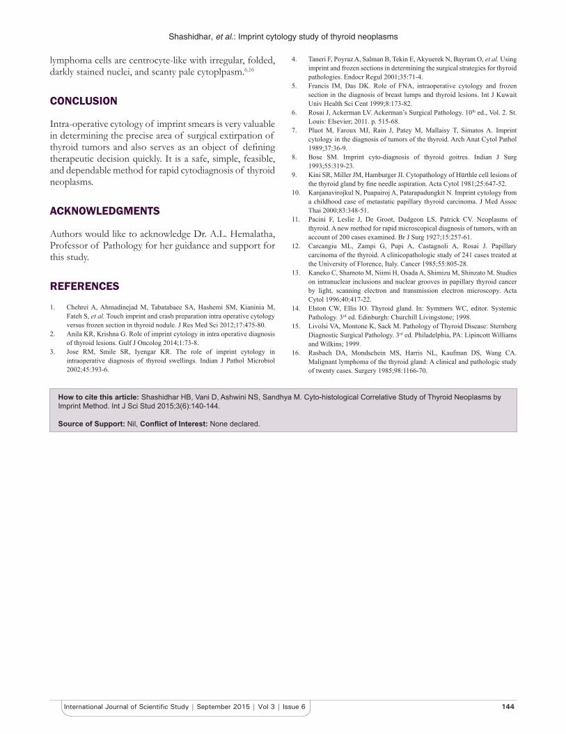
The follicular adenomas (Figures 4-6) accounted to 93% of benign cases (54/58) in the present study. The follicular adenomas are the most common among thyroid neoplasms. The cytology shows follicular cells in sheets and dispersed cells. The presence of micro follicular aggregates implies a follicular neoplasm. Since the presence of capsule cannot be assessed in aspirates, these are better termed as follicular neoplasms.3,6-8
Four cases of Hurthle cell neoplasm (Figures 7 and 8) were reported which on cytology showed cellular aspirates
Figure 2: Sex incidences in the present study
Figure 3: Distribution of benign and malignant cases diagnosed by imprint cytology
Figure 4: Follicular neoplasm showing gray white encapsulated mass with occasional areas of hemorrhage and degeneration
Figure 5: Imprint smear of Follicular neoplasm showing numerous microfollicles
Shashidhar, et al.: Imprint cytology study of thyroid neoplasms
143 International Journal of Scientifi c Study | September 2015 | Vol 3 | Issue 6
of tumor cells in loosely cohesive groups and papillaroid architecture. These tumor cells were large, polygonal to
oval with characteristic abundant fi nely granular blue gray cytoplasm.3,9
Among the malignant tumors, papillary carcinoma (Figures 8 and 9) was the most common encountered constituting 78.94% of malignant cases (30/38). It is the most common malignancy of thyroid with a female sex predominance of 3:1. The spread of the tumor is by lymphatics. Cytologically, aspirates are cellular and are comprised of papillary fronds with a fi brovascular core and lined by tumor cells. These cells are large with nuclei showing intranuclear cytoplasmic inclusions, nuclear grooves, and clefts.10-14
Two cases of anaplastic carcinoma (Figure 10) were reported which on cytology shows bizarre giant or spindle cells in singles and small clusters. The cells have pleomorphic nuclei and maybe multinucleate with coarse clumped chromatin.6,11,15,16
Two rare cases of NHL maltoma (Figure 11) were encountered. It is an indolent tumor in the thyroid which remains localized for a long time before dissemination. The
Figure 6: Histopathology of follicular adenoma depicting a well circumscribed lesion composed of micro and macrofollicles
with surrounding compressed normal thyroid tissue
Figure 7: Imprint smear of Hurthle cell neoplasm showing oxyphil cells in follicular patterns and clusters
Figure 8: Imprint smear showing papillary structures with nuclear features
Figure 10: Imprint smear (a) and histopathology (b) of anaplastic carcinoma thyroid
ba
Figure 9: Gross (a) and microscopy (b) of well encapsulated variant of papillary carcinoma of thyroid
ba
Figure 11: Gross (a) and imprint smear of non-Hodgkins lymphoma
ba
Shashidhar, et al.: Imprint cytology study of thyroid neoplasms
144International Journal of Scientifi c Study | September 2015 | Vol 3 | Issue 6
lymphoma cells are centrocyte-like with irregular, folded, darkly stained nuclei, and scanty pale cytoplpasm.6,16
CONCLUSION
Intra-operative cytology of imprint smears is very valuable in determining the precise area of surgical extirpation of thyroid tumors and also serves as an object of defi ning therapeutic decision quickly. It is a safe, simple, feasible, and dependable method for rapid cytodiagnosis of thyroid neoplasms.
ACKNOWLEDGMENTS
Authors would like to acknowledge Dr. A.L. Hemalatha, Professor of Pathology for her guidance and support for this study.
REFERENCES
1. Chehrei A, Ahmadinejad M, Tabatabaee SA, Hashemi SM, Kianinia M, Fateh S, et al. Touch imprint and crash preparation intra operative cytology versus frozen section in thyroid nodule. J Res Med Sci 2012;17:475-80.
2. Anila KR, Krishna G. Role of imprint cytology in intra operative diagnosis of thyroid lesions. Gulf J Oncolog 2014;1:73-8.
3. Jose RM, Smile SR, Iyengar KR. The role of imprint cytology in intraoperative diagnosis of thyroid swellings. Indian J Pathol Microbiol 2002;45:393-6.
4. Taneri F, Poyraz A, Salman B, Tekin E, Akyuerek N, Bayram O, et al. Using imprint and frozen sections in determining the surgical strategies for thyroid pathologies. Endocr Regul 2001;35:71-4.
5. Francis IM, Das DK. Role of FNA, intraoperative cytology and frozen section in the diagnosis of breast lumps and thyroid lesions. Int J Kuwait Univ Health Sci Cent 1999;8:173-82.
6. Rosai J, Ackerman LV. Ackerman’s Surgical Pathology. 10th ed., Vol. 2. St. Louis: Elsevier; 2011. p. 515-68.
7. Pluot M, Faroux MJ, Rain J, Patey M, Mallaisy T, Simatos A. Imprint cytology in the diagnosis of tumors of the thyroid. Arch Anat Cytol Pathol 1989;37:36-9.
8. Bose SM. Imprint cyto-diagnosis of thyroid goitres. Indian J Surg 1993;55:319-23.
9. Kini SR, Miller JM, Hamburger JI. Cytopathology of Hürthle cell lesions of the thyroid gland by fi ne needle aspiration. Acta Cytol 1981;25:647-52.
10. Kanjanavirojkul N, Puapairoj A, Patarapadungkit N. Imprint cytology from a childhood case of metastatic papillary thyroid carcinoma. J Med Assoc Thai 2000;83:348-51.
11. Pacini F, Leslie J, De Groot, Dudgeon LS, Patrick CV. Neoplasms of thyroid. A new method for rapid microscopical diagnosis of tumors, with an account of 200 cases examined. Br J Surg 1927;15:257-61.
12. Carcangiu ML, Zampi G, Pupi A, Castagnoli A, Rosai J. Papillary carcinoma of the thyroid. A clinicopathologic study of 241 cases treated at the University of Florence, Italy. Cancer 1985;55:805-28.
13. Kaneko C, Shamoto M, Niimi H, Osada A, Shimizu M, Shinzato M. Studies on intranuclear inclusions and nuclear grooves in papillary thyroid cancer by light, scanning electron and transmission electron microscopy. Acta Cytol 1996;40:417-22.
14. Elston CW, Ellis IO. Thyroid gland. In: Symmers WC, editor. Systemic Pathology. 3rd ed. Edinburgh: Churchill Livingstone; 1998.
15. Livolsi VA, Montone K, Sack M. Pathology of Thyroid Disease: Sternberg Diagnostic Surgical Pathology. 3rd ed. Philadelphia, PA: Lipincott Williams and Wilkins; 1999.
16. Rasbach DA, Mondschein MS, Harris NL, Kaufman DS, Wang CA. Malignant lymphoma of the thyroid gland: A clinical and pathologic study of twenty cases. Surgery 1985;98:1166-70.
How to cite this article: Shashidhar HB, Vani D, Ashwini NS, Sandhya M. Cyto-histological Correlative Study of Thyroid Neoplasms by Imprint Method. Int J Sci Stud 2015;3(6):140-144.
Source of Support: Nil, Confl ict of Interest: None declared.
![Correlative Studies between Computed Tomography and ... · growing soft tissue neoplasms. They can be benign or malignant. [8,9] They are asymptomatic, ... muscle, pattern of enhancement,](https://img.pdfslide.us/doc/110x75/5ffc5759efb696189801c415/correlative-studies-between-computed-tomography-and-growing-soft-tissue-neoplasms.jpg)