Embed Size (px)
Citation preview
SUDANESE JOURNAL OF PAEDIATRICS 2014; Vol 14, Issue No. 1
http://www.sudanjp.org39
Original ArticleCystic fibrosis in Sudanese children: First report of 35 cases Salah A Ibrahim (1), Munadhil A Fadl Elmola (2), Zain A Karrar (1), Ali M E Arabi (1), Mohamed A Abdullah (1), Sulafa K Ali (1), Fathelrahman Elawad (3), Tag Elsir A Ali (4), Mashair B Abdulrahman (5), Salma O Ahmed (6), Abelrazzag S Gundi (7) (1) Department of Paediatrics and Child Health, Faulty of Medicine, University of Khartoum, Khartoum,
Sudan.(2) Department of Radiology, Omdurman Teaching Hospital, Omdurman, Sudan. (3) Department of Paediatrics and Child Health, Faulty of Medicine, Elnileen University, Khartoum, Sudan. (4) Department of Paediatrics and Child Health, Faulty of Medicine, International University of Africa,
Khartoum, Sudan. (5) Department of Paediatrics, Ahmed Gasim Hospital, Khartoum North, Sudan. (6) Department of Paediatrics, Gaafar Ibn Oaf Children’s Hospital, Khartoum, Sudan. (7) Faculty of Medicine, University of Khartoum, Khartoum, Sudan.
Correspondence to:Salah A IbrahimDepartment of Paediatrics and Child Health, Faulty of Medicine, University of Khartoum,P.O.Box 102, Khartoum, Sudan.Email: [email protected]
How to cite this article: Ibrahim SA, Fadl Elmola MA, Karrar ZA, Arabi AME, Abdullah MA, Ali SK, Elawad F, Ali TEA, Abdulrahman MB, Ahmed SO, Gundi AS. Cystic fibrosis in Sudanese children: First report of 35 cases. Sudan J Pae diatr 2014; 14(1):39-44.
ABSTRACT Cystic fibrosis is the most common severe genetic disorder among children of European descent. It is much less common in Africans and Asians. It affects most critically the lungs causing chronic lung disease, failure to thrive and social deprivation. This is a retrospective review of 35 Sudanese patients with confirmed cystic fibrosis. About 60% of cases presented before the age of 5 years and male to female ratio was 1.7:1.0. Consanguinity was reported in 25 of the families. The main presenting features were productive cough, wheeze and clubbing. The chest X-ray showed variable degrees of hyperinflation, collapse, cystic, fibrotic changes and bronchiectasis involving both upper and lower lobes with blurring
of cardiac border and hilar vasculature in the majority of cases. The sweat chloride was between 70 and 140 mmol/l in 83% of the patients (positive > 60 mmol/l). Three patients underwent DNA study and confirmed to have cystic fibrosis transmembrane conductance regulator (CFTR) gene mutations. Gene study was not available for the rest of the patients. To our knowledge this is the first report of confirmed cases of cystic fibrosis in Sudanese patients.
Key words: Cystic fibrosis; Children, Africa; Sudan; Non-Caucasians.
SUDANESE JOURNAL OF PAEDIATRICS 2014; Vol 14, Issue No. 1
http://www.sudanjp.org 40
INTRODUCTIONCystic fibrosis (CF) is the most common life-shortening genetic disorder among children of Caucasians descent. It is an autosomal recessive disorder with an incidence of 1:2500 and a carrier state varying between 1:20 and 1:30 [1]. The world highest incidence of 1:1353 is reported from Ireland [2]. It is less common in pure Africans or Asians. The incidence among white Americans is 1:3200, among African Americans is 1:15000 and among Asians Americans is 1:32000 [3, 4]. Database of the CF United Kingdom (UK) register of 5,274 CF patients showed that 96.3% are Caucasians and 196 non-Caucasians out of whom 63 and 12 were of Pakistani and Indian origins, respectively; there were six Black Caribbeans and three Black Africans [5]. In a genetic study done in South Africa out of 218 cases only 12 (5.5%) were black [6]. The commonest genetic mutation is delta F508 which is also the most prevalent among Caucasians. The next most prevalent alleles are 3102 and 1200, which are the most common among the Africans (in South, Central and West Africa), and African-Americans [3, 4, 7-10]. The name CF describes the fibrosis and the cystic changes of the pancreas, however; the complete clinical picture of CF was not recognized until 1930. In 1952, the sweat electrolyte abnormalities were first described [1, 11]. Cystic fibrosis (Mucovisidosis) is caused by genetic mutation of the protein cystic fibrosis transmembrane conductance regulator (CFTR) causing abnormal transport of chloride and sodium across the epithelium leading to thick viscous secretions (in sweat, digestive fluids and mucus); it affects most critically the lungs, pancreas and the intestine. For a long time the diagnosis of cystic fibrosis in Sudan used to be based on history, clinical and radiological findings. In 2008, sweat test was introduced in the Faculty of Medicine, University of Khartoum. Since then out of over one hundred samples tested, less
MATERIALS AND METHODSThis is a descriptive, retrospective, hospital based study. It was conducted in Gaafar Ibn Oaf Children’s Hospital. The Study population included children under 18 years of age, who were diagnosed as cystic fibrosis based on clinical features and confirmed by sweat chloride test. This test was considered positive if the sweat chloride (using the filter paper method) is 60 mmol/l or more. Data were collected from patients medical and laboratory records by using a case report form. The data were analyzed using statistical package for social science (SPSS), version 15. Ethical clearance was obtained from hospital involved. Informed verbal consent was obtained from families of participants.
RESULTSThirty five patients with confirmed cystic fibrosis were studied. Table 1 shows that the male: female ratio is 1.7:1.0 and 60% of cases presented at or before the age of 5 years. Those who belong to tribes living in Northern and Central Sudan comprised 46 and 40 percent of the cases, respectively (Table 2). Consanguinity is predominant and the parents of 25 of the cases are either first or second cousins (Table 3).
than one third had high chloride levels. The aim of this study was to review the clinical characteristics of Sudanese patients diagnosed with CF.
Number ofAge groupFemalesMales(Years)
13< 12151 - 5616 – 104311 – 151322Total
Table 1 - Sex distribution by age groups (n=35)
SUDANESE JOURNAL OF PAEDIATRICS 2014; Vol 14, Issue No. 1
http://www.sudanjp.org41
The main presentation is respiratory as shown in Tables 3 and 4. Recurrent hospitalization with bronchiolitis or acute episodic asthma, cough productive of copious sputum, clubbing, chest signs (wheeze and crackles), and an abnormal chest radiological findings are presenting features in almost all (82-100%) cases. Staphylococcus aureus was isolated repeatedly from sputum of four of the cases but Pseudomonas aeruginosa was isolated only once from a fifth case. There is one patient who had lobectomy for a bronchiectatic left lower lobe.
Number of cases (%)Residence16 (46)Northern Sudan14 (40)Central Sudan3 (8)Eastern Sudan2 (6)Weastern sudan
35 (100)Total
Table 2 - Distribution of patients by residence of origin (n=35)
Number of casesItem
Past history
19Recurrent pneumonia
9Recurrent wheezy episodes
5Anti-tuberculous therapy2Gluten free diet
2Failure to thrive
2Bronchoscopy / Thoracotomy
Family history
25Consanguinity
4Sibling / Cousin affected
5Sibling died (cough +/- diarrhea)
Table 3 - Medical and family history of patients (n=35)
%Number of casesClinical feature
8831Weight less than 2 SD*
7526Height less than 2 SD
8630Wasting
8228Clubbing
269Chest deformity
10035Crackles +/- wheeze
5419Diarrhea (offensive stools)
4616Abdominal pain +/- distension
114Vomiting
269Developmental / pubertal delay
8228Radiological changes on chest imaging
*SD= standard deviation
Table 4 - Clinical and radiological features of patients (n-35)
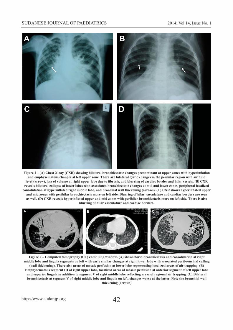
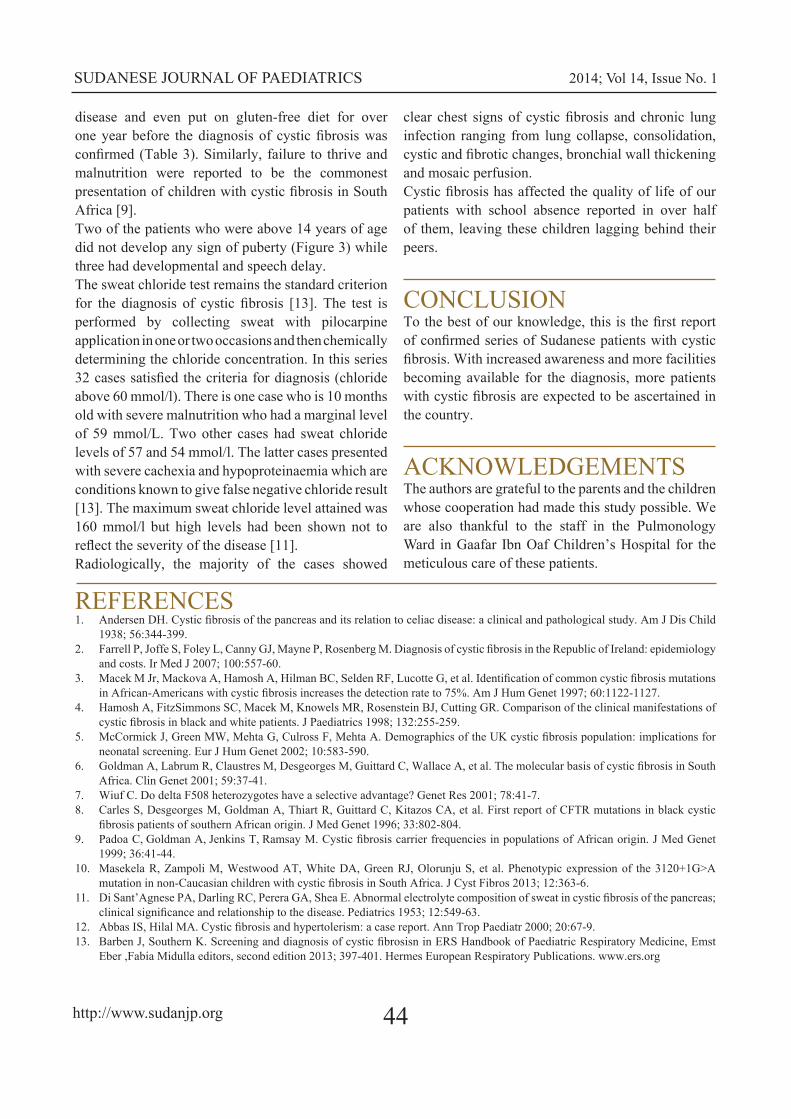
Abdominal pain, diarrhea and vomiting were a presenting feature in 16, 19 and 11 of the cases, respectively. Growth retardation (wasting) and developmental delay were seen in 30 and 9 of the cases, respectively. Radiological changes were also seen in 28 (82%) cases. The chest X-ray showed variable degrees of hyperinflation, collapse, cystic, fibrotic changes and bronchiectasis involving both upper and lower lobes with blurring of cardiac border and hilar vasculature in the majority of patients (Figure 1). The chest computed tomography (CT) clarified the bronchiectasis, peribronchial cuffing, areas of mosaic perfusion and air trapping in some of the patients (Figure 2).
SUDANESE JOURNAL OF PAEDIATRICS 2014; Vol 14, Issue No. 1
http://www.sudanjp.org 42
Figure 1 – (A) Chest X-ray (CXR) showing bilateral bronchiectatic changes predominant at upper zones with hyperinflation and emphysematous changes at left upper zone. There are bilateral cystic changes in the perihilar region with air fluid
level (arrow), loss of volume at right upper lobe due to fibrosis, and blurring of cardiac border and hilar vessels. (B) CXR reveals bilateral collapse of lower lobes with associated bronchiectatic changes at mid and lower zones, peripheral localized
consolidation at hyperinflated right middle lobe, and bronchial wall thickening (arrows). (C) CXR shows hyperinflated upper and mid zones with perihilar bronchiectasis more on left side. Blurring of hilar vasculature and cardiac borders are seen as well. (D) CXR reveals hyperinflated upper and mid zones with perihilar bronchiectasis more on left side. There is also
blurring of hilar vasculature and cardiac borders.
Figure 2 – Computed tomography (CT) chest lung window. (A) shows florid bronchiectasis and consolidation at right middle lobe and lingula segments on left with early similar changes at right lower lobe with associated peribronchial cuffing
(wall thickening). There also areas of mosaic perfusion at lower lobe representing localized areas of air trapping. (B) Emphysematous segment III of right upper lobe, localized areas of mosaic perfusion at anterior segment of left upper lobe
and superior lingula in addition to segment V of right middle lobe reflecting areas of regional air trapping. (C) Bilateral bronchiectasis at segment V of right middle lobe and lingula on left, changes worse at the latter. Note the bronchial wall
thickening (arrows)

SUDANESE JOURNAL OF PAEDIATRICS 2014; Vol 14, Issue No. 1
http://www.sudanjp.org43
The sweat chloride (using the filter paper method) is considered positive for a homozygous patient if it is 60 mmol/l or more. In 83% of the patients the level was between 70 and 140 mmol/l (Table 5). One patient had a border line sweat chloride of 59 mmol/l. Two other cases who had sweat chloride of 70 and 73 mmol/l were confirmed to have CFTR gene mutations. An additional case whose sweat chloride was 82 mmol/l was also confirmed to have p.D579G and p.R1102K/G>A mutations. Gene study was not available for the rest of patients.
Sweat chlorideNumber of cases (%)(mmol/l)
4 (11)< 6921 (60)70 - 998 (23)100 - 1402 (6) >140
35 (100)Total
Table 5 - Levels of sweat chloride of the study group (n=35)
Figure 3 - This is a 15.5 year-old-girl showing short stature and delayed sexual maturation.
DISCUSSION In this report we present for the first time 35 Sudanese children with confirmed cystic fibrosis. The vast majority (30 out of 35) of the cases reported in this series are originally from Northern Sudan or from areas in central Sudan which had been known to have mix marriages with the Turkish, British or Egyptians during the colonial periods (1821-1883 and 1898-1956). It is probable that the ancestors of this group are of Mediterranean / Caucasian descent. Because of the small sample size and the mix marriages it is difficult to conclude how many of these patients are not of Caucasian descent or pure Africans. Studies done in South, Central and West Africa had documented that cystic fibrosis is present in black African population [10]. Abbas in the year 2000 reported a case of suspected cystic fibrosis in association with multiple skeletal defects in a consanguineous family in Sudan but the ethnic origin was not reported [12]. The majority of the cases in this report, presented before the age of five years. This is the usual age of presentation of CF elswhere [4]. The male preponderance shown in this report has also been seen in other series [4]. It is reported from the CF UK register that male predominance significantly increases with age [5]. It is also well documented that the respiratory symptoms in patients with CF start first and early as seen in this series. Almost all (82-100%) cases reported started coughing in infancy or thereabout; and recurrent cough presenting as slow resolving pneumonia, recurrent bronchopneumonia, bronchiolitis and episodic asthma. Because of the endemicity of tuberculosis in the country and the difficulty to diagnose pulmonary tuberculosis in children, not surprisingly, five of the patients in this series received antituberculous treatment. Steatorrhoea had not been a striking presenting feature in this series. About half of the cases presented with abdominal distension, abdominal pain, vomiting, and recurrent bouts of loose, greasy stools. However, over half of the patients had foul-smelly stools, even in the absence of diarrhea. Growth assessment of this cohort revealed that the vast majority (75 – 88%) had stunted growth. Some had severe growth retardation with weights and heights well below 4 standard deviations. Two of these patients were suspected to have celiac
SUDANESE JOURNAL OF PAEDIATRICS 2014; Vol 14, Issue No. 1
http://www.sudanjp.org 44
disease and even put on gluten-free diet for over one year before the diagnosis of cystic fibrosis was confirmed (Table 3). Similarly, failure to thrive and malnutrition were reported to be the commonest presentation of children with cystic fibrosis in South Africa [9]. Two of the patients who were above 14 years of age did not develop any sign of puberty (Figure 3) while three had developmental and speech delay. The sweat chloride test remains the standard criterion for the diagnosis of cystic fibrosis [13]. The test is performed by collecting sweat with pilocarpine application in one or two occasions and then chemically determining the chloride concentration. In this series 32 cases satisfied the criteria for diagnosis (chloride above 60 mmol/l). There is one case who is 10 months old with severe malnutrition who had a marginal level of 59 mmol/L. Two other cases had sweat chloride levels of 57 and 54 mmol/l. The latter cases presented with severe cachexia and hypoproteinaemia which are conditions known to give false negative chloride result [13]. The maximum sweat chloride level attained was 160 mmol/l but high levels had been shown not to reflect the severity of the disease [11]. Radiologically, the majority of the cases showed
CONCLUSIONTo the best of our knowledge, this is the first report of confirmed series of Sudanese patients with cystic fibrosis. With increased awareness and more facilities becoming available for the diagnosis, more patients with cystic fibrosis are expected to be ascertained in the country.
ACKNOWLEDGEMENTSThe authors are grateful to the parents and the children whose cooperation had made this study possible. We are also thankful to the staff in the Pulmonology Ward in Gaafar Ibn Oaf Children’s Hospital for the meticulous care of these patients.
clear chest signs of cystic fibrosis and chronic lung infection ranging from lung collapse, consolidation, cystic and fibrotic changes, bronchial wall thickening and mosaic perfusion. Cystic fibrosis has affected the quality of life of our patients with school absence reported in over half of them, leaving these children lagging behind their peers.
REFERENCES 1. Andersen DH. Cystic fibrosis of the pancreas and its relation to celiac disease: a clinical and pathological study. Am J Dis Child
1938; 56:344-399. 2. Farrell P, Joffe S, Foley L, Canny GJ, Mayne P, Rosenberg M. Diagnosis of cystic fibrosis in the Republic of Ireland: epidemiology
and costs. Ir Med J 2007; 100:557-60. 3. Macek M Jr, Mackova A, Hamosh A, Hilman BC, Selden RF, Lucotte G, et al. Identification of common cystic fibrosis mutations
in African-Americans with cystic fibrosis increases the detection rate to 75%. Am J Hum Genet 1997; 60:1122-1127. 4. Hamosh A, FitzSimmons SC, Macek M, Knowels MR, Rosenstein BJ, Cutting GR. Comparison of the clinical manifestations of
cystic fibrosis in black and white patients. J Paediatrics 1998; 132:255-259. 5. McCormick J, Green MW, Mehta G, Culross F, Mehta A. Demographics of the UK cystic fibrosis population: implications for
neonatal screening. Eur J Hum Genet 2002; 10:583-590. 6. Goldman A, Labrum R, Claustres M, Desgeorges M, Guittard C, Wallace A, et al. The molecular basis of cystic fibrosis in South
Africa. Clin Genet 2001; 59:37-41. 7. Wiuf C. Do delta F508 heterozygotes have a selective advantage? Genet Res 2001; 78:41-7. 8. Carles S, Desgeorges M, Goldman A, Thiart R, Guittard C, Kitazos CA, et al. First report of CFTR mutations in black cystic
fibrosis patients of southern African origin. J Med Genet 1996; 33:802-804. 9. Padoa C, Goldman A, Jenkins T, Ramsay M. Cystic fibrosis carrier frequencies in populations of African origin. J Med Genet
1999; 36:41-44. 10. Masekela R, Zampoli M, Westwood AT, White DA, Green RJ, Olorunju S, et al. Phenotypic expression of the 3120+1G>A
mutation in non-Caucasian children with cystic fibrosis in South Africa. J Cyst Fibros 2013; 12:363-6. 11. Di Sant’Agnese PA, Darling RC, Perera GA, Shea E. Abnormal electrolyte composition of sweat in cystic fibrosis of the pancreas;
clinical significance and relationship to the disease. Pediatrics 1953; 12:549-63. 12. Abbas IS, Hilal MA. Cystic fibrosis and hypertolerism: a case report. Ann Trop Paediatr 2000; 20:67-9. 13. Barben J, Southern K. Screening and diagnosis of cystic fibrosisn in ERS Handbook of Paediatric Respiratory Medicine, Emst
Eber ,Fabia Midulla editors, second edition 2013; 397-401. Hermes European Respiratory Publications. www.ers.org