Embed Size (px)
Citation preview

Division of Molecular Structural Biology
Department of Medical Biochemistry and Biophysics
Karolinska Institutet, Stockholm, Sweden
CYSTEINE BIOSYNTHESIS IN MYCOBACTERIUM TUBERCULOSIS AS
POTENTIAL DRUG TARGET
Katharina Brunner
Stockholm 2017

All previously published papers were reproduced with permission from the publisher.
Published by Karolinska Institutet.
Printed by E-Print AB 2017
© Katharina Brunner, 2017
ISBN 978-91-7676-604-0

Institutionen för medicinsk biokemi och biofysik
Cysteine biosynthesis in Mycobacterium tuberculosis as potential drug target
THESIS FOR DOCTORAL DEGREE (Ph.D.)
AKADEMISK AVHANDLING
som för avläggande av medicine doktorsexamen vid
Karolinska Institutet offentligen försvaras i
Samuelssonsalen, Scheelelaboratoriet, Tomtebodavägen 6,
Karolinska Institutet, Solna
Fredagen den 12 maj, 2017, kl 10:00
Av
Katharina Brunner Principal Supervisor:
Prof. Gunter Schneider
Karolinska Institutet
Department of Medical Biochemistry and
Biophysics
Division of Molecular Structural Biology
Co-supervisor:
Dr. Robert Schnell
Karolinska Institutet
Department of Medical Biochemistry and
Biophysics
Division of Molecular Structural Biology
Opponent:
Prof.ssa Barbara Campanini
Università degli Studi di Parma
Dipartimento di Scienze degli Alimenti e del
Farmaco
Examination Board:
Prof. Ralf Morgenstern
Karolinska Institutet
Institute for Environmental Medicine
Dr. Pål Stenmark
Stockholms Universitet
Department of Biochemistry and Biophysics
Dr. Ylva Ivarsson
Uppsala Universitet
Department of Chemistry


To
Gottfrank & Dorian Stain


ABSTRACT
The bacterium Mycobacterium tuberculosis is the underlying cause of tuberculosis, one of the
most devastating infectious diseases, causing 1.4 million deaths in 2015. In addition one third
of the global population is infected with latent tuberculosis, during which the bacilli remain
viable in a dormant state, characterized by slow growth rates and changes in metabolism. It
was shown that the underlying genes of sulfur assimilation and L-cysteine biosynthesis are
essential for the survival of Mtb during dormancy. The availability of sulfur and the
subsequently produced L-cysteine is directly linked to increased survival of Mtb inside host
macrophages, because the first line of defense of Mtb against reactive oxygen and nitrogen
species relies on mycothiol, the functional analog to glutathione in mycobacteria and the
redox active sulfhydryl group of mycothiol is directly derived from L-cysteine. Hence the
enzymes of the sulfur assimilation pathway and L-cysteine biosynthesis have been proposed
as potential targets for novel antimycobacterial drugs.
Within the scope of this thesis, mycobacterial CysC, the APS kinase domain of the
mycobacterial sulfur activation complex was mechanistically and structurally characterized.
Phosphoryl group transfer from ATP to APS was shown to follow a conserved mechanism
found in several species. In addition mutation of an L-cysteine residue in the lid region
closing off the ATP binding site resulted in impaired ATP binding and prevented catalysis.
The structural characterization included a comparison of mycobacterial CysC and the crystal
structures to the two human homologs and it was found that the substrate binding sites for
APS and ATP in the three enzymes shared a high degree of sequence identity, hence inhibitor
development specifically targeting the mycobacterial enzyme was considered to be very
challenging.
The three mycobacterial L-cysteine synthases, CysK1, CysK2 and CysM are upregulated
during different metabolic states of Mtb. A high-throughput screening campaign of CysM
identified a class of urea-based active site binders. Subsequent in vitro validation and organic
synthesis of compounds to establish structure activity relationships in combination with
structural based methods allowed the identification of seven potent CysM inhibitors. The
affinities were found to be in the low micromolar range. The identified compounds did not
only decrease CysM activity, but also showed bactericidal potency in a nutrient starvation
model. CysK1 and CysK2 were also tested against a library of 71 compounds that were used
for in vitro validation of CysM. In total four compounds were found to inhibit CysK1 and
CysK2 and were also among the best inhibitors targeting CysM, suggesting that the identified
inhibitors might provide a valuable starting point towards the development of a drug targeting
all three L-cysteine synthases simultaneously.

LIST OF SCIENTIFIC PAPERS
I. Ömer Poyraz, Katharina Brunner, Bernhard Lohkamp, Hanna Axelsson, Lars GJ
Hammarström, Robert Schnell, Gunter Schneider. (2015). ”Crystal Structures of the
Kinase Domain of the Sulfate-Activating Complex in Mycobacterium tuberculosis”
PLoS One 10(3):e0121494.
II. Katharina Brunner, Selma Maric, Rudraraju Srilakshmi Reshma, Helena Almqvist,
Brinton Seashore-Ludlow, Anna-Lena Gustavsson, Ömer Poyraz, Perumal
Yogeeswari, Thomas Lundbäck, Michaela Vallin, Dharmarajan Sriram, Robert
Schnell and Gunter Schneider. (2016). “Inhibitors of the Cysteine Synthase CysM
with Antibacterial Potency against Dormant Mycobacterium tuberculosis.” Journal of
Medicinal Chemistry 59(14):6848–6859.
III. Katharina Brunner, Eva Maria Steiner, Rudraraju Srilakshmi Reshma,
Dharmarajan Sriram, Robert Schnell and Gunter Schneider. “Profiling of in vitro
Activities of Urea-based Inhibitors against Cysteine Synthases from Mycobacterium
tuberculosis”. Manuscript.

CONTENTS
1 Mycobacterium tuberculosis ........................................................................................... 1
1.1 The history of tuberculosis .................................................................................... 1
1.2 Pathogenesis of tuberculosis infection .................................................................. 2
1.2.1 Immune response against invading bacteria ............................................. 4
1.2.2 Influence of macrophage phenotype on progression of tuberculosis ...... 4
1.2.3 Granuloma formation – a hallmark of tuberculosis ................................. 4
1.2.4 Active and latent tuberculosis ................................................................... 5
1.2.5 Mtb interferes with the immune response of the host .............................. 6
1.2.6 Adaptation of Mtb to the harsh environment inside macrophages .......... 6
1.3 Maintenance of redox homeostasis in Mtb ........................................................... 7
1.4 Tuberculosis treatment and challenges ................................................................. 8
1.5 Early stage drug discovery .................................................................................. 10
1.6 L-cysteine biosynthesis as drug target ................................................................ 13
1.7 Sulfur assimilation in Mtb ................................................................................... 13
1.7.1 The sulfur assimilation pathway in other organisms.............................. 15
1.7.2 The mycobacterial sulfate activation complex ....................................... 16
1.7.3 APS kinase .............................................................................................. 16
1.8 De novo L-cysteine biosynthesis......................................................................... 18
1.8.1 The classical pathway to L-cysteine ....................................................... 19
1.8.2 A salvage pathway to L-cysteine formation ........................................... 21
1.8.3 L-cysteine formation during dormancy .................................................. 22
2 Aims of the thesis .......................................................................................................... 25
3 Results and discussion ................................................................................................... 27
3.1 PAPER I – Crystal Structures of the Kinase Domain of the Sulfate-
Activating Complex in Mycobacterium tuberculosis ......................................... 27
3.1.1 ADP binding site of mycobacterial APS kinase .................................... 27
3.1.2 APS binding pocket ................................................................................ 28
3.1.3 Magnesium binding site .......................................................................... 28
3.1.4 Mechanistic proposal for phosphoryl group transfer ............................. 28
3.1.5 Comparison to the human PAPS synthetases ......................................... 29
3.1.6 Is CysC regulated by disulfide bond formation? .................................... 30
3.2 Paper II – Inhibitors of the L-cysteine Synthase CysM with Antibacterial
Potency against Dormant Mycobacterium tuberculosis ..................................... 32
3.2.1 High-throughput screening and selection of hits .................................... 32
3.2.2 Hit expansion ........................................................................................... 32
3.2.3 In vitro validation .................................................................................... 33
3.2.4 Crystal structures of the enzyme-ligand inhibitor complexes................ 34
3.2.5 Common features of best inhibitors ........................................................ 34
3.2.6 Antimycobacterial activity of urea-based inhibitors .............................. 37
3.2.7 Compound selectivity ............................................................................. 37

3.3 Paper III – Profiling of in vitro activities of urea-based inhibitors against
L-cysteine synthase from Mycobacterium tuberculosis ..................................... 38
3.3.1 Identification of inhibitors targeting CysK1 and CysK2 ....................... 38
3.3.2 Urea-based compounds inhibit all three mycobacterial L-cysteine
synthases .................................................................................................. 38
3.3.3 Comparison of the CysK1 specific inhibitors: urea-based scaffold
versus thiazolidine-based scaffold .......................................................... 40
3.3.4 Targeting all three L-cysteine synthases at once .................................... 41
3.4 Conclusion ........................................................................................................... 42
4 Acknowledgements ....................................................................................................... 43
5 References ..................................................................................................................... 47

LIST OF ABBREVIATIONS
ADP
AMP
AMP-PNP
APS
ATP
adenosine 5′-diphosphate
adenosine 5′-monophosphate
adenosine 5′-(β,γ-imido) triphosphate
adenosine 5′-phosphosulfate
adenosine 5′-triphosphate
BCG
CD
DNA
DTT
ESI-MS
FDA
HIV
HTS
IFN
IL
ITC
LAM
LHS
NADH
NADPH
NAS
NMR
MDR
MSH
MSSM
Mtb
NOD
OAS
OPS
PAINS
PAMP
PAPS
PAPSS
PIM
PLP
PPi
RHS
RNS
ROS
SAM
SAR
SL
TDM
TLR
TNF
WHO
XDR
Bacille Calmette Guérin
circular dichroism
deoxyribonucleic acid
dithiothreitol
electrospray ionization mass spectrometry
Food and Drug Administration
human immunodeficiency virus
high-throughput screening
interferon
interleukin
isothermal titration calorimetry
lipoarabinomannan
left-hand side
β-nicotinamide adenine dinucleotide
β-nicotinamide adenine dinucleotide 2’-phosphate
N-acetyl serine
nuclear magnetic resonance
multidrug-resistant
mycothiol
mycothiol disulfide
Mycobacterium tuberculosis
nucleotide-binding oligomerization domain like receptor
O-acetyl-L-serine
O-phospho-L-serine
pan assay interference compounds
pathogen associated molecular patterns
adenosine 3′-phosphate-5′-phosphosulfate
adenosine 3′-phosphate-5′-phosphosulfate synthetase
phosphatidylinositol mannan
pyridoxal 5′-phosphate
pyrophosphate
right-hand side
reactive nitrogen species
reactive oxygen species
S-5′-adenosyl-L-methionine
structure-activity relationship
sulfolipid
trehalose dimycolate
toll-like receptor
tumor necrosis factor
World Health Organization
extensively drug-resistant


1
1 MYCOBACTERIUM TUBERCULOSIS
1.1 THE HISTORY OF TUBERCULOSIS
Mycobacterium tuberculosis (Mtb) and humans share a common history for at least 70,000
years. The spread of the disease was accompanied by the migration of early humans out of
Africa, as shown by population genomics studies (Comas, Coscolla et al. 2013). The first
physical evidence of tuberculosis infection was found in human skeletons from the Neolithic
era excavated in Israel and Syria showing typical bone lesions for spinal tuberculosis,
underpinned by the presence of mycolic acids and ancient mycobacterial DNA extracted from
bones (Baker, Lee et al. 2015; Spigelman, Donoghue et al.). During the Neolithic era the
increase of human population and density explain the successful spread of Mtb (Comas,
Coscolla et al. 2013), rather than transmission from domesticated animals as proposed earlier
(Diamond 1999).
Tuberculosis, also known as consumption or the white plague had its peak between the 17th
and 19th
century. At this time every fourth adult in Europe died from tuberculosis (Wilson
2004). It took until 1882 to understand the underlying cause of tuberculosis (Koch 1882).
Robert Koch was the first to show that tuberculosis was an infectious disease caused by the
bacterium Mycobacterium tuberculosis by performing animal experiments on guinea pigs and
was subsequently awarded the Nobel Prize in Physiology and Medicine in 1905 (Cambau &
Drancourt 2014).
In 1921, the bacille Calmette-Guérin (BCG) vaccine was developed, which is still the sole
effective and licensed vaccine against tuberculosis today (Calmette 1922; Hatherill, Tait et al.
2016). Calmette and Guérin were sub-culturing virulent Mycobacterium bovis isolates for
thirteen years before the strain showed sufficient attenuation to be safe enough to be used in
humans (Luca & Mihaescu 2013). In 1943, the first antimycobacterial drug, the antibiotic
streptomycin became available. This, in combination with the BCG vaccine, resulted in
mortality rates starting to drop and, hope to fully eradicate tuberculosis emerged. Funding and
interest in tuberculosis research declined until (Keshavjee & Farmer 2012) co-infections of
HIV and tuberculosis and infections with multidrug-resistant Mtb (MDR) were observed in
the early 1990s and death tolls started to rise again (Comas & Gagneux 2009). In 1998 the
first genome of Mtb was sequenced (Cole, Brosch et al. 1998). To date there are seven known
Mtb strain lineages that affect humans. The Mtb lineages are associated with different
geographical regions and all evolved from one common ancestral strain. Whole genome
sequencing revealed that the different Mtb lineages are genetically much more diverse than
expected, and this diversity can be linked to human migration events (Hershberg, Lipatov et
al. 2008). One example is the successful spread of the Beijing lineage from East Asia towards
Europe along the Silk Road (Merker, Blin et al. 2015). Tuberculosis infections caused by the
Beijing lineage had high prevalence in Asia and the former Soviet Union in the past. To date,
this lineage has spread over the entire world and more recent tuberculosis outbreaks caused
by the Beijing lineage are associated with drug resistance (Rufai, Sankar et al. 2016).
In 2015 about two million people died from tuberculosis worldwide and additional 10.4
million newly infected were counted globally. On top of that, one third of the global
population carries an asymptomatic latent tuberculosis infection that is reactivated in

2
approximately ten percent of all cases and MDR Mtb is on the rise. The observation that
currently used antibiotics are ineffective in treating latent tuberculosis infections or MDR-
infections indicates that novel antimycobacterial drugs are urgently needed to achieve the
goal of the WHO to eradicate tuberculosis by 2035 (World Health Organization 2016).
1.2 PATHOGENESIS OF TUBERCULOSIS INFECTION
After initial contact between host and Mtb, the infection can either be cleared, controlled by
the immune system or progress into active tuberculosis (Salgame, Geadas et al. 2015). The
active form of tuberculosis, known as pulmonary tuberculosis, mostly affects the lungs and is
initiated by a characteristic lung inflammation (Hopewell, Kato-Maeda et al. 2016). If
tuberculosis remains untreated, it eventually results in death (Fitzgerald, Sterling et al. 2015).
Individuals carrying active tuberculosis can easily spread the infection by expelling Mtb-
containing air droplets, produced through coughing and sneezing (Fig. 1). Symptoms of
active tuberculosis include fever, coughing, with occasional blood in the sputum, weight loss,
night sweats and changes in the opacity of certain areas in chest radiographs (Fitzgerald,
Sterling et al. 2015).
Specific human sub-populations show an increased risk of developing tuberculosis. Amongst
those are individuals living in developing countries, suffering from malnutrition, who are co-
infected with HIV, or health care workers, facing higher exposure to infectious individuals. In
addition, young adults in their early twenties, smokers and patients treated with immune
suppressants are part of this group. Interestingly gender also plays a role, as men are more
prone to acquire infection (World Health Organization 2016) (Fogel 2015).
Latent tuberculosis is defined as measurable specific immune response against Mtb in the
absence of active tuberculosis symptoms and is considered to be controlled by the host
immune system (Gengenbacher & Kaufmann 2012) and individuals carrying a latent
tuberculosis infection are not contagious (Ernst 2012). Two billion people, which account for
one third of the world population, are carrying such a latent tuberculosis infection, which
represents a huge infectious cohort, because ten percent of latent tuberculosis infections are
reactivated. The reasons for reactivation of latent tuberculosis are poorly understood, but rely
on the complex cross-talk between the pathogen and the host immune system (Philips &
Ernst 2012; Vynnycky & Fine 2000; World Health Organization 2016).

3
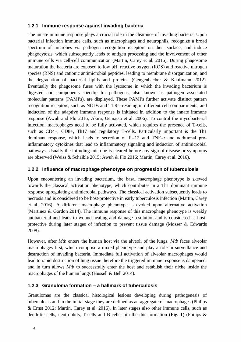
Figure 1. The upper panel shows the route of tuberculosis transmission between infected
individuals and transformation into latent tuberculosis. The lower panel illustrates the progress of
Mtb infection from phagocytosis through granuloma formation and necrosis. After phagocytosis
Mtb survives inside the host macrophages by interfering with phagolysosomal fusion and induces
formation of a solid granuloma. The granuloma matures via the necrotic phase to a caseous
granuloma, lower panel. Reprinted by permission from Oxford University Press: FEMS
Microbiology Reviews (Gengenbacher & Kaufmann 2012), copyright 2012.
4
1.2.1 Immune response against invading bacteria
The innate immune response plays a crucial role in the clearance of invading bacteria. Upon
bacterial infection immune cells, such as macrophages and neutrophils, recognize a broad
spectrum of microbes via pathogen recognition receptors on their surface, and induce
phagocytosis, which subsequently leads to antigen processing and the involvement of other
immune cells via cell-cell communication (Martin, Carey et al. 2016). During phagosome
maturation the bacteria are exposed to low pH, reactive oxygen (ROS) and reactive nitrogen
species (RNS) and cationic antimicrobial peptides, leading to membrane disorganization, and
the degradation of bacterial lipids and proteins (Gengenbacher & Kaufmann 2012).
Eventually the phagosome fuses with the lysosome in which the invading bacterium is
digested and components specific for pathogens, also known as pathogen associated
molecular patterns (PAMPs), are displayed. These PAMPs further activate distinct pattern
recognition receptors, such as NODs and TLRs, residing in different cell compartments, and
induction of the adaptive immune response is initiated in addition to the innate immune
response (Awuh and Flo 2016; Akira, Uematsu et al. 2006). To control the mycobacterial
infection, macrophages need to be fully activated, which requires the presence of T-cells,
such as CD4+, CD8+, Th17 and regulatory T-cells. Particularly important is the Th1
dominant response, which leads to secretion of IL-12 and TNF-α and additional pro-
inflammatory cytokines that lead to inflammatory signaling and induction of antimicrobial
pathways. Usually the intruding microbe is cleared before any sign of disease or symptoms
are observed (Weiss & Schaible 2015; Awuh & Flo 2016; Martin, Carey et al. 2016).
1.2.2 Influence of macrophage phenotype on progression of tuberculosis
Upon encountering an invading bacterium, the basal macrophage phenotype is skewed
towards the classical activation phenotype, which contributes in a Th1 dominant immune
response upregulating antimicrobial pathways. The classical activation subsequently leads to
necrosis and is considered to be host-protective in early tuberculosis infection (Martin, Carey
et al. 2016). A different macrophage phenotype is evoked upon alternative activation
(Martinez & Gordon 2014). The immune response of this macrophage phenotype is weakly
antibacterial and leads to wound healing and damage resolution and is considered as host-
protective during later stages of infection to prevent tissue damage (Mosser & Edwards
2008).
However, after Mtb enters the human host via the alveoli of the lungs, Mtb faces alveolar
macrophages first, which comprise a mixed phenotype and play a role in surveillance and
destruction of invading bacteria. Immediate full activation of alveolar macrophages would
lead to rapid destruction of lung tissue therefore the triggered immune response is dampened,
and in turn allows Mtb to successfully enter the host and establish their niche inside the
macrophages of the human lungs (Hussell & Bell 2014).
1.2.3 Granuloma formation – a hallmark of tuberculosis
Granulomas are the classical histological lesions developing during pathogenesis of
tuberculosis and in the initial stage they are defined as an aggregate of macrophages (Philips
& Ernst 2012; Martin, Carey et al. 2016). In later stages also other immune cells, such as
dendritic cells, neutrophils, T-cells and B-cells join the this formation (Fig. 1) (Philips &

5
Ernst 2012). Initially it was assumed that granulomas are host-protective, by walling-off
mycobacteria and keeping them from disseminating. Whereas this might be true in the later
stage of infection in which granulomas become fibrotic and calcify, it is rather the opposite
during early infection, in which they act as infection promotor. Close proximity of the cells in
the granuloma facilitate cell-to-cell spread and expansion of the mycobacterial population.
For initiation of granuloma formation one infected alveolar macrophage is sufficient (Davis
& Ramakrishnan 2008). In this stage the infected macrophage recruits other macrophages
which subsequently differentiate. Mycobacterial ketomycolic acids direct the differentiation
towards the foamy type of macrophages which are associated with necrotic tissue (Peyron,
Vaubourgeix et al. 2008). Characteristic for the foamy type is a high amount of lipids, for
example cholesterol that is imported in order to serve as a carbon source during chronic
infection (Pandey & Sassetti 2008). The mycobacterial cell wall components
phosphatidylinositol mannan (PIM), lipoarabinomannan (LAM) and trehalose dimycolate
(TDM) cause formation of multinucleated giant cells (Puissegur, Lay et al. 2007). Infected
dendritic cells allow migration of Mtb from the lungs to lymph nodes to present antigens to
naïve T-cells (Lay, Poquet et al. 2007). T-cells can either be diffusely spread in between the
other cells of the granuloma or form an outer layer around the aggregate structure and are
responsible in maintaining organized granulomas and control Mtb progression after the active
phase of tuberculosis. Granulomas were shown to be highly dynamic structures in which cells
can freely diffuse in and out (Fig. 1) (Philips & Ernst 2012). The center of a granuloma is
usually called caseum and comprises a cheese-like consistency resulting from coagulative
tissue necrosis. If the host is capable of controlling Mtb, then necrosis is stalled and the
caseum is eventually replaced by calcified and fibrotic tissue and infection is considered to be
latent. However if tuberculosis progresses, the caseum is expanded leading to necrosis and
cavity formation in the lungs. The breakdown of the surrounding lung tissue allows spreading
of Mtb into the nearby airways and facilitates dissemination and transmission, referred to as
active tuberculosis (Martin, Carey et al. 2016).
1.2.4 Active and latent tuberculosis
The sharp discrimination between active tuberculosis and latent tuberculosis infection is
heavily disputed and considered as outdated, because it draws the picture of an
oversimplified, two-state binary model of disease that opposes the knowledge we gained
about tuberculosis (Salgame, Geadas et al. 2015; Martin, Carey et al. 2016; Mack, Migliori et
al. 2009; Horsburgh, Barry et al. 2015). For example, inside the lungs of patients carrying
either variant of tuberculosis, both caseous and calcified granulomas have been observed in
parallel (Lenaerts, Barry et al. 2015). Furthermore, Mtb itself is considered to be present in
different metabolic states during active and latent tuberculosis. Gengenbacher and Kaufmann
proposed a dynamic model of the two disease states, in which latent tuberculosis is
characterized by a majority of dormant bacilli and a minority of some actively dividing Mtb
that sense the environment in terms of nutrient availability and presence of oxygen. Inside the
calcified granulomas the environment prohibits the presence of a large number of actively
dividing Mtb and the majority remains dormant. If the granuloma transists into the caseous
state, the bacilli awake from dormancy. Some remain in their dormant state though and still
comprise the phenotypic inherent antimycobacterial drug resistance, which explains the

6
required long lasting tuberculosis treatment of at least six months (Gengenbacher &
Kaufmann 2012).
1.2.5 Mtb interferes with the immune response of the host
Mtb has evolved to survive the host immune response, due to their long common
evolutionary history. The bacterium interferes with well-established immune cell functions
and survives inside host phagocytes including macrophages (Armstrong & Hart 1971).
Degradation and processing of invading bacteria require fusion of the phagosome and the
lysosome, which is achieved by a well order mechanism of interactions between phagosomal
membrane proteins and phospholipids. Part of this mechanism is the recruitment of the
GTPase, Rab7 to the phagosomal membrane and subsequent phosphorylation of
phosphatidylinositol. Mtb however abolishes both steps and thereby evades digestion by
macrophages (Philips & Ernst 2012). In addition Mtb interferes with proper functioning of
the autophagy system that impedes intracellular growth of mycobacteria after it has been
stimulated by IFNγ. Through disruption of membranes with the help of several type VII
secretion systems, Mtb can escape into the cytoplasm, avoid necrosis and even infect adjacent
cells (Hopewell, Kato-Maeda et al. 2016). Phagosome permeabilization triggers the release of
IFNα and -β that in turn suppress expression of IL-1β, essential for controlling the Mtb
infection (Mayer-Barber, Andrade et al. 2011; Novikov, Cardone et al. 2011). The
mycobacterial cell wall contains various lipids that do not only protect Mtb from polar
molecules and serve as a waxy barrier preventing antimycobacterial drugs to enter the
cytoplasm, but they are also involved in direct interaction with the host. PIM and LAM
stimulate innate immune and inflammatory response and serve as toll like receptor 2 agonists
and LAM alters phagosome maturation. TDM, also known as mycobacterial cord-factor is
recognized by two lectin receptors in macrophages and dendritic cells and transduces pro-
inflammatory signals (Hopewell, Kato-Maeda et al. 2016). TDM alone is also sufficient to
induce transient granuloma formation in mice (Copenhaver, Sepulveda et al. 2004).
1.2.6 Adaptation of Mtb to the harsh environment inside macrophages
Upon phagocytosis Mtb has to withstand drastic environmental changes, characterized by low
pH, nutrient starvation, lack of oxygen and presence of ROS and RNS (Gengenbacher &
Kaufmann 2012; Martin, Carey et al. 2016). In response to these challenges the
mycobacterial metabolism changes completely. Mtb switches from an actively dividing state
to a non-replicating persistent state during which also the susceptibility to antimycobacterial
drugs is decreased, known as phenotypic drug resistance, a form of resistance conferred by a
metabolic state that is not encoded in the genome. During the non-replicating state, which is
also termed dormancy, the bacilli remain fully viable but at a very low metabolic rate
(Gengenbacher & Kaufmann 2012).
Mtb is capable of withstanding low pH within the phagolysosome (Schaible, Sturgill-
Koszycki et al. 1998), which is to some degree caused by the unique composition of the
mycobacterial cell wall. The absence of full acidification of the phagolysosome in
macrophages however is attributed to inhibition of the phagosomal ATPase by Mtb and the
presence of a urease that cleaves urea. The buffering property of the produced ammonia
stabilizes the pH at a slightly higher level than usual (Gengenbacher & Kaufmann 2012).

7
While engulfed inside the macrophage, Mtb scavenges nutrients from the host environment
(Niederweis 2008; Martin, Carey et al. 2016). Upon switching from carbohydrates as a
carbon source to lipids from foamy giant cells, the acetyl moiety of acetyl-CoA is replaced by
propionyl and leads to toxic by-products for Mtb (Gengenbacher & Kaufmann 2012). In
order to avoid harming itself, the bacterium uses propionyl-CoA to produce PDIM, which are
considered a virulence factor (Lee, VanderVen et al. 2013). In good agreement with this is
the observation that genes involved in fatty acid biosynthesis are upregulated (Schnappinger,
Ehrt et al. 2003).
Inside caseous and calcified granulomas Mtb faces hypoxia (Via, Lin et al. 2008). To
counterstrike hypoxia a complex transcriptional network including the DosR regulon is
upregulated and genes are transcribed to stabilize proteins, avoid DNA damage, switch on
alternative electron transport chains and induce angiogenesis inside the granulomas to
increase oxygen content (Leistikow, Morton et al. 2010; Martin, Carey et al. 2016).
Inside the macrophages Mtb also faces damage of DNA, cell wall and proteins by ROS and
RNS. The phagocytosed bacteria usually are killed by these radicals that primarily target Fe-S
clusters and sulfhydryl groups in proteins and nucleotide bases in nucleic acids (Martin,
Carey et al. 2016). ROS and RNS induce transcription of the DosR regulon also and
additional detoxifying enzymes are expressed in Mtb (Kumar, Toledo et al. 2007). Reactive
nitrogen species are not only produced by macrophages, but also by Mtb itself, and are
considered to act bacteriostatic rather than bactericidal (Cunningham-Bussel, Zhang et al.
2013). The detoxifying enzyme superoxide dismutase disarms superoxide radicals and is part
of a macromolecular oxidoreductase complex that copes with oxidative stress and maintains
the cytosolic redox homeostasis in Mtb. To cope with hydrogen peroxide, Mtb uses catalase-
peroxidase (Nambi, Long et al. 2015; Martin, Carey et al. 2016). NADH-dependent
peroxidase and peroxynitrite reductase is another complex involved in detoxification of ROS
and RNS, consisting of an alkyl hydroperoxide reductase, an oxidoreductase, a
dihydrolipoamide acyltransferase and lipoamide dehydrogenase (Ehrt & Schnappinger 2009).
1.3 MAINTENANCE OF REDOX HOMEOSTASIS IN MTB
To maintain intracellular redox homeostasis during the exposure to oxidative stress, Mtb
produces mycothiol (MSH) which is the functional analog of glutathione in Mtb and related
genera (Fig. 2A) (Bornemann, Jardine et al. 1997). MSH is the major low-molecular-weight
thiol produced in Mtb and present in millimolar concentration inside the mycobacterial cell
(Bhave, Muse et al. 2007). MSH biosynthesis involves five enzyme-catalyzed reactions. The
fourth step of MSH biosynthesis directly links maintenance of redox homeostasis to the
availability of L-cysteine and emphasizes the importance of L-cysteine biosynthesis also
during the dormant state of Mtb, while engulfed inside the macrophage. MSH maintains the
reducing intracellular environment and thereby acts as a thiol redox buffer (Jothivasan &
Hamilton 2008; Fan, Vetting et al. 2009). Toxic oxidants, which are produced inside
macrophages to combat Mtb, are reduced by MSH, which in turn is oxidized to MSSM. In
order to maintain high concentrations of MSH, the NADPH-dependent flavoprotein
mycothiol disulfide reductase constantly reduces MSSM to MSH (Fig 2B). Mutants deficient
in MSH biosynthesis have been shown to be more sensitive to oxidative stress, confirming
the importance of this system in tuberculosis pathogenesis (Jothivasan & Hamilton 2008).

8
Figure 2. A) Chemical structure of mycothiol (MSH), the dominant low-molecular-weight thiol in
mycobacteria. The L-cysteine derived redox-active part is encircled in yellow. B) RNS and ROS
oxidize MSH to MSSM which is regenerated by the NADPH-dependent flavoprotein mycothiol
disulfide reductase to maintain a high intracellular level of MSH.
1.4 TUBERCULOSIS TREATMENT AND CHALLENGES
The current recommendation to treat new cases of pulmonary tuberculosis is a daily regimen
of multiple antibiotics for at least six months and is considered standard treatment for
presumable drug-susceptible Mtb (World Health Organization 2016). The initial induction
phase takes two months and includes treatment with isoniazid, rifampicin and pyrazinamide.
In addition, ethambutol or streptomycin is administered to circumvent unrecognized drug
resistance against any of the three core drugs. Ethambutol is avoided in young children, if
possible, because toxic adverse drug effects are difficult to detect in this patient group. As
soon as drug-susceptibility to the three core drugs of the regimen is confirmed, the fourth
antimycobacterial drug is discontinued. The induction phase is followed by a consolidation

9
phase, in which treatment with isoniazid and rifampicin is continued for at least four months
(Horsburgh, Barry et al. 2015).
Treatment of tuberculosis takes much longer compared to treatment of any other bacterial
infection. This is attributed to the high rate of spontaneous mutations in the genome of Mtb
and the subsequent development of drug resistance (Fogel 2015). In particular the Beijing
lineage of Mtb and strain W and related W-like families are associated with a high occurrence
of drug resistance (Kremer, Glynn et al. 2004). Although much effort is put in identifying the
underlying genetic differences between lineages, no genetic advantages have been identified
that entails a higher occurrence of drug resistance in this particular lineage (Lawn & Zumla
2011). In general Mtb does not appear to gain mutations associated with drug resistance via
transposition or horizontal gene transfer (Smith, Wolff et al. 2013). However previous
treatment with antimycobacterial drugs, incomplete treatment, not complying with treatment
recommendations or inadequate treatment regimens can increase the mutation rate and
leading to faster emerge of drug resistances (Fogel 2015). In addition to spontaneous
mutations resulting in drug resistance, also drug-susceptibility towards certain
antimycobacterial drugs varies, dependent on the metabolic state of Mtb itself. Current
treatments target non-replicating Mtb only to a small extent and leave two billion people
untreated (Wallis, Maeurer et al. 2016).
Although the overall numbers of tuberculosis deaths have been decreasing over the past
years, an increase of drug resistance incidences has been observed over the past thirty years,
which emphasizes the urge to develop new drugs targeting Mtb (Lawn & Zumla 2011). In the
year 2015, 480,000 new cases of MDR Mtb have been reported, among which ten percent
account for extensively drug-resistant (XDR) Mtb. MDR strains are resistant against at least
isoniazid and rifampicin, whereas XDR strains are in addition resistant to fluoroquinolone
and one of the three injectable antibiotics, capreomycin, kanamycin or amikacin. In addition
to MDR Mtb, cases of totally drug resistant Mtb, characterized by resistance against all
currently used antimycobacterial drugs have been reported (World Health Organization
2016).
The highest numbers of XDR incidences have been counted in India, China and the Russian
Federation and together they account for a total of 45% of all MDR tuberculosis cases (World
Health Organization 2016). The WHO recommends drug-susceptibility testing for all treated
patients however infrastructures for culturing Mtb are not always available, especially not in
areas with very high incidence rates, which often appear to be in developing countries. These
patients often undergo blind therapy in combination with intermittent treatment leading to
increased rates of drug resistance. Treatment of MDR Mtb, especially, is very costly and long
lasting and also shows higher rates of toxic adverse effects, resulting in drug-induced
irreversible destruction of the lung tissue, associated with impaired organ functionality (Lawn
& Zumla 2011).
10
1.5 EARLY STAGE DRUG DISCOVERY
The development of a novel drug is a very time consuming and costly process. It is estimated
that the entire process from discovery to approval of a drug takes approximately fifteen years
with associated costs of one billion dollar. Drug discovery is the first stage of the drug
development process that in the best case results in the approval of a candidate drug. One
method to discover a new drug is the target-based approach, which is initiated by the choice
of a target that requires extensive characterization and evaluation of disease association. This
step, also known as target validation, employs laboratory techniques that are required for in
vitro and in vivo validation, such as expression profiling, biochemical assays, whole cell and
animal models. Target validation is followed by hit identification, which is often achieved by
high-through put screening. The through-put can vary, dependent on the choice of screening
method. In the target-based approach an already established biochemical or activity assay that
proofs interaction with or inhibition of the target of choice is optimized to fit the purpose of
high-throughput liquid handling and is used for screening of compound libraries against the
target of interest (Hughes, Rees et al. 2011). The compound libraries are composed of
chemicals with drug-like properties which usually obey Lipinski’s rule of five, which states
that a drug which is orally active does not comprise more than five hydrogen bond donors
and not more than ten hydrogen bond acceptors. Furthermore, the molecular mass does not
exceed 500 Da and the ClogP is not higher than 5 (Lipinski, Lombardo et al. 2001; Hughes,
Rees et al. 2011). The screening campaign for CysM (paper II) was performed in a target-
based fashion. Fig. 3 shows the different stages of the drug development process and also
shows which stages of the drug development process were covered during inhibitor
development targeting CysM, which will be discussed later in chapter 3.2. The advantages of
target-based screens are a much higher throughput, reduced variability in biological samples
and the rapid assessment of the effect on the target. Disadvantages of target-based approaches
are the difficulty to select disease-relevant targets and that the observed direct effects on the
target cannot be translated to in vivo activities, due to redundancy of pathways or drug
permeability issues (Zuniga, Early et al. 2015; Hughes, Rees et al. 2011; Chai & Mátyus
2015).
Alternatively, a phenotypic whole cell assay can be performed, in which the compound is
added to live cells and the effect of the added compound is validated based on the phenotypic
changes after treatment. In this approach, the target is not necessarily validated or not even
known. Target identification, characterization and validation are performed in subsequent
steps. The major advantage of phenotypic-based approaches is the direct observation of in
vivo activity. Disadvantages are a much lower through put, the unknown targets and the
associated effort to understand the mode of action (Chai & Mátyus 2015; Hughes, Rees et al.
2011; Zuniga, Early et al. 2015). Phenotypic approaches are more successful, in the discovery
of novel first-in-class drugs compared to target-based approaches, which switches when
follow up drugs are discovered (Swinney & Anthony 2011).
The next stage is the hit refinement process, in which the hits obtained are compared and
further characterized to demonstrate dose-dependency and generate dose-response curves
with orthogonal assays. The hits are ranked according to their potency and clustered into
groups based on their chemical properties. During this stage also initial structure-activity
relationships are established, which may include reiterative synthesis of compounds that are

11
not available in the compound library, to determine the common scaffold of hit series. In the
hit to lead stage the identified hit series are evaluated in an in vivo model with the aim to
increase potency and selectivity of the hits, but also with regard to their pharmacokinetic and
toxicological properties. Structure-activity relationships are established in a systematic way to
assign the functional groups of a hit with increased potency and/or selectivity. In this stage
also structure based drug design, such as X-ray crystallography and NMR are employed.
Crucial at this point is the assessment of the potential of a compound becoming a drug, which
is indicated by solubility and permeability of the compound. The final stage of the drug
discovery process is the lead optimization stage, during which suboptimal properties of the
lead compounds are improved, while the desired properties are maintained (Hughes, Rees et
al. 2011). The number of compounds decreases tremendously during the drug discovery
process and often not a single compound makes the transition into a clinical candidate that
enters the drug development stage, including clinical trials (Bleicher, Bohm et al. 2003).
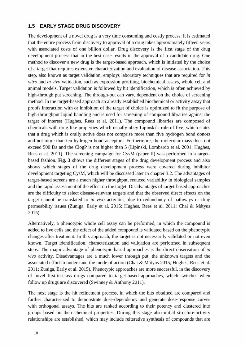
Figure 3. Flow scheme of the different stages of the drug development process (red), with focus on
early stage drug discovery (purple). The lower panel of the figure summarizes the major steps towards
development of inhibitors targeting CysM (paper II), including high-throughput screening, in vitro and
in vivo validation. Scatterplot from the target-based HTS, which was based on the PLP fluorescence
signal and the emission spectrum of the ligand free (red) and ligand bound (blue) enzyme are shown
in comparison to the background (black). The in vitro target specific interaction is illustrated by the
binding curve of the top inhibitor from an ITC experiment, and the crystal structures of target ligand
complexes. In vivo validation of the antibacterial activity is illustrated by the antibacterial killing
effect of the top CysM inhibitor in comparison to isoniazid.

12
Drug discovery of novel antimycobacterial drugs faces several organism specific challenges.
One example is the slow growth rate that slows down the evaluation of all live-bacteria based
tests. In order to perform research on Mtb, a laboratory of biosafety level 3 is required, which
results in very costly facilities needed for the drug discovery process. The waxy cell wall
containing many different lipids acts as a natural diffusion barrier that prevents penetration of
drugs through the bacterial cell wall. The presence of efflux pumps that actively transport
certain types of molecules out of the mycobacterial cell are in particular hard to tackle.
Another obstacle is the presence of various microenvironments in which Mtb can survive and
the simultaneous existence of several metabolic states of Mtb. (Zuniga, Early et al. 2015).
Several animal models are used to study latent tuberculosis, however these models do not
truly mimic infection in humans. One example is the mouse model, although frequently used
immune response towards Mtb is different because granulomas do not progress into the
caseous or calcified type. The bacterial burden can also differ a lot between mice and
humans. Zebrafish are also often used as model organisms as they show caseation of
granulomas however Mycobacterium marinum establishes the infection instead of Mtb. In
vitro models such as the Wayne hypoxia model suffer from the absence of standardization,
because different protocols exist and this model does not address adaptation of Mtb to other
stress inducers (Alnimr 2015).
Even though mentioned challenges exist in the development of novel antimycobacterial
drugs, several advances in drug development have been made in recent years. One example is
bedaquiline, which was FDA-approved in 2012 for treatment of adults suffering from MDR
pulmonary tuberculosis, where no alternative treatments are available (Cox & Laessig 2014).
Bedaquiline was discovered by applying a phenotypic whole cell screen, using
Mycobacterium smegmatis as a surrogate strain. The target was identified by isolating
resistant mutants, followed by whole genome sequencing (Andries, Verhasselt et al. 2005).
This approach has the advantage that it not only identifies the target, but also identifies
mechanisms responsible for resistance or activation of the drug. Bedaquiline was shown to
inhibit a mycobacterial ATP synthase found in the mycobacterial cell membrane and thereby
preventing energy production of Mtb (Mikušová & Ekins 2017). During clinical trials of
bedaquiline a mortality imbalance between the treated and placebo group was observed,
however a direct link between bedaquiline and mortality of the patients could not be
established (Cox & Laessig 2014; Diacon, Pym et al. 2014). Bedaquiline is one of four novel
compounds that are currently in the advanced stages of clinical development. In addition to
these four novel compounds, the three drugs, rifampicin, rifapentine and linezolid, which are
already approved for treatment of tuberculosis, are currently undergoing further clinical trials.
Rifampicin and rifapentine are tested if high doses are feasible for treatment at all and
linezolid is tested in new drug regimens with already used antimycobacterial drugs (World
Health Organization 2016; Wallis, Maeurer et al. 2016). Very recently, the drug-like
molecule spiroisoxazoline SMARt-420 was shown to fully reverse resistance against
ethionamide in Mtb, a prodrug that requires activation by mycobacterial enzymes. SMARt-
420 activates an alternative bioactivation pathway for ethionamide that increased sensitivity
towards the drug. Although SMARt-420 is still in its discovery stage, it opens interesting
possibilities, for example temporary treatment with SMARts, to destroy resistant Mtb
subpopulations during ordinary treatment, as proposed by the authors of this study
(Blondiaux, Moune et al. 2017).

13
1.6 L-CYSTEINE BIOSYNTHESIS AS DRUG TARGET
The availability of L-cysteine has been shown to be of high importance for Mtb during
infection. Up-regulation of underlying genes of L-cysteine biosynthesis under conditions
reflecting dormancy revealed a link between L-cysteine necessity and latent tuberculosis
(Voskuil, Visconti et al. 2004; Schnappinger, Ehrt et al. 2003). The genes significantly up-
regulated included cysD, cysNC and sirA, which encode for enzymes important for sulfur
assimilation and cysM and cysK2, genes encoding for two of the three mycobacterial L-
cysteine synthases (Voskuil, Visconti et al. 2004; Voskuil, Bartek et al. 2011; Schnappinger,
Ehrt et al. 2003; Betts, Lukey et al. 2002; Hampshire, Soneji et al. 2004). In addition, the
redox-active moiety of mycothiol is a sulfhydryl group that is derived from L-cysteine and
directly linked to mycobacterial redox defense against RNS and ROS inside macrophages
(Bhave, Muse et al. 2007). Randomized transposon mutagenesis showed that mutants
deficient in producing CysD, CysH, CysE, CysNC, CysO and CysM were severely attenuated
in a macrophage and mouse model of dormancy (Rengarajan, Bloom et al. 2005). Inhibitors
targeting mycobacterial CysH, the APS reductase providing sulfite for subsequent L-cysteine
formation, have been successfully identified by a target-based screening approach. The
strongest inhibitors were based on a 6H-pyrido[4,3-b]carbazole scaffold. These compounds
reduced cellular levels of sulfur containing metabolites, such as sulfite, L-cysteine and
mycothiol in vivo and showed bactericidal activity against dormant Mtb (Palde, Bhaskar et al.
2016). Hence targeting L-cysteine biosynthesis and the preceding sulfur assimilation pathway
might be a valuable route for the discovery of novel antimycobacterial drugs. The
mycobacterial L-cysteine biosynthesis is of particular interest, because of the complete
absence of this pathway in humans (Schnell, Sriram et al. 2015).
1.7 SULFUR ASSIMILATION IN MTB
The first step in sulfur assimilation in Mtb (Fig. 4) is transport of inorganic sulfate by the
CysT●W●A●SubI ABC transporter complex through the cytoplasmic membrane into the
mycobacterial cytoplasm from the environment. The sulfate transporter consists of the two
membrane spanning subunits CysW and CysT. The sulfate binding subunit, SubI, is
connected via a lipid tail to the cytoplasmic membrane. On the cytoplasmic site, CysW and
CysT each bind one copy of the nucleotide binding protein CysA (Wooff, Michell et al.
2002). Sulfate is thought to follow a similar transport route to that of phosphate, which first is
imported via porins and subsequently transported through the cytoplasmic membrane by a
specific phosphate transporter (Wolschendorf, Mahfoud et al. 2007; Rengarajan, Bloom et al.
2005).
In the cytoplasm, sulfate is further processed by ATP sulfurylase, a trifunctional enzyme
complex consisting of two polypeptide chains, the sulfurylase CysD and a GTPase CysN
(Fig. 5) (Schelle & Bertozzi 2006). In Mtb CysN carries the APS kinase, CysC, as its C-
terminal domain (Sun, Andreassi et al. 2005). ATP sulfurylase can either produce APS,
which is the starting material for the reductive branch of the sulfur assimilation pathway
subsequently leading to the synthesis of L-cysteine and MSH (Williams, Senaratne et al.
2002). Alternatively ATP sulfurylase can produce PAPS, the substrate for sulfotransferases to
produce sulfolipids, membrane components that have been shown to be associated with
increased mycobacterial virulence (Goren, Brokl et al. 1974). In particular sulfolipid 1
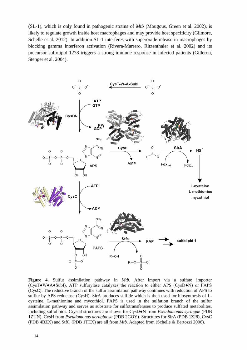
14
(SL-1), which is only found in pathogenic strains of Mtb (Mougous, Green et al. 2002), is
likely to regulate growth inside host macrophages and may provide host specificity (Gilmore,
Schelle et al. 2012). In addition SL-1 interferes with superoxide release in macrophages by
blocking gamma interferon activation (Rivera-Marrero, Ritzenthaler et al. 2002) and its
precursor sulfolipid 1278 triggers a strong immune response in infected patients (Gilleron,
Stenger et al. 2004).
Figure 4. Sulfur assimilation pathway in Mtb. After import via a sulfate importer
(CysT●W●A●SubI), ATP sulfurylase catalyzes the reaction to either APS (CysD●N) or PAPS
(CysC). The reductive branch of the sulfur assimilation pathway continues with reduction of APS to
sulfite by APS reductase (CysH). SirA produces sulfide which is then used for biosynthesis of L-
cysteine, L-methionine and mycothiol. PAPS is used in the sulfation branch of the sulfur
assimilation pathway and serves as substrate for sulfotransferases to produce sulfated metabolites,
including sulfolipids. Crystal structures are shown for CysD●N from Pseudomonas syringae (PDB
1ZUN), CysH from Pseudomonas aeruginosa (PDB 2GOY). Structures for SirA (PDB 1ZJ8), CysC
(PDB 4BZX) and Stf0, (PDB 1TEX) are all from Mtb. Adapted from (Schelle & Bertozzi 2006).

15
The remnant of the sulfotransferase reaction is 3’-phosphoadenosine-5’-phosphate (PAP),
which is processed by CysQ, a PAP phosphatase hydrolyzing PAP to adenosine
monophosphate, and phosphate (Hatzios, Iavarone et al. 2008), which in addition is a
competitive inhibitor of sulfotransferases. Alternatively CysQ also uses PAPS as a substrate,
resulting in the formation of APS (Pi, Hoang et al. 2005). Disruption of cysQ in Mtb resulted
in a phenotype comprising reduced growth in liquid culture. In these mutants the content of
reduced sulfur species remained the same, however presence of SL-1 and its precursor were
significantly less abundant compared to the wild type (Hatzios, Schelle et al. 2011).
If APS is utilized for L-cysteine production, the pathway is directed towards the production
of reduced sulfur. The first step of the reductive branch is performed by CysH, also called
APS reductase which catalyzes the reduction of APS to sulfite and AMP. The necessary
reduction potential is provided by the co-factor thioredoxin. The catalyzed reaction follows a
two-step mechanism in which APS undergoes a nucleophilic attack, yielding a thiosulfonated
enzyme intermediate (Carroll, Gao et al. 2005). The enzyme contains an iron sulfur cluster,
which is essential for catalysis, but its exact function is not fully understood. Most likely, it
plays a role in sulfuryl group transfer through formation of a electrostatic bridge to the sulfate
group of APS and by orienting the catalytic residues and activating the substrate. (Bhave,
Hong et al. 2011). In the second step sulfite is released in a thioredoxin dependent manner
(Carroll, Gao et al. 2005).
The produced sulfite is then further reduced via a six electron transfer reaction by sulfite
reductase SirA, previously denoted NirA (Schnell, Sandalova et al. 2005; Pinto, Harrison et
al. 2007). Reduced ferredoxin binds temporarily and delivers electrons via the iron sulfur
cluster to the siroheme group to sulfite, which thereby is reduced to sulfide (Kuznetsova,
Knaff et al. 2004; Schnell, Sandalova et al. 2005).
1.7.1 The sulfur assimilation pathway in other organisms
The sulfur assimilation pathway in Mtb is most similar to the corresponding pathway in the
higher plant Arabidopsis thaliana, which follows the same steps from sulfur uptake by a
transporter via activation as APS to follow the reductive branch of the pathway towards
sulfite or as PAPS towards the sulfation branch (Takahashi H 2011; Ravilious and Jez 2012).
Another organism in which sulfur assimilation is well characterized is the γ-proteobacterium
Escherichia coli. The major difference to Mtb is that APS kinase is not fused to the C-
terminus of CysN, but expressed as a single polypeptide chain. Instead of employing APS
that is reduced by APS reductase to sulfite, in E. coli the sulfite that eventually is
incorporated in L-cysteine is produced from PAPS by the aid of PAPS reductase (Carroll,
Gao et al. 2005). Furthermore the genome of E. coli does not encode the necessary genes for
PAPS-dependent sulfotransferases that are present in many plants and other bacteria
(Sekowska, Kung et al. 2000; Carroll, Gao et al. 2005). To produce sulfated metabolites E.
coli is dependent on aryl sulfate sulfotransferases that transfer sulfuryl groups from phenolic
esters to phenol (Malojčić, Owen et al. 2014).

16
1.7.2 The mycobacterial sulfate activation complex
In the trifunctional sulfate activation complex (Fig. 5) CysD catalyzes the adenylyl transfer
from ATP to activate the inorganic sulfate. The sulfate ion performs a nucleophilic attack on
the α-phosphorous of ATP to synthesize adenosine 5’-phosphosulfate. The formation of the
anhydride bond in APS is energetically disadvantageous, therefore APS production is linked
to energy-providing GTP hydrolysis, catalyzed by CysN (Sun, Andreassi et al. 2005).
Figure 5. Model of the trifunctional ATP sulfurylase with substrates and products formed. CysD
performs the formation of APS from SO42-
and ATP, which is subsequently used for L-cysteine
production. CysN provides the necessary energy to build the sulfuric phosphoric acid anhydride
bond in APS by hydrolyzing GTP. CysC, which is fused to the C-terminus of CysN phosphorylates
APS to PAPS, which is then used as substrate for sulfotransferases.
ATP sulfurylase lies at a metabolic crossroad that can either direct produced APS towards the
reductive branch of the pathway, resulting in the de novo biosynthesis of L-cysteine (Schnell,
Sriram et al. 2015) and subsequently mycothiol (Jothivasan & Hamilton 2008), or towards
the sulfation branch by production of PAPS by CysC. In Mtb, the formation of PAPS by
CysC is almost 6000 fold more efficient than the formation of APS by CysD, which has been
shown by enzymatic studies of ATP sulfurylase in vitro (Sun, Andreassi et al. 2005).
1.7.3 APS kinase
The APS kinase domain of mycobacterial ATP sulfurylase, denoted as CysC, has a molecular
weight of 21 kDa (Poyraz, Brunner et al. 2015) and belongs to the fold-class of P-loop
NTPases. Common in all P-loop NTPases is the presence of the Walker A motif, also known
17
as the P-loop comprising a consensus sequence of GxxxxGK. For a sequence alignment of
CysC with its closest structural homologs, see paper I, Fig. 2. The P-loop is responsible for
binding the β-phosphate of the substrate ATP (Leipe, Koonin et al. 2003). In CysC of Mtb
this loop can be found between β1 and α1 and starts with G450, however with an additional
fifth residue between the two glycine residues in the consensus sequence followed by a
conserved serine residue, which coordinates Mg2+
(Poyraz, Brunner et al. 2015). The Walker
B motif is the second characteristic of P-loop NTPases and can be found at the C-terminal
end of a hydrophobic β-strand and coordinates the divalent metal ion Mg2+
that in turn
coordinates the β- and γ-phosphate of ATP, comprised by D478 in the mycobacterial CysC.
(Leipe, Koonin et al. 2003; Poyraz, Brunner et al. 2015). The loop region, from residue L477
to D480, found between β2 and α2 is conserved in APS kinases (Poyraz, Brunner et al. 2015).
In many organisms the APS kinases are highly conserved, however, they can be expressed as
a single polypeptide chain instead of a fusion protein (Hell 2008), which is the case in E. coli.
In Pseudomonas aeruginosa CysN and CysC are as well fused, but one additional copy of
CysC is expressed as a single polypeptide chain (Williams, Senaratne et al. 2002). The APS
kinase of Mtb shares a high degree of sequence identity with several APS kinases from
different organisms, amongst these are the two human homologs PAPS synthetase 1 and 2.
The overall sequence identity between mycobacterial CysC and the kinase domains of both
human PAPS synthetases is approximately 50%. In contrast to Mtb, the APS kinase domain
and the adenylyl transferase domain form one bifunctional enzyme on a single polypeptide
chain in humans, the APS kinase domain however is found at the N-terminus. The
energetically unfavorable phosphoric-sulfuric anhydride bond formation in APS is driven by
the separate enzyme, inorganic pyrophosphatase by hydrolysis of PPi (Venkatachalam 2003).
Penicillium chrysogenum APS kinase follows a sequential ordered reaction mechanism in
which ATP has to bind before APS and PAPS has to be released before ADP can leave the
active site (Renosto, Seubert et al. 1984). APS leads to uncompetitive substrate inhibition in
P. chrysogenum, A. thaliana, human and rat APS kinase, during which the enzyme is trapped
in a dead end complex in which ADP and APS are bound in the active site interfering with
the sequential ordered mechanism and leaving the enzyme inactive (Lansdon, Fisher et al.
2004).
In E. coli the usually followed sequential ordered reaction mechanism can alternatively
follow a ping pong mechanism during which a phosphorylated enzyme intermediate at S109
is produced, which is situated between the two nucleotide binding sites. In the presence of
low APS concentration the ternary CysC●ATP●APS complex is not formed (Satishchandran
& Markham 1989). Later it was shown that the phosphorylated S109 can participate in either
of the two reaction mechanisms (Satishchandran, Hickman et al. 1992; Satishchandran &
Markham 2000).
In A. thaliana, four isoenzymes of APS kinase can be found, which are expressed as single
polypeptide chains, however comprise different sizes, the smaller variant with 23 kDa for
cytoplasmic expression and a larger variant of 32 kDa, which is a pre-protein, carrying a large
transit peptide for export. In addition, in all isoenzymes six conserved L-cysteine residues are
found. In APS kinase 1, four of these L-cysteine residues are in close proximity that
potentially can form two disulfide bonds. Isoform 1 is activated in the presence of thiols and
18
thioredoxin, whereas the presence of oxidants results in the opposite, hence it was proposed
that activity is dependent on the redox state of these L-cysteine residues (Lillig, Schiffmann
et al. 2001). APS kinase 1 and 2 produce the majority of PAPS, which is used for sulfation to
produce glucosinolates, essential in plant defense mechanisms (Halkier & Gershenzon 2006).
Mutant studies showed that APS kinase 1 would be sufficient for normal growth and APS
kinase 3 and 4 are functionally redundant. APS kinase 2 however was shown to be essential
for A. thaliana germination, because its absence results in inviable pollen (Mugford,
Matthewman et al. 2010). Extensive ITC studies of APS kinase 1 confirmed the proposed
sequential ordered mechanism. Although binding of APS as the first substrate would be
possible and lead to catalysis, the reaction would be slower, because initial ATP binding
results in proper positioning of the γ-phosphate in close proximity to the acceptor hydroxyl
group in APS and assures the prerequisites for catalysis, by providing optimal conformations
of the active sites (Ravilious & Jez 2012).
1.8 DE NOVO L-CYSTEINE BIOSYNTHESIS
In Mtb three de novo L-cysteine biosynthesis pathways are known (Fig. 6). The key players
are the three PLP-dependent L-cysteine synthases, CysK1, CysM and CysK2, each of them is
characteristic for one pathway (Schnell, Sriram et al. 2015). Although all three enzymes
produce L-cysteine, they show clear differences in their substrate preference. While CysM
and CysK2 use O-phospho-L-serine (OPS) as their preferred acceptor substrate, CysK1
exclusively uses O-acetyl-L-serine (OAS) (Ågren, Schnell et al. 2008; Steiner, Böth et al.
2014; Schnell, Oehlmann et al. 2007; O’Leary, Jurgenson et al. 2008). CysK1 and CysK2
utilize the sulfide provided by the reductive branch of the sulfur assimilation pathway
(Steiner, Böth et al. 2014; Schnell, Oehlmann et al. 2007). CysM is dependent on a small
sulfur delivering protein CysO that is thiocarboxylated at its C-terminus (O’Leary, Jurgenson
et al. 2008). The three L-cysteine synthases show a sequence identity of approximately 30%
with each other and they have been shown to be active during different metabolic states of
Mtb. While CysK1 represents the classical pathway that is also found in other organisms,
such as bacteria (Kredich, Hulanicka et al. 1979; Rabeh & Cook 2004) and higher plants
(Bonner, Cahoon et al. 2005), the underlying genes of the two alternative pathways cysM,
cysO and cysK2 are upregulated under conditions mimicking dormancy, such as hypoxia and
oxidative stress (Schnappinger, Ehrt et al. 2003; Voskuil, Visconti et al. 2004; Betts, Lukey et
al. 2002; Hampshire, Soneji et al. 2004). Randomized transposon mutagenesis resulted in
attenuated mutants deficient in CysO and CysM in an in vitro model of dormancy and a
mouse model of tuberculosis infection, which lead to the conclusion that the pathway based
on CysM is the major route to L-cysteine during dormancy (Rengarajan, Bloom et al. 2005;
Sassetti & Rubin 2003). Mammals including humans produce L-cysteine from L-methionine.
L-methionine undergoes SAM-dependent transmethylation followed by transsulfuration to
yield L-cysteine. L-methionine is an essential amino acid that mammals need to acquire from
their diet (Stipanuk 2004).

19
Figure 6. The three routes of de novo L-cysteine biosynthesis in Mtb. Each route is characterized by
one cysteine synthase. In the classical pathway which is shown in red CysK1 utilizes sulfide from
the reductive branch of the sulfur assimilation pathway and O-acetyl-L-serine (OAS) produced by
CysE. In the salvage pathway CysK2 produces L-cysteine from the same sulfur source as CysK1
but uses O-phospho-L-serine (OPS) as its acceptor substrate. The preferred reaction of CysK2
however is the formation of S-sulfocysteine from OPS and thiosulfate. L-cysteine formation during
dormancy is performed by CysM that uses thiocarboxylated CysO as the sulfur donor and OPS.
Crystal structures of CysK1 (PDB 2Q3C) and the CysM●CysO complex (PDB 3DWG) are shown
in red and blue and purple, respectively. Adapted from (Schnell, Sriram et al. 2015)
1.8.1 The classical pathway to L-cysteine
The classical pathway which is found in most bacteria and plants involves two enzymatic
reactions (Campanini, Benoni et al. 2015). CysE first catalyzes the reaction to produce OAS
followed by the formation of L-cysteine by CysK1, a bona fide O-acetyl-L-serine
sulfhydrylase (Rabeh & Cook 2004). CysE catalyzes acetylation of L-serine utilizing acetyl-
CoA leading to OAS. The C-terminal end of CysE plays an important role in regulation of L-
cysteine formation. CysE is active in complex with CysK1, often referred to as L-cysteine
synthase complex in the literature (Campanini, Speroni et al. 2005). While CysE is
catalytically active, CysK1 remains inactive. Upon complex formation the C-terminal end of
CysE is inserted into the active site of CysK1 blocking the binding site for OAS, leading to
inhibition of CysK1 (Fig. 7B). Increasing concentration of OAS in the presence of reduced
sulfur results in dissociation of the complex allowing CysK1 to become catalytically active
and produce L-cysteine, while CysE becomes inactive (Campanini, Speroni et al. 2005)

20
Figure 7. Complex formation between CysK1 and CysE, and the regulation of cysteine biosynthesis.
A) Magnification of the active site of CysK1 (PDB 2Q3C) colored in red with the inserted C-terminal
tetrapeptide DFSI of CysE colored in turquois. B) Model of the interplay between CysK1 and CysE.
CysE is active while in complex with CysK1 and produces L-serine. When OAS starts to accumulate,
the complex dissociates and CysK1 catalyzes L-cysteine formation. Adapted from (Schnell, Sriram et
al. 2015).
CysK1 belongs to the fold family of type II PLP-dependent enzymes (Schneider, Käck et al.
2000), with an overall structure divided into two domains, where each shows a typical α/β
fold. The domain at the N-terminus is made up by a four stranded parallel β-sheet, which is
flanked by three α-helices at one side and one α-helix on the other side. The domain at the C-
terminus comprises a mixed β-sheet consisting of six β-strands that are surrounded by four α-
helices. The cofactor PLP is bound between the two domains. The enzymatic reaction can be
divided into two half reactions following a ping-pong mechanism (Rege, Kredich et al. 1996).
In the ground state of CysK1, the PLP cofactor forms an internal aldimine with the catalytic
residue K44 via a covalent bond. The first half reaction starts with the formation of an
external aldimine between the incoming substrate OAS and the co-factor PLP. CysK1
performs an elimination reaction at the β-carbon position by abstracting one proton at the α-
position and releasing the acetate moiety yielding the α-aminoacrylate intermediate and
protonated K44 as the product of the first half reaction. The second half reaction is initiated
by a nucleophilic attack of sulfide on the β-carbon of the α-aminoacrylate intermediate.
Reprotonation of the α-carbon by K44 and release of L-cysteine regenerate the initial ground
state of CysK1 (Schnell, Oehlmann et al. 2007).
The classical route of de novo L-cysteine biosynthesis is strictly regulated at three levels,
which has been demonstrated in Salmonella. At the first level, gene expression is under
control of CysB, a LysR-type transcription factor controlling gene transcription of the entire
cysteine regulon cysPUWAM (cysM in E. coli and Salmonella most likely corresponds to the
mycobacterial cysK2 gene). However the genes that are not controlled by CysB are the
underlying genes of CysE and CysK1. The product OAS of the CysE catalyzed reaction is
prone to non-enzymatic rearrangement to N-acetyl serine (NAS). Rising of the NAS level
activates CysB and induces transcription of the genes in the cysteine regulon. In contrast

21
sulfide and thiosulfate act as anti-inducers of gene expression. The second level of regulation
is through competitive feedback inhibition of CysE through increasing L-cysteine
concentration triggering a rearrangement of the C-terminus that subsequently blocks the
binding site of acetyl CoA. The third level is the regulation of the CysE●CysK1 complex
formation itself. High OAS concentration leads to dissociation of the complex. Binding of
OAS in the active site of CysK1 directly competes with binding of the C-terminal end of
CysE comprising the amino acids DFSI in the mycobacterial enzyme. The C-terminal end of
CysE in other organisms follows the consensus sequence N, X1, X2, I where X1 corresponds
to an aromatic residue and X2 to a strong hydrogen bond donor (Campanini, Benoni et al.
2015). The highly conserved isoleucine at the C-terminal end of CysE probably induces the
closing of the active site of CysK1 (Zhao, Moriga et al. 2006; Raj, Mazumder et al. 2013;
Huang, Vetting et al. 2005). The tetrapeptide DFSI, which was derived from the C-terminus
of mycobacterial CysE was shown to act as a strong inhibitor of CysK1 (Schnell, Oehlmann
et al. 2007). From the co-crystal structure it can be seen that DFSI is bound between the two
domains of CysK1. The carboxylate group of isoleucine is anchored in the interior of the
active site by the conserved residues T71, N74, S72 and Q144 usually binding the
carboxylate group of the substrate OAS, whereas the residual amino acids of the tetrapeptide
fill the active site pocket (Fig. 7A). The conserved residues binding the substrate form part of
a loop that with two additional loops between β5 and α3 and β6 and α4, close over the active
site upon domain rotation caused by the formation of the α-aminoacrylate reaction
intermediate complex. Upon binding of the C-terminal tetrapeptide of CysE, domain rotation
is prevented and the active site is blocked (Schnell, Oehlmann et al. 2007).
Paralogs of CysK1 have been shown to gain moonlighting functions upon pairing up with
other proteins via the insertion of the C-terminal residues of the binding partner into the
active site of CysK1. CysK1 in C. elegans, B. subtilis and S. aureus have been shown to alter
gene transcription. In uropathogenic E.coli, CysK1 is involved in activation of an
antibacterial protein toxin that inhibits the growth of neighboring bacteria (Campanini,
Benoni et al. 2015).
1.8.2 A salvage pathway to L-cysteine formation
CysK2 is the key player of another potential pathway leading to L-cysteine formation. In
contrast to CysK1 and CysM, no crystal structure of the enzyme is available, however CD
spectroscopy confirmed a mixed α/β fold and spectrophotometry detected the characteristic
absorbance maximum of internal aldimine formation of CysK2 and the co-factor PLP,
suggesting that it is also belongs to the fold type II family of PLP-dependent enzymes. CysK2
uses OPS and sulfide as a sulfur donor to produce L-cysteine, however with a very low
catalytic efficiency. The preferred sulfur donor is thiosulfate instead, reflected in an almost
tenfold increase of affinity and a fivefold higher turnover increase when compared to sulfide.
The product of the preferred enzymatic reaction is S-sulfocysteine, which has been shown to
be a direct precursor of L-cysteine in bacteria and plants. The exact physiological role of S-
sulfocysteine, however, remains enigmatic. One hypothesis is that thiosulfate is produced as a
side product of the sulfur assimilation pathway, due to interfering oxidative stress, when the
pathogen is engulfed inside the host macrophages and could then act as a sensing molecule of
oxidative stress (Steiner, Both et al. 2014). In A. thaliana exposure to light induces oxidative
stress in chloroplasts. The S-sulfocysteine synthase CS26 produces S-sulfocysteine a signal

22
molecule activating redox defense mechanisms, to cope with oxidative stress (Gotor &
Romero 2013). Alternatively S-sulfocysteine might act as the storage variant of L-cysteine,
due to its lower toxicity towards the bacterial cells in comparison to L-cysteine (Nakatani,
Ohtsu et al. 2012).
Based on the unique combination of utilizing OPS and sulfide and the low catalytic efficiency
of L-cysteine formation via the CysK2 route, this pathway might play a role as a dormancy-
induced salvation pathway rather than a major route to L-cysteine production (Steiner, Böth
et al. 2014).
1.8.3 L-cysteine formation during dormancy
An alternative pathway leading to L-cysteine production during dormancy is exclusively
found in Actinobacteria (Burns, Baumgart et al. 2005). The L-cysteine synthase that is the
key enzyme catalyzing the reaction to form L-cysteine in this alternative route is CysM. In
contrast to CysK1 that uses OAS as its primary substrate, CysM uses OPS to form the α-
aminoacrylate reaction intermediate (Ågren, Schnell et al. 2008). Instead of using sulfide ions
provided by the reductive branch of the sulfur assimilation pathway, CysM uses the small
sulfur-delivery protein CysO that is thiocarboxylated at its C-terminus as a sulfur donor
(Burns, Baumgart et al. 2005; Ågren, Schnell et al. 2008; O’Leary, Jurgenson et al. 2008). It
was also shown that CysM is capable of using OAS or OPS, however the rate constant of the
reaction is more than 600 times higher with OPS, clearly indicating the substrate specificity
(Jurgenson, Burns et al. 2008; Ågren, Schnell et al. 2008; O’Leary, Jurgenson et al. 2008).
The overall structure of CysM is similar to CysK1, following the same fold family of type II
PLP-dependent enzymes, where K51 forms the internal aldimine with the PLP cofactor in the
active site. The active site of CysM compared to CysK1 shows several residues which are
different, in particular R220 in CysM, found to be responsible for OPS specificity (Ågren,
Schnell et al. 2008). CysM can adapt to an open or closed conformation, as has been seen in
several crystal structures (Jurgenson, Burns et al. 2008; Ågren, Schnell et al. 2009).
Characteristic for the open conformation is a solvent accessible active site. In contrast, in the
closed conformation three loop regions at the entrance of the active site comprise different
arrangements and the C-terminus is inserted into the active site, making it solvent
inaccessible. The α-aminoacrylate of CysM has a half-life eight times longer than the same
reaction intermediate of CysK1. This property is due to the firm closure that depends on the
conformation of the C-terminus in order to protect the oxidation sensitive reaction
intermediate from damage by hydrogen peroxide and other small nucleophiles. Protection of
the reaction intermediate may be relevant for survival of Mtb in vivo, to assure L-cysteine
production inside the oxidizing environment of host macrophages (Ågren, Schnell et al.
2009).
CysO is a small sulfur carrier protein comprising an ubiquitin-like fold. The core of this
protein is a four stranded mixed β-sheet, with an α-helix inserted between β2 and β3
(Jurgenson, Burns et al. 2008). Thiocarboxylation of the C-terminus starts with the acyl-
adenylation of a diglycyl moiety by MoeZR, which is subsequently converted by the same
enzyme through a sulfane group bound at its C-terminus (Voss, Nimtz et al. 2011).

23
Figure 8. A) Catalytic cycle of CysM indicating the ground state with the internal aldimine formed
with K51 (I), the external aldimine with the OPS (II), the α-aminoacrylate intermediate (III), and
the CysO-Cys-PLP external aldimine (IV). B) Superposition of the open and closed conformation
of CysM, colored in light and dark blue, respectively. The cofactor PLP is colored in yellow. The C-
terminal end of the monomer in the closed conformation is labeled. (PDB 3FGP). C) In the
CysM●CysO complex, CysM and CysO (PDB 3DWG) are colored in light blue and purple,
respectively. Secondary structure elements that are involved in interaction between CysM and CysO
are labeled and three 310 helices found in the mobile active site loop (residue 211–237) of CysM are
colored in beige.

24
The overall reaction, catalyzed by CysM, follows a mechanism similar to the CysK1 reaction.
(Fig. 8A) After conversion of the internal aldimine (I) of K51 and the cofactor PLP to an
external aldimine (II) upon binding of OPS, the phosphate group of OPS is abstracted at the
β-carbon via an elimination reaction, yielding the α-aminoacrylate intermediate (III).
Complex formation between CysM and CysO allows nucleophilic attack on the β-carbon by
thiocarboxylate, resulting in a CysO-L-cysteine-PLP adduct (IV). Reprotonation of the α-
carbon induces release of CysO carrying the reaction product L-cysteine as a C-terminal
adduct, while the initial ground state with the bound internal aldimine (I) is restored. Prior to
cleavage of the L-cysteine moiety by the zinc dependent metalloprotease Mec+, a
spontaneous N-acyl shift in L-cysteine is observed (Jurgenson, Burns et al. 2008). A crystal
structure of the CysM●CysO complex comprised an asymmetric CysM dimer, where one
monomer adapted to the closed conformation and the second monomer interacted with CysO.
The main differences between the two monomers were observed in the loop region stretching
from residue 211 to residue 237, which fold into three 310 helices in the CysM●CysO
monomer instead of one 310 helix in the closed conformation. In addition part of the N-
terminal domain extends away from the core of CysM to allow interaction with CysO. The
loop region is also responsible for the majority of interactions with CysO. Other interactions
include hydrogen bonding between helix α4 and strand β8 of CysM with a loop region
preceding β3 and the C-terminus of CysO, respectively. After β-elimination of phosphate by
CysM, the thiocarboxylated C-terminus of CysO is placed into the initial binding pocket of
the leaving group of the first substrate (Jurgenson, Burns et al. 2008). Using thiocarboxylated
CysO instead of sulfide ions is likely to be beneficial for Mtb, especially during dormancy,
because thiocarboxylate is more resistant to oxidation and less toxic to the cell (O’Leary,
Jurgenson et al. 2008).
In summary, the mycobacterial sulfur assimilation pathway supplies the sulfur required for
the production of sulfated metabolites, while the three L-cysteine synthases use sulfur
according to their substrate profile. CysM seems to be a dormancy specific L-cysteine
synthase and inhibition of CysM is expected to interfere with the availability of mycothiol,
crucial for intracellular redox homeostasis.

25
2 AIMS OF THE THESIS
L-cysteine biosynthesis and the preceding sulfur assimilation pathway have been shown to be
essential for Mtb in order to maintain its metabolism during active and latent tuberculosis.
With this in mind, and with the lack of novel antimycobacterial drugs, we studied four
mycobacterial enzymes to determine whether it is possible to discover potent inhibitors with
the potential to be further developed into novel antimycobacterial drug candidates.
Paper I – Crystal structures of the Kinase domain of the Sulfate-activating complex of
Mycobacterium tuberculosis
Structural and mechanistic characterization of CysC, the APS-kinase domain of
mycobacterial ATP sulfurylase
Comparison to human homologs to evaluate feasibility of developing specific
inhibitors targeting the mycobacterial enzyme only
Paper II – Inhibitors of the L-cysteine Synthase CysM with Antibacterial Potency against
Dormant Mycobacterium tuberculosis
Performance of a CysM-based HT screening campaign to identify novel inhibitors
targeting the L-cysteine synthase CysM, including hit confirmation and hit expansion
In vitro validation of the obtained hits with several orthogonal enzymatic assays and
crystallographic methods, including establishment of SAR
In vivo validation of the most potent inhibitors in actively dividing Mtb and a nutrient
starvation model, reflecting the dormant state of Mtb
Paper III – Profiling of in vitro activities of urea-based inhibitors against L-cysteine
synthases from Mycobacterium tuberculosis
In vitro validation of a subset of inhibitors tested during the experiments to generate
paper II against the two remaining mycobacterial L-cysteine synthases CysK1 and
CysK2 by two orthogonal enzymatic assays
Identification of inhibitors specific to CysK1 and/or CysK2
Identification of compounds with triple inhibitory effect against CysK1, CysK2 and
CysM


27
3 RESULTS AND DISCUSSION
3.1 PAPER I – CRYSTAL STRUCTURES OF THE KINASE DOMAIN OF THE
SULFATE-ACTIVATING COMPLEX IN MYCOBACTERIUM
TUBERCULOSIS
The mycobacterial APS kinase domain CysC has been co-crystallized with several
nucleotide combinations, ADP alone, ADP and APS and AMP-PNP, a non-hydrolysable
analog to ATP, and APS, respectively. Co-crystal structures were solved with the molecular
replacement method using the structure of the human PAPS synthetase 1 as a search model
(Sekulic, Konrad et al. 2007).
The overall structure of CysC follows the classical α/β purine nucleotide binding fold (Fig.
9A) (Schulz, Elzinga et al. 1974). On both sides two α-helices flank the five-stranded
parallel β-sheet core. In addition β2 and α2 are connected via one short α-helix and β4 and
β5 via two α-helices, respectively. Analytical gel filtration revealed that CysC forms a
dimer in solution which is consistent with the crystallographically determined dimeric
structure for all three CysC complexes. Analysis of the molecular contacts suggests a
buried surface area of 3300 Å2 for the dimer interface formed by helices α2 and α3 with
their corresponding symmetry mates.
The closest structural homologs were three bacterial APS kinases from Aeropyrum pernix,
(PDB 2YVU) , Thiobacillus denitrificans (Gay, Segel et al. 2009), Aquifex aelicus (Yu,
Lansdon et al. 2007), one from the fungus P. chrysogenum (MacRae, Segel et al. 2000),
one from the plant A. thaliana (Ravilious & Jez 2012) and the APS kinase domain from
human PAPS synthetase 1 (Harjes, Bayer et al. 2005) with r.m.s.d. values ranging from 0.9-
1.5 Å. Sequence identities of CysC with the five homologs ranged from 34 to 52%, sharing
the highest sequence identity with human PAPS synthetase 1.
3.1.1 ADP binding site of mycobacterial APS kinase
The ADP binding pocket is found in a cleft that is formed by the conserved P-loop stabilizing
the two phosphate groups of ADP on the bottom, loop T551 to K562 followed by helix α5
interacting with the ribose and the adenine base and is completed by the loop formed from
L593 to I599 that binds the adenine base on the opposite site. After analysis of the two
subunits of the binary complex it became apparent that the ADP molecules are bound in anti
in two slightly different conformations. The rotation of the glycosidic bond results in a shift
of the adenine ring and the α-phosphate. In this complex the major part of the adenine base
and the ribose moiety are accessible from the solvent, the bisphosphate group however is
tightly fixed to CysC by the P-loop. In the ternary complex CysC●ADP●APS, ADP is bound
in one defined conformation and the interactions between ADP and CysC are fully developed
as compared to the binary complex. In the second ternary complex, in which ADP was
replaced by AMP-PNP in the crystallization mixture, the purine nucleotide binds in the same
fashion as ADP. The only difference is the γ-phosphate group that interacts with K456 from
the P-loop and K562 on one side and is further stabilized from the opposite site by a bound
magnesium cation (Fig. 9B).

28
Figure 9. A) Overall structure of the Michaelis complex of the CysC dimer colored in beige and
purple with the bound substrates AMP-PNP and APS shown in purple stick representation. The
magnesium ion stabilizing the phosphate group is shown as a grey ball. The P-loop is colored in
pink and the DGDN loop in purple. The sidechain of C556 is shown in purple stick representation.
(PDB 4BZX) B) Detailed view of the interactions between CysC and the bound substrates AMP-
PNP and APS.
3.1.2 APS binding pocket
The APS binding pocket is found opposite of the ADP/AMP-PNP binding site and formed by
loop 490-495 and loop 562-582. APS is oriented with its ribose moiety towards the terminal
phosphate group of ADP and AMP-PNP, respectively. The adenine ring of APS is π-stacked
between F492 and F576. The former phenylalanine is part of loop 477-499, which is
displaced by approximately 2 Å upon APS binding in comparison to the binary complex and
thereby closes off the APS binding pocket. Loop displacement explains the relatively high
overall r.m.s.d. values of 0.9 Å, between the binary complex and the two ternary complexes,
although the protein core retains the same conformation in all three complexes.
3.1.3 Magnesium binding site
In spite of adding Mg2+
to the crystallization mixture, no electron density was observed for
the divalent metal ion in the ternary complex CysC●ADP●APS. In the second ternary
complex CysC●AMP-PNP●APS, where ADP is replaced by AMP-PNP, electron density for
Mg2+
was observed, which is probably due to the presence of the γ-phosphate in AMP-PNP
that contributes to the octahedral coordination of the magnesium ion via its oxygen. The
coordination of magnesium is completed by a second oxygen provided by the β-phosphate,
the side chain oxygen of S457 and three water molecules.
3.1.4 Mechanistic proposal for phosphoryl group transfer
The ternary complex of CysC in the crystal structure of CysC●AMP-PNP●APS provides a
detailed view of the interactions with the substrate and residues that are involved in catalysis
and allows a proposal for phosphoryl group transfer (Fig. 10). The γ-phosphate of AMP-PNP
is in close vicinity of the 3’ hydroxyl group of the receiving substrate APS. During catalysis,
orientation of AMP-PNP and charge stabilization of its phosphate groups is achieved by the

29
P-loop and the magnesium ion. Through progression of the enzymatic reaction, a charge
accumulation around the γ-phosphate occurs and is stabilized by K562, which is perfectly
positioned between the γ-phosphate group of AMP-PNP and the oxygen of 3’ hydroxyl group
of APS. The conserved residue D480 probably acts as a catalytic base by abstracting a proton
from the 3’ hydroxyl group of APS, and is attacked by the stabilized γ-phosphate. The
mechanism for the APS kinases is preserved in mice, humans, fungi and plants and also CysC
follows this mechanism (Singh & Schwartz 2003; Sekulic, Konrad et al. 2007; Lansdon,
Fisher et al. 2004; Ravilious & Jez 2012).
Figure 10. A) Magnification of the CysC active site with bound substrates AMP-PNP and APS
shown in purple stick representation and magnesium shown as a grey ball. Residues K562 and
D480 which are involved in phosphoryl group transfer are shown in stick representation. (PDB
4BZX) B) Proposed mechanism of phosphoryl group transfer in mycobacterial CysC. D480 acts as
the catalytic base that abstracts a proton from the 3’ hydroxyl group of APS that subsequently reacts
with the γ-phosphate of ATP. Charge stabilization during catalysis is achieved by K562.
3.1.5 Comparison to the human PAPS synthetases
PAPS synthetases are the closest human homologs, sharing a sequence identity of
approximately 50% with the mycobacterial CysC. Two isoforms have been characterized,
PAPS synthetase 1 and 2, which are sharing more than 70% sequence identity with each other
(Fuda, Shimizu et al. 2002).
One of the aims of this study was to analyze whether the architectures of the active sites of
mycobacterial CysC and the kinase domain in human PAPS synthase 1 and 2 are different

30
enough to allow inhibitor design to specifically inhibit only the mycobacterial enzyme.
Inhibition of CysC would prevent the formation of PAPS, and subsequently would impede
formation of sulfated lipids, which is associated with increased virulence in Mtb (Williams,
Senaratne et al. 2002). The APS binding sites of all three enzymes are very similar. Out of the
fifteen amino acids making up the APS binding site, twelve are conserved. An expected
increase in binding pocket volume by replacement of A522 (CysC) with F131 (hPAPSS) was
minimal. In the loop that binds the adenosine ring of APS T574 and H575 of CysC are
exchanged to K183 and G184 in the human enzyme, where only the backbone carbonyl
group of K183 participates in interaction with the ligand.
For the ATP binding site, the situation is similar. Although only six out of fourteen residues
are conserved, five additional residues comprise similar size and chemical properties as the
mycobacterial CysC. Only three amino acids are significantly distinct, which is P561,
replaced by V170 in the human homolog, R597 with C/S207 and Q602 with C212. Given the
high degree of conserved residues between human PAPS synthetase and CysC the design of
specific inhibitors targeting the mycobacterial enzyme only appears to be a challenging task
for medicinal chemistry, and was therefore not pursued further.
3.1.6 Is CysC regulated by disulfide bond formation?
During the performance of enzymatic activity assays to demonstrate that mycobacterial CysC
is functional as a single unit, it was observed that CysC activity is increased upon addition of
reductants, which was also reported for CysC homologs in A. thaliana (Sekowska, Kung et
al. 2000) and A. aelicus (Yu, Lansdon et al. 2007). In A. thaliana the formation of a
regulatory disulfide bond between an L-cysteine in the N-terminal domain and an L-cysteine
residue in the core domain of the adjacent monomer was observed. The disulfide bond
formation is dependent on the redox state of the surrounding. Reduction results in activation
of APS kinase, whereas disulfide bond formation gives a 20-fold less efficient enzyme
(Herrmann, Nathin et al. 2015; Lillig, Schiffmann et al. 2001). In the monomer of the
mycobacterial CysC three L-cysteine residues can be found, which raised the question if
CysC is also regulated upon disulfide bond formation. Five CysC mutants (C514A, C549A,
C556A, C556S and a triple mutant, in which all three cysteine residues were mutated to
alanine residues) were produced to probe the role of the three L-cysteine residues, since after
inspection of the crystal structures it became apparent that none of the L-cysteine residues is
involved in the formation of disulfide bonds. Amino acid replacement at position C514 and
C549 did result in enzyme variants that show the same activation behavior as the wild type
variant, suggesting that neither of these two L-cysteine residues alter CysC activity based on
their redox states.
The amino acid replacement at position 556 resulted in a catalytically inactive enzyme.
C556S was completely inactive and could not be reactivated upon addition of reductants. The
same behavior was observed for the triple mutant. In contrast, the activity of the C556A
mutant could be rescued by addition of DTT, but to a smaller extend than it was observed for
the wild type. The C556S mutant was sensitive to non-specific proteolysis by thrombin,
which was also observed during protein purification of the wild type enzyme. In case of the
wild type however, sensitivity could be circumvented by addition of ADP, but not in the case
of the C556S mutant.

31
Co-crystallization of the C556S mutant resulted in a crystal structure with an r.m.s.d. value of
0.5 Å between the mutant and wild type crystal structure of the binary CysC●ADP complex
indicating little structural deviations between the two of them. In none of the subunits of the
mutant ADP was bound and one segment from residue 552 to 581, including the residue of
interest in the C556S mutant, could not be modeled, due to lack of electron density. This
stretch belongs to the lid region that closes off the ATP binding site and allows positioning of
R559 close to the adenine ring of ATP and is conserved in APS kinases.
Mutagenesis, observed thrombin sensitivity despite addition of ADP and structural analysis
indicate that mutation of C556 to S556 results in deficient ATP binding, due to increased
flexibility of the lid closing the active site. It is known that enzyme catalysis is linked to
protein dynamics. Altered contributions of hydrogen bonds or van der Waals interactions are
capable of influencing protein dynamics in such a way that catalysis can be prevented or
slowed down (Henzler-Wildman & Kern 2007). Studies on the dynamics of two orthologous
adenylate kinases revealed that reduced catalytic activity in one of the kinases could be
attributed to slower opening of the lid region (Wolf-Watz, Thai et al. 2004). In CysC the
mutation from L-cysteine to L-serine might have a similar effect on the lid closing the ATP
binding site, but instead of reducing catalytic activity the enzyme remains entirely inactive.
Overall, oxidative stress, caused by macrophages in order to clear Mtb infections, might
partially oxidize the thiol groups of L-cysteine residues present in CysC and thereby direct
sulfur assimilation towards the reductive branch to produce L-cysteine and subsequently
mycothiol. Imagining such a type of enzyme regulation would allocate CysC the role of a
switch that decides upon the fate of activated sulfate.
32
3.2 PAPER II – INHIBITORS OF THE L-CYSTEINE SYNTHASE CYSM WITH
ANTIBACTERIAL POTENCY AGAINST DORMANT MYCOBACTERIUM
TUBERCULOSIS
3.2.1 High-throughput screening and selection of hits
To screen for potential hits targeting CysM in a high-throughput format, a target-based
approach was chosen. The high-throughput screening assay employed was based on the PLP
fluorescence emission signal increase upon binding of compound in close vicinity to the
covalently bound PLP cofactor in the active site (Campanini, Speroni et al. 2005). The
advantage of the PLP fluorescence emission assay was that it was easily adaptable to high
throughput format.
The PLP fluorescence emission assay, however, faces several limitations as screening assay
and method to reliably determine Kd values. Compounds that fail to induce significant
changes in PLP fluorescence cannot be identified as potential binders. This is particularly true
for allosteric inhibitors binding far from the cofactor PLP. Enzymatic assays, using OPS and
the small sulfur delivery protein CysO as a sulfur donor to identify inhibitors would be
capable of identifying allosteric inhibitors as well, but would meet major obstacles in terms of
producing sufficient amounts of pure thiocarboxylated CysO in vitro. In addition this assay is
not suitable to be adapted to high-throughput format. The sensitivity of the assay limits the
range of Kd values that can be determined reliably. The enzyme concentration required to
obtain an acceptable signal to background ratio sets a limit for binding constants to the low
micromolar range. The used CysM concentration of 2 µM would require the addition of the
corresponding concentration of inhibitor to saturate all inhibitor binding sites and hence sets
the limit for a Kd value that can be determined to ≥ 1 µM.
Screening of a chemically diverse library, consisting of 17,312 lead- to drug-like molecules
against CysM, resulted in the identification of twenty concentration dependent active site
binders. After passing the initial hits through filters to remove potential aggregators,
unwanted reactive and pan-assay interference compounds (PAINS), six compounds
remained. Focus was put on two closely related compounds based on a urea scaffold, because
several structural analogs were accessible through the compound library of the screening
facility. The PLP fluorescence assay was also employed to determine Kd values of these
initial two urea scaffold hits, which were found to be in the low micromolar range.
3.2.2 Hit expansion
Hit expansion was an iterative process, including several rounds of compound selection and
synthesis. Insights from co-crystal structures of enzyme-inhibitor complexes, combined with
obtained biochemical data from five orthogonal enzymatic assays, guided the design of
structural analogs. The compound library used for in vitro validation contained 71
compounds, of which 54 were synthesized by our collaborators from BITS-Pilani,
Hyderabad, India, and the remaining seventeen were either available through the compound
library of the screening facility CBCS or purchased from commercial vendors.

33
3.2.3 In vitro validation
Five orthogonal assays were exploited to characterize the 71 compounds in the prepared
compound library to derive structure-activity relationships (SAR). Kd values were determined
by exploiting the PLP fluorescence emission assay and wherever necessary due to affinities in
the low micromolar range, the alternative method ITC was used. In addition first order rate
constants (kobs) of the first half reaction were determined by spectrophotometrically
monitoring the formation of the α-aminoacrylate reaction intermediate. Derived kobs values in
the presence of 100 µM compound and 500 µM OPS were then compared to kobs values of
the uninhibited reaction to determine the remaining activity of CysM. The strongest inhibitors
were tested if they fully inhibit the enzymatic reaction of CysM, by preventing the formation
of the reaction product L-cysteine-CysO adduct. The reaction products were analyzed by
ESI-MS. Compounds 1 and 2 fully inhibited the CysM●CysO reaction, indicated by the
presence of only one peak in the MS spectrum, corresponding to the thiocarboxylated CysO.
Compound 3 and 5-7, showed partial inhibition, indicated by the presence of two peaks, one
corresponding to the mass of thiocarboxylated CysO only and the other corresponding to the
L-cysteine-CysO adduct. Although compound 17 was showing relatively high affinity and
decreased CysM activity, only one peak corresponding to the molecular weight of L-cysteine-
CysO adduct was detected, indicating that this compound had little potential to inhibit the
overall CysM●CysO reaction. Compound 1 was the strongest inhibitor characterized, with an
apparent Kd of 0.3 µM, determined by ITC and an IC50 value of approximately 0.5 µM of the
multiple turnover enzyme reaction using thiocarboxylated CysO as the sulfur donor.
Phosphate release after β-elimination from OPS was monitored spectrophotometrically, after
derivatization with malachite green reagent to demonstrate the concentration dependency of
inhibition on the multiple turnover enzyme reaction. Biochemical data and resolution of the
obtained co-crystal structures of the strongest inhibitors are summarized in Tab. 1 below.
Further details on the biochemical data of all tested compounds, and descriptions of the five
orthogonal assays, can be found in Tab. 1 in paper II and Tab. S2 and the materials and
methods section in the supplementary information of paper II.

34
Table 1 Summary of the obtained biochemical data for the strongest inhibitors targeting CysM and
corresponding resolutions of the complex crystal structures.
Compound Kd (µM) kobs (min-1
) Activity (%) CysO-CYS Resolution (Å)
1 0.3 ± 0.01 0.1 ± 0.01 0.3 INH 2.1
2 1.7 ± 0.1 0.24 ±0.06 0.6 INH 1.7
3 4.5 ± 0.2 0.48 ± 0.18 1.2 partial INH 2.7
4 2.2 ± 0.1 0.23 ± 0.05 0.6 n/d 2.0
5 8.0 ± 0.6 1.92 ± 1.08 4.9 partial INH 1.6
6 1.4 ± 0.4 0.48 ± 0.12 1.2 partial INH 2.6
7 3.4 ± 0.1 0.24 ± 0.06 0.6 partial INH 2.5
3.2.4 Crystal structures of the enzyme-ligand inhibitor complexes
The overall structure of CysM in complex with the best inhibitors showed little deviation
from the crystal structure of CysM in complex with the external aldimine, as indicated by
r.m.s.d values between 0.7–1.4 Å after superposition of the Cα carbons. The fold of type II
PLP-dependent enzymes, comprising two domains, where each shows the typical α/β fold, is
maintained in all the inhibitor complexes. After manual inspection of the CysM●inhibitor
complexes, it became apparent that the open and the closed conformation are observed within
the same dimer. The open conformation comprises a disordered C-terminus (residues 310-
323) and a disordered mobile active site loop, containing residue R220, which results in an
open active site. In the co-crystal structures in which CysM is in complex with the inhibitors,
the open conformation allows partial solvent exposure of the inhibitor. In the closed
conformation however the active site loop and the C-terminus are well defined and they fold
over the inhibitor binding site, preventing solvent exposure. In order to describe the CysM-
inhibitor interactions the closed conformation was chosen, in which the active site loop and
the C-termini are resolved and hence interactions between CysM and the inhibitors are fully
established.
3.2.5 Common features of best inhibitors
The most potent inhibitors share all common structural features and consist of three parts
(Fig. 11A). The left-hand side (LHS) is always a phenyl ring substituted with a carboxylate
group. The LHS is connected via a urea linker to the right-hand side (RHS), which is either a
substituted monophenyl. (type I inhibitors, compounds 1, 5-7) or a bisphenyl group (type II

35
inhibitors, compounds 2-4) (Fig. 11B-F). All seven inhibitors bind in the same orientation
with their carboxylated LHS, binding in a similar fashion as the carboxylate group of the
substrate OPS, as can be seen from the crystal structures of CysM in complex with
compounds 1 – 7. Type I inhibitors interact via hydrogen bonds with the conserved residues
T78, S79, N81 and T82 involved in carboxylate group binding of the natural substrate OPS.
The characteristic carboxylate group is common in all potent inhibitors identified for the
mycobacterial homolog CysK1 and paralogs of L-cysteine synthases in other organisms, such
as H. influenzae and S. enterica (Schnell, Oehlmann et al. 2007; Poyraz, Jeankumar et al.
2013; Salsi, Bayden et al. 2010; Amori, Katkevica et al. 2012; Pieroni, Annunziato et al.
2016). A substitution of the LHS with a tetrazolidine group resulted in complete loss of
binding, which further underpins the carboxylate group as a hallmark of inhibitors targeting
L-cysteine synthases. It became apparent that introduction of additional substitutions on the
LHS of type I inhibitors resulted in decreased binding ability as demonstrated by the
introduction of one additional hydroxyl or repositioning of the carboxylate group from meta
to para. Interactions of the carboxylate binding pocket of CysM with the type II inhibitor
compound 3, comprising a carboxylate substituted LHS only involve binding of the
conserved residues N81 and T82. In general type II inhibitors are more tolerant towards
substitutions on their LHS, as shown by retained binding affinities and no loss in inhibitory
power upon introduction of the additional hydroxyl group or repositioning of the carboxylate
group, which is due to conformational changes caused by the urea linker in combination with
the RHS of type II inhibitors, as can be seen in compound 3 and 4.
The urea linker forms stacking interactions with PLP, orienting itself in parallel to the plane
of the pyrimidine ring of the co-factor in the active site. The carbonyl group of the urea linker
interacts via a hydrogen bond with R220, which is part of the mobile active site loop and both
amides interact with two internal water molecules in type II inhibitors. In type I inhibitors, the
interactions between enzyme and urea linker differ, in fact the carbonyl group of urea
interacts with N221, one residue upstream in the polypeptide chain as compared to type II
inhibitors. Amide interactions are formed with the terminal residue A323 and one internal
water molecule. Exchanging the central linker against thiourea, sulfonamide, amide or an
elongated urea group resulted in compounds not binding to CysM.
The RHS occupies a rather flexible hydrophobic pocket that adapts to the different sizes of
type I and type II inhibitors, which is due to the high flexibility of the active site loop and the
C-terminus that folds over the active site pocket. Binding of type II inhibitors induce a
different conformation of the active site loop that increases the size of the hydrophobic
binding pocket of the RHS. In addition the different conformation of the active site loop does
not allow insertion of the C-terminus into the active site pocket and thus results in differences
in interaction with the urea linker (Fig. 11C and 11D).

36
Figure 11. A) General scaffold of the urea-based CysM inhibitors. B) Superposition of
CysM in complex with compound 5 (PDB 5I6D) and compound 2 (5I7R) showing the two
different conformations of the active site loop and the C-terminus upon binding of the two
types of the urea-based inhibitors. The complex structure of CysM and compound 5 is
colored in yellow and compound 5 is shown in stick representation in orange. The
corresponding active site loop and the C-terminus are colored in red. The complex structure
of CysM and compound 2 is colored in light blue, compound 2 is shown in stick
representation colored in marine and the corresponding active site loop and the C-terminus
are colored in dark blue. C) Binding pocket of type I inhibitors with compound 1 shown in
orange. (PDB 5I7A) D) Expanded binding pocket upon binding of type II inhibitors E)
Detailed interaction of compound 1 with CysM. F) Detailed interaction of compound 2
with CysM.

37
Binding affinity of type I inhibitors were negatively affected by introducing electron
withdrawing groups, clearly indicating that halogen substitutions were preferred, as could be
seen for the best inhibitor, compound 1, that is chlorine-substituted in meta and para position.
Type II inhibitors did not allow substitutions on the terminal phenyl group without sacrificing
binding affinity. From the co-crystal structures of CysM and compounds 2, 3, and 4 it is
obvious that binding of type II inhibitors does not allow further size increase of the RHS, due
to less than 4 Å of space in either direction.
3.2.6 Antimycobacterial activity of urea-based inhibitors
The strongest inhibitors were tested against actively dividing Mtb and showed MIC values
between 2-20 µM. In the lower micromolar range, these MIC values are not as potent as MIC
values achieved by culture treatment with isoniazid and rifampicin, which were determined to
0.4 µM and 0.5 µM, respectively. However, CysM is expected to be most active during the
dormant phase of Mtb, therefore a nutrient starvation model was chosen. The conditions in
this model reflect conditions inside host macrophages and as a result Mtb remains in a non-
replicating state. Compound 2 and 5 showed comparable killing activities to the antibiotics
rifampicin and moxifloxacin, known to have a killing effect on non-replicating Mtb.
Compound 1 and 3 even showed improved killing activities over rifampicin and
moxifloxacin by decreasing the bacterial count by three orders of magnitude. In order to
address whether the observed bactericidal effect was due to targeting of the L-cysteine
biosynthesis the cultures treated with compound 1 and 3 were supplemented with L-cysteine
and a complete loss of antimycobacterial activity was observed, which hints towards L-
cysteine biosynthesis, as the actual target of the identified inhibitors. The inhibitors were
tested for cytotoxicity against three human cell lines and one mouse cell line. None of the
tested inhibitors showed significant cytotoxicity towards the mammalian cells.
3.2.7 Compound selectivity
A homology search of the human genome identified cystathionine beta synthase as the closest
human homolog, with a sequence identity of 29%. The affinity of compound 1 was 300 times
lower than for CysM and compound 2 did not bind at all. The low cytotoxicity and the
selectivity for CysM over the closest human paralog make the identified set of inhibitors a
valuable starting point for developing a drug to specifically target dormant Mtb.

38
3.3 PAPER III – PROFILING OF IN VITRO ACTIVITIES OF UREA-BASED
INHIBITORS AGAINST L-CYSTEINE SYNTHASE FROM
MYCOBACTERIUM TUBERCULOSIS
The presence of the two additional L-cysteine synthases in Mtb raised the question of whether
the identified compounds described in paper II selectively inhibit CysM or also target the two
other homologous mycobacterial L-cysteine synthases CysK1 and CysK2. The 71
compounds used for determining SAR for CysM, were rescreened against CysK1 and CysK2
with two orthogonal assays. One assay was based on a fluorescence emission signal increase
or decrease upon binding of an inhibitor in close proximity of the PLP-cofactor in the active
site (Campanini, Speroni et al. 2005). The second assay was based on the spectrophotometric
detection of L-cysteine, the product of the enzymatic reaction, after derivatization with
ninhydrin. Traditionally, ninhydrin is used to derivatize amino groups in compounds, such as
amino acids. However, if the reaction is performed in acidic milieu the derivatization is
specific for L-cysteine and results in the formation of a distinct pink derivative, which can be
detected at a wavelength of 560 nm (Gaitonde 1967). The same derivatization method could
be used to monitor formation of S-sulfocysteine, the reaction product of CysK2, since it was
shown that S-sulfocysteine also results in the formation of the same pink derivative, due to
acidic hydrolysis to L-cysteine and sulfide (Steiner, Böth et al. 2014). For the initial screening
of the 71 compounds both assays were exploited to determine the fact of binding and
inhibition. For identified hits the PLP-fluorescence-based assay was used to determine Kd
values and the assay based on ninhydrin derivatization was used for IC50 determination.
3.3.1 Identification of inhibitors targeting CysK1 and CysK2
During screening of the 71 compounds, it became apparent that the PLP-fluorescence-based
assay was not sufficient to identify all compounds that also showed inhibition of CysK1 and
CysK2. Some of the compounds that did not result in an increase or decrease of fluorescence
signal did nevertheless show inhibition of CysK1 and CysK2, and IC50 values could be
determined. For CysK1 six compounds comprising IC50 values between 2.5–29.4 µM were
identified, with compounds 9 and 2 being the strongest inhibitors. In the case of CysK2 seven
compounds with IC50 values between 6.6–29.9 µM were determined. The best inhibitors for
CysK2 were shown to be compounds 1 and 2.
3.3.2 Urea-based compounds inhibit all three mycobacterial L-cysteine
synthases
In total, four inhibitors, compounds 1 to 4, were identified that target both L-cysteine
synthases, CysK1 and CysK2 (Fig. 12). These compounds were also among the top inhibitors
targeting CysM, however, the determined IC50 for CysM with compound 1 was 20 times
lower than the IC50 for CysK1 and CysK2, which were shown to be approximately 10 µM.
All the inhibitors of the L-cysteine synthases identified so far carry a carboxylic acid
substitution that interacts with the conserved residues T71, S72, N74, T75 and Q144 (residue
numbering based on CysK1) usually binding the carboxylate group of the natural substrates
OAS and OPS (Cox & Laessig 2014; Poyraz, Jeankumar et al. 2013; Pieroni, Annunziato et
al. 2016; Amori, Katkevica et al. 2012; Salsi, Bayden et al. 2010). The loops, however, which
are responsible for closing off the active site, are different in sequence and conformation in

39
all three L-cysteine synthases and explain the preference of one L-cysteine synthase for a
certain inhibitor over the other. The urea-based compounds identified as inhibitors within the
frame of this study follow the same pattern, carrying the characteristic carboxylic acid moiety
on their LHS. The RHS, however, allows some variation, since both L-cysteine synthases,
CysK1 and CysK2, show affinity for both types of inhibitors, the mono-substituted phenyl
and the bisphenyl variant. Halogen substituted phenyls show stronger inhibition of CysK2
and CysM. They also inhibit CysK1, but to a much weaker extent.
Figure 12. Venn diagram illustrating the inhibitory profile of the urea-based compounds of
mycobacterial cysteine synthases. The blue circle represents CysM, the red circle CysK1 and the
green circle CysK2. Below the compound structures IC50 values for CysK1 and CysK2 are shown in
red and green, respectively. For comparative reasons remaining activity in percent for CysM are
shown in blue.
In general L-cysteine synthases seem to be tolerant towards the introduction of an additional
hydroxyl group, with the exception of compound 9, belonging to the type I inhibitors of L-
cysteine synthases. Compound 9 only inhibits CysK1 and CysK2, but not CysM, indicating
that introduction of the hydroxyl group on the LHS of type I inhibitors prevents binding of
CysM. This observation is underpinned by the effective binding and strong inhibition of all
three L-cysteine synthases by compound 4, which belongs to type II inhibitors with the same
LHS substitutions as compound 9.

40
3.3.3 Comparison of the CysK1 specific inhibitors: urea-based scaffold
versus thiazolidine-based scaffold
To understand the structural basis of selectivity, extensive attempts to generate crystal
structures of CysK1 and CysK2 with bound inhibitors were made. Using an established
crystallization condition to obtain complex crystals of CysK1 and compounds 2 and 9
resulted in the formation of protein crystals with no inhibitor bound. 96-well HTS in presence
of compounds 2 or 9 to identify novel crystallization conditions did not result in any
formation of protein crystals. The crystal structure of CysK2 has not been solved yet,
although excessive efforts by various researchers in our research group have been made to
obtain crystals over the past 10 years. HTS for crystallization conditions for CysK2 in
complex with compound 2, unfortunately, did not result in any formation of well-diffracting
crystals.
Figure 13. A) Superposition of CysM and CysK1. CysM is colored in dark blue and CysK1 in
bordeaux. The PLP-cofactor is colored in yellow, and the thiazolidine inhibitor is shown in pink and
compound 2 in light blue. B) Magnification of the superposed inhibitor binding sites of CysM and
CysK1. Conserved active site residues binding the carboxylic acid moiety of the two inhibitors are
shown in stick representation and colored blue and bordeaux for CysM and CysK1, respectively. A
loop segment that closes off the active site of CysK1 is shown in bordeaux. Binding of compound 2
by CysK1 in the same orientation as by CysM is not possible, due to severe clashes indicated by the
arrow, suggesting that the urea-based compounds bind to CysK1 in a different manner compared to
CysM.
Although all potent inhibitors of L-cysteine synthases identified so far carry a carboxylic acid
group that is anchored through interactions with highly conserved active site residues, a
structural basis to identify more similarities and/or differences in how the inhibitors interact
with each of the mycobacterial L-cysteine synthases would be valuable. Understanding which
parts of the inhibitors can be modified in order to increase affinity for one target, while not
sacrificing it for the other, could potentially result in more potent inhibitors targeting all three
L-cysteine synthases at once. Superposition of the crystals structures of CysK1 in complex
with thiazolidine, and CysM in complex with compound 2, shows the potential binding
orientation of the urea-based inhibitors in the active site of CysK1 (Fig. 13). The carboxylate
group on the inhibitors LHS probably binds in the same orientation to CysK1 as to CysM,
based on the conserved active-site residues binding the carboxylate group on all so far
identified L-cysteine synthase inhibitors. The RHS, however, is very unlikely to bind in the

41
same fashion in CysK1 as observed in CysM, because this orientation would result in severe
clashes between inhibitor and a loop segment, stretching from P217 to G224.
3.3.4 Targeting all three L-cysteine synthases at once
Although the three L-cysteine synthases are active during different metabolic states, it would
be of great importance to inhibit all three L-cysteine synthases at once. The three pathways
are redundant and could potentially provide back-up systems for each other, taking over L-
cysteine production upon specific inhibition of only one of the three L-cysteine synthases and
thus leading to drug resistance. By inhibiting all three L-cysteine synthases at once, the onset
of drug resistance caused by the redundancy of L-cysteine synthases could potentially be
postponed.

42
3.4 CONCLUSION
In paper I, CysC, the APS kinase domain of mycobacterial ATP sulfurylase was
characterized in a mechanistic and structural manner. The study further identified an L-
cysteine residue that controls lid dynamics important for catalytic activity. The overall
structure of the human homologs PAPSS1 and PAPSS2 and the mycobacterial CysC are very
similar and so are the substrate binding sites, which share a significant degree of sequence
identity.
Paper II describes the successful identification of inhibitors targeting CysM, the L-cysteine
synthase most active during dormancy. Seven compounds have been identified that bind in
the active site of CysM with affinities clustering in the low micromolar range. These
compounds also inhibit the enzyme, and the high resolution structures of the protein ligand
complexes in combination with the inhibition data from the set of 71 compounds designed
and selected by systematic ligand expansion allows for description of SAR. Four of the
identified inhibitors were shown to outperform currently used antibiotics in terms of killing
non-replicating Mtb shown in a nutrient starvation model.
In paper III, it was shown that inhibitors targeting CysM also show inhibitory power against
CysK1 and CysK2. Superposition of the complex structures of CysK1 and thiazolidine and
CysM and compound 2 underpin the hypothesis that the carboxylate group on the LHS of the
urea-based compounds bind in the same cleft in CysK1 as the carboxylate group of the
thiazolidine inhibitor does. The RHS of the urea-based inhibitors however is unlikely to bind
in the same orientation as observed in CysM, because this would result in severe clashes with
CysK1.
In conclusion, developing specific inhibitors targeting only the mycobacterial CysC, without
targeting human homologs, seems very challenging, due to a high degree of similarity
between the enzymes of the two species, as has been shown in paper I. The identified
inhibitors targeting CysM, CysK1 and CysK2 in paper II and III however could provide a
valuable starting point to develop a drug that inhibits all three L-cysteine synthases at once.

43
4 ACKNOWLEDGEMENTS
I started a Ph.D. because I had the feeling that so many questions were unanswered. Now,
almost six years later, I am not sure if I got the answers I was looking for or if I have more
questions than I have ever had before. I am very grateful for this time that not only taught
me science, but also taught me a lot about myself and shaped me as a person.
I would like to thank my main supervisor Gunter, for giving me the possibility to perform
my doctoral studies at MSB. Thank you for your scientific input, your guidance and support
through the meanders of biochemistry and biophysics. Although there is a long way ahead,
I know you prepared me well for upcoming scientific challenges. I think one of the most
important things you taught me is never to lose your principles, no matter how tempting the
reward might be. Mit Wehmut verabschiede ich mich, ich bin aus dem Schneider.
Robi, I am not sure if any other Ph.D. student was forced to learn as many methods as I
was. It was amazing what you taught me and I cannot thank you enough for this toolbox. I
never made a secret out of the fact that you truly became the uncle I never had. The reasons
are manifold and the list is too long to be written, but I need to make some things clear
before I go for good, once and for all so that you never forget. Listen to the end! And no
Robi, Deep Purple did not have their peak time when David Coverdale was their singer!
Miss you already and I hope you catch the real big fish, in whatever sense.
Thank you, Bernie, for being a bit of an off-the-record supervisor. I appreciate all the Coot
tricks that I could scavenge from you and that you often tried to challenge me during our
seminars. There should definitely be more of your kind. I am utterly sad that I never had the
chance to join the Mars mission team.
My evaste Eva, it is very sad that our time as fellow Ph.D. students is now coming to an
end. Through blood, sweat and sometimes close to tears, mostly because of laughter, I
made some of the best memories of my life in your presence. If we only would have
recorded the 24 hours before your wedding, we would be rich by now.
Thank you to all the present and past members of MSB that I now can call friends in all
parts of the world. It was a pleasure to work and laugh with you. In particular, I want to
thank you, Ahmad for being the helping hand in the lab and for making sure that we never
drowned in our own mess. Dominic, even though you are so far away now, I want to thank
you for teaching me the necessary skills to survive in a research group at KI. I miss you
quite many times. I also want to mention the PSF gang, Helena, Tomas, Martin, Ida and
Ruairi, without you, most of the experiments would not be possible, thank you for
teaching, expressing and lending! Thank you to all the other scientists that I have had the
pleasure of meeting and collaborating with.
One more person that should not be missing here, Erika, you have been a mentor for me
from when I was a Bachelor student, thank you for always taking your time, when I
spontaneously crashed your office.

44
Dominik, you are definitely among my favorite people, with the best taste in music, I
thought at least until the last Friday in March 2017. I know, I neglected our weekly
meetings lately, but this is no excuse to ever listen to Eros and enjoy it. I cannot even
properly acknowledge you. Dr. Juha, my private dancer and medical advisor, I am glad
you do not fit into a drawer and I assure you, you will never have a more entertaining
patient than me. Maria, it feels like ages ago that we sat in the same office and got make-
up cake, what a journey from then to now, thank you for being part of it.
Irina, I have to say, I did not miss stastics (for all the others, this is not a typo) too much,
but sometimes coffee or ice cream with you would be more than nice. Hamburg is calling
me. Rozbeh, I think your mother will move on now and no, she is not paying allowance.
Essam, without you the acknowledgements would be much shorter. Thank you for
integrating me into the social life of KI and so much more. Will, you truly raised the level
on the trashy scale to 22. Olle, thank you for the coffee! Håkan, my favorite dandy, you
still owe Laura and me margaritas, we put it on ice for now, but we won’t forget.
Francesca, my complaints department, thank you for always being on my side. Andrea,
you have no idea how many lives cow foot has saved so far.
Gottfrank, leider war deine Zeit um, bevor ich dir dieses Buch überreichen konnte. Danke,
für deinen tiefen Respekt, für den Weg, den ich gewählt habe. Du bist immer bei mir, in
Gedanken, in meiner Sturheit und meinem Durchhaltevermögen. Doris, die Tänzerin im
Sturm. Thank you for your unshakeable belief in me. You always motivated me and
inspired me to accept new challenges and to never give up. Without you, I would not be
where I am now. Gyross, you are the best second mum on this planet and you were so
damn right, when you said, “If you are riding a dead horse, jump off and absolutely do not
try to put two dead horses together if you want to get any further” and thank you that the
Cava pipeline never ran dry. My dear RoBru Sistaz, it feels very safe knowing that there is
a place to come home to that is always filled with joy and laughter. Danke, dass du den
Forschergeist in mir geweckt hast, Papa, und mich trotzdem jedes Mal wieder zum
Wundern bringst, wenn das rationale Denken überhand nimmt.
There are people that fall from the sky and become so fundamental that one ends up in
joining them in the most meaningful quest of their lives. One is allowed to witness an event
that turns everything upside down and suddenly everything starts to make sense. Thank you
for your trust, Shiromi! Sanja, my rock, with you at my side, I will never lose a war. We
started this Nordic journey together and it feels strange that it soon might come to an end. I
would go to the end of the world with you, but only with hallonsaft and falafel.
Laurent, even though we are hardly ever of the same opinion, I enjoy our discussions so
much. They are always deeply meaningful and always create something novel. Christine, I
would have never thought that there are people on this planet that understand without
explanation, without judgement. Unexpected that two windows open at the same time can
result in something so precious. To more Cava, more songs and more concerts. Laura, you
are the only one with whom I would ever hitchhike again. I really felt like Alice falling into

45
the rabbit hole while meeting the king of Ha-Ha. When I think about it, I went through the
most dangerous situations of my life with you. For now, to more songs and more concerts.
Tina, it is a pity that we cannot sit on each other’s balconies spontaneously. I miss you very
often, especially when I do stupid things. I always would want to tell you in person and
read in your face what you think. Thank you for being one of my most truthful companions.
Babsi, Russian ball pit is only awesome in your presence, maybe tigers and pigeons would
double the fun. Anta, we need to go to Porto. Can we please finally bring all the unicorns
together and start riding towards the West. It is really about time, I miss the laughter. Dita,
I always feel like a child when we go for new adventures. I am glad that strawberry yoghurt
still does not have any fish bones. Thank you for being my biggest fan and for pushing the
creative half of me. Markus, it is a pity that our travel plans did not match this time, but I
insist we give it at least one more try.
Dorian Stain, I am very grateful that you unleash the creativity from deep within.


47
5 REFERENCES Akira, S., Uematsu, S. et al. (2006). "Pathogen Recognition and Innate Immunity." Cell
124(4):783-801.
Alnimr, A. M. (2015). "Dormancy models for Mycobacterium tuberculosis: A minireview."
Brazilian Journal of Microbiology 46(3):641-647.
Amori, L., Katkevica, S. et al. (2012). "Design and synthesis of trans-2-substituted-
cyclopropane-1-carboxylic acids as the first non-natural small molecule inhibitors of O-
acetylserine sulfhydrylase." Medicinal Chemical Communications 3(9):1111-1116.
Andries, K., Verhasselt, P. et al. (2005). "A Diarylquinoline Drug Active on the ATP
Synthase of Mycobacterium tuberculosis." Science 307(5707):223-227.
Armstrong, J. A. & Hart P. D. A. (1971). "Response of cultured macrophages to
Mycobacterium tuberculosis, with observations on fusion of lysosomes with phagosomes."
The Journal of Experimental Medicine 134(3):713-740.
Awuh, J. A. & Flo T. H. (2016). "Molecular basis of mycobacterial survival in
macrophages." Cellular and Molecular Life Sciences:1-24.
Baker, O., Lee, O. Y. C. et al. (2015). "Human tuberculosis predates domestication in ancient
Syria." Tuberculosis 95, Supplement 1:S4-S12.
Betts, J. C., Lukey, P. T. et al. (2002). "Evaluation of a nutrient starvation model of
Mycobacterium tuberculosis persistence by gene and protein expression profiling." Molecular
Microbiology 43(3):717-731.
Bhave, D. P., Hong, J. A. et al. (2011). "Spectroscopic Studies on the [4Fe-4S] Cluster in
Adenosine 5′-Phosphosulfate Reductase from Mycobacterium tuberculosis." The Journal of
Biological Chemistry 286(2):1216-1226.
Bhave, D. P., Muse, W. B. et al. (2007). "Drug Targets in Mycobacterial Sulfur Metabolism."
Infectious disorders drug targets 7(2):140-158.
Bleicher, K. H., Bohm, H. J. et al. (2003). "Hit and lead generation: beyond high-throughput
screening." Nature Reviews Drug Discovery 2(5):369-378.
Blondiaux, N., Moune, M. et al. (2017). "Reversion of antibiotic resistance in Mycobacterium
tuberculosis by spiroisoxazoline SMARt-420." Science 355(6330):1206-1211.
Bonner, E. R., Cahoon, R. E. et al. (2005). "Molecular basis of cysteine biosynthesis in
plants: structural and functional analysis of O-acetylserine sulfhydrylase from Arabidopsis
thaliana." Journal of Biological Chemistry 280(46):38803-38813.
Bornemann, C., Jardine, M. A. et al. (1997). "Biosynthesis of mycothiol: elucidation of the
sequence of steps in Mycobacterium smegmatis." Biochemical Journal 325(Pt 3):623-629.
Burns, K. E., Baumgart, S. et al. (2005). "Reconstitution of a new Cysteine biosynthetic
pathway in Mycobacterium tuberculosis." Journal of the American Chemical Society
127(33):11602-11603.
Calmette, A. (1922). "L'infection bacillaire et la tuberculose chez l'homme et chez les
animaux processus d'infection, et de defense, étude biologique et expérimentale." from
http://books.google.com/books?id=PBk1AQAAMAAJ, accessed 15th
of March, 2017.

48
Cambau, E. & Drancourt M. (2014). "Steps towards the discovery of Mycobacterium
tuberculosis by Robert Koch, 1882." Clinical Microbiology and Infection 20(3):196-201.
Campanini, B., Benoni, R. et al. (2015). "Moonlighting O-acetylserine sulfhydrylase: New
functions for an old protein." Biochimica et biophysica acta 1854(9):1184-1193.
Campanini, B., Speroni, F. et al. (2005). "Interaction of serine acetyltransferase with O-
acetylserine sulfhydrylase active site: Evidence from fluorescence spectroscopy." Protein
Science 14(8):2115-2124.
Carroll, K. S., Gao, H. et al. (2005). "A Conserved Mechanism for Sulfonucleotide
Reduction." PLOS Biology 3(8):e250.
Chai, C. L. L. & Mátyus, P. (2015). "One size does not fit all: Challenging some dogmas and
taboos in drug discovery." Future Medicinal Chemistry 8(1):29-38.
Cole, S. T., Brosch, R. et al. (1998). "Deciphering the biology of Mycobacterium tuberculosis
from the complete genome sequence." Nature 393(6685):537-544.
Comas, I., Coscolla, M. et al. (2013). "Out-of-Africa migration and Neolithic coexpansion of
Mycobacterium tuberculosis with modern humans." Nature Genetics 45(10):1176-1182.
Comas, I. & Gagneux, S. (2009). "The Past and Future of Tuberculosis Research." PLoS
Pathogens 5(10):e1000600.
Copenhaver, R. H., Sepulveda, E. et al. (2004). "A Mutant of Mycobacterium tuberculosis
H37Rv That Lacks Expression of Antigen 85A Is Attenuated in Mice but Retains
Vaccinogenic Potential." Infection and Immunity 72(12):7084-7095.
Cox, E. & Laessig, K. (2014). "FDA Approval of Bedaquiline — The Benefit–Risk Balance
for Drug-Resistant Tuberculosis." New England Journal of Medicine 371(8):689-691.
Cunningham-Bussel, A., Zhang, T. et al. (2013). "Nitrite produced by Mycobacterium
tuberculosis in human macrophages in physiologic oxygen impacts bacterial ATP
consumption and gene expression." Proceedings of the National Academy of Sciences of the
United States of America 110(45):E4256-E4265.
Davis, J. M. & Ramakrishnan L. (2008). "“The Very Pulse of the Machine”: The
Tuberculous Granuloma in Motion." Immunity 28(2):146-148.
Davis, J. M. & Ramakrishnan L. (2009). "The Role of the Granuloma in Expansion and
Dissemination of Early Tuberculous Infection." Cell 136(1):37-49.
Diacon, A. H., Pym, A. et al. (2014). "Multidrug-Resistant Tuberculosis and Culture
Conversion with Bedaquiline." New England Journal of Medicine 371(8):723-732.
Diamond, J. M. (1999). “Guns, Germs, and Steel: The Fates of Human Societies.” New York:
W. W. Norton & Company.
Ehrt, S. & Schnappinger D. (2009). "Mycobacterial survival strategies in the phagosome:
Defense against host stresses." Cellular microbiology 11(8):1170-1178.
Ernst, J. D. (2012). "The immunological life cycle of tuberculosis." Nature Reviews
Immunology 12(8):581-591.
Fan, F., Vetting, M. W. et al. (2009). "Structures and Mechanisms of the Mycothiol
Biosynthetic Enzymes." Current opinion in chemical biology 13(4):444-452.

49
Fitzgerald, D. W., Sterling, T. R. et al. (2015). “Mycobacterium tuberculosis” In Mandell,
Douglas, and Bennett's Principles and Practice of Infectious Diseases (pp. 2787-2818)
Philadelphia: ELSEVIER.
Fogel, N. (2015). "Tuberculosis: A disease without boundaries." Tuberculosis 95(5):527-531.
Fuda, H., Shimizu, C. et al. (2002). "Characterization and expression of human bifunctional
3'-phosphoadenosine 5'-phosphosulphate synthase isoforms." Biochemical Journal 365(Pt
2):497-504.
Gaitonde, M. K. (1967). "A spectrophotometric method for the direct determination of
cysteine in the presence of other naturally occurring amino acids." Biochemical Journal
104(2):627-633.
Gay, S. C., Segel, I. H. et al. (2009). "Structure of the two-domain hexameric APS kinase
from Thiobacillus denitrificans: structural basis for the absence of ATP sulfurylase activity."
Acta Crystallographica Section D: Biological Crystallography 65(Pt 10):1021-1031.
Gengenbacher, M. & Kaufmann, S. H. E. (2012). "Mycobacterium tuberculosis: success
through dormancy." FEMS Microbiology Reviews 36(3):514-532.
Gilleron, M., Stenger, S. et al. (2004). "Diacylated Sulfoglycolipids Are Novel Mycobacterial
Antigens Stimulating CD1-restricted T Cells during Infection with Mycobacterium
tuberculosis." The Journal of Experimental Medicine 199(5):649-659.
Gilmore, S. A., Schelle, M. W. et al. (2012). "Sulfolipid-1 Biosynthesis Restricts
Mycobacterium tuberculosis Growth in Human Macrophages." ACS Chemical Biology
7(5):863-870.
Goren, M. B., Brokl, O. et al. (1974). "Lipids of Putative Relevance to Virulence in
Mycobacterium tuberculosis: Correlation of Virulence with Elaboration of Sulfatides and
Strongly Acidic Lipids." Infection and Immunity 9(1):142-149.
Gotor, C. & Romero, L. C. (2013). "S-sulfocysteine synthase function in sensing chloroplast
redox status." Plant Signaling & Behavior 8(3):e23313.
Halkier, B. A. & Gershenzon, J. (2006). "Biology and biochemistry of glucosinulates."
Annual Review of Plant Biology 57(1):303-333.
Hampshire, T., Soneji, S. et al. (2004). "Stationary phase gene expression of Mycobacterium
tuberculosis following a progressive nutrient depletion: a model for persistent organisms?"
Tuberculosis (Edinburgh, Scotland) 84(3-4):228-238.
Harjes, S., Bayer, P. et al. (2005). "The Crystal Structure of Human PAPS Synthetase 1
Reveals Asymmetry in Substrate Binding." Journal of Molecular Biology 347(3):623-635.
Hatherill, M., Tait, D. et al. (2016). "Clinical Testing of Tuberculosis Vaccine Candidates."
Microbiology Spectrum 4(5):1-18.
Hatzios, S. K., Iavarone, A. T. et al. (2008). "Rv2131c from Mycobacterium tuberculosis Is a
CysQ 3′-Phosphoadenosine-5′-phosphatase." Biochemistry 47(21):5823-5831.
Hatzios, S. K., Schelle, M. W. et al. (2011). "The Mycobacterium tuberculosis CysQ
phosphatase modulates the biosynthesis of sulfated glycolipids and bacterial growth."
Bioorganic & medicinal chemistry letters 21(17):4956-4959.

50
Hell R. D., Knaff, C. et al. (2008). Sulfur Metabolism in Phototrophic Organisms. 249
Supplement 2:S147-55.
Henzler-Wildman, K. & Kern D. (2007). "Dynamic personalities of proteins." Nature
450(7172):964-972.
Herrmann, J., Nathin, D. et al. (2015). "Recapitulating the Structural Evolution of Redox
Regulation in Adenosine 5′-Phosphosulfate Kinase from Cyanobacteria to Plants." The
Journal of Biological Chemistry 290(41):24705-24714.
Hershberg, R., Lipatov, M. et al. (2008). "High Functional Diversity in Mycobacterium
tuberculosis Driven by Genetic Drift and Human Demography." PLOS Biology 6(12):e311.
Hopewell, P. C., Kato-Maeda, M. et al. (2016). “Tuberculosis.” In Murray and Nadel's
Textbook of Respiratory Medicine. (pp. 593-628) Philadelphia: ELSEVIER.
Horsburgh, C. R., Barry, C. E. et al. (2015). "Treatment of Tuberculosis." New England
Journal of Medicine 373(22):2149-2160.
Huang, B., Vetting, M. W. et al. (2005). "The Active Site of O-Acetylserine Sulfhydrylase Is
the Anchor Point for Bienzyme Complex Formation with Serine Acetyltransferase." Journal
of Bacteriology 187(9):3201-3205.
Hughes, J. P., Rees, S. et al. (2011). "Principles of early drug discovery." British Journal of
Pharmacology 162(6):1239-1249.
Hussell, T. & Bell, T. J. (2014). "Alveolar macrophages: plasticity in a tissue-specific
context." Nat Rev Immunol 14(2):81-93.
Jothivasan, V. K. & Hamilton, C. J. (2008). "Mycothiol: synthesis, biosynthesis and
biological functions of the major low molecular weight thiol in actinomycetes." Natural
Product Reports 25(6):1091-1117.
Jurgenson, C. T., Burns, K. E. et al. (2008). "Crystal Structure of a Sulfur Carrier Protein
Complex Found in the Cysteine Biosynthetic Pathway of Mycobacterium tuberculosis."
Biochemistry 47(39):10354-10364.
Keshavjee, S. & Farmer, P. E. (2012). "Tuberculosis, Drug Resistance, and the History of
Modern Medicine." New England Journal of Medicine 367(10):931-936.
Koch, R. (1882). "Die Aetiologie der Tuberkulose" Berliner klinische Wochenschrift 15:221–
230.
Kredich, N. M. M., Hulanicka, D. et al. (1979). "Synthesis of L-cysteine in Salmonella
typhimurium." Ciba Found Symp 72:87-99.
Kremer, K., Glynn, J. R. et al. (2004). "Definition of the Beijing/W Lineage of
Mycobacterium tuberculosis on the Basis of Genetic Markers." Journal of Clinical
Microbiology 42(9):4040-4049.
Kumar, A., Toledo, J. C. et al. (2007). "Mycobacterium tuberculosis DosS is a redox sensor
and DosT is a hypoxia sensor." Proceedings of the National Academy of Sciences of the
United States of America 104(28):11568-11573.
Kuznetsova, S., Knaff, D. B. et al. (2004). "Mechanism of Spinach Chloroplast Ferredoxin-
Dependent Nitrite Reductase: Spectroscopic Evidence for Intermediate States." Biochemistry
43(2):510-517.

51
Lansdon, E. B., Fisher, A. J. et al. (2004). "Human 3‘-Phosphoadenosine 5‘-Phosphosulfate
Synthetase (Isoform 1, Brain): Kinetic Properties of the Adenosine Triphosphate Sulfurylase
and Adenosine 5‘-Phosphosulfate Kinase Domains." Biochemistry 43(14):4356-4365.
Lawn, S. D. & Zumla, A. I. (2011). "Tuberculosis." The Lancet 378(9785):57-72.
Lay, G., Poquet, Y. et al. (2007). "Langhans giant cells from M. tuberculosis-induced human
granulomas cannot mediate mycobacterial uptake." The Journal of Pathology 211(1):76-85.
Lee, W., VanderVen, B. C. et al. (2013). "Intracellular Mycobacterium tuberculosis Exploits
Host-derived Fatty Acids to Limit Metabolic Stress." The Journal of Biological Chemistry
288(10):6788-6800.
Leipe, D. D., Koonin, E. V. et al. (2003). "Evolution and Classification of P-loop Kinases and
Related Proteins." Journal of Molecular Biology 333(4):781-815.
Leistikow, R. L., Morton, R. A. et al. (2010). "The Mycobacterium tuberculosis DosR
Regulon Assists in Metabolic Homeostasis and Enables Rapid Recovery from Nonrespiring
Dormancy." Journal of Bacteriology 192(6):1662-1670.
Lenaerts, A., Barry, C. E. et al. (2015). "Heterogeneity in tuberculosis pathology,
microenvironments and therapeutic responses." Immunological Reviews 264(1):288-307.
Lillig, C. H., Schiffmann, S. et al. (2001). "Molecular and Catalytic Properties of Arabidopsis
thaliana Adenylyl Sulfate (APS)-Kinase." Archives of Biochemistry and Biophysics
392(2):303-310.
Lipinski, C. A., Lombardo, F. et al. (2001). "Experimental and computational approaches to
estimate solubility and permeability in drug discovery and development settings1." Advanced
Drug Delivery Reviews 46(1–3):3-26.
Luca, S. & Mihaescu T. (2013). "History of BCG Vaccine." Mædica 8(1):53-58.
Mack, U., Migliori, G. B. et al. (2009). "LTBI: latent tuberculosis infection or lasting immune
responses to M. tuberculosis? A TBNET consensus statement." European Respiratory Journal
33(5):956-973.
MacRae, I. J., Segel, I. H. et al. (2000). "Crystal Structure of Adenosine 5‘-Phosphosulfate
Kinase from Penicillium chrysogenum." Biochemistry 39(7):1613-1621.
Malojčić, G., Owen, R. L. et al. (2014). "Structural and Mechanistic Insights into the PAPS-
Independent Sulfotransfer Catalyzed by Bacterial Aryl Sulfotransferase and the Role of the
DsbL/DsbI System in Its Folding." Biochemistry 53(11):1870-1877.
Martin, C. J., Carey, A. F. et al. (2016). "A bug’s life in the granuloma." Seminars in
immunopathology 38(2):213-220.
Martinez, F. O. & Gordon, S. (2014). "The M1 and M2 paradigm of macrophage activation:
time for reassessment." F1000Prime Reports 6:1-13.
Mayer-Barber, K. D., Andrade, B. B. et al. (2011). "Innate and adaptive interferons suppress
IL-1α and IL-1β production by distinct pulmonary myeloid subsets during Mycobacterium
tuberculosis infection." Immunity 35(6):1023-1034.
Merker, M., Blin, C. et al. (2015). "Evolutionary history and global spread of the
Mycobacterium tuberculosis Beijing lineage." Nature Genetics 47(3):242-249.

52
Mikušová, K. & Ekins, S. (2017). "Learning from the past for TB drug discovery in the
future." Drug Discovery Today 22(3):534-545.
Mosser, D. M. & Edwards, J. P. (2008). "Exploring the full spectrum of macrophage
activation." Nature Reviews Immunology 8(12):958-969.
Mougous, J. D., Green, R. E. et al. (2002). "Sulfotransferases and Sulfatases in
Mycobacteria." Chemistry & Biology 9(7):767-776.
Mugford, S. G., Matthewman, C. A. et al. (2010). "Adenosine-5′-phosphosulfate kinase is
essential for Arabidopsis viability." FEBS Letters 584(1):119-123.
Nakatani, T., Ohtsu, I. et al. (2012). "Enhancement of thioredoxin/glutaredoxin-mediated L-
cysteine synthesis from S-sulfocysteine increases L-cysteine production in Escherichia coli."
Microbial Cell Factories 11:62-62.
Nambi, S., Long, J. E. et al. (2015). "The oxidative stress network of Mycobacterium
tuberculosis reveals coordination between radical detoxification systems." Cell host &
microbe 17(6):829-837.
Niederweis, M. (2008). "Nutrient acquisition by mycobacteria." Microbiology 154(3):679-
692.
Novikov, A., Cardone, M. et al. (2011). "Mycobacterium tuberculosis triggers host type I
interferon signaling to regulate IL-1β production in human macrophages." Journal of
immunology 187(5):2540-2547.
O’Leary, S. E., Jurgenson, C. T. et al. (2008). "O-Phospho-L-serine and the Thiocarboxylated
Sulfur Carrier Protein CysO-COSH are Substrates for CysM, a Cysteine Synthase from
Mycobacterium tuberculosis." Biochemistry 47(44):11606-11615.
Palde, P. B., Bhaskar, A. et al. (2016). "First-in-Class Inhibitors of Sulfur Metabolism with
Bactericidal Activity against Non-Replicating M. tuberculosis." ACS Chemical Biology
11(1):172-184.
Pandey, A. K. & Sassetti, C. M. (2008). "Mycobacterial persistence requires the utilization of
host cholesterol." Proceedings of the National Academy of Sciences of the United States of
America 105(11):4376-4380.
Peyron, P., Vaubourgeix, J. et al. (2008). "Foamy Macrophages from Tuberculous Patients'
Granulomas Constitute a Nutrient-Rich Reservoir for M. tuberculosis Persistence." PLoS
Pathogens 4(11):e1000204.
Philips, J. A. & Ernst, J. D. (2012). "Tuberculosis Pathogenesis and Immunity." Annual
Review of Pathology: Mechanisms of Disease 7(1):353-384.
Pi, N., Hoang, M. B. et al. (2005). "Kinetic measurements and mechanism determination of
Stf0 sulfotransferase using mass spectrometry." Analytical Biochemistry 341(1):94-104.
Pieroni, M., Annunziato, G. et al. (2016). "Rational Design, Synthesis, and Preliminary
Structure–Activity Relationships of α-Substituted-2-Phenylcyclopropane Carboxylic Acids as
Inhibitors of Salmonella typhimurium O-Acetylserine Sulfhydrylase." Journal of Medicinal
Chemistry 59(6):2567-2578.
Pinto, R., Harrison, J. S. et al. (2007). "Sulfite Reduction in Mycobacteria." Journal of
Bacteriology 189(18):6714-6722.

53
Poyraz, Ö., Brunner, K. et al. (2015). "Crystal Structures of the Kinase Domain of the
Sulfate-Activating Complex in Mycobacterium tuberculosis." PLOS ONE 10(3):e0121494.
Poyraz, Ö., Jeankumar, V. U. et al. (2013). "Structure-Guided Design of Novel Thiazolidine
Inhibitors of O-Acetyl Serine Sulfhydrylase from Mycobacterium tuberculosis." Journal of
medicinal chemistry 56(16):6457-6466.
Puissegur, M.-P., Lay, G. et al. (2007). "Mycobacterial Lipomannan Induces Granuloma
Macrophage Fusion via a TLR2-Dependent, ADAM9- and β1 Integrin-Mediated Pathway."
The Journal of Immunology 178(5):3161-3169.
Rabeh, W. M. & Cook, P. F. (2004). "Structure and Mechanism of O-Acetylserine
Sulfhydrylase." Journal of Biological Chemistry 279(26):26803-26806.
Raj, I., Mazumder, M. et al. (2013). "Molecular basis of ligand recognition by OASS from E.
histolytica: Insights from structural and molecular dynamics simulation studies." Biochimica
et Biophysica Acta (BBA) - General Subjects 1830(10):4573-4583.
Ravilious, G. E. & Jez, J. M. (2012). "Nucleotide Binding Site Communication in
Arabidopsis thaliana Adenosine 5′-Phosphosulfate Kinase." The Journal of Biological
Chemistry 287(36):30385-30394.
Ravilious, G. E. & Jez, J. M. (2012). "Structural biology of plant sulfur metabolism: From
assimilation to biosynthesis." Natural Product Reports 29(10):1138-1152.
Rege, V. D., Kredich, N. M. et al. (1996). "A Change in the Internal Aldimine Lysine (K42)
in O-Acetylserine Sulfhydrylase to Alanine Indicates Its Importance in Transimination and as
a General Base Catalyst." Biochemistry 35(41):13485-13493.
Rengarajan, J., Bloom, B. R. et al. (2005). "Genome-wide requirements for Mycobacterium
tuberculosis adaptation and survival in macrophages." Proceedings of the National Academy
of Sciences of the United States of America 102(23):8327-8332.
Renosto, F., Seubert, P. A. et al. (1984). "Adenosine 5'-phosphosulfate kinase from
Penicillium chrysogenum. Purification and kinetic characterization." Journal of Biological
Chemistry 259(4):2113-2123.
Rivera-Marrero, C. A., Ritzenthaler, J. D. et al. (2002). "Molecular cloning and expression of
a novel glycolipid sulfotransferase in Mycobacterium tuberculosis." Microbiology
148(3):783-792.
Rufai, S. B., Sankar, M. M. et al. (2016). "Predominance of Beijing lineage among pre-
extensively drug-resistant and extensively drug-resistant strains of Mycobacterium
tuberculosis: A tertiary care center experience." International Journal of Mycobacteriology 5,
Supplement 1:S197-S198.
Salgame, P., Geadas, C. et al. (2015). "Latent tuberculosis infection – Revisiting and revising
concepts." Tuberculosis 95(4):373-384.
Salsi, E., Bayden, A. S. et al. (2010). "Design of O-acetylserine sulfhydrylase inhibitors by
mimicking Nature." Journal of medicinal chemistry 53(1):345-356.
Sassetti, C. M. & Rubin, E. J. (2003). "Genetic requirements for mycobacterial survival
during infection." Proceedings of the National Academy of Sciences of the United States of
America 100(22):12989-12994.

54
Satishchandran, C., Hickman, Y. et al. (1992). "Characterization of the phosphorylated
enzyme intermediate formed in the adenosine 5'-phosphosulfatekinase reaction."
Biochemistry 31(47):11684-11688.
Satishchandran, C. & Markham, G. D. (1989). "Adenosine-5'-phosphosulfate kinase from
Escherichia coli K12. Purification, characterization, and identification of a phosphorylated
enzyme intermediate." Journal of Biological Chemistry 264(25):15012-15021.
Satishchandran, C. & Markham, G. D. (2000). "Mechanistic Studies of Escherichia coli
Adenosine-5′-phosphosulfate Kinase." Archives of Biochemistry and Biophysics 378(2):210-
215.
Schaible, U. E., Sturgill-Koszycki, S. et al. (1998). "Cytokine Activation Leads to
Acidification and Increases Maturation of Mycobacterium avium-Containing Phagosomes in
Murine Macrophages." The Journal of Immunology 160(3):1290-1296.
Schelle, M. W. & Bertozzi, C. R. (2006). "Sulfate Metabolism in Mycobacteria."
ChemBioChem 7(10):1516-1524.
Schnappinger, D., Ehrt, S. et al. (2003). "Transcriptional Adaptation of Mycobacterium
tuberculosis within Macrophages: Insights into the Phagosomal Environment." The Journal
of Experimental Medicine 198(5):693-704.
Schneider, G., Käck, H. et al. (2000). "The manifold of vitamin B6 dependent enzymes."
Structure 8(1):R1-R6.
Schnell, R., Oehlmann, W. et al. (2007). "Structural insights into catalysis and inhibition of
O-acetylserine sulfhydrylase from Mycobacterium tuberculosis. Crystal structures of the
enzyme alpha-aminoacrylate intermediate and an enzyme-inhibitor complex." The Journal of
biological chemistry 282(32):23473-23481.
Schnell, R., Sandalova, T. et al. (2005). "Siroheme- and [Fe4-S4]-dependent NirA from
Mycobacterium tuberculosis Is a Sulfite Reductase with a Covalent Cys-Tyr Bond in the
Active Site." Journal of Biological Chemistry 280(29):27319-27328.
Schnell, R., Sriram, D. et al. (2015). "Pyridoxal-phosphate dependent mycobacterial cysteine
synthases: Structure, mechanism and potential as drug targets." Biochimica et biophysica acta
1854(9):1175-1183.
Schulz, G., Elzinga, M. et al. (1974). "Three dimensional structure of adenyl kinase." Nature
250(462):120-123.
Sekowska, A., Kung, H.-F. et al. (2000). "Sulfur metabolism in Escherichia coli and related
bacteria: facts and fiction." Journal of Molecular Microbiology and Biotechnology 2(2):145-
177.
Sekulic, N., Konrad, M. et al. (2007). "Structural Mechanism for Substrate Inhibition of the
Adenosine 5′-Phosphosulfate Kinase Domain of Human 3′-Phosphoadenosine 5′-
Phosphosulfate Synthetase 1 and Its Ramifications for Enzyme Regulation." Journal of
Biological Chemistry 282(30):22112-22121.
Singh, B. & Schwartz, N. B. (2003). "Identification and Functional Characterization of the
Novel BM-motif in the Murine Phosphoadenosine Phosphosulfate (PAPS) Synthetase."
Journal of Biological Chemistry 278(1):71-75.

55
Smith, T., Wolff, K. A. et al. (2013). "Molecular Biology of Drug Resistance in
Mycobacterium tuberculosis." Current topics in microbiology and immunology 374:53-80.
Spigelman, M., Donoghue, H. D. et al. "Evolutionary changes in the genome of
Mycobacterium tuberculosis and the human genome from 9000 years BP until modern
times." Tuberculosis 95:S145-S149.
Steiner, E. M., Böth, D. et al. (2014). "CysK2 from Mycobacterium tuberculosis is an O-
phospho-L-serine-dependent S-sulfocysteine synthase." Journal of bacteriology
196(19):3410-3420.
Stipanuk, M. H. (2004). "Sulfur amino acid metabolism: Pathways for Production and
Removal of Homocysteine and Cysteine." Annual Review of Nutrition 24(1):539-577.
Sun, M., Andreassi, J. L. et al. (2005). "The Trifunctional Sulfate-activating Complex (SAC)
of Mycobacterium tuberculosis." Journal of Biological Chemistry 280(9):7861-7866.
Swinney, D. C. & Anthony, J. (2011). "How were new medicines discovered?" Nature
Reviews Drug Discovery 10(7):507-519.
Takahashi H. K. S., Giordano M. Et al. (2011). "Sulfur Assimilation in Photosynthetic
Organisms: Molecular Functions and Regulations of Transporters and Assimilatory
Enzymes." Annual Review of Plant Biology 62(1):157-184.
Wallis, R. S., Maeurer, M. et al. (2016). "Tuberculosis—advances in development of new
drugs, treatment regimens, host-directed therapies, and biomarkers." The Lancet Infectious
Diseases 16(4):e34-e46.
Weiss, G. & Schaible, U. E. (2015). "Macrophage defense mechanisms against intracellular
bacteria." Immunological Reviews 264(1):182-203.
Venkatachalam, K. V. (2003). "Human 3'‐phosphoadenosine 5'‐phosphosulfate (PAPS)
Synthase: Biochemistry, Molecular Biology and Genetic Deficiency." IUBMB Life 55(1):1-
11.
Via, L. E., Lin, P. L. et al. (2008). "Tuberculous Granulomas Are Hypoxic in Guinea Pigs,
Rabbits, and Nonhuman Primates." Infection and Immunity 76(6):2333-2340.
Williams, S. J., Senaratne, R. H. et al. (2002). "5′-Adenosinephosphosulfate Lies at a
Metabolic Branch Point in Mycobacteria." Journal of Biological Chemistry 277(36):32606-
32615.
Wilson, L. G. (2004). "Commentary: Medicine, population, and tuberculosis." International
Journal of Epidemiology 34(3):521-524.
Wolf-Watz, M., Thai, V. et al. (2004). "Linkage between dynamics and catalysis in a
thermophilic-mesophilic enzyme pair." Nature Structural & Molecular Biology 11(10):945-
949.
Wolschendorf, F., Mahfoud, M. et al. (2007). "Porins Are Required for Uptake of Phosphates
by Mycobacterium smegmatis." Journal of Bacteriology 189(6):2435-2442.
Wooff, E., Michell, S. L. et al. (2002). "Functional genomics reveals the sole sulphate
transporter of the Mycobacterium tuberculosis complex and its relevance to the acquisition of
sulphur in vivo." Molecular Microbiology 43(3):653-663.

56
World Health Organization (2016). "Global tuberculosis report." from
http://www.who.int/tb/publications/global_report/en/, accessed 25th
March 2017.
Voskuil, M. I., Bartek, I. L. et al. (2011). "The Response of Mycobacterium tuberculosis to
Reactive Oxygen and Nitrogen Species." Frontiers in Microbiology 2:105.
Voskuil, M. I., Visconti, K. C. et al. (2004). "Mycobacterium tuberculosis gene expression
during adaptation to stationary phase and low-oxygen dormancy." Tuberculosis 84(3–4):218-
227.
Voss, M., Nimtz, M. et al. (2011). "Elucidation of the Dual Role of Mycobacterial MoeZR in
Molybdenum Cofactor Biosynthesis and Cysteine Biosynthesis." PLOS ONE 6(11):e28170.
Vynnycky, E. & Fine, P. E. (2000 ). "Lifetime risks, incubation period, and serial interval of
tuberculosis." American Journal of Epidemiology 152(3):247-263
Yu, Z., Lansdon, E. B. et al. (2007). "Crystal Structure of the Bifunctional ATP Sulfurylase –
APS kinase from the Chemolithotrophic Thermophile Aquifex aeolicus." Journal of
Molecular Biology 365(3):732-743.
Zhao, C., Moriga, Y. et al. (2006). "On the interaction site of serine acetyltransferase in the
cysteine synthase complex from Escherichia coli." Biochemical and Biophysical Research
Communications 341(4):911-916.
Zuniga, E. S., Early, J. et al. (2015). "The future for early-stage tuberculosis drug discovery."
Future microbiology 10(2):217-229.
Ågren, D., Schnell, R. et al. (2008). "Cysteine Synthase (CysM) of Mycobacterium
tuberculosis Is an O-Phosphoserine Sulfhydrylase: Evidence for an alternative cysteine
biosynthesis pathway in Mycobacteria." Journal of Biological Chemistry 283(46):31567-
31574.
Ågren, D., Schnell, R. et al. (2009). "The C-terminal of CysM from Mycobacterium
tuberculosis protects the aminoacrylate intermediate and is involved in sulfur donor
selectivity." FEBS letters 583(2):330-336.


![Mass Spectrometric Analysis of l-Cysteine Metabolism: … · tion of [U-13C3, 15N]L-cysteine to the culture, the levels of [13C3,15N]L-cysteine increased, and [13C3, 15N]L-cysteine](https://img.pdfslide.us/doc/110x75/5fe663421198753c202620ce/mass-spectrometric-analysis-of-l-cysteine-metabolism-tion-of-u-13c3-15nl-cysteine.jpg)