Embed Size (px)
Citation preview

HAL Id: hal-02335539https://hal.archives-ouvertes.fr/hal-02335539
Submitted on 19 Mar 2021
HAL is a multi-disciplinary open accessarchive for the deposit and dissemination of sci-entific research documents, whether they are pub-lished or not. The documents may come fromteaching and research institutions in France orabroad, or from public or private research centers.
L’archive ouverte pluridisciplinaire HAL, estdestinée au dépôt et à la diffusion de documentsscientifiques de niveau recherche, publiés ou non,émanant des établissements d’enseignement et derecherche français ou étrangers, des laboratoirespublics ou privés.
Cutting-edge mass spectrometry methods for themulti-level structural characterization of antibody-drug
conjugatesAlain Beck, Guillaume Terral, François Debaene, Elsa Wagner-Rousset, Julien
Marcoux, Marie-Claire Janin-Bussat, Olivier Colas, Alain Van Dorsselaer,Sarah Cianférani
To cite this version:Alain Beck, Guillaume Terral, François Debaene, Elsa Wagner-Rousset, Julien Marcoux, et al..Cutting-edge mass spectrometry methods for the multi-level structural characterization of antibody-drug conjugates. Expert Review of Proteomics, Taylor & Francis, 2016, 13 (2), pp.157-183.�10.1586/14789450.2016.1132167�. �hal-02335539�

1
Publisher: Taylor & Francis
Journal: Expert Review of Proteomics
DOI: 10.1586/14789450.2016.1132167
Review
Cutting-edge mass spectrometry methods for the multi-level structural characterization
of antibody-drug conjugates
Alain Beck(1)*, Guillaume Terral(2,3), François Debaene(2,3)≠, E. Wagner-Rousset(1), Julien
Marcoux(2,3)¥, Marie-Claire Janin-Bussat(1), Olivier Colas(1), Alain Van Dorsselaer(2,3) and
Sarah Cianférani(2,3)*
(1)Centre d’Immunologie Pierre-Fabre (CIPF), 5 Av. Napoléon III, BP 60497, 74164 Saint-
Julien-en-Genevois, France
(2)BioOrganic Mass Spectrometry Laboratory (LSMBO), IPHC, Université de Strasbourg, 25
rue Becquerel, 67087 Strasbourg, France
(3)IPHC, CNRS, UMR7178, 67087 Strasbourg, France
*[email protected], *[email protected]
Present address:
¥ Institute of Pharmacology and Structural Biology - UMR 5089 - 205 Route de Narbonne,
31077 Toulouse, France
≠ Sanofi, Analytics & Formulation Dpt - 9, quai Jules Guesde, 94403 Vitry-sur-Seine, France

2
Abstract
Antibody drug conjugates (ADCs) are highly cytotoxic drugs covalently attached via conditionally
stable linkers to monoclonal antibodies (mAbs) and are among the most promising next-generation
empowered biologics for cancer treatment. ADCs are more complex than naked mAbs, as the
heterogeneity of the conjugates adds to the inherent microvariability of the biomolecules. The
development and optimization of ADCs rely on improving their analytical and bioanalytical
characterization by assessing several critical quality attributes, namely the distribution and position of
the drug, the amount of naked antibody, the average drug to antibody ratio, and the residual drug-
linker and related product proportions. Here brentuximab vedotin (Adcetris®) and trastuzumab
emtansine (Kadcyla®), the first and gold-standard hinge-cysteine and lysine drug conjugates,
respectively, were chosen to develop new mass spectrometry (MS) methods and to improve multiple-
level structural assessment protocols.
Keywords
Antibody drug conjugate, bioanalysis, brentuximab vedotin, drug to antibody ratio, IdeS, ion
mobility MS, native MS, mass spectrometry, trastuzumab emtansine
Abbreviation List
MS, mass spectrometry; ESI, electrospray ionization; ADC, antibody-drug-conjugate; DAR,
drug-to-antibody ratio; HDX, hydrogen/deuterium exchange; BV, brentuximab vedotin; T-
DM1, trastuzumab emtansine; HOS, higher order structure; mAb, monoclonal antibody;
HER2, human epidermal growth factor receptor 2; human immunoglobilin G, IgG; UHPLC,
ultra-high performance liquid chromatography; MS/MS, tandem mass spectrometry; SEC,
size exclusion chromatography; HIC, hydrophobic interaction chromatography; SMCC,
succinimidyl-4-(N-maleimidomethyl)cyclohexane-1-carboxylate; CQA, critical quality
attribute; rpHPLC, reversed-phase high performance liquid chromatography; CE, capillary
electrophoresis; CE-SDS, capillary electrophoresis-sodium dodecyl sulphate ; IEF,

3
isolelectrofocusing; icIEF, imaged capillary isoelectrofocusing (icIEF); SRM, selected
reaction monitoring; Q-TOF, quadrupole time-of-flight; IM-MS, ion mobility mass
spectrometry; ACN, acetonitrile; FA, formic acid; DTT, dithiothreitol; TCEP, tris(2-
carboxyethyl)phosphine; IdeS, immunoglobulin-degrading enzyme from Streptococcus
pyogenes; mcMMAF, maleimidocaproyl-monomethyl Auristatin F; vcMMAE, valine-
citrulline-monomethyl Auristatin E; XRD, X-ray diffraction; NMR, nuclear magnetic
resonance; PK, pharmacokinetics; CD, circular dichroism; DSC, differential scanning
calorimetry; UV, ultraviolet; Vis, visible; ELISA, enzyme-like immunosorbent assay.

4
Introduction
With advances in protein engineering technologies, monoclonal antibodies (mAbs) and their
derivatives have emerged as the largest drug class in human therapeutics [1]. More than 60
antibody based pharmaceuticals such as ADCs, radio-immunoconjugates, bispecific
antibodies, Fab fragments, Fc-fusion proteins and peptides have already been approved to
treat cancer, autoimmune diseases, and more recently, to lower cholesterol levels [2]. More
than 50 others are under phase III clinical trials [3], with an approval rate (~20%) which
compares favorably with that of other new chemical entities (~5%). The success of mAbs
stems from their unique properties namely their high specificity and affinity (in the nM to pM
range), their long circulating half-lives (up to 20 days) and their ability to induce immune cell
effector response. For oncology therapy however, first generation mAbs are often inefficient
or face resistance. To overcome these limitations, several families of armed antibodies are
emerging—including radio-immunoconjugates [4], antibody-drug conjugates (ADCs, another
class of immunoconjugates) [5], immunotoxins [6] and immunocytokines [7], among which
ADCs have so far proved the most successful, with two drugs already on the market.
Brentuximab vedotin (BV, marketed as Adcetris® by Seattle Genetics/Takeda) is indicated
for the treatment of hematological malignancies (Hodgkin’s lymphoma and systemic
anaplastic large-cell lymphoma) while trastuzumab emtansine (T-DM1, marketed as
Kadcyla® by Genentech/Roche) has been approved for the treatment of breast cancer patients
resistant to human epidermal growth factor receptor 2 (HER2) [8,9]. The ADCs in
development target a wide range of cancers [10,11]. Many of these new compounds have
emerged from a better understanding of structure-function relationships, which have mainly
been achieved thanks to state-of-the art mass spectrometry (MS) methods [12,13], but also
from lessons learned from pharmaceutical and clinical developments. An example of this
process is Mylotarg® (gemtuzumab ozogamicin), which in 2000 became the first ADC

5
approved by the FDA but was then withdrawn by the sponsor in 2010. One of the motivations
for its withdrawal was its poor chemistry manufacturing and control characteristics, chiefly
the presence of ~50% unconjugated mAbs competing with the ADC for antigen recognition
and cell internalization [14-16]. This has been corrected for inotuzumab ozogamicin, another
ADC from the same family, currently in clinical phase III, whose production process ensures
the removal of naked antibodies.
The present review aims to draw an exhaustive picture of MS-based methodologies
available for the characterization of ADCs at different levels, those of the intact conjugated
protein, subunits thereof, peptides, and free drug-linker and related products (respectively the
top, middle, bottom, and drug-linker levels, respectively). A particular focus will be MS
approaches that allow ADCs to be analyzed in near native conditions and provide an
assessment of their higher order structures.
ADC architecture
Antibody-drug conjugates (∼154 kDa) consist of a recombinant monoclonal antibody (∼148
kDa) covalently attached by a synthetic linker to a highly cytotoxic agent (0.3–1.5 kDa) [17].
The three structural components of an ADC are thereby (Figure 1): i) an antibody, which
specifically recognizes cancer cells by binding to an overexpressed membrane antigen; ii) a
payload, which is a potent warhead that inhibits the growth of the cancer cells upon its
release; and iii) a cleavable or non-cleavable linker, which covalently binds the two moieties
together. In ADCs therefore, the proven antigen-specific selectivity of mAbs is complemented
by the potency of highly cytotoxic molecule [18]. As a result, the systemic toxicity associated
with traditional chemotherapeutic treatments is reduced and the therapeutic index improved.

6
Antibody carrier
From a structural standpoint, therapeutic chimeric, humanized, and human G
immunoglobilins (IgGs) are tetrameric glycoproteins with molecular weights of
approximately 150 kDa. They comprise two heavy chains (~50 kDa each) and two light
chains (~25 kDa each). Disulfide bridges (16 for IgG1 and IgG4; 18 for IgG2) and
noncovalent interactions maintain their three-dimensional (3D) structure (H2L2
homoheterodimers). The heavy and light chains are linked by one disulfide bond and the
heavy chains by two (for IgG1 and IgG4) or three (for IgG2) disulfide bonds located in a short
hinge domain. The other 12 disulfide bridges are intramolecular and delimit six different
globular domains. Antigen binding is mediated by the variable domains, mainly by three
loops connecting individual β-strands in each domain named complementarity determining
regions. Like natural IgGs, all recombinant antibodies contain an Asn-X-Ser/Thr-X′ (where X
and X′ are any amino acid other than proline) consensus sequence for N-glycosylation in their
heavy chain CH2 constant domain. On average, IgG glycans account for just 2−3 % of the
total mass of the antibody.
Cytotoxic warheads
The vast majority of ADCs either approved or at the clinical trial stage utilize auristatin and
maytansine derivatives—tubulin polymerization inhibitors—as payloads. Nevertheless an
increasing number of clinical-stage ADCs exploit calicheamycins (DNA cleaving agents),
duocarmycins (DNA alkylating agents), doxorubicins (DNA intercalating agents),
pyrrolobenzodiazepines (DNA cross-linking agents), or SN38, an irinotecan metabolite
(topoisomerase I inhibitor). These compounds are all highly toxic for cells and are more
hydrophobic than the antibodies [17,19].

7
Linker properties
A plethora of linkers have been designed to connect the drug and antibody moieties. Most are
stable in the bloodstream at physiological pH (7.4) and temperature but are labile once the
ADC is inside the cells targeted by the antibody. They may contain conditional cleavage sites
(acid-, lysosomal protease- or glutathione-sensitive) or not (depend on degradation of the
mAb component in the lysosomes). In addition, their polarity (the number of charged
residues) can be optimized to limit aggregation and increase or reduce the number of
bystander cells killed.
Figure 1. Schematic representation of the structure of the two FDA- and EMA-approved ADCs, which
comprise an antibody, a linker and a payload. (a) Brentuximab vedotin (auristatin, protease cleavable linker,
Cys-conjugation); (b) trastuzumab emtansine (maytansinoid, non-reducible thioether linkage, Lys-conjugation).

8
Drug conjugation
Drug conjugation is most frequently achieved via reactions on side chains of two different
amino acids: lysine side-chain amines or cysteine thiol groups after reduction of the interchain
disulfide bonds [20]. The conjugated drugs already on the market—BV, a hinge cysteine-
linked ADC, and T-DM1, a surface exposed lysine conjugate—are illustrative of two main
ADC families currently in clinical trials (Figure 1).
Cysteine-linked ADCs are typically generated by partially reducing the interchain
disulfides of the antibody hinge before alkylation with a cytotoxic agent, most frequently
through maleimide chemistry [21,22]. This class of ADCs forms controlled mixtures of
molecular species with a variable number of drug molecules per antibody (known as the drug-
to-antibody ratio, DAR), a different location of the cytotoxic drug for a particular DAR, and a
mixture of covalently and non-covalently associated light- and heavy-chain subdomains
(Figure 2a).
Lysine conjugates are most commonly assembled via the formation of amide bonds
between the epsilon amino group of endogenous lysine residues and activated esters [23,24].
Most IgGs contain ~90 lysine residues but only 32 cysteines, among which 8 only are
involved in the interchain disulfide bridges of chimeric, humanized and human IgG1.
Therefore, although the number of drug molecules incorporated on average per antibody is
similar [25], lysine conjugation yields a much more heterogeneous ADC population than
cysteine conjugation does. In the reference lysine conjugate T-DM1 for instance, 40 out of the
88 lysine residues were found to be solvent-exposed. Lysine conjugation of T-DM1 proceeds
through the reaction of trastuzumab with the heterobifunctional linker succinimidyl-4-(N-
maleimidomethyl)cyclohexane-1-carboxylate (SMCC), producing a linker-modified
intermediate. The cytotoxic agent DM1, which contains a free thiol group, is added in a

9
second step and reacts with the maleimide to produce the thioether-linked drug conjugate.
This results in an average DAR for T-DM1 of 3.5.
The instability of the chemical link between the mAb and the drug in systemic
circulation is problematic (eg retro-Michael deconjugation) however. This drug loss modifies
the composition of the ADC, potentially diminishing both the amount of drug delivered to the
tumor site and its off-target toxicity and overall safety. Other points of concern stem from
current bioconjugation methods that yield mixtures of antibodies with different DARs, and
within each DAR species, different conjugation sites, each species potentially having different
toxicities and ADME properties [26]. Last but not least, the lack of high-resolution analytical
techniques for their structural characterization is hampering the development of next-
generation ADCs.
To extend the therapeutic window and to produce more homogeneous ADCs, a
number of strategies have recently been developed allowing more site-specific conjugation
[27]. This can be achieved either by adding engineered cysteine residues at specific sites
without disruption of the interchain disulfide bonds [28-31] or by adding engineered peptide
tags recognized by microbial transglutaminases to specifically transamidate amine-containing
drug linkers attached to glutamine residues [32,33]. Alternatively, unnatural amino acids can
be inserted into the primary sequence of mAbs to provide a chemical handle on their
conjugation [34,35]. As an alternative to maleimide conjugation moreover, the ongoing
development of new heterobifunctional reagents should facilitate the production of more
stable ADCs [36,37]. To our knowledge, two to four of these next-generation ADCs have
already reached the stage of phase I clinical trials [31,38,39] and have shown an improved
therapeutic index in pre-clinical toxicology studies [40].
10
The ADC analytical toolbox: a combination of native and denaturing methods
A large number of orthogonal analytical and bioanalytical methods are needed for the
characterization of ADCs [9,41]. Table 1 lists the analytical, structural and functional assays
reported in ADC quality control monographs as having been used either for batch release or for
characterization and structural assessment [13,20,42,43].
ADCs present an increased level of complexity compared to naked mAbs.
As discussed above, most of the current clinical-stage ADCs consist of a heterogeneous
population of species with a variable number of drug molecules per antibody (Figure 2a,b).
Most frequently, an average number of 4 cytotoxic payloads are covalently attached to the
mAb. For cysteine ADCs for example, conjugates with zero, two, four, six or eight cytotoxic
payloads per mAb are obtained, such that the average DAR is four [22]. A discrete
distribution of odd and even drug loads is observed for lysine ADCs, as a result of the greater
heterogeneity of the population. However, whereas lysine ADCs are assemblies maintained
by covalent interactions between heavy and light chains (no reducing step that opens the
disulfide bridges), cysteine ADC populations are mixtures of covalent and non-covalent
compounds due to the presence of drugs at the interchain cysteine residues, making their
analysis more complex. Analytical techniques and workflows therefore need to be adapted to
obtain a clear and comprehensive understanding of the relationships between conjugation and
the resulting product quality and heterogeneity.

11
Figure 2. Schematic representation of the different types of ADCs : (a) cysteine hinge, (b) lysine hinge and
(c) site-specific ADCs, and their corresponding drug-to-antibody ratio (DAR) distributions. The strategies that have
been proposed to date for site-specific conjugation include (1) the covalent labeling of engineered cysteines; (2)
inserting non-natural amino acids; engineering (3) the glycans or (4) the N-terminus of heavy and light chains, (5)
adding engineered tags; (6) non-covalent binding using the affinity properties of Fc-binding domain; (7)
photoaffinity labeling on the nucleotide binding site; and (8) the conjugation of a pharmacophore on the catalytic
site.
Main quality attributes for the characterization of ADCs
The most important ADC characteristics required for process and formulation development,
routine lot-release, and stability testing are the following [44]. The average DAR determines
the amount of payload that can be delivered to the tumor cell and affects both the toxicity and
safety of the ADC. The distribution of drug-loads (i.e. the fraction of antibodies containing
zero, one, two, three, … n drugs) is also an important characteristic since the different forms

12
may have different toxicological and pharmacological properties. Because the cytotoxic drugs
linked to antibodies are hydrophobic, conjugates are more likely to aggregate during
manufacturing and storage. Furthermore, size variants—in particular those with high
molecular weights (aggregates)—can modify the pharmacokinetics (that is, accelerate
clearance) and reduce drug exposure. Finally, the concentrations of free drug (from cleavable
linkers) or drug-linker compounds (from non-cleavable linkers) have to be controlled, since
the unconjugated drug and its related products may pose serious toxicity and safety threats.
Residual quantities of unconjugated drug or drug-related impurities may remain in the final
product as a result of incomplete purification or of degradation during long-term storage. The
proportion of unconjugated mAb must also be controlled.
State-of-the-art analytical methods for the characterization of ADCs
Average DAR measurements
A variety of methods have been used to measure the average DAR, the most appropriate
depending on the properties of the cytotoxic drug and how it is linked to the antibody. The
simplest method for estimating DARs is based on ultraviolet/visible (UV/Vis) spectroscopy
[45], but the limitations of this approach (namely the UV photolability of calicheamicin, the
lability of the linker, over-quantitation due to high concentrations of non-covalently bound
drug) make confirmation using an orthogonal method necessary [46]. To measure the DAR of
cysteine-linked drug conjugates, the most widely employed technique is hydrophobic
interaction chromatography (HIC) [30,46-49]. This is performed under non-denaturing
conditions at neutral pH with a gradient from high to low salt concentration. A low amount of
organic modifier can also be included in the (low salt) mobile phase to improve the elution of
mAbs loaded with lipophilic drugs. Under these conditions, each peak in the resulting
chromatogram corresponds to an intact mAb species bound to a specific number of bound

13
drug molecules (Figure 3b). Finally, native MS has recently emerged as an alternative
technique in this context, as discussed below.
Drug load distribution
As for the average DAR, chromatography is the technique most commonly used to measure
the drug-load distribution, sometimes coupled to MS. Chromatographic methods separate
mAbs with different payloads based on the increased hydrophobicity imparted to the antibody
upon conjugation with the drug linker. When the drug is connected to the antibody through
inter-chain disulfide bonds, HIC (performed under non-denaturing and non-reducing
conditions) is the benchmark method used to resolve the drug-load variants [41,50]. Reverse
phase liquid chromatography (rpHPLC) (under reducing conditions) and sodium dodecyl
sulphate capillary electrophoresis (SDS-CE, under both non reducing and reduction
conditions)s can serve as orthogonal techniques to confirm the drug loading profile of
cysteine ADCs [47,48]. When the conjugation is made through lysine amine residues, ion
isolelectrofocusing (IEF) or imaged capillary isoelectrofocusing (icIEF) can be used to
resolve the different ADC products. In this case, conjugation decreases the net positive charge
of the mAb by one for each attached drug linker, if the drug-linker is itself uncharged. Finally,
rpHPLC works well for ADC forms conjugated at specific sites (e.g., at inter-chain cysteines),
but is less useful for more heterogeneous lysine-linked conjugates.
Size variants
These are typically determined by size exclusion chromatography (SEC), as is the case for
intact mAbs. It is worth mentioning however that regular SEC using phosphate buffered
mobile phase yields poor peak shapes for ADCs and an insufficient resolution between
aggregates and monomeric ADC products [51]. This is probably the result of non-specific
interactions between the cytotoxic drugs and the surface of the stationary phase. To overcome
these difficulties, various organic modifiers have been added to the SEC mobile phase—such

14
as 25% propylene glycol [52] or 10% DMSO [53]—to improve the shape of the resulting
peaks. Alcohol-type organic modifiers may also prove valuable in improving the SEC
performance of ADC products. For reduced fragments on the other hand—that is, variants
with reduced charge namely light chains and heavy chains with different drug loads—generic
rpHPLC has successfully been used [50].
Free drug-linker quantification:
Concentrations of free drug molecules can be determined by enzyme-like immunosorbent
assay (ELISA) and CE, but rpHPLC is currently considered the gold standard technique since
it often provides the best selectivity, sensitivity and precision for cytotoxic drugs, which are
typically lipophilic, low molecular weight compounds [54]. This approach can be followed by
UV/MS, allowing the identification and quantification of various cytotoxic species released
from the conjugate in stability testing studies.
In summary, the analytical methods most appropriate to characterize a given ADC
strongly depend on the chemical properties of the linker, the cytotoxic payload and the
attachment anchor (i.e. lysine or inter-chain cysteines) to the antibody. One should bear in
mind that the assay (cationic exchange chromatography, for instance) used for the parent mAb
may not always work for the corresponding ADC.
A multi-level workflow for the comprehensive characterization of ADCs
Dozens of mAb microvariants have been identified and reported in the literature, which differ
in terms of their glycoform, charge, cysteine attachment, oxidation state, size, and gene
sequence [55-58]. These microvariants and payloads-mAb conjugation both increase the
structural complexity of the final drug substance [59], with over-deamidation typically
occurring due to conjugation at basic pHs [59].
Figure 3 presents a general workflow for determining the critical quality attributes of
ADCs. This multi-level approach allows the quality and heterogeneity of ADCs to be assessed

15
at the level of the primary structure, but also probes the changes to the higher order structure
arising from payload conjugation. The top level of the analysis involves mass measurements
of intact ADCs (> 150 kDa), typically using electrospray sources, either under native or
classical denaturing conditions, which yield accuracies in the 30–100 ppm range and reveal
the glycoform heterogeneity, drug-load profile and the average DAR of covalent conjugates.
If performed under classical denaturing conditions, this analysis is readily coupled to liquid
chromatography (HPLC), enabling automated HPLC-MS experiments. On the other hand,
native MS provides the drug or antigen binding stoichiometries. Top-down approaches can
also be used to sequence intact mAbs, without proteolytic digestion [60-62].
The next (middle) involves the analysis of large ADC fragments (25-50 kDa). The
subunits obtained by reduction or enzymatic cleavage are amenable to LC-MS analysis, which
with latest-generation spectrometers provides isotopic resolution and mass accuracies under 5
ppm. Improved fragmentation capabilities (using collision induced dissociation, and/or
electron transfer dissociation) make middle-level approaches less demanding than intact
mAb/ADC top-down analysis [63].
A peptide-level (bottom level) analysis of the ADC (0.7–7 kDa fragments) is still
required if information on the primary sequence and/or the conjugation site are required.
Classically, the cysteines are reduced and alkylated before the ADC is digested (with trypsin
or endoproteinases such as Lys-C, Asp-N, Glu-C, or pepsin) and the resulting peptide
fragments are identified through LC-MS/MS experiments and further searches in protein
databases. Tandem mass spectrometry is also usually performed to sequence the peptides and
to locate post-translational modifications such as glycosylation and the formation of disulfide
bridges, notable at the conjugation sites.
The higher order structures (HOSs) of proteins (secondary, tertiary, and quaternary
arrangements) are often responsible for their uniqueness and can govern their function, by

16
preventing binding to antigens or Fcgamma and FcRn receptors for example. In this context,
recent reports from the EMA or FDA have pointed out that knowledge of the HOSs of
biopharmaceutical compounds is crucial for meaningful comparative studies between
innovator products, biosimilars, and biobetters, but also, in case of ADCs, to assess the impact
of conjugation on the overall structure of the mAb. As discussed below, this structural
assessment can be performed by IM-MS and hydrogen/deuterium exchange coupled to MS
(HDX-MS).
The fifth level of characterization presented here stems from the potential safety
threats from residual drug-linker and related products in ADC batches, as mentioned above.
These compounds can be quantified using the MS strategies for selected reaction monitoring
(SRM) that have been developed for similar bioanalyses.
The relevance of this multi-level ADC characterization workflow is outlined in the
next sections with two example applications, for BV and T-DM1.

17
Figure 3: Multi-level workflow affording a comprehensive characterization of ADCs.
Hinge-cysteine linked ADCs: BV as a case study
Brentuximab vedotin is a potent ADC composed of the monoclonal antibody cAC10, which
targets the CD30 antigen on Hodgkin lymphoma and systemic anaplastic large-cell lymphoma
cells; a highly stable valine-citrulline linker; and a potent chemotherapeutic agent
monomethyl auristatin E, which inhibits microtubule polymerization (Figure 1a). The
conjugation strategy used for BV involves partially reducing the interchain disulfides of the
anitbody prior to conjugation, yielding an ADC with 0–8 drug molecules loaded per antibody
and an average DAR of four [22]. The presence of drug molecules at the interchain cysteine
residues means that BV is a composite of covalently and non-covalently associated light and
heavy chain subdomains (Figure 4a).
Top level: intact BV analysis
Classical intact mass measurements under denaturing conditions are not appropriate for the
characterization of cysteine-linked ADCs
The first step in the analysis of intact proteins is often a classical ESI-MS analysis under
denaturing conditions, typically in an acidified H2O/acetonitrile solvent, either through direct
infusion or by rpHPLC-MS analysis. For BV however, these harsh solvent conditions disrupt
the non-covalent interactions between the light and heavy chain, hampering the detection of
the intact ADC. As a result, the most intense MS signals usually arise from drug-linked
antibody fragments, from both the heavy and the light chain, while minor signals are obtained
from the free unconjugated mAb (Figure 4c) [64,65]. This suggests that denaturing MS
analyses are inappropriate for the characterization of intact cysteine-linked ADCs and that a
one-step determination of the drug load profile and average DAR is not possible for these
compounds in classical denaturing conditions.

18
Native and Ion Mobility MS as alternative methods for drug load profiling and average DAR
measurements
Native MS has been used extensively for more than 25 years to study protein/ligand
complexes from a variety of systems (for reviews, see [66,67]) but also to detect very large
protein assemblies weighing several million Daltons [68,69]. It has more recently been shown
to be a powerful technique for the analysis of intact mAbs [55,57,58,70-75]. Valliere-
Douglass et al. were the first to report the use of ESI-MS coupled to SEC under native
conditions to record the mass of a cysteine-linked ADC and assess the relative distribution of
drug-linked species [64]. The masses of the SEC-desalted ADCs were determined, using an
ESI source and standard desolvation and ionization conditions, on a Q-TOF instrument for
IgG1 mAbs conjugated with maleimidocaproyl-monomethyl Auristatin F (mcMMAF) and
valine-citrulline-monomethyl Auristatin E (vcMMAE) at inter-chain cysteine residues.
Elsewhere, Chen et al. employed nano- instead instead of conventional ESI in combination
with limited enzymatic digestion to directly determine DARs [76]. Since then, several papers
have used this technique to characterize the drug load distribution and to evaluate the average
DAR for several cysteine linked ADCs, with results in good agreement with those obtained by
HIC or UV/vis spectroscopy [65,73,77,78]. More recently, Valliere-Douglas described how
native SEC-MS could be used in vivo for DAR measurements and to monitoring changes over
time in the drug load distribution [79]. Debaene et al. performed an extensive native MS
characterization of BV in comparison with its unconjugated form brentuximab, highlighting
the efficiency and accuracy (<30 ppm in routine analysis) of native MS on the latest high-
resolution Orbitrap instruments for drug distribution profiling and the determination of
average DARs [65]. A relatively simple but efficient analytical workflow was described,
consisting of desalting, optional deglycosylation, then native MS analysis of the ADC and
semi-quantitative interpretation of the data obtained. The authors also highlighted the value of
high-resolution native MS (Figure 4d) for one-shot ADC characterization, the limited overlap

19
in the peak distribution obtained offering a unique description of intact glycosylated ADCs.
Debaene et al. have also shown for the first time how native and IM-MS can be combined for
the characterization of ADCs. The average DAR and the drug load profile were obtained
through a direct extrapolation of semi-quantitative IM-MS data (Figure 4e). The native MS
and IM-MS measurements were confirmed by HIC (Figure 4b) demonstrating the analytical
potential of native MS strategies.
The main advantage of using native MS to characterize cysteine-linked ADCs lies in
its ability to detect non-covalent associations of light and heavy chains, as these are not
revealed by classical rpHPLC-MS methods. Furthermore, native MS data obtained on most
quadrupole time-of-flight or orbirtrap instruments are sufficiently resolved to confirm the
identity of the ADCs, and the relative distributions of the drug-loaded species derived from
these data are in good agreement with those measured by HIC. Two additional advantages of
native MS are its efficiency—with measurements completed in minutes rather than an hour
for HIC—and the limited sample handling that is required, which limits the risk of artifacts
appearing in the data. The process could indeed be automated by using one of several
commercially-available SEC-native MS workflows.
Native Top Down MS of BV
Top-down MS is an emerging approach for the amino acid sequencing of intact proteins,
providing valuable information on protein isoforms (proteoforms) [80,81], with the limited
sample manipulation again reducing the occurrence of artifacts in the data. Top–down
approaches have so far been largely performed on Fourier transform ion cyclotron resonance
and Orbitrap instruments. While top-down analysis has been used to sequence mAbs [60-
62,82-84], their (large) size means that currently achievable resolutions only provide 30%
sequence coverage at best and incomplete sequencing of the complementarity determining

20
regions [60]. The top-down (middle-level) analysis of smaller mAb fragments has proved
more successful however, with 70% sequence coverage having been achieved [63].
A recent interest in combining top-down proteoform analysis and structural
characterization has lead to native top-down approaches being combined with ion mobility
spectrometry either Q-TOF [85] or Orbitrap instruments [86]. Tandem native MS was
successfully used by Dyachenko et al. for BV to show that drug conjugation takes place
inhomogeneously at cysteine residues on both the light and heavy chains [86]. Thanks to the
implementation of a high mass quadrupole on a high resolution Orbitrap instrument, precursor
ions corresponding to one specific DAR were selected and then sequenced thanks to the
MS/MS capabilities of the instrument. This located the conjugation site of the drugs in the
antibodies and even revealed the positional isomers of the DAR 2 and DAR 6 species.

21

22
Figure 4. Top level analysis of brentuximab vedotin (BV). (a) Schematic representation of cysteine ADCs with
a variable number of drug molecules per antibody. S-S bonds are represented by yellow lines while drug loads
appear as red stars. (b) Reference hydrophobic interaction chromatogram revealing the drug loading profile and
the average drug-to-antibody ratio (DAR). (c, d) Deconvoluted electrospray ionization mass spectra of
deglycosylated BV (c) under classical denaturing conditions (H2O:ACN:FA 50:50:1) and (d) under native
conditions (AcONH4 150 mM pH 7.4). (e) Ion mobility mass spectrometry (IM-MS) derived drug load profile for BV:
relative intensities of each drug load as a function of the number of drug molecules loaded onto the mAb.
Middle level: BV subunit analysis
Even if structural insights can be obtained for intact ADCs by native MS and IM-MS, the
higher mass accuracy provided by the more straightforward rpHPLC-MS analysis of their
subunits remains valuable. As for unconjugated mAbs, ADC profiles can be simplified by
reduction [87] (yielding the light and heavy chains at ~25 and ~50 kDa, respectively) or
enzymatic treatments, such as N-deglycosylation (IgGZERO® or PNGase-F) [88],
carboxypeptidase B digestion [87] or glutaminyl–peptide cyclotransferase treatment [89].
Smaller mAb fragments can also be generated by papain digestion (producing ~50 kDa
Fab/Fc fragments) or IdeS digestion (Fabricator®, the immunoglobulin-degrading enzyme of
Streptococcus pyogenes) followed by reduction with dithiothreitol (DTT, for Fc/2, LC and Fd
fragments of ~25 kDa), as illustrated in Figure 4. This approach has the advantages of being
fast (less than 2 h for the entire analysis including digestion and rpHPLC-MS analysis),
informative, and inexpensive in terms of materials. Reduction experiments leading to
individual light and heavy chains or IdeS treatment are to our knowledge currently mostly
used for middle level analysis.
Characterization of BV subunits under denaturing and reducing conditions (middle level, 23–54 kDa
fragments)
Reducing treatments are a routine way to divide the analysis of mAbs and ADCs into more
manageable pieces. The drug loading profile and average DAR can then be obtained by
rpHPLC with MS, as an orthogonal method to HIC [90]—as for the latter, rp-HPLC relies on

23
differences in hydrophobicity. This middle-up strategy can be implemented on any current
HPLC-MS instrumentation and is therefore available in most labs. Treatment of cysteine-
linked ADCs with DTT or tris(2-carboxyethyl)phosphine (TCEP) fully reduces the remaining
inter-chain disulfides and yields six species: light chains with zero or one drug molecule
attached (L0 and L1), and heavy chains with zero, one, two or three drug molecules attached
(H0, H1, H2 and H3). These species are stable in the denaturing organic solvent and can be
successfully separated on a reversed phase column. The average DAR is obtained by
calculating the weighting the proportions of the total integrated intensity under the light and
heavy chain peaks according to their associated drug loads (Figure 5) [91,92].
Figure 5. Middle level characterization of brentuximab vedotin (BV) after reduction. (a) Schematic
representation of the workflow for middle-level analysis. (b) Reverse-phase high-pressure liquid chromatogram of
BV fragments obtained after reduction with dithiothreitol and alkylation with idoacetic acid. (c, d) Deconvoluted

24
mass spectra of (c) the light- and (d) the heavy-chain fragments of BV. (e) Table listing the masses, proportions,
and average drug-to-antibody ratios of the different BV fragments, as measured from the corresponding peak
areas.
Characterization of BV subunits after enzymatic cleavage (middle-level, 23–28 kDa fragments)
Downsized mAb or ADC fragments an also be obtained by limited proteolytic cleavage under
non-denaturing conditions in the hinge region of the heavy chain, yielding Fab or (Fab’)2, and
Fc fragments, whose reduction (with DTT) produces even smaller fragments of approximately
25 kDa: the light chain and the two halves of the heavy chain (Fc/2 and Fd). Formerly
conducted with proteases with a limited specificity, such as papain [93], pepsin [94], and
endoprotease Lys-C [95], the enzymatic cleavage for middle level analyses is mostly
conducted using IdeS, a bacterial protease that specifically cleaves IgGs under the hinge
region [96]. The potency of IdeS has been demonstrated for cysteine-linked ADCs on an
antibody-fluorophore conjugate [88]. A rapid IdeS- and rpHPLC-MS-based procedure was
also recently employed by Firth et al. for the characterization of two auristatin ADCs [97].
With IdeS, a complete middle level characterization can be completed within a few hours,
providing the primary sequence and the glycoprofiles of the Fab and Fc fragments. The data
can also be used for biosimilar comparability studies and Fc-fusion protein studies. More
recently, IdeS digestion has been shown to be preferable to DTT treatment for the
characterization of BV, the LC fragments being better separated in the subsequent rpHPLC-
MS analysis allowing the identification of positional isomers (Figure 6) [92]. In addition to
the seven expected major peaks (from the Fc/2, L0, L1, Fd0, Fd1, Fd2 and Fd3 fragments)
two minor satellite peaks with identical masses are observed close to those from Fd1 and Fd2
(Figure 6) and tentatively assigned to positional isomers. This was confirmed by peptide
mapping with nanoLC-MS/MS following the digestion with endoprotease Lys-C of isolated
fragments, as discussed below.

25
Figure 6. Middle level characterization of brentuximab vedotin (BV) after digestion with IdeS (the
immunoglobulin-degrading enzyme from Streptococcus pyogenes). (a) Schematic representation of the
workflow for middle level analysis. (b) Reverse-phase high-pressure liquid chromatogram of BV fragments
obtained after IdeS cleavage and dithiothreitol reduction. (c–e) Deconvoluted mass spectra of (c) the light chain,
(d) the Fc/2 and (e) the Fd fragments of BV. (f) Table listing the masses, proportions, and average drug-to-
antibody ratios of the different BV fragments, as measured from the corresponding peak areas.
Bottom level: peptide mapping of BV and the characterization of positional isomers (0.3–7 kDa
fragments)
Following denaturation, reduction, and alkylation of the Cys residues, mAbs or recombinant
proteins are commonly digested with trypsin or endoproteinases such as Lys-C, Asp-N, or
Glu-C. Samples can be prepared automatically thus facilitating multiple analyses. The extra

26
hydrophobicity from the drug molecule means however that peptide mapping is more difficult
for cysteine-linked ADCs than for mAbs, especially for species bearing two or more
conjugated drugs [44]. As a result, ADCs have seldom been characterized at the peptide level.
Junutula et al. describe the site-specific conjugation of vcMMAE to an antibody through
engineered cysteine substitutions on the light and heavy chains resulting in only tree isoforms
with zero, one or two drug molecules and no positional isomer [30]. The tryptic peptide
mapping of these conjugates, with LC-MS detection, identified four drug-conjugated peptides
via a characteristic in-source fragmentation ion (m/z = 718.5) that is observed in all the mass
spectra of molecules containing vcMMAE. The peptides were all found to be fragments of
either complete or partial tryptic cleavage around the engineered cysteines. More recently,
Janin-Bussat et al. reported an improved BV peptide mapping protocol allowing the drug-
loaded peptides to be identified by LC-MS analysis [92]. Because of the hydrophobicity of the
drug, all steps of the peptide mapping protocol were adjusted to maintain the hydrophobic
drug-loaded peptides in solution and enable their unambiguous identification by LC-MS. In
particular, solvents were added immediately before (10% acetonitrile) and after (40%
isopropanol) the enzymatic digestion step. The resulting data showed that as expected, the
drug molecules were linked to the inter-chain cysteines of the heavy and light chains.
Furthermore, after IdeS digestion and reduction of the ADC, the positional isomers were
identified for the first time by LC-MS/MS analysis, unambiguously demonstrating that when
only one drug molecule binds, the link forms on Cys220 of the L15 peptide of the heavy chain.
Conversely, when two drug molecules attach to the heavy chain, they bind preferentially to
Cys226 and Cys229 of the L16 peptide [92]. Interestingly, Birdsall et al. recently
demonstrated a rapid on-line method based on MS and multidimensional chromatography,
which allowed them to confirm the structure of a cysteine-conjugated ADC base on an IgG1
[98]. The HIC peaks in the 1st dimension were successfully assigned to specific subunits via

27
MS following dissociation under denaturing reverse-phase conditions (the 2nd dimension).
Elsewhere, a comparative characterization by LC-MS of the cysteine-linked conjugation
profiles of IgG1 and IgG2 ADCs has been performed by Wiggins et al. [99]. The results
demonstrated that IgG1 monoclonal antibodies favor conjugation to the cysteines between the
light and heavy chains, whereas IgG2s link preferentially to cysteines in the hinge region.
Finally, as an example, application of new enzymes that should also prove valuable for the
characterization of ADCs, Srzentic et al. used secreted aspartic protease 9 (SAP9) from C.
albicans, which generates 3-4 kDa peptide fragments, and bottom-up proteomics to achieve
near 100% sequence coverage on a mAb in a single LC-MS run [100].
Lysine linked ADCs: T-DM1 as a case study
Trastuzumab emtansine is the third ADC to have received market approval and is indicated
for the treatment of HER2-positive breast cancer patients [23]. Trastuzumab (Herceptin®)
which mitigates the effects of HER2 overexpression, is coupled to a maytansine derivate
(DM1) which inhibits the elongation of microtubules. In contrast with BV, T-DM1 can be
conjugated without reduction, so that T-DM1 exists only as covalent species. Zero to eight
drug molecules are attached with an average DAR of 3.5 [101].
This suggests that T-DM1 is easier to characterize than BV; however, the methods
(primarily HIC) that are used to determine the average DAR for cysteine-conjugates are
difficult to adapt for the analysis of lysine-conjugates. Indeed, the high degree of
heterogeneity that characterizes lysine ADCs, which have at least 40 lysines available for
conjugation, makes UV/vis spectroscopy and MS approaches more suitable [45]. The systems
amenable to the former are rather few in number as i) the drug must be detectable by UV/vis
wavelengths; ii) the drug and mAb must have different maximum absorptions and iii) the
absorption of light should not influence the interaction between the drug and the mAb.
28
Lysine-linked conjugate species with different drug loads can be resolved by icIEF, but the
presence of charge-associated variants of the antibody intermediate and any cross-linking or
differences in the UV responses of the differentially drug-loaded forms may prevent
quantification [102,103].
Mass spectrometry based techniques are therefore pivotal for the characterization of
lysine-conjugated ADCs [25,90,104-106]. Several papers have demonstrated the potential of
LC-MS and SEC-MS to respectively measure the average DAR and drug load distribution on
maytansin-conjugated mAbs [25,90,101]. Marcoux et al. have recently demonstrated the
ability of native MS and native IM-MS to characterize lysine-conjugated ADCs, notably T-
DM1 [78]. These authors first compared the potential of native MS versus denaturing MS for
the determination of average DARs and drug load distributions. The presence of highly
charged species of T-DM1 leads to charge state overlap, especially under denaturing
conditions, such that only species with DARs of zero to seven (at best) can be observed
(Figure 7a). High-resolution native MS is nonetheless beneficial as it reveals DAR 8 species
at the intact protein level (Figure 7b). Charge reduction using imidazole also allows the
detection of all the species from DAR 0 to DAR 8, and the average DAR thus obtained for T-
DM1, 3.5 ± 0.1, is in agreement with the value reported by Lazar et al. [101]. As for cysteine
ADCs, native IM-MS highlights the increase in heterogeneity that occurs when trastuzumab is
conjugated with DM1 (Figure 7c). The drug distribution profile obtained and the average
DAR (3.4 ± 0.2) are consistent with values obtained from other MS methods. Huang et al.
have also illustrated the potential of IM-MS in a routine rpHPLC-MS analysis of an IgG1
lysine conjugate, showing in particular how a “cleanup" step significantly improves the
signal-to-noise ratios in the mass spectra of the intact ADC, and increases the accuracy of the
associated DAR measurements [107].
29
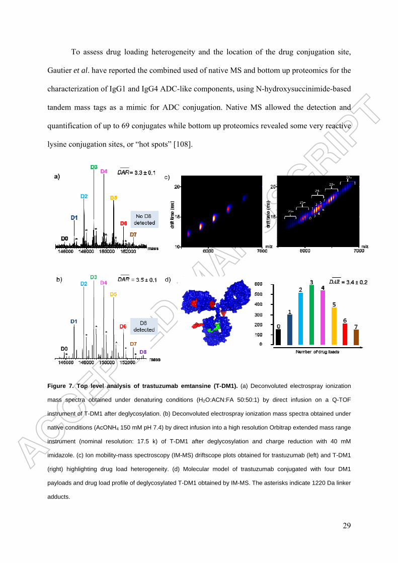
To assess drug loading heterogeneity and the location of the drug conjugation site,
Gautier et al. have reported the combined used of native MS and bottom up proteomics for the
characterization of IgG1 and IgG4 ADC-like components, using N-hydroxysuccinimide-based
tandem mass tags as a mimic for ADC conjugation. Native MS allowed the detection and
quantification of up to 69 conjugates while bottom up proteomics revealed some very reactive
lysine conjugation sites, or “hot spots” [108].
Figure 7. Top level analysis of trastuzumab emtansine (T-DM1). (a) Deconvoluted electrospray ionization
mass spectra obtained under denaturing conditions (H2O:ACN:FA 50:50:1) by direct infusion on a Q-TOF
instrument of T-DM1 after deglycosylation. (b) Deconvoluted electrospray ionization mass spectra obtained under
native conditions (AcONH4 150 mM pH 7.4) by direct infusion into a high resolution Orbitrap extended mass range
instrument (nominal resolution: 17.5 k) of T-DM1 after deglycosylation and charge reduction with 40 mM
imidazole. (c) Ion mobility-mass spectroscopy (IM-MS) driftscope plots obtained for trastuzumab (left) and T-DM1
(right) highlighting drug load heterogeneity. (d) Molecular model of trastuzumab conjugated with four DM1
payloads and drug load profile of deglycosylated T-DM1 obtained by IM-MS. The asterisks indicate 1220 Da linker
adducts.

30
Site specific ADCs
As discussed and illustrated above, the three market-approved ADCs, and most of those under
clinical trial are controlled mixtures of molecules. In 2008, Junutula et al. reported the design
and production of a new class of homogeneous ADCs, thiomab-drug conjugates (TDCs),
prepared by conjugation to engineered IgGs containing additional exposed cysteine residues
whose position were identified using phage display methods [30]. After reducing the blocked
cysteine residues and interchain disulfides, the latter were regenerated and maleimide reagents
were conjugated to the reactive cysteine thiol group to generate site-specifically modified
ADCs with 92.1 % of the population having two loaded molecules and only 0.2, 3.3, and
4.3 % having zero, one, or three, respectively (average DAR = 2.0), as assessed by rpHPLC-
MS. Importantly, these TDCs are better tolerated at higher doses in animals, indicating that
their therapeutic index is higher than that of existing ADCs. A plethora of other site specific
techniques have subsequently been reported, as recently reviewed (amongst others) by
Panowksi et al. [109], Agarwal and Bertozzi [110] and Dennler et al. [111], and summarized
in Figure 2c. Interestingly, Junutula and colleagues published another paper, in 2012 [112],
indicating that their first-generation TDCs were not very stable in the blood stream. Thanks to
MS, the degradation mechanism was shown to be a retro-Michael reaction, resulting in the
transfer of the drug linker to circulating molecules bearing free thiol groups such as albumin
and glutathione [113]. In addition, based again on mass shift measurements, succimide ring
hydrolysis was shown to stabilize the drug linker on the antibody was observed, with a mass
increase of 18 Da (H20). This ring opening was correlated with a positively charged local site.
Succinimide hydrolysis has since been employed to improve the stability (and hence the
safety) of several ADCs [114]; different strategies have been adopted, notably the process-
favored mild method based on a basic pH treatment [115], but also self-hydrolyzing
maleimides [114] and electron withdrawal N-substituents [116].

31
Alternatively, the specific conjugation to N-glycans of IgG (at Asn297) can be
achieved by periodate oxidation of fucose followed by hydrazone condensation, metabolic
incorporation of 6-thiofucose and then maleimide conjugation [117]. The process is
completed using specific enzymes such as enzymatic transfer of galactose and sialic acid
followed by periodate oxidation and oxime condensation, enzymatic transfer of galactose and
9-azidosialic acid followed by Cu-free click reaction, enzymatic removal of terminal
galactose followed by enzymatic transfer of GalNAz and Cu-free click reaction [118]. Each
step in the reaction can be monitored by LC-MS, which can then also be used to assess the
structures [119], and the glyco-profiles [120].
Conjugation can also be achieved through the incorporation of non canonical amino-
acids—such as p-acetylphenylalanine sometimes with an azido-lysine derivative—and then
oxime condensation and Cu-free click chemistry. The cell-free incorporation of p-
azidomethylphenylalanine followed by Cu-free click chemistry, and the incorporation of
selenocysteine followed by mild reduction and alkylation have also been described [121]. As
for other site-specific conjugation techniques, the choice of attachment site may affect the
stability of the resulting ADCs [39].
Conjugation methods based on the enzymatic modification of peptide tags have also
been explored extensively. One method involves glycosidase treatment—to access Q295 (and
N297Q mutants)—prior to the transglutaminase-mediated conjugation of amine-
functionalized small molecules or drugs to engineered LLQGA sites [33]. The sortase-
mediated conjugation of a glycine-functionalized chelator near the C-terminus of a single-
chain antibody (scFv) and the formylglycine generating enzyme-mediated conversion of
cysteine to formylglycine followed by hydrazino-Pictet−Spengler ligation have also been
successfully generated [122].

32
One of the caveats of these site-specific approaches is that many commonly used
bioorthogonal reagents do not react stereospecifically and typically yield at least two
diastereomeric products, resulting in several regioisomers. In addition, since these conjugation
methods are applied to heterogeneous antibodies (e.g. glyco- and charge-variants), the
homogeneity of the final products depends on the resolving power of the analytical and
structural methods employed [58].
ADC higher order structure and aggregates
The HOS of a protein, namely its secondary, tertiary and quaternary assemblies, is often
essential for its function [123,124]. While disruption of the native fold can also impact the
function of the molecule, the main immunogenic consequences of misfolded proteins stem
from their propensity to aggregate [125,126]. In solution, mAbs and ADCs tend to self-
associate, forming a range species from oligomers to visible particles. Changes in the HOS of
ADCs can alter their quality attributes, and thereby their safety, efficacy and
pharamacokinetics. A requirement of regulatory agencies is therefore that the effects of
manufacturing, storage and delivery on the HOSs of mAbs and ADCs be well characterized
[127].
Determining the HOSs of proteins using analytical biophysical techniques is still
challenging however. The classical methods for protein structure determination, namely X-ray
diffraction (XRD) and nuclear magnetic resonance (NMR) [128], provide atomic resolution
but can only tackle relatively small proteins, let alone protein assemblies, and consume large
amounts of biological material. For mAbs and ADCs furthermore, the flexibility of the hinge
region hinders the growth of crystals (for XRD) and complicates NMR analyses. So far, only
a small number of mAb crystal structures have been published [129] and no 3D structure of
an ADC has been solved. In this context, lower-resolution methods, notably those based on
MS (see Figure 8 and the following sections), can provide valuable insights.

33
Native MS and IM-MS
Native MS has been used to reveal the presence of mAb multimers (dimers, trimers,
tetramers) [130] and to study the structural dynamics of IgG4 [74]. As non-covalent
interactions are maintained, a single native MS analysis can be used to both characterize the
ADC (average DAR, drug load distribution, proportion of unconjugated drug molecules) and
check for the presence of multimers. Native MS has also been used to unambiguously
determine antibody/antigen binding stoichiometries [131] and to monitor the dynamics of
IgG4 Fab-arm exchange [74]. Furthermore, Haberger et al. recently reported a linear
correlation between results obtained by quantitative native MS and IgG-FcRn functional
analysis, a larger amount of higher order complexes being detected in the absence of
methionine oxidation in both cases [132].
The structural analysis of proteins can also be facilitated by IM-MS [133-136]. In this
approach, ions are separated based on their collisional cross section (CCS) as they drift
through an inert buffer gas in an electrical field, providing information on the charge and
conformation of a protein complementary to the associated mass data. The measured drift
times can be converted into CCSs, which correspond to the averaged rotational projection on
a 2D space of the biomolecule 3D structure. IM-MS adds an additional level of
conformational characterization to that provided by native MS. The analysis of the HOSs of
mAbs by IM-MS remains a relatively novel endeavor [56,57,72,74,131,137-140].
To our knowledge, only three publications have so far reported the use of native IM-
MS for the characterization of ADCs [65,78,107]. Debaene et al. extensively analyzed the
higher order structure characterization of BV by IM-MS [65], comparing the heterogeneity of
BV with that of unconjugated brentuximab. Five different species were observed, having zero
to eight drug molecules attached but similar gas phase conformations—viz. CCSs of 71.1 ±
0.1 nm2 and 68.1 ± 0.1 nm2 for the DAR 8 and DAR 0 species, respectively. The constant shift

34
in CCS between two consecutive even drug conjugates points to a small increase in mass
rather than to structural changes and indicating that the conformation of the protein is not
drastically altered upon binding.
Similarly, Marcoux et al. performed a global conformational analysis of T-DM1 [78]
and presented the first attempt 3D model of an ADC based on the structure of human IgG1
and the Fab structure of trastuzumab (PDB IDs 1HZH and 1N8Z, respectively). The CCSs
derived from IM-MS were found to be slightly higher than those estimated from the masses of
the species, but considerably lower than those expected from molecular modeling. These
discrepancies, also observed for unconjugated mAb and attributed to a collapse of T-DM1 in
the gas-phase [65,138], contrast with the results obtained for BV, for which the values
calculated assuming spherical proteins match those measured by IM-MS. This suggests that
the surface-exposure of lysine-linked payloads makes them more susceptible to gas-phase
collapse than equivalent payloads in cysteine-linked conjugates. Each drug-loading event
induced a CCS increment of ~25 Å2, which matches the binding contribution expected from
the mass of a single DM1 molecule (~100 Å2). This suggests that the conformational changes
that occur in trastuzumab upon drug conjugation are very slight.
As mentioned above in the discussion of analytical methods for T-DM1, Huang et al.
[107] have also employed IM-MS to perform DAR measurements. The protein drift time was
used as a second dimension to monitor the changes in DAR in lot-to-lot analyses of both a
site-specifically and randomly conjugated ADC.
Although it remains an emerging technique native IM-MS offers a simple and direct
means to assess the size and shape of ADCs and thereby obtain a rough conformational
picture. The examples above also illustrate how IM-MS can be used to identify all the species
with different drug loads present in solution and study the effects of drug conjugation on the
global conformation of the mAb. In the medium term, the development of molecular

35
dynamics algorithms able to cope with > 150 kDa proteins should facilitate the structural
interpretation of IM-MS data on mAbs and ADCs.
HDX-MS
Technical developments over the past 10 years have brought HDX-MS to the forefront of
methods for the structural characterization of proteins. Indeed, HDX-MS now fills a niche in
their therapeutic analysis, notably for epitope mapping and comparability studies [141-147],
but also in the investigation of mAb/Fc receptor interactions [148]. Papers have also been
published on the use of HDX-MS to study mAb aggregation [149-151], to optimize
formulation stability [152], to monitor reversible mAb self-assembly [153], and to
characterize the structural effects of glycosylation or chemical modifications on mAbs [154-
157].
In a typical HDX-MS experiment, the protein of interest is deuterated over different
lengths of time, after which the reaction is quenched under strong acidic conditions at low
temperature (0 °C). A classic proteolytic digestion is then performed using pepsin, which
cleaves non-specifically but with a high reproducibility and under acidic conditions. The
digested peptides are then analyzed by rpLC−MS to assess their level of deuteration. An
additional level of IM separation can help to assign the peptides and increases the sequence
coverage (Figure 8). HDX-MS can be performed on proteins or assemblies with only a dozen
picomoles of material (vs. milligrams for crystallography) and no size limitations in theory (in
practice HDX-MS analysis of assemblies up to 300kDa were reported [151,158] ).
Its other main strength is its ability to monitor any structural changes that occur upon
labeling in solution.
A field of application of HDX-MS not mentioned above is the study of mAb HOSs.
Circular dichroism (CD), fluorescence spectroscopy and differential scanning calorimetry
(DSC) are the methods most frequently used at present [159,160]. For instance, Guo et al.

36
reported similar CD profiles for vcMMAE and the isolated mAb (MMAE), suggesting that
the secondary and tertiary structures of cysteine-linked ADCs do not differ significantly from
those of the parent mAb [161]. However, a key limitation of these biophysical techniques is
that the information obtained is an average both over all the protein conformers in solution
and across the sequence of the protein. The better spatial resolution offered by HDX-MS (at
the peptide level at least and at the amino acid level when combined with electron transfer
dissociation) and its capacity to generate dynamic data for solution samples, makes HDX-MS
an attractive alternative to probe the HOSs of mAbs and ADCs.
By comparing the deuterium uptake plots of the conjugated and unconjugated MMAE,
Pan et al. showed that almost 90% of the primary sequence of vcMMAE has the same HDX
kinetics as its parent mAb, the only differences occurring for the two peptides in the Fc
domain which have faster kinetics after conjugation [162]. This suggests that vcMMAE and
MMAE have very similar conformations and dynamics in solution. A detailed comparison of
the HDX-MS profiles of the ADC and its corresponding TCEP-reduced mAb indicated that
the partial reduction of the IgG1 interchain disulfides induced some minor, local structural
differences in the conformation and dynamics of the mAb Fc region. However, alkylation of
the reduced cysteine residues by the drug molecule does not further impact the local structural
of the domains where the differences were observed [163]. The same group also used HDX-
MS to demonstrate that site specific conjugation on engineered cysteine has only minor
structural effects on other ADCs (with pyrrolobenzodiazepine or mcMMAF as the conjugated
drug), the only difference being a slightly higher deuterium uptake after conjugation in the
vicinity of the mutation (264VDSV) because of the destabilization of hydrogen bonds between
C239 and V264 induced by the drug-linker [164].
Since HDX-MS can provide information the conformations and dynamics of ADCs in
solution—information that would be more difficult to obtain via other analytical methods—it

37
should become a technique of choice for ADC comparisons, the optimization of conjugation
sites, and the choice of the best drug-linkers in the development of therapeutic candidates.
Aggregation
As aggregation is the main cause of drastic decrease in the therapeutic efficiency of proteins,
with potentially deleterious effects on patient health, a crucial characteristic of ADCs is the
proportion of aggregates in the final product [124]. Both mAbs and ADCs can become
unstable during conjugation [160,165,166] or under environmental stress (from changes in
temperature or ionic strength, exposure to light, etc.). However, aggregation is more common
for ADCs, because the solubility of the mAb is reduced upon conjugation to hydrophobic
drug-linkers [44,161].
For example, Becley et al. found increased aggregation of a cysteine-linked ADC at
higher temperatures and for the species with a higher DAR [166]. This trend was attributed to
the drug conjugation in the hinge region, as revealed by DSC and CD data. The authors note
that for the species with a higher drug loading, while conjugation does not measurably alter
the secondary structure, it does reduce the thermal stability of the CH2 domain, with high
molecular weight aggregates forming rapidly. Using SEC, Adem et al. showed that the
aggregation propensity of an auristatin ADC increases with the ionic strength of the solution,
especially again for the high drug load species [167]. The authors then used DSC enables to
directly correlate the thermal unfolding of the protein with the loading of the drug.
Regarding the photosensitivity of ADCs, although those currently on the market are
conjugated with non-photosensitive drugs, many of those under clinical trial contain
photosensitive drugs such as doxorubicin or duocarmycin derivatives. Cockrell et al. reported
that binding an eosin photosensitizer to trastuzumab promotes the formation of aggregates,
with high-mass components being detected by SEC analysis and dynamic light scattering
highlighting the formation of large soluble particles and aggregates [168]. Since aggregation

38
occurred for the conjugate but not on the mAb control, this indicates that drug conjugation is
specifically responsible for the observed aggregation.
The presence of charge variants can also affect the stability, solubility,
pharmacokinetic and tissue distribution of an ADC. Boylan et al. used SEC-MS to determine
the DAR and evaluate the proportion of aggregates for different charge variants of a cysteine-
linked Fc fragment, the heterogeneity originating from degradation and different drug-linkers
and conjugation sites [106]. For a species at a given DAR, several acidic bands are observed
in the IEF data, revealing the presence of charge variants. The authors did not find any
correlation between particular conjugation sites and charge variants.
In the future, native MS approaches should become more widespread for the analysis
of ADC and mAb aggregates, as the on-line coupling of SEC to native MS makes the the
analysis of aggregates straightforward. A few groups have already exploited HDX to study
mAb aggregation. Zhang et al. used HDX-MS to compare the aggregation mechanism and the
resulting aggregate structures of a mAb under freeze-thaw and thermal stress, showing that
bevacizumab aggregation increases with the number of F/T cycles and decreases with the
protein concentration [150]. The reduced hydrogen exchange recorded for three
complementarity determining regions suggests that these residues may form strong
intermolecular bonds in the antibody aggregates, while the regions with enhanced HDX rates
are most probably partially unfolded. An attempt was made to predict the aggregation patterns
using several residue level modeling methods, but this proved unsuccessful [150].

39
Figure 8. Mass spectrometry techniques for the analysis of higher order structure and aggregation for
ADCs.
Residual drug linker and related products
As highlighted in Table 1 and already mentioned above as a potential health hazard, the
amount of residual drug-linker and related products in ADC drug substance batches is a
critical quality attribute. The unconjugated drug or drug-related impurities that remain in the
final product are typically the result of incomplete purification down-stream of the
conjugation reaction. Related forms of the unconjugated drug, such as linker-drug species or
other degradation products, may also be released while the conjugate is in storage. Sensitive
methods are therefore required in ADC development and production facilities to monitor and

40
validate the cleaning procedures for cytotoxic products, both for the safety of operators and to
demonstrate the absence of cross contamination in multi-cytotoxic production plants [169].
Wakanhar et al. have reviewed ELISA, HPLC-UV/Vis and CE-LIF (capillary
electrophoresis laser induced fluorescence) methods that have been used to determine the
concentrations of free drug molecules in various ADCs [44]. Chih et al. also emphasize the
importance of monitoring drug-linker release with an example MS application [170]. This
method was successfully used to explain an unexpected release of free drug during the
stability testing of ADCs.
One of the major practical challenges of these kinds of analyses is removing the large
ADC excess from the sample to allow the very small amounts of multiple small organic
molecules to be quantified. This issue was solved by Fleming et al. for free DM1 dosages by
directly injecting the ADC onto a single rpHPLC column without prior sample preparation
[54]. The ADC flows through the column without interacting with the stationary phase, which
combines a hydrophobic core with a hydrophilic outer layer. The hydrophilic layer shields the
C18-like core from interactions with mAbs and ADCs, while the small organic molecules
present in solution readily interact with the hydrophobic portion. Li et al. recently reported
another elegant method based on two-dimensional LC-MS [171]. The SEC method in the 1st
dimension separated the small molecule impurities (the free drug, drug-linker, and drug-
linker-N-Acetylcystein adduct) from the intact ADC, and provided simultaneously
information on size variants, namely monomers, dimers and aggregates. The small molecules
giving rise to the peak in the 1st dimension were isolated and further analyzed by rpHPLC in a
2nd dimension for identification and quantitation by MS.

41
Bioanalysis of ADC biotransformation
Bioanalytical methods are rapidly being developed to quantitatively monitor the
transformation of ADCs in various in vitro or in vivo biological matrices such as
serum/plasma and tumor tissues [172]. Indeed, a crucial property of conjugates is their
stability in biofluids as the release over time of cytotoxic drugs into the bloodstream
constitutes a considerable health threat [173]. This drug loss also affects the composition of
the ADC, potentially altering the amount of drug delivered to the tumor site and posing
another substantial safety risk because of its off-target toxicity [174]. Tumey et al. have
recently published an interesting survey of the biotransformation events that have been
elucidated in recent years [175], as well as one of the resulting strategies to optimize the
design of next generation ADCs [115].
Traditionally, pharmacokinetic studies of mAb candidates are performed using ligand-
binding assays (LBAs) [172,176,177], a technique also used to study the catabolites [178] and
immunogenicity of ADCs [179,180]. The limitations of this approach are well known
however. The specific assay reagents required for LBA are often not available early on in a
program; moreover, interference can occur from endogenous proteins, antidrug antibodies and
soluble target ligands [176,181]. Complementary or alternative data from liquid
chromatography coupled to MS-based methods can therefore facilitate the analysis of mAbs
in biological matrixes. Selected reaction monitoring (SRM)-MS combined with stable isotope
dilution is thus increasingly employed in pharmacokinetic studies of recombinant proteins,
notably mAbs, in highly complex matrices (serum, plasma, tumor tissues, and other body
fluids) [181-187]. In addition, while LC-SRM has been used for decades to quantify small
molecules, tremendous improvements in the technique over the past few years have
broadened its scope to include the quantitation of peptides and proteins, including ADC
catabolites [188]. Optimized LC-SRM assays offer unequalled sensitivity, high analyte

42
specificity, a high multiplexing capacity and precision, and robust quantitation of analytes
down to high ng/ml–low µg/ml concentrations in unfractionated plasma.
Most of the methods described above rely on enzymatic digestion of the mAb to yield
at least one peptide with a unique sequence to be quantified as a surrogate for the whole mAb
[189]. Multidimensional chromatography or immunoenrichment can be used to separate the
mAb of interest from those of the endogenous matrix, thereby increasing the relative
concentration of the analyte of interest [190,191]. The quantitative bioanalysis of ADCs in
plasma has also be achieved by hybrid immuno-capture LC-MS/MS [192,193].
ADCs are administered as intravenous infusions, and, following in vivo processing,
multiple analytes are detected in systemic circulation. According to a recent paper from the
American Association of Pharmaceutical Scientists ADC Working Group [194] the most
commonly observed analytes are conjugated antibodies (i.e. with a DAR of one or more), total
antibodies (conjugated, partially or fully deconjugated), antibody-conjugated drugs (small
molecules conjugated to an antibody), and unconjugated drug molecules. Metabolites of the
drug molecule, with or without the linker, may also be detected. As discussed above, most of
the ADCs currently under clinical trial exist as a heterogeneous mixture of antibody species
with DARs varying from zero to eight. Each of these species has its own distinct in vivo
pharmacokinetic profile, efficacy, and safety [46] and specific quantitative analytical
workflows have to be developed. Pharmacokinetic profiling highlights the impact of the DAR
and other perfectible parameters on the biological properties of ADCs, notably the rate of
drug loss (from deconjugation and instability) and the clearance of the species with different
DARs. Ultimately through, establishing the relationships between pharamacokinetic
exposures and the efficacy and toxicity of the drug is the most helpful guide for the
optimization and development of ADCs. This information is also mandatory for the
pharmacokinetic-pharmacodynamic modeling that helps to determine the first-in human

43
dosage for clinical trials [195-198]. In this context, Deslandes has recently compared the
pharmacokinetics of ADCs in Phase I clinical studies [199], while toxicology issues have
been discussed by Saber and Leighton from the FDA [200].
Final conclusions
The development and optimization of ADCs are increasingly reliant on the analytical and
bioanalytical characterization of their main quality attributes, namely the drug load
distribution, proportion of naked antibody, and DAR. These needs have recently been fulfilled
by a number of cutting-edge mass spectrometry methods, with workflows optimized to be
used at different levels. Electrophoretic and chromatographic methods, in association or not
with MS, are still the main techniques used for the characterization of mAbs and ADC.
However, recent developments in native MS mean that this technique, in combination with
classical MS approaches, offers the means to complete a multi-level characterization of ADCs
and other protein-based therapeutic agents.
At the top level, native MS and native IM-MS complement the data provided by HIC
and rpHPLC-MS, the reference methods for the quality control of interchain cysteine- and
lysine-linked ADCs, respectively.
At the middle level, the LC-MS analysis of reduced or IdeS-digested ADCs provides
structural insights into the ADC subunits. The use of IdeS ensures the complete separation of
the Fc/2 and Fd fragments. This facilitates the glycoprofiling of the Fc/2 fragment and the
detection of any additional conjugation.
At the bottom level, high-quality ADC peptide mapping via improved (nano)HPLC-
MS is now available in most analytical labs. This can be used to characterize the drug-loaded
peptides and to identify positional isomers at cysteine residues. Because of the hydrophobicity
of the drug molecules, organic solvents are added at all stages of the bottom-up approach,
including enzymatic digestion, to ensure that the drug-loaded peptides remain in solution.

44
For studies of aggregation and the HOSs of ADCs, native MS and IM-MS along with
HDX-MS are emerging as promising techniques to complement already established methods
such as SEC.
Regarding MS-based quantitation finally, it is noteworthy that SRM-MS now offers
the same sensitivity as ELISA assays, but with a superior specificity.
Expert commentary & five year view
The state-of-the-art MS methods currently used to characterize mAbs, notably for bottom-
level analysis [201], are readily applicable for the study of ADCs. Capillary electrophoresis
coupled to MS is now established as a valuable method, orthogonal to LC, for peptide
mapping at the bottom [202-204] and middle [205] levels, and progress toward top level
analysis is forthcoming, including under native conditions. Another emerging technique, CE-
UV/MALDI-MS/MS (MALDI: matrix-assisted laser desorption ionization), has recently been
applied to study mAbs [206] and should prove valuable in the future.
Top-down MS and sequencing [207], middle-down MS with electron transfer
dissociation fragmentation [63], and extended bottom-up proteomics with enzymes [100] are
all in constant progress. An alternative middle-down avenue toward a full sequence coverage
of IdeS-digested and reduced fragments of IgG may be LC-MALDI with in-source decay
[207]. A common objective of all approaches based on MS/MS is to limit both the number of
sample preparation steps and the use of enzymes for peptide mapping to avoid the
introduction of artifacts into the data (e.g. over-deamidation from the use of trypsin at basic
pH).
Two-dimensional LC-MS should facilitate future studies of large molecules such as
antibodies and ADCs, the extra resolution enabling the direct identification of different
species. The first dimension can be adapted to the problem at hand via a plethora of
chromatographic techniques developed for the characterization of antibodies, while rpHPLC

45
is frequently used in the second dimension as it desalts the sample before its introduction into
the mass spectrometer. Recent applications of this approach reported by Li et al. [171], Stoll
et al. [208] and Birdsall et al. [98], have involved SEC x rpHPLC-MS, CEX x rpHPLC-MS,
and HIC x rpHPLC-MS, respectively. Recent improvements made to chromatographic
methods such as SEC [209], cation exchange [210], rpHPLC [211], and HIC (Fekete S et al,
submitted) will no doubt also benefit the application of 2D-LC-MS for the analysis of mAbs,
ADCs and related products.
Imaging mass spectrometry has successfully been used to reveal the spatial
distribution of small drugs and metabolites in biological tissues [212], and has recently been
demonstrated for the study of large biological molecules [213], notably to monitor mAbs in
brain tumors [214]. One assumes therefore that this technique will also be applied for the
characterization of ADCs.
Focusing now on the samples under study, while on the one hand, next-generation
ADCs should be more homogeneous and thereby easier to characterize, their more complex
formulation will pose new analytical questions. Indeed, bi-specific antibody conjugates [215],
antibody-dual-conjugates [216], treatment combinations (eg. ADCs + immune check points
mAbs) as well as alternatives to IgG formats [217] have already been developed.
Finally, a feasible objective for the analytical methods used to study ADCs during
pharmaceutical development, is that they should be translated to the bioanalytical field, for the
analysis of blood and/or tumor tissues. This would allow the quantitation of known and
unknown catabolites for large and small molecules and help to optimize the next generation of
ADCs.

46
Key issues
• High resolution native MS provides accurate mass measurements (within 30 ppm) of
intact ADCs, and can also yield the average DAR and drug load distribution. Native MS is
furthermore unique in its ability to simultaneous detect covalent and non-covalent species in a
mixture.
• Native IM-MS reveals the drug loading profile of ADCs, the CCCs of each payload
species highlighting slight conformational differences.
• As an orthogonal method, LC-MS following IdeS digestion of ADCs can be used to
measure the drug load distribution on light chain and Fd fragments, as well as the average
DAR for both monomeric and multimeric species. In addition, the Fc fragment can be
analyzed in the same run, providing a complete glyco-profile and demonstrating the presence
or absence of additional conjugation.
• Because the drug molecules are hydrophobic, all enzymatic digestion steps used to
prepare the sample for peptide mapping have to be adapted to keep the drug-loaded peptides
soluble. When this procedure is properly optimized however, unambiguous maps are obtained
by LC-MS, while the positional isomers of the cytotoxic drug can be determined by rpHPLC
after digestion with IdeS and reduction.
Acknowledgments
The authors would like to thank all the members of the CIPF and LSMBO that have been
involved in the characterization of ADCs: A. Boeuf, M. Excoffier, L. Maurel-Chevillet, C.
Klinguer-Hamour, T. Champion. M. Dillenbourg, M. Fumex, S. Tanguy, F. Delalande, H.
Diemer, J.-M. Strub, C. Carapito and C. Schaeffer.
They would also like to thank Paul Guerry from Green Grow Scientific for editing of the
manuscript.

47
Financial and competing interests disclosure
This work was supported by the bioclusters of the OptimAbs network (the Lyon Biopole and
Alsace Biovalley) and sponsors (DGCIS, Oseo, Feder, Région Rhône-Alpes, Région Alsace,
Communauté Urbaine de Strasbourg, CNRS, and the Université de Strasbourg).
We thank GIS IBiSA and Région Alsace for financial support in purchasing respectively a
Synapt G2 HDMS instrument and a Exactive Plus EMR Orbitrap spectrometer.
François Debaene is an employee of Sanofi. The other authors have no other relevant
affiliations or financial involvement with any organization or entity with a financial interest in
or financial conflict with the subject matter or materials discussed in the manuscript. This
includes employment, consultancies, honoraria, stock ownership or options, expert testimony,
grants or patents received or pending, or royalties.

48
Table 1. Analytical, structural and functional assays reported in quality control monographs
for the characterization of ADCs. They may be used for drug substance/drug product batch
release or characterization as well as for comparability, stability studies and bioanalysis).
Mass spectrometry-related methods appear in bold.
General aspect/
Physico-chemistry
• Appearance
• Color
• Opalescence
• pH
• Osmolality
• Water content (lyophilized)
Identity/ Structure
• Native ADC: HIC, native MS, native IM-MS,
native CE-MS
• Denaturated ADC (surfactant, solvant): SDS-
PAGE, SDS-CE, rpHPLC-UV/MS, MALDI-TOF
• Subunits (reduced, IdeS+red.): SDS-PAGE, SDS-
CE, rpHPLC-UV/MS, ETD
• Peptide mapping/ disulfide bridges/ conjugation
sites (Lys-C, trypsin, Asp-N, Glu-C, chemotrypsin,
SAP9): rpHPLC-UV/MS, CE-MS
• Higher order structures: CD, HDX-MS, DSC (Tm),
FTIR, fluorescence
• Immuno-identification: ELISA, immunoblots,
FACS, SPR
Quantification • Protein quantitation: UV, BCA, Protein A HPLC,

49
ELISA
• Extinction coefficient: calculation, amino acid
analysis
Purity/ Impurities
• Mass variants/ Aggregates: SDS-CE, SDS-PAGE,
SEC-UV/MALS/IR, LC-MS, A4F-UV/MALS/IR,
DLS, SV-AUC, native MS, IM-MS
• Subvisible/visible particles: light obscuration,
MFI/MCM (Flow Cell Microscopy)
• Charge variants (pI): IEF, cIEF, icIEF/iCE, CEX
• Drug loading and distribution, average DAR,
positional isomers: UV, HIC, LC-UV/MS (red;
IdeS/red), 2D-LC-UV/MS, native MS
• Free antibody: HIC, cIEF, icIEF/iCE
• Free drug(linker) and related products: LC-
UV/MS, 2D-LC-UV/MS, ELISA
• Free thiol groups: Ellman’s reagent
• Quenching agent (N-acetyl cysteine): LC-UV/MS
• Residual solvent: LC-UV, GC
• Heavy metals: ICP-MS
• Endotoxins
• Bioburden
Functional assays/ Potency • Antigen binding: ELISA, FACS, SPR, native MS
• Cytotoxicity (IC50): cell based assays (FACS,
electroluminescence, UV-visible)

50
• ADCC (when applied)
A4F, asymmetrical flow field-flow fractionation; ADCC, antibody-dependent cell-mediated cytotoxicity; BCA,
bicinchoninic acid; CD, circular dichroism; CEX, cation exchange; DAR, drug-to-antibody ratio; DSC,
differential scanning calorimetry; ELISA, enzyme-like immunosorbent assay; ETD, electron transfer dissociation;
FACS, Fluorescence-activated cell sorting; FTIR, Fourier transform infrared; HDX, hydrogen/deuterium
exchange; HIC, hydrophobic interaction chromatography; (i)CE, (imaged) capillary electrophoresis; (i)(c)IEF,
(imaged) (capillary) isoelectrofocusing; ICP, inductively coupled plasma; IdeS, the immunoglobulin-degrading
enzyme of Streptococcus pyogenes; IM, ion mobility; IR, infrared; MALDI, matrix-assisted laser desorption
ionization; MALS, multiangle light scattering; MS, mass spectrometry; PAGE, polyacrylamide gel
electrophoresis; rpHPLC, reverse-phase high-performance liquid chromatography; SDS, sodium dodecyl sulphate;
SEC, size exclusion chromatography; SPR, surface plasmon resonance; SV-AUC, sedimentation velocity
analytical ultracentrifugation; TOF, time of flight; UV, ultraviolet.

51
References Papers of special note have been highlighted as:
. of interest
.. of considerable interest
1. Beck A, Wurch T, Bailly C, Corvaia N. Strategies and challenges for the nextgeneration of therapeutic antibodies. Nat Rev Immunol, 10(5), 345-352 (2010).
2. Reiner Z. PCSK9 inhibitors - past, present and future. Expert Opin Drug MetabToxicol, 1-5 (2015).
3. Reichert JM. Antibodies to watch in 2015. MAbs, 7(1), 1-8 (2015).4. Kraeber-Bodere F, Bodet-Milin C, Rousseau C et al. Radioimmunoconjugates for the
treatment of cancer. Semin Oncol, 41(5), 613-622 (2014).5. Beck A, Haeuw JF, Wurch T, Goetsch L, Bailly C, Corvaia N. The next generation of
antibody-drug conjugates comes of age. Discov Med, 10(53), 329-339 (2010).6. Madhumathi J, Devilakshmi S, Sridevi S, Verma RS. Immunotoxin therapy for
hematologic malignancies: where are we heading? Drug Discov Today, (2015).7. Danielli R, Patuzzo R, Ruffini PA et al. Armed antibodies for cancer treatment: a
promising tool in a changing era. Cancer Immunol Immunother, 64(1), 113-121(2015).
8. Mendes D, Alves C, Afonso N et al. The benefit of HER2-targeted therapies onoverall survival of patients with metastatic HER2-positive breast cancer - a systematicreview. Breast Cancer Res, 17(1), 140 (2015).
9. Beck A. Review of Antibody-Drug Conjugates, Methods in Molecular Biology series: Abook edited by Laurent Ducry. MAbs, 6(1), 30-33 (2014).
10. Klinguer-Hamour C, Strop P, Shah DK, Ducry L, Xu A, Beck A. World Antibody-DrugConjugate Summit, October 15-16, 2013, San Francisco, CA. MAbs, 6(1), 18-29(2014).
11. Beck A, Reichert JM. Antibody-drug conjugates: present and future. MAbs, 6(1), 15-17 (2014).
12. Sochaj AM, Swiderska KW, Otlewski J. Current methods for the synthesis ofhomogeneous antibody-drug conjugates. Biotechnol Adv, 33(6 Pt 1), 775-784 (2015).
13. Beck A, Senter P, Chari R. World Antibody Drug Conjugate Summit Europe: February21-23, 2011; Frankfurt, Germany. MAbs, 3(4), 331-337 (2011).
14. Maeda E, Urakami K, Shimura K, Kinoshita M, Kakehi K. Charge heterogeneity of atherapeutic monoclonal antibody conjugated with a cytotoxic antitumor antibiotic,calicheamicin. Journal of Chromatography A, 1217(45), 7164-7171 (2010).
15. Labrijn AF, Buijsse AO, van den Bremer ET et al. Therapeutic IgG4 antibodiesengage in Fab-arm exchange with endogenous human IgG4 in vivo. Nat Biotechnol,27(8), 767-771 (2009).
16. Bross PF, Beitz J, Chen G et al. Approval summary: gemtuzumab ozogamicin inrelapsed acute myeloid leukemia. Clin Cancer Res, 7(6), 1490-1496 (2001).
17. Chari RV, Miller ML, Widdison WC. Antibody-drug conjugates: an emerging conceptin cancer therapy. Angew Chem Int Ed Engl, 53(15), 3796-3827 (2014).
18. Ornes S. Antibody-drug conjugates. P Natl Acad Sci USA, 110(34), 13695-13695(2013).
19. Flygare JA, Pillow TH, Aristoff P. Antibody-drug conjugates for the treatment ofcancer. Chem Biol Drug Des, 81(1), 113-121 (2013).
20. Beck A, Lambert J, Sun M, Lin K. Fourth World Antibody-Drug Conjugate Summit:February 29-March 1, 2012, Frankfurt, Germany. MAbs, 4(6), 637-647 (2012).

52
21. Doronina SO, Toki BE, Torgov MY et al. Erratum: Development of potent monoclonalantibody auristatin conjugates for cancer therapy (vol 21, pg 778, 2003). NatureBiotechnology, 21(8), 941-941 (2003).
22. Senter PD, Sievers EL. The discovery and development of brentuximab vedotin foruse in relapsed Hodgkin lymphoma and systemic anaplastic large cell lymphoma. NatBiotechnol, 30(7), 631-637 (2012).**Adcetris®: from bench to the FDA approval
23. Lewis Phillips GD, Li G, Dugger DL et al. Targeting HER2-positive breast cancer withtrastuzumab-DM1, an antibody-cytotoxic drug conjugate. Cancer Res, 68(22), 9280-9290 (2008).
24. Blanc V, Bousseau A, Caron A, Carrez C, Lutz RJ, Lambert JM. SAR3419: an anti-CD19-Maytansinoid Immunoconjugate for the treatment of B-cell malignancies. ClinCancer Res, 17(20), 6448-6458 (2011).
25. Wang L, Amphlett G, Blattler WA, Lambert JM, Zhang W. Structural characterizationof the maytansinoid-monoclonal antibody immunoconjugate, huN901-DM1, by massspectrometry. Protein Sci, 14(9), 2436-2446 (2005).
26. Roberts SA, Andrews PA, Blanset D et al. Considerations for the nonclinical safetyevaluation of antibody drug conjugates for oncology. Regul Toxicol Pharmacol, 67(3),382-391 (2013).
27. Gordon M, Canakci M, Li L, Zhuang J, Osborne BA, Thayumanavan S. A Field Guideto Challenges and Opportunities in Antibody-Drug Conjugates for Chemists.Bioconjug Chem, (2015).
28. McDonagh CF, Turcott E, Westendorf L et al. Engineered antibody-drug conjugateswith defined sites and stoichiometries of drug attachment. Protein Eng Des Sel, 19(7),299-307 (2006).
29. Hartley JA, Hamaguchi A, Coffils M et al. SG2285, a novel C2-aryl-substitutedpyrrolobenzodiazepine dimer prodrug that cross-links DNA and exerts highly potentantitumor activity. Cancer Res, 70(17), 6849-6858 (2010).
30. Junutula JR, Raab H, Clark S et al. Site-specific conjugation of a cytotoxic drug to anantibody improves the therapeutic index. Nat Biotechnol, 26(8), 925-932 (2008).**Reference paper for site-specific antibody drug conjugates.
31. Kung Sutherland MS, Walter RB, Jeffrey SC et al. SGN-CD33A: a novel CD33-targeting antibody-drug conjugate using a pyrrolobenzodiazepine dimer is active inmodels of drug-resistant AML. Blood, 122(8), 1455-1463 (2013).
32. Dennler P, Chiotellis A, Fischer E et al. Transglutaminase-Based Chemo-EnzymaticConjugation Approach Yields Homogeneous Antibody-Drug Conjugates.Bioconjugate Chem, 25(3), 569-578 (2014).
33. Farias SE, Strop P, Delaria K et al. Mass spectrometric characterization oftransglutaminase based site-specific antibody-drug conjugates. Bioconjug Chem,25(2), 240-250 (2014).
34. Axup JY, Bajjuri KM, Ritland M et al. Synthesis of site-specific antibody-drugconjugates using unnatural amino acids. Proc Natl Acad Sci U S A, 109(40), 16101-16106 (2012).
35. Smith EL, Giddens JP, Iavarone AT, Godula K, Wang LX, Bertozzi CR.Chemoenzymatic Fc glycosylation via engineered aldehyde tags. Bioconjug Chem,25(4), 788-795 (2014).
36. Kolodych S, Koniev O, Baatarkhuu Z et al. CBTF: new amine-to-thiol couplingreagent for preparation of antibody conjugates with increased plasma stability.Bioconjug Chem, 26(2), 197-200 (2015).
37. Koniev O, Kolodych S, Baatarkhuu Z et al. MAPN: First-in-Class Reagent forKinetically Resolved Thiol-to-Thiol Conjugation. Bioconjug Chem, (2015).
38. Jeffrey SC, Burke PJ, Lyon RP et al. A potent anti-CD70 antibody-drug conjugatecombining a dimeric pyrrolobenzodiazepine drug with site-specific conjugationtechnology. Bioconjug Chem, 24(7), 1256-1263 (2013).

53
39. Dorywalska M, Strop P, Melton-Witt JA et al. Effect of attachment site on stability ofcleavable antibody drug conjugates. Bioconjug Chem, 26(4), 650-659 (2015).
40. Strop P, Delaria K, Foletti D et al. Site-specific conjugation improves therapeuticindex of antibody drug conjugates with high drug loading. Nat Biotechnol, 33(7), 694-696 (2015).
**Site-specific drug conjugation technology that overcome the previously reported limitations of conventional high-loaded ADCs..
41. Alley SC, Anderson KE. Analytical and bioanalytical technologies for characterizingantibody-drug conjugates. Curr Opin Chem Biol, 17(3), 406-411 (2013).42. Reichert JM, Beck A, Lugovskoy AA, Wurch T, Coats S, Brezski RJ. 9th annual
European Antibody Congress, November 11-13, 2013, Geneva, Switzerland. MAbs,6(2), 309-326 (2014).
43. Hamilton GS. Antibody-drug conjugates for cancer therapy: The technological andregulatory challenges of developing drug-biologic hybrids. Biologicals, (2015).
44. Wakankar A, Chen Y, Gokarn Y, Jacobson FS. Analytical methods forphysicochemical characterization of antibody drug conjugates. MAbs, 3(2), 161-172(2011).
** Reference review for analytical characterization of ADCs. 45. Chen Y. Drug-to-antibody ratio (DAR) by UV/Vis spectroscopy. Methods Mol Biol,
1045, 267-273 (2013).46. Hamblett KJ, Senter PD, Chace DF et al. Effects of drug loading on the antitumor
activity of a monoclonal antibody drug conjugate. Clin Cancer Res, 10(20), 7063-7070 (2004).
47. Le LN, Moore JMR, Ouyang J, Chen XY, Nguyen MDH, Galush WJ. ProfilingAntibody Drug Conjugate Positional Isomers: A System-of-Equations Approach.Analytical Chemistry, 84(17), 7479-7486 (2012).
*Mathematical approach using inputs from capillary electrophoresis and hydrophobicinteraction chromatography to determine the positional isomer distribution within a population of ADCs
48. Ouyang J. Drug-to-antibody ratio (DAR) and drug load distribution by hydrophobicinteraction chromatography and reversed phase high-performance liquidchromatography. Methods Mol Biol, 1045, 275-283 (2013).
49. Haverick M, Mengisen S, Shameem M, Ambrogelly A. Separation of mAbs molecularvariants by analytical hydrophobic interaction chromatography HPLC: overview andapplications. MAbs, 6(4), 852-858 (2014).
*Reference review for hydrophobic interaction chromatography of mAbs and ADCs.50. Sun MM, Beam KS, Cerveny CG et al. Reduction-alkylation strategies for the
modification of specific monoclonal antibody disulfides. Bioconjug Chem, 16(5), 1282-1290 (2005).
**Reference research paper for interchain cysteine-drug conjugates characterization. 51. King HD, Dubowchik GM, Mastalerz H et al. Monoclonal antibody conjugates of
doxorubicin prepared with branched peptide linkers: inhibition of aggregation bymethoxytriethyleneglycol chains. J Med Chem, 45(19), 4336-4343 (2002).
52. Hollander I, Kunz A, Hamann PR. Selection of reaction additives used in thepreparation of monomeric antibody-calicheamicin conjugates. Bioconjug Chem, 19(1),358-361 (2008).
53. Quiles S, Raisch KP, Sanford LL, Bonner JA, Safavy A. Synthesis and preliminarybiological evaluation of high-drug-load paclitaxel-antibody conjugates for tumor-targeted chemotherapy. J Med Chem, 53(2), 586-594 (2010).
54. Fleming MS, Zhang W, Lambert JM, Amphlett G. A reversed-phase high-performanceliquid chromatography method for analysis of monoclonal antibody-maytansinoidimmunoconjugates. Anal Biochem, 340(2), 272-278 (2005).
55. Beck A, Sanglier-Cianferani S, Van Dorsselaer A. Biosimilar, biobetter, and nextgeneration antibody characterization by mass spectrometry. Anal Chem, 84(11),4637-4646 (2012).

54
56. Beck A, Wagner-Rousset E, Ayoub D, Van Dorsselaer A, Sanglier-Cianferani S.Characterization of therapeutic antibodies and related products. Anal Chem, 85(2),715-736 (2013).
**Comprehensive review of mAbs analytical and structural characterization. 57. Beck A, Diemer H, Ayoub D et al. Analytical characterization of biosimilar antibodies
and Fc-fusion proteins. Trac-Trend Anal Chem, 48, 81-95 (2013).58. Beck A, Debaene F, Diemer H et al. Cutting-edge mass spectrometry characterization
of originator, biosimilar and biobetter antibodies. J Mass Spectrom, 50(2), 285-297(2015).
59. Beck A, Cianférani S. Harness the benefits of mass spectrometry for in-depthAntibody Drug Conjugates analytical characterization. LCGC, in press (2015).
60. Fornelli L, Damoc E, Thomas PM et al. Analysis of Intact Monoclonal Antibody IgG1by Electron Transfer Dissociation Orbitrap FTMS. Molecular & Cellular Proteomics,11(12), 1758-1767 (2012).
61. Tsybin YO, Fornelli L, Stoermer C et al. Structural analysis of intact monoclonalantibodies by electron transfer dissociation mass spectrometry. Anal Chem, 83(23),8919-8927 (2011).
62. Mao Y, Valeja SG, Rouse JC, Hendrickson CL, Marshall AG. Top-down structuralanalysis of an intact monoclonal antibody by electron capture dissociation-Fouriertransform ion cyclotron resonance-mass spectrometry. Anal Chem, 85(9), 4239-4246(2013).
63. Fornelli L, Ayoub D, Aizikov K, Beck A, Tsybin YO. Middle-Down Analysis ofMonoclonal Antibodies with Electron Transfer Dissociation Orbitrap Fourier TransformMass Spectrometry. Analytical Chemistry, 86(6), 3005-3012 (2014).
64. Valliere-Douglass JF, McFee WA, Salas-Solano O. Native intact mass determinationof antibodies conjugated with monomethyl Auristatin E and F at interchain cysteineresidues. Anal Chem, 84(6), 2843-2849 (2012).
** First reasearch paper on native MS of ADCs 65. Debaene F, Boeuf A, Wagner-Rousset E et al. Innovative Native MS Methodologies
for Antibody Drug Conjugate Characterization: High Resolution Native MS and IM-MSfor Average DAR and DAR Distribution Assessment. Analytical Chemistry, 86(21),10674-10683 (2014).
** First reasearch paper on native Ion Mobility MS of ADCs 66. Schmidt C, Robinson CV. Dynamic protein ligand interactions--insights from MS.
FEBS J, 281(8), 1950-1964 (2014).67. Pacholarz KJ, Garlish RA, Taylor RJ, Barran PE. Mass spectrometry based tools to
investigate protein-ligand interactions for drug discovery. Chem Soc Rev, 41(11),4335-4355 (2012).
68. Rajabi K, Ashcroft AE, Radford SE. Mass spectrometric methods to analyze thestructural organization of macromolecular complexes. Methods, (2015).
69. Bereszczak JZ, Havlik M, Weiss VU et al. Sizing up large protein complexes byelectrospray ionisation-based electrophoretic mobility and native mass spectrometry:morphology selective binding of Fabs to hepatitis B virus capsids. Anal Bioanal Chem,406(5), 1437-1446 (2014).
70. Rosati S, Yang Y, Barendregt A, Heck AJ. Detailed mass analysis of structuralheterogeneity in monoclonal antibodies using native mass spectrometry. Nat Protoc,9(4), 967-976 (2014).
**Detailed protocol for mAbs native MS analytical characterizaion 71. Thompson NJ, Hendriks LJ, de Kruif J, Throsby M, Heck AJ. Complex mixtures of
antibodies generated from a single production qualitatively and quantitativelyevaluated by native Orbitrap mass spectrometry. MAbs, 6(1), 197-203 (2014).
72. Zhang H, Cui W, Gross ML. Mass spectrometry for the biophysical characterization oftherapeutic monoclonal antibodies. FEBS Lett, 588(2), 308-317 (2014).
73. Thompson NJ, Rosati S, Heck AJ. Performing native mass spectrometry analysis ontherapeutic antibodies. Methods, 65(1), 11-17 (2014).

55
74. Debaene F, Wagner-Rousset E, Colas O et al. Time Resolved Native Ion-MobilityMass Spectrometry to Monitor Dynamics of IgG4 Fab Arm Exchange and "Bispecific"Monoclonal Antibody Formation. Analytical Chemistry, 85(20), 9785-9792 (2013).
* 75. Rosati S, Rose RJ, Thompson NJ et al. Exploring an orbitrap analyzer for thecharacterization of intact antibodies by native mass spectrometry. Angew Chem Int Ed Engl, 51(52), 12992-12996 (2012).
76. Chen J, Yin S, Wu Y, Ouyang J. Development of a native nanoelectrospray massspectrometry method for determination of the drug-to-antibody ratio of antibody-drugconjugates. Anal Chem, 85(3), 1699-1704 (2013).
77. Rosati S, van den Bremer ET, Schuurman J, Parren PW, Kamerling JP, Heck AJ. In-depth qualitative and quantitative analysis of composite glycosylation profiles andother micro-heterogeneity on intact monoclonal antibodies by high-resolution nativemass spectrometry using a modified Orbitrap. MAbs, 5(6), 917-924 (2013).
78. Marcoux J, Champion T, Colas O et al. Native mass spectrometry and ion mobilitycharacterization of trastuzumab emtansine, a lysine-linked antibody drug conjugate.Protein Science, 24(8), 1210-1223 (2015).
**First research paper of Kadcyla® Native Ion Mobility MS characterization 79. Hengel SM, Sanderson R, Valliere-Douglass J, Nicholas N, Leiske C, Alley SC.
Measurement of in vivo drug load distribution of cysteine-linked antibody-drugconjugates using microscale liquid chromatography mass spectrometry. Anal Chem,86(7), 3420-3425 (2014).
**First demonstration that native MS characterization of ADCs may be used as a potent bioanalysis method.
80. Smith LM, Kelleher NL. Proteoform: a single term describing protein complexity. NatMethods, 10(3), 186-187 (2013).
81. Tran JC, Zamdborg L, Ahlf DR et al. Mapping intact protein isoforms in discoverymode using top-down proteomics. Nature, 480(7376), 254-258 (2011).
82. Zhang Z, Shah B. Characterization of variable regions of monoclonal antibodies bytop-down mass spectrometry. Anal Chem, 79(15), 5723-5729 (2007).
83. Bondarenko PV, Second TP, Zabrouskov V, Makarov AA, Zhang Z. Massmeasurement and top-down HPLC/MS analysis of intact monoclonal antibodies on ahybrid linear quadrupole ion trap-Orbitrap mass spectrometer. J Am Soc MassSpectrom, 20(8), 1415-1424 (2009).
84. Ren D, Pipes GD, Hambly D, Bondarenko PV, Treuheit MJ, Gadgil HS. Top-down N-terminal sequencing of Immunoglobulin subunits with electrospray ionization time offlight mass spectrometry. Anal Biochem, 384(1), 42-48 (2009).
85. Lermyte F, Sobott F. Electron transfer dissociation provides higher-order structuralinformation of native and partially unfolded protein complexes. Proteomics, 15(16),2813-2822 (2015).
** 86. Dyachenko A, Wang G, Belov M et al. Tandem Native Mass-Spectrometry on Antibody-Drug Conjugates and Submillion Da Antibody-Antigen Protein Assemblies on an Orbitrap EMR Equipped with a High-Mass Quadrupole Mass Selector. Anal Chem, 87(12), 6095-6102 (2015).
87. Beck A, Bussat MC, Zorn N et al. Characterization by liquid chromatographycombined with mass spectrometry of monoclonal anti-IGF-1 receptor antibodiesproduced in CHO and NS0 cells. J Chromatogr B Analyt Technol Biomed Life Sci,819(2), 203-218 (2005).
* 88. Wagner-Rousset E, Janin-Bussat MC, Colas O et al. Antibody-drug conjugate modelfast characterization by LC-MS following IdeS proteolytic digestion. MAbs, 6(1), 273-285 (2014).
*First research report showing that IdeS may be used for ADCs subunit characterization byMS.
89. Xu W, Peng Y, Wang F, Paporello B, Richardson D, Liu H. Method to convert N-terminal glutamine to pyroglutamate for characterization of recombinant monoclonalantibodies. Anal Biochem, 436(1), 10-12 (2013).

56
90. Basa L. Drug-to-antibody ratio (DAR) and drug load distribution by LC-ESI-MS.Methods Mol Biol, 1045, 285-293 (2013).
91. Lyon RP, Meyer DL, Setter JR, Senter PD. Conjugation of Anticancer Drugs throughEndogenous Monoclonal Antibody Cysteine Residues. Method Enzymol, 502, 123-138 (2012).
** 92. Janin-Bussat MC, Dillenbourg M, Corvaia N, Beck A, Klinguer-Hamour C. Characterization of antibody drug conjugate positional isomers at cysteine residues by peptide mapping LC-MS analysis. J Chromatogr B Analyt Technol Biomed Life Sci, 981-982, 9-13 (2015).
**First research report on Adcetris® peptide mapping. 93. Yan B, Valliere-Douglass J, Brady L et al. Analysis of post-translational modifications
in recombinant monoclonal antibody IgG1 by reversed-phase liquidchromatography/mass spectrometry. J Chromatogr A, 1164(1-2), 153-161 (2007).
94. Gadgil HS, Bondarenko PV, Pipes G et al. The LC/MS analysis of glycation of IgGmolecules in sucrose containing formulations. J Pharm Sci, 96(10), 2607-2621 (2007).
95. Gadgil HS, Bondarenko PV, Pipes GD et al. Identification of cysteinylation of a freecysteine in the Fab region of a recombinant monoclonal IgG1 antibody using Lys-Climited proteolysis coupled with LC/MS analysis. Anal Biochem, 355(2), 165-174(2006).
96. Chevreux G, Tilly N, Bihoreau N. Fast analysis of recombinant monoclonal antibodiesusing IdeS proteolytic digestion and electrospray mass spectrometry. Anal Biochem,415(2), 212-214 (2011).
97. Firth D, Bell L, Squires M, Estdale S, McKee C. A rapid approach for characterizationof thiol-conjugated antibody-drug conjugates and calculation of drug-antibody ratio byliquid chromatography mass spectrometry. Anal Biochem, 485, 34-42 (2015).
98. Birdsall RE, Shion H, Kotch FW, Xu A, Porter TJ, Chen W. A rapid on-line method formass spectrometric confirmation of a cysteine conjugated antibody-drug-conjugatestructure using multidimensional chromatography. mAbs, in press (2015).
**First 2D-LC characterization of cysteine ADCs positional isomers. 99. Wiggins B, Liu-Shin L, Yamaguchi H, Ratnaswamy G. Characterization of cysteine-
linked conjugation profiles of immunoglobulin g1 and immunoglobulin g2 antibody-drug conjugates. J Pharm Sci, 104(4), 1362-1372 (2015).
100. Srzentic K, Fornelli L, Laskay UA et al. Advantages of extended bottom-upproteomics using Sap9 for analysis of monoclonal antibodies. Anal Chem, 86(19),9945-9953 (2014).
101. Lazar AC, Wang LT, Blattler WA, Amphlett G, Lambert JM, Zhang W. Analysis of thecomposition of immunoconjugates using size-exclusion chromatography coupled tomass spectrometry. Rapid Commun Mass Sp, 19(13), 1806-1814 (2005).
**Key paper for the MS characterization of Antibody Maytansinoid Conjugates. 102. Lin J, Lazar AC. Determination of charge heterogeneity and level of unconjugated
antibody by imaged cIEF. Methods Mol Biol, 1045, 295-302 (2013).103. Kim MT, Chen Y, Marhoul J, Jacobson F. Statistical modeling of the drug load
distribution on trastuzumab emtansine (Kadcyla), a lysine-linked antibody drugconjugate. Bioconjug Chem, 25(7), 1223-1232 (2014).
104. Lu SX, Takach EJ, Solomon M, Zhu Q, Law SJ, Hsieh FY. Mass spectral analyses oflabile DOTA-NHS and heterogeneity determination of DOTA or DM1 conjugated anti-PSMA antibody for prostate cancer therapy. J Pharm Sci, 94(4), 788-797 (2005).
105. Wang L, Amphlett G, Lambert JM, Blattler W, Zhang W. Structural characterization ofa recombinant monoclonal antibody by electrospray time-of-flight mass spectrometry.Pharm Res, 22(8), 1338-1349 (2005).
106. Boylan NJ, Zhou W, Proos RJ, Tolbert TJ, Wolfe JL, Laurence JS. Conjugation SiteHeterogeneity Causes Variable Electrostatic Properties in Fc Conjugates.Bioconjugate Chem, 24(6), 1008-1016 (2013).

57
107. Huang RY, Deyanova EG, Passmore D et al. Utility of Ion Mobility Mass Spectrometryfor Drug-to-Antibody Ratio Measurements in Antibody-Drug Conjugates. J Am SocMass Spectrom, (2015).
108. Gautier V, Boumeester AJ, Lossl P, Heck AJ. Lysine conjugation properties in humanIgGs studied by integrating high-resolution native mass spectrometry and bottom-upproteomics. Proteomics, 15(16), 2756-2765 (2015).
109. Panowksi S, Bhakta S, Raab H, Polakis P, Junutula JR. Site-specific antibody drugconjugates for cancer therapy. MAbs, 6(1), 34-45 (2014).
110. Agarwal P, Bertozzi CR. Site-specific antibody-drug conjugates: the nexus ofbioorthogonal chemistry, protein engineering, and drug development. BioconjugChem, 26(2), 176-192 (2015).
111. Dennler P, Fischer E, Schibli R. Antibody conjugates: from heterogeneouspopulations to defined reagents. Antibodies, 4, 197-224 (2015).
112. Shen BQ, Xu K, Liu L et al. Conjugation site modulates the in vivo stability andtherapeutic activity of antibody-drug conjugates. Nat Biotechnol, 30(2), 184-189(2012).
**Key paper to undestand retro-Michael deconjugation of ADCs in vivo. 113. Alley SC, Benjamin DR, Jeffrey SC et al. Contribution of linker stability to the activities
of anticancer immunoconjugates. Bioconjugate Chem, 19(3), 759-765 (2008).114. Lyon RP, Setter JR, Bovee TD et al. Self-hydrolyzing maleimides improve the stability
and pharmacological properties of antibody-drug conjugates. Nat Biotechnol, 32(10),1059-1062 (2014).
**Technology that improved ADC antitumor activity and reduced neutropenia 115. Tumey LN, Charati M, He T et al. Mild method for succinimide hydrolysis on ADCs:
impact on ADC potency, stability, exposure, and efficacy. Bioconjug Chem, 25(10),1871-1880 (2014).
116. Fontaine SD, Reid R, Robinson L, Ashley GW, Santi DV. Long-term stabilization ofmaleimide-thiol conjugates. Bioconjug Chem, 26(1), 145-152 (2015).
117. Okeley NM, Toki BE, Zhang XQ et al. Metabolic Engineering of Monoclonal AntibodyCarbohydrates for Antibody-Drug Conjugation. Bioconjugate Chem, 24(10), 1650-1655 (2013).
118. van Geel R, Wijdeven MA, Heesbeen R et al. Chemoenzymatic Conjugation of ToxicPayloads to the Globally Conserved N-Glycan of Native mAbs ProvidesHomogeneous and Highly Efficacious Antibody-Drug Conjugates. Bioconjug Chem,(2015).
119. Qasba PK. Glycans of Antibodies as a Specific Site for Drug Conjugation UsingGlycosyltransferases. Bioconjug Chem, (2015).
120. Janin-Bussat MC, Tonini L, Huillet C et al. Cetuximab Fab and Fc N-glycan fastcharacterization using IdeS digestion and liquid chromatography coupled toelectrospray ionization mass spectrometry. Methods Mol Biol, 988, 93-113 (2013).
121. Hallam TJ, Smider VV. Unnatural amino acids in novel antibody conjugates. FutureMed Chem, 6(11), 1309-1324 (2014).
122. Perez HL, Cardarelli PM, Deshpande S et al. Antibody-drug conjugates: currentstatus and future directions. Drug Discov Today, 19(7), 869-881 (2014).
123. Joubert MK, Hokom M, Eakin C et al. Highly aggregated antibody therapeutics canenhance the in vitro innate and late-stage T-cell immune responses. J Biol Chem,287(30), 25266-25279 (2012).
124. Rosenberg AS. Effects of protein aggregates: an immunologic perspective. AAPS J,8(3), E501-507 (2006).
125. Joubert MK, Luo Q, Nashed-Samuel Y, Wypych J, Narhi LO. Classification andcharacterization of therapeutic antibody aggregates. J Biol Chem, 286(28), 25118-25133 (2011).
126. van Beers MM, Bardor M. Minimizing immunogenicity of biopharmaceuticals bycontrolling critical quality attributes of proteins. Biotechnol J, 7(12), 1473-1484 (2012).

58
127. Valliere-Douglass JF, Lewis P, Salas-Solano O, Jiang S. Solid-state mAbs and ADCssubjected to heat-stress stability conditions can be covalently modified with buffer andexcipient molecules. J Pharm Sci, 104(2), 652-665 (2015).
*Demonstration that freeze-dried mAbs and ADCs can be covalently modified with buffer andexcipient molecules on the side chains of Glu, Asp, Thr, and Ser amino acids when subjected to temperature stress.
128. Poppe L, Jordan JB, Rogers G, Schnier PD. On the Analytical Superiority of 1D NMRfor Fingerprinting the Higher Order Structure of Protein Therapeutics Compared toMultidimensional NMR Methods. Anal Chem, 87(11), 5539-5545 (2015).
129. Rayner LE, Hui GK, Gor J, Heenan RK, Dalby PA, Perkins SJ. The solution structuresof two human IgG1 antibodies show conformational stability and accommodate theirC1q and FcgammaR ligands. J Biol Chem, 290(13), 8420-8438 (2015).
130. Kukrer B, Filipe V, van Duijn E et al. Mass spectrometric analysis of intact humanmonoclonal antibody aggregates fractionated by size-exclusion chromatography.Pharm Res, 27(10), 2197-2204 (2010).
131. Atmanene C, Laux A, Glattard E et al. Characterization of human and bovinephosphatidylethanolamine-binding protein (PEBP/RKIP) interactions with morphineand morphine-glucuronides determined by noncovalent mass spectrometry. Med SciMonit, 15(7), BR178-187 (2009).
132. Haberger M, Heidenreich AK, Schlothauer T et al. Functional assessment of antibodyoxidation by native mass spectrometry. MAbs, 1-10 (2015).
133. Hall Z, Politis A, Bush MF, Smith LJ, Robinson CV. Charge-state dependentcompaction and dissociation of protein complexes: insights from ion mobility andmolecular dynamics. J Am Chem Soc, 134(7), 3429-3438 (2012).
134. Scarff CA, Patel VJ, Thalassinos K, Scrivens JH. Probing hemoglobin structure bymeans of traveling-wave ion mobility mass spectrometry. J Am Soc Mass Spectrom,20(4), 625-631 (2009).
135. Bowers MT, Kemper PR, Vonhelden G, Vankoppen PAM. Gas-Phase IonChromatography - Transition-Metal State Selection and Carbon Cluster Formation.Science, 260(5113), 1446-1451 (1993).
136. Ruotolo BT, Giles K, Campuzano I, Sandercock AM, Bateman RH, Robinson CV.Evidence for macromolecular protein rings in the absence of bulk water. Science,310(5754), 1658-1661 (2005).
* 137. Bagal D, Valliere-Douglass JF, Balland A, Schnier PD. Resolving disulfide structuralisoforms of IgG2 monoclonal antibodies by ion mobility mass spectrometry. Anal Chem, 82(16), 6751-6755 (2010).
138. Pacholarz KJ, Porrini M, Garlish RA et al. Dynamics of intact immunoglobulin Gexplored by drift-tube ion-mobility mass spectrometry and molecular modeling. AngewChem Int Ed Engl, 53(30), 7765-7769 (2014).
139. Jones LM, Zhang H, Cui W et al. Complementary MS methods assist conformationalcharacterization of antibodies with altered S-S bonding networks. J Am Soc MassSpectrom, 24(6), 835-845 (2013).
140. Pritchard C, Groves KJ, Biesenbruch S et al. Quantification of human growthhormone in serum with a labeled protein as an internal standard: essentialconsiderations. Anal Chem, 86(13), 6525-6532 (2014).
* 141. Houde D, Berkowitz SA, Engen JR. The utility of hydrogen/deuterium exchange massspectrometry in biopharmaceutical comparability studies. J Pharm Sci, 100(6), 2071-2086 (2011).
142. Kaltashov IA, Bobst CE, Abzalimov RR, Berkowitz SA, Houde D. Conformation anddynamics of biopharmaceuticals: transition of mass spectrometry-based tools fromacademe to industry. J Am Soc Mass Spectrom, 21(3), 323-337 (2010).
143. Guan X, Noble KA, Tao Y et al. Epitope mapping of 7S cashew antigen in complexwith antibody by solution-phase H/D exchange monitored by FT-ICR massspectrometry. J Mass Spectrom, 50(6), 812-819 (2015).

59
144. Faleri A, Santini L, Brier S et al. Two cross-reactive monoclonal antibodies recognizeoverlapping epitopes on Neisseria meningitidis factor H binding protein but havedifferent functional properties. FASEB J, 28(4), 1644-1653 (2014).
145. Sevy AM, Healey JF, Deng W, Spiegel PC, Meeks SL, Li R. Epitope mapping ofinhibitory antibodies targeting the C2 domain of coagulation factor VIII by hydrogen-deuterium exchange mass spectrometry. J Thromb Haemost, 11(12), 2128-2136(2013).
146. Obungu VH, Gelfanova V, Huang L. Epitope mapping of antibodies by massspectroscopy: a case study. Methods Mol Biol, 988, 291-302 (2013).
147. Pandit D, Tuske SJ, Coales SJ et al. Mapping of discontinuous conformationalepitopes by amide hydrogen/deuterium exchange mass spectrometry andcomputational docking. J Mol Recognit, 25(3), 114-124 (2012).
148. Jensen PF, Larraillet V, Schlothauer T, Kettenberger H, Hilger M, Rand KD.Investigating the interaction between the neonatal Fc receptor and monoclonalantibody variants by hydrogen/deuterium exchange mass spectrometry. Mol CellProteomics, 14(1), 148-161 (2015).
149. Zhang A, Qi W, Singh SK, Fernandez EJ. A new approach to explore the impact offreeze-thaw cycling on protein structure: hydrogen/deuterium exchange massspectrometry (HX-MS). Pharm Res, 28(5), 1179-1193 (2011).
150. Zhang A, Singh SK, Shirts MR, Kumar S, Fernandez EJ. Distinct aggregationmechanisms of monoclonal antibody under thermal and freeze-thaw stressesrevealed by hydrogen exchange. Pharm Res, 29(1), 236-250 (2012).
** 151. Iacob RE, Bou-Assaf GM, Makowski L, Engen JR, Berkowitz SA, Houde D. Investigating monoclonal antibody aggregation using a combination of H/DX-MS and other biophysical measurements. J Pharm Sci, 102(12), 4315-4329 (2013).
152. Majumdar R, Middaugh CR, Weis DD, Volkin DB. Hydrogen-deuterium exchangemass spectrometry as an emerging analytical tool for stabilization and formulationdevelopment of therapeutic monoclonal antibodies. J Pharm Sci, 104(2), 327-345(2015).
153. Arora J, Hickey JM, Majumdar R et al. Hydrogen exchange mass spectrometryreveals protein interfaces and distant dynamic coupling effects during the reversibleself-association of an IgG1 monoclonal antibody. MAbs, 7(3), 525-539 (2015).
154. Houde D, Peng Y, Berkowitz SA, Engen JR. Post-translational modificationsdifferentially affect IgG1 conformation and receptor binding. Mol Cell Proteomics, 9(8),1716-1728 (2010).
155. Burkitt W, Domann P, O'Connor G. Conformational changes in oxidatively stressedmonoclonal antibodies studied by hydrogen exchange mass spectrometry. ProteinSci, 19(4), 826-835 (2010).
156. Zhang A, Hu P, MacGregor P et al. Understanding the conformational impact ofchemical modifications on monoclonal antibodies with diverse sequence variationusing hydrogen/deuterium exchange mass spectrometry and structural modeling.Anal Chem, 86(7), 3468-3475 (2014).
157. Tsuchida D, Yamazaki K, Akashi S. Characterization of stress-exposed granulocytecolony stimulating factor using ELISA and hydrogen/deuterium exchange massspectrometry. J Am Soc Mass Spectrom, 25(10), 1747-1754 (2014).
** 158. Engen JR, Wales TE. Analytical Aspects of Hydrogen Exchange Mass Spectrometry. Annu Rev Anal Chem (Palo Alto Calif), 8, 127-148 (2015).
159. Ionescu RM, Vlasak J, Price C, Kirchmeier M. Contribution of variable domains to thestability of humanized IgG1 monoclonal antibodies. J Pharm Sci, 97(4), 1414-1426(2008).
160. Wakankar AA, Feeney MB, Rivera J et al. Physicochemical stability of the antibody-drug conjugate Trastuzumab-DM1: changes due to modification and conjugationprocesses. Bioconjug Chem, 21(9), 1588-1595 (2010).

60
161. Guo J, Kumar S, Prashad A, Starkey J, Singh SK. Assessment of physical stability ofan antibody drug conjugate by higher order structure analysis: impact of thiol-maleimide chemistry. Pharm Res, 31(7), 1710-1723 (2014).
* 162. Pan LY, Salas-Solano O, Valliere-Douglass JF. Conformation and dynamics ofinterchain cysteine-linked antibody-drug conjugates as revealed by hydrogen/deuterium exchange mass spectrometry. Anal Chem, 86(5), 2657-2664 (2014).
163. Valliere-Douglass JF, Hengel SM, Pan LY. Approaches to Interchain Cysteine-LinkedADC Characterization by Mass Spectrometry. Mol Pharm, 12(6), 1774-1783 (2015).
*Key review for interchain Cysteine-linked ADC Characterization.164. Pan J, Zhang S, Chou A, Hardie DB, Borchers CH. Fast Comparative Structural
Characterization of Intact Therapeutic Antibodies Using Hydrogen-DeuteriumExchange and Electron Transfer Dissociation. Anal Chem, 87(12), 5884-5890 (2015).
165. Acchione M, Kwon H, Jochheim CM, Atkins WM. Impact of linker and conjugationchemistry on antigen binding, Fc receptor binding and thermal stability of modelantibody-drug conjugates. MAbs, 4(3), 362-372 (2012).
166. Beckley NS, Lazzareschi KP, Chih HW, Sharma VK, Flores HL. Investigation intotemperature-induced aggregation of an antibody drug conjugate. Bioconjug Chem,24(10), 1674-1683 (2013).
167. Adem YT, Schwarz KA, Duenas E, Patapoff TW, Galush WJ, Esue O. AuristatinAntibody Drug Conjugate Physical Instability and the Role of Drug Payload.Bioconjugate Chem, 25(4), 656-664 (2014).
168. Cockrell GM, Wolfe MS, Wolfe JL, Schoneich C. Photoinduced aggregation of amodel antibody-drug conjugate. Mol Pharm, 12(6), 1784-1797 (2015).
169. Singh SK, Luisi DL, Pak RH. Antibody-Drug Conjugates: Design, Formulation andPhysicochemical Stability. Pharm Res, (2015).
170. Chih HW, Gikanga B, Yang Y, Zhang B. Identification of Amino Acid ResiduesResponsible for the Release of Free Drug from an Antibody-Drug Conjugate UtilizingLysine-Succinimidyl Ester Chemistry. J Pharm Sci-Us, 100(7), 2518-2525 (2011).
171. Li Y, Gu C, Gruenhagen J et al. A size exclusion-reversed phase two dimensional-liquid chromatography methodology for stability and small molecule related species inantibody drug conjugates. J Chromatogr A, 1393, 81-88 (2015).
172. Gorovits B. Bioanalysis of antibody-drug conjugates. Bioanalysis, 7(13), 1559-1560(2015).
**Reference review for ADCs bioanalysis. 173. Zhou Q, Kim J. Advances in the Development of Site-Specific Antibody-Drug
Conjugation. Anticancer Agents Med Chem, 15(7), 828-836 (2015).174. Rao C, Rangan VS, Deshpande S. Challenges in antibody-drug conjugate discovery:
a bioconjugation and analytical perspective. Bioanalysis, 7(13), 1561-1564 (2015).175. Tumey LN, Rago B, Han X. In vivo biotransformations of antibody-drug conjugates.
Bioanalysis, 7(13), 1649-1664 (2015).176. Kumar S, King LE, Clark TH, Gorovits B. Antibody-drug conjugates nonclinical
support: from early to late nonclinical bioanalysis using ligand-binding assays.Bioanalysis, 7(13), 1605-1617 (2015).
177. Kelley M, Stevenson L, Golob M et al. Workshop Report: AAPS Workshop on MethodDevelopment, Validation, and Troubleshooting of Ligand-Binding Assays in theRegulated Environment. AAPS J, 17(4), 1019-1024 (2015).
178. Salomon PL, Singh R. Sensitive ELISA Method for the Measurement of Catabolites ofAntibody-Drug Conjugates (ADCs) in Target Cancer Cells. Mol Pharm, 12(6), 1752-1761 (2015).
179. Carrasco-Triguero M. Insights on the immunogenicity of antibody-drug conjugates.Bioanalysis, 7(13), 1565-1568 (2015).
180. Carrasco-Triguero M, Yi JH, Dere R et al. Immunogenicity assays for antibody-drugconjugates: case study with ado-trastuzumab emtansine. Bioanalysis, 5(9), 1007-1023 (2013).

61
181. Ezan E, Bitsch F. Critical comparison of MS and immunoassays for the bioanalysis oftherapeutic antibodies. Bioanalysis, 1(8), 1375-1388 (2009).
**Reference review for the complementarity of MS and ILA method of the bioanalysis of mAbs.
182. Dubois M, Becher F, Herbet A, Ezan E. Immuno-mass spectrometry assay of EPI-HNE4, a recombinant protein inhibitor of human elastase. Rapid Commun MassSpectrom, 21(3), 352-358 (2007).
183. Hagman C, Ricke D, Ewert S, Bek S, Falchetto R, Bitsch F. Absolute quantification ofmonoclonal antibodies in biofluids by liquid chromatography-tandem massspectrometry. Anal Chem, 80(4), 1290-1296 (2008).
184. Heudi O, Barteau S, Zimmer D et al. Towards absolute quantification of therapeuticmonoclonal antibody in serum by LC-MS/MS using isotope-labeled antibody standardand protein cleavage isotope dilution mass spectrometry. Anal Chem, 80(11), 4200-4207 (2008).
**Reference research paper for quantification by LC-MS/MS mAbs in complexe biological matrixes.
185. Lesur A, Varesio E, Hopfgartner G. Accelerated tryptic digestion for the analysis ofbiopharmaceutical monoclonal antibodies in plasma by liquid chromatography withtandem mass spectrometric detection. J Chromatogr A, 1217(1), 57-64 (2010).
186. Mesmin C, Fenaille F, Ezan E, Becher F. MS-based approaches for studying thepharmacokinetics of protein drugs. Bioanalysis, 3(5), 477-480 (2011).
187. Becher F, Dubois M, Fenaille F, Ezan E. Mass spectrometry protocol for the absolutequantification of a monoclonal antibody in serum with immunopurification. MethodsMol Biol, 988, 345-352 (2013).
188. Saad OM, Shen BQ, Xu K, Khojasteh SC, Girish S, Kaur S. Bioanalytical approachesfor characterizing catabolism of antibody-drug conjugates. Bioanalysis, 7(13), 1583-1604 (2015).
**Key review for ADCs catabolism. 189. Lebert D, Picard G, Beau-Larvor C et al. Absolute and multiplex quantification of
antibodies in serum using PSAQ (TM) standards and LC-MS/MS. Bioanalysis, 7(10),1237-1251 (2015).
*Demontration of the usefulness of PSAQ for multiplex quanntification of mAbs.190. Ezan E, Dubois M, Becher F. Bioanalysis of recombinant proteins and antibodies by
mass spectrometry. Analyst, 134(5), 825-834 (2009).191. Liu H, Manuilov AV, Chumsae C, Babineau ML, Tarcsa E. Quantitation of a
recombinant monoclonal antibody in monkey serum by liquid chromatography-massspectrometry. Anal Biochem, 414(1), 147-153 (2011).
192. Xu K, Liu L, Saad OM et al. Characterization of intact antibody-drug conjugates fromplasma/serum in vivo by affinity capture capillary liquid chromatography-massspectrometry. Anal Biochem, 412(1), 56-66 (2011).
193. Liu A, Pasquali M. Acidified acetonitrile and methanol extractions for quantitativeanalysis of acylcarnitines in plasma by stable isotope dilution tandem massspectrometry. J Chromatogr B Analyt Technol Biomed Life Sci, 827(2), 193-198(2005).
194. Gorovits B, Alley SC, Bilic S et al. Bioanalysis of antibody-drug conjugates: AmericanAssociation of Pharmaceutical Scientists Antibody-Drug Conjugate Working Groupposition paper. Bioanalysis, 5(9), 997-1006 (2013).
195. Lu D, Jin JY, Girish S et al. Semi-mechanistic Multiple-Analyte PharmacokineticModel for an Antibody-Drug-Conjugate in Cynomolgus Monkeys. Pharm Res, 32(6),1907-1919 (2015).
196. Xu K, Liu L, Dere R et al. Characterization of the drug-to-antibody ratio distribution forantibody-drug conjugates in plasma/serum. Bioanalysis, 5(9), 1057-1071 (2013).
**Key paper to undestand ADCs deconjugation in vivo.

62
197. Khot A, Sharma S, Shah DK. Integration of bioanalytical measurements using PK-PDmodeling and simulation: implications for antibody-drug conjugate development.Bioanalysis, 7(13), 1633-1648 (2015).
198. Wang H, Rangan VS, Sung MC et al. Pharmacokinetic characterization of BMS-936561, an anti-CD70 antibody-drug conjugate, in preclinical animal species andprediction of its pharmacokinetics in humans. Biopharm Drug Dispos, (2015).
199. Deslandes A. Comparative clinical pharmacokinetics of antibody-drug conjugates infirst-in-human Phase 1 studies. MAbs, 6(4), 859-870 (2014).
**Reference translational review for ADCs moving from the preclinic to FIH. 200. Saber H, Leighton JK. An FDA oncology analysis of antibody-drug conjugates. Regul
Toxicol Pharmacol, 71(3), 444-452 (2015).**Reference survey of ADCs toxicity. 201. Ayoub D, Bertaccini D, Diemer H et al. Characterization of the N-terminal
heterogeneities of monoclonal antibodies using in-gel charge derivatization of alpha-amines and LC-MS/MS. Anal Chem, 87(7), 3784-3790 (2015).
202. Gahoual R, Burr A, Busnel JM et al. Rapid and multi-level characterization oftrastuzumab using sheathless capillary electrophoresis-tandem mass spectrometry.MAbs, 5(3), 479-490 (2013).
**Reference research paper for the characterization of mAbs by CESI-MS. 203. Gahoual R, Biacchi M, Chicher J et al. Monoclonal antibodies biosimilarity
assessment using transient isotachophoresis capillary zone electrophoresis-tandemmass spectrometry. MAbs, 6(6), 1464-1473 (2014).
204. Gahoual R, Busnel JM, Beck A, Francois YN, Leize-Wagner E. Full antibody primarystructure and microvariant characterization in a single injection using transientisotachophoresis and sheathless capillary electrophoresis-tandem mass spectrometry.Anal Chem, 86(18), 9074-9081 (2014).
205. Biacchi M, Gahoual R, Said N, Beck A, Leize-Wagner E, Francois YN. GlycoformSeparation and Characterization of Cetuximab Variants by Middle-up Off-LineCapillary Zone Electrophoresis-UV/Electrospray Ionization-MS. Anal Chem, 87(12),6240-6250 (2015).
206. Biacchi M, Bhajun R, Said N, Beck A, Francois YN, Leize-Wagner E. Analysis ofmonoclonal antibody by a novel CE-UV/MALDI-MS interface. Electrophoresis, 35(20),2986-2995 (2014).
207. Ayoub D, Jabs W, Resemann A et al. Correct primary structure assessment andextensive glyco-profiling of cetuximab by a combination of intact, middle-up, middle-down and bottom-up ESI and MALDI mass spectrometry techniques. MAbs, 5(5),699-710 (2013).
208. Stoll DR, Harmes DC, Danforth J et al. Direct Identification of Rituximab MainIsoforms and Subunit Analysis by Online Selective Comprehensive Two-DimensionalLiquid Chromatography-Mass Spectrometry. Anal Chem, 87(16), 8307-8315 (2015).
209. Fekete S, Beck A, Veuthey JL, Guillarme D. Theory and practice of size exclusionchromatography for the analysis of protein aggregates. J Pharm Biomed Anal, 101,161-173 (2014).
**Comprehensive review of the use of SEC for mAbs characterization. 210. Fekete S, Beck A, Fekete J, Guillarme D. Method development for the separation of
monoclonal antibody charge variants in cation exchange chromatography, Part I: saltgradient approach. J Pharm Biomed Anal, 102, 33-44 (2015).
211. Fekete S, Beck A, Wagner E, Vuignier K, Guillarme D. Adsorption and recoveryissues of recombinant monoclonal antibodies in reversed-phase liquidchromatography. J Sep Sci, 38(1), 1-8 (2015).
212. Zhao YS, Li C. Mass Spectrometry Imaging: Applications in Drug Distribution Studies.Curr Drug Metab, (2015).
213. Neubert P, Walch A. Current frontiers in clinical research application of MALDIimaging mass spectrometry. Expert Rev Proteomics, 10(3), 259-273 (2013).
63
214. Ait-Belkacem R, Berenguer C, Villard C et al. Monitoring therapeutic monoclonalantibodies in brain tumor. MAbs, 6(6), 1385-1393 (2014).
**First demontrastion that MALDI Imaging can be applied to mAbs. 215. Kontermann RE, Brinkmann U. Bispecific antibodies. Drug Discov Today, 20(7), 838-
847 (2015).216. Li X, Patterson JT, Sarkar M et al. Site-Specific Dual Antibody Conjugation via
Engineered Cysteine and Selenocysteine Residues. Bioconjug Chem, (2015).217. Merten H, Brandl F, Pluckthun A, Zangemeister-Wittke U. Antibody-Drug Conjugates
for Tumor Targeting-Novel Conjugation Chemistries and the Promise of non-IgGBinding Proteins. Bioconjug Chem, (2015).