Embed Size (px)
Citation preview
ORIGINAL ARTICLES
Denise Carolina Valdivia Montero1, Mariana Guzzi Maqueda2, María Carolina Baztán Piñero3, Alicia Kowalczuk4, Ricardo Galimberti5
Cutaneous expression of systemic candidiasis in immunosuppressed patients
ABSTRACT
Systemic candidiasis is a significant cause of morbidity and mortality in immunosuppressed hospitalized patients. Skin signs of systemic candidiasis are numerous although they can be observed in a small percentage of patients.We report five cases of systemic candidiasis with cutaneous manifestations in immunosuppressed patients: one case of bone marrow transplantation, two patients with acute myeloid leukaemia, one with bacterial sepsis and one patient with lymphocytic lymphoma. The patients presented persistent fever and cutaneous lesions. Skin biopsy for histopathological study and cultures were carried out. The histological examination showed yeasts in three of the cases. Skin culture revealed: Candida tropicalis, Candida krusei and Candida albicans in two cases.Due to the increase number of resistant species new drugs such as echinocandins are currently available for the treatment of this entity (Dermatol Argent 2010;16(5):337-343).
Keywords:
systemic candidiasis, resistant species, echinocandins.
1 Resident MD.2 Associate MD.3 Chief of Residents.4 Deputy chief of the Department of Dermatology.5 Chief of Dermatology.Department of Dermatology. Hospital Italiano de Buenos Aires. Buenos Aires, Argentina.Correspondence Author: Dr. Denise Carolina Valdivia Montero. e-mail: [email protected] | [email protected]
Date Received: 13/4/2010 | Date accepted: 10/6/2010
IntroductionSystemic fungal infections have increased over the past two decades, the most common are those attri-buted to the genus Candida, mainly affecting immunosuppressed patients.Candida albicans is the causal pathogen of most of these infections, but in recent years non-albicans Candida species have been observed as opportunistic emergents involved in the etiology of systemic candidiasis.1
Cutaneous manifestations of systemic candidiasis are variable, there is no characteristic lesion, and it depends on the patient's clinical condition and lesions are often late appearance or not present at all. The appropriate dermatological examination is vital as it provides tools that allow an early diagnosis and initiate an effective treatment, and thus decrease mortality.We report five cases of immunosuppressed patients (Table 1) with systemic candidiasis and cutaneous manifestations.
338MANIAC: clinico-pathological correlation of an acral nevus with difficult interpretation
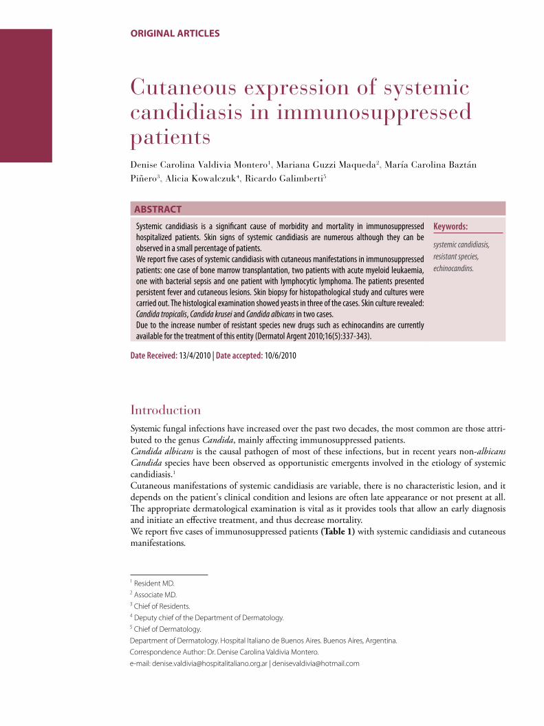
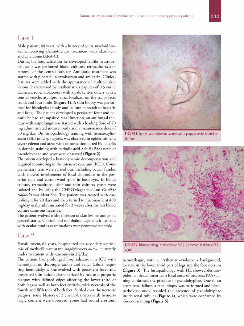
Case 1Male patient, 44 years, with a history of acute myeloid leu-kemia receiving chemotherapy treatment with idarubicin and cytarabine (ARA-C).During his hospitalization he developed febrile neutrope-nia, so it was perfomed blood cultures, retrocultures and removal of the central catheter. Antibiotic treatment was started with piperacillin-tazobactam and amikacin. Clinical features were added with the appearance of multiple skin lesions characterized by erythematous papules of 0.5 cm in diameter, some violaceous, with a pale center, others with a central vesicle, asymptomatic, localized on the scalp, face, trunk and four limbs (Figure 1). A skin biopsy was perfor-med for histological study and culture in search of bacteria and fungi. The patient developed a persistent fever and be-cause he had an impaired renal function, an antifungal the-rapy with caspofunginwas started with a loading dose of 70 mg administrated intravenously and a maintenance dose of 50 mg/day. On histopathology staining with hematoxylin-eosin (HE) mild spongiosis was observed in epidermis, and severe edema and areas with extravasation of red blood cells in dermis; staining with periodic acid-Schiff (PAS) nests of pseudohyphae and yeast were observed (Figure 2).The patient developed a hemodynamic decompensation and required monitoring in the intensive care unit (ICU). Com-plementary tests were carried out, including ocular fundus wich showed involvement of focal choroiditis in the pos-terior pole and cotton-wool spots in both eyes. In blood culture, retroculture, urine and skin cultures yeasts were isolated and by using the CHROMagar medium Candida tropicalis was identified. The patient was treated with cas-pofungin for 20 days and then turned to fluconazole at 400 mg/day orally administrated for 2 weeks after the last blood culture came out negative.The patient evolved with remission of skin lesions and good general status. Clinical and ophthalmologic check ups and with ocular fundus examinations were performed monthly.
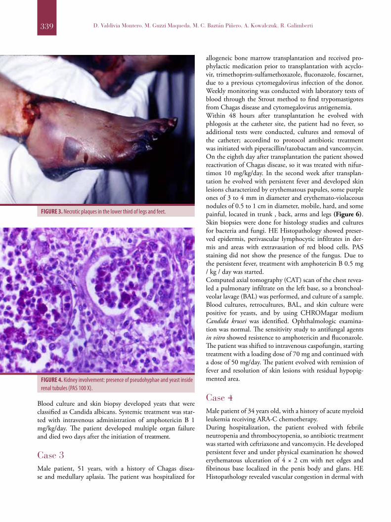
Case 2Female patient, 64 years, hospitalized for secondary septice-mia of methicillin-resistant Staphylococcus aureus, currently under treatment with vancomycin 2 g/day.The patient had prolonged hospitalization in ICU with hemodynamic decompensation and renal failure requi-ring hemodialysis. She evolved with persistent fever and presented skin lesions characterized by necrotic purpuric plaques with defined edges affecting the lower third of both legs as well as both feet entirely, with necrosis of the fourth and fifth toes of both feet. Settled over the necrotic plaques, tense blisters of 2 cm in diameter with hemorr-hagic content were observed, some had round erosions,
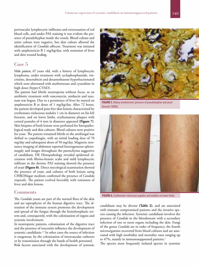
hemorrhagic, with a erythemato-violaceous background, located in the lower third part of legs and the foot dorsum (Figure 3). The histopathology with HE showed dermoe-pidermal detachment with focal areas of necrosis; PAS stai-ning confirmed the presence of pseudohyphae. Due to an acute renal failure, a renal biopsy was performed and histo-pathology study revealed the presence of pseudohyphae inside renal tubules (Figure 4), which were confirmed by Grocott staining (Figure 5).
FIGURE 1. Erythemato-violaceous papules with a purpuric center located in the face.
FIGURE 2. Histopathology. Nests of yeast PAS (+) observed in dermis (PAS 400X).
Cutaneous expression of systemic candidiasis in immunosuppressed patients
339 D. Valdivia Montero, M. Guzzi Maqueda, M. C. Baztán Piñero, A. Kowalczuk, R. Galimberti
Blood culture and skin biopsy developed yeats that were classified as Candida albicans. Systemic treatment was star-ted with intravenous administration of amphotericin B 1 mg/kg/day. The patient developed multiple organ failure and died two days after the initiation of treatment.
Case 3Male patient, 51 years, with a history of Chagas disea-se and medullary aplasia. The patient was hospitalized for
allogeneic bone marrow transplantation and received pro-phylactic medication prior to transplantation with acyclo-vir, trimethoprim-sulfamethoxazole, fluconazole, foscarnet, due to a previous cytomegalovirus infection of the donor. Weekly monitoring was conducted with laboratory tests of blood through the Strout method to find trypomastigotes from Chagas disease and cytomegalovirus antigenemia.Within 48 hours after transplantation he evolved with phlogosis at the catheter site, the patient had no fever, so additional tests were conducted, cultures and removal of the catheter; accordind to protocol antibiotic treatment was initiated with piperacillin/tazobactam and vancomycin. On the eighth day after transplantation the patient showed reactivation of Chagas disease, so it was treated with nifur-timox 10 mg/kg/day. In the second week after transplan-tation he evolved with persistent fever and developed skin lesions characterized by erythematous papules, some purple ones of 3 to 4 mm in diameter and erythemato-violaceous nodules of 0.5 to 1 cm in diameter, mobile, hard, and some painful, located in trunk , back, arms and legs (Figure 6). Skin biopsies were done for histology studies and cultures for bacteria and fungi. HE Histopathology showed preser-ved epidermis, perivascular lymphocytic infiltrates in der-mis and areas with extravasation of red blood cells. PAS staining did not show the presence of the fungus. Due to the persistent fever, treatment with amphotericin B 0.5 mg / kg / day was started.Computed axial tomography (CAT) scan of the chest revea-led a pulmonary infiltrate on the left base, so a bronchoal-veolar lavage (BAL) was performed, and culture of a sample. Blood cultures, retrocultures, BAL, and skin culture were positive for yeasts, and by using CHROMagar medium Candida krusei was identified. Ophthalmologic examina-tion was normal. The sensitivity study to antifungal agents in vitro showed resistence to amphotericin and fluconazole. The patient was shifted to intravenous caspofungin, starting treatment with a loading dose of 70 mg and continued with a dose of 50 mg/day. The patient evolved with remission of fever and resolution of skin lesions with residual hypopig-mented area.
Case 4Male patient of 34 years old, with a history of acute myeloid leukemia receiving ARA-C chemotherapy.During hospitalization, the patient evolved with febrile neutropenia and thrombocytopenia, so antibiotic treatment was started with ceftriaxone and vancomycin. He developed persistent fever and under physical examination he showed erythematous ulceration of 4 × 2 cm with net edges and fibrinous base localized in the penis body and glans. HE Histopathology revealed vascular congestion in dermal with
FIGURE 3. Necrotic plaques in the lower third of legs and feet.
FIGURE 4. Kidney involvement: presence of pseudohyphae and yeast inside renal tubules (PAS 100 X).
340Cutaneous expression of systemic candidiasis in immunosuppressed patients
perivascular lymphocytic infiltrates and extravasation of red blood cells, and under PAS staining it was evident the pre-sence of pseudohyphae inside the vessels. Blood culture and urine culture were negative, but skin culture allowed the identification of Candida albicans. Treatment was initiated with amphotericin B 1 mg/kg/day, with remission of fever and skin wound healing.
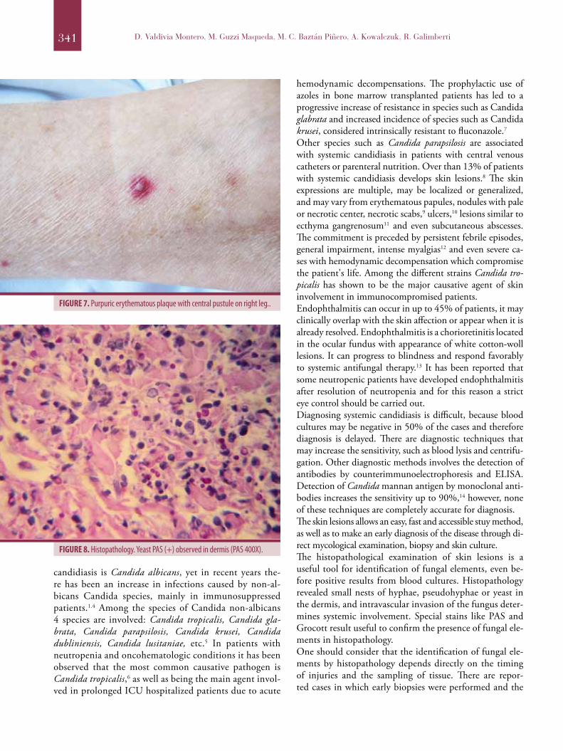
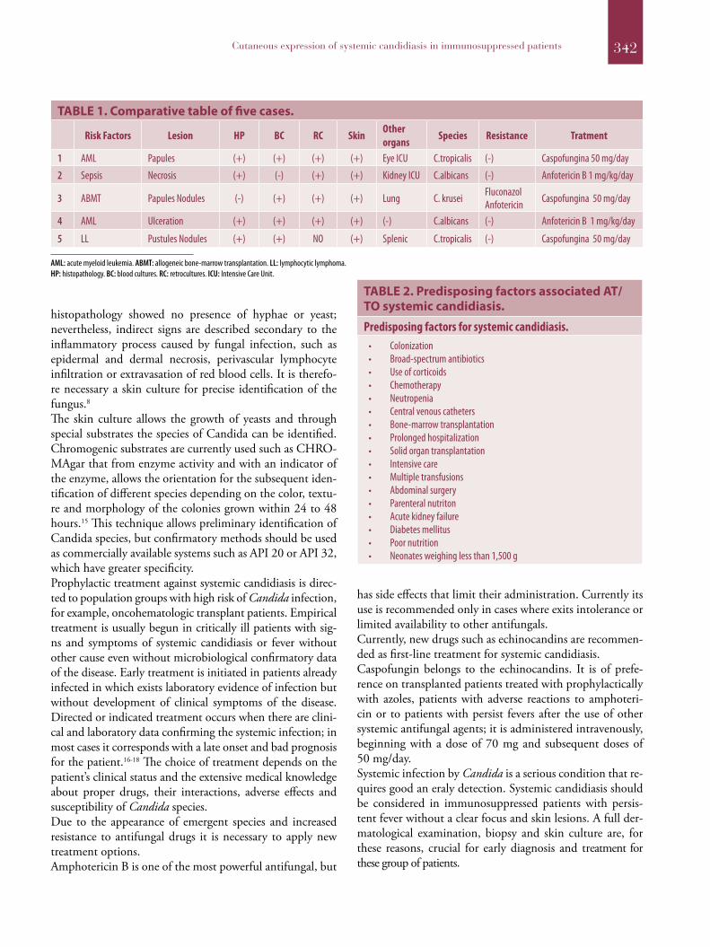
Case 5Male patient, 67 years old, with a history of lymphocytic lymphoma, under treatment with cyclophosphamide, vin-cristine, doxorubicin and dexamethasone hyperfractionated which were alternated with methotrexate and cytarabine in high doses (hyper-CVAD).The patient had febrile neutropenia without focus, so an antibiotic treatment with vancomycin, amikacin and tazo-nam was begun. Due to a persistence of fever he started on amphotericin B at doses of 1 mg/kg/day. After 72 hours, the patient developed pain-free skin lesions characterized by erythemato-violaceous nodules 1 cm in diameter on his left forearm, and on lower limbs, erythematous plaques with central pustules of 4 mm in diameter appeared (Figure 7). Skin biopsies of both lesions were perfomed for histopatho-logical study and skin cultures. Blood cultures were positive for yeast. The patient remained febrile so the antifungal was shifted to caspofungin, with an initial loading dose of 70 mg/day and subsequent doses of 50 mg/day. Magnetic reso-nance imaging of abdomen reported heterogeneous spleno-megaly and images throughout the parenchyma suggestive of candidiasis. HE Histopathology revealed epidermal ul-ceration with fibrino-hemtic scabs and mild lymphocytic infiltrate in the dermis; PAS staining showed the presence of yeast (Figure 8). Direct mycological examination showed the presence of yeast, and cultures of both lesions using CHROMagar medium confirmed the presence of Candida tropicalis. The patient evolved favorably with remission of fever and skin lesions.
CommentsThe Candida yeasts are part of the normal flora of the skin and are saprophytes of the human digestive tract. The al-teration of the immune system promotes the development and spread of the fungus through the hemolymphatic sys-tem and, consequently, with the colonization of organs and systemic involvement.In neutropenic patients, colonization of the digestive tract and the presence of mucositis influence the development of systemic candidiasis.2.3 In other cases the source of infection is exogenous, by the colonization of intravascular catheters or by transmission through the hands of health personnel.Risk factors associated with the development of systemic
candidiasis may be diverse (Table 2), and are associated with immune compromised patients and the invasive spe-cies causing the infection. Systemic candidiasis involves the presence of Candida in the bloodstream with a secondary infection of one or more organs including the skin. Fungi of the genus Candida are in order of frequency, the fourth microorganism recovered from blood cultures and are asso-ciated with high morbidity and mortality rates ranging up to 47%, mainly in immunosuppressed patients.2
The species most frequently isolated species in systemic
FIGURE 5. Kidney involvement: presence of pseudohyphae and yeast (Grocott 1000X).
FIGURE 6. Erythemato-violaceous papules and nodules on lower limbs.
341 D. Valdivia Montero, M. Guzzi Maqueda, M. C. Baztán Piñero, A. Kowalczuk, R. Galimberti
candidiasis is Candida albicans, yet in recent years the-re has been an increase in infections caused by non-al-bicans Candida species, mainly in immunosuppressed patients.1.4 Among the species of Candida non-albicans 4 species are involved: Candida tropicalis, Candida gla-brata, Candida parapsilosis, Candida krusei, Candida dubliniensis, Candida lusitaniae, etc.5 In patients with neutropenia and oncohematologic conditions it has been observed that the most common causative pathogen is Candida tropicalis,6 as well as being the main agent invol-ved in prolonged ICU hospitalized patients due to acute
hemodynamic decompensations. The prophylactic use of azoles in bone marrow transplanted patients has led to a progressive increase of resistance in species such as Candida glabrata and increased incidence of species such as Candida krusei, considered intrinsically resistant to fluconazole.7
Other species such as Candida parapsilosis are associated with systemic candidiasis in patients with central venous catheters or parenteral nutrition. Over than 13% of patients with systemic candidiasis develops skin lesions.8 The skin expressions are multiple, may be localized or generalized, and may vary from erythematous papules, nodules with pale or necrotic center, necrotic scabs,9 ulcers,10 lesions similar to ecthyma gangrenosum11 and even subcutaneous abscesses. The commitment is preceded by persistent febrile episodes, general impairment, intense myalgias12 and even severe ca-ses with hemodynamic decompensation which compromise the patient's life. Among the different strains Candida tro-picalis has shown to be the major causative agent of skin involvement in immunocompromised patients.Endophthalmitis can occur in up to 45% of patients, it may clinically overlap with the skin affection or appear when it is already resolved. Endophthalmitis is a chorioretinitis located in the ocular fundus with appearance of white cotton-woll lesions. It can progress to blindness and respond favorably to systemic antifungal therapy.13 It has been reported that some neutropenic patients have developed endophthalmitis after resolution of neutropenia and for this reason a strict eye control should be carried out.Diagnosing systemic candidiasis is difficult, because blood cultures may be negative in 50% of the cases and therefore diagnosis is delayed. There are diagnostic techniques that may increase the sensitivity, such as blood lysis and centrifu-gation. Other diagnostic methods involves the detection of antibodies by counterimmunoelectrophoresis and ELISA. Detection of Candida mannan antigen by monoclonal anti-bodies increases the sensitivity up to 90%,14 however, none of these techniques are completely accurate for diagnosis.The skin lesions allows an easy, fast and accessible stuy method, as well as to make an early diagnosis of the disease through di-rect mycological examination, biopsy and skin culture.The histopathological examination of skin lesions is a useful tool for identification of fungal elements, even be-fore positive results from blood cultures. Histopathology revealed small nests of hyphae, pseudohyphae or yeast in the dermis, and intravascular invasion of the fungus deter-mines systemic involvement. Special stains like PAS and Grocott result useful to confirm the presence of fungal ele-ments in histopathology.One should consider that the identification of fungal ele-ments by histopathology depends directly on the timing of injuries and the sampling of tissue. There are repor-ted cases in which early biopsies were performed and the
FIGURE 7. Purpuric erythematous plaque with central pustule on right leg..
FIGURE 8. Histopathology. Yeast PAS (+) observed in dermis (PAS 400X).
342Cutaneous expression of systemic candidiasis in immunosuppressed patients
histopathology showed no presence of hyphae or yeast; nevertheless, indirect signs are described secondary to the inflammatory process caused by fungal infection, such as epidermal and dermal necrosis, perivascular lymphocyte infiltration or extravasation of red blood cells. It is therefo-re necessary a skin culture for precise identification of the fungus.8
The skin culture allows the growth of yeasts and through special substrates the species of Candida can be identified. Chromogenic substrates are currently used such as CHRO-MAgar that from enzyme activity and with an indicator of the enzyme, allows the orientation for the subsequent iden-tification of different species depending on the color, textu-re and morphology of the colonies grown within 24 to 48 hours.15 This technique allows preliminary identification of Candida species, but confirmatory methods should be used as commercially available systems such as API 20 or API 32, which have greater specificity.Prophylactic treatment against systemic candidiasis is direc-ted to population groups with high risk of Candida infection, for example, oncohematologic transplant patients. Empirical treatment is usually begun in critically ill patients with sig-ns and symptoms of systemic candidiasis or fever without other cause even without microbiological confirmatory data of the disease. Early treatment is initiated in patients already infected in which exists laboratory evidence of infection but without development of clinical symptoms of the disease. Directed or indicated treatment occurs when there are clini-cal and laboratory data confirming the systemic infection; in most cases it corresponds with a late onset and bad prognosis for the patient.16-18 The choice of treatment depends on the patient’s clinical status and the extensive medical knowledge about proper drugs, their interactions, adverse effects and susceptibility of Candida species.Due to the appearance of emergent species and increased resistance to antifungal drugs it is necessary to apply new treatment options.Amphotericin B is one of the most powerful antifungal, but
has side effects that limit their administration. Currently its use is recommended only in cases where exits intolerance or limited availability to other antifungals.Currently, new drugs such as echinocandins are recommen-ded as first-line treatment for systemic candidiasis.Caspofungin belongs to the echinocandins. It is of prefe-rence on transplanted patients treated with prophylactically with azoles, patients with adverse reactions to amphoteri-cin or to patients with persist fevers after the use of other systemic antifungal agents; it is administered intravenously, beginning with a dose of 70 mg and subsequent doses of 50 mg/day.Systemic infection by Candida is a serious condition that re-quires good an eraly detection. Systemic candidiasis should be considered in immunosuppressed patients with persis-tent fever without a clear focus and skin lesions. A full der-matological examination, biopsy and skin culture are, for these reasons, crucial for early diagnosis and treatment for these group of patients.
TABLE 1. Comparative table of five cases.
Risk Factors Lesion HP BC RC Skin Other organs Species Resistance Tratment
1 AML Papules (+) (+) (+) (+) Eye ICU C.tropicalis (-) Caspofungina 50 mg/day
2 Sepsis Necrosis (+) (-) (+) (+) Kidney ICU C.albicans (-) Anfotericin B 1 mg/kg/day
3 ABMT Papules Nodules (-) (+) (+) (+) Lung C. krusei Fluconazol Anfotericin Caspofungina 50 mg/day
4 AML Ulceration (+) (+) (+) (+) (-) C.albicans (-) Anfotericin B 1 mg/kg/day
5 LL Pustules Nodules (+) (+) NO (+) Splenic C.tropicalis (-) Caspofungina 50 mg/day
AML: acute myeloid leukemia. ABMT: allogeneic bone-marrow transplantation. LL: lymphocytic lymphoma.HP: histopathology. BC: blood cultures. RC: retrocultures. ICU: Intensive Care Unit.
TABLE 2. Predisposing factors associated AT/TO systemic candidiasis.
Predisposing factors for systemic candidiasis.• Colonization• Broad-spectrum antibiotics• Use of corticoids• Chemotherapy• Neutropenia• Central venous catheters• Bone-marrow transplantation• Prolonged hospitalization• Solid organ transplantation• Intensive care• Multiple transfusions• Abdominal surgery• Parenteral nutriton• Acute kidney failure• Diabetes mellitus• Poor nutrition• Neonates weighing less than 1,500 g
343 D. Valdivia Montero, M. Guzzi Maqueda, M. C. Baztán Piñero, A. Kowalczuk, R. Galimberti
References1. Pappas PG. Invasive candidiasis. Infect Dis Clin North Am 2006;20:485-
506.2. Gudlaugsson O. Attributable mortality of nosocomial candidemia,
revisited. Clin Infect Dis 2003;37:1172-1177.3. Pfaller MA, Jones RN, Messer SA, Edmond MB et ál. National
epidemiology of mycoses survey: a multicenter study of strain variaton and antifungal susceptibility among isolates on Candida species. Diagn Microbiol Infect Dis 1998;31:327-332.
4. Wisplinghoff H, Seifert H, Wenzel RP, Edmond MB. Current trends in the epidemiology of nosocomial bloodstream infections in patients with hematological malignancies and solid neoplasms in hospitals in the United States. Clin Infect Dis 2003;36:1103-1110.
5. Pfaller MA, Diekema DJ. Epidemiology of invasive candidiasis: a persistent public health problem. Clin Microbiol Rev 2000;20:133-163.
6. Kontoyiannis DP, Vaziri I, Hanna HA, Boktour M et ál. Risk factors for Candida tropicalis fungemia in patients with cancer. Clin Infect Dis 2001; 33:1676-1681.
7. Wingard J, Merz W, Rinaldi M, Johnson T et ál. Increase in Candida krusei infections among patients with bone marrow transplantation and neutropenia treated prophylactically with fl uconazole. N Engl J Med 1991; 325:1274-1277.
8. Slater DN, Wylde P, Harrington CI, Worth R. Systemic candidiasis: diagnosis from cutaneous manifestations. J R Soc Med 1982;75:875-878.
9. File TM Jr, Marina OA, Flowers FP. Necrotic skin lesions associated with disseminated candidiasis. Arch Dermatol 1979;114:214-215.
10. Galimberti RL, Flores V, González Ramos MC, Villalba LI. Cutaneous ulcers due to Candida albicans in an immunocompromised patient-response to therapy with itraconazole. Clin Exp Dermatol 1989;14:295-297.
11. Fine JD, Miller JA, Harrist TJ, Haynes HA. Cutaneous lesions in disseminated candidiasis mimicking ecthyma gangrenosum. Am J Med 1981;70: 1133-1135.
12. Jarowski CI, Fialk MA, Murray HW, Gottlieb GJ et ál. Fever, rash, and muscle tenderness: a distinctive clinical presentation of disseminated candidiasis. Arch Intern Med 1978;138: 544-546.
13. Sallam A, Lynn W, McCluskey P, Ligtman S et ál. Endogenous Candida endophthalmitis. Expert Rev Anti Infect Ther 2006;4:675-685.
14. Nakamura A, Ishikawa N, Suzuki H. Diagnosis of invasive candidiasis by detection of mannan antigen by using the avidin-biotin enzyme immunoassay. J Clin Microbiol 1991;29:2363-2327.
15. Yücesoy M, Marol S. Performance of CHROMagar Candida and BIGGY agar for identifi cation of yeast species. Ann Clin Microb Antimicrob 2003;2:8
16. Allevato MA, Negroni R, Galimberti R. Antifúngicos. Ayer, hoy y mañana. Act Terap Dermatol 2007;30:8.
17. Del Palacio A, Alhambra A, Cuétara MS. Estrategias de tratamiento: profilaxis, tratamiento empírico, precoz (anticipado) y dirigido de candidiasis invasora en el enfermo crítico no neutropénico. Rev Iberoam Micol 2006;23:35-38.
18. Pappas PG, Kauff man CA, Andes D, Benjamin DK Jr et ál. Clinical practice guidelines for the management of candidiasis: 2009 update by the Infectious Diseases Society of America. Clin Infect Dis 2009;48:503-535.