Embed Size (px)
Citation preview
CELL DIVISION IN A SPECIES OF ERWINIA1 2
II. INHIBITION OF DIVISION BY D-AMINO ACIDS
E. A. GRULA
Department of Bacteriology, Oklahoma State University, Stillwater, Oklahoma
Received for publication February 3, 1960
Cell division in a species of Erwinia can beinhibited by certain carbon-energy sources whenthe cells are grown in nutrient broth (Grula,1960). Because nutrient broth is an ill-definedcomplex, it was decided that nutritional relation-ships and the accumulation or the disappearanceof a compound(s) essential to cell division couldbest be studied using a chemically definedmedium.
MATERIALS AND METHODS
Basic techniques and conditions have been re-ported (Grula, 1960). All chemicals used in thisstudy were of the highest quality obtainable and,in many instances, the same chemical was ob-tained and tested from several supply houses.During the early phase of this investigation,
the cells appeared, at times, to lose their abilityto elongate unless they were periodically trans-ferred to nutrient agar containing 1 per cent glu-cose. Therefore, the cells were routinely trans-ferred from nutrient agar to glucose nutrient agarand vice versa every time a transfer was involved(usually every 24 hr during active periods ofexperimentation and at least monthly duringless active periods). Cells grown on nutrient agar(no glucose) were always used as inoculum forstudies involving defined media. Lack of con-sistent elongation was probably due more tolack of knowledge concerning the process thanto the medium used, since, at the end of thisstudy, a culture that had been transferred rou-tinely on nutrient agar 52 times during a 3-monthinterval formed long cells when transferred backto nutrient agar glucose medium.
1 This investigation was supported by the Re-search Foundation, Oklahoma State University,Project no. 91 and a National Science Foundationgrant G9848.
2 Portions of this study were presented at the59th General Meeting of the Society of AmericanBacteriologists, St. Louis, Missouri, May 10 to 15,1959.
RESULTSFormulation of a chemically defined medium
for growth of Erwinia and other phytopathogenshas been reported (Starr, 1946; Starr and Mandel,1950). The medium has the following composi-tion per 100 ml: glucose, 0.5 g (autoclavedseparately); NH4Cl, 0.1 g; KH2PO4, 0.2 g;MgSO4 7HOH, 0.02 g (solution I); and the fol-lowing as trace mineral salts, H3BO3, 0.5 ,ug;CaCO3, 10.0 J,g; CuSO4.5HOH, 1.0 ,ug;FeSO4(NH4)2SO4 6HOH, 10.0 or 50.0 ,ug (weused the 50 ,ug level); KI, 1.0 MAg; MnSO4-HOH.2.0 pig; MoO3, 1.0 ,ug; and ZnSO4.7HOH, 5.0,ug (solution II). The pH was adjusted to 6.8with'NaOH.When washed cells of Erwinia were added to
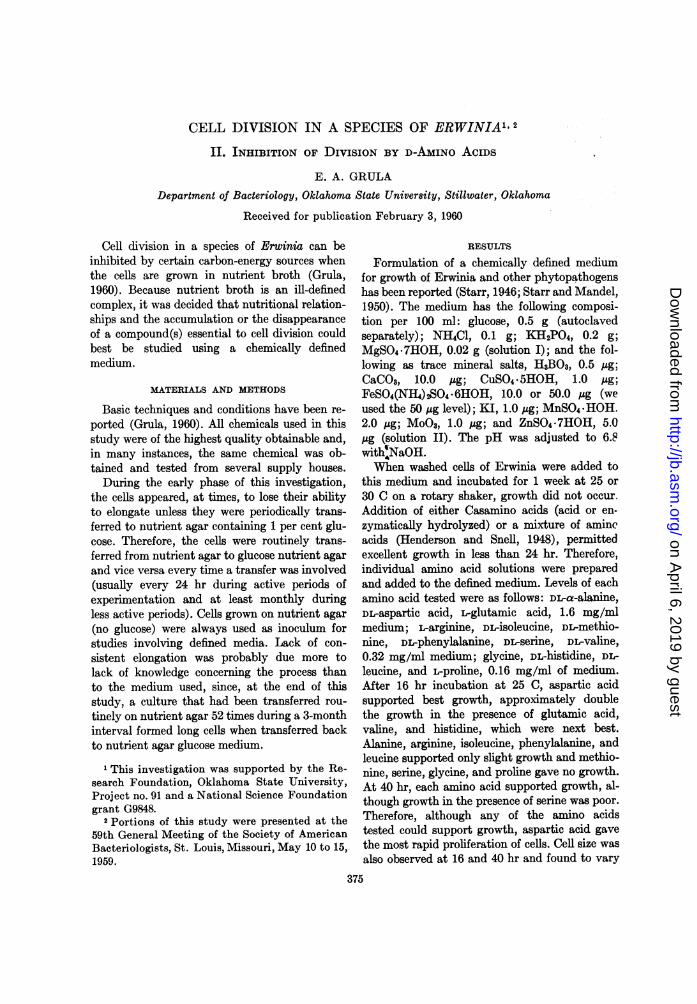
this medium and incubated for 1 week at 25 or30 C on a rotary shaker, growth did not occur.Addition of either Casamino acids (acid or en-zymatically hydrolyzed) or a mixture of amineacids (Henderson and Snell, 1948), permittedexcellent growth in less than 24 hr. Therefore,individual amino acid solutions were preparedand added to the defined medium. Levels of eachamino acid tested were as follows: DL-a-alanine,DL-aspartic acid, L-glutamic acid, 1.6 mg/mlmedium; L-arginine, DL-isoleucine, DL-methio-nine, DL-phenylalanine, DL-serine, DL-valine,0.32 mg/ml medium; glycine, DL-histidine, DL-leucine, and L-proline, 0.16 mg/ml of medium.After 16 hr incubation at 25 C, aspartic acidsupported best growth, approximately doublethe growth in the presence of glutamic acid,valine, and histidine, which were next best.Alanine, arginine, isoleucine, phenylalanine, andleucine supported only slight growth and methio-nine, serine, glycine, and proline gave no growth.At 40 hr, each amino acid supported growth, al-though growth in the presence of serine was poor.Therefore, although any of the amino acidstested could support growth, aspartic acid gavethe most rapid proliferation of cells. Cell size wasalso observed at 16 and 40 hr and found to vary
375
on April 6, 2019 by guest
http://jb.asm.org/
Dow
nloaded from
GRULA
between 3 and 5 ,u for all amino acids except DL-serine. Cells grown in the presence of serine, as thesole source of organic nitrogen, were small (1 to2 IA).
Various combinations of several amino acids(aspartic, glutamic, valine, methionine. and histi-dine) were added in an attempt to increase celllength in the defined medium. At no time couldelongation greater than 5 ,u be obtained. Thethinking at this stage of the experimentation was:if the correct conditions for inhibition of celldivision could be obtained, cells should become100 ,u or greater in length rather than 5 or 10 ,.Because elongation could not be increased
using the various amino acid combinations, in-dividual solutions of B-vitamins were added tothe defined medium containing aspartic acid eventhough it was obvious that B-vitamins were notrequired for growth. Although none of the vita-mins were capable of increasing cell size to anygreat extent (none greater than 8 ,u), it was notedthat p-aminobenzoic acid (40 to 400 ,ug/ml) re-tarded growth (turbidimetric) and cell size wasagain small (1 to 2 ,u). Addition of individualpurine and pyrimidine bases (adenine, guanine,xanthine, uracil, and cytosine) to a level of 20,ug/ml also did not enhance elongation whenadded to the defined medium.
Because certain sugars interfere with cell di-vision (Grula, 1960), various acids or their salts,occurring as intermediates in sugar metabolism,were tested. The sodium salts of formate, acetate,butyrate, pyruvate, potassium fumarate, andsuccimic, malic, and a-ketoglutaric acids werefilter sterilized, the acids adjusted to neutral pHwith KOH, and added to the medium in place ofglucose to a level of 0.0336 M concentration.Pyruvate was the only organic acid able to re-place glucose and cause long cell formation; how-ever, some'elongation occurred in the presence ofsuccinic acid. With most of the compounds tested,cell size was intermediate (2 to 4 ,.), and at notime were cells longer than those grown in thepresence of glucose. Butyrate, acetate, formate,and malic acid were toxic, whereas the other com-pounds allowed good growth.
Because various amino acid combinations, B-vitamins, purines, and pyrimidines or acid meta-bolic intermediates would not allow elongationin the defined medium any greater than that ob-tained using glucose, each constituent of themedium was studied since it was not known if all
constituents were needed, and the possibilityexisted that one of them was preventing the cellsfrom becoming extremely long. Therefore, eachcompound (glucose, MgSO4, KH2PO4, asparticacid, and NH4CI) and the trace mineral solutionwas individually titrated against all other con-stituents of the medium in "rule-out" experi-ments. The following observations were madeafter 16 hr incubation at 25 C: (1) aspartic acid,KH2PO4, and MgSO4 were essential for growth,(2) glucose stimulated, but was not required forgrowth, (3) the trace mineral solution and NH4Clwere not needed for growth. Because glucose wasinvolved in elongation, it was retained in mostfuture media manipulations and also, the tracemineral solution was retained since it was feltthat the solution would compensate for possiblefluctuations in the purity of distilled water. Phos-phate was added as an equimolar solution ofKH2PO4 and K2HPO4 to give a starting pH of6.8 without adjustment. Therefore, the basalmedium was modified to contain the followingper 100 ml of distilled water: glucose, 300 mg(added aseptically); MgSO4-7HOH, 3 mg;K2HPO4, 174 mg; KH2PO4, 136 mg; asparticacid, 280 mg (made up separately and adjustedto pH of about 7.0 using NaOH); and the tracemineral salt solution (II) as listed at the be-ginning of this report. Length of cells grown inthis medium in the presence of glucose is 5 to 10A, with some cells growing to about 20 IA. In theabsence of glucose, cell length is uniform and inthe 2 to 4 ,u range.
Because glucose caused greater elongation inthe modified medium, several compounds werere-checked for ability to enhance elongation, par-ticularly amino acids since NH4C1 had now beenexcluded. Data for DL forms of the amino acids(except glycine) are given in table 1. After 16 hrincubation, six amino acids, DL-serine, DL-methio-nine, DL-phenylalanine, DL-histidine, DL-threo-nine, and DL-tryptophan, caused formation ofextremely long cells. The cells grew to 100 to 300,u in length and appeared as long filaments someof which had division areas every 10 to perhaps30 Iu. A wide variation in growth response to thedifferent amino acids is also evident as shown intable 1.
Addition of DL-serine at the same concentra-tion to nutrient broth in the absence of glucosegave rise to cells of approximately the samelength as seen previously using glucose. Addition
376 [VOL. 80
on April 6, 2019 by guest
http://jb.asm.org/
Dow
nloaded from
CELL DIVISION IN ERWINIA. II
TABLE 1Response of Erwinia sp. to various amino acids*
Amino Acid Concn ODt Cell Size in p
Aspartic acid (noglucose) ........ 0.0169 0.83 2-4
Aspartic acid..... 0.0169 1.2 5-10; 10% 10-20Arginine*HC.... 0.0336 1.5 3-5Threonine........ 0.0336 0.17 10-150Glycine .......... 0.0336 1.05 4-5; 20% 5-10Methionine ....... 0.0168 0.85 5-100Glutamic acid.... 0.0336 1.3 3-4Valine............ 0.0168 0.38 3-10; 10% 10-20Leucine .......... 0.0336 0.15 3-5; 10% 5-15Serine... .... 0.0420 0.11 20-300Phenylalanine .... 0.0168 0.76 10-150Lysine.HCl...... 0.0336 1.1 2-5; 10% 5-10Isoleucine. . 0.0336 0.32 2-4Histidine....... .. 0.0336 0.69 10-150Proline........... 0.0336 0.92 3-7Tryptophan...... 0.0063 0.27 10-100Tyrosine ......... 0.0042 1.2 3-5a-Alanine ........ 0.0336 0.5 2-5
* All tubes contained trace mineral solution,phosphate, magnesium sulfate, aspartic acid andglucose as given in the text except tube 1 whereglucose was omitted.
t Optical density read at 540 my after16 hr at 25 C. OD of inoculum was 0.47.
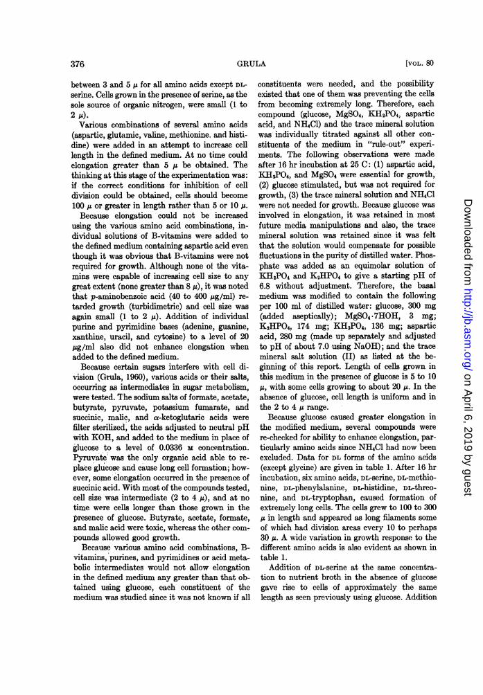
Of DL-serine to the defined medium in the absenceof glucose also gave rise to long cells althoughtotal cell mass was less and cell size was more inthe 20 to 100 A range rather than the large andtangled masses of 100 to 300 ,u filaments seen inthe presence of glucose. A titration of DL-serinein the presence of glucose is given in table 2.Increasing concentrations of DL-serine (up to0.0504 M) allows for a progressive increase incell length. At higher concentrations, growth is,for all practical purposes, completely inhibited.A concentration of 0.0336 M DL-serine was chosenfor future studies because fairly good growth aswell as long cell formation occur at this level.Results similar to these were also obtained usingeither DL-methionine or DL-phenylalanine.The D and L isomers of serine, methionine, and
phenylalanine were then tested to determine ifelongation was due to the L or D form of the aminoacid. Long cell development occurred only in thepresence of the D isomer with all three compounds(0.0168 M for D-serine, 0.0067 M for D-phenylala-
TABLE 2Titration of DL-serine in the presence of 0.0166 m
glucose*Concn DLSerine ODt Average Size in i
None 1.05 5-200.0084 0.96 10-200.0168 0.80 20-1000.0336 0.40 100-3000.0504 0.12 100-3000.0672 0.03 No growth0.0840 0.02 No growth
* All tubes contained trace mineral solution,aspartic acid, magnesium sulfate and phosphateas given in the text.
t Optical density read at 540 mu after 15 hr at25 C on a rotary shaker.
nine or D-methionilne). Although some "bulb" orsphereoplast formation occurs after about 12 hrincu-bation (size and number vary), it should bepointed out that at about 24 hr and thereafter,cell size appears to progressively decrease (5 to30 ,u size predominates) and many poorly stained"ghost" filaments remain. The bulb formation isunusual in that one or up to three bulbs can formanywhere along the length of a cell. This observa-tion is similar to that reported by Bachmann andBonner (1959); however, it has not been demon-strated that these filaments are completely coeno-cytic. Free spheroplasts appear to be relativelystable in 17 per cent maltose.D and L forms of aspartic acid were tested in
the presence and absence of either glucose, DL-serine, or both to determine if either isomer hadan effect on the division mechanism of the cells.Observation of the cultures and cells-,at 16 hrshowed that total cell mass (optical density) wasapproximately the same in the presence of eitherisomer and cell size was the same as though theDL form was present in the medium.
Attempts to reverse long cell formation. Studieswere initiated to determine how to reverse elonga-tion in order to obtain information concerninginvolvement of specific metabolites in the divisionprocess.Although critical studies have not been done, it
has been observed that cells from the top ofliquid cultures containing D-serine not aeratedduring growth are typically long (100 to 300 IA),whereas cells taken from the bottom of unshaken
3771960]
on April 6, 2019 by guest
http://jb.asm.org/
Dow
nloaded from
GRULA
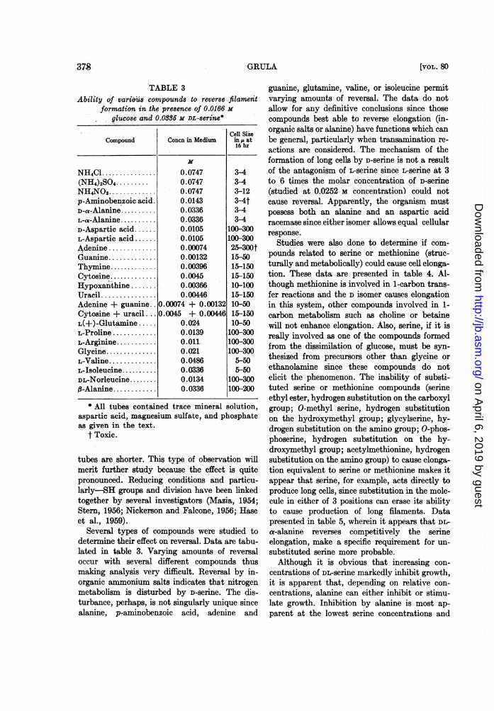
TABLE 3Ability of various compounds to reverse filament
formation in the presence of 0.0166 m
glucose and 0.0386 M DL-serine*
Cell SizeCompound Concn in Medium in at
NH4Cl ............... 0.0747 3-4(NH4)2SO4 ......... 0.0747 3-4NH4NO3............. 0.0747 3-12p-Aminobenzoic acid. 0.0143 3-4tD-a-Alamnne.. 0.0336 3-4L-a-Alafllfle......'0.03363-4D-Aspartic acid.'..... 0.0105 100-300L-Aspartic acid ...... 0.0105 100-300Adenine ............. 0.00074 25-300tGuanine............. 0.00132 15-50Thymine.... 0.00396 15-150Cytosine... 0.0045 15-150Eypoxanthine ........ 0.00366 10-100Uracil ............... 0.00446 15-150Adenine + guanine.. 0.00074 + 0.00132 10-50Cytosine + uracil . .. 0.0045 + 0.00446 15-150L(+)-Glutamine ..... 0.024 10-50L-Proline ............ 0.0139 100-300L-Arginine ........... 0.011 100-300Glycine............... 0.021 100-300L-Valine............. 0.0486 5-50L-Isoleucine.......... 0.0336 5-50DL-Norleucine ........ 0.0134 100-300B-Alanine............ 0.0336 100-200
* All tubes contained trace mineral solution,aspartic acid, magnesium sulfate, and phosphateas given in the text.
t Toxic.
tubes are shorter. This type of observation willmerit further study because the effect is quitepronounced. Reducing conditions and particu-larly-SH groups and division have been linkedtogether by several investigators (Mazia, 1954;Stern, 1956; Nickerson and Falcone, 1956; Haseet al., 1959).
Several types of compounds were studied todetermine their effect on reversal. Data are tabu-lated in table 3. Varying amounts of reversaloccur with several different compounds thusmaking analysis very difficult. Reversal by in-organic ammonium salts indicates that nitrogenmetabolism is disturbed by D-serine. The dis-turbance, perhaps, is not singularly unique sincealanine, p-aminobenzoic acid, adenine and
guanine, glutamine, valine, or isoleucine permitvarying amounts of reversaL The data do notallow for any definitive conclusions since thosecompounds best able to reverse elongation (in-organic salts or alanine) have functions which canbe general, particularly when transamination re-actions are considered. The mechanism of theformation of long cells by D-serine is not a resultof the antagonism of L-serine since -serine at 3to 6 times the molar concentration of D-serine(studied at 0.0252 M concentration) could notcause reversal. Apparently, the organism mustpossess both an alanine and an aspartic acidracemase since either isomer allows equal cellularresponse.
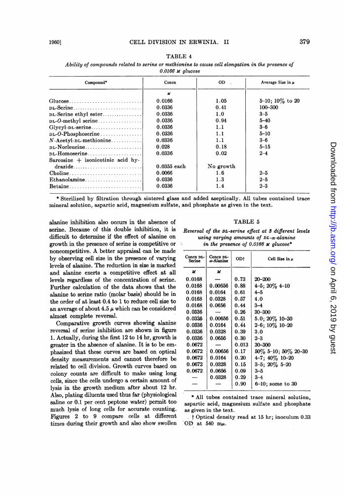
Studies were also done to determine if com-pounds related to serine or methionine (struc-turally and metabolically) could cause cell elonga-tion. These data are presented in table 4. Al-though methionine is involved in 1-carbon trans-fer reactions and the D isomer causes elongationin this system, other compounds involved in 1-carbon metabolism such as choline or betainewill not enhance elongation. Also, serine, if it isreally involved as one of the compounds formedfrom the dissimilation of glucose, must be syn-thesized from precursors other than glycine orethanolamine since these compounds do notelicit the phenomenon. The inability of substi-tuted serine or methionine compounds (serineethyl ester, hydrogen substitution on the carboxylgroup; 0-methyl serine, hydrogen substitutionon the hydroxymethyl group; glycylserine, hy-drogen substitution on the amino group; 0-phos-phoserine, hydrogen substitution on the hy-droxymethyl group; acetylmethionine, hydrogensubstitution on the amino group) to cause elonga-tion equivalent to serine or methionine makes itappear that serine, for example, acts directly toproduce long cells, since substitution in the mole-cule in either of 3 positions can erase its abilityto cause production of long filaments. Datapresented in table 5, wherein it appears that DL-a-alanine reverses competitively the serineelongation, make a specific requirement for un-substituted serine more probable.
Although it is obvious that increasing con-centrations of DL-serine markedly inhibit growth,it is apparent that, depending on relative con-centrations, alanine can either inhibit or stimu-late growth. Inhibition by alanine is most ap-parent at the lowest serine concentrations and
37$ [VOL. 803
on April 6, 2019 by guest
http://jb.asm.org/
Dow
nloaded from
1960]: CELL DIVISION IN ERWINIA. II
TABLE 4
379
Ability of compounds related to serine or methionine to cause cell elongation in the presence of0.0166 m glucose
Compound* Concn OD Average Size in,u
Glucose ........................... 0.0166 1.05 5-10; 10% to 20DL-Serine........................... 0.0336 0.41 100-300DL-Serine ethyl ester. 0.0336 1.0 3-5DL-O-methyl serine ................. 0.0336 0.94 5-40Glycyl-DL-serine.................... 0.0336 1.1 3-6DL-O-Phosphoserine ................ 0.0336 1.1 5-10N-Acetyl-DL-methionine ........... 0.0336 1.1 3-6DL-Norleucine...................... 0.028 0.18 5-15DL-Homoserine..................... 0.0336 0.02 2-4Sarcosine + isonicotinic- acid hy-
drazide........................... 0.0355 each No growthCholine ........................... 0.0066 1.6 2-5Ethanolamine ...................... 0.0336 1.3 2-5Betaine ........................... 0.0336 1.4 2-3
* Sterilized by filtration through sintered glass and added aseptically. All tubes contained tracemineral solution, aspartic acid, magnesium sulfate, and phosphate as given in the text.
alanine inhibition also occurs in the absence ofserine. Because of this double inhibition, it isdifficult to determine if the effect of alanine on
growth in the presence of serine is competitive or
noncompetitive. A better appraisal can be madeby observing cell size in the presence of varyinglevels of alanine. The reduction in size is markedand alanine exerts a competitive effect at alllevels regardless of the concentration of serine.Further calculation of the data shows that thealanine to serine ratio (molar basis) should be inthe order of at least 0.4 to 1 to reduce cell size toan average of about 4.5 ,u which can be consideredalmost complete reversal.
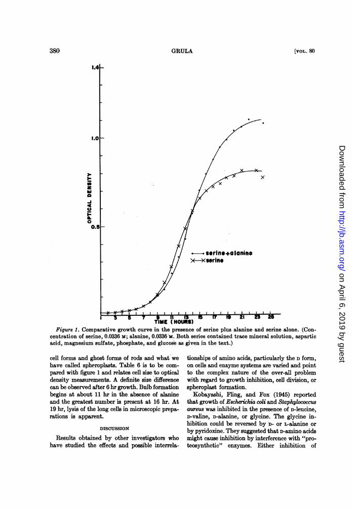
Comparative growth curves showilng alaninereversal of serine inhibition are shown in figure1. Actually, during the first 12 to 14 hr, growth isgreater in the absence of alanine. It is to be em-
phasized that these curves are based on opticaldensity measurements and cannot therefore berelated to cell division. Growth curves based on
colony counts are difficult to make using longcells, since the cells undergo a certain amount oflysis in the growth medium after about 12 hr.Also, plating diluents used thus far (physiologicalsaline or 0.1 per cent peptone water) permit toomuch lysis of long cells for accurate counting.Figures 2 to 9 compare cells at differenttimes during their growth and also show swollen
TABLE 5Reversal of the DL-serine effect at $ different levels
using varying amounts of DL-a-alaninein the presence of 0.0166 X glucose*
Concn DL- Concn DL- ODt Cell Size inuSerine a-Alanine
0.0168 - 0.73 20-2000.0168 0.00656 0.88 4-5; 20% 4-100.0168 0.0164 0.61 4-50.0168 0.0328 0.57 4.00.0168 0.0656 0.44 3-40.0336 - 0.26 30-3000.0336 0.00656 0.51 5.0; 20% 10-300.0336 0.0164 0.44 2-6; 10% 10-200.0336 0.0328 0.39 3.00.0336 0.0656 0.30 2-30.0672 - 0.013 30-3000.0672 0.00656 0.17 50% 5-10; 50% 20-300.0672 0.0164 0.20 4-7; 40% 10-200.0672 0.0328 0.15 3-5; 20% 5-200.0672 0.0656 0.09 3-5
0.0328 0.29 3-40.90 6-10; some to 30
* All tubes contained trace mineral solution,aspartic acid, magnesium sulfate and phosphateas given in the text.
t Optical density read at 15 hr; inoculum 0.33OD at 540 mIA.
on April 6, 2019 by guest
http://jb.asm.org/
Dow
nloaded from
GRULA
3-1
hiaAI4t
0.
0-
x
- serine+alannine>*-4rine
Figure 1. Comparative growth curve in the presence of serine plus alanine and serine alone. (Con-centration of serine, 0.0336 M; alanine, 0.0336 M. Both series contained trace mineral solution, asparticacid, magnesium sulfate, phosphate, and glucose as given in the text.)
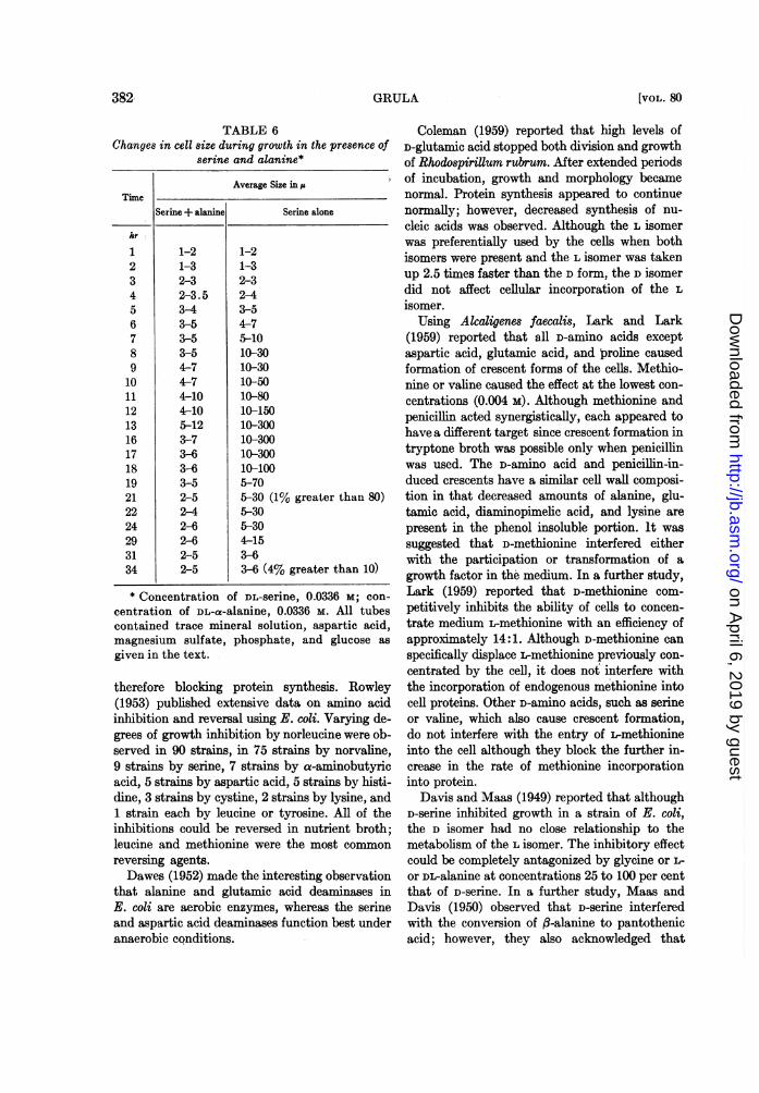
cell forms and ghost forms of rods and what wehave called spheroplasts. Table 6 is to be com-pared with figure 1 and relates cell size to opticaldensity measurements. A definite size differencecan be observed after 6 hr growth. Bulb formationbegins at about 11 hr in the, absence of alanineand the greatest number is present at 16 hr. At19 hr, lysis of the long celLs in microscopic prepa-rations is apparent.
DISCUSSION
Results obtained by other investigators whohave studied the effects and possible interrela-
tionships of amino acids, particularly the D form,on cells and enzyme systems are varied and pointto the complex nature of the over-all problemwith regard to growth inhibition, cell division, orspheroplast formation.
Kobayashi, Fling, and Fox (1945) reportedthat growth of Escherichia coli and Staphylococcusaureus was inhibited in the presence of D-leucine,D-valine, D-alanine, or glycine. The glycine in-hibition could be reversed by D- or -alanine orby pyridoxine. They suggested that D-amino acidsmight cause inhibition by interference with "pro-teosynthetic" enzymes. Either inhibition of
380 [VOL. 830
on April 6, 2019 by guest
http://jb.asm.org/
Dow
nloaded from
CELL DIVISION IN ERWINIA. II
M '
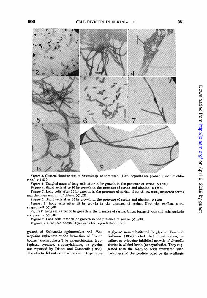
Figure B. Control showing size of Erwinia sp. at zero time. (Dark deposits are probably sodium chlo-ride.) X1,230.
Figure S. Tangled mass of long cells after 10 hr growth in the presence of serine. X1,230.Figure 4. Short cells after 10 hr growth in the presence of serine and alanine. X1,230.Figure 5. Long cells after 35 hr growth in the presence of serine. Note the swollen, distorted forms
and the large amount of debris. X1,230.Figure 6. Short cells after 35 hr growth in the presence of serine and alanine. XI ,230.Figure. 7. Long cells after 35 hr growth in the presence of serine. Note the swollen, club-
shaped cell. X1,230.Figure 8. Long cells after 56 hr growth in the presence of serine. Ghost forms of rods and spheroplasts
are present. X1,230.Figure 9. Long cells after 24 hr growth in the presence of serine. X 1,230.Figures 2-9 reduced about 32 per cent for reproduction here.
growth of Salmonella typhimurium and Hae-mophilus influenzae or the formation of "roundbodies" (spheroplasts?) by DL-methionine, tryp-tophan, tyrosine, L-phenylalanine, or glycinewas reported by Dienes and Zamecnik (1952).The effects did not occur when di- or tripeptides
of glycine were substituted for glycine. Yaw andKakawas (1952) noted that D-methiornne, D-valine, or D-leucine inhibited growth of Brucellaabortus in Albimi broth (nonsynthetic). They sug-gested that the D-amino acids interfered withhydrolysis of the peptide bond or its synthesis
19601 381
on April 6, 2019 by guest
http://jb.asm.org/
Dow
nloaded from
GRULA
TABLE 6Changes in cell size during growth in the presence of
serine and alanine*
Average Size in ,
TimeSerine+ alanine Serine alone
hr
1 1-2 1-22 1-3 1-33 2-3 2-34 2-3.5 2-45 3-4 3-56 3-5 4-77 3-5 5-108 3-5 10-309 4-7 10-3010 4-7 10-5011 4-10 10-8012 4-10 10-15013 5-12 10-30016 3-7 10-30017 3-6 10-30018 3-6 10-10019 3-5 5-7021 2-5 5-30 (1% greater than 80)22 2-4 5-3024 2-6 5-3029 2-6 4-1531 2-5 3-634 2-5 3-6 (4% greater than 10)
* Concentration of DL-serine, 0.0336 M; con-centration of DL-a-alanine, 0.0336 M. All tubescontained trace mineral solution, aspartic acid,magnesium sulfate, phosphate, and glucose asgiven in the text.
therefore blocking protein synthesis. Rowley(1953) published extensive data on amino acidinhibition and reversal using E. coli. Varying de-grees of growth inhibition by norleucine were ob-served in 90 strains, in 75 strains by norvaline,9 strains by serine, 7 strains by a-aminobutyricacid, 5 strains by aspartic acid, 5 strains by histi-dine, 3 strains by cystine, 2 strains by lysine, and1 strain each by leucine or tyrosine. All of theinhibitions could be reversed in nutrient broth;leucine and methionine were the most commonreversing agents.Dawes (1952) made the interesting observation
that alanine and glutamic acid deaminases inE. coli are aerobic enzymes, whereas the serineand aspartic acid deaminases function best underanaerobic conditions.
Coleman (1959) reported that high levels ofD-glutamic acid stopped both division and growthof Rhodospirillum rubrum. After extended periodsof incubation, growth and morphology becamenormal. Protein synthesis appeared to continuenormally; however, decreased synthesis of nu-cleic acids was observed. Although the L isomerwas preferentially used by the cells when bothisomers were present and the L isomer was takenup 2.5 times faster than the D form, the D isomerdid not affect cellular incorporation of the Lisomer.
Using Alcaligenes faecalis, Lark and Lark(1959) reported that all D-amino acids exceptaspartic acid, glutamic acid, and proline causedformation of crescent forms of the cells. Methio-nine or valine caused the effect at the lowest con-centrations (0.004 M). Although methionine andpenicillin acted synergistically, each appeared tohave a different target since crescent formation intryptone broth was possible only when penicillinwas used. The D-amino acid and penicillin-in-duced crescents have a similar cell wall composi-tion in that decreased amounts of alanine, glu-tamic acid, diaminopimelic acid, and lysine arepresent in the phenol insoluble portion. It wassuggested that D-methionine interfered eitherwith the participation or transformation of agrowth factor in the medium. In a further study,Lark (1959) reported that D-methionine com-petitively inhibits the ability of cells to concen-trate medium L-methionine with an efficiency ofapproximately 14:1. Although D-methionine canspecifically displace L-methionine previously con-centrated by the cell, it does not interfere withthe incorporation of endogenous methionine intocell proteins. Other D-amino acids, such as serineor valine, which also cause crescent formation,do not interfere with the entry of L-methionineinto the cell although they block the further in-crease in the rate of methionine incorporationinto protein.
Davis and Maas (1949) reported that althoughD-serine inhibited growth in a strain of E. coli,the D isomer had no close relationship to themetabolism of the L isomer. The inhibitory effectcould be completely antagonized by glycine or L-or DL-alanine at concentrations 25 to 100 per centthat of D-serine. In a further study, Maas andDavis (1950) observed that D-serine interferedwith the conversion of fl-alanine to pantothenicacid; however, they also acknowledged that
382, [VOL. 80
on April 6, 2019 by guest
http://jb.asm.org/
Dow
nloaded from
CELL DIVISION IN ERWINIA. II
higher levels of serine affected metabolic processesother than those involving pantothenic acid syn-thesis. Their strain was not able to convert D-serine to pyruvic acid, could not readilydeaminate D-serine, and ethanolamine did notinhibit growth at a level of 200 ,ug/ml.
Mueller and Miller (1949) reported that D-serine decreased the yield titer of tetanus toxin,and Kavanagh, Tunin, and Wild (1958) reportthe increased biosynthesis of cephalosporin N(synnematin B) when D-methionine is added tothe growth medium. Although exotic substitutedamino acids were used, Martel and Berlinguet(1959) report impairment by some on the de-velopment of transplantable Novikoff hepatomatumor cells.
Eisenstadt et al. (1959) reported that D-asparticacid inhibited the formation of amylase in cellsof Pseudomonas saccharophila grown in the pres-ence of maltose, sucrose, or cellobiose. The in-hibition could be reversed completely when theratio of L- to D-aspartic acid was 6:1. The in-hibition could also be reversed by adenosine butnot inosine and appeared to be due to the inhibi-tion of adenosine monophosphate formationwhich, in turn, affected the synthesis of adenosinetriphosphate, amino acid activation, and synthe-sis of ribonucleic acid.A direct effect by a D-amino acid on an enzyme
has been studied by Murachi and Tashiro (1958).They observed that the D-isomer of lysine in-hibited D-amino acid oxidase by competing withthe substrate D-amino acid for the apo-oxidaseprotein.Normal synthesis of cellular constituents spe-
cifically requiring D-amino acids did not receiveserious consideration until D-isomers were foundin bacterial cell walls. Recently, a D-alanine ac-tivating enzyme has been reported by Baddileyand Neuhaus (1959).The synthesis of vitamin B6 by mutants of E.
coli involves serine, glycine, and glycoaldehyde,particularly at 37 C (Morris, 1959). He suggeststhat interference with B6 biosynthesis may beonly one consequence of the presence of an in-complete metabolic block which also controls thesynthesis of serine and glycine.
Andrejew, Gernez-Rieux, and Tacquet (1958)reported that catalase of mycobacteria and beefliver was inhibited by D-cycloserine. Shockman(1959) was able to reverse the effects of cyclo-serine on cell wall synthesis in Streptococcus
faecalis with D but not the L form of alanine. Hesuggested that the competition between cyclo-serine and D-alanine might take place at the siteof incorporation of D-alanine into a wall precursor.Park (1958) suggested that cycloserine might pre-vent normal incorporation of D-alanine into thecell wall.
Because D- or L-alanine can reverse D-serine inwhat appears to be a competitive manner in thesecells, and because some spheroplast formationoccurs in the presence of D-amino acids, it wouldappear that some inhibition of cell wall synthesisis occurring. It should be pointed out, however,that cell division is inhibited and the cells becomequite long hours before spheroplast formation isevident and, also, many filaments never form anyspheroplasts. Because cell wall and other analyseshave not yet been done, it is too early to say thatthe effect of D-amino acids in this system is adirect one which will relate only to uniform cellwall synthesis. The relationships between cellwall synthesis and cell division remain obscure.Although the accumulation of uridine-aminosugar-peptides in the presence of penicillin andinterference with cell wall synthesis leading tospheroplast formation have been elegantly de-scribed (Park and Strominger, 1957; Strominger,Park, and Thompson, 1959; Lederberg, 1957), therelationship of these amino sugar-peptides to celldivision remains to be explained. It is well knownthat penicillin, for example, will also inhibit celldivision causing formation of filamentous cellswithout concomitant formation of spheroplasts.Is it possible that two somewhat "different"cell walls are synthesized by a cell, one being thewall that is laid down only in the division zone,the other being cell wall that is synthesized atother growing areas or a growing point?
ACKNOWLEDGMENT
The writer wishes to acknowledge the aid ofMr. Fred Rowe of Research and Development,Continental Oil Company, Ponca City, Okla-homa, in preparation of the electron micrographs.
SUMMARY
The D forms of serine, methionine, phenylal-anine, threonine, tryptophan, or histidine eitheralone or in combination with glucose, profoundlyinhibit cell division in a species of Erwiniagrowing in a chemically defined medium. Higherlevels of the D-amino acid can also completely
1960] 383
on April 6, 2019 by guest
http://jb.asm.org/
Dow
nloaded from
384 GRULA
inhibit growth. Division inhibition by serine canbe reversed almost completely by inorganic am-monium salts, D- or L-alanine, or p-aminoben-zoic acid and, to a lesser extent, by other com-pounds such as guanine, L-valine, L-isoleucine, orglutamine. Reversal of division inhibition byalanine appears to be competitive. Substitutionin any of 3 positions in the serine molecule negatesdivision inhibition by serine.
REFERENCES
ANDREJEW, A., CH. GERNEZ-RIEUX, ANDA. TACQUET 1958 L'effect de la D-CYCIO-serine et de L'INH sur l'activit6 catalasiquedes mycobacteries. Biochim. et Biophys.Acta, 30, 102-111.
BACHMANN, B. J., AND D. M. BONNER 1959 Pro-toplasts from Neurospora crassa. J. Bac-teriol., 78, 550-556.
BADDILEY, J., AND F. C. NEIUHAUS 1959 Theenzymic activation of D-alanine in Lactobacil-lus arabinosus 17-5. Biochim. et Biophys.Acta, 33, 277-279.
COLEMAN, G. S. 1959 The effect of DL-glutamicacid on the growth of Rhodospirillum rubrum.Biochim. et Biophys. Acta, 31, 55-65.
DAVIS, B. D., AND W. K. MAAS 1949 Inhibitionof Escherichia coli by D-serine and the produc-tion of serine-resistant mutants. J. Am.Chem. Soc., 71, 1865-1866.
DAWES, E. A. 1952 Observations on the growthof Escherichia coli in media containing aminoacids as the sole source of nitrogen. J.Bacteriol., 63, 647-660.
DIENES, L., AND P. C. ZAMECNIK 1952 Trans-formation of bacteria into L forms by aminoacids. J. Bacteriol., 64, 770-771.
EISENSTADT, J. M., L. GROSSMAN, AND H. P.KLEIN 1959 Inhibition of protein synthesisby D-aspartate and a possible site of its action.Biochim. et Biophys. Acta, 36, 292-294.
GRULA, E. A. 1960 Cell division in a species ofErwinia. I. Initial observations relating tonutritional dependency. J. Bacteriol., 80,369-374.
HASE, E., H. OTSUKA, S. MIHARA, AND H. TAMIYA1959 Role of sulfur in the cell division ofchlorella, studied by the technique of syn-chronous culture. Biochim. et Biophys.Acta, 35, 180-189.
HENDERSON, L. M., AND E. E. SNELL 1948 Auniform medium for determination of aminoacids with various microorganisms. J. Biol.Chem., 172, 15-29.
KAVANAGH, F., D. TUNIN, AND G. WILD 1958
[VOL. 80
D-methionine and the biosynthesis of cephalo-sporin N. Arch. Biochem. Biophys., 77,268-274.
KOBAYASHI, Y., M. FLING, AND S. W. Fox 1945Antipodal specificity in the inhibition ofgrowth of Escherichia coli by amino acids. J.Biol. Chem., 174, 391-398.
LARK, K. G. 1959 Isotopic competition betweenD- and L-methionine in Alcaligenes faecalis.Can. J. Microbiol., 5, 381-394.
LARK, C., AND K. G. LARK 1959 The effects ofD-amino acids on Alcaligenes fecalis. Can.J. Microbiol., 5, 369-379.
LEDERBERG, J. 1957 Mechanism of action ofpenicillin. J. Bacteriol., 73, 144.
MAAS, W. K., AND B. D. DAVIS 1950 Panto-thenate studies. I. Interference by D-serineand L-aspartic acid with pantothenate syn-thesis in Escherichia coli. J. Bacteriol., 60,733-745.
MARTEL, F., AND L. BERLINGUET 1959 Impair-ment of tumor growth by unnatural aminoacids. Can. J. Biochem. Physiol., 37, 433-439.
MAZIA, D. 1954 SH and growth. In Symposiumon glutathione, pp. 209-228. Academic Press,Inc., New York.
MORRIS, J. G. 1959 The synthesis of vitaminB6 by some mutant strains of Escherichia coli.J. Gen. Microbiol., 20, 597-604.
MUELLER, J. H., AND P. A. MILLER 1949 Inhibi-tion of tetanus toxin formation by D-serine.J. Am. Chem. Soc., 71, 1865-1866.
MURACHI, T., AND M. TASHIRO 1958 The inhibi-tion of D-amino acid oxidase by D-lysine.Biochim. et Biophys. Acta, 29, 645-646.
NICKERSON, W. J., AND G. FALCONE 1956 Iden-tification of protein disulfide reductase as acellular division enzyme in yeast. Science,124, 722-723.
PARK, J. T. 1958 Inhibition of cell-wall synthe-sis in Staphylococcus aureus by chemicalswhich cause accumulation of wall precursors.Biochem. J., 70, 2P.
PARK, J. T., AND J. L. STROMINGER 1957 Modeof action of penicillin. Science, 125, 99-101.
ROWLEY, D. 1953 Inhibition of E. coli strainsby amino acids. Nature, 171, 80-81.
SHOCKMAN, G. D. 1959 Reversal of cycloserineinhibition by D-alanine. Proc. Soc. Exptl.Biol. Med., 101, 693-695.
STARR, M. P. 1946 The nutrition of phyto-pathogenic bacteria. I. Minimal nutritiverequirements of the genus Xanthomonas.J. Bacteriol., 51, 131-143.
STARR, M. P., AND M. MANDEL 1950 The nutri-tion of phytopathogenic bacteria. IV. Mini-
on April 6, 2019 by guest
http://jb.asm.org/
Dow
nloaded from
CELL DIVISION IN ERWINIA. II
mal nutritive requirements of the genus
Erwinia. J. Bacteriol., 60, 669-672.STERN, H. 1956 Sulfhydryl groups and cell
division. Science, 124, 1292-1293.STROMINGER, J. L., J. T. PARK, AND R. E. THOMP-
SON 1959 Composition of cell wall of
Staphylococcus aureus; its relationship to themechanism of action of penicillin. J. Biol.Chem., 234, 3263-3268.
YAW, E. K., AND J. C. KAKAWAS 1952 Studieson the effects of D-amino acids on Brucellaabortus. J. Bacteriol., 63, 263-268.
19601 385
on April 6, 2019 by guest
http://jb.asm.org/
Dow
nloaded from


![RESEARCHARTICLE CompetitionandHabitatQualityInfluence ...Introduction Habitatrequirementsdiffer formanymigratory birdspecies betweenthebreeding and non-breeding season[1,2]and aspecies](https://img.pdfslide.us/doc/110x75/613e497659df642846166ea7/researcharticle-competitionandhabitatqualityinfluence-introduction-habitatrequirementsdiffer.jpg)