Embed Size (px)
Citation preview
DIAGNOSTIC RADIOLOGY RESIDENCY ULTRASOUND CURRICULUM
From the curriculum developed by: THE SOCIETY OF RADIOLOGISTS IN ULTRASOUND Curriculum Committee Rick I. Feld, M.D., Chair Teresita L. Angtuaco, M. D. , Edward I. Bluth, M.D., John K. Crowe, M.D , Theodore Dubinsky, M.D. , Sujata Ghate, M.D. , Robert D. Harris, M.D(DHMC) , Beth C. Kleiner, M.D. , Christopher R. B. Merritt, M.D. , Harriet J. Paltiel, M.D. , John Pellerito, M.D. , Leslie M .Scoutt, M.D. , Sheila Sheth, M.D. , Therese M. Weber, M.D. , Beverly G. Coleman, M.D., Ex- Officio , Douglas L. Brown, MD. Ex-Officio, Ulrike M. Hamper, M.D., Executive Board Liaison, Peter M. Doubilet, M.D.,Ph.D,. Executive Board Liaison
This ultrasound curriculum is intended to serve as a guideline for diagnostic radiology residency training programs, utilizing the goals and objectives format required by the American Council of Graduate Medical Education (ACGME).
At the conclusion of each one-month rotation, the resident should be able to demonstrate competence in these six specific areas: medical knowledge, patient care, practice-based learning and improvement, interpersonal and communication skills, professionalism, and systems-based practice, as outlined below.
The resident should understand this material through a “hands-on” clinical experience, including ultrasound scanning, combined with formal didactic teaching such as conferences, and independent learning utilizing teaching files, textbooks, and on-line electronic web-based tools such as medical journal articles, etc.
There are two parts to the medical knowledge portion of the curriculum. The first, Section A, lists hands-on scanning objectives to be mastered by the end of each clinical rotation. The second, Section B, is a more comprehensive list of entities that the resident should understand by the end of each rotation. Each institution may have its own individual system for acquiring this knowledge base.
1. Vascular:
--Lower extremity DVT.
Core Curriculum
--Abdomen: venous thrombosis (hepatic and renal veins, inferior vena cava); portal hypertension; TIPS; renal stenosis. --Aortic diseases (aneurysm) --Carotid artery duplex evaluation (vascular surgery rotation) --Complications of percutaneous vascular interventions: pseudoaneurysm and AV fistula --Upper extremity DVT -- vascular characterization of a mass
2. Biliary Tree and Gall Bladder --Normal anatomy --Biliary obstruction : benign and malignant causes

--Congenital anomalies of the common bile duct—choledochal cysts --Gallstones --Inflammatory disease of the gall bladder—acute cholecystitis and complications --Gall bladder carcinoma --Cholecystoses
3. Liver and Spleen --Normal anatomy --Neoplastic disease—hepatoma, metastases, cysts. --Infection—Abscess --Cirrhosis and portal hypertension. --Other parenchymal disease (steatosis) --Trauma---biloma, hematoma
4.Pancreas --Normal anatomy --Inflammatory disease
Complications of acute pancreatitis--pseudocyst Chronic pancreatitis Ductal calculi
--Neoplasms: adenocarcinoma, micro- and macro cystic adenomas, IPMN
5. Gastrointestinal --Appendicitis --Inflammatory bowel processes—colitis, Crohn’s
Disease --Bowel Obstruction
Bowel related mass: neoplasm, phlegmon 6. Kidney
--Normal anatomy --Congenital Anomalies: ectopia, horseshoe kidney, duplex collecting system, UPJ
obstruction --Infection: renal abscess and pyonephrosis --Calculous disease -- Neoplasms: renal cell carcinoma, transitional cell carcinoma, angiomyolipoma,
metastasis, cystic neoplasms. --Cystic Diseases: cortical cysts, polycystic kidney disease, medullary cysts. --Hydronephrosis
7. Retroperitoneum --Anatomy: perirenal and pararenal spaces, pelvic extraperitoneal spaces. -- Adrenal neoplasms and other masses --Lymphadenopathy --Neoplasms --Hematoma --Abscess
8. Peritoneal cavity --Anatomy --Intraperitoneal collections: ascites and abscess
--Neoplasm: carcinomatosis --Hemoperitoneum --Peritoneal inclusion cyst
9. Gynecology --Normal Anatomy --Uterus
Cyclic change in the endometrium Leiomyoma Leiomysarcoma Adenomyosis Endometrial pathology: carcinoma, polyp, hyperplasia, changes related to tamoxifan Gestational trophoblastic disease Postpartum—retained products of conception Cervical carcinoma Intrauterine contraceptive device
--Adnexa Polycystic ovarian syndrome Ovarian cysts; follicular, corpus luteum, theca lutein Acute conditions
Hemorrhagic cyst Ovarian torsion Ectopic pregnancy
Neoplasms: Surface epithelial—cystadenoma/carcinoma Germ Cell—teratoma Metastatic (Sex cord/stromal)
Pelvic inflammatory disease Hydro/pyo salpinx Tuboovarian abscess and complex
10. Thyroid/Head and Neck --Normal Anatomy --Nodules: patterns suggestive of benignity --Neoplasms --Nodular goiter and other conditions such as Thyroiditis --Parathyroid mass: adenoma --Submandibular and parotid gland masses
Benign and malignant lymph nodes 11. Scrotum
--Normal Anatomy --Neoplasms
Seminoma and other germ cell tumors Metastastic
--Benign masses
Simple cysts Epidermoid cysts Epididymal cysts and sperm granuloma
--Infections Epididymitis/orchitis Testicular and scrotal abscess/pyocele Fournier’s gangrene
--Trauma Testicular hematoma/rupture Scrotal wall hematoma Hematocele
--Varicocele --Torsion --Hernia --Extratesticular masses: spermatocele, adenomatoid tumor
12. Obstetrics First Trimester
--Diagnosis of intrauterine pregnancy and relationship to bHCG levels --Basic anatomy and normal developmental features of embryo and early fetus --Dating—CRL --Complications
Ectopic pregnancy Anembryonic pregnancy Subchorionic hematoma Intrauterine demise
--Anomalies identifiable in first trimester Second and third trimester
--Basic evaluation --Anatomy—Guidelines of AIUM, ACR, ACOG
kidneys, stomach, urinary bladder, umbilical cord insertion, spine, cerebral ventricles and posterior fossa, 4-chambered heart
--Dating—BPD, FL, AC, --Growth—EFW --Placenta, location and morphology --Amniotic Fluid --Cervix—status, length, confirmation of IUG --Diagnosis of chromosomal abnormalities
Biochemical screening Genetic sonogram—markers of aneuploidy
--Fetal anomalies CNS Thorax/cardiac Genitourinary Gastrointestinal MSK

I. MEDICAL KNOWLEDGE
A. “HANDS-ON” SCANNING By the end of each level of training, the resident should be able to scan most of the clinical scenarios listed below in each training category. Rotation 1 Gallbladder (gallstones/acute cholecystitis) Liver (masses) Kidney (hydronephrosis, stones) Transabdominal/transvaginal pelvis (mass/cyst/free fluid) Abdominal aorta (aneurysm) Pleural effusion and ascites Pregnancy (normal early intrauterine pregnancy) Thyroid nodules Rotation 2 Pancreas (pancreatitis, mass) Biliary (common bile duct, biliary ductal dilatation) Abdominal mass/adenopathy Kidney (mass/cyst) Basic Doppler (portal vein, pseudoanerysm, arteriovenous fistula) Pregnancy (first trimester, failed pregnancy, ectopic pregnancy) Adnexal mass (ovarian and non-ovarian) Testis (pain and masses) Basics obstetrics (basic fetal biometry, basic second/third trimester fetal anatomy, placental localization, amniotic fluid volume) Neonatal brain Rotation 3 Advanced obstetrics (comprehensive second/third trimester) Pediatrics (abdomen, spine, hips) Ultrasound-guided interventional procedures Parathyroid Advanced abdominal Doppler (visceral organs, organ transplants) Rotation 4 Advanced obstetrics (comprehensive second/third trimester) Pediatrics (abdomen, spine, hips) Ultrasound-guided interventional procedures Parathyroid Carotid artery Advanced abdominal Doppler (visceral organs, organ transplants) Peripheral vessels (arterial bypass grafts, upper extremity veins) Lower extremity (deep vein thrombosis)
B. COMPREHENSIVE KNOWLEDGE
PHYSICS/INSTRUMENTATION The resident should understand the basic principles of physics that form the foundation of clinical ultrasound. Rotation 1 Define ultrasound, including the relationship of sound waves used in imaging Straight narrow sound beams, simple reflection, constant sound speed Beam shape: linear, sector, curved array Probes: transabdominal, endocavitary Endocavitary imaging: transvaginal, transrectal, endoscopic, laparoscopic Display: Gray scale, M-mode, pulsed wave Doppler, color and power Doppler Image orientation: standard images in different planes Image optimization: power output, gain, time gain compensation Image recording options: electronic (digital), film, paper Acoustic properties of fluid, cyst, calcification, complex fluid and solid structures Tissue characteristics: acoustic shadowing and enhancement Focal zone Rotation 2 Transducer choice: curvilinear, linear, sector, vector Frequency, sound speed, wavelength, intensity, decibels, beam width, Fresnel zone, Fraunhoffer zone Interaction of sound waves with tissues: reflection, attenuation, scattering, refraction, absorption, acoustic impedance pulse-echo principles Generation/detection of ultrasound waves Doppler phenomenon, Doppler formula Beam formation/focusing Gray scale, M-mode, pulsed wave Doppler, color Doppler imaging, power Doppler imaging Rotation 3 Beamwidth, sidelobe, slice thickness artifacts Multiple reflection artifacts - mirror image/reverberation Refractive artifacts Doppler artifacts- pulse wave, color imaging, including aliasing Gray scale versus Doppler (trade-off of penetration and resolution) 3-D volumetric imaging Thermal/non-thermal effects on tissue: biological health risks Image optimization Harmonic imaging Equipment quality assurance: phantoms, spatial/contrast resolution Rotation 4 Beamwidth, sidelobe, slice thickness artifacts Multiple reflection artifacts - mirror image/reverberation Refractive artifacts Doppler artifacts- pulse wave, color imaging, including aliasing Gray scale versus Doppler (trade-off of penetration and resolution) 3-D volumetric imaging Thermal/non-thermal effects on tissue: biological health risks Image optimization
Harmonic imaging Ultrasound contrast agents Equipment quality assurance: phantoms, spatial/contrast resolution
CLINICAL APPLICATIONS GENERAL The resident should understand the importance of clinical ultrasound protocols. Published protocols/standards from the American College of Radiology (ACR) or the American Institute of Ultrasound in Medicine (AIUM) with or without local modification are acceptable frames of reference. Residents should also be familiar with ACR appropriateness criteria as a guide for appropriate clinical use of ultrasound and other imaging modalities. The resident should gain a general understanding of both the clinical uses and limitations of ultrasound as well as the appropriate integration of other complementary cross-sectional imaging studies, particularly CT and MRI. The resident should understand the importance of documentation and reporting skills/ requirements, including the electronic applications in their institution. The resident should understand the importance of clinical quality assurance, including radiologic-pathologic correlation, as well as sonographer-physician discrepancies. ABDOMINAL Rotation 1 Liver: normal echotexture, size, and shape (including anatomic variants), diffuse disease, (fatty infiltration, acute and chronic hepatitis, cirrhosis, edema), focal masses, metastases, granuloma Gallbladder: normal appearance, wall thickening, gallstones, including supine, decubitus and erect positions, sludge, acute cholecystitis (calculous/acalculous), sonographic Murphy’s sign, other etiologies of wall thickening, polyp Bile ducts: normal intra- and extrahepatic bile duct diameters and dilatation Pancreas: normal anatomy, pancreatic duct, mass Spleen: normal echotexture, size and shape (including anatomic variants), focal masses (cystic versus solid), lymphoma, abscess, infarction, granuloma Peritoneal cavity: ascites, fluid localization/quantification (free/loculated)
Pleural effusion
Rotation 2 Liver: hematoma, biloma, abscess Post-liver transplantation collections: hematoma, biloma, abscess (see vascular section) Gallbladder: hyperplastic cholecystoses, carcinoma Bile ducts: bile duct stones, inflammatory disease, cholangitis, pneumobilia Pancreas: neoplasm, cysts Pancreatitis complications: abscess, pseudocyst and pseudoaneurysm, chronic pancreatitis Peritoneal cavity: abscess, hemorrhage, omental mass, metastasis, carcinomatosis Spleen: varices Rotation 3 Liver: trauma Bile ducts: neoplasm (cholangiocarcinoma) Spleen: trauma Chest: pericardial effusion, mass, atelectasis/pneumonia
Organ transplants: see vascular section Gastrointestinal tract: normal gut ultrasound signature, acute appendicitis, diverticulitis, Crohn’s disease Peritoneal cavity: free air Abdominal wall hernia, inguinal hernia Rotation 4 Liver: trauma Bile ducts: neoplasm (cholangiocarcinoma) Spleen: trauma Chest: pericardial effusion, mass, atelectasis/pneumonia Organ transplants: see vascular section Gastrointestinal tract: normal gut ultrasound signature, acute appendicitis, diverticulitis, Crohn’s disease Peritoneal cavity: free air Abdominal wall hernia, inguinal hernia
****************************************************************************** KIDNEYS, URINARY BLADDER AND PROSTATE Rotation 1 Renal: Normal renal cortical echotexture, size and shape, glomerulointerstitial renal disease, simple renal cyst , renal stones, hydronephrosis, pyonephrosis Ureters: hydroureter Urinary bladder: calculi, wall thickening, ureteral jets, bladder volume, including post-void residual Rotation 2 Abscess/pyelonephritis, perinephric fluid Post-renal transplant collections: hematoma, urinoma, abscess, lymphocele (see vascular section) Complex renal cyst, adult polycystic disease and acquired renal cystic disease, renal cell carcinoma, angiomyolipoma Urinary bladder: mass, infection, hemorrhage, wall thickening, bladder outlet obstruction, diverticula, ureterocele Transabdominal prostate Ureters: hydroureter Rotation 3 Kidneys: xanthogranulomatous pyelonephritis, emphysematous pyelonephritis, congenital anomalies, pelvic kidney (see pediatrics section), medullary nephrocalcinosis Adrenal glands: mass Retroperitoneum: adenopathy, mass Ureters: ureteral stone Urinary bladder: ectopic ureterocele Renal artery stenosis, renal vein thrombosis (see vascular section section) Transrectal prostate Rotation 4 Kidneys: xanthogranulomatous pyelonephritis, emphysematous pyelonephritis, congenital anomalies, pelvic kidney,medullary sponge kidney, nephrocalcinosis Adrenal glands: mass

Retroperitoneum: adenopathy, mass Ureters: ureteral stone Urinary bladder: ectopic ureterocele Renal artery stenosis, renal vein thrombosis (see vascular section section) Transrectal prostate with biopsy
************************************************************************
GYNECOLOGY Rotation 1 Uterus: normal size, shape, position, echogenicity, fibroid identification Endometrium: normal appearance during phases of menstrual cycle and thickness measurement (pre-menopausal, post-menopausal, effects of hormone replacement), intrauterine device, fluid Ovary: normal size, shape, echogenicity, physiologic variation during phases of menstrual cycle (follicles, corpus luteum, hemorrhagic ovarian cyst) Free pelvic fluid Rotation 2 Uterus: congenital anomalies, endometrial polyp, endometrial hyperplasia, endometrial carcinoma, endometritis, pyometrium, fibroid localization (submucous, intramural, subserosal), adenomyosis Ovarian cyst: hemorrhagic/ruptured cyst, endometrioma, polycystic ovarian disease, ovarian hyperstimulation syndrome Ovarian neoplasm: cystic/solid adnexal masses, cystadenoma/carcinoma, dermoid, fibroma, germ cell tumor, Doppler evaluation Ovarian torsion Pelvic inflammatory disease, tubo-ovarian abscess Cervix: mass, stenosis, endometrial obstruction Fallopian tube: hydrosalpinx, pyosalpinx Post-hysterectomy Rotation 3 Uterus: congenital anomalies, endometrial polyp, endometrial hyperplasia, endometrial carcinoma, endometritis, pyometrium, fibroid localization (submucous, intramural, subserosal), adenomyosis Ovarian cyst: hemorrhagic/ruptured cyst, endometrioma, polycystic ovarian disease, ovarian hyperstimulation syndrome Ovarian neoplasm: cystic/solid adnexal masses, cystadenoma/carcinoma, dermoid, fibroma, germ cell tumor, Doppler evaluation Peritoneal inclusion cyst Ovarian neoplasm and cancer staging Saline hysterosonography Rotation 4 Uterus: congenital anomalies, endometrial polyp, endometrial hyperplasia, endometrial carcinoma, endometritis, pyometrium, fibroid localization (submucous, intramural, subserosal), adenomyosis Ovarian cyst: hemorrhagic/ruptured cyst, endometrioma, polycystic ovarian disease, ovarian hyperstimulation syndrome Ovarian neoplasm: cystic/solid adnexal masses, cystadenoma/carcinoma, dermoid, fibroma, germ cell tumor, Doppler evaluation Peritoneal inclusion cyst Ovarian neoplasm and cancer staging Saline hysterosonography
****************************************************************************** OBSTETRICS
FIRST TRIMESTER Rotation 1 Normal findings: gestational sac appearance, size, gestational sac growth, yolk sac, embryo, cardiac activity including normal embryonic heart rate, amnion, chorion, normal early fetal anatomy/growth, crown-rump length measurement, correlation with BHCG levels and menstrual dates Rotation 2 Multiple gestations (chorionicity and amnionicity), failed early pregnancy, spontaneous complete/incomplete abortion, ectopic pregnancy, blighted ovum, embryonic death, subchorionic hematoma, gestational trophoblastic disease, gross embryonic structural abnormalities, anencephaly Rotation 3 Multiple gestations (chorionicity and amnionicity), failed early pregnancy, spontaneous complete/incomplete abortion, ectopic pregnancy, blighted ovum, embryonic death, subchorionic hematoma, gestational trophoblastic disease, gross and subtle embryonic structural abnormalities, anencephaly Unusual ectopic pregnancy: interstitial, cervical, ovarian, scar, abdominal, rudimentary horn Nuchal translucency Chorionic villous sampling Rotation 4 Multiple gestations (chorionicity and amnionicity), failed early pregnancy, spontaneous complete/incomplete abortion, ectopic pregnancy, blighted ovum, embryonic death, subchorionic hematoma, gestational trophoblastic disease, gross and subtle embryonic structural abnormalities, anencephaly Unusual ectopic pregnancy: interstitial, cervical, ovarian, scar, abdominal, rudimentary horn Nuchal translucency Chorionic villous sampling
SECOND AND THIRD TRIMESTER Rotation 1 Normal findings: normal fetal anatomy/situs/development, placenta, biometry, amniotic fluid volume, multiple gestations Anencephaly Oligohydramnios (spontaneous premature rupture of membranes, renal disease, fetal death, intrauterine growth retardation, infection) Polyhydramnios, placenta previa Cervical appearance and length Rotation 2 Recognition of fetal abnormalities that require high risk obstetrics referral, including intrauterine growth retardation, hydrops, holoprosencephaly, hydrocephalus, neural tube defects, multicystic dysplastic kidney, hydronephrosis Placental abruption, placental masses, two-vessel umbilical cord, cord masses, retained products of conception Rotation 3 Recognition of fetal abnormalities that require high risk obstetrics referral, including
congenital anomalies/chromosomal abnormalites and syndromes such as Down’s syndrome and Turner’s syndrome, hydrops, congenital infections, chest masses, cardiac malformations and arrhythmias, diaphragmatic hernia, abdominal wall defects, abdominal masses, gastrointestinal tract obstruction/abnormalities, ascites, skeletal dysplasias, cleft lip/palate, complications of twin pregnancy, hydrancephaly Borderline findings: nuchal thickening, choroid plexus cyst, echogenic cardiac focus, echogenic bowel, borderline hydrocephalus Placental cord insertion site/vasa previa, velamentous cord insertion, cord prolapse, succenturiate placenta, cervical incompetence Umbilical cord Doppler, fetal cranial Doppler, biophysical profile Guidance for amniocentesis Placenta accreta, percreta, increta Rotation 4 Recognition of fetal abnormalities that require high risk obstetrics referral, including congenital anomalies/chromosomal abnormalites and syndromes such as Down’s syndrome and Turner’s syndrome, hydrops, congenital infections, chest masses, cardiac malformations and arrhythmias, diaphragmatic hernia, abdominal wall defects, abdominal masses, gastrointestinal tract obstruction/abnormalities, ascites, skeletal dysplasias, cleft lip/palate, complications of twin pregnancy, hydrancephaly Borderline findings: nuchal thickening, choroid plexus cyst, echogenic cardiac focus, echogenic bowel, borderline hydrocephalus Placental cord insertion site/vasa previa, velamentous cord insertion, cord prolapse, succenturiate placenta, cervical incompetence Umbilical cord Doppler, fetal cranial Doppler, biophysical profile Guidance for amniocentesis Placenta accreta, percreta, increta
THYROID/NECK Rotation 1 Normal thyroid echotexture, size and shape Thyroid disease: diffuse and focal disease Multinodular thyroid Rotation 2 Thyroid nodule characterization: echotexture, calcifications including microcalcifications, margins, recommendations for fine needle aspiration biopsy Hashimoto’s thyroiditis/Graves’ disease Rotation 3 Parathyroid mass: adenoma Congenital cysts: branchial cleft Lymph nodes: benign and malignant characterization Post-thyroidectomy recurrence Submandibular and parotid glands: normal and abnormal Fine needle aspiration Rotation 4 Parathyroid mass: adenoma Congenital cysts: branchial cleft
Lymph nodes: benign and malignant characterization Post-thyroidectomy recurrence Submandibular and parotid glands: normal and abnormal Fine needle aspiration
****************************************************************************** VASCULAR/DOPPLER Rotation 1 Abdominal aorta: normal appearance and measurement, aneurysm Inferior vena cava: normal appearance, thrombosis Hematoma Iatrogenic pseudoaneurysm Rotation 2 Peripheral vascular aneurysm, including iliac and popliteal arteries Hepatic vasculature: pulsed Doppler and color Doppler imaging of the portal veins, splenic vein, hepatic arteries and hepatic veins, including normal direction of flow Hemodynamics of cirrhosis, portal hypertension and varices, portal vein thrombosis Upper extremity venous thrombosis: subclavian and internal jugular vein thrombosis, axillary and brachial vein thrombosis Carotid artery: normal, atherosclerotic plaque, carotid artery stenosis and occlusion Renal vein thrombosis Iatrogenic arteriovenous fistula Rotation 3 Renal transplant: arterial resistive index (rejection, acute tubular necrosis), transplant vein thrombosis, renal infarction, post-biopsy complications, renal arterial stenosis Liver transplants, including hepatic artery stenosis or thrombosis (resistive index), portal vein thrombosis, post-biopsy complications, inferior vena cava stenosis Pancreas transplant: arterial and venous anastomosis, patency and stenosis TIPS evaluation and complications Lower extremities: chronic venous insufficiency Arterial bypass graft Mesenteric ischemia Renal artery stenosis Rotation 4 Liver transplants, including hepatic artery stenosis or thrombosis (resistive index), portal vein thrombosis, post-biopsy complications, inferior vena cava stenosis Pancreas transplant: arterial and venous anastomosis, patency and stenosis TIPS evaluation and complications Vascular Lab/IR: Lower extremities: chronic venous insufficiency Arterial bypass graft Hemodialysis graft/fistula Carotid artery: waveform analysis, stenosis, dissection, pseudoaneurysm, stent Vertebral artery: subclavian steal syndrome Mesenteric ischemia Lower extremity deep vein thrombosis
Pre-graft vein mapping ****************************************************************************** SCROTUM Rotation 1 Testes: normal echotexture, shape and size Epididymes Testicular mass Hydrocele Rotation 2 Epididymitis, orchitis Testicular torsion Testicular mass characterization: microlithiasis, germ cell tumor, lymphoma, metastasis Cystic ectasia of rete testis Extratesticular masses/cysts, spermatocele, adenomatoid tumor, epidydimal head cyst Varicocele Trauma Rotation 3 Testicular mass characterization: microlithiasis, germ cell tumor, lymphoma, metastasis Cystic ectasia of rete testis Extratesticular masses/cysts, spermatocele, adenomatoid tumor, epidydimal head cyst Varicocele Hernia Non-descended testis Fournier’s gangrene trauma Rotation 4 Testicular mass characterization: microlithiasis, germ cell tumor, lymphoma, metastasis Cystic ectasia of rete testis Extratesticular masses/cysts, spermatocele, adenomatoid tumor, epidydimal head cyst Varicocele Fournier’s gangrene trauma
****************************************************************************** PEDIATRICS Rotation 1 Normal abdominal anatomy Normal renal anatomy Normal brain anatomy Normal neck anatomy Rotation 2 Brain: intracranial hemorrhage and complications, including periventricular leukomalacia and hydrocephalus, shunt evaluation Kidneys: hydronephrosis, stones, hydroureters, anomalies of position and fusion, renal scarring, masses, cystic disease
Adrenal hemorrhage, masses (neuroblastoma) Liver: cirrhosis, choledochal cysts, liver masses, hepatitis/biliary atresia Gallbladder: gallstones, biliary stones, hydrops Pancreatitis Normal hip Intussusception Acute appendicitis Acute pancreatitis Hypertrophic pyloric stenosis Scrotal: torsion, epdidymitis, orchitis, masses, undescended testis, mass , trauma Ovarian torsion Rotation 3 Organ transplant Polysplenia, asplenia Kidneys: hydronephrosis, stones, hydroureters, anomalies of position and fusion, renal scarring, masses, cystic disease Adrenal hemorrhage, masses (neuroblastoma) Liver: cirrhosis, choledochal cysts, liver masses, hepatitis/biliary atresia Hip dislocation Congenital brain malformations, agenesis of corpus callosum, vein of Galen aneurysm, Dandy Walker Malformation, aqueductal stenosis Neonatal spine: tethered cord, intraspinal mass Liver Doppler Imperforate hymen, uterine anomalies Rotation 4 Organ transplant Polysplenia, asplenia Hip dislocation Congenital brain malformations, agenesis of corpus callosum, vein of Galen aneurysm, Dandy Walker Malformation, aqueductal stenosis Neonatal spine: tethered cord, intraspinal mass Liver Doppler Imperforate hymen, uterine anomalies
****************************************************************************** MUSCULOSKELETAL Rotation 1 Mass Hematoma Baker’s cyst, including rupture Cellulitis Abscess Rotation 2 Normal tendon appearance Foreign body Soft tissue gas
Joint fluid Muscle tear Rotation 3 Tendon tear, inflammation Rotator cuff tear Rotation 4 Tendon tear, inflammation Rotator cuff tear Foreign bodies ****************************************************************************** BREAST Rotation 1 Sonomammographic anatomy Cystic versus solid mass Mastitis/abscess Rotation 2 Characterization of cysts Lymph node characterization: axillary, supraclavicular, intramammary Rotation 3 (There are three rotations in Mammography, including Breast US) Characterization of solid masses: benign versus malignant Architectural distortion Intraductal masses/abnormalities Galactocele Screening Multifocal malignancy Elastography ****************************************************************************** INTERVENTIONAL Rotation 1 Informed consent Sterile technique Localization of fluid for paracentesis or thoracentesis to be performed by another service Ultrasound-guided paracentesis Rotation 2 Pre-procedural evaluation: coagulation laboratory studies, anticoagulation medication Stratification of risk for percutaneous procedures Techniques for ultrasound-guided invasive procedures: understanding important landmarks and pitfalls of percutaneous procedures, including recognition of critical structures to be avoided Biopsy of soft tissue masses Random core liver biopsy Aspiration of fluid collections, cysts and catheter placement for abscess and fluid drainage (pleural, peritoneal and other spaces) Ultrasound-guided thoracentesis Post-procedural evaluation: radiographic studies, patient monitoring, management of complications Rotation 3
Aspiration of fluid collections, cysts and catheter placement for abscess and fluid drainage (pleural, peritoneal and other spaces) Ultrasound-guided thoracentesis Post-procedural evaluation: radiographic studies, patient monitoring, management of complications Fine needle biopsy versus core biopsy in specific application, such as focal liver mass, renal mass, thyroid/parathyroid mass, retroperitoneal lymphadenopathy Rotation 4 Aspiration of fluid collections, cysts and catheter placement for abscess and fluid drainage (pleural, peritoneal and other spaces) Ultrasound-guided thoracentesis Post-procedural evaluation: radiographic studies, patient monitoring, management of complications Fine needle biopsy versus core biopsy in specific application, such as focal liver mass, renal mass, thyroid/parathyroid mass, retroperitoneal lymphadenopathy Pseduoaneurysm management: contraindications and technique of non-surgical treatment with ultrasound-guided compression repair versus thrombin injection Intraoperative ultrasound guidance Assessment ACR In-Service exam ABR Exam Faculty Evaluations 360 degree evaluations Learning Portfolio Pre-call skills list (Rotation 1) Pre-call exam (Rotation 1)
II. PATIENT CARE Rotation 1 Gather essential and accurate clinical and radiologic information about patients relevant to the interpretation of the ultrasound examination Communicate effectively and demonstrate caring, respectful behavior when interacting with patients and their families, answering their questions and helping them to understand the ultrasound procedure as well as its clinical significance Use information technology to support patient care decisions Become familiar with common indications for ultrasound exams as well as limitation and capabilities of the modality for specific indications. Rotation 2 Communicate effectively and demonstrate caring, respectful behavior when interacting with patients and their families, answering their questions and helping them to understand the ultrasound procedure as well as its clinical significance Screen and supervise more complex ultrasound studies Understand the importance of the physician/patient interaction during an ultrasound examination Advise referring physicians (emergency staff) as to most appropriate diagnostic examinations. Expedite more urgent cases referred on an emergency basis Contact clinicians referring examinations to the ultrasound department if additional
information is needed. Review prior radiologic studies and clinical information Be responsive to individual patient needs. Rotation 3 Communicate effectively and demonstrate caring, respectful behavior when interacting with patients and their families, answering their questions and helping them to understand the ultrasound procedure as well as its clinical significance Screen and supervise, with increasing level of responsibility, most ultrasound studies Prioritize exams based on urgency Expedite cases Gather the pertinent information for interventional cases Understand the bioeffects and safety issues in diagnostic ultrasound Review prior radiologic studies and clinical information Be responsive to individual patient needs. Rotation 4 Communicate effectively and demonstrate caring, respectful behavior when interacting with patients and their families, answering their questions and helping them to understand the ultrasound procedure as well as its clinical significance Screen ultrasound requests for appropriateness. Interview patients for procedure as to allergies, medications, medical history Review prior radiologic studies and clinical information Be responsive to individual patient needs. Assessment Faculty evaluation 360 degree evaluation ACR In-Service Exam ABR Exam Learning Portfolio
III. PRACTICE-BASED LEARNING AND IMPROVEMENTS Rotation 1 Use information technology to manage information, to access on-line medical information, and for self learning Concentrate on acquiring technical competence in sonography. Observe the ultrasound technologists and backscan. Accompany the attending radiologist when he/she is obtaining additional views. List interesting cases on database Rotation 2 Use information technology to manage information, to access on-line medical information, and for self learning Demonstrate knowledge of principles of research methods, statistical methods, study design and their implementation Demonstrate critical assessment of the scientific literature Demonstrate knowledge and application of the principles of evidence-based medicine in practice List interesting cases on database Follow up on interesting cases, including post-surgical and post-biopsy
Rotation 3 Use information technology to manage information, to access on-line medical information, and for self learning Facilitate teaching of medical students, sonographers, other residents and other health care professionals Participate in quality assurance programs for sonographers and physicians Learn about equipment quality assurance programs Apply basic knowledge of study design and statistical methods to the appraisal of clinical studies and other information on diagnostic and therapeutic effectiveness List interesting cases on database Follow up on interesting cases, including post-surgical and post-biopsy
Rotation 4 Use information technology to manage information, to access on-line medical information, and for self learning Facilitate teaching of medical students, sonographers, other residents and other health care professionals Participate in quality assurance programs for sonographers and physicians Learn about equipment quality assurance programs Apply basic knowledge of study design and statistical methods to the appraisal of clinical studies and other information on diagnostic and therapeutic effectiveness List interesting cases on database Follow up on interesting cases, including post-surgical and post-biopsy
Assessment Faculty evaluation ACR in service examination ABR Exam Medical Student evaluation Learning Portfolio
IV. . INTERPERSONAL AND COMMUNICATION SKILLS Rotation 1 Dictate prompt, accurate and concise radiologic reports for basic ultrasound studies using available electronic software applications Develop effective communication skills with patients, patients’ families, physicians and other members of the health care team Promptly communicate urgent, critical or unexpected ultrasound findings to residents, referring physicians or clinicians and document the communication in the radiological report Provide basic explanations of ultrasound examinations to patients. Establish a working relationship with ultrasound technologists. Rotation 2 Interact with residents and attending physicians in consultation when clinical-radiologic correlation is necessary
Dictate accurate and concise radiologic reports for more complex ultrasound studies with concise impression including diagnosis and/or differential diagnoses Explain exams and, where appropriate, results to patients and families Rotation 3 Dictate accurate and concise reports for the most complex ultrasound studies with concise impression including diagnosis and/or differential diagnoses as well as recommendations for further imaging and/or management, when appropriate Consult effectively with senior residents and attending physician in most aspects of ultrasound Explain the exams and results to medical students and other learners Rotation 4 Dictate accurate and concise reports for all, including the most complex ultrasound studies Consult effectively with senior residents and attending physicians in most aspects of ultrasound Explain the exams and results to medical students and other learners Assessment Faculty Evaluation 360 degree Evaluation Medical Student Evaluation ACR In-Service Exam ABR Exam Dictation Review
V. PROFESSIONALISM Rotation 1 Demonstrate honor, integrity, respect and compassion to patients, other physicians and other health care professionals Demonstrate positive work habits, including punctuality and professional appearance Be discrete in discussing individual patient histories and findings. Respect individual ethnic or religious preferences. Rotation 2 Demonstrate a commitment to the ethical principles pertaining to confidentiality of patient information Demonstrate responsiveness to the needs of patients that supercedes self-interest (altruism) Demonstrate honor, integrity, respect and compassion to patients, other physicians and other health care professionals Demonstrate positive work habits, including punctuality and professional appearance Rotation 3 Demonstrate a commitment to the ethical principles pertaining to confidentiality of patient information Demonstrate responsiveness to the needs of patients that supercedes self-interest (altruism) Demonstrate honor, integrity, respect and compassion to patients, other physicians and other health care professionals Demonstrate positive work habits, including punctuality and professional appearance Demonstrate accountability to patients, society and the profession Rotation 4

Demonstrate a commitment to the ethical principles pertaining to confidentiality of patient information Demonstrate responsiveness to the needs of patients that supercedes self-interest (altruism) Demonstrate honor, integrity, respect and compassion to patients, other physicians and other health care professionals Demonstrate positive work habits, including punctuality and professional appearance Demonstrate accountability to patients, society and the profession
Assessment Faculty Evaluation 360 degree Evaluation Medical Student Evaluation ACR In-Service Exam ABR Exam Patient/Family Feedback, where appropriate
VI. SYSTEMS BASED PRACTICE Residents must demonstrate an awareness of and responsiveness to the larger context and system of health care and the ability to effectively call on system resources to provide care that is of optimal value. Rotation 1 Start to understand how their professional practice affects other health care professionals, the health care organization, and the larger society, and how these elements affect their own practice Assist referring clinicians in providing cost-effective health care Begin to learn about practice cost-effective health care and resource allocationBe prepared to Begin to evaluate the request for imaging as regards cost, effectiveness, and appropriateness, and to facilitate performance of an alternative study if indicated. Become familiar with the ACR Appropriateness Criteria Rotation 2 Understand how their professional practice affects other health care professionals, the health care organization, and the larger society Learn how these elements affect DHMC Help referring clinicians provide cost-effective health care practice cost-effective health care and resource allocation that does not compromise quality of care Be prepared to evaluate the US request for cost, effectiveness, and appropriateness Understand the ACR Appropriateness Criteria
Rotation 3 Understand how their professional practice affects other health care professionals, the health care organization, and the larger society Know how these elements affect their own practice Help referring clinicians provide cost-effective health care Practice cost-effective health care and resource allocation that does not compromise quality of care
Evaluate the US request for cost and effectiveness, and appropriateness Facilitate performance of an alternative study if indicated. Understand the ACR Appropriateness Criteria
Rotation 4 Understand how their professional practice affects other health care professionals, the health care organization, and the larger society Know how these elements affect their own practice Help referring clinicians provide cost-effective health care Practice cost-effective health care and resource allocation that does not compromise quality of care Evaluate the US request for cost, effectiveness, and appropriateness Facilitate performance of an alternative study if indicated. Understand the ACR Appropriateness Criteria
Assessment Faculty Evaluation 360 degree Evaluation Medical Student Evaluation ACR In-Service Exam ABR Exam Patient/Family Feedback, where appropriate
SUGGESTED READING Year I Rotations Requisites, Middleton, Kurtz, Hertzberg, 2nd ed. Ch 1-9, 13-20 Diagnostic Ultrasound, 2nd ed. McGahan, Goldberg Ch. 39
Year II Rotation Requisites Ch 6, 10, 12, 21-23 Diagnostic US Ch 25, 27-28, 32, 38
Year III Rotation Diagnostic U/S Ch 1-2, 11,15,40, 43-51
Year IV Rotation Diagnostic U/S Ch 4,5,7,10,12.18.19
Ultrasound Conferences
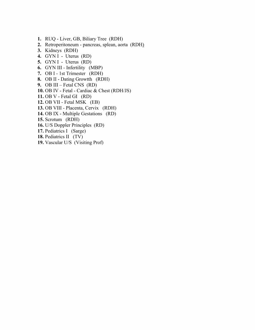
1. RUQ - Liver, GB, Biliary Tree (RDH) 2. Retroperitoneum - pancreas, splean, aorta (RDH) 3. Kidneys (RDH) 4. GYN I - Uterus (RD) 5. GYN I - Uterus (RD) 6. GYN III - Infertility (MBP) 7. OB I - 1st Trimester (RDH) 8. OB II - Dating Growtth (RDH) 9. OB III – Fetal CNS (RD) 10. OB IV - Fetal - Cardiac & Chest (RDH/JS) 11. OB V - Fetal GI (RD) 12. OB VII - Fetal MSK (EB) 13. OB VIII - Placenta, Cervix (RDH) 14. OB IX - Multiple Gestations (RD) 15. Scrotum (RDH) 16. U/S Doppler Principles (RD) 17. Pediatrics I (Sarge) 18. Pediatrics II (TV) 19. Vascular U/S (Visiting Prof)