Embed Size (px)
Citation preview
w
Journal of the American College of Cardiology Vol. 60, No. 6, 2012© 2012 by the American College of Cardiology Foundation ISSN 0735-1097/$36.00
STATE-OF-THE-ART PAPER
Current Status ofTranscatheter Aortic Valve Replacement
John G. Webb, MD, David A. Wood, MD
Vancouver, British Columbia, Canada
Surgical aortic valve replacement (SAVR) has long been the mainstay of therapy for severe aortic stenosis. How-ever, transcatheter aortic valve replacement (TAVR) is now generally accepted as the new standard of care forpatients with symptomatic aortic stenosis who are not candidates for open surgery. Arguably TAVR may also bea preferred alternative to SAVR in carefully selected high-risk, but still operable, patients in whom morbidity andmortality may be reduced. Although TAVR outcomes continue to improve, concerns remain with respect to vas-cular injury, stroke, paravalvular regurgitation, and valve durability. However, it seems likely that with ongoingrefinement of transcatheter valve systems, techniques, and patient selection TAVR is becoming an increasinglyappealing option for a much broader range of patients. Randomized trials and ongoing surveillance will play animportant role as we enter a new era of rigorous clinical evaluation for minimally invasive therapies for struc-tural heart disease. (J Am Coll Cardiol 2012;60:483–92) © 2012 by the American College of CardiologyFoundation
Published by Elsevier Inc. http://dx.doi.org/10.1016/j.jacc.2012.01.071
ewdp
oefccnTtwl(t(SStCIntTpAts
When patients develop symptoms due to aortic stenosis,functional limitation is inevitably followed by physicaldeterioration, heart failure, and mortality. Aortic valvereplacement (AVR), specifically surgical AVR (SAVR),improves symptoms and is generally accepted to prolongsurvival on the basis of historical comparisons and longexperience (1). However, many symptomatic patients do notundergo surgery, because they are not referred, are refused,or are declined as candidates (1).
Since transcatheter AVR (TAVR) was first accomplishedjust over a decade ago, clinical outcomes have steadilyimproved (2). Recently TAVR became the only interven-tion for aortic stenosis shown to prolong life in a random-ized trial (3). Arguably TAVR is now the standard of carefor extremely high-risk or “inoperable” patients and is avalid alternative to surgery for many high-risk but “opera-ble” patients (4). As we pass 10 years of clinical experiencewith TAVR and over 50,000 implants in over 40 countries,a review of contemporary TAVR seems appropriate.
Transcatheter Valves
The SAPIEN valve. The current state-of-the-art Ed-wards SAPIEN XT transcatheter heart valve (THV) (Ed-
ards Lifesciences Inc., Irvine, California) utilizes a balloon-
From the St. Paul’s and Vancouver General Hospitals, University of BritishColumbia, Vancouver, British Columbia, Canada. Drs. Webb and Wood areconsultants to Edwards Lifesciences and St. Jude Medical. Dr. Webb is also aconsultant to Siemens Medical, Entourage Medical, Cardiapex, Endoluminal Solu-tions, and Paieon Medical.
nManuscript received October 4, 2011; revised manuscript received December 20,
2011, accepted January 2, 2012.
xpandable cobalt chromium alloy tubular frame withinhich are sewn leaflets constructed from bovine pericar-ium. The inflow of the frame is covered with fabric torovide an annular seal (Fig. 1).For transarterial implantation the THV is compressed
nto a low-profile NovaFlex (Edwards Lifesciences) deliv-ry catheter and introduced through a sheath placed in theemoral artery. Alternatively a sheath can be placed surgi-ally in the left ventricular apex or ascending aorta. In eitherase the THV is balloon-expanded within the diseasedative valve displacing the diseased native leaflets (Fig. 2).he low-profile (16-F to 19-F) SAPIEN XT/NovaFlex
ransfemoral system is in widespread clinical use around theorld. However, availability in the United States remains
imited to patients enrolled in the randomized PARTNER-2Placement of AoRTic TraNscathetER Valve-2) trial. At thisime only the earlier generation RetroFlex transfemoral systemEdwards Lifesciences) is clinically available in the Unitedtates (Figs. 1 and 2). These systems utilize the earlierAPIEN THV, which requires the use of larger diameter 22-Fo 24-F sheaths.
oreValve. The CoreValve ReValving System (Medtronicnc., Minneapolis, Minnesota) utilizes a self-expandingitinol (a nickel-titanium alloy that is malleable at lowemperature, but relatively rigid at body temperature) frame.he leaflets and annular seal are constructed of porcineericardium (Fig. 3). This THV is compressed within itsccutrak delivery catheter (Medtronic) and introduced
hrough an 18-F sheath into the common femoral orubclavian artery. Once positioned within the diseased
ative valve the delivery catheter is withdrawn, releasing the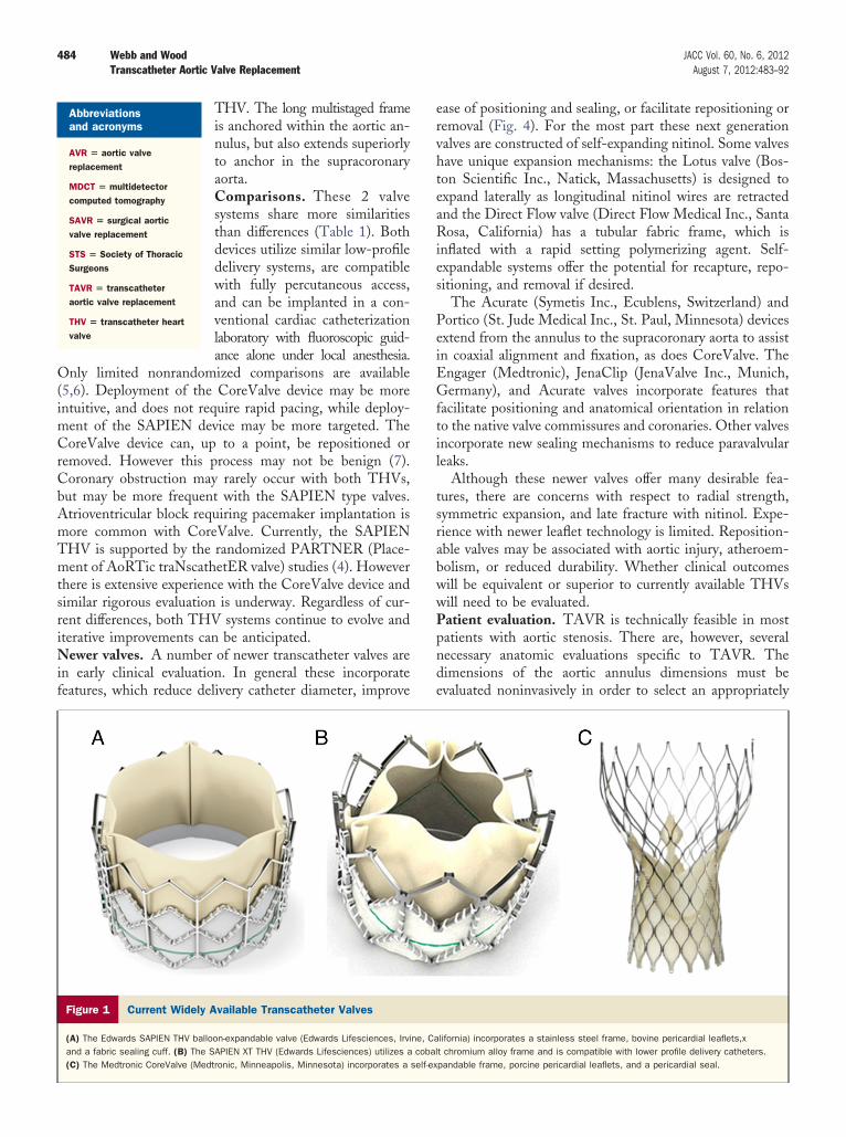
484 Webb and Wood JACC Vol. 60, No. 6, 2012Transcatheter Aortic Valve Replacement August 7, 2012:483–92
THV. The long multistaged frameis anchored within the aortic an-nulus, but also extends superiorlyto anchor in the supracoronaryaorta.Comparisons. These 2 valvesystems share more similaritiesthan differences (Table 1). Bothdevices utilize similar low-profiledelivery systems, are compatiblewith fully percutaneous access,and can be implanted in a con-ventional cardiac catheterizationlaboratory with fluoroscopic guid-ance alone under local anesthesia.
Only limited nonrandomized comparisons are available(5,6). Deployment of the CoreValve device may be moreintuitive, and does not require rapid pacing, while deploy-ment of the SAPIEN device may be more targeted. TheCoreValve device can, up to a point, be repositioned orremoved. However this process may not be benign (7).Coronary obstruction may rarely occur with both THVs,but may be more frequent with the SAPIEN type valves.Atrioventricular block requiring pacemaker implantation ismore common with CoreValve. Currently, the SAPIENTHV is supported by the randomized PARTNER (Place-ment of AoRTic traNscathetER valve) studies (4). Howeverthere is extensive experience with the CoreValve device andsimilar rigorous evaluation is underway. Regardless of cur-rent differences, both THV systems continue to evolve anditerative improvements can be anticipated.Newer valves. A number of newer transcatheter valves arein early clinical evaluation. In general these incorporatefeatures, which reduce delivery catheter diameter, improve
Figure 1 Current Widely Available Transcatheter Valves
(A) The Edwards SAPIEN THV balloon-expandable valve (Edwards Lifesciences, Irviand a fabric sealing cuff. (B) The SAPIEN XT THV (Edwards Lifesciences) utilizes a(C) The Medtronic CoreValve (Medtronic, Minneapolis, Minnesota) incorporates a
Abbreviationsand acronyms
AVR � aortic valvereplacement
MDCT � multidetectorcomputed tomography
SAVR � surgical aorticvalve replacement
STS � Society of ThoracicSurgeons
TAVR � transcatheteraortic valve replacement
THV � transcatheter heartvalve
ease of positioning and sealing, or facilitate repositioning orremoval (Fig. 4). For the most part these next generationvalves are constructed of self-expanding nitinol. Some valveshave unique expansion mechanisms: the Lotus valve (Bos-ton Scientific Inc., Natick, Massachusetts) is designed toexpand laterally as longitudinal nitinol wires are retractedand the Direct Flow valve (Direct Flow Medical Inc., SantaRosa, California) has a tubular fabric frame, which isinflated with a rapid setting polymerizing agent. Self-expandable systems offer the potential for recapture, repo-sitioning, and removal if desired.
The Acurate (Symetis Inc., Ecublens, Switzerland) andPortico (St. Jude Medical Inc., St. Paul, Minnesota) devicesextend from the annulus to the supracoronary aorta to assistin coaxial alignment and fixation, as does CoreValve. TheEngager (Medtronic), JenaClip (JenaValve Inc., Munich,Germany), and Acurate valves incorporate features thatfacilitate positioning and anatomical orientation in relationto the native valve commissures and coronaries. Other valvesincorporate new sealing mechanisms to reduce paravalvularleaks.
Although these newer valves offer many desirable fea-tures, there are concerns with respect to radial strength,symmetric expansion, and late fracture with nitinol. Expe-rience with newer leaflet technology is limited. Reposition-able valves may be associated with aortic injury, atheroem-bolism, or reduced durability. Whether clinical outcomeswill be equivalent or superior to currently available THVswill need to be evaluated.Patient evaluation. TAVR is technically feasible in mostpatients with aortic stenosis. There are, however, severalnecessary anatomic evaluations specific to TAVR. Thedimensions of the aortic annulus dimensions must beevaluated noninvasively in order to select an appropriately
lifornia) incorporates a stainless steel frame, bovine pericardial leaflets,xt chromium alloy frame and is compatible with lower profile delivery catheters.pandable frame, porcine pericardial leaflets, and a pericardial seal.
ne, Cacobal
self-ex
Appltcartc
tfdia
485JACC Vol. 60, No. 6, 2012 Webb and WoodAugust 7, 2012:483–92 Transcatheter Aortic Valve Replacement
sized valve. Transesophageal echocardiographic (8,9), mul-tidetector computed tomography (MDCT), and magneticresonance imaging measurements are widely utilized (10–12).
rterial access is generally assessed with invasive angiogra-hy or contrast MDCT. Most arteries are somewhat com-liant and can accommodate sheaths slightly (�1 to 2 mm)
arger than their internal diameter, although this may not behe case when the artery is diffusely diseased, tortuous, oralcified. The aorta should be evaluated with invasivengiography or contrast MDCT to assess technical issueselated to the delivery and implantation of the specific valveype, aortic root and valvular calcification, and the risk oforonary obstruction.
Whether TAVR is advisable depends not only on variousechnical considerations, but also on the likelihood ofunctional and survival benefit. Increasingly evaluation isirected on identifying patients in whom a significant
mprovement in quality and duration of life is likely andvoiding unnecessary intervention in patients where the
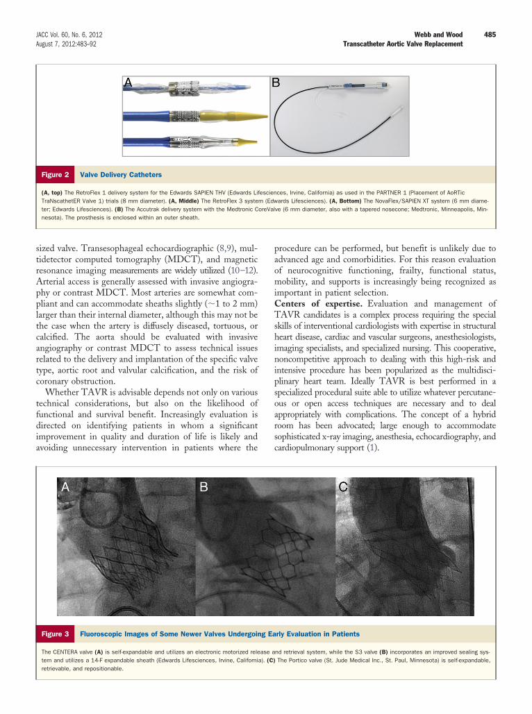
Figure 2 Valve Delivery Catheters
(A, top) The RetroFlex 1 delivery system for the Edwards SAPIEN THV (Edwards LiTraNscathetER Valve 1) trials (8 mm diameter). (A, Middle) The RetroFlex 3 systeter; Edwards Lifesciences). (B) The Accutrak delivery system with the Medtronic Cnesota). The prosthesis is enclosed within an outer sheath.
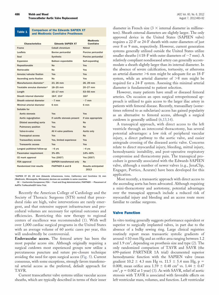
Figure 3 Fluoroscopic Images of Some Newer Valves Undergoin
The CENTERA valve (A) is self-expandable and utilizes an electronic motorized reletem and utilizes a 14-F expandable sheath (Edwards Lifesciences, Irvine, Californiretrievable, and repositionable.
procedure can be performed, but benefit is unlikely due toadvanced age and comorbidities. For this reason evaluationof neurocognitive functioning, frailty, functional status,mobility, and supports is increasingly being recognized asimportant in patient selection.Centers of expertise. Evaluation and management ofTAVR candidates is a complex process requiring the specialskills of interventional cardiologists with expertise in structuralheart disease, cardiac and vascular surgeons, anesthesiologists,imaging specialists, and specialized nursing. This cooperative,noncompetitive approach to dealing with this high-risk andintensive procedure has been popularized as the multidisci-plinary heart team. Ideally TAVR is best performed in aspecialized procedural suite able to utilize whatever percutane-ous or open access techniques are necessary and to dealappropriately with complications. The concept of a hybridroom has been advocated; large enough to accommodatesophisticated x-ray imaging, anesthesia, echocardiography, andcardiopulmonary support (1).
ces, Irvine, California) as used in the PARTNER 1 (Placement of AoRTicards Lifesciences). (A, Bottom) The NovaFlex/SAPIEN XT system (6 mm diame-
ve (6 mm diameter, also with a tapered nosecone; Medtronic, Minneapolis, Min-
rly Evaluation in Patients
nd retrieval system, while the S3 valve (B) incorporates an improved sealing sys-The Portico valve (St. Jude Medical Inc., St. Paul, Minnesota) is self-expandable,
fescienm (EdworeVal
g Ea
ase aa). (C)
486 Webb and Wood JACC Vol. 60, No. 6, 2012Transcatheter Aortic Valve Replacement August 7, 2012:483–92
Recently the American College of Cardiology and theSociety of Thoracic Surgeons (STS) noted that proce-dural risks are high, valve interventions are rarely emer-gent, and that extensive support infrastructure and pro-cedural volumes are necessary for optimal outcomes andefficiencies. Restricting this new therapy to regionalcenters of excellence was recommended (1). With wellover 1,000 cardiac surgical programs in the United Stateswith an average volume of 60 aortic cases per year, thiswill undoubtedly be controversial.Endovascular access. The femoral artery has been themost popular access site. Although originally requiring asurgical cutdown most experienced groups now utilize apercutaneous puncture and suture pre-closure techniqueavoiding the need for open surgical access (Fig. 5). Currentconsensus, with some exceptions, strongly favors transfemo-ral arterial access as the preferred, default approach forTAVR.
Current transcatheter valve systems utilize vascular access
Comparison of the Edwards SAPIEN XTand Medtronic CoreValve ProsthesesTable 1 Comparison of the Edwards SAPIEN XTand Medtronic CoreValve Prostheses
Characteristics Edwards SAPIEN XTMedtronicCoreValve
Frame Cobalt chromium Nitinol
Leaflets Bovine pericardial Porcine pericardial
Seal Synthetic Porcine pericardial
Expansion Balloon expandable Self-expanding
Repositionable No Yes
Retrievable No Yes
Annular/valvular fixation Yes Yes
Ascending aorta fixation No Yes
Manufacturers diameter* 23, 26 mm 26, 29 mm
Treatable annulus diameter* 18–25 mm 20–27 mm
Length 15–17 mm 53–55 mm
Sheath internal diameter 18-F, 19-F 18-F
Sheath external diameter �7 mm �7 mm
Minimal arterial diameter 6 mm 6 mm
Suitable for
Aortic stenosis Yes Yes
Aortic regurgitation If calcific stenosis present If size appropriate
Dilated ascending aorta Yes No
Pulmonary position Yes No
Valve-in-valve All 4 valve positions Aortic only
Transapical access Yes No
Transaxillary access Yes, limited experience Yes
Transaortic access Yes Yes
Longest published follow-up �6 yrs �4 yrs
Pacemaker requirement 3%–8% 14%–40%
CE mark approval Yes (2007) Yes (2007)
FDA approval SAPIEN transfemoral only No
Randomized trial results PARTNER A and B Results anticipated2013
*SAPIEN XT 20, 29 mm (Edwards Lifesciences, Irvine, California); and CoreValve 31 mm(Medtronic, Minneapolis, Minnesota) devices are available in some countries.
CE � Conformite Europeenne; FDA � Food and Drug Administration; PARTNER � Placement ofAoRTic TraNscathetER Valve Trial.
sheaths, which are typically described in terms of their inner
diameter in French size (3 � internal diameter in millime-ters). Sheath external diameters are slightly larger. The onlyapproved device in the United States (SAPIEN valve)requires a 22-F or 24-F sheath with outer diameters of justover 8 or 9 mm, respectively. However, current generationsystems generally utilized outside the United States utilizesmaller sheaths (�18-F with outer diameters of �7 mm). Arelatively compliant nondiseased artery can generally accom-modate a sheath slightly larger than its internal diameter. Inthe absence of severe calcification, tortuosity, or atheromaan arterial diameter �6 mm might be adequate for an 18-Fsystem, while an arterial diameter of �8 mm might berequired for a 24-F system. Assessing this minimal arterialdiameter is fundamental to patient selection.
However, many patients have small or diseased femoralarteries. On occasion an open surgical retroperitoneal ap-proach is utilized to gain access to the larger iliac artery inpatients with femoral disease. Recently, transaxillary (some-times referred to as subclavian) access has gained popularityas an alternative to femoral access, although a surgicalcutdown is generally utilized (6,13,14).
A transapical approach, with direct access to the leftventricle through an intercostal thoracotomy, has severalpotential advantages: a low risk of peripheral vascularinjury, a direct pathway to the aortic valve, and easierantegrade crossing of the diseased aortic valve. Concernsrelate to direct myocardial injury, bleeding, mitral injury,hemodynamic instability, and post-operative respiratorycompromise and thoracotomy pain. The transapical pro-cedure is generally associated with the Edwards SAPIENvalve, although a number of newer valves (e.g., JenaClip,Engager, Portico, Acurate) have been developed for thisapplication.
Most recently, a transaortic approach with direct access tothe ascending aorta has been advocated. Although requiringa mini-thoracotomy and aortotomy, potential advantagesover the transapical approach include a reduced risk ofmyocardial injury and bleeding and an access route morefamiliar to cardiac surgeons.
Valve Function
In vitro testing generally suggests performance equivalent orsuperior to surgically implanted valves, in part due to theabsence of a bulky sewing ring. Large clinical registriesroutinely report mean transaortic systolic gradients ofaround �10 mm Hg and an orifice area ranging between 1.2and 1.9 cm2, depending on prosthesis size and type (2). Theonly randomized comparison of TAVR and SAVR (the699-patient PARTNER 1A trial) documented superiorhemodynamic function with the SAPIEN valve (meangradient 10.2 � 4.3 mm Hg vs. 11.5 � 5.4 mm Hg, p �0.008; mean orifice area 1.59 � 0.48 cm2 vs. 1.44 � 0.47cm2, p � 0.002 at 1 year) (3). As with SAVR, relief of aorticstenosis with TAVR is associated with favorable effects on
left ventricular mass, volumes, and function. Left ventricular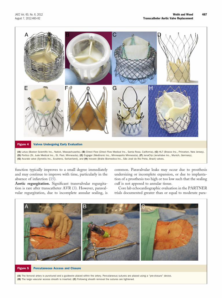
487JACC Vol. 60, No. 6, 2012 Webb and WoodAugust 7, 2012:483–92 Transcatheter Aortic Valve Replacement
function typically improves to a small degree immediatelyand may continue to improve with time, particularly in theabsence of infarction (15).Aortic regurgitation. Significant transvalvular regurgita-tion is rare after transcatheter AVR (3). However, paraval-vular regurgitation, due to incomplete annular sealing, is
Figure 4 Valves Undergoing Early Evaluation
(A) Lotus (Boston Scientific Inc., Natick, Massachusetts), (B) Direct Flow (Direct F(D) Portico (St. Jude Medical Inc., St. Paul, Minnesota), (E) Engager (Medtronic In(G) Acurate valve (Symetis Inc., Ecublens, Switzerland), and (H) Inovare (Braile Bio
Figure 5 Percutaneous Access and Closure
(A) The femoral artery is punctured and a guidewire placed within the artery. Percu(B) The large vascular access sheath is inserted. (C) Following sheath removal th
common. Paravalvular leaks may occur due to prosthesisundersizing or incomplete expansion, or due to implanta-tion of a prosthesis too high or too low such that the sealingcuff is not apposed to annular tissue.
Core lab echocardiographic evaluation in the PARTNERtrials documented greater than or equal to moderate para-
edical Inc., Santa Rosa, California), (C) HLT (Bracco Inc., Princeton, New Jersey),neapolis Minnesota), (F) JenaClip (JenaValve Inc., Munich, Germany),a Inc., São José do Rio Preto, Brazil) valves.
us sutures are placed using a “pre-closure” device.res are tightened.
low Mc., Minmedic
taneoe sutu

pfoha
rrem
488 Webb and Wood JACC Vol. 60, No. 6, 2012Transcatheter Aortic Valve Replacement August 7, 2012:483–92
valvular aortic regurgitation after TAVR in 11.8% of theinoperable patients and 12.2% of operable patients (3,4).However, net aortic regurgitation (both valvular and para-valvular combined) was actually reduced after TAVR(3,4,9,13).
Severe paravalvular regurgitation may result in severehemodynamic consequences, although improved implanta-tion techniques and more accurate annulus sizing have madesuch severe leaks increasingly less common. Most leaks are,in fact, mild to moderate, well tolerated, not associated withhemolysis, and do not worsen with time (13,14). Neverthe-less, it is clear that moderate and even mild leaks areassociated with a less favorable late survival than no leak(16,17). Whether this association represents cause and effectis unknown. When paravalvular regurgitation is excessiveredilation, repositioning, or implantation of a second, over-lapping transcatheter valve can often reduce or correct theproblem (13,18). However, these interventions may beassociated with a poorly understood increased risk of em-bolic stroke and should not be taken too lightly (7,19).Valve durability. Clinical and echocardiographic follow-upof both the SAPIEN and CoreValve prostheses beyond 3and up to 5 years has been well documented (15–17,20). Todate, late leaflet failure has been exceedingly rare and invitro accelerated wear testing is consistent with durabilitycomparable to surgical bioprostheses. Mid term durability ofthe SAPIEN and CoreValve frames has been documented andframe fracture has not been observed. However, only time willtell if durability matches that of surgical bioprostheses.
Clinical Outcomes
Survival. Contemporary registry series routinely documentrocedural success (defined loosely here as implantation of aunctional valve with the patient surviving the procedure) inver 95% and procedural survival (30 days) in over 90% ofigh-risk patients (2,5,18,21). More rigorous assessmentsre now available from the randomized PARTNER trials.
As the first of 2 parallel trials completed, PARTNER 1Bandomized 358 “inoperable” patients (in whom surgicalisk was prohibitive as judged by experienced surgeons) toither transarterial (femoral artery access) TAVR or bestedical management (Fig. 6) (4). Despite early generation
large-diameter systems and minimal operator experiencewith TAVR, the 30-day mortality of 6.4% among patientsassigned to TAVR compared very favorably with the STSmortality estimate of 11.6% with surgery. By way ofcomparison, the STS National Cardiac Database reportsthat among patients (low- and high-risk combined) actuallyundergoing SAVR in the United States, mortality wassimilarly 6.4%. Patients managed medically had a mortalityexceeding 50% at 1 year. The PARTNER 1B trial docu-mented a dramatic 20% absolute reduction in mortality at 1year with transarterial TAVR, meeting statistical tests for
superiority. At 2 years the survival curves continue todiverge with an additional 16.9% difference in mortalityaccruing between 1 and 2 years (16).
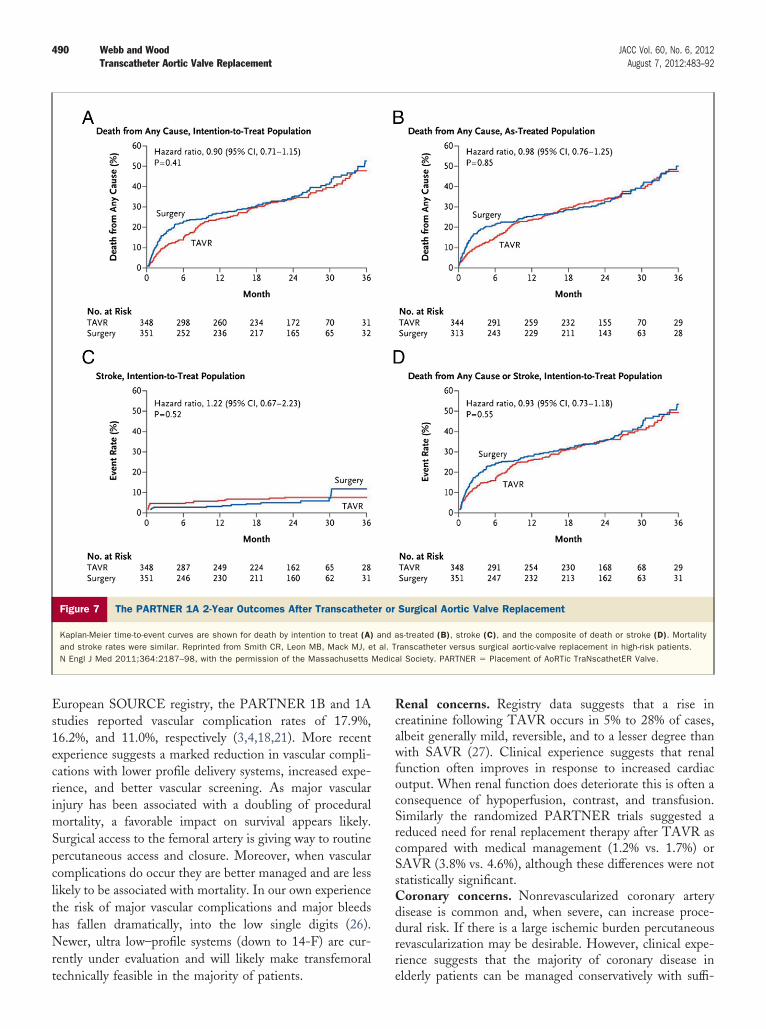
The parallel trial, PARTNER 1A, was completed a yearlater (Fig. 7) (3). In this trial 699 high-risk patients wererandomized to either TAVR (either a transfemoral ortransapical as determined by access considerations) orSAVR. The mean STS score was just under 12%, repre-senting the top 5% of patients in terms of operative risk.Mortality at 30 days was actually lower with TAVR thanSAVR (3.4% vs. 6.5%, p � 0.07) and at 1 year (24.2% vs.26.8%, p � 0.44); not statistically significant, but stillmeeting tests for noninferiority.
In addition to the previous intention-to-treat analysis(treatment assigned, 30 days from randomization), theas-treated analysis (treatment received, 30 days from theprocedure) has assumed relevance as surgical patientswere less likely to undergo their assigned procedurewithin 30 days, thereby reducing procedural deathswithin 30 days of randomization. In the as-treatedanalysis femoral transarterial TAVR 30-day mortalitywas less than one half that with SAVR (3.7% vs. 8.2%,p � 0.05). As in previous observational studies of TAVR,mortality was higher among patients that underwent atransapical procedure. The transapical procedure did not,on its own, meet the test of noninferiority. To whatdegree this difference is attributable to selection ofhigher-risk patients and a learning curve in the transapi-cal group is controversial.Functional benefit. For many elderly patients morbiditymay be a greater concern than mortality. A marked anddurable improvement in functional class and quality of lifeafter TAVR has been well documented (15,22,23). Therandomized PARTNER trials documented a marked re-duction in rehospitalization with transfemoral TAVR ascompared with medical management and, in comparisonwith surgery, a significantly shorter length of stay as well asearlier improvement in functional status (3,4,22).Late survival. Registries report survival 1 year followingTAVR ranging from 69% to 85% (3–5,15,18,21). Similarly,1-year survival in the PARTNER trials following TAVRwas 69% in inoperable patients and 76% in high-riskoperable patients. Late mortality was primarily the resultof advanced age and debilitating comorbidities such assevere pulmonary and renal disease (3,4,15). Given theinvasive nature of the procedure and limited resources itmay be as important to decide who will not benefit, aswho will.
Specific Risks of TAVR
Neurological concerns. Stroke is a known potential com-plication with both SAVR and TAVR (24). For perspective,the recent European SOURCE (SAPIEN Aortic Biopros-thesis Multi-Region Outcome Registry) and United King-dom high-risk TAVR registries reported stroke rates of
2.4% and 4.0%, respectively. The rigorously monitored
nitT
tnyi(w((
bipeAco
489JACC Vol. 60, No. 6, 2012 Webb and WoodAugust 7, 2012:483–92 Transcatheter Aortic Valve Replacement
PARTNER 1B “inoperable” trial reported an increase inmajor stroke rate of 4%, arguably justified given the overall21.9% reduction in death and/or stroke at 2 years (numberneeded to treat � 4.6) (16).
The PARTNER 1A trial allows direct comparison ofstroke rates between TAVR and SAVR. Major strokes(strokes with permanent deficit) were similar: 3.8% versus2.1% at 30 days (p � 0.20) (25). As there were relatively fewstrokes, an analysis of all neurological events (includingtransient ischemic attacks and minor strokes without per-manent deficit) was performed. Neurological events (withand without clinical deficit) were increased with TAVR(5.5% vs. 2.4% at 30 days, p � 0.04). Predictors of
eurological events with TAVR included a prior neurolog-cal event, more severe atherosclerotic burden, worse func-ional disability, smaller valve area, and transapical access.
Figure 6 The PARTNER 1B Trial Compared Transarterial TAVR
Time to event analyses of death (A), death due to cardiac causes (B), rehospLeon MR, Smith CR, Mack M, et al. Transcatheter aortic-valve implantation for1597–607, with information from Makkar RR, Fontana GP, Jilaihawi H, et al. TJ Med 2012;366:1696–704, with permission from the Massachusetts Medicacatheter aortic valve replacement.
he temporal pattern of neurological events was similar for d
he 2 groups, with an increased risk over the first week, buto subsequent increased hazard over SAVR out beyond 2ears. At 2-year follow up there was no significant differencen the overall numbers of strokes with TAVR and SAVR17). The composite of death and/or stroke with disabilityas actually lower (although nonsignificantly) with TAVR
femoral and apical combined) as compared to SAVR26.5% vs. 28.0% at 1 year, p � 0.68) (Fig. 7).
Although data are preliminary, the risk of stroke seems toe falling with smaller, less traumatic catheters (Fig. 3),mproved technique, and lower-risk patients (13,24). Aotential role for devices designed to capture or deflectmbolic material is currently being evaluated (2,21).ccess concerns. Vascular events have been the most
ommon major complication associated with TAVR and areften implicated in procedural mortality. Using the large-
dical Management in Extremely High-Risk (Inoperable) Patients
ion (C), and the combined endpoint of death or stroke (D). Redrawn fromstenosis in patients who cannot undergo surgery. N Engl J Med 2010;363:theter aortc-valve replacement for inoperable severe aortic stenosis. N Englty. PARTNER � Placement of AoRTic TraNscathetER Valve; TAVR � trans-
to Me
italizataorticranscal Socie
iameter 22-F and 24-F RetroFlex delivery system, the
490 Webb and Wood JACC Vol. 60, No. 6, 2012Transcatheter Aortic Valve Replacement August 7, 2012:483–92
European SOURCE registry, the PARTNER 1B and 1Astudies reported vascular complication rates of 17.9%,16.2%, and 11.0%, respectively (3,4,18,21). More recentexperience suggests a marked reduction in vascular compli-cations with lower profile delivery systems, increased expe-rience, and better vascular screening. As major vascularinjury has been associated with a doubling of proceduralmortality, a favorable impact on survival appears likely.Surgical access to the femoral artery is giving way to routinepercutaneous access and closure. Moreover, when vascularcomplications do occur they are better managed and are lesslikely to be associated with mortality. In our own experiencethe risk of major vascular complications and major bleedshas fallen dramatically, into the low single digits (26).Newer, ultra low–profile systems (down to 14-F) are cur-rently under evaluation and will likely make transfemoral
Figure 7 The PARTNER 1A 2-Year Outcomes After Transcathet
Kaplan-Meier time-to-event curves are shown for death by intention to treat (A)and stroke rates were similar. Reprinted from Smith CR, Leon MB, Mack MJ, eN Engl J Med 2011;364:2187–98, with the permission of the Massachusetts
technically feasible in the majority of patients.
Renal concerns. Registry data suggests that a rise increatinine following TAVR occurs in 5% to 28% of cases,albeit generally mild, reversible, and to a lesser degree thanwith SAVR (27). Clinical experience suggests that renalfunction often improves in response to increased cardiacoutput. When renal function does deteriorate this is often aconsequence of hypoperfusion, contrast, and transfusion.Similarly the randomized PARTNER trials suggested areduced need for renal replacement therapy after TAVR ascompared with medical management (1.2% vs. 1.7%) orSAVR (3.8% vs. 4.6%), although these differences were notstatistically significant.Coronary concerns. Nonrevascularized coronary arterydisease is common and, when severe, can increase proce-dural risk. If there is a large ischemic burden percutaneousrevascularization may be desirable. However, clinical expe-rience suggests that the majority of coronary disease in
Surgical Aortic Valve Replacement
s-treated (B), stroke (C), and the composite of death or stroke (D). Mortalityranscatheter versus surgical aortic-valve replacement in high-risk patients.al Society. PARTNER � Placement of AoRTic TraNscathetER Valve.
er or
and at al. TMedic
elderly patients can be managed conservatively with suffi-
491JACC Vol. 60, No. 6, 2012 Webb and WoodAugust 7, 2012:483–92 Transcatheter Aortic Valve Replacement
cient reduction in angina accrued from relief of aorticstenosis alone. TAVR facilitates a strategy of staged revas-cularization as required.
Acute coronary obstruction may rarely occur, generallydue to displacement of a bulky native valve leaflet over acoronary ostium (usually the left). Large series suggest a riskof �0.5% with the SAPIEN valve and a somewhat lowerrisk with CoreValve (2,21). When coronary obstructiondoes occur successful management may require temporarycardiopulmonary support and revascularization (28). Factorsthat increase the risk of coronary obstruction include anunusually bulky native leaflet (adjacent to a coronaryostium), a low origin of the coronary ostium (oftendefined as �12 mm from the basal leaflet insertion asassessed by MDCT), a shallow sinus of Valsalva (offeringless room for the native leaflet), an oversized prosthesis,and high implantation.Concomitant mitral regurgitation. Mitral regurgitation iscommon in patients with aortic stenosis, increasing bothTAVR and SAVR procedural risk. However, mitral regur-gitation may be better tolerated following successful AVRby either modality (29,30). Furthermore mitral regurgita-tion may sometimes improve following AVR, particularlywhen regurgitation is functional. In the PARTNER 1Btrial, 22% of patients had moderate or severe mitral regur-gitation. Mitral regurgitation was a marker of higher TAVRprocedural mortality, but also of greater procedural benefitin comparison to medical management. Similarly, in thePARTNER 1A study, 20% of patients had moderate orsevere mitral regurgitation. Mitral regurgitation was a pre-dictor of increased procedural mortality; however, this riskwas less with TAVR than SAVR (24.2% vs. 35%) (3). Atherapeutic strategy of replacing the aortic valve alone maybe reasonable in some patients in whom the risk of doublevalve surgery might be prohibitive.The conduction system. The atrioventricular conductionsystem passes superficially through the interventricular sep-tum immediately below the aortic valve. Injury during valveimplantation may result in partial or complete heart block.Risk factors include advanced age, right bundle branchblock, atrioventricular delay, along with prosthesis over-sizing and ventricular positioning.
In the PARTNER randomized studies new pacemakerswere no more frequent 1 year following SAPIEN valveimplantation than with medical management (4.5% vs.7.8%, p � 0.27) or with SAVR (5.7% vs. 5.0%, p � 0.68)(3,4). However, CoreValve implantation is more frequentlyassociated early and late atrioventricular block, presumablydue to greater extension into the left ventricular outflowtract with compression of the septal conduction tissues. Therequirement for new pacemakers was 3-fold higher follow-ing CoreValve, as compared with SAPIEN, implantation inboth the United Kingdom and French national registries(5,6). For this reason routine prophylactic temporary pacingleads are routinely used, with more prolonged electrocar-
diographic monitoring (31).Failed Surgical Prostheses
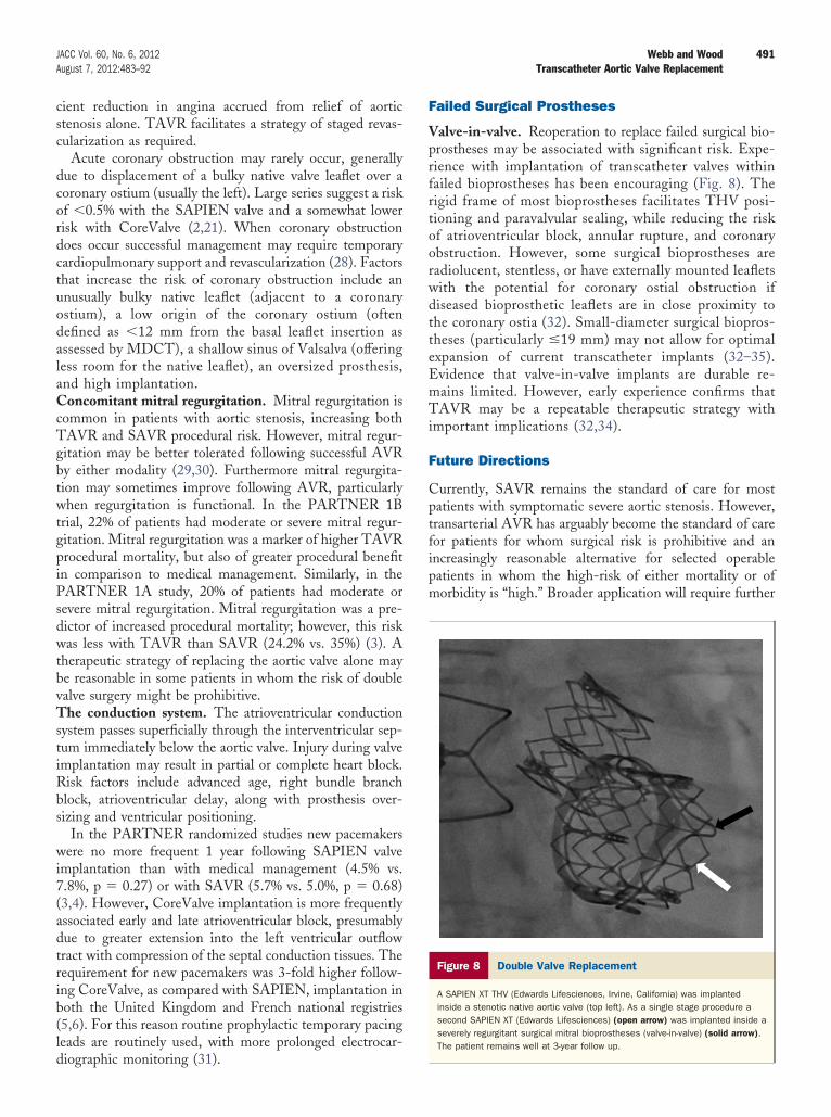
Valve-in-valve. Reoperation to replace failed surgical bio-prostheses may be associated with significant risk. Expe-rience with implantation of transcatheter valves withinfailed bioprostheses has been encouraging (Fig. 8). Therigid frame of most bioprostheses facilitates THV posi-tioning and paravalvular sealing, while reducing the riskof atrioventricular block, annular rupture, and coronaryobstruction. However, some surgical bioprostheses areradiolucent, stentless, or have externally mounted leafletswith the potential for coronary ostial obstruction ifdiseased bioprosthetic leaflets are in close proximity tothe coronary ostia (32). Small-diameter surgical biopros-theses (particularly �19 mm) may not allow for optimalexpansion of current transcatheter implants (32–35).Evidence that valve-in-valve implants are durable re-mains limited. However, early experience confirms thatTAVR may be a repeatable therapeutic strategy withimportant implications (32,34).
Future Directions
Currently, SAVR remains the standard of care for mostpatients with symptomatic severe aortic stenosis. However,transarterial AVR has arguably become the standard of carefor patients for whom surgical risk is prohibitive and anincreasingly reasonable alternative for selected operablepatients in whom the high-risk of either mortality or ofmorbidity is “high.” Broader application will require further
Figure 8 Double Valve Replacement
A SAPIEN XT THV (Edwards Lifesciences, Irvine, California) was implantedinside a stenotic native aortic valve (top left). As a single stage procedure asecond SAPIEN XT (Edwards Lifesciences) (open arrow) was implanted inside aseverely regurgitant surgical mitral bioprostheses (valve-in-valve) (solid arrow).The patient remains well at 3-year follow up.

492 Webb and Wood JACC Vol. 60, No. 6, 2012Transcatheter Aortic Valve Replacement August 7, 2012:483–92
refinement as well as more rigorous and longer follow-up. Itis possible that, with time, TAVR will become a preferredoption for a much broader group of patients. A majorconcern may well be the following: When are patients tooill, frail, or old to gain significant benefit in terms ofduration or quality of life?
Reprint requests and correspondence: Dr. John G. Webb, St.Paul’s Hospital, 1081 Burrard Street, Vancouver, British Columbia,V6Z 1Y6, Canada. E-mail: [email protected].
REFERENCES
1. Holmes DR Jr., Mack MJ. Transcatheter valve therapy: A professionalsociety overview from the American College of Cardiology Foundationand the Society of Thoracic Surgeons. J Am Coll Cardiol 2011;58:445–55.
2. Webb JG, Cribier A. Percutaneous transarterial aortic valve implan-tation: what do we know? Eur Heart J 2011;32:140–7.
3. Smith CR, Leon MB, Mack MJ, et al. Transcatheter versus surgicalaortic-valve replacement in high-risk patients. N Engl J Med 2011;364:2187–98.
4. Leon MB, Smith CR, Mack M, et al. Transcatheter aortic-valveimplantation for aortic stenosis in patients who cannot undergosurgery. N Engl J Med 2010;363:1597–607.
5. Moat NE, Ludman P, de Belder MA, et al. Long-term outcomes aftertranscatheter aortic valve implantation in high-risk patients with severeaortic stenosis: The U.K. TAVI (United Kingdom TranscatheterAortic Valve Implantation) Registry. J Am Coll Cardiol 2011;58:2130–8.
6. Eltchaninoff H, Prat A, Gilard M, et al. Transcatheter aortic valveimplantation: early results of the FRANCE (FRench Aortic NationalCoreValve and Edwards) registry. Eur Heart J 2011;32:191–7.
7. Geisbusch S, Bleiziffer S, Mazzitelli D, Ruge H, Bauernschmitt R,Lange R. Incidence and management of CoreValve dislocation duringtranscatheter aortic valve implantation. Circ Cardiovasc Interv 2010;3:531–6.
8. Zamorano JL, Badano LP, Bruce C, et al. EAE/ASE recommenda-tions for the use of echocardiography in new transcatheter interven-tions for valvular heart disease. J Am Soc Echocardiogr 2011;24:937–65.
9. Jayasuriya C, Moss RR, Munt B. Transcatheter aortic valve implan-tation in aortic stenosis: the role of echocardiography. J Am SocEchocardiogr 2011;24:15–27.
10. Leipsic J, Gurvitch R, LaBounty T, et al. Multidetector computedtomography in transcatheter aortic valve implantation. J Am CollCardiol Img 2011;4:416–29.
11. Jabbour A, Ismail TF, Moat N, et al. Multimodality imaging intranscatheter aortic valve implantation and post-procedural aorticregurgitation comparison among cardiovascular magnetic resonance,cardiac computed tomography, and echocardiography. J Am CollCardiol 2011;58:2165–73.
12. Willson A, Webb JG, Leipsic J. Three-dimensional aortic annularassessment by multidetector computed tomography predicts moderateor severe paravalvular regurgitation after transcatheter aortic valvereplacement: implications for sizing of transcatheter heart valves. J AmColl Cardiol 2012;59:1287–94.
13. Tamburino C, Capodanno D, Ramondo A, et al. Incidence andpredictors of early and late mortality after transcatheter aortic valveimplantation in 663 patients with severe aortic stenosis. Circulation2011;123:299–308.
14. Petronio AS, De Carlo M, Bedogni F, et al. Safety and efficacy of thesubclavian approach for transcatheter aortic valve implantation with
the CoreValve revalving system. Circ Cardiovasc Interv 2010;3:359–66.15. Gurvitch R, Wood DA, Tay EL, et al. Transcatheter aortic valveimplantation. Durability of clinical and hemodynamic outcomes be-yond 3 years in a large patient cohort. Circulation 2010;122:1319–27.
16. Makkar RR, Fontana GP, Jilaihawi H, et al. Transcatheter aortic-valve replacement for inoperable severe aortic stenosis. N Engl J Med2012;366:1696–704.
17. Kodali S, Williams M, Smith CR, et al. Two year outcomes aftertranscatheter or surgical replacement in high risk patients with aorticstenosis. N Engl J Med 2012;366:1686–95.
18. Thomas M, Schymik G, Walther T, et al. One-year outcomes ofcohort 1 in the Edwards SAPIEN Aortic Bioprosthesis EuropeanOutcome (SOURCE) registry: the European registry of transcatheteraortic valve implantation using the Edwards SAPIEN valve. Circula-tion 2011;124:425–33.
19. Webb JG, Binder RK. Post-dilating transcatheter heart valves. J AmColl Cardiol Intv 2012;5:513–4.
20. Piazza N, Grube E, Gercken U, et al. A clinical protocol for analysisof the structural integrity of the Medtronic CoreValve System frameand its application in patients with 1-year follow-up. EuroIntervention2010;5:680–6.
21. Thomas M. The global experience with percutaneous aortic valvereplacement. J Am Coll Cardiol Intv 2010;3:1103–9.
22. Reynolds MR, Magnuson EA, Lei Y, et al. Health-related quality oflife after transcatheter aortic valve replacement in inoperable patientswith severe aortic stenosis. Circulation 2011;124:1964–72.
23. Cohen D, Reynolds M, Magnuson, et al. Health related quality of lifeafter transcatheter or surgical aortic valve replacement in high riskpatients with severe aortic stenosis: results from the PARTNER trial(cohort A). J Am Coll Cardiol 2011;58:1557–65.
24. Daneault B, Kirtane AJ, Kodali SK, et al. Stroke associated withsurgical and transcatheter treatment of aortic stenosis. A comprehen-sive review. J Am Coll Cardiol 2011;58:2143–50.
25. Miller DC, Blackstone EH, Mack MJ SL, et al. Transcatheter(TAVR) versus surgical (AVR) aortic valve replacement: occurrence,hazard, risk factors, and consequences of neurological events in thePARTNER trial. J Thorac Cardiovasc Surg 2012;143:832–43.e13.
26. Toggweiler S, Gurvitch R, Leipsic J, et al. Percutaneous aortic valvereplacement. Improved vascular outcomes with a fully percutaneousprocedure. J Am Coll Cardiol 2012;59:113–8.
27. Bagur R, Webb JG, Nietlispach F, et al. Acute kidney injury followingtranscatheter aortic valve implantation: predictive factors, prognosticvalue, and comparison with surgical aortic valve replacement. EurHeart J 2010;31:865–74.
28. Masson JB, Kovac J, Schuler G, et al. Transcatheter aortic valveimplantation: review of the nature, management and avoidance ofprocedural complications. J Am Coll Cardiol Intv 2009;2:811–20.
29. Tzikas A, Piazza N, van Dalen BM, et al. Changes in mitralregurgitation after transcatheter aortic valve implantation. CatheterCardiovasc Interv 2010;75:43–9.
30. Toggweiler S, Boone RH, Rodés-Cabau J, et al. Transcatheter aorticvalve replacement. Outcomes of patients with moderate or severemitral regurgitation. J Am Coll Cardiol 2012;59:2068–74.
31. Piazza N, Nuis RJ, Tzikas A, et al. Persistent conduction abnormal-ities and requirements for pacemaking six months after transcatheteraortic valve implantation. EuroIntervention 2010;6:475–84.
32. Gurvitch R, Cheung A, Ye J, et al. Transcatheter valve-in-valveimplantation for failed surgical bioprosthetic valves. J Am Coll Cardiol2011;58:2196–209.
33. Webb JG, Wood DA, Ye J, et al. Transcatheter valve-in-valveimplantation for failed bioprosthetic heart valves. Circulation 2010;121:1848–57.
34. Piazza N, Bleiziffer S, Brockmann G, et al. Transcatheter aortic valveimplantation for failing surgical aortic bioprosthetic valve: from con-cept to clinical application and evaluation (part 2). J Am Coll Cardiol2011;4:733–42.
35. Piazza N, Bleiziffer S, Brockmann G, et al. Transcatheter aortic valveimplantation for failing surgical aortic bioprosthetic valve: from con-cept to clinical application and evaluation (part 1). J Am Coll Cardiol2011;4:721–32.
Key Words: aortic y replacement y valve.