Embed Size (px)
Citation preview
REVIEW Open Access
Current status of plant metabolite-basedfabrication of copper/copper oxidenanoparticles and their applications: areviewKhwaja Salahuddin Siddiqi1 and Azamal Husen2*
Abstract
Since green mode of nanoparticles (NPs) synthesis is simple, advantageous and environment friendly relative tochemical and physical procedures, various plant species have been used to fabricate copper and copper oxidenanoparticles (Cu/CuO-NPs) owing to the presence of phytochemicals which often act as capping as well asstabilizing agent. These Cu/CuO-NPs are highly stable and used in the degradation of organic dyes like methyleneblue and reduction of organic compounds such as phenols. They are also used as antibacterial, antioxidant andantifungal agent due to their cytotoxicity. They are also examined for agricultural crops growth and productivity.Cu-NPs increased the root and shoot growth of mung bean. In wheat plants, these particles reduced shoot growth;and enhanced the grain yield and stress tolerance through starch degradation. Similarly, CuO-NPs treated seedlingshave shown reduced chlorophyll, carotenoid and sugar content, whereas proline and anthocyanins were increasedin Brassica rapa seedlings. Overall, this review presents the recent understanding of plant-mediated Cu and CuO-NPs fabrication and their application in biomedicine, environmental remediation and agricultural practices. Acomparison of the traditional/conventional method of fabrication of NPs with those of green protocols has alsobeen made. Some misconception of copper chemistry has also been critically discussed in terms of oxidation andreduction reactions.
Keywords: Cu/CuO NPs, Biogenic synthesis, Biomedical, Crop growth, Applications
IntroductionRecent advancement in nanotechnology has acceleratedour interest in designing nano sized particles of desiredshape and size. Since their property changes with morph-ology they have been used in various areas of medicine,agriculture and environmental remediation [1–4]. The‘green synthesis’ of nanoparticles (NPs) using plant is ad-vantageous over chemical, physical and or microbial syn-thesis as it removes the complicated protocol; and canalso meet the large-scale production requirement. Further,
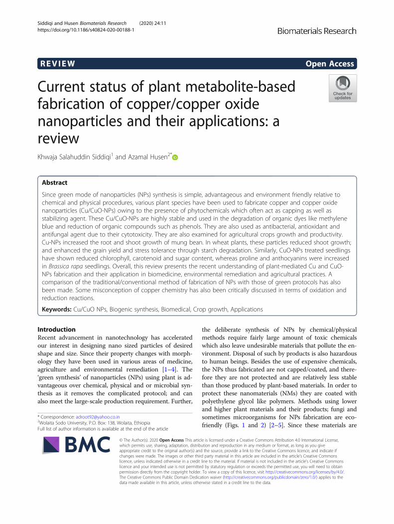
the deliberate synthesis of NPs by chemical/physicalmethods require fairly large amount of toxic chemicalswhich also leave undesirable materials that pollute the en-vironment. Disposal of such by products is also hazardousto human beings. Besides the use of expensive chemicals,the NPs thus fabricated are not capped/coated, and there-fore they are not protected and are relatively less stablethan those produced by plant-based materials. In order toprotect these nanomaterials (NMs) they are coated withpolyethylene glycol like polymers. Methods using lowerand higher plant materials and their products; fungi andsometimes microorganisms for NPs fabrication are eco-friendly (Figs. 1 and 2) [2–5]. Since these materials are
© The Author(s). 2020 Open Access This article is licensed under a Creative Commons Attribution 4.0 International License,which permits use, sharing, adaptation, distribution and reproduction in any medium or format, as long as you giveappropriate credit to the original author(s) and the source, provide a link to the Creative Commons licence, and indicate ifchanges were made. The images or other third party material in this article are included in the article's Creative Commonslicence, unless indicated otherwise in a credit line to the material. If material is not included in the article's Creative Commonslicence and your intended use is not permitted by statutory regulation or exceeds the permitted use, you will need to obtainpermission directly from the copyright holder. To view a copy of this licence, visit http://creativecommons.org/licenses/by/4.0/.The Creative Commons Public Domain Dedication waiver (http://creativecommons.org/publicdomain/zero/1.0/) applies to thedata made available in this article, unless otherwise stated in a credit line to the data.
* Correspondence: [email protected] Sodo University, P.O. Box: 138, Wolaita, EthiopiaFull list of author information is available at the end of the article
Siddiqi and Husen Biomaterials Research (2020) 24:11 https://doi.org/10.1186/s40824-020-00188-1
easily available and do not require organic solvent as reac-tion medium, they are easy to handle and economical. Inmajor cases the NMs thus synthesized are capped by bio-molecules like phenols, tannin, flavonoids and ascorbatepresent in the plant materials. They enhance stability ofNPs and also prevent their interaction with atmosphericoxygen. These NMs are thus not oxidized and can be keptfor long period of time without undergoing any change intheir properties. Green synthetic methods make use ofmany waste materials like banana peels, lemon rind,dried leaves of medicinal plants and algae etc. The pre-cursor even in crude form may react with these mate-rials to produce NPs. The noble metal NPs for examplesilver, gold, platinum and their alloys have been fre-quently biosynthesized by various workers [6–12].Among the coinage metal NPs, copper nanoparticles(Cu-NPs) are of great interest due to their low cost,easy availability and high electrical conductivity [13–15]. Borkow and Gabbay [16] and Zheng et al. [17] havereported that the copper ions are used as pesticides,fungicides and fertilizers. Copper oxide (CuO) is also acheaper material in comparison to silver/gold and caneasily be mixed with polymers due to their stability[18]. Recently, Din et al. [19] have discussed the synthe-sis and characterization of cupric-oxide nanoparticles(CuO-NPs) together with their application as antioxi-dant, antibacterial and antifungal agent.
In general, Cu-NPs are used as an antimicrobial, anti-oxidant, antidiabetic, anti-inflammatory and antifoulingagent when integrated in coatings, plastics and textiles[20–23]. They also find application in heat transferfluids, sensors, e-sensitized solar cells, lithium ion batter-ies, gas -sensing, heterogeneous catalysis and as antican-cer agent [24–28].Based on the current information, the present review
unfolds the protocol of plant-mediated bio-fabrication ofCu-NPs/CuO-NPs followed by their characterizationand application in different areas. Attempt has also beenmade to enlist the impact of NPs on crops as growthpromoter and the effect of size of NPs on their efficiencyas catalyst and antibacterial agent.
Fabrication of cu-NPs/CuO-NPsCu-NPsSynthesis of spherical Cu-NPs of 23 ± 1.10 nm fromaqueous flower extract of Millettia pinnata and theircharacterization by ultraviolet–visible spectroscopy (UV-Vis), scanning electron microscopy (SEM), transmisionelectron microscopy (TEM), x-ray diffraction (XRD), Fou-rier transform infrared spectroscopy (FTIR) and selectedarea electron diffraction (SAED) has been reported byThiruvengadam et al. [21]. UV-Vis study has confirmedthe reduction of copper acetate to Cu-NPs. The maximumabsorption was recorded at 384 nm which confirmed the
Fig. 1 Benefits of bio-derived fabrication/green synthesis of nanoparticles over chemical and physical procedures
Siddiqi and Husen Biomaterials Research (2020) 24:11 Page 2 of 15

surface plasmon resonance (SPR) of the NPs. Their FTIRspectrum indicated the presence of proteins, acids, flavo-noids, polyphenols, carboxylic acid and alkaloids which re-duced the copper ions into Cu-NPs.Cu-NPs of 50–250 nm diameters have been fabricated
from Magnolia kobus leaf extract at room temperature[29]. Most of these NPs were spherical in shape andtheir formation was confirmed from UV-Vis spectrumexhibiting a peak at 560 nm. As the concentration ofCu-NPs increases the intensity of absorption peak mea-sured at 560 nm also increases. It has also been observedthat with increase in temperature yield of the Cu-NPsincreases. However, complete conversion was achievedat about 90–95 °C. It was observed that when
concentration of Magnolia leaf extract was increased upto 20%, smaller and spherical NPs were formed [30]. Itis suggested that organic molecules/metabolites acting ascapping agents also cause aggregation of NPs but it takeslong time. Energy-dispersive x-ray (EDX) spectra showedsignals for copper along with oxygen and carbon. Signalsfor other elements may not be due to biomolecules ad-hering to the surface of Cu-NPs but they may be due toimpurity present in the colloidal mixture of NPs and leafextract. It has been noted that chemically fabricated NPswere air oxidized while those synthesized by Magnolialeaf extract were stable for more than 30 days [30]. It isbecause the biomolecules acting as capping agent pro-tect the Cu-NPs from oxidation by air.
Fig. 2 Detailed scheme of bio-derived fabrication/green synthesis of nanoparticlesusing lower/higher plant materials and their products; fungusand microorganisms
Siddiqi and Husen Biomaterials Research (2020) 24:11 Page 3 of 15
Kulkarni et al. [31] have examined the fabrication ofCu-NPs from copper sulfate and Eucalyptus sp. leaf ex-tract at room temperature. A slight change in pH of thecopper sulfate solution from 2.16 to 2.83 was observedwhen leaf extract at pH 6.96 was added followed by achange in colour of the resulting mixture which showedabsorption at 572 nm in its Uv-Vis spectrum. FTIRspectrum of the Cu-NPs showed the presence of phenol,amine, amino acid and flavonoids. It has been suggestedthat the biomolecules present in the eucalyptus leaf ex-tract act both as reducing and capping agent. The NPswere highly crystalline with face centred cubic (FCC)structure. Their average size was found to be 38.62 nm.It is worth observing that the concentration and pH ofthe reducing agent is mainly responsible for the produc-tion of NPs.Biosynthesis of Cu-NPs from leaf broth of Azadirachta
indica and the influence of concentration of precursorsalt, concentration of leaf broth, temperature and pH ofthe medium were reported on the conversion rate of Cu-NPs [32] which are similar to those found above [31].Ultrasmall Cu-NPsof 2.90 ± 0.64 nm diameter have
been synthesized from lemongrass tea by one potmethod [33]. Although, colour of the mixture containingboth the Cu-NPs and lemongrass was yellow it did notexhibit any absorption in the UV-Vis region (350-950nm). It has been ascribed to extremely small size of Cu-NPs which are devoid of surface plasmon resonance [34,35]. It is strange and unusual for NPs to exhibit colourbut no absorption. Generally, a sharp peak is observed in560 to 570 nm region for Cu-NPs if they are larger than5 nm [34–36]. Their IR spectra showed the presence ofpolyphenols, proteins and carbohydrate [37, 38] as reducingagents. SEM and TEM images showed the presence offinely divided and dispersed Cu-NPs. However, they werenot found to be oxidized either to cuprous oxide or cupricoxide which is quite obvious because in presence of redu-cing agents oxidation cannot occur. It has been observedthat polyethyleneglycol or polyvinyl pyr olidone can alsoprevent the NPs from oxidation and aggregation [39–41].Cu-NPs synthesized using aqueous leaf extract of
henna (Lawsonia inermis) showed an absorption peak at570 nm which is the signature peak of CuO-NPs [42].Blue copper sulfate absorbs at 810 nm but Cu-NPs arereddish brown if all copper ions have been reduced toNPs. It has been reported that Cu-NPs are surroundedby a thin film of copper oxide (CuO and Cu2O) which ischaracterized by absorption at 800 nm [43]. It may bedue to the presence of already oxidized copper sulfatepresent as impurity because in solution it cannot be oxi-dized unless it is heated in open at about 900 °C. Cu-NPs are efficiently formed at pH 11. Perhaps reductionof copper ions is facilitated in highly alkaline medium asthe lawsones are stabilized at higher pH. Electrical
conductivity of calcined Cu-NPs was enhanced becauseall particles come closer as a result of which the mobilityof electrons increases [44]. Authors have found that theabsorption of Cu-NPsat 570 nm was lost and a new peakappeared at 285 nm when Cu-NPs were calcined. It ishighly improbable for a calcined material to absorb inUV region of spectrum. It may be quite likely that thespectrum was not carefully run otherwise any transitionmetal or its oxide will not show any absorption in UVrange. Their interpretation of electronic transition frominner to the outermost shell of copper [45] is highly un-likely because such situation occurs only when copperion forms a coordination compound with a very strongligand and forces one of the 3d9 electrons to be trans-ferred to the outer 4d orbital. Since there are no copperions this peak at 284 nm is most likely to be due to someimpurity. SEM and EDAX showed agglomerated spher-ical Cu-NPs. The suggestion that Cu-NPs are oxidizedto CuO on exposure to air is author’s lack of knowledgeof copper chemistry. If the Cu-NPs are exposed to airthey will form CuSO4 which is evidenced by a distinctblue colour on its surface but it is not oxidized to copperoxides.Biogenic synthesis and characterization of Cu-NPs
from Citrus medica juice has been reported [46]. UV-Visspectrum showed absorption peak at 631 nm which hasbeen taken as an evidence for the formation of Cu-NPs.Since the reduction reaction was carried out in analuminum vessel instead of a glass vessel some fractionof aluminum would have dissolved in the citron juice.Blue CuSO4 solution turned reddish brown due to for-mation of Cu2SO4 after the addition of citron juice,followed by deposition of shiny brown precipitate on thewall of aluminum vessel. It is suggested that a more re-active metal displaces a less reactive metal from its com-pound in aqueous medium [47] according to thefollowing reaction.
2Al sð Þ þ 3Cu2SO4 aqð Þ→ 6Cu sð Þ þ Al2 SO4ð Þ3 aqð Þð1Þ
Also, it has been stated that usually, copper cannot beobtained through the reduction of CuSO4 in water [48].The reduction stops at Cu2O stage due to aggregation ofwater molecules around copper core [49]. However, re-duction of CuSO4 to Cu2O is highly improbable becauseit is unstable and quickly oxidized to CuSO4 again.Addition of a surfactant at this stage yields Cu-NPs. Sizeof Cu-NPs ranged between 10 and 60 nm although aver-age size was found to be 33 nm.Fabrication of Cu-NPs from aqueous leaf extract of
Euphorbia esula has been reported [50]. Catalytic activ-ity of the NPs for the reduction of 4-nitrophenol and lig-and free Ullmann-coupling reaction has also been
Siddiqi and Husen Biomaterials Research (2020) 24:11 Page 4 of 15
investigated. Phenols and flavonoids in the leaf extract ofE. esula act as reducing and capping agent for Cu-NPs.It has been shown from TEM images and size distribu-tion studies that Cu-NPs are of 20–110 nm diameter.However, particles with 40 nm diameter are in abun-dance with FCC structure. Catalytic activity of Cu-NPsfor the reduction of 4-nitrophenol to 4-aminophenol hasbeen studied in presence NaBH4. The reduction of nitro-phenol to aminophenol was ascertained from a changein colour from light yellow to deep yellow followed byshift of absorption from 317 to 403 nm due to the for-mation of nitrophenolate ions. When Cu-NPs wereadded, the solution became colourless after 580 s withthe appearance of a new peak at 300 nm, as a result ofthe formation of 4-amino phenol.Fabrication of Cu-NPs of 15–20 nm from aqueous
peel extract of Punica granatum has been achievedwhich also acts as reducing as well as capping agent[51]. The UV-Vis spectrum of Cu-NPs practicallyshows a flat line without any absorption in the vicin-ity of 500–700 nm. Although there is a very faintelevation at 585 it cannot be considered as an ab-sorption corresponding to Cu-NPs [52]. The otherabsorptions below 400 nm have been assigned to thepresence of proteins, enzymes and flavonoids. Sincethe mixture containing extract and CuSO4 washeated at 80 °C, the enzymes would have certainlydenatured and therefore the absorption in the aboveregion may be only due to other stable phytochemi-cals present in the extract.
Activated carbon microfibers (ACF) coated with cop-per and stabilized with sodium dodecyl sulphate havebeen used as carrier for copper transport [53]. It was cal-cined at 350 °C to CuO-ACF and finally reduced in acurrent of hydrogen to generate Cu-ACF carbon nanofi-bers (CNF). They were then grown on Cu-ACF to pro-duce Cu-CNF/ACF [53]. The Cicer arietinum planttreated with Cu-CNF grew well relative to the untreatedplant. Shoots of the plant showed the presence of Cu-CNF. Presence of copper and carbon fibers was seen inthe shoot of plant. It is supposed to be translocated viaxylem of the plant. This material is a source of transportof copper as a micronutrient. Cu-NP released is thoughtto be converted to Cu2+ ions and Cu-CNF is consideredas growth stimulant to plants. Further details of variousplant species and their parts used in Cu-NPs synthesisand other associated activities are presented in Table 1.
CuO-NPsBiosynthesis of CuO-NPs from gum karaya has been re-ported [54]. A mixture of CuCl2 and gum in aqueous so-lution was made alkaline and heated at 75 °C withcontinuous stirring. Blue copper chloride turned blackafter 1 h which was separated as black powder. CuO-NPs were deposited on the surface of gum karaya per-haps due to their affinity for gum. XRD analysis showedthe presence of crystalline CuO-NPs scattered all overthe gum matrix which is identical to those reported earl-ier [55]. FTIR spectrum showed peaks in the lower re-gion of the spectrum corresponding to Cu-O stretching
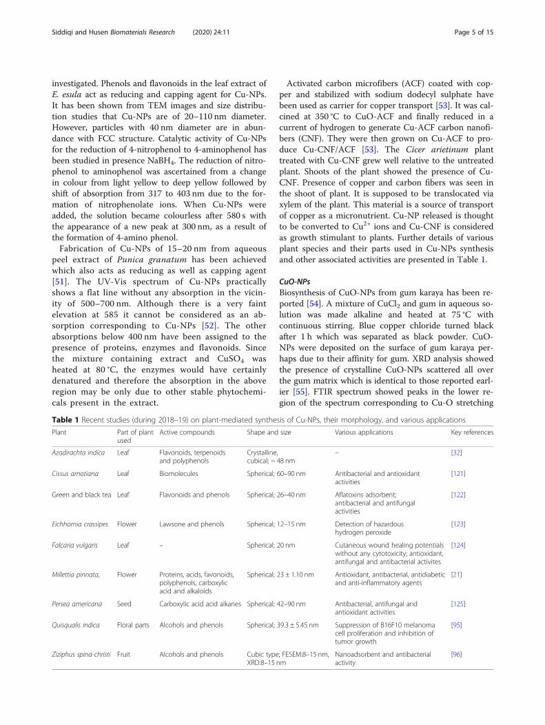
Table 1 Recent studies (during 2018–19) on plant-mediated synthesis of Cu-NPs, their morphology, and various applications
Plant Part of plantused
Active compounds Shape and size Various applications Key references
Azadirachta indica Leaf Flavonoids, terpenoidsand polyphenols
Crystalline,cubical; ~ 48 nm
– [32]
Cissus arnotiana Leaf Biomolecules Spherical; 60–90 nm Antibacterial and antioxidantactivities
[121]
Green and black tea Leaf Flavonoids and phenols Spherical; 26–40 nm Aflatoxins adsorbent;antibacterial and antifungalactivities
[122]
Eichhornia crassipes Flower Lawsone and phenols Spherical; 12–15 nm Detection of hazardoushydrogen peroxide
[123]
Falcaria vulgaris Leaf – Spherical; 20 nm Cutaneous wound healing potentialswithout any cytotoxicity; antioxidant,antifungal and antibacterial activites
[124]
Millettia pinnata, Flower Proteins, acids, favonoids,polyphenols, carboxylicacid and alkaloids
Spherical; 23 ± 1.10 nm Antioxidant, antibacterial, antidiabeticand anti-inflammatory agents
[21]
Persea americana Seed Carboxylic acid acid alkanes Spherical; 42–90 nm Antibacterial, antifungal andantioxidant activities
[125]
Quisqualis indica Floral parts Alcohols and phenols Spherical; 39.3 ± 5.45 nm Suppression of B16F10 melanomacell proliferation and inhibition oftumor growth
[95]
Ziziphus spina-christi Fruit Alcohols and phenols Cubic type; FESEM:8–15 nm,XRD:8–15 nm
Nanoadsorbent and antibacterialactivity
[96]
Siddiqi and Husen Biomaterials Research (2020) 24:11 Page 5 of 15
frequency. It is difficult to distinguish between Cu-O ofcupric oxide and those of cuprous oxide as the NPs maybe a mixture of both the CuO and Cu2O. Since the gumused in this work is a naturally occurring bio-polymer itcontains sugars and amino acids which act as reducingas well as capping agent for CuO-NPs. Also they remainadhered to the surface of the gum matrix through elec-trostatic force. Abboud et al. [56] have reported the fab-rication of CuO-NPs from Bifurcaria bifurcate (brownalga) purely in aqueous medium. Formation of NPs wasascertained from a change in colour from dark blue→colourless→deep red→black. It was a slow processbut on heating at 100 °C the reduction of Cu2+ to CuO-NPs was facilitated [57]. UV-Vis spectrum of CuO-NPsexhibited peaks at 260 and 650 nm. The former wasassigned to cuprous oxide NPs and the latter has beenattributed to cupric oxide NPs [58, 59]. Terpenoidspresent in the algal extract are responsible for the reduc-tion ofCu2+to CuO-NPs. However, the peak observed at260 nm in the UV-region cannot be assigned to the pres-ence of cuprous oxide NPs because metal oxides (owingto their colour) can absorb only in the visible region ofthe spectrum. The colour of the colloidal mixture is nei-ther purely black nor reddish. It is reddish black owingto the presence of both the cuprous and cupric oxideNPs which is also supported by uneven distribution andtwo crystalline phases corresponding to mono-clinic cu-pric oxide (CuO) and cubic cuprous oxide Cu2O NPs[45, 59, 60]. There were two types of NPs identified fromTEM images. Spherical ones were in abundance alongwith some elongated CuO-NPs ranging between 5 and45 nm diameters. Diterpenoids are the major compo-nents in alga which work as reductant and stabilizer forCuO-NPs [61].Fabrication of CuO-NPs from leaf extract (Aloe vera)
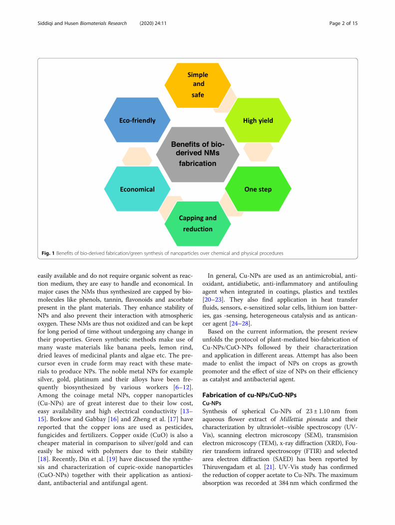
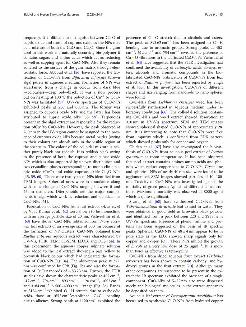
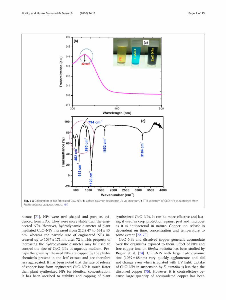
by Vijay Kumar et al. [62] were shown to be monoclinicwith an average particle size of 20 nm. Vishveshvar et al.[63] have shown CuO-NPs (obtained from Ixoro cocci-nea leaf extract) of an average size of 300 nm because ofthe formation of NP clusters. CuO-NPs obtained fromRuellia tuberosa aqueous extract were characterized byUV-Vis, FTIR, TEM, FE-SEM, EDAX and DLS [64]. Inthis experiment, the aqueous copper sulphate solutionwas added to the leaf extract showing a pale yellow tobrownish black colour which had indicated the forma-tion of CuO-NPs Fig. 3a). The absorption peak at 327nm was confirmed by SPR (Fig. 3b) and also the forma-tion of CuO nanorods of ~ 83.23 nm. Further, the FTIRstudies have shown the characteristic peaks at 452 cm− 1,612 cm− 1, 794 cm− 1, 893 cm− 1, 1120 cm− 1, 1652 cm− 1
and 3184 cm− 1 in 500–4000 cm− 1 range (Fig. 3c). Bandsat 3184 cm− 1exhibited O −H stretch due to carboxylicacids, those at 1652 cm− 1established −C=C− bendingdue to alkenes. Strong bands at 1120 cm− 1exhibited the
presence of C −O stretch due to alcohols and esters.The peak at 893.62 cm− 1 has been assigned to C −Hbending due to aromatic groups. Strong peaks at 452cm− 1, 612 cm− 1 and 794 cm− 1 revealed the presence ofCu −O vibrations in the fabricated CuO-NPs. Vasantharajet al. [64] have suggested that the FTIR investigation hadconfirmed the availability of carboxylic acids, alkenes, es-ters, alcohols and aromatic compounds in the bio-fabricated CuO-NPs. Fabrication of CuO-NPs from leafextract of Psidium guajava has been reported by Singhet al. [65]. In this investigation, CuO-NPs of differentshapes and size ranging from nanorods to nano sphereswere found.CuO-NPs from Eichhornia crassipes weed has been
successfully synthesized in aqueous medium under la-boratory conditions [66]. The colloidal solution contain-ing CuO-NPs and weed extract showed absorption at310 nm in UV-Vis spectrum. SEM and TEM imagesshowed spherical shaped CuO-NPs of approximately 28nm. It is interesting to note that CuO-NPs were freefrom impurity which is confirmed from EDX patternwhich showed peaks only for copper and oxygen.Ghidan et al. [67] have also investigated the biosyn-
thesis of CuO-NPs from aqueous peel extract of Punicagranatum at room temperature. It has been observedthat peel extract contains amines amino acids and phe-nols which reduce copper ions to CuO-NPs. Crystallineand spherical NPs of nearly 40 nm size were found to beagglomerated. SEM images showed particles of 10–100nm. Toxicity of CuO-NPs was investigated against themortality of green peach Aphids at different concentra-tions. Maximum mortality was observed at 8000 μg/mlwhich is quite significant.Sivaraj et al. [68] have synthesized CuO-NPs from
Tabernaemontana divaricate leaf extract in water. Theywere obtained in good yield as brownish black powderand identified from a peak between 220 and 225 nm inUV-Vis spectrum. Presence of phenol, amine and pro-teins has been suggested on the basis of IR spectralpeaks. Spherical CuO-NPs of 48 ± 4 nm appear to be inpure state as the EDX showed sharp signals only forcopper and oxygen [69]. These NPs inhibit the growthof E. coli at a very low dose of 25 μgml− 1. It is morethan twice as effective as tetracycline.CuO-NPs from dried aqueous fruit extract (Tribulus
terrestris) has been shown to contain carbonyl and hy-droxyl groups in the fruit extract [70]. Although manyother compounds are suspected to be present in the ex-tract the IR spectrum exhibited the presence of a singlecomponent. CuO-NPs of 5–22 nm size were dispersednicely and biological molecules in the extract appear tobe deposited on them.Aqueous leaf extract of Pterospermum acerifolium has
been used to synthesize CuO-NPs from hydrated copper
Siddiqi and Husen Biomaterials Research (2020) 24:11 Page 6 of 15
nitrate [71]. NPs were oval shaped and pure as evi-denced from EDX. They were more stable than the engi-neered NPs. However, hydrodynamic diameter of plantmediated CuO-NPs increased from 212 ± 47 to 634 ± 40nm, whereas the particle size of engineered NPs in-creased up to 1037 ± 171 nm after 72 h. This property ofincreasing the hydrodynamic diameter may be used tocontrol the size of CuO-NPs in aqueous medium. Per-haps the green synthesized NPs are capped by the phyto-chemicals present in the leaf extract and are thereforeless aggregated. It has been noted that the rate of releaseof copper ions from engineered CuO-NP is much fasterthan plant synthesized NPs for identical concentration.It has been ascribed to stability and capping of plant
synthesized CuO-NPs. It can be more effective and last-ing if used in crop protection against pest and microbesas it is antibacterial in nature. Copper ion release isdependent on time, concentration and temperature tosome extent [72, 73].CuO-NPs and dissolved copper generally accumulate
over the organisms exposed to them. Effect of NPs andfree copper ions on Elodea nuttallii has been studied byRegier et al. [74]. CuO-NPs with large hydrodynamicsize (1059 ± 88 nm) very quickly agglomerate and didnot change even when irradiated with UV light. Uptakeof CuO-NPs in suspension by E. nuttallii is less than thedissolved copper [75]. However, it is contradictory be-cause large quantity of accumulated copper has been
Fig. 3 a Colouration of bio-fabricated CuO-NPs, b surface plasmon resonance UV-vis spectrum, c FTIR spectrum of CuO-NPs as fabricated fromRuellia tuberosa aqueous extract [64]
Siddiqi and Husen Biomaterials Research (2020) 24:11 Page 7 of 15
found in Landoltia punctata when exposed to CuO-NPsthan when they were exposed to similar concentrationof dissolved copper [76]. It has been argued that copperis solubilized from CuO-NPs due to acid exuded fromgrowing shoots. However, copper ions would be avail-able only if reduction of CuO-NPs occurs followed by itsoxidation.
CuOþH2Redn → CuþH2O ð2Þ
Cu Oxdn→Cu2þ þ 2e ð3Þ
It is obvious that more copper ions will be availablefrom copper salts because it is highly ionized in waterand can be transported to different parts of plantsthrough osmosis. CuO-NPs get accumulated due tolarge size.
CuSO4⇋Cu2þ þ 2e− ð4Þ
CuO-NPs from Gloriosa superb leaf extract in waterhave been synthesized [77]. Spherical CuO-NPs of 8–17nm were obtained in pure state as the XRD patternshows single phase monoclinic structure although SEMimages show particles of smaller size (5–10 nm).Formation of CuO-NPs from Carica papaya leaf ex-
tract has been reported [78]. A sharp peak in the FTIRspectrum of colloidal cupric oxide NPs at 473 cm− 1 hasbeen taken as an evidence for its formation. Presence ofcuprous oxide NPs has been eliminated due to absenceof any characteristic peak around 605–660 cm− 1. Rodshaped CuO-NPs of 140 nm are crystalline and haveFCC structure [79]. Larger particles (614 nm) are notmonodispersed perhaps due to capping by the phyto-chemicals in the papaya leaf extract.Leaf extract of Calotropis gigantea has also been used
for the fabrication of CuO-NPs. Steriods and polyphe-nols in the extract reduce Cu2+ ions to CuO-NPs andalso act as stabilizer for them. It has been stated [26]that oxygen of ester and phenols form metal chelate withcopper ion. On heating the chelate complex, CuO-NPsare obtained. It is surprising that the authors have saidthat CuO-NPs are formed by the phytochemicals andpure NPs are obtained after heating at 400 °C. A metalcomplex can be formed by a metal ion only. Free metalin atomic state cannot form a complex. A chelate withphenol cannot be formed as it is a monodentate ligandand cannot form a ring. By heating copper nitrate at400 °C, CuO-NPs cannot be obtained because oxidationof copper to CuO can occur only at 900 °C. CuO-NPswere already formed by the leaf extract, and there wereno complex whatsoever. Such hypothesis is baseless.Heating at 400 °C will cause burning of any organic mat-ter left in excess and shall leave soot or carbon as impur-ity. Spherical CuO-NPs of 20–30 nm were formed as the
EDX showed peaks only for copper and oxygen separ-ately. The NPs were not well scattered.Natural phytochemicals are frequently used for the
fabrication of metal and or metal oxide NPs. A facilesynthesis of CuO-NPs from Gundelia tournefortii aque-ous extract of has been reported [80]. It has been shownthat Cu2+ to CuO-NPs conversion occurs after heatingthe mixture of plant extract and CuCl2 at 60 °C for 2 h.IR spectra of extract and NPs exhibited the presence ofphenolic compounds absorbed on the surface of CuO-NPs [81, 82]. NPs are highly crystalline [80] and spher-ical. Authors have concluded from EDX pattern that thepresence of oxygen refers to the oxidation of Cu-NPswhen exposed to air. It is not true because the Cu andoxygen peaks are due to CuO-NPs.CuO-NPs fabricated from the aqueous flower extract
of Anthemis nobilis had FCC, crystalline structure [83].These NPs were found to be useful for the synthesis ofpropargylamines in moderate yield. Vegetable peels havealso been used for the synthesis of CuO-NPs [84]. Cauli-flower (Brassica oleracea), potato (Solanum tuberosum)and pea (Pisum sativum) peel extracts yield CuO-NPsfrom CuCl2. 2H2O.This reaction was very slow as it took24 h at 60 °C to yield NPs. All NPs obtained from theabove sources were monoclinic ranging from 22.2 to31.60 nm. Since IR spectra did not show any peak at610 cm− 1 the presence of Cu2O was ruled out [85]. Asthe Cu2O is deep red it could have been easily noticed.Distinction between Cu2O and CuO can hardly be madeon the basis of Cu-O stretching frequencies as they arevery closely spaced. Morphology of CuO-NPs preparedfrom different vegetable wastes vary in size. Their cata-lytic activity is also different from each other due to theirshape, size and concentration.Mixed metal nanomaterials have also gained popular-
ity due to their multidisciplinary application. Biofabrica-tion of Pd/CuO-NPs from Theobroma cacao seedextract and their application as catalyst has been re-ported [86]. The Pd/CuO-NPs were highly stable be-cause no change was observed even when it was left for30 days. Antioxidants like epicatechin, catechin and theirderivatives in cocoa seed extract act as strong reducingas well as capping agent. Pd has face centered cubicstructure mixed with CuO-NPs of about 40 nm. Ag-glomeration is prevented due to capping of Pd/CuO-NPs. Presence of oxygen in EDS spectrum has been as-cribed to oxidation of Cu NPs to CuO-NPs. When Pd/CuO does not contain free copper ions what is the needof oxygen for oxidation of CuO-NPs. However, oxidationof metallic copper in presence of water can take severalhours nevertheless it would give CuSO4 with a charac-teristic blue colour. However, authors assumption needsverification. The mechanism proposed for the formationof metal NPs and their nucleation is not convincing. All
Siddiqi and Husen Biomaterials Research (2020) 24:11 Page 8 of 15
the phenolic –OH groups cannot be oxidized even inmultiple steps as the drainage of electron from it willmake the ring unstable. If such nucleation occurs ametal cluster would be formed.Seeds of many leguminous plants also contain fairly
reasonable amount of proteins, phenols, flavonoids, alka-loids and amino acids [87–89] and hence they are used asreducing agent to synthesize metal NPs. Crystalline mono-clinic CuO-NPs of 26.6 nm have been synthesized fromblack been extract and their anti-cancer activity againstHeLa cells has been reported [90]. They were effective in avery low concentration range (0.5–1mgml− 1) and shortduration of time (12–48 h). It has been ascribed to intra-cellular ROS generation in a dose dependent manner.The mechanism suggested for the formation of CuO-
NPs is beyond imagination and conceptually non con-vincing. Authors have argued that the precursor CuSO4
reacts with hydroxyl anion OH-, generated by theionization of water molecules and eventually reduced byphytochemicals present in the seed extract. It must beclarified at this stage that:(a) It is universally known that water is not ionized
until acidified water is electrolysed and (b) Cu (OH)2can be formed only if NaOH or requisite amount ofNH4OH is added to a copper salt as shown below.
CuSO4 þ 2NaOH→Cu OHð Þ2 þNa2SO4 ð5Þ
CuSO4 þ 2NH4OH→Cu OHð Þ2 þ NH4ð Þ2SO4 ð6Þ
However, CuSO4 remains ionized in aqueous mediumor it may form hexaaquo copper complex, [Cu(H2O)6].
CuSO4⇋Cu2þ þ SO42− ð7Þ
Free Cu2+ ion is then reduced by protein or polyphe-nol available in the black bean extract. Phenol is
oxidized to phenolate ion which provides electron toCu2+ ions to from CuO-NPs.Bawadi [91] and Bobe et al. [92] have reported that
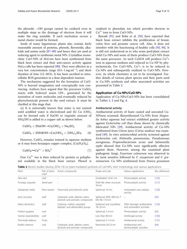
black bean extract inhibits the proliferation of breast,colon liver and prostate cancer cell. Also, it does notinterfere with the functioning of healthy cells [93, 94]. Itis still not understood as to why some peel/plant extractyield Cu-NPs and some of them produce CuO-NPs fromthe same precursor. As such CuSO4 will produce Cu2+ions in aqueous medium and reduced to Cu-NP by phy-tochemicals. For CuO-NPs, Cu2+ is to be reduced toCu-NPs and subsequently oxidized to CuO-NPs. How-ever, its whole chemistry is yet to be investigated. Fur-ther details of various plant species and their parts usedin Cu-NPs synthesis and other associated activities arepresented in Table 2.
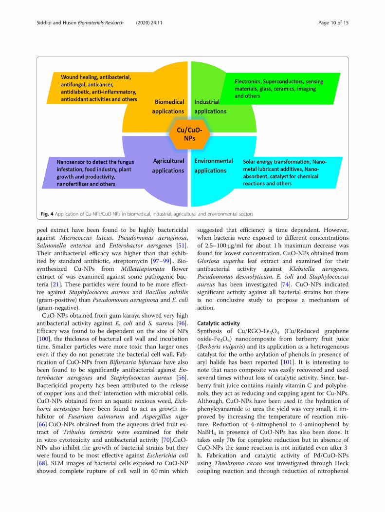
Application of Cu-NPs/CuO-NPsApplication of Cu-NPs/CuO-NPs has been consolidatedin Tables 1, 2 and Fig. 4.
Antibacterial activityAntibacterial activity of foam coated and uncoated Cu-NPswas screened. Biosynthesized Cu-NPs from Magno-lia kobus aqueous leaf extract exhibited greater activityagainst Escherichia coli than those shown by chemicallyfabricated NPs [29]. Antibacterial activity of Cu-NPssynthesized from Citron juice (Citrus medica) was exam-ined [49]. In vitro antimicrobial activity screened againstEscherichia coli, Klebsiella pneumoniae, Pseudomonasaeruginosa, Propionibacterium acnes and Salmonellatyphi showed that Cu-NPs were significantly effectiveagainst them. However, among the examined plantpathogenic fungi, Fusarium culmorum was observed tobe most sensitive followed by F. oxysporum and F. gra-minearum. Cu NPs synthesized from Punica granatum
Table 2 Recent studies (during 2018–19) on plant-mediated synthesis of CuO-NPs, their morphology and various applications
Plant Part of plantused
Active compounds Shape and size Various applications Key references
Aloe vera Leaf – Octahedral; 24-61 nm Photocatalytic activity [126]
Ferulago angulate Aerial part – Shell like sheet structure;44 nm
Photocatalytic activity [127]
Galeopsidis herba Plant extract Flavonoids and phenolic acids Spherical; 10 nm Antioxidant and catalyticactivity
[128]
Ixoro coccinea Leaf Carboxylic acids, alkenes, esters,alcohols and aromatic compounds
Spherical; SEM: 300 nm; TEM: 80–110 nm
– [63]
Malus domestica Leaf Carbonyl, methyl, saturatedaliphatic and alkane/alkyl groups
Spherical and crystalline;18–20 nm
DNA cleavage, antibacterialand antioxidant activities
[129]
Psidium guajava Leaf – Spherical; 2–6 nm Photocatalytic activity [65]
Stachys lavandulifolia Leaf – Less than 80 nm Antifungal activity [130]
Terminalia belerica Fruits – Spherical; 9–14 nm Antibacterial activity [131]
Ruellia tuberosa Leaf Carboxylic acids, alkenes, esters,alcohols and aromatic compounds
Nanorods; 83.23 nm Antibacterial andphotocatalytic activities
[64]
Siddiqi and Husen Biomaterials Research (2020) 24:11 Page 9 of 15
peel extract have been found to be highly bactericidalagainst Micrococcus luteus, Pseudomonas aeruginosa,Salmonella enterica and Enterobactor aerogenes [51].Their antibacterial efficacy was higher than that exhib-ited by standard antibiotic, streptomycin [97–99].. Bio-synthesized Cu-NPs from Millettiapinnata flowerextract of was examined against some pathogenic bac-teria [21]. These particles were found to be more effect-ive against Staphylococcus aureus and Bacillus subtilis(gram-positive) than Pseudomonas aeruginosa and E. coli(gram-negative).CuO-NPs obtained from gum karaya showed very high
antibacterial activity against E. coli and S. aureus [96].Efficacy was found to be dependent on the size of NPs[100], the thickness of bacterial cell wall and incubationtime. Smaller particles were more toxic than larger oneseven if they do not penetrate the bacterial cell wall. Fab-rication of CuO-NPs from Bifurcaria bifurcate have alsobeen found to be significantly antibacterial against En-terobacter aerogenes and Staphylococcus aureus [56].Bactericidal property has been attributed to the releaseof copper ions and their interaction with microbial cells.CuO-NPs obtained from an aquatic noxious weed, Eich-horni acrassipes have been found to act as growth in-hibitor of Fusarium culmorum and Aspergillus niger[66].CuO-NPs obtained from the aqueous dried fruit ex-tract of Tribulus terrestris were examined for theirin vitro cytotoxicity and antibacterial activity [70].CuO-NPs also inhibit the growth of bacterial strains but theywere found to be most effective against Escherichia coli[68]. SEM images of bacterial cells exposed to CuO-NPshowed complete rupture of cell wall in 60 min which
suggested that efficiency is time dependent. However,when bacteria were exposed to different concentrationsof 2.5–100 μg/ml for about 1 h maximum decrease wasfound for lowest concentration. CuO-NPs obtained fromGloriosa superba leaf extract and examined for theirantibacterial activity against Klebsiella aerogenes,Pseudomonas desmolyticum, E. coli and Staphylococcusaureus has been investigated [74]. CuO-NPs indicatedsignificant activity against all bacterial strains but thereis no conclusive study to propose a mechanism ofaction.
Catalytic activitySynthesis of Cu/RGO-Fe3O4 (Cu/Reduced grapheneoxide-Fe3O4) nanocomposite from barberry fruit juice(Berberis vulgaris) and its application as a heterogeneouscatalyst for the ortho arylation of phenols in presence ofaryl halide has been reported [101]. It is interesting tonote that nano composite was easily recovered and usedseveral times without loss of catalytic activity. Since, bar-berry fruit juice contains mainly vitamin C and polyphe-nols, they act as reducing and capping agent for Cu-NPs.Although, CuO-NPs have been used in the hydration ofphenylcyanamide to urea the yield was very small, it im-proved by increasing the temperature of reaction mix-ture. Reduction of 4-nitrophenol to 4-aminophenol byNaBH4 in presence of CuO-NPs has also been done. Ittakes only 70s for complete reduction but in absence ofCuO-NPs the same reaction is not initiated even after 3h. Fabrication and catalytic activity of Pd/CuO-NPsusing Theobroma cacao was investigated through Heckcoupling reaction and through reduction of nitrophenol
Fig. 4 Application of Cu-NPs/CuO-NPs in biomedical, industrial, agricultural and environmental sectors
Siddiqi and Husen Biomaterials Research (2020) 24:11 Page 10 of 15

to aminophenol [86]. The catalyst was reused up to 6 cy-cles. Mechanistic pathway is yet to be established. Cu-NPs from aqueous leaf extract of Euphorbia esula hasalso been used for catalytic reduction of 4-nitrophenoland ligand free Ullmann-coupling reaction [50].The CuO-NPs fabricated from the leaf extract of Psi-
dium guajava has exhibited a remarkable degradation effi-ciency against the industrial dyes. It degraded Nile blue by93%, and reactive yellow 160 by 81% in just 120min withapparent rate constants of 0.023 and 0.014min− 1, respect-ively [65].CuO-NPs obtained from Carica papaya leaf ex-tract has been exploited in the photocatalytic degradationof Coomassive brilliant blue dye in sunlight [78].
Plant growth responsePlants are very susceptible to minor changes in the en-vironment (abiotic stress) and supply of any trace elem-ent/NPs either as a nutrient or as a foreign matter [28,102–108]. Cu-NPs enhanced the root and shoot growthof mung bean [109], but reduced shoot growth in wheat[110]. Yasmeen et al. [111] synthesized Cu and Fe-NPsand demonstrated their role on the growth as well asyield of wheat varieties. The metal NPs synthesized fromonion extract of 15–30 nm diameters had irregularshape. Wheat varieties were treated with 20, 25, 30, 35and 40 ppm Cu-NPs at different stages of growth untilproduction of seeds. Spike length was invariably in-creased or remained unchanged when treated with 25ppm Cu-NPs but significantly increased the number ofgrains. However, higher Cu concentration reduced thenumber of grains/spike and overall 1000 grain weight.Copper and iron NPs together at 25 ppm concentrationhave better effect on production and yield of wheat thancopper or iron alone. Cu-NPs increased the grain yieldand stress tolerance in wheat plant through starch deg-radation. Sugar content and SOD activity was enhancedin seeds treated with copper and iron NPs. Quantity ofcopper was increased while iron remained unchanged inseeds treated with Cu and Fe NPs [111].ACF coated with copper and stabilized with sodium
dodecyl sulphate have been used as carrier for coppertransport [53]. It was calcined at 350 to CuO-ACF andfinally reduced in a current of hydrogen to generate Cu-ACF carbon nanofibers. They were then grown on Cu-ACF to produce Cu-CNF/ACF [112]. The Cicer arieti-num plant treated with Cu-CNF grew well in compari-son to control plants.Impact of engineered CuO-NPs on growth and devel-
opment of Arabidopsis thaliana at molecular level hasbeen explored [113]. Their seedlings were treated withdifferent doses of CuO-NPs for 3 weeks. Plant biomass,total chlorophyll content and root elongation invariablydecreased while an increase in anthocyanin, lipid peroxi-dation and proline were observed. Lignin was also found
to be deposited in root. CuO-NPs treated seedlings alsoshowed an increase in superoxide and hydrogen perox-ide in leaves and roots which is directly proportional toits concentration. As a consequence of oxidative stress,the main root growth was inhibited but lateral root for-mation was initiated. This is mainly due to metabolicimbalance and as a response to resistance caused byCuO-NPs [114]. However, the plant system in responseto such stress produces antioxidants to prevent the dam-age by ROS [104, 105, 115] which subsequently causessome changes in metabolic functions. Very recently,Chung et al. [116] have examined the effect of CuO-NPson Brassica rapa seedlings. CuO-NPs treated seedlingshave shown reduced root and shoot length, chlorophyll,carotenoid and sugar content, while proline and antho-cyanins were increased. Additionally, production of mal-ondialdehyde and hydrogen peroxide were increased inCuO-NPs treated seedlings which has been associatedwith DNA damage.
Miscellaneous responseRecently, the biosynthesized Cu-NPs from flower extractof Millettia pinnata have also been shown to exhibitanti-diabetic and anti-inflammatory activities [21]. Hu-man mesenchymal stem cells exposed to CuO-NPs at aconcentration of 25 μg ml− 1 reduced their viability.CuO-NPs at a dose of or below10 μg/ml were non toxicto normal cells and therefore, a safe concentration maybe used to prevent the interaction of CuO-NPs with nor-mal mesenchymal cells. It has been suggested that CuO-NPs release Cu ions which are antibacterial and antican-cerous [117, 118]. It also means that reduction of Cu-NPs to copper ions occur which are toxic to normal bac-terial cells and cancer cells in mammals. Toxicity ofCuO-NPs obtained from aqueous leaf extract of Pteros-permum acerifolium was determined against Daphniamagna at several concentrations [71]. The engineeredNPs were several times more toxic to Daphnia thangreen synthesized NPs [119, 120]. Release of copper ionsfrom CuO-NPs is the main cause of toxicity to Daphnia.Dissolved copper ions from CuO-NPs get accumulatedaround Daphnia which cause toxicity.
ConclusionPlenty of nano particles are synthesized every year butonly some of them are put to beneficial use. Green syn-thesized NPs of desired shape, size and stability can en-hance their overall qualities for future applications. Itappears that all plant species contain some type of phy-tochemicals which reduce the metal salts and metal ox-ides into their NPs. Cu and CuO-NPshave shown greatpromise as biocide and mild antibacterial agent. Eventhough, biofabrication of metal NPs from plants and mi-crobes do not leave toxic residues in environment, their
Siddiqi and Husen Biomaterials Research (2020) 24:11 Page 11 of 15
safe disposal is necessary. Further, it is also essential toutilize the fabricated Cu and CuO-NPs as biomedicine,environmental remediation and agricultural practices.Attention must be focused on their biocompatibility.
AbbreviationsNPs: Nanoparticles; Cu-NPs: Copper nanoparticles; CuO-NPs: Cupric-oxidenanoparticles; UV-Vis: Ultraviolet–visible spectroscopy; SEM: Scanningelectron microscopy; TEM: Transmission electron microscopy; XRD: X-raydiffraction; FTIR: Fourier transform infrared spectroscopy; SAED: Selected area(electron) diffraction; EDAX: Energy-dispersive x-ray spectroscopy;ACF: Activated carbon microfibers.
AcknowledgementsAuthors are thankful to publishers for permission to adopt figures in thisreview.
Authors’ contributionsAH gathered the research data. KSS and AH analyzed these data and wrotethis review paper. Both authors read and approved the final manuscript.
FundingNot applicable.
Availability of data and materialsNot applicable.
Ethics approval and consent to participateNot applicable.
Consent for publicationNot applicable.
Competing interestsThe authors declare that they have no competing interests.
Author details1Department of Chemistry, Aligarh Muslim University, Aligarh, Uttar Pradesh202002, India. 2Wolaita Sodo University, P.O. Box: 138, Wolaita, Ethiopia.
Received: 11 March 2020 Accepted: 5 May 2020
References1. Husen A, Siddiqi KS. Phytosynthesis of nanoparticles: concept, controversy
and application. Nano Res Lett. 2014;9:229.2. Siddiqi KS, Husen A, Rao RAK. A review on biosynthesis of silver
nanoparticles and their biocidal properties. J Nanobiotechnol. 2018;16:14.3. Husen A. Introduction and techniques in nanomaterials formulation: An
overview. In: Husen A, Jawaid M, editors. Nanomaterials for Agriculture andForestry Applications. Cambridge: Elsevier Inc; 2020. p. 1–14.
4. Siddiqi KS, Husen A. Fabrication of metal and metal oxide nanoparticles byalgae and their toxic effects. Nano Res Lett. 2016;11:363.
5. Siddiqi KS, Husen A. Fabrication of metal nanoparticles from fungi andmetal salts: scope and application. Nano Res Lett. 2016;11:98.
6. Philip D, Unni C, Aromal SA, Vidhu VK. Murraya koenigii leaf-assisted rapidgreen synthesis of silver and gold nanoparticles. Spectrochem Acta A MolBiomol Spectrosc. 2011;78:899–904.
7. Banerjee P, Satapathy M, Mukhopahayay A, Das P. Leaf extract mediatedgreen synthesis of silver nanoparticles from widely available Indian plants:synthesis, characterization, antimicrobial property and toxicity analysis.Bioresour Bioprocess. 2014;1:3.
8. Nagar N, Jain S, Kachhawah P, Devra V. Synthesis and characterization ofsilver nanoparticles via green route. Korean J Chem Eng. 2016;33:2990–7.
9. Husen A. Gold nanoparticles from plant system: synthesis, characterizationand their application. In: Ghorbanpour M, Manika K, Varma A, editors.Nanoscience and Plant–Soil Systems. Soil Biology. Cham: Springer, 2017;48:455–479.
10. Siddiqi KS, Husen A. Recent advances in plant-mediated engineered goldnanoparticles and their application in biological system. J Trace ElementsMed Biol. 2017;40:10–23.
11. Siddiqi KS, Rashid M, Rahman A, Tajuddin HA, Rehman S. Biogenicfabrication and characterization of silver nanoparticles using aqueous-ethanolic extract of lichen (Usnea longissima) and their antimicrobial activity.Biomat Res. 2018;22:23.
12. Siddiqi KS, Rashid M, Tajuddin RS, Husen A. Synthesis of silver nanoparticlesusing aqueous leaf extract of Diospyros montana Roxb. And theirantimicrobial activity against some clinical isolates. BioNanoSci. 2019;9:302–12.
13. Umer A, Naveed S, Ramzan N, Rafiqui MS. Selection of a suitable methodfor the synthesis of copper nanoparticles. Nano. 2012;7:1230005.
14. Jain S, Jain A, Kachhawah P, Devra V. Synthesis and size control of coppernanoparticles and their catalytic application. Trans Nonferrous Met SocChina. 2015;25:3995–4000.
15. Tiwari M, Jain P, Hariharapura RC, Narayanan K, Udaya BK, Udupa N, Rao JV.Biosynthesis of copper nanoparticles using copper-resistant Bacillus cereus, asoil isolate. Process Biochem. 2016;51:1348–56.
16. Borkow G, Gabbay J. Copper, an ancient remedy returning to fightmicrobial, fungal and viral infections. Cur Chem Biol. 2009;3:272–8.
17. Zheng XG, Xu CN, Tomokiyo Y, Tanaka E, Yamada H, Soejima Y. Observationof charge stripes in cupric oxide. Phys Rev Lett. 2000;85:5170–3.
18. Ren G, Hu D, Cheng EW, Vargas-Reus MA, Reip P, Allaker RP.Characterisation of copper oxide nanoparticles for antimicrobialapplications. Int J Antimicrob Agent. 2009;33:587–90.
19. Din MI, Arshad F, Hussain Z, Mukhtar M. Green adeptness in the synthesisand stabilization of copper nanoparticles: catalytic, antibacterial, cytotoxicity,and antioxidant activities. Nano Res Lett. 2017;12:638.
20. Apostolov AT, Apostolova IN, Wesselinowa JM. Dielectric constant ofmultiferroic pure and doped CuO nanoparticles. Solid State Commun. 2014;192:71–4.
21. Thiruvengadam M, Chung IM, Gomathi T, Ansari MA, Khanna VG, Babu V,Rajakumar G. Synthesis, characterization and pharmacological potential ofgreen synthesized copper nanoparticles. Bioprocess Biosyst Eng. 2019;42:1769–77.
22. Pariona N, Mtz-Enriquez AI, Sanchez-Rangel D, Carrion G, Paraguay-DelgadoF, Rosas-Saito G. Green-synthesized copper nanoparticles as a potentialantifungal against plant pathogens. RSC Adv. 2019;9:18835–43.
23. Lee Y, Choi JR, Lee KJ, Stott NE, Kim D. Large-scale synthesis of coppernanoparticles by chemically controlled reduction for applications of inkjet-printed electronics. Nanotechnology. 2008;19:598–604.
24. Rubilar O, Rai M, Tortella G, Diez MC, Seabra AB, Durán N. Biogenicnanoparticles: copper, copper oxides, copper sulphides, complex coppernanostructures and their applications. Biotechnol Lett. 2013;35:1365–75.
25. Waser O, Hess M, Güntner A, Novák P, Pratsinis SE. Size controlled CuOnanoparticles for Li-ion batteries. J Power Sour. 2013;241:415–22.
26. Sharma JK, Akhtar MS, Ameen S, Srivastava P, Singh G. Green synthesis ofCuO nanoparticles with leaf extract of Calotropis gigantea and its dye-sensitized solar cells applications. J All Comp. 2015;632:321–5.
27. Wang F, Li H, Yuan H, Sun Y, Chang F, Deng H, Xie L, Li H. A highly sensitivegas sensor based on CuO nanoparticles synthetized via a sol–gel method.RSC Adv. 2016;6:79343–9.
28. Joshi A, Sharma A, Bachheti RK, Husen A, Mishra VK. Plant-mediatedsynthesis of copper oxide nanoparticles and their biological applications. In:Husen A, Iqbal M, editors. Nanomaterials and Plant Potential. Cham:Springer International Publishing AG; 2019. p. 221–37.
29. Lee HJ, Song JY, Kim BS. Biological synthesis of copper nanoparticles usingMagnolia kobus leaf extract and their antibacterial activity. J Chem TechnolBiotechnol. 2013;88:1971–7.
30. Song JY, Jang HK, Kim BS. Biological synthesis of gold nanoparticles usingMagnolia kobus and Diopyros kaki leaf extracts. Process Biochem. 2009;44:1133–8.
31. Kulkarni V, Suryawanshi S, Kulkarni P. Biosynthesis of copper nanoparticlesusing aqueous extract of Eucalyptus sp. plant leaves. Curr Sci. 2015;109:255–27.
32. Nagar N, Devra V. Green synthesis and characterization of coppernanoparticles using Azadirachta indica leaves. Mat Chem Phys. 2018;213:44–51.
33. Brumbaugh AD, Cohen KA, Angelo SKS. Ultrasmall copper nanoparticlessynthesized with a plant tea reducing agent. ACS Sustain Chem Eng. 2014;2:1933–9.
Siddiqi and Husen Biomaterials Research (2020) 24:11 Page 12 of 15

34. Pileni MP, Lisiecki I. Nanometer metallic copper particle synthesis in reversemicelles. Colloids Surf A Physicochem Eng Asp. 1993;80:63–8.
35. Singh M, Sinha I, Premkumar M, Singh AK, Mandal RK. Structural and surfaceplasmon behavior of cu nanoparticles using different stabilizers. Colloid SurfA. 2010;359:88–94.
36. Vazquez-Vazquez C, Banobre-Lopez M, Mitra A, Arturo Lopez-Quintela M,Rivas J. Synthesis of small atomic copper clusters in microemulsions.Langmuir. 2009;25:8208–16.
37. Narayanan KB, Sakthivel N. Coriander leaf mediated biosynthesis of goldnanoparticles. Mater Lett. 2008;62:4588–90.
38. Sheny DS, Mathew J, Philip D. Phytosynthesis of au, Ag and au−Agbimetallic nanoparticles using aqueous extract and dried leaf of Anacardiumoccidentale. Spectrochim Acta Part A. 2011;79:254–62.
39. Yu W, Xie H, Chen L, Li Y, Zhang C. Synthesis and characterization ofmonodispersed copper colloids in polar solvents. Nano Res Lett. 2009;4:465–70.
40. Engels V, Benaskar F, Jefferson DA, Johnson BFG, Wheatley AEH.Nanoparticulate copper - routes towards oxidative stability. Dalton Trans.2010;39:6496–502.
41. Zhang Y, Zhu P, Li G, Zhao T, Fu X, Sun R, Zhou F, Wong C. Facilepreparation of monodisperse, impurity-free, and antioxidation coppernanoparticles on a large scale for application in conductive ink. ACS ApplMater Interf. 2014;6:560–7.
42. Cheirmadurai K, Biswas S, Murali R, Thanikaivelan P. Green synthesis ofcopper nanoparticles and conducting nanobiocomposites using plant andanimal sources. RSC Adv. 2016;4:19507–11.
43. Wang Y, Biradar AV, Wang G, Sharma KK, Duncan CT, Rangan S, Asefa T.Controlled synthesis of water-dispersible faceted crystalline coppernanoparticles and their catalytic properties. Chemistry. 2010;16:10735–43.
44. Demirskyi D, Agrawal D, Ragulya A. Neck formation between copperspherical particles under single-mode and multimode microwave sintering.Mat Sci Eng: A. 2010;A527:2142–5.
45. Swarnkar RK, Singh SC, Gopal R. Effect of aging on copper nanoparticlessynthesized by pulsed laser ablation in water: structural and opticalcharacterizations. Bull Mater Sci. 2011;34:1363–9.
46. Shende S, Ingle AP, Gade A, Rai M. Green synthesis of copper nanoparticlesby Citrus medica Linn. (Idilimbu) juice and its antimicrobial activity. World JMicrobiol Biotechnol. 2015;31:865–73.
47. Sastry ABS, Aamanchi RBK, Rama Linga Prasad CS, Murty BS. Large-scalegreen synthesis of cu nanoparticles. Environ Chem Lett. 2013;11:183–7.
48. Hirai H, Wakabayashi H, Komiyama M. Preparation of polymer-protectedcolloidal dispersions of copper. Bull Chem Soc Japan. 1986;59:367–72.
49. Zhu YJ, Qian YT, Zhang MW, Chen ZY, Xu DF. Preparation andcharacterization of nanocrystalline powders of cuprous oxide by using C-radiation. Mater Res Bull. 1994;29:377–83.
50. Nasrollahzadeh M, Sajadi SM, Khalaj M. Green synthesis of coppernanoparticles using aqueous extract of the leaves of Euphorbia esula L andtheir catalytic activity for ligand-free Ullmanncoupling reaction andreduction of 4-nitrophenol. RSC Adv. 2014;4:47313–8.
51. Kaur P, Thakur R, Chaudhury A. Biogenesis of copper nanoparticles usingpeel extract of Punica granatum and their antimicrobial activity againstopportunistic pathogens. Green Chem Lett Rev. 2016;9:33–8.
52. Hashemipour H, Zadeh ME, Pourakbari R, Rahimi P. Investigation onsynthesis and size control of copper nanoparticle via electrochemical andchemical reduction method. Int J Phys Sci. 2011;6:4331–6.
53. Ashfaq M, Verma N, Khan S. Carbon nanofibers as a micronutrient carrier inplants: efficient translocation and controlled release of cu nanoparticles.Environ Sci: Nano. 2017;4:138–48.
54. Padil VVT, Černík M. Green synthesis of copper oxide nanoparticles usinggum karaya as a biotemplate and their antibacterial application. Int JNanomedicine. 2013;8:889–98.
55. Das D, Nath BC, Phukon P, Dolui SK. Synthesis and evaluation of antioxidantand antibacterial behavior of CuO nanoparticles. Coll Surf B Biointerf. 2013;101:430–3.
56. Abboud Y, Saffaj T, Chagraoui A, El Bouari A, Brouzi K, Tanane O, Ihssane B.Biosynthesis, characterization and antimicrobial activity of copper oxidenanoparticles (CONPs) produced using brown alga extract (Bifurcariabifurcata). Appl Nanosci. 2014;4:571–6.
57. Krithiga N, Jayachitra A, Rajalakshmi A. Synthesis, characterization andanalysis of the effect of copper oxide nanoparticles in biological systems.Ind J Nano Sci. 2013;1:6–15.
58. Borgohain K, Murase N, Mahamuni S. Synthesis and properties of Cu2Oquantum particles. J Appl Phys. 2002;92:1292–7.
59. Yin M, Wu CK, Lou Y, Burda C, Koberstein JT, Zhu Y, O’Brien S. Copper oxidenanocrystals. J Am Chem Soc. 2005;127:9506–11.
60. Rahman A, Ismail A, Jumbianti D, Magdalena S, Sudrajat H. Synthesis ofcopper oxide nanoparticles by using Phormidium cyanobacterium. Indo JChem. 2009;9:355–60.
61. Kiruba Daniel SCG, Nehru K, Sivakumar M. Rapid biosynthesis of silvernanoparticles using Eichornia crassipes and its antibacterial activity. CurrNanosci. 2012;8:1–5.
62. Vijay Kumar PPN, Shameem U, Kollu P, Kalyani RL, Pammi SVN. Greensynthesis of copper oxide nanoparticles using Aloe vera leaf extract and itsantibacterial activity against fish bacterial pathogens. BioNanoSci. 2015;5:135–9.
63. Vishveshvar K, Aravind Krishnan MV, Haribabu K, Vishnuprasad S. Greensynthesis of copper oxide nanoparticles using Ixiro coccinea plant leavesand its characterization. BioNanoSci. 2018;8:554–8.
64. Vasantharaj S, Sathiyavimal S, Saravanan M, Senthilkumar P, Kavitha G,Shanmugavel M, Manikandan E, Pugazhendhi A. Synthesis of ecofriendlycopper oxide nanoparticles for fabrication over textile fabrics:characterization of antibacterial activity and dye degradation potential. JPhotochem Photobiol B Biol. 2018;191:149.
65. Singh J, Kumar V, Kim KH, Rawat M. Biogenic synthesis of copper oxidenanoparticles using plant extract and its prodigious potential forphotocatalytic degradation of dyes. Environ Res. 2019;177:108569.
66. Vanathi P, Rajiv P, Sivaraj R. Synthesis and characterization of Eichhornia-mediated copper oxide nanoparticles and assessing their antifungal activityagainst plant pathogens. Bull Mater Sci. 2016;39:1165–70.
67. Ghidan AY, Al-Antary TM, Awwad AM. Green synthesis of copper oxidenanoparticles using Punica granatum peels extract: effect on green peachaphid. Environ Nanotechnol Monit Manag. 2016;6:95–8.
68. Sivaraj R, Rahman PK, Rajiv P, Salam HA, Venckatesh R. Biogenic copperoxide nanoparticles synthesis using Tabernaemontana divaricate leaf extractand its antibacterial activity against urinary tract pathogen. SpectrochimActa A Mol Biomol Spectrosc. 2014;133:178–81.
69. Huang J, Li Q, Sun D, Lu Y, Su Y, Yang X, Wang H, Wang Y, Shao W, He N,Hong J, Chen C. Biosynthesis of silver and gold nanoparticles by novel sundried Cinnamomum camphora leaf. Nanotechnology. 2007;18:105104–15.
70. Gopinath V, Priyadarshini S, Al-Maleki AR, Alagiri M, Yahya R, Saravanan S,Vadivelu J. In vitro toxicity, apoptosis and antimicrobial effects of phyto-mediated copper oxide nanoparticles. RSC Adv. 2016;6:110986–95.
71. Saif S, Tahir A, Asim T, Chen Y. Plant mediated green synthesis of CuOnanoparticles: comparison of toxicity of engineered and plant mediatedCuO nanoparticles towards Daphnia magna. Nanomaterials. 2016;6:205.
72. Odzak N, Kistler D, Behra R, Sigg L. Dissolution of metal and metal oxidenanoparticles in aqueous media. Environ Pollut. 2014;191:132–8.
73. Adam N, Leroux F, Knapen D, Bals S, Blust R. The uptake of ZnO and CuOnanoparticles in the water-flea Daphnia magna under acute exposurescenarios. Environ Pollut. 2014;194:130–7.
74. Regier N, Cosio C, von Moos N, Slaveykova VI. Effects of copper-oxidenanoparticles, dissolved copper and ultraviolet radiation on copperbioaccumulation, photosynthesis and oxidative stress in the aquaticmacrophyte Elodea nuttallii. Chemosphere. 2015;128:56–61.
75. Perreault F, Oukarroum A, Melegari SP, Matias WG, Popovic R. Polymercoating of copper oxide nanoparticles increases nanoparticles uptake andtoxicity in the green alga Chlamydomonas reinhardtii. Chemosphere. 2012;87:1388–94.
76. Shi J, Abid AD, Kennedy IM, Hristova KR, Silk WK. To duckweeds (Landoltiapunctata), nanoparticulate copper oxide is more inhibitory than the solublecopper in the bulk solution. Environ Pollut. 2011;159:1277–82.
77. Raja Naika H, Lingaraju K, Manjunath K, Kumar D, Nagaraju G, Suresh D,Nagabhushana H. Green synthesis of CuO nanoparticles using Gloriosa superbaL. extract and their antibacterial activity. J Taibah Univ Sci. 2015;9:7–12.
78. Sankar R, Manikandan P, Malarvizhi V, Fathima T, Shivashangari KS,Ravikumar V. Green synthesis of colloidal copper oxide nanoparticles usingCarica papaya and its application in photocatalytic dye degradation.Spectrochim Acta A Mol Biomol Spectrosc. 2014;121:746–50.
79. Ethiraj AS, Kang DJ. Synthesis and characterization of CuO nanowires by asimple wet chemical method. Nano Res Lett. 2012;7:70.
80. Nasrollahzadeh M, Maham M, Sajadi SM. Green synthesis of CuOnanoparticles by aqueous extract of Gundelia tournefortii and evaluation of
Siddiqi and Husen Biomaterials Research (2020) 24:11 Page 13 of 15
their catalytic activity for the synthesis of N-monosubstituted ureas andreduction of 4-nitrophenol. J Colloid Interface Sci. 2015;455:245–53.
81. Adzet T, Puigmacia M. High-performance liquid chromatography ofcaffeoylquinic acid derivatives of Cynara scolymus L. leaves. JChromatograph A. 1985;348:447–53.
82. Haghi G, Hatami A, Arshi R. Distribution of caffeic acid derivatives inGundelia tournefortii L. Food Chem. 2011;124:1029–35.
83. Nasrollahzadeh M, Mohammad Sajadi S, Rostami-Vartooni A. Greensynthesis of CuO nanoparticles by aqueous extract of Anthemis nobilisflowers and their catalytic activity for the A3 coupling reaction. J ColloidInterface Sci. 2015;459:183–8.
84. Ullah H, Ullah Z, Fazal A, Irfan M. Use of vegetable waste extracts forcontrolling microstructure of CuO nanoparticles: green synthesis,characterization, and photocatalytic applications. J Chem. 2017;2721798:5.
85. Ranjbar-Karimi R, Bazmandegan-Shamili A, Aslani A, Kaviani K. Sonochemicalsynthesis, characterization and thermal and optical analysis of CuOnanoparticles. Phys B Condens Matter. 2010;405:3096–100.
86. Nasrollahzadeh M, Sajadi SM, Rostami-Vartooni A, Bagherzadeh M. Greensynthesis of Pd/CuO nanoparticles by Theobroma cacao L. seeds extract andtheir catalytic performance for the reduction of 4-nitrophenol andphosphine-free heck coupling reaction under aerobic conditions. J ColloidInterface Sci. 2015;448:106–13.
87. Geil P, Anderson J. Nutrition and health implications of dry beans: a review.J Am Coll Nutr. 1994;13:549–58.
88. Mishra SB, Rao CV, Ojha SK, Vijayakumar M, Verma A. An analytical review ofplants for anti diabetic activity with their phytoconstituent and mechanismof action: a review. Int J Pharmacol Sci Res. 2010;1:29–44.
89. Bachheti A, Sharma A, Bachheti RK, Husen A, Pandey DP. Plantallelochemicals and their various application. In: Mérillon JM, Ramawat KG,editors. Co-Evolution of Secondary Metabolites, Reference Series inPhytochemistry. Cham: Springer International Publishing AG. https://doi.org/10.1007/978-3-319-76887-8_14-1 (2019).
90. Nagajyothi PC, Muthuraman P, Sreekanth TVM, Kim DH, Shim J. Greensynthesis: in-vitro anticancer activity of copper oxide nanoparticles againsthuman cervical carcinoma cells. Arab J Chem. 2017;10:215–25.
91. Bawadi HA. Inhibition of Caco-2 colon, MCF-7, and Hs578T breast, and DU145 prostatic cancer cell proliferation by water soluble black beancondensed tannins. Can Lett. 2005;218:153–62.
92. Bobe G, Barret KG, Mentor-Marcel RA, Saffiotti U, Young MR, Colburn NH,Albert PS, Bennink MR, Lanza E. Dietary cooked navy beans and theirfractions attenuate colon carcinogenesis in azoxymethane-induced Ob/Obmice. Nutri Cancer. 2008;60:373–81.
93. Hangen L, Bennik MR. Consumption of black beans and navy beans(Phaseolus vulgaris) reduced azoxymethane-induced colon cancer in rats.Nutr Cancer. 2002;44:60–5.
94. Thompson MD, Mensack MM, Jiang W, Zhu Z, Lewis MR, McGinley JN, BrickMA, Thompson HJ. Cell signaling pathways associated with a reduction inmammary cancer burden by dietary common bean (Phaseolus vulgaris L.).Carcinogenesis. 2012;33:226–32.
95. Mukhopadhyay R, Kazi J, Debnath MC. Synthesis and characterization ofcopper nanoparticles stabilized with Quisqualis indica extract: evaluation ofits cytotoxicity and apoptosis in B16F10 melanoma cells. BiomedPharmacother. 2018;97:1373–85.
96. Khani R, Roostaei B, Bagherzade G, Moudi M. Green synthesis of coppernanoparticles by fruit extract of Ziziphus spina-christi (L.) Willd.: applicationfor adsorption of triphenylmethane dye and antibacterial assay. J Mol Liq.2018;255:541–9.
97. Machado TDB, Leal ICR, Amaral ACF, Dos Santos KRN, Da Silva MG, KusterRM. Antimicrobial Ellagitannin of Punica granatum Fruits. J Braz Chem Soc.2002;13:606–10.
98. Voravuthikunchai SP, Sririrak T, Limsuwan S, Supawita T, Iida T, Honda T.Inhibitory effects of active compounds from Punica granatum pericarp onVerocytotoxin production by Enterohemorrhagic Escherichia coli O157: H7. JHealth Sci. 2005;51:590–6.
99. Naz S, Siddiqi R, Ahmad S, Rasool SA, Sayeed SAJ. Antibacterial activity directedisolation of compounds from Punica granatum. J Food Sci. 2007;72:341–5.
100. Azam A, Ahmed AS, Oves M, Khan MS, Memic A. Size-dependentantimicrobial properties of CuO nanoparticles against gram-positive and-negative bacterial strains. Int J Nanomedicine. 2012;7:3527–35.
101. Nasrollahzadeh M, Maham M, Rostami-Vartooni A, Bagherzadeh M,Sajadi SM. Barberry fruit extract assisted in situ green synthesis of cu
nanoparticles supported on a reduced graphene oxide–Fe3O4
nanocomposite as a magnetically separable and reusable catalyst forthe O-arylation of phenols with aryl halides under ligand-freeconditions. RSC Adv. 2015;5:64769–80.
102. Getnet Z, Husen A, Fetene M, Yemata G. Growth, water status, physiological,biochemical and yield response of stay green sorghum {Sorghum bicolor (L.)Moench} varieties-a field trial under drought-prone area in Amhara regionalstate, Ethiopia. J Agron. 2015;14:188–202.
103. Embiale A, Hussein M, Husen A, Sahile S, Mohammed K. Differentialsensitivity of Pisum sativum L. cultivars to water-deficit stress: changes ingrowth, water status, chlorophyll fluorescence and gas exchange attributes.J Agron. 2016;15:45–57.
104. Siddiqi KS, Husen A. Engineered gold nanoparticles and plant adaptationpotential. Nano Res Lett. 2016;11:400.
105. Siddiqi KS, Husen A. Plant response to engineered metal oxidenanoparticles. Nano Res Lett. 2017;12:92.
106. Husen A, Iqbal M, Aref IM. Plant growth and foliar characteristics of fababean (Vicia faba L.) as affected by indole-acetic acid under water-sufficientand water-deficient conditions. J Environ Biol. 2017;38:179–86.
107. Husen A, Iqbal M, Sohrab SS, Ansari MKA. Salicylic acid alleviates salinity-caused damage to foliar functions, plant growth and antioxidant system inEthiopian mustard (Brassica carinata A. Br.). Agri Food Sec. 2018;7:44.
108. Husen A, Iqbal M, Khanum N, Aref IM, Sohrab SS, Meshresa G. Modulationof salt-stress tolerance of Niger (Guizotia abyssinica), an oilseed plant, byapplication of salicylic acid. J Environ Biol. 2019;40:94–104.
109. Dimkpa CO, McLean JE, Latta DE, Manangon E, Britt DW, Johnson WP,Boyanov MI, Anderson AJ. CuO and ZnO nanoparticles: phtotoxicity, metalspeciation, and induction of oxidative stress in sand-grown wheat. JNanopart Res. 2012;14:1125–9.
110. Pradhan SP, Patra S, Das S, Chandra S, Mitra KK, Dey S, Akbar P, Palit A,Goswani A. Photochemical modulation of biosafe manganese nanoparticleson Vigna radiata: a detailed molecular, biochemical, and biophysical study.Environ Sci Technol. 2013;47:13122–31.
111. Yasmeen F, Raja NI, Razzaq A, Komatsu S. Proteomic and physiologicalanalyses of wheat seeds exposed to copper and iron nanoparticles. BiochimBiophys Acta. 1865;2017:28–42.
112. Ashfaq M, Singh S, Sharma A, Verma N. Cytotoxic evaluation of thehierarchical web of carbon micronanofibers. Ind Eng Chem Res. 2013;52:4672–82.
113. Nair PMG, Chung IM. Impact of copper oxide nanoparticles exposure onArabidopsis thaliana growth, root system development, root lignificaion, andmolecular level changes. Environ Sci Pollut Res. 2014;21:12709–22.
114. Lequeux H, Hermans C, Lutts S, Nathalie V. Response to copper excess inArabidopsis thaliana: impact on the root system architecture, hormonedistribution, lignin accumulation and mineral profile. Plant Physiol Biochem.2010;48:673–82.
115. Husen A. Growth characteristics, physiological and metabolic responses ofteak (Tectona grandis Linn. F.) clones differing in rejuvenation capacitysubjected to drought stress. Silvae Gene. 2010;59:124–36.
116. Chung IM, Rekha K, Venkidasamy B, Thiruvengadam M. Effect of copperoxide nanoparticles on the physiology, bioactive molecules, andtranscriptional changes in Brassica rapa ssp. rapa seedlings. Water Air SoilPollut. 2019;230:48.
117. Karlsson HL, Cronholm P, Gustafsson J, Moller L. Copper oxide nanoparticlesare highly toxic: a comparison between metal oxide nanoparticles andcarbon nanotubes. Chem Res Toxicol. 2008;21:1726–32.
118. Heinlaan M, Ivask A, Blinova I, Dubourguier HC, Kahru A. Toxicity ofnanosized and bulk ZnO, CuO and TiO2 to bacteria Vibrio fischeri andcrustaceans Daphnia magna and Thamnocephalus platyurus. Chemosphere.2008;71:1308–16.
119. Tavares KP, Caloto-Oliveira Á, Vicentini DS, Melegari SP, Matias WG, BarbosaS, Kummrow F. Acute toxicity of copper and chromium oxide nanoparticlesto Daphnia similis. Ecotoxicol Environ Contam. 2014;9:43–50.
120. Adam N, Vakurov A, Knapen D, Blust R. The chronic toxicity of CuOnanoparticles and copper salt to Daphnia magna. J Hazard Mater. 2015;283:416–22.
121. Rajeshkumar S, Menon S, Venkat Kumar S, Tambuwala MM, Bakshi HA,Mehta M, Satija S, Gupta G, Chellappan DK, Thangavelu L, Dua K.Antibacterial and antioxidant potential of biosynthesized coppernanoparticles mediated through Cissus arnotiana plant extract. J PhotochemPhotobiol B. 2019;197:111531.
Siddiqi and Husen Biomaterials Research (2020) 24:11 Page 14 of 15

122. Asghar MA, Zahir E, Shahid SM, Khan MN, Asghar MA, Iqbal J, Walker G. Iron,copper and silver nanoparticles: green synthesis using green and black tealeaves extracts and evaluation of antibacterial, antifungal and aflatoxin B1adsorption activity. LWT. 2018;90:98–107.
123. Roy K, Ghosh CK, Sarkar CK. Rapid detection of hazardous H2O2 by biogeniccopper nanoparticles synthesized using Eichhornia crassipes extract.Microsyst Technol. 2019;25:1699–703.
124. Zangeneh MM, Ghaneialvar H, Akbaribazm H, Ghanimatdan M, Abbasi N,Goorani S, Pirabbasi E, Zangeneh A. Novel synthesis of Falcaria vulgaris leafextract conjugated copper nanoparticles with potent cytotoxicity,antioxidant, antifungal, antibacterial, and cutaneous wound healingactivities under in vitro and in vivo condition. J Photochem Photobiol BBiol. 2019;197:111556.
125. Rajeshkumar S, Rinith G. Nanostructural characterization of antimicrobial andantioxidant copper nanoparticles synthesized using novel Persea americanaseeds. OpenNano. 2018;3:18–27.
126. Kerour A, Boudjadar S, Bourzami R, Allouche B. Eco-friendly synthesis ofcuprous oxide (Cu2O) nanoparticles and improvement of their solarphotocatalytic activities. J Solid State Chem. 2018;263:79–83.
127. Mehr ES, Sorbiun M, Ramazani A, Fardood ST. Plant-mediated synthesis ofzinc oxide and copper oxide nanoparticles by using Ferulago angulata(Schlecht) Boiss extract and comparison of their photocatalytic degradationof Rhodamine B (RhB) under visible light irradiation. J Mater Sci MaterElectron. 2018;29:1333–40.
128. Dobrucka R. Antioxidant and catalytic activity of biosynthesized CuOnanoparticles using extract of Galeopsidis herba. J Inorg Organomet PolymMat. 2018;28:812–9.
129. Jadhav MS, Kulkarni S, Raikar P, Barretto DA, Vootla SK, Raikar US. Greenbiosynthesis of CuO & Ag–CuO nanoparticles from Malus domestica leafextract and evaluation of antibacterial, antioxidant and DNA cleavageactivities. New J Chem. 2018;42:204–13.
130. Khatami M, Varma RS, Heydari M, Peydayesh M, Sedighi A, Askari HA, RohaniM, Baniasadi M, Arkia S, Seyedi F, Khatami S. Copper oxide nanoparticlesgreener synthesis using tea and its antifungal efficiency on Fusarium solani.Geomicrobiol J. 2019;36:777–81.
131. Akhter SMH, Mohammad F, Ahmad S. Terminalia belerica mediated greensynthesis of nanoparticles of copper, iron and zinc metal oxides as thealternate antibacterial agents against some common pathogens.BioNanoSci. 2019;9:365–72.
Publisher’s NoteSpringer Nature remains neutral with regard to jurisdictional claims inpublished maps and institutional affiliations.
Siddiqi and Husen Biomaterials Research (2020) 24:11 Page 15 of 15


![Coordinate Regulation of Metabolite Glycosylation and · Coordinate Regulation of Metabolite Glycosylation and StressHormoneBiosynthesisbyTT8inArabidopsis1[OPEN] Amit Rai2,3, Shivshankar](https://img.pdfslide.us/doc/110x75/60342c778ae2d32d91662064/coordinate-regulation-of-metabolite-glycosylation-coordinate-regulation-of-metabolite.jpg)