Embed Size (px)
Citation preview

C
urre
nt P
rote
in &
Pep
tide
Scie
nce
� �*��@A����@��� �*��A�B�BBB�
������������ ���
��������������
Brett D. Hill, Andrew Zak, Eshita Khera and Fei Wen*
Department of Chemical Engineering, University of Michigan, Ann Arbor, MI 48109 USA
A R T I C L E H I S T O R Y
Received: May 05, 2017 Revised: August 30, 2017 Accepted: October 05, 2017 DOI: 10.2174/1389203718666161122113041
Abstract: Virus-like particles (VLPs) are nanoscale biological structures consisting of viral proteins assembled in a morphology that mimic the native virion but do not contain the viral genetic material. The possibility of chemically and genetically modifying the proteins contained within VLPs makes them an attractive system for numerous applications. As viruses are potent immune activators as well as natu-ral delivery vehicles of genetic materials to their host cells, VLPs are especially well suited for antigen and drug delivery applications. Despite the great potential, very few VLP designs have made it through clinical trials. In this review, we will discuss the challenges of developing VLPs for antigen and drug delivery, strategies being explored to address these challenges, and the genetic and chemical approaches available for VLP engineering.
Keywords: Virus-like particles, drug delivery, vaccines, epitope, immunity, protein engineering, chemistry, nanoparticles.
1. INTRODUCTION
In the mid 1960s, pathologists first observed the forma-tion of nanoscale particles from virus-infected human and animal tissue samples that resembled the virus in morphol-ogy but were non-infectious [1-3]. These virus-like particles (VLPs) were soon discovered to consist of viral proteins but do not contain the viral genetic material (Fig. 1). Further studies showed that VLPs have the ability to present viral epitopes via authentic, repetitive, highly organized structures similar to those in a native virion but without the risk of in-fection. These features have attracted significant interest in developing VLPs as novel immunogens in prophylactic vac-cines. More importantly, the ability of VLP to self-assemble into a virion-mimicking structure in the absence of the viral genetic material has led to a major departure of its develop-ment as vaccines from the previously adopted “isolate-inactivate-inject” approach, which had spawned the success-ful influenza, polio, and MMR vaccines [4]. The first VLP vaccine was approved in 1981 and was composed of purified hepatitis B virus surface antigen (HBsAg) VLPs purified from the plasma of chronically infected individuals [5]. Al-though the vaccine was costly to produce and was sourced from a limited supply of donors [6], it was very effective and proved the efficacy of VLP for use as prophylactic vaccines. With the advent of recombinant DNA technology, it was discovered that HBsAg VLPs could be produced in the yeast Saccharomyces cerevisiae, and these VLPs were formulated into the Recombinvax HB vaccine licensed in 1986 [6]. The use of recombinant methods to produce VLP marked the
*Address correspondence to this author at the Department of Chemical Engineering, University of Michigan, 2800 Plymouth Rd., NCRC B028-G058W, Ann Arbor, MI 48109, USA; Tel: (734) 764-8723; Fax: (734) 763-0459; E-mail: [email protected]
second generation in VLP production, leading to higher pro-duction yields and the ability to produce VLPs derived from viruses of diverse genetic backgrounds (Fig. 1). The high tailorability of VLP proteins has since been the subject of genetic and chemical engineering to expand their applica-tions beyond the simple presentation of native antigenic epi-topes. These third-generation chimeric VLPs have been de-veloped for various purposes including imaging reagents [7-10], template synthesis [11, 12], and catalysts [13-15] (Fig. 1). Nevertheless, arguably the most studied applications of chimeric VLP lies in their use as targeted antigen and/or drug delivery vehicles. This is not surprising as viruses (and therefore VLPs) are natural nanoparticles that have perfected the art of cellular delivery of biomolecules over eons of evo-lution and present many advantages over synthetic nanopar-ticle systems. For example, VLPs are biocompatible [16, 17], less toxic [18], and functionally tunable through genetic en-gineering. In the past four decades, much progress has been made in the field of VLP research with many recombinant production systems developed for a wide range of applica-tions extensively reviewed elsewhere [19-24]. In this paper, we focus on the design rationale for developing VLP to suit various antigen and drug delivery applications as well as the corresponding engineering strategies to introduce these modifications.
2. DESIGN CONSIDERATION
Antigen and drug delivery using VLPs often require dis-tinct and opposing design considerations. The goal of antigen delivery is to achieve a sufficiently strong and long-lasting adaptive immune response against the antigen, which may entail the selective activation of humoral or cellular immu-nity. This often requires enhancement of immune activation and/or fine-tuning of the selection and presentation of the
1875-5550/18 $58.00+.00 © 2018 Bentham Science Publishers
Send Orders for Reprints to [email protected] 112
Current Protein and Peptide Science, 2018, 19, 112-127
REVIEW ARTICLE
Engineering Virus-like Particles for Antigen and Drug Delivery

Virus-like Particles for Antigen and Drug Delivery Current Protein and Peptide Science, 2018, Vol. 19, No. 1 113
antigenic epitopes. On the other hand, the goal of targeted drug delivery is to maximize delivery efficiency. To this end, the design of VLPs as drug delivery vehicles should aim to evade immune detection to prolong systemic circulation, recognize and accumulate at the target site, and exhibit con-trolled drug release functionalities. In this section, we will put forward VLP design considerations to achieve the goals for antigen and drug delivery (Fig. 2).
2.1. Antigen Delivery
As viruses often display many copies of only a few pro-teins [25], it has been postulated that the co-evolution of viruses and vertebrates has led to the ability of the immune system to rapidly detect, discriminate, and respond to the repeated and ordered structure of a virus [20]. Therefore, antigen delivery on the organized and repetitive structure of a VLP often provides enhanced immunogenicity compared to other delivery system designs, such as subunit vaccines (a comparison of antigen delivery systems has been reviewed elsewhere [26, 27]). In fact, VLPs are such potent immune stimulators of B cells that self-antigen displayed on VLP has been shown to break B-cell self-tolerance and induce the production of autoreactive antibodies [28-31]. The particu-late nature of VLPs also leads to efficient uptake by antigen presenting cells (APCs) to activate both CD8+ and CD4+ T cells, which can further enhance the antibody response [32, 33]. However, unlike viruses, VLPs often elicit weak T-cell responses due to the lack of additional stimuli presented by viral replication [20, 34]. Yet, recent studies have suggested the necessity of targeting T cells in developing effective vac-cines. For example, the conventional influenza vaccine de-sign relying on antibody responses is inadequate; instead, pre-existing influenza-specific T-cells correlate with protec-tion [35-37]. Therefore, additional design considerations are required to enhance the capability of VLPs to induce T-cell immune responses.
2.1.1. Enhancing T-cell Activation
Adjuvants like alum [38] and oil-water emulsions [39] have been routinely used in vaccine formulations for almost a century to enhance the immune response. Not surprisingly, VLP-based vaccines such as Gardasil® and Cervarix® co-administer human papillomavirus-like particles with alumi-num salts [40] and aluminum hydroxide/monophosphoryl lipid A (MPLA) [41] adjuvants, respectively. Due to the re-combinant and synthetic nature of VLPs, the functionaliza-tion of immunostimulatory molecules on the VLP has emerged as a new vaccine strategy to enhance their immuno-genicity [42]. These self-adjuvanting VLPs have been shown to generate similar immunostimulatory effects as co-administration with significantly less quantities of the same adjuvant [43], thus minimizing adverse immunotoxicity. Below, we will present various classes of immunostimula-tory molecules that can be used in self-adjuvanting VLP vac-cine designs.
Although not fully understood, studies have suggested that adjuvants improve the vaccine efficacy partly by en-hancing the activation and maturation of APCs, such as den-dritic cells (DCs) and macrophages [44]. These activated APCs then induce the maturation of T cells and B cells that are crucial in adaptive immune responses and immunological memory [45, 46]. It is well established that APC activation is frequently triggered by the recognition of pathogen-associated molecular patterns (PAMPs) by toll-like receptors (TLRs) expressed on the APC surface [47]. In addition to upregulating the expression of major histocompatibility complex (MHC) for enhanced antigen presentation to T cells, activated APCs provide a cytokine and chemokine microenvironment that supports strong antigen-specific cel-lular and humoral immune responses [48]. Therefore, TLR ligands and their synthetic analogs represent an attractive class of adjuvants that can enhance APC maturation, which
Fig. (1). An overview of the evolutionary stages in the development of VLPs. Wild-type VLPs were first discovered in virus infected tissue samples, but advances in recombinant engineering enabled the production of engineered VLPs in a variety of recombinant host systems. More recently, additional functionalities not present in VLPs composed of native viral proteins have been introduced by chemical and ge-netic engineering approaches. These modifications have allowed increased tunability and greatly expanded applications including their use as antigen and drug delivery vehicles.
��������� �����
����������������������
����� �������������
������ �����!����
������ � ��������
���
������� �
��� ��������
����������� ����������
"�#���� ��#���$%�&���
����� ��� ������� ��� !�� ������ ������"�������������� # �����$��������� ������ �������
%������ ��������&����� ������������ ��������� �� ���� ��������!�����'(������ �� ����������������� )* �������+!#$ ������# �����$,*
# ������������ ��������- ������� ��� ���'��� ��������������������� �� � �� ./��0�#1� ���� ����������� � ������������2�� ������������ 34��5����+

114 Current Protein and Peptide Science, 2018, Vol. 19, No. 1 Hill et al.
in turn improves the activation of antigen-specific T cells [49, 50].
TLRs are activated by specifc ligands (listed in [51]), which can be exploited as potential VLP adjuvants. Oligonucleotides (CpG DNA, ssRNA), lipopeptides (E8Pam2Cys), and peptides (flaggelin) are TLR agonists that have been introduced into VLPs to significantly increase specific T-cell responses (Table 1). Although T cell activation through TLRs is thought to primarily occur indirectly through the activation of APCs, there is increasing evidence that a range of T-cell subsets including CD8+ T cells [52-54], CD45RO+ memory CD4+ T cells [55], CD4+CD25+ Treg cells [56, 57], and Th17 cells [58] also express TLR2 and/or TLR5 on their surface, thus can be activated directly. The relative contribution of direct (T-cell TLR activation) and indirect (APC TLR activation) mecha-nisms is unknown and warrants further investigation. It should also be noted that molecules activating other immune signaling pathways that are independent of TLRs can also be displayed on VLPs to enhance T-cell activation [59, 60]. For instance, the activation of natural killer T cells by α-galactosylceramide (α-GalCer), the activation of DCs by CD40L, and VLP directly functionalized with supporting cytokines have been shown to increase antigen-specific T-cell responses (Table 1).
Table 1. Immune stimulators utilized in self-adjuvanting
VLPs.
Stimulant Receptor VLP
CpG DNA TLR9 Hepatitis B virus [61], Qβ [61-63]
ssRNA TLR7/8 Papaya mosaic [64]
E8Pam2Cys TLR2 Hepatitis C virus [65]
Flagellin TLR5 Influenza [66], Rabies [67]
α-GalCer NKT TCR Rabbit hemorrhagic disease [43]
CD40L CD40 HIV [68], SIV [69]
GM-CSF CD116 Rabies [70], SIV [71]
2.1.2. Epitope Recognition Enhancement
The adaptive immune response is predicated on the abil-ity of B cells and T cells to recognize antigenic protein frag-ments known as epitopes. The identification of B-cell and T-cell epitopes that can be exploited for vaccination is a major research field that presents many challenges [72-76]. In cases where epitopes are unknown for a given antigenic protein, the whole or nearly whole protein can be delivered in its
Fig. (2). Design considerations for developing VLPs for antigen and drug delivery. The efficient delivery of antigen by VLPs can be engi-neered by (a) enhancement of T cell activation via the use of immunostimulating adjuvants and (b) the rational design of whole antigen or epitopes. Efficient drug delivery using VLPs can be achieved by (c) surface modification of the VLPs to shield the viral antigens from the immune system, (d) introduction of cell-specific targeting biomolecules on the surface of the VLPs and (e) implementing strategies for the effective and controlled release of the drug.

Virus-like Particles for Antigen and Drug Delivery Current Protein and Peptide Science, 2018, Vol. 19, No. 1 115
native form for immune recognition. Indeed, by means of genetic fusion [67, 77-86], conjugation [87, 88], or pseudo-typing [78, 89-92], large antigenic proteins have been suc-cessfully incorporated in VLPs for vaccination, eliciting ef-fective antigen-specific humoral and/or cellular immune re-sponses. In cases where the epitopes have been identified, the delivery of selected epitopes presents several advantages over that of large antigens. First, epitopes are often easier to be incorporated into a VLP than larger antigens thanks to their short sequences imposing minimal interference with VLP protein function and assembly (see Section 3.1 for de-tails). Many sites in a range of VLP proteins have been suc-cessfully used for epitope insertion (Table 2). Second, the small size of epitopes also entails the possibility of incorpo-rating multiple different epitopes or multiple copies of the same epitope into one or more sites in a VLP protein to elicit an improved immune response. For example, multiple epi-topes have been inserted into HBcAg VLPs to improve cyto-toxic T-cell responses against hepatitis B virus [93] and Plasmodium falciparum [94] as well as to increase the breadth of antibody response against influenza virus [95]. Other studies have demonstrated that increasing the number of repeats of an influenza M2 epitope displayed on HBcAg [96] or nodavirus [97] VLPs increases M2-specific antibody titers in mice. Finally, epitopes that are specific for certain immune cells such as B cells, CD4+ T cells or CD8+ T cells have been incorporated into VLPs to selectively acti-vate different arms of the adaptive immune system [98] pro-viding further control over the desired immune response.
Despite these advantages, there are several challenges as-sociated with the design of epitope-based vaccines. In the case of B-cell epitopes, their location in a VLP carrier pro-tein can greatly affect the magnitude of the resulting anti-body response. It has been shown that the region around amino acid 80 of the HBcAg protein is the major antigenic determinant of the HBcAg VLP [99], and the placement of heterologous epitopes in this immunodominant region results in a stronger epitope-specific antibody response than in other locations of the HBcAg [100]. Another challenge is associ-ated with the conformational structure of a significant frac-tion of B-cell epitopes [101], making it more difficult to pre-sent them on the VLP surface in an antigenic form than lin-ear epitopes. Yet, vaccines targeting conformational B-cell epitopes provide a means of generating broadly neutralizing antibodies for viruses that have proven difficult to vaccinate against such as influenza [102] and RSV [103, 104]. One class of the most common conformational epitopes are those with α helical structure found in coiled-coil motifs of many enveloped viral glycoproteins [105]. Antibodies targeting these α helix epitopes have been shown to inhibit cellular entry of HIV [106], Ebola [107], and influenza [108] viruses. To achieve conformational presentation of these α helix epi-topes, the leucine zipper domain of the yeast transcription factor GCN4 has been used as the flanking sequence to stabi-lize their conformation [106, 109-111]. Presentation of epi-topes with more complex conformations, such as a helix-turn-helix structure, is not as straightforward and may re-quire computational design of specialized scaffolds to obtain the correct epitope conformation (reviewed in [112]).
T-cell epitopes, although linear, also present unique chal-lenges. The sequences flanking T-cell epitopes have been
implicated in the efficiency of antigen processing and pres-entation [113, 114], thus present an engineering opportunity for modulating epitope-specific T-cell activation. Analysis of the flanking sequences of highly presented T-cell epitopes has led to the identification of both natural and synthetic sequences that can either promote or inhibit the epitope pres-entation to T cells [115, 116]. Additionally, oligoalanine residues flanking an epitope have been shown to increase the epitope presentation efficiency, possibly by providing a buffer from nearby interfering sequences [114]. Rueda et al. applied this concept when designing parvovirus-like particles to present an immunodominant CD8+ T-cell epitope of chicken ovalbumin (OVA). By inserting an additional 3-5 aa of the natural OVA sequence flanking the epitope, the result-ing parvovirus-like particles demonstrated considerable im-provement of the epitope presentation compared to the OVA epitope alone [117]. In addition to epitope flanking se-quences, the selection of T-cell epitopes themselves is chal-lenging due to their dynamic nature. It is believed that the long-lasting protective memory T cells respond to only a few peptides derived from the pathogen, termed immunodomi-nant epitopes [118-120]. However, the immunodominant epitopes can change depending on an individual’s disease state [121-123], and subdominant T-cell epitopes are also shown to be important in controlling viral replication [124]. To address this challenge, further cooperative advancements in high throughput T-cell epitope mapping [125, 126], dis-ease pathology [127, 128], and personalized approaches in vaccine development [129] are required.
2.2. Drug Delivery
Effective drug delivery vehicles should ideally be safe, encapsulate cargo, evade the immune system, specifically target cells or tissues, and release the cargo at the destination in a controlled manner. VLPs lend themselves well to these objectives, as viruses are essentially delivery vehicles for nucleic acids. In addition, VLPs are versatile structures ame-nable to chemical and genetic modifications that result in predictable, highly defined, and homogenous alterations. These traits have generated numerous interests in using VLPs as vehicles for drug delivery [130, 131]; however re-purposing VLPs as drug delivery vehicles still presents chal-lenges such as their inherent immunogenicity, their cell/tissue targeting specificity, as well as the cargo release. We will discuss design strategies and recent progress in ad-dressing these challenges.
2.2.1. Immune Evasion
As discussed in section 2.1, VLPs are inherently immu-nogenic. Furthermore, recall activation of the immune re-sponse caused by pre-existing immunity or repetitive doses can result in rapid clearance of the VLPs from the host, lead-ing to reduced circulation time and reduced drug delivery efficiency. Therefore, the intrinsic immunogenicity of VLPs presents a significant hurdle to their use as drug delivery vehicles, and necessitates modifications to facilitate immune evasion. Surface attachment of polyethylene glycol (PEG), i.e. PEGylation, is the most established approach for immune stealthing of delivery vehicles and has been widely em-ployed to protect synthetic nanoparticles [132-134], viruses [135-138] and VLP [18, 139] from host immune re-

116 Current Protein and Peptide Science, 2018, Vol. 19, No. 1 Hill et al.
sponses. However, alternatives to PEGylation are being sought due to drawbacks such as reduced binding affinity to cellular receptors [140], unexpected pharmacokinetics due to changes in the VLP physiochemical properties [141] and particularly its non-biodegradability [142]. Polyketal shells, formed by photopolymerized cross-linking of amino ketal methacrylamide monomers and ketal bismethacrylamide, have been found to be effective in encapsulating and shield-ing adenovirus vectors designed for the targeted delivery of DNA and siRNA [143] while being completely biodegrad-able and non-toxic. Polyketal shells, as well as other natural [144-148] and synthetic [149] non-immunogenic polymers that have provided immune stealthing of synthetic nanoparticles, could potentially be employed to impart im-mune evasion properties to VLPs. In addition to physical masking with non-immunogenic materials, recent studies have investigated an “active stealthing” approach that dis-guises synthetic nanoparticles and VLPs with the “self” marker CD47, a glycoprotein present in all mammalian cell membranes used by macrophages and leukocytes as a marker to distinguish self from non-self structures [150]. Nanobeads [151] or P22 VLPs [152] displaying this computationally derived CD47 self-peptide significantly reduced immune activation and immune clearance in mouse models [152]. Active stealthing with self-peptides may present several ad-vantages in that these short peptide sequences introduce minimal alteration in the structure thus the function of VLP proteins and targeting ligands, as well as the size of the modified VLPs is minimally affected.
2.2.2. Targeting Specificity
VLPs often retain the natural tropism of the wild-type vi-rus, which can be exploited to deliver therapeutic cargoes to specific tissues or organs. For example, JC polyomavirus and rotavirus VLPs have been shown to target xenografted hu-man bladder tumor nodules or intestinal cells [153, 154] in mice, respectively. However, the natural tropism of VLPs presents a disadvantage when targeting other sites is desir-able. In these cases, the VLPs can be retargeted by the display of cell-specific targeting ligands. Cancer cells, one of the most common targets for drug delivery, often overexpress receptors that help promote their growth such as folate, epidermal growth factor, and transferrin receptors [155]. Therefore, VLPs presenting respective ligands have been widely used for targeted delivery and uptake by various cancer cells [156-159]. However, it should be noted that these receptors are also expressed, to a lesser extent, on healthy cells, resulting in cytotoxicity associated with off-target delivery and reduced delivery efficiency due to competion with natural ligands found in circulation [160]. To improve targeting specificity and delivery efficiency, other targeting ligands have been utilized.
Antibodies are a classic example of targeting ligand with exquisite specificity and have been used to retarget VLPs for drug delivery [77, 90]. Full-length antibodies (~150 KDa) are potentially immunogenic and their large size may limit tissue penetration [161]. These drawbacks can be partially addressed by smaller antibody derivatives such as scFv (~30KDa) and Fab (~50KDa). However, antibodies and their derivatives are generally expensive and too big to be geneti-cally fused to VLP proteins (with some exceptions [90,
162]). Due to these limitations, VLPs have been conjugated with other smaller (<20KDa) and less expensive targeting ligands in the form of DNA aptamers [163-165] and peptides [166, 167], which can achieve similar binding specificity and affinity as antibodies [155]. While DNA aptamers still re-quire conjugation to the VLP surface, targeting peptides have the option of being genetically fused to viral capsid proteins [168, 169], greatly simplifying the VLP production. More importantly, large combinatorial libraries of DNA aptamers and peptides are easily created and screened for desired bind-ing affinity and targeting specificity, even when the cell re-ceptor is unknown [170, 171]. This high-throughput engi-neering approach not only provides the flexibility in the choices of targeting ligands, but also enables the tuning of their binding affinity to cell surface receptors, which is criti-cal in overcoming the “binding barrier” that inhibits the tissue penetration of nanoparticles and VLPs [172, 173]. To this end, lower affinity ligands might be useful for the penetration of solid tumors while high affinity ligands may be more useful for applications such as haematological cancers where tissue penetration is not an issue [160].
2.2.3. Drug Release
The cellular entry and transport of VLPs proceeds ac-cording to the life cycle of the native viruses. Initial cell en-try by enveloped viruses typically proceeds by fusion of the virus lipid membrane with either the plasma membrane or endosomal membrane [174]. Regardless of the mechanism, the release of the cargo by enveloped viruses occurs almost instantly and directly into the cytoplasm. Non-enveloped (i.e. capsid) viruses generally gain entry into the cell by receptor-mediated endocytosis but cannot escape the endosome by membrane fusion and therefore must rely on non-fusion strategies for release of their molecular payload into the cell [175]. Consequently, capsid VLPs have been engineered to take advantage of the unique endosomal environment for triggered drug release in acidic [176, 177] and reducing con-ditions [178, 179] typically found within an endosome. In another strategy, fusogenic peptides can be displayed on the capsid VLP surface to insert themselves into the lipid bilayer of the host cell membrane, bypassing endocytosis to achieve direct entry into the cytosol [180, 181]. This approach may be useful for sensitive cargos such as siRNA that are not stable in the harsh conditions of the endosomal environment [182].
Activation strategies encompassing in situ stimuli includ-ing endosomal pH change, proteases and redox molecules provide limited control over drug release. There has been recent interest in photosensitive chemistries where controlled irradiation of light can enable better and precise spatio-temporal control over the activation and/or release of the drug molecules from nanoparticles [183]. The deep tissue penetration, low cellular toxicity, and highly tunable nature of light therapy (wavelength, intensity, beam diameter, loca-tion and duration) make it well-suited for biomedical appli-cations [184]. Therefore, light activated release of molecular cargo from synthetic nanoparticles [185-187], viruses [163, 188] and VLPs [164, 189-191] has been reported as an at-tractive approach for controlled drug release. In addition to initiating drug release, photo-activation is another effective strategy for the activation of photosensitive therapeutics.

Virus-like Particles for Antigen and Drug Delivery Current Protein and Peptide Science, 2018, Vol. 19, No. 1 117
Bacteriophage Qβ [190] and MS2 VLPs [164] have been reported as vehicles for the targeted delivery of light acti-vated porphyrins, which upon photo-induction release singlet oxygen radicals into the cells for photodynamic therapy. Various other photochemical mechanisms have demonstrated controlled drug release from nanoparticles [184, 192, 193], and therefore represent valuable approaches to design VLPs for improved spatio-temporal control of drug release.
3. VLP ENGINEERING APPROACHES
The extent to which VLPs can be engineered to improve antigen and drug delivery is often dictated by the methods available for their synthesis. Therefore, much effort has been devoted to developing novel and increasingly refined ap-proaches for VLP modification with the aim to expand their functional capability. As the main molecular determinant of VLP assembly, targeting, and immune recognition, the pro-tein component of VLPs often serves as the engineering tar-get. By means of genetic fusion, conjugation, and non-covalent interactions, the VLP proteins can be modified with functional entities such as peptides, proteins, drugs or other bioactive molecules. Depending on the engineering objec-tives, characteristics of the protein(s) present in the VLPs should be evaluated to select the most suitable approach.
3.1. Genetic Fusion
Proteins and peptides can perform diverse functions in a delivery system such as acting as the targeting entity, the stealth reagent, the antigen to be delivered, or the therapeutic drug [23, 130]. Therefore, genetic fusion of the gene encod-ing the protein/peptides in frame with the gene encoding a VLP protein is widely used to expand the VLP functional capability. Genetic fusion allows for the production of ho-mogeneous VLPs without the additional downstream proc-essing steps that are often required for conjugation (see sec-tion 3.2). In the meantime, it presents numerous challenges for researchers. The genomes of viruses are often small and encode few proteins [20], suggesting that viral proteins may have evolved over millennia to embody the optimal se-quences necessary for carrying out the viral lifecycle. There-fore, modifying the protein sequences by inserting even short sequences can cause issues with protein folding, VLP as-sembly, and VLP stability. Even if the resulting fusion pro-tein can assemble into a particle, this does not guarantee that it will present epitopes in the intended conformation [194]. The factors affecting the success of a genetic fusion are dis-cussed below.
Although internally-displayed fusions are useful for T-cell antigen delivery, they are rarely utilized due their inabil-ity to interact with the external environment or to allow re-lease of peptide drugs in a bioactive form (with exception [195]). When acting as targeting entities, stealth reagents, or extracellular antigens for immune cells (e.g. B cells), the protein/peptide fusions have to be exposed on the VLP surface to interact with the external environment. Sur-face exposed fusions are commonly inserted into the surface loops in VLP proteins as opposed to the N or C terminus. Compared to terminal fusions, which only constrain the in-serted poplypeptide at one end, a surface loop insertion im-poses constraints on both ends of the polypeptide. This is
usually not a problem for the display of small fusions such as linear epitopes [196], but can cause difficulties for inserting larger proteins, which may be required for applications such as antigen presentation when epitopes have not been identi-fied from the antigenic protein sequence [225].
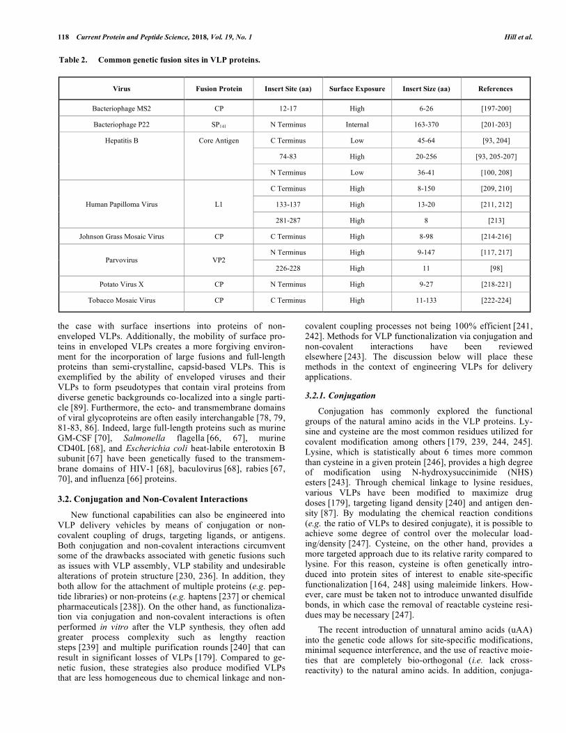
A survey of the literature suggests that VLPs derived from different viruses exhibit varying degree of tolerance for the size of fusions they can incorporate (Table 2). In addi-tion, the choice of virus can be further facilitated by careful evaluation of geometries of the fusion sites in the VLP pro-tein to match the distance between the N and C termini of the insert. This is exemplified by the ability of hepatitis B core antigen (HBcAg) VLPs to display a green fluorescence pro-tein consisting of 238 amino acids (aa) on its surface utiliz-ing only a short linker [225, 226], whereas prior studies had only succeeded in inserting short sequences (less than 50 aa) [227]. The same strategy has enabled the functional in-sertion of the outer surface protein C of Borrelia burgdorferi (188 aa) [228], mature IL-33 (158 aa) [80] and envelope domain III of dengue virus (104 aa) [229] into the major immunodominant region of HBcAg with or without short linkers. If the polypeptide fusion to be inserted and the target VLP carrier protein have incompatible geometries, creative engineered linkers can be employed to successfully display the insert. For example, a 256 aa protein with N and C ter-mini at opposing ends could be displayed on the surface of HBcAg using either long, flexible, glycine-rich linkers [230] or a digestible linker that was cleaved in situ after coexspres-sion of the corresponding protease [231]. Therefore, rational design of protein linkers aided by detailed crystal structures can circumvent issues of insert conformation. Various flexi-ble, rigid, or cleavable linkers that can be potentially used to enable the insertion of large proteins in a VLP protein have been well reviewed elsewhere [232].
Another factor affecting the success of genetic fusions is the physical properties of the insert and VLP proteins such as charge, isoelectric point, and hydrophobicity. Abidin et al. reported that hydrophobic peptide insertions in the VP1 cap-sid protein caused severe aggregation of the resulting polyomavirus VLPs, and that the introduction of charged aspartic acid residues flanking the insert improved the solu-bility and recovery [233]. In another study, Bendahmane et al. showed that the coat protein of tobacco mosaic virus (TMV) is highly sensitive to the insertion of short peptide sequences that increase the positive charge and isoelectric point of the coat protein, ultimately leading to assembly in-hibition and impaired function [234]. Lu et al. also reported poor attachment of negatively charged antigens and nucleic acids to the anionic surface of HBcAg VLPs that could be overcome by decreasing the negative charge of the capsid [235]. Although the last study does not involve ge-netic fusion, these examples illustrate the possibility of aug-menting the physical properties of either the insert or the VLP proteins to overcome unfavorable interactions.
Compared to capsid VLPs, enveloped VLPs generally of-fer a number of advantages for the presentation of large sur-face fusions. First, at least one terminus of a glycoprotein is often exposed on the surface of enveloped VLPs allowing surface-displayed insertions to be introduced at a terminus rather than a position in the middle of the protein, as is often
118 Current Protein and Peptide Science, 2018, Vol. 19, No. 1 Hill et al.
the case with surface insertions into proteins of non-enveloped VLPs. Additionally, the mobility of surface pro-teins in enveloped VLPs creates a more forgiving environ-ment for the incorporation of large fusions and full-length proteins than semi-crystalline, capsid-based VLPs. This is exemplified by the ability of enveloped viruses and their VLPs to form pseudotypes that contain viral proteins from diverse genetic backgrounds co-localized into a single parti-cle [89]. Furthermore, the ecto- and transmembrane domains of viral glycoproteins are often easily interchangable [78, 79, 81-83, 86]. Indeed, large full-length proteins such as murine GM-CSF [70], Salmonella flagella [66, 67], murine CD40L [68], and Escherichia coli heat-labile enterotoxin B subunit [67] have been genetically fused to the transmem-brane domains of HIV-1 [68], baculovirus [68], rabies [67, 70], and influenza [66] proteins.
3.2. Conjugation and Non-Covalent Interactions
New functional capabilities can also be engineered into VLP delivery vehicles by means of conjugation or non-covalent coupling of drugs, targeting ligands, or antigens. Both conjugation and non-covalent interactions circumvent some of the drawbacks associated with genetic fusions such as issues with VLP assembly, VLP stability and undesirable alterations of protein structure [230, 236]. In addition, they both allow for the attachment of multiple proteins (e.g. pep-tide libraries) or non-proteins (e.g. haptens [237] or chemical pharmaceuticals [238]). On the other hand, as functionaliza-tion via conjugation and non-covalent interactions is often performed in vitro after the VLP synthesis, they often add greater process complexity such as lengthy reaction steps [239] and multiple purification rounds [240] that can result in significant losses of VLPs [179]. Compared to ge-netic fusion, these strategies also produce modified VLPs that are less homogeneous due to chemical linkage and non-
covalent coupling processes not being 100% efficient [241, 242]. Methods for VLP functionalization via conjugation and non-covalent interactions have been reviewed elsewhere [243]. The discussion below will place these methods in the context of engineering VLPs for delivery applications.
3.2.1. Conjugation
Conjugation has commonly explored the functional groups of the natural amino acids in the VLP proteins. Ly-sine and cysteine are the most common residues utilized for covalent modification among others [179, 239, 244, 245]. Lysine, which is statistically about 6 times more common than cysteine in a given protein [246], provides a high degree of modification using N-hydroxysuccinimide (NHS) esters [243]. Through chemical linkage to lysine residues, various VLPs have been modified to maximize drug doses [179], targeting ligand density [240] and antigen den-sity [87]. By modulating the chemical reaction conditions (e.g. the ratio of VLPs to desired conjugate), it is possible to achieve some degree of control over the molecular load-ing/density [247]. Cysteine, on the other hand, provides a more targeted approach due to its relative rarity compared to lysine. For this reason, cysteine is often genetically intro-duced into protein sites of interest to enable site-specific functionalization [164, 248] using maleimide linkers. How-ever, care must be taken not to introduce unwanted disulfide bonds, in which case the removal of reactable cysteine resi-dues may be necessary [247].
The recent introduction of unnatural amino acids (uAA) into the genetic code allows for site-specific modifications, minimal sequence interference, and the use of reactive moie-ties that are completely bio-orthogonal (i.e. lack cross-reactivity) to the natural amino acids. In addition, conjuga-
Table 2. Common genetic fusion sites in VLP proteins.
Virus Fusion Protein Insert Site (aa) Surface Exposure Insert Size (aa) References
Bacteriophage MS2 CP 12-17 High 6-26 [197-200]
Bacteriophage P22 SP141 N Terminus Internal 163-370 [201-203]
C Terminus Low 45-64 [93, 204]
74-83 High 20-256 [93, 205-207]
Hepatitis B Core Antigen
N Terminus Low 36-41 [100, 208]
C Terminus High 8-150 [209, 210]
133-137 High 13-20 [211, 212] Human Papilloma Virus L1
281-287 High 8 [213]
Johnson Grass Mosaic Virus CP C Terminus High 8-98 [214-216]
N Terminus High 9-147 [117, 217] Parvovirus VP2
226-228 High 11 [98]
Potato Virus X CP N Terminus High 9-27 [218-221]
Tobacco Mosaic Virus CP C Terminus High 11-133 [222-224]

Virus-like Particles for Antigen and Drug Delivery Current Protein and Peptide Science, 2018, Vol. 19, No. 1 119
tion with uAA can proceed through much more efficient chemical reactions to produce a more homogeneous product than conjugation with the canonical amino acids [241]. For example, uAAs presenting alkyne or azide motifs are espe-cially popular and have been incorporated into Qβ [249, 250], TMV [251], MS2 [250], and HBcAg [235] VLPs to enable the cycloaddition click chemistry. The synthesis of VLPs containing uAA is outside the scope of this review but methods are described elsewhere [252, 253].
Although chemical conjugation has been used for encap-sulation [164, 239, 254], the permanence of covalent bonds makes it more suitable for VLP surface functionalization for targeting [159, 165, 240], antigen presentation [247], or exte-rior drug loading [179], especially in the case of non-protein conjugates. Notably, VLP displaying folate [240] or nucleic acid aptamers [163, 165] have been used to target cancerous cells while other applications have included nicotine conju-gates for the treatment of nicotine dependence [237] and the stealthing of VLPs by PEGylation [240]. Reversible chemis-tries can also be employed to achieve different release kinet-ics. For example, Aljabali et al. showed that CPMV VLPs conjugated to the chemotherapeutic drug doxorubicin by a stable amide bond were taken up by HeLa cells in vitro and released the drug after proteolytic degradation associated with the endolysosomal pathway. On the other hand, conju-gation of doxorubicin by a labile disulfide bond resulted in most of the drug being released into the culture medium after 24 hours, essentially acting as free doxorubicin [179].
3.2.2. Non-covalent Interactions
Methods of non-covalent functionalization of VLPs have simpler implementation than conjugation, as no chemical reactions are required. A wide range of materials can take part in non-covalent interactions with VLP proteins such as proteins [255], nucleotides [256-258], metals [10, 259], and other bioactive molecules [260]. In addition, the membrane of enveloped VLPs can interact with lipoproteins [71, 90, 261]. The varying strengths of different non-covalent inter-actions affect their utility for engineering various aspects of VLP delivery vehicles. Relatively strong non-covalent inter-actions, such as biotin/streptavidin [31] or glycosylphos-phatidylinositol-anchored (GPI-anchored) proteins, have been used for surface presentation of targeting [90] or im-munostimulatory [71, 261] motifs. However, being weak in nature, most non-covalent interactions do not provide the stability of covalent bonds, making them more suitable for delivery of encapsulated cargos, especially larger molecules that cannot diffuse through the VLP capsid once assembled [159].
The inherent affinities present in VLPs can be explored to encapsulate desirable materials. For instance, the inner surface of the viral capsid is often positively charged and thus amenable to encapsulating genomic materials, therapeu-tic nucleotides [256-258], and other negatively-charged sub-strates such as polymers [189] , nanoparticles [262], and enzymes [255]. In addition, the pre-existing mechanisms that viruses use to bind nucleic acids can be repurposed to incor-porate diverse cargos in VLPs. Ashley et al. took advantage of the ability of MS2 viral capsid to internally bind an RNA sequence known as the pac site [263]. Through conjugation
of this pac site to quantum dots, siRNA, doxorubicin, or ricin toxin-A chain, these cargos were encapsulated in the MS2 capsids by disassembly and reassembly in vitro [166]. For a more simplified production strategy, intracellular VLP assembly and cargo encapsulation was accomplished utiliz-ing bi-functional RNA aptamers that bind both Qβ coat pro-tein and an arginine rich peptide (Rev) fused to fluorescent proteins [264] or luciferase and peptidase E enzymes [265].
Native VLP affinities may prove insufficient for the en-capsulation of some materials, and therefore some investiga-tors have introduced their own mechanisms to enable new non-covalent interactions for improved encapsulation effi-ciency. In one study, an internal hydrophobic binding pocket was created by genetically manipulating protein cages de-rived from the pyruvate of dehydrogenase E2 subunit to en-able efficient encapsulation doxorubicin [260]. The genetic introduction of polyhistadine tags provides another strategy for introducing specific interactions for encapsulation of metal-containing cargos such as magnetic nanoparticles [10], quantum dots [266], and nitrilotriacetic-acid (NTA) conju-gated compounds [176]. Protein/protein interactions can also be introduced as demonstrated in one study, where the CPMV capsid protein was fused to one strand of a coiled coil motif to enable encapsulation of fluorescent proteins fused to a complimentary coiled coil strand in the CCMV VLPs [267]. These examples demonstrate a rational design approach to introduce non-covalent interactions for efficient cargo encapsulation. When multiple, complex, non-covalent interactions are involved, directed evolution has been em-ployed successfully for additional functional enhancement. In an elegant study, Wörsdörfer et al. developed an intracel-lular encapsulation system in E. coli driven by electrostatic interactions between lumazine synthase capsid and a polyar-ginine tag appended to the C terminus of the cytotoxic HIV protease [268]. When the tagged protease was co-expressed with the capsid proteins, a growth advantage was conferred to cells producing capsids that could better sequester the protease, thereby providing a selective pressure that led to the identification of a mutant VLP with encapsulation capac-ity increased by 5 to 10 fold [268].
CONCLUSION
VLPs exhibit properties such as self-assembly, natural tropism, and intrinsic immunogenicity that make them an attractive technology platform for antigen and drug delivery. To achieve a satisfactory clinical outcome, however, these properties often need to be augmented, altered, or even masked, which is a challenging task from the design and synthesis perspectives. For antigen delivery, VLPs have demonstrated potential as witnessed by the FDA approval of VLP-based hepatitis B and HPV vaccines. However, outside of these examples, VLPs have seen very limited success of-ten due to their inability to elicit effective immune responses, especially T-cell responses that are critical for long-lasting immune memories against many pathogens and cancers. Many recent studies have focused on enhancing T-cell re-sponses by the inclusion of immune activators found in viral replication processes or other pathogens in the VLP design. In addition, mutation-prone viruses such as influenza and HIV as well as non-viral targets such as malaria and cancers require the immunogenic presentation of cross-protective

120 Current Protein and Peptide Science, 2018, Vol. 19, No. 1 Hill et al.
epitopes. Rational epitope design aided by identification of immunogenic insertion sites, epitope flanking sequences, and multivalent epitope display has increased the versatility of VLPs and has allowed them to initiate tailored and specific immune responses to a variety of targets. In the case of drug delivery, VLPs still face largely the same challenges of syn-thetic delivery vehicles in that they must evade the immune system, specifically target the cells of interest, and deliver the therapeutic cargo. Therefore, most of the recent pro-gresses in engineering VLPs for drug delivery have taken advantage of the innovations in the field of synthetic nanoparticle-based delivery. Additionally, the proteinaceous nature and self-assembly of VLPs gives them a distinct ad-vantage over synthetic nanoparticles in that they are very homogeneous, biodegradable, and easily subjected to pre-dictable alterations through genetic manipulations.
Despite these recent advancements, VLPs still present a few drawbacks compared to other delivery systems, most notably their synthesis and modification. VLP synthesis in-volves several complex variables such as the host of choice, the complexity of the VLP proteins, and purification (re-viewed elsewhere [24, 269, 270]). Further, VLP modification can be achieved by genetic alteration; however, its success is largely unpredictable and highly dependent on the choice of VLP, the site of modification, and the characteristics of the fused peptides (size, hydrophobicity, isoelectric point, etc.). Although conjugation is a relatively robust alternative to genetic manipulation for VLP modification, it is usually lim-ited to chemistries based on the canonical amino acids. The introduction of non-natural amino acids into VLPs allows for simplified production, increased homogeneity and site-specific modification but requires specialized production capabilities.
Regardless of the specific application, recent VLP de-signs have benefited significantly from an ever-increasing understanding of the immune system, pathogen-host interac-tions, and cancer biology. Likewise, without the advance-ments in protein engineering, materials science, and chemis-try, the synthesis of these VLP designs would not be possi-ble. Therefore, only continued breakthroughs at these inter-disciplinary interfaces will enable VLPs to achieve their clinical potential.
CONSENT FOR PUBLICATION
Not applicable.
CONFLICT OF INTEREST
The authors declare no conflict of interest, financial or otherwise.
ACKNOWLEDGEMENTS
The authors would like to thank all members of the Fei Wen lab for helpful feedback and comments on the manu-script, especially Dr. S. Rizvi for valuable input and editing of section 2.2. We would also like to acknowledge the finan-cial support provided by National Institutes of Health grant OD020053, National Science Foundation grant 1511720, and the MCubed program at the University of Michigan.
REFERENCES
[1] Blumberg, B.S.; Alter, H.J.; Visnich, S. A "New" Antigen in Leukemia Sera. JAMA, 1965, 191, 541-546.
[2] Millman, I.; Loeb, L.A.; Bayer, M.E.; Blumberg, B.S. Australia antigen (a hepatitis-associated antigen): purification and physical properties. J. Exp. Med., 1970, 131(6), 1190-1199.
[3] Bayer, M.E.; Blumberg, B.S.; Werner, B. Particles associated with Australia antigen in the sera of patients with leukaemia, Down's Syndrome and hepatitis. Nature, 1968, 218(5146), 1057-1059.
[4] Rappuoli, R. Bridging the knowledge gaps in vaccine design. Nat. Biotechnol., 2007, 25(12), 1361-1366.
[5] Krugman, S. The newly licensed hepatitis B vaccine. Character-istics and indications for use. JAMA, 1982, 247(14), 2012-2015.
[6] McAleer, W.J.; Buynak, E.B.; Maigetter, R.Z.; Wampler, D.E.; Miller, W.J.; Hilleman, M.R. Human hepatitis B vaccine from recombinant yeast. Nature, 1984, 307(5947), 178-180.
[7] Cormode, D.P.; Skajaa, T.; Fayad, Z.A.; Mulder, W.J. Nanotechnology in medical imaging: probe design and applications. Arterioscler. Thromb. Vasc. Biol., 2009, 29(7), 992-1000.
[8] Manchester, M.; Singh, P. Virus-based nanoparticles (VNPs): platform technologies for diagnostic imaging. Adv. Drug Deliv. Rev., 2006, 58(14), 1505-1522.
[9] Schwarz, B.; Douglas, T. Development of virus-like particles for diagnostic and prophylactic biomedical applications. Wiley Interdiscip. Rev. Nanomed. Nanobiotechnol., 2015, 7(5), 722-7735.
[10] Shen, L.; Zhou, J.; Wang, Y.; Kang, N.; Ke, X.; Bi, S.; Ren, L. Efficient encapsulation of Fe(3)O(4) nanoparticles into genetically engineered hepatitis B core virus-like particles through a specific interaction for potential bioapplications. Small, 2015, 11(9-10), 1190-1196.
[11] Li, F.; Wang, Q. Fabrication of nanoarchitectures templated by virus-based nanoparticles: strategies and applications. Small, 2014, 10(2), 230-245.
[12] Wnek, M.; Gorzny, M.L.; Ward, M.B.; Walti, C.; Davies, A.G.; Brydson, R.; Evans, S.D.; Stockley, P.G. Fabrication and characterization of gold nano-wires templated on virus-like arrays of tobacco mosaic virus coat proteins. Nanotechnology, 2013, 24(2), 025605.
[13] Maity, B.; Fujita, K.; Ueno, T. Use of the confined spaces of apo-ferritin and virus capsids as nanoreactors for catalytic reactions. Curr. Opin. Chem. Biol., 2015, 25, 88-97.
[14] Patterson, D.; Edwards, E.; Douglas, T. Hybrid Nanoreactors: Coupling Enzymes and Small-Molecule Catalysts within Virus-Like Particles. Israel J. Chem., 2015, 55(1), 96-101.
[15] Patterson, D.P.; McCoy, K.; Fijen, C.; Douglas, T. Constructing catalytic antimicrobial nanoparticles by encapsulation of hydrogen peroxide producing enzyme inside the P22 VLP. J. Mater. Chem. B, 2014, 2(36), 5948-5951.
[16] Zhan, W.; Tang, L.-J.; Zhang, X.-B.; Jiang, J.-H.; Tan, W. Aptamer-Modified Nandodrug Delivery Systems. ACS Nano., 2011, 5, 7696-7699.
[17] Zhao, L.; Seth, A.; Wibowo, N.; Zhao, C.X.; Mitter, N.; Yu, C.; Middelberg, A.P. Nanoparticle vaccines. Vaccine, 2014, 32(3), 327-337.
[18] Steinmetz, N.F. Viral nanoparticles as platforms for next-generation therapeutics and imaging devices. Nanomedicine, 2010, 6(5), 634-641.
[19] Glasgow, J.; Tullman-Ercek, D. Production and applications of engineered viral capsids. Appl. Microbiol. Biotechnol., 2014, 98(13), 5847-5858.
[20] Jennings, G.T.; Bachmann, M.F. The coming of age of virus-like particle vaccines. Biol. Chem., 2008, 389(5), 521-536.
[21] Pushko, P.; Pumpens, P.; Grens, E. Development of virus-like particle technology from small highly symmetric to large complex virus-like particle structures. Intervirology, 2013, 56(3), 141-165.
[22] Rodriguez-Limas, W.A.; Sekar, K.; Tyo, K.E. Virus-like particles: the future of microbial factories and cell-free systems as platforms for vaccine development. Curr. Opin. Biotechnol., 2013, 24(6), 1089-1093.
[23] Roldao, A.; Mellado, M.C.; Castilho, L.R.; Carrondo, M.J.; Alves, P.M. Virus-like particles in vaccine development. Expert Rev. Vaccines, 2010, 9(10), 1149-1176.
[24] Zeltins, A. Construction and characterization of virus-like particles: a review. Mol. Biotechnol., 2013, 53(1), 92-107.

Virus-like Particles for Antigen and Drug Delivery Current Protein and Peptide Science, 2018, Vol. 19, No. 1 121
[25] Bachmann, M.F.; Zinkernagel, R.M. Neutralizing antiviral B cell responses. Annu. Rev. Immunol., 1997, 15, 235-270.
[26] Trovato, M.; De Berardinis, P. Novel antigen delivery systems. World J. Virol., 2015, 4(3), 156-168.
[27] Rosenthal, J.A.; Chen, L.; Baker, J.L.; Putnam, D.; DeLisa, M.P. Pathogen-like particles: biomimetic vaccine carriers engineered at the nanoscale. Curr. Opin. Biotechnol., 2014, 28, 51-58.
[28] Spohn, G.; Keller, I.; Beck, M.; Grest, P.; Jennings, G.T.; Bachmann, M.F. Active immunization with IL-1 displayed on virus-like particles protects from autoimmune arthritis. Eur. J. Immunol., 2008, 38(3), 877-887.
[29] Ambuhl, P.M.; Tissot, A.C.; Fulurija, A.; Maurer, P.; Nussberger, J.; Sabat, R.; Nief, V.; Schellekens, C.; Sladko, K.; Roubicek, K.; Pfister, T.; Rettenbacher, M.; Volk, H.D.; Wagner, F.; Muller, P.; Jennings, G.T.; Bachmann, M.F. A vaccine for hypertension based on virus-like particles: preclinical efficacy and phase I safety and immunogenicity. J. Hypertens., 2007, 25(1), 63-72.
[30] Rohn, T.A.; Jennings, G.T.; Hernandez, M.; Grest, P.; Beck, M.; Zou, Y.; Kopf, M.; Bachmann, M.F. Vaccination against IL-17 suppresses autoimmune arthritis and encephalomyelitis. Eur. J. Immunol., 2006, 36(11), 2857-2867.
[31] Chackerian, B.; Lenz, P.; Lowy, D.R.; Schiller, J.T. Determinants of autoantibody induction by conjugated papillomavirus virus-like particles. J. Immunol., 2002, 169(11), 6120-6126.
[32] Braun, M.; Jandus, C.; Maurer, P.; Hammann-Haenni, A.; Schwarz, K.; Bachmann, M.F.; Speiser, D.E.; Romero, P. Virus-like particles induce robust human T-helper cell responses. Eur. J. Immunol., 2012, 42(2), 330-340.
[33] Swain, S.L.; McKinstry, K.K.; Strutt, T.M. Expanding roles for CD4(+) T cells in immunity to viruses. Nat. Rev. Immunol., 2012, 12(2), 136-148.
[34] Storni, T.; Lechner, F.; Erdmann, I.; Bachi, T.; Jegerlehner, A.; Dumrese, T.; Kundig, T.M.; Ruedl, C.; Bachmann, M.F. Critical role for activation of antigen-presenting cells in priming of cytotoxic T cell responses after vaccination with virus-like particles. J. Immunol., 2002, 168(6), 2880-2886.
[35] Appay, V.; Douek, D.C.; Price, D.A. CD8+ T cell efficacy in vaccination and disease. Nat. Med., 2008, 14(6), 623-628.
[36] Doherty, P.C.; Kelso, A. Toward a broadly protective influenza vaccine. J. Clin. Invest., 2008, 118(10), 3273-3275.
[37] Wilkinson, T.M.; Li, C.K.; Chui, C.S.; Huang, A.K.; Perkins, M.; Liebner, J.C.; Lambkin-Williams, R.; Gilbert, A.; Oxford, J.; Nicholas, B.; Staples, K.J.; Dong, T.; Douek, D.C.; McMichael, A.J.; Xu, X.N. Preexisting influenza-specific CD4+ T cells correlate with disease protection against influenza challenge in humans. Nat. Med., 2012, 18(2), 274-280.
[38] Marrack, P.; McKee, A.S.; Munks, M.W. Towards an understanding of the adjuvant action of aluminium. Nat. Rev. Immunol., 2009, 9(4), 287-293.
[39] Ott, G.; Barchfeld, G.L.; Van Nest, G. Enhancement of humoral response against human influenza vaccine with the simple submicron oil/water emulsion adjuvant MF59. Vaccine, 1995, 13(16), 1557-1562.
[40] Paavonen, J.; Naud, P.; Salmeron, J.; Wheeler, C.M.; Chow, S.N.; Apter, D.; Kitchener, H.; Castellsague, X.; Teixeira, J.C.; Skinner, S.R.; Hedrick, J.; Jaisamrarn, U.; Limson, G.; Garland, S.; Szarewski, A.; Romanowski, B.; Aoki, F.Y.; Schwarz, T.F.; Poppe, W.A.; Bosch, F.X.; Jenkins, D.; Hardt, K.; Zahaf, T.; Descamps, D.; Struyf, F.; Lehtinen, M.; Dubin, G.; Group, H.P.S. Efficacy of human papillomavirus (HPV)-16/18 AS04-adjuvanted vaccine against cervical infection and precancer caused by oncogenic HPV types (PATRICIA): final analysis of a double-blind, randomised study in young women. Lancet, 2009, 374(9686), 301-314.
[41] Casella, C.R.; Mitchell, T.C. Putting endotoxin to work for us: monophosphoryl lipid A as a safe and effective vaccine adjuvant. Cell. Mol. Life Sci., 2008, 65(20), 3231-3240.
[42] Gupta, S.; Termini, J.M.; Niu, L.; Kanagavelu, S.K.; Schmidtmayerova, H.; Snarsky, V.; Kornbluth, R.S.; Stone, G.W. EBV LMP1, a viral mimic of CD40, activates dendritic cells and functions as a molecular adjuvant when incorporated into an HIV vaccine. J. Leukoc. Biol., 2011, 90, 389-398.
[43] McKee, S.J.; Young, V.L.; Clow, F.; Hayman, C.M.; Baird, M.A.; Hermans, I.F.; Young, S.L.; Ward, V.K. Virus-like particles and alpha-galactosylceramide form a self-adjuvanting composite particle that elicits anti-tumor responses. J. Control Release, 2012, 159(3), 338-345.
[44] De Gregorio, E.; D'Oro, U.; Wack, A. Immunology of TLR-independent vaccine adjuvants. Curr. Opin. Immunol., 2009, 21(3), 339-345.
[45] van Montfoort, N.; van der Aa, E.; Woltman, A.M. Understanding MHC class I presentation of viral antigens by human dendritic cells as a basis for rational design of therapeutic vaccines. Front. Immunol., 2014, 5, 182.
[46] Neefjes, J.; Jongsma, M.L.; Paul, P.; Bakke, O. Towards a systems understanding of MHC class I and MHC class II antigen presentation. Nat. Rev. Immunol., 2011, 11(12), 823-836.
[47] Tipping, P.G. Toll-like receptors: the interface between innate and adaptive immunity. J. Am. Soc. Nephrol., 2006, 17(7), 1769-1771.
[48] Kasturi, S.P.; Skountzou, I.; Albrecht, R.A.; Koutsonanos, D.; Hua, T.; Nakaya, H.I.; Ravindran, R.; Stewart, S.; Alam, M.; Kwissa, M.; Villinger, F.; Murthy, N.; Steel, J.; Jacob, J.; Hogan, R.J.; García-Sastre, A.; Compans, R.; Pulendran, B. Programming the magnitude and persistence of antibody responses with innate immunity. Nature, 2011, 470, 543-547.
[49] Rahman, A.H.; Taylor, D.K.; Turka, L.A. The contribution of direct TLR signaling to T cell responses. Immunol. Res., 2009, 45(1), 25-36.
[50] Singh, M.; O'Hagan, D. Advances in vaccine adjuvants. Nat. Biotechnol., 1999, 17(11), 1075-1081.
[51] Bhardwaj, N.; Gnjatic, S.; Sawhney, N. TLR AGONISTS: Are They Good Adjuvansts? Cancer J., 2010, 16, 382-391.
[52] Lee, S.-M.; Joo, Y.-D.; Seo, S.-K. Expression and Function of TLR2 on CD4 Versus CD8 T Cells. Immune Network, 2009, 9, 127-132.
[53] Cottalorda, A.; Mercier, B.C.; Mbitikon-Kobo, F.M.; Arpin, C.; Teoh, D.Y.L.; McMichael, A.; Marvel, J.; Bonnefoy-Bérard, N. TLR2 engagement on memory CD8+ T cells improves their cytokine-mediated proliferation and IFN-γ secretion in the absence of Ag. Eur. J. Immunol., 2009, 39, 2673-2681.
[54] Mercier, B.C.; Cottalorda, A.; Coupet, C.-A.; Marvel, J.; Bonnefoy-Berard, N. TLR2 Engagement on CD8 T Cells Enables Generation of Functional Memory Cells in Response to a Suboptimal TCR Signal. J. Immunol., 2009, 182, 1860-1867.
[55] Caron, G.; Duluc, D.; Fremaux, I.; Jeannin, P.; David, C.; Gascan, H.; Delneste, Y. Direct Stimulation of Human T Cells via TLR5 and TLR7/8: Flagellin and R-848 Up-Regulate Proliferation and IFN- Production by Memory CD4+ T Cells. J. Immunol., 2005, 175, 1551-1557.
[56] Crellin, N.K.; Garcia, R.V.; Hadisfar, O.; Allan, S.E.; Steiner, T.S.; Levings, M.K. Human CD4+ T cells express TLR5 and its ligand flagellin enhances the suppressive capacity and expression of FOXP3 in CD4+CD25+ T regulatory cells. J. Immunol., 2005, 175(12), 8051-8059.
[57] Liu, H.; Komai-Koma, M.; Xu, D.; Liew, F.Y. Toll-like receptor 2 signaling modulates the functions of CD4+ CD25+ regulatory T cells. Proc. Natl. Acad. Sci. USA, 2006, 103(18), 7048-7053.
[58] Reynolds, J.M.; Pappu, B.P.; Peng, J.; Martinez, G.J.; Zhang, Y.; Chung, Y.; Ma, L.; Yang, X.O.; Nurieva, R.I.; Tian, Q.; Dong, C. Toll-like receptor 2 signaling in CD4(+) T lymphocytes promotes T helper 17 responses and regulates the pathogenesis of autoimmune disease. Immunity, 2010, 32(5), 692-702.
[59] Bendelac, A.; Medzhitov, R. Adjuvants of immunity: harnessing innate immunity to promote adaptive immunity. J. Exp. Med., 2002, 195(5), F19-F23.
[60] Coffman, R.L.; Sher, A.; Seder, R.A. Vaccine adjuvants: putting innate immunity to work. Immunity, 2010, 33(4), 492-503.
[61] Storni, T.; Ruedl, C.; Schwarz, K.; Schwendener, R.A.; Renner, W.A.; Bachmann, M.F. Nonmethylated CG motifs packaged into virus-like particles induce protective cytotoxic T cell responses in the absence of systemic side effects. J. Immunol., 2004, 172(3), 1777-1785.
[62] Goldinger, S.M.; Dummer, R.; Baumgaertner, P.; Mihic-Probst, D.; Schwarz, K.; Hammann-Haenni, A.; Willers, J.; Geldhof, C.; Prior, J.O.; Kundig, T.M.; Michielin, O.; Bachmann, M.F.; Speiser, D.E. Nano-particle vaccination combined with TLR-7 and -9 ligands triggers memory and effector CD8(+) T-cell responses in melanoma patients. Eur. J. Immunol., 2012, 42(11), 3049-3061.
[63] Speiser, D.E.; Schwarz, K.; Baumgaertner, P.; Manolova, V.; Devevre, E.; Sterry, W.; Walden, P.; Zippelius, A.; Conzett, K.B.; Senti, G.; Voelter, V.; Cerottini, J.P.; Guggisberg, D.; Willers, J.; Geldhof, C.; Romero, P.; Kundig, T.; Knuth, A.; Dummer, R.; Trefzer, U.; Bachmann, M.F. Memory and effector CD8 T-cell

122 Current Protein and Peptide Science, 2018, Vol. 19, No. 1 Hill et al.
responses after nanoparticle vaccination of melanoma patients. J. Immunother., 2010, 33(8), 848-858.
[64] Lebel, M.-È.; Daudelin, J.-F.; Chartrand, K.; Tarrab, E.; Kalinke, U.; Savard, P.; Labrecque, N.; Leclerc, D.; Lamarre, A. Nanoparticle adjuvant sensing by TLR7 enhances CD8+ T cell-mediated protection from Listeria monocytogenes infection. J. Immunol., 2014, 192, 1071-1078.
[65] Chua, B.Y.; Johnson, D.; Tan, A.; Earnest-Silveira, L.; Sekiya, T.; Chin, R.; Torresi, J.; Jackson, D.C. Hepatitis C VLPs Delivered to Dendritic Cells by a TLR2 Targeting Lipopeptide Results in Enhanced Antibody and Cell-Mediated Responses. PLoS One, 2012, 7, e47492.
[66] Wang, B.Z.; Quan, F.S.; Kang, S.M.; Bozja, J.; Skountzou, I.; Compans, R.W. Incorporation of Membrane-Anchored Flagellin into Influenza Virus-Like Particles Enhances the Breadth of Immune Responses. J. Virol., 2008, 82(23), 11813-11823.
[67] Qi, Y.; Kang, H.; Zheng, X.; Wang, H.; Gao, Y.; Yang, S.; Xia, X. Incorporation of membrane-anchored flagellin or Escherichia coli heat-labile enterotoxin B subunit enhances the immunogenicity of rabies virus-like particles in mice and dogs. Front. Microbiol., 2015, 6, 169.
[68] Franco, D.; Liu, W.M.; Gardiner, D.F.; Hahn, B.H.; Ho, D.D. CD40L-Containing Virus-Like Particle as a Candidate HIV-1 Vaccine Targeting Dendritic Cells. J. Acquir. Immune Defic. Syndr., 2011, 56(5), 393-400.
[69] Skountzou, I.; Quan, F.-S.; Gangadhara, S.; Ye, L.; Vzorov, A.; Selvaraj, P.; Jacob, J.; Compans, R. W.; Kang, S.-M. Incorporation of glycosylphosphatidylinositol-anchored granulocyte- macrophage colony-stimulating factor or CD40 ligand enhances immuno-genicity of chimeric simian immunodeficiency virus-like particles. J. Virol., 2007, 81(3), 1083-1094.
[70] Kang, H.; Qi, Y.; Wang, H.; Zheng, X.; Gao, Y.; Li, N.; Yang, S.; Xia, X. Chimeric rabies virus-like particles containing membrane-anchored GM-CSF enhances the immune response against rabies virus. Viruses, 2015, 7(3), 1134-1152.
[71] Skountzou, I.; Quan, F.S.; Gangadhara, S.; Ye, L.; Vzorov, A.; Selvaraj, P.; Jacob, J.; Compans, R.W.; Kang, S.M. Incorporation of glycosylphosphatidylinositol-anchored granulocyte- macrophage colony-stimulating factor or CD40 ligand enhances immuno-genicity of chimeric simian immunodeficiency virus-like particles. J. Virol., 2007, 81(3), 1083-1094.
[72] Davidson, E.; Doranz, B.J. A high-throughput shotgun mutagenesis approach to mapping B-cell antibody epitopes. Immunology, 2014, 143(1), 13-20.
[73] Li Pira, G.; Ivaldi, F.; Moretti, P.; Manca, F. High throughput T epitope mapping and vaccine development. J. Biomed. Biotechnol., 2010, 2010, 325720.
[74] Patronov, A.; Doytchinova, I. T-cell epitope vaccine design by immunoinformatics. Open. Biol., 2013, 3(1), 120139.
[75] Wen, F.; Esteban, O.; Zhao, H. Rapid identification of CD4+ T-cell epitopes using yeast displaying pathogen-derived peptide library. J. Immunol. Methods, 2008, 336(1), 37-44.
[76] Wen, F.; Zhao, H. Construction and screening of an antigen-derived peptide library displayed on yeast cell surface for CD4+ T cell epitope identification. Methods Mol. Biol., 2013, 1061, 245-264.
[77] Kato, T.; Yui, M.; Deo, V.K.; Park, E.Y. Development of rous sarcoma virus-like particles displaying hCC49 scFv for specific targeted drug delivery to human colon carcinoma cells. Pharm. Res., 2015, 32(11), 3699-3707.
[78] Kirchmeier, M.; Fluckiger, A.C.; Soare, C.; Bozic, J.; Ontsouka, B.; Ahmed, T.; Diress, A.; Pereira, L.; Schodel, F.; Plotkin, S.; Dalba, C.; Klatzmann, D.; Anderson, D.E. Enveloped virus-like particle expression of human cytomegalovirus glycoprotein B antigen induces antibodies with potent and broad neutralizing activity. Clin. Vaccine Immunol., 2014, 21(2), 174-180.
[79] Lagging, L.M.; Meyer, K.; Owens, R.J.; Ray, R. Functional role of hepatitis C virus chimeric glycoproteins in the infectivity of pseudotyped virus. J. Virol., 1998, 72(5), 3539-3546.
[80] Long, Q.; Huang, W.; Yao, Y.; Yang, X.; Sun, W.; Jin, X.; Li, Y.; Chu, X.; Liu, C.; Peng, Z.; Ma, Y. Virus-like particles presenting interleukin-33 molecules: immunization characteristics and potentials of blockingIL-33/ST2 pathway in allergic airway inflammation. Hum. Vaccin. Immunother., 2014, 10(8), 2303-2311.
[81] Lv, L.; Li, X.; Liu, G.; Li, R.; Liu, Q.; Shen, H.; Wang, W.; Xue, C.; Cao, Y. Production and immunogenicity of chimeric virus-like
particles containing the spike glycoprotein of infectious bronchitis virus. J. Vet. Sci., 2014, 15(2), 209-216.
[82] Ogembo, J.G.; Muraswki, M.R.; McGinnes, L.W.; Parcharidou, A.; Sutiwisesak, R.; Tison, T.; Avendano, J.; Agnani, D.; Finberg, R.W.; Morrison, T.G.; Fingeroth, J.D. A chimeric EBV gp350/220-based VLP replicates the virion B-cell attachment mechanism and elicits long-lasting neutralizing antibodies in mice. J. Transl. Med., 2015, 13, 50.
[83] Schmidt, M.R.; McGinnes-Cullen, L.W.; Kenward, S.A.; Willems, K.N.; Woodland, R.T.; Morrison, T.G. Modification of the respiratory syncytial virus f protein in virus-like particles impacts generation of B cell memory. J. Virol., 2014, 88(17), 10165-10176.
[84] Shahana, P.V.; Das, D.; Gontu, A.; Chandran, D.; Maithal, K. Efficient production of Tymovirus like particles displaying immunodominant epitopes of Japanese Encephalitis Virus envelope protein. Protein Expr. Purif., 2015, 113, 35-43.
[85] Shen, H.; Xue, C.; Lv, L.; Wang, W.; Liu, Q.; Liu, K.; Chen, X.; Zheng, J.; Li, X.; Cao, Y. Assembly and immunological properties of a bivalent virus-like particle (VLP) for avian influenza and Newcastle disease. Virus Res., 2013, 178(2), 430-436.
[86] Xue, C.; Wang, W.; Liu, Q.; Miao, Z.; Liu, K.; Shen, H.; Lv, L.; Li, X.; Chen, X.; Cao, Y. Chimeric influenza-virus-like particles containing the porcine reproductive and respiratory syndrome virus GP5 protein and the influenza virus HA and M1 proteins. Arch. Virol., 2014, 159(11), 3043-3051.
[87] Jegerlehner, A.; Tissot, A.; Lechner, F.; Sebbel, P.; Erdmann, I.; Kundig, T.; Bachi, T.; Storni, T.; Jennings, G.; Pumpens, P.; Renner, W.A.; Bachmann, M.F. A molecular assembly system that renders antigens of choice highly repetitive for induction of protective B cell responses. Vaccine, 2002, 20(25-26), 3104-3112.
[88] Peacey, M.; Wilson, S.; Baird, M.A.; Ward, V.K. Versatile RHDV virus-like particles: incorporation of antigens by genetic modification and chemical conjugation. Biotechnol. Bioeng., 2007, 98(5), 968-977.
[89] Cronin, J.; Zhang, X.Y.; Reiser, J. Altering the tropism of lentiviral vectors through pseudotyping. Curr. Gene Ther., 2005, 5(4), 387-398.
[90] Deo, V.K.; Kato, T.; Park, E.Y. Chimeric virus-like particles made using GAG and M1 capsid proteins providing dual drug delivery and vaccination platform. Mol. Pharm., 2015, 12(3), 839-845.
[91] Pushko, P.; Pearce, M.B.; Ahmad, A.; Tretyakova, I.; Smith, G.; Belser, J.A.; Tumpey, T.M. Influenza virus-like particle can accommodate multiple subtypes of hemagglutinin and protect from multiple influenza types and subtypes. Vaccine, 2011, 29(35), 5911-5918.
[92] Tretyakova, I.; Pearce, M.B.; Florese, R.; Tumpey, T.M.; Pushko, P. Intranasal vaccination with H5, H7 and H9 hemagglutinins co-localized in a virus-like particle protects ferrets from multiple avian influenza viruses. Virology, 2013, 442(1), 67-73.
[93] Ding, F.X.; Wang, F.; Lu, Y.M.; Li, K.; Wang, K.H.; He, X.W.; Sun, S.H. Multiepitope peptide-loaded virus-like particles as a vaccine against hepatitis b virus-related hepatocellular carcinoma. Hepatology, 2009, 49(5), 1492-1502.
[94] Birkett, A.; Lyons, K.; Schmidt, A.; Boyd, D.; Oliveira, G.A.; Siddique, A.; Nussenzweig, R.; Calvo-Calle, J.M.; Nardin, E. A modified hepatitis B virus core particle containing multiple epitopes of the Plasmodium falciparum circumsporozoite protein provides a highly immunogenic malaria vaccine in preclinical analyses in rodent and primate hosts. Infect. Immun., 2002, 70(12), 6860-6870.
[95] Kim, M.C.; Song, J.M.; O, E.; Kwon, Y.M.; Lee, Y.J.; Compans, R.W.; Kang, S.M. Virus-like particles containing multiple M2 extracellular domains confer improved cross-protection against various subtypes of influenza virus. Mol. Ther., 2013, 21(2), 485-492.
[96] De Filette, M.; Min Jou, W.; Birkett, A.; Lyons, K.; Schultz, B.; Tonkyro, A.; Resch, S.; Fiers, W. Universal influenza A vaccine: optimization of M2-based constructs. Virology, 2005, 337(1), 149-161.
[97] Yong, C.Y.; Yeap, S.K.; Ho, K.L.; Omar, A.R.; Tan, W.S. Potential recombinant vaccine against influenza A virus based on M2e displayed on nodaviral capsid nanoparticles. Int. J. Nanomedicine, 2015, 10, 2751-2763.
[98] Rueda, P.; Martinez-Torrecuadrada, J.L.; Sarraseca, J.; Sedlik, C.; del Barrio, M.; Hurtado, A.; Leclerc, C.; Casal, J.I. Engineering
Virus-like Particles for Antigen and Drug Delivery Current Protein and Peptide Science, 2018, Vol. 19, No. 1 123
parvovirus-like particles for the induction of B-cell, CD4(+) and CTL responses. Vaccine, 1999, 18(3-4), 325-332.
[99] Salfeld, J.; Pfaff, E.; Noah, M.; Schaller, H. Antigenic determinants and functional domains in core antigen and e antigen from hepatitis B virus. J. Virol., 1989, 63(2), 798-808.
[100] Schodel, F.; Moriarty, A.M.; Peterson, D.L.; Zheng, J.A.; Hughes, J.L.; Will, H.; Leturcq, D.J.; McGee, J.S.; Milich, D.R. The position of heterologous epitopes inserted in hepatitis B virus core particles determines their immunogenicity. J. Virol., 1992, 66(1), 106-114.
[101] Forsstrom, B.; Axnas, B.B.; Rockberg, J.; Danielsson, H.; Bohlin, A.; Uhlen, M. Dissecting antibodies with regards to linear and conformational epitopes. PLoS One, 2015, 10(3), e0121673.
[102] Wu, Y.; Cho, M.; Shore, D.; Song, M.; Choi, J.; Jiang, T.; Deng, Y.Q.; Bourgeois, M.; Almli, L.; Yang, H.; Chen, L.M.; Shi, Y.; Qi, J.; Li, A.; Yi, K.S.; Chang, M.; Bae, J.S.; Lee, H.; Shin, J.; Stevens, J.; Hong, S.; Qin, C.F.; Gao, G.F.; Chang, S.J.; Donis, R.O. A potent broad-spectrum protective human monoclonal antibody crosslinking two haemagglutinin monomers of influenza A virus. Nat. Commun., 2015, 6, 7708.
[103] Correia, B.E.; Bates, J.T.; Loomis, R.J.; Baneyx, G.; Carrico, C.; Jardine, J.G.; Rupert, P.; Correnti, C.; Kalyuzhniy, O.; Vittal, V.; Connell, M.J.; Stevens, E.; Schroeter, A.; Chen, M.; Macpherson, S.; Serra, A.M.; Adachi, Y.; Holmes, M. A.; Li, Y.; Klevit, R.E.; Graham, B.S.; Wyatt, R.T.; Baker, D.; Strong, R.K.; Crowe, J.E., Jr.; Johnson, P.R.; Schief, W.R. Proof of principle for epitope-focused vaccine design. Nature, 2014, 507(7491), 201-206.
[104] Magro, M.; Mas, V.; Chappell, K.; Vazquez, M.; Cano, O.; Luque, D.; Terron, M.C.; Melero, J.A.; Palomo, C. Neutralizing antibodies against the preactive form of respiratory syncytial virus fusion protein offer unique possibilities for clinical intervention. Proc. Natl. Acad. Sci. USA, 2012, 109(8), 3089-3094.
[105] Skehel, J.J.; Wiley, D.C. Coiled coils in both intracellular vesicle and viral membrane fusion. Cell, 1998, 95(7), 871-874.
[106] Sia, S.K.; Kim, P.S. Protein grafting of an HIV-1-inhibiting epitope. Proc. Natl. Acad. Sci. USA, 2003, 100(17), 9756-9761.
[107] Chakraborty, S.; Rao, B.J.; Asgeirsson, B.; Dandekar, A. Characterizing alpha helical properties of Ebola viral proteins as potential targets for inhibition of alpha-helix mediated protein-protein interactions. F1000Research, 2014, 3, 251.
[108] Ekiert, D.C.; Bhabha, G.; Elsliger, M.A.; Friesen, R.H.; Jongeneelen, M.; Throsby, M.; Goudsmit, J.; Wilson, I.A. Antibody recognition of a highly conserved influenza virus epitope. Science, 2009, 324(5924), 246-251.
[109] Anggraeni, M.R.; Connors, N.K.; Wu, Y.; Chuan, Y.P.; Lua, L.H.; Middelberg, A.P. Sensitivity of immune response quality to influenza helix 190 antigen structure displayed on a modular virus-like particle. Vaccine, 2013, 31(40), 4428-4435.
[110] Relf, W.A.; Cooper, J.; Brandt, E.R.; Hayman, W.A.; Anders, R.F.; Pruksakorn, S.; Currie, B.; Saul, A.; Good, M.F. Mapping a conserved conformational epitope from the M protein of group A streptococci. Pept. Res., 1996, 9(1), 12-20.
[111] Yan, Z.; Hartsock, W.J.; Qian, Z.; Holmes, K.V.; Hodges, R.S. Strategies for Designing Peptide Immunogens To Elicit α -Helical Conformation-Specific Antibodies Reactive with Native Proteins. In: Small Wonders: Peptides for Disease Control, Rajasekaran, K.; Cary, J.W.; Jaynes, J.M.; Montesinos, E., Eds.; American Chemical Society: Washington, DC, 2012; Vol. 1095, pp. 93-136.
[112] He, L.; Zhu, J. Computational tools for epitope vaccine design and evaluation. Curr. Opin. Virol., 2015, 11, 103-112.
[113] De Berardinis, P.; Guardiola, J.; Manca, F. Epitope context and reshaping of activated T helper cell repertoire. Hum. Immunol., 1997, 54(2), 189-193.
[114] Del Val, M.; Schlicht, H.J.; Ruppert, T.; Reddehase, M.J.; Koszinowski, U.H. Efficient processing of an antigenic sequence for presentation by MHC class I molecules depends on its neighboring residues in the protein. Cell, 1991, 66(6), 1145-1153.
[115] Le Gall, S.; Stamegna, P.; Walker, B.D. Portable flanking sequences modulate CTL epitope processing. J. Clin. Invest., 2007, 117(11), 3563-3575.
[116] Steers, N.J.; Currier, J.R.; Jobe, O.; Tovanabutra, S.; Ratto-Kim, S.; Marovich, M.A.; Kim, J.H.; Michael, N.L.; Alving, C.R.; Rao, M. Designing the epitope flanking regions for optimal generation of CTL epitopes. Vaccine, 2014, 32(28), 3509-3516.
[117] Rueda, P.; Moron, G.; Sarraseca, J.; Leclerc, C.; Casal, J.I. Influence of flanking sequences on presentation efficiency of a
CD8+ cytotoxic T-cell epitope delivered by parvovirus-like particles. J. Gen. Virol., 2004, 85(Pt 3), 563-572.
[118] Akram, A.; Inman, R.D. Immunodominance: a pivotal principle in host response to viral infections. Clin. Immunol., 2012, 143(2), 99-115.
[119] Sant, A.J.; Chaves, F.A.; Krafcik, F.R.; Lazarski, C.A.; Menges, P.; Richards, K.; Weaver, J.M. Immunodominance in CD4 T-cell responses: implications for immune responses to influenza virus and for vaccine design. Expert Rev. Vaccines, 2007, 6(3), 357-368.
[120] Wilson, E.H.; Hunter, C.A. Immunodominance and recognition of intracellular pathogens. J. Infect. Dis., 2008, 198(11), 1579-1581.
[121] Frickel, E.M.; Sahoo, N.; Hopp, J.; Gubbels, M.J.; Craver, M.P.; Knoll, L.J.; Ploegh, H.L.; Grotenbreg, G.M. Parasite stage-specific recognition of endogenous Toxoplasma gondii-derived CD8+ T cell epitopes. J. Infect. Dis., 2008, 198(11), 1625-1633.
[122] Recker, M.; Nee, S.; Bull, P.C.; Kinyanjui, S.; Marsh, K.; Newbold, C.; Gupta, S. Transient cross-reactive immune responses can orchestrate antigenic variation in malaria. Nature, 2004, 429(6991), 555-558.
[123] Schreiber, H.; Wu, T.H.; Nachman, J.; Kast, W.M. Immunodominance and tumor escape. Semin. Cancer Biol., 2002, 12(1), 25-31.
[124] Friedrich, T.C.; Valentine, L.E.; Yant, L.J.; Rakasz, E.G.; Piaskowski, S.M.; Furlott, J.R.; Weisgrau, K.L.; Burwitz, B.; May, G.E.; Leon, E.J.; Soma, T.; Napoe, G.; Capuano, S.V., 3rd; Wilson, N.A.; Watkins, D.I. Subdominant CD8+ T-cell responses are involved in durable control of AIDS virus replication. J. Virol., 2007, 81(7), 3465-3476.
[125] Newell, E.W. Higher throughput methods of identifying T cell epitopes for studying outcomes of altered antigen processing and presentation. Front. Immunol., 2013, 4, 430.
[126] Newell, E.W.; Davis, M.M. Beyond model antigens: high-dimensional methods for the analysis of antigen-specific T cells. Nat. Biotechnol., 2014, 32(2), 149-157.
[127] Corbiere, V.; Chapiro, J.; Stroobant, V.; Ma, W.; Lurquin, C.; Lethe, B.; van Baren, N.; Van den Eynde, B.J.; Boon, T.; Coulie, P.G. Antigen spreading contributes to MAGE vaccination-induced regression of melanoma metastases. Cancer Res., 2011, 71(4), 1253-1262.
[128] Venkatesha, S.H.; Durai, M.; Moudgil, K.D. Infection and Autoimmunity. Infection and Autoimmunity, 2015, 45-68.
[129] Kreiter, S.; Vormehr, M.; van de Roemer, N.; Diken, M.; Löwer, M.; Diekmann, J.; Boegel, S.; Schrörs, B.; Vascotto, F.; Castle, J.C.; Tadmor, A.D.; Schoenberger, S.P.; Huber, C.; Türeci, Ö.; Sahin, U. Mutant MHC class II epitopes drive therapeutic immune responses to cancer. Nature, 2015, 520, 692-696.
[130] Ma, Y.; Nolte, R.J.; Cornelissen, J.J. Virus-based nanocarriers for drug delivery. Adv. Drug Deliv. Rev., 2012, 64(9), 811-825.
[131] Molino, N.M.; Wang, S.W. Caged protein nanoparticles for drug delivery. Curr. Opin. Biotechnol., 2014, 28, 75-82.
[132] Wongpinyochit, T.; Uhlmann, P.; Urquhart, A.J.; Seib, F.P. PEGylated Silk Nanoparticles for Anticancer Drug Delivery. Biomacromolecules, 2015, 16(11), 3712-3722.
[133] Otsuka, H.; Nagasaki, Y.; Kataoka, K. PEGylated nanoparticles for biological and pharmaceutical applications. Adv. Drug Deliv. Rev., 2003, 55(3), 403-419.
[134] Cu, Y.; Saltzman, W.M. Drug delivery: Stealth particles give mucus the slip. Nat. Mater., 2009, 8(1), 11-13.
[135] O'Riordan, C.R.; Lachapelle, A.; Delgado, C.; Parkes, V.; Wadsworth, S.C.; Smith, A.E.; Francis, G.E. PEGylation of adenovirus with retention of infectivity and protection from neutralizing antibody in vitro and in vivo. Hum. Gene Ther., 1999, 10(8), 1349-1358.
[136] Chillon, M.; Lee, J.H.; Fasbender, A.; Welsh, M.J. Adenovirus complexed with polyethylene glycol and cationic lipid is shielded from neutralizing antibodies in vitro. Gene Ther., 1998, 5(7), 995-1002.
[137] Raja, K.S.; Wang, Q.; Gonzalez, M.J.; Manchester, M.; Johnson, J.E.; Finn, M.G. Hybrid virus-polymer materials. 1. Synthesis and properties of PEG-decorated cowpea mosaic virus. Biomacro-molecules, 2003, 4(3), 472-476.
[138] Lee, K.L.; Shukla, S.; Wu, M.; Ayat, N.R.; El Sanadi, C.E.; Wen, A.M.; Edelbrock, J.F.; Pokorski, J.K.; Commandeur, U.; Dubyak, G.R.; Steinmetz, N.F. Stealth filaments: Polymer chain length and conformation affect the in vivo fate of PEGylated potato virus X. Acta. Biomater., 2015, 19, 166-179.

124 Current Protein and Peptide Science, 2018, Vol. 19, No. 1 Hill et al.
[139] Steinmetz, N.F.; Manchester, M. PEGylated viral nanoparticles for biomedicine: the impact of PEG chain length on VNP cell interactions in vitro and ex vivo. Biomacromolecules, 2009, 10(4), 784-792.
[140] Harris, J.M.; Martin, N.E.; Modi, M. Pegylation: a novel process for modifying pharmacokinetics. Clin. Pharmacokinet., 2001, 40(7), 539-551.
[141] Perry, J.L.; Reuter, K.G.; Kai, M.P.; Herlihy, K.P.; Jones, S.W.; Luft, J.C.; Napier, M.; Bear, J.E.; DeSimone, J.M. PEGylated PRINT nanoparticles: the impact of PEG density on protein binding, macrophage association, biodistribution, and pharmaco-kinetics. Nano. Lett., 2012, 12(10), 5304-5310.
[142] Knop, K.; Hoogenboom, R.; Fischer, D.; Schubert, U.S. Poly(ethylene glycol) in drug delivery: Pros and cons as well as potential alternatives. Angewand. Chem. Int. Ed., 2010, 49, 6288-6308.
[143] Cho, S.K.; Kwon, Y.J. Simultaneous gene transduction and silencing using stimuli-responsive viral/nonviral chimeric nanoparticles. Biomaterials, 2012, 33(11), 3316-3323.
[144] Hu, C.M.; Zhang, L.; Aryal, S.; Cheung, C.; Fang, R.H.; Zhang, L. Erythrocyte membrane-camouflaged polymeric nanoparticles as a biomimetic delivery platform. Proc. Natl. Acad. Sci. USA, 2011, 108(27), 10980-10985.
[145] Pain, D.; Das, P.K.; Ghosh, P.; Bachhawat, B.K. Increased circulatory half-life of liposomes after conjunction with dextran. J. Biosci., 1984, 6, 811-816.
[146] Surolia, A.; Bachhawat, B.K. Monosialoganglioside liposome-entrapped enzyme uptake by hepatic cells. Biochim. Biophys. Acta, 1977, 497, 760-765.
[147] Socha, M.; Lamprecht, A.; El Ghazouani, F.; Emond, E.; Maincent, P.; Barre, J.; Hoffman, M.; Ubrich, N. Increase in the vascular residence time of propranolol-loaded nanoparticles coated with heparin. J. Nanosci. Nanotechnol., 2008, 8(5), 2369-2376.
[148] Parodi, A.; Quattrocchi, N.; van de Ven, A.L.; Chiappini, C.; Evangelopoulos, M.; Martinez, J.O.; Brown, B.S.; Khaled, S.Z.; Yazdi, I.K.; Enzo, M.V.; Isenhart, L.; Ferrari, M.; Tasciotti, E. Synthetic nanoparticles functionalized with biomimetic leukocyte membranes possess cell-like functions. Nat. Nanotechnol., 2013, 8(1), 61-68.
[149] Amoozgar, Z.; Yeo, Y. Recent advances in stealth coating of nanoparticle drug delivery systems. Wiley Interdiscip. Rev. Nanomed. Nanobiotechnol., 2012, 4(2), 219-233.
[150] Oldenborg, P.A.; Zheleznyak, A.; Fang, Y.F.; Lagenaur, C.F.; Gresham, H.D.; Lindberg, F.P. Role of CD47 as a marker of self on red blood cells. Science, 2000, 288(5473), 2051-2054.
[151] Rodriguez, P.L.; Harada, T.; Christian, D.A.; Pantano, D.A.; Tsai, R.K.; Discher, D.E. Minimal "Self" peptides that inhibit phagocytic clearance and enhance delivery of nanoparticles. Science, 2013, 339(6122), 971-975.
[152] Schwarz, B.; Madden, P.; Avera, J.; Gordon, B.; Larson, K.; Miettinen, H.M.; Uchida, M.; LaFrance, B.; Basu, G.; Rynda-Apple, A.; Douglas, T. Symmetry Controlled, Genetic Presentation of Bioactive Proteins on the P22 Virus-like Particle Using an External Decoration Protein. ACS Nano., 2015, 9(9), 9134-9147.
[153] Cortes-Perez, N.G.; Sapin, C.; Jaffrelo, L.; Daou, S.; Grill, J.P.; Langella, P.; Seksik, P.; Beaugerie, L.; Chwetzoff, S.; Trugnan, G. Rotavirus-like particles: a novel nanocarrier for the gut. J. Biomed. Biotechnol., 2010, 2010, 317545.
[154] Fang, C.Y.; Tsai, Y.D.; Lin, M.C.; Wang, M.; Chen, P.L.; Chao, C.N.; Huang, Y.L.; Chang, D.; Shen, C.H. Inhibition of human bladder cancer growth by a suicide gene delivered by JC polyomavirus virus-like particles in a mouse model. J. Urol., 2015, 193(6), 2100-2106.
[155] Toporkiewicz, M.; Meissner, J.; Matusewicz, L.; Czogalla, A.; Sikorski, A.F. Toward a magic or imaginary bullet? Ligands for drug targeting to cancer cells: principles, hopes, and challenges. Int. J. Nanomedicine, 2015, 10, 1399-1414.
[156] Galaway, F.A.; Stockley, P.G. MS2 viruslike particles: a robust, semisynthetic targeted drug delivery platform. Mol. Pharm., 2013, 10(1), 59-68.
[157] Kitai, Y.; Fukuda, H.; Enomoto, T.; Asakawa, Y.; Suzuki, T.; Inouye, S.; Handa, H. Cell selective targeting of a simian virus 40 virus-like particle conjugated to epidermal growth factor. J. Biotechnol., 2011, 155(2), 251-256.
[158] Pokorski, J.K.; Hovlid, M.L.; Finn, M.G. Cell targeting with hybrid Qbeta virus-like particles displaying epidermal growth factor. Chembiochem, 2011, 12(16), 2441-2447.
[159] Ren, Y.; Wong, S.M.; Lim, L.Y. Folic acid-conjugated protein cages of a plant virus: a novel delivery platform for doxorubicin. Bioconjug. Chem., 2007, 18(3), 836-843.
[160] Allen, T.M. Ligand-targeted therapeutics in anticancer therapy. Nat. Rev. Cancer, 2002, 2(10), 750-763.
[161] Chames, P.; Van Regenmortel, M.; Weiss, E.; Baty, D. Therapeutic antibodies: successes, limitations and hopes for the future. Br. J. Pharmacol., 2009, 157, 220-233.
[162] Poulin, K.L.; Lanthier, R.M.; Smith, A.C.; Christou, C.; Quiroz, M.R.; Powell, K.L.; O'Meara, R.W.; Kothary, R.; Lorimer, I.A.; Parks, R.J. Retargeting of Adenovirus Vectors through Genetic Fusion of a Single-Chain or Single-Domain Antibody to Capsid Protein IX. J. Virol., 2010, 84(19), 10074-10086.
[163] Cohen, B.A.; Bergkvist, M. Targeted in vitro photodynamic therapy via aptamer-labeled, porphyrin-loaded virus capsids. J. Photochem. Photobiol. B., 2013, 121, 67-74.
[164] Stephanopoulos, N.; Tong, G.J.; Hsiao, S.C.; Francis, M.B. Dual-surface modified virus capsids for targeted delivery of photodynamic agents to cancer cells. ACS Nano., 2010, 4(10), 6014-6020.
[165] Tong, G.J.; Hsiao, S.C.; Carrico, Z.M.; Francis, M.B. Viral capsid DNA aptamer conjugates as multivalent cell-targeting vehicles. J. Am. Chem. Soc., 2009, 131(31), 11174-11178.
[166] Ashley, C.E.; Carnes, E.C.; Phillips, G.K.; Durfee, P.N.; Buley, M.D.; Lino, C.A.; Padilla, D.P.; Phillips, B.; Carter, M.B.; Willman, C.L.; Brinker, C.J.; Caldeira Jdo, C.; Chackerian, B.; Wharton, W.; Peabody, D.S. Cell-specific delivery of diverse cargos by bacteriophage MS2 virus-like particles. ACS Nano., 2011, 5(7), 5729-5745.
[167] Shishido, T.; Mieda, H.; Hwang, S.Y.; Nishimura, Y.; Tanaka, T.; Ogino, C.; Fukuda, H.; Kondo, A. Affibody-displaying bionanocapsules for specific drug delivery to HER2-expressing cancer cells. Bioorg. Med. Chem. Lett., 2010, 20(19), 5726-5731.
[168] Hagen, S.; Baumann, T.; Wagner, H.J.; Morath, V.; Kaufmann, B.; Fischer, A.; Bergmann, S.; Schindler, P.; Arndt, K.M.; Muller, K.M. Modular adeno-associated virus (rAAV) vectors used for cellular virus-directed enzyme prodrug therapy. Scientific Rep, 2014, 4, 3759.
[169] Munch, R.C.; Janicki, H.; Volker, I.; Rasbach, A.; Hallek, M.; Buning, H.; Buchholz, C.J. Displaying high-affinity ligands on adeno-associated viral vectors enables tumor cell-specific and safe gene transfer. Mol. Ther., 2013, 21(1), 109-118.
[170] Koivunen, E.; Arap, W.; Rajotte, D.; Lahdenranta, J.; Pasqualini, R. Identification of receptor ligands with phage display peptide libraries. J. Nucl. Med., 1999, 40(5), 883-888.
[171] Darmostuk, M.; Rimpelova, S.; Gbelcova, H.; Ruml, T. Current approaches in SELEX: An update to aptamer selection technology. Biotechnol. Adv., 2015, 33(6 Pt 2), 1141-1161.
[172] Adams, G.P.; Schier, R.; McCall, A.M.; Simmons, H.H.; Horak, E.M.; Alpaugh, R.K.; Marks, J.D.; Weiner, L.M. High affinity restricts the localization and tumor penetration of single-chain fv antibody molecules. Cancer Res., 2001, 61(12), 4750-4755.
[173] Juweid, M.; Neumann, R.; Paik, C.; Perez-Bacete, M.J.; Sato, J.; van Osdol, W.; Weinstein, J.N. Micropharmacology of monoclonal antibodies in solid tumors: direct experimental evidence for a binding site barrier. Cancer Res., 1992, 52(19), 5144-5153.
[174] Chou, T. Stochastic entry of enveloped viruses: fusion versus endocytosis. Biophys. J., 2007, 93(4), 1116-1123.
[175] Mudhakir, D.; Harashima, H. Learning from the viral journey: how to enter cells and how to overcome intracellular barriers to reach the nucleus. AAPS J., 2009, 11(1), 65-77.
[176] Ohtake, N.; Niikura, K.; Suzuki, T.; Nagakawa, K.; Mikuni, S.; Matsuo, Y.; Kinjo, M.; Sawa, H.; Ijiro, K. Low pH-triggered model drug molecule release from virus-like particles. Chembiochem., 2010, 11(7), 959-962.
[177] Dalmau, M.; Lim, S.; Wang, S.W. Design of a pH-dependent molecular switch in a caged protein platform. Nano. Lett., 2009, 9(1), 160-166.
[178] Niikura, K.; Sugimura, N.; Musashi, Y.; Mikuni, S.; Matsuo, Y.; Kobayashi, S.; Nagakawa, K.; Takahara, S.; Takeuchi, C.; Sawa, H.; Kinjo, M.; Ijiro, K. Virus-like particles with removable cyclodextrins enable glutathione-triggered drug release in cells. Mol. Biosyst., 2013, 9(3), 501-507.

Virus-like Particles for Antigen and Drug Delivery Current Protein and Peptide Science, 2018, Vol. 19, No. 1 125
[179] Aljabali, A.A.; Shukla, S.; Lomonossoff, G.P.; Steinmetz, N.F.; Evans, D.J. CPMV-DOX delivers. Mol. Pharm., 2013, 10(1), 3-10.
[180] Wei, B.; Wei, Y.; Zhang, K.; Wang, J.; Xu, R.; Zhan, S.; Lin, G.; Wang, W.; Liu, M.; Wang, L.; Zhang, R.; Li, J. Development of an antisense RNA delivery system using conjugates of the MS2 bacteriophage capsids and HIV-1 TAT cell-penetrating peptide. Biomed. Pharmacother., 2009, 63(4), 313-318.
[181] Nigatu, A.S.; Vupputuri, S.; Flynn, N.; Ramsey, J.D. Effects of cell-penetrating peptides on transduction efficiency of PEGylated adenovirus. Biomed. Pharmacother., 2015, 71, 153-160.
[182] Dominska, M.; Dykxhoorn, D.M. Breaking down the barriers: siRNA delivery and endosome escape. J. Cell. Sci., 2010, 123(Pt 8), 1183-1189.
[183] Wong, P.T.; Choi, S.K. Mechanisms of drug release in nanotherapeutic delivery systems. Chem. Rev., 2015, 115(9), 3388-3432.
[184] Fomina, N.; Sankaranarayanan, J.; Almutairi, A. Photochemical mechanisms of light-triggered release from nanocarriers. Adv. Drug Deliv. Rev., 2012, 64(11), 1005-1020.
[185] Tong, R.; Hemmati, H.D.; Langer, R.; Kohane, D.S. Photoswitchable nanoparticles for triggered tissue penetration and drug delivery. J. Am. Chem. Soc., 2012, 134(21), 8848-8855.
[186] Lin, M.; Gao, Y.; Hornicek, F.; Xu, F.; Lu, T.J.; Amiji, M.; Duan, Z. Near-infrared light activated delivery platform for cancer therapy. Adv. Colloid Interface Sci., 2015, 226(Pt B), 123-137.
[187] Liu, J.; Bu, W.; Pan, L.; Shi, J. NIR-triggered anticancer drug delivery by upconverting nanoparticles with integrated azobenzene-modified mesoporous silica. Angew. Chem. Int. Ed. Engl., 2013, 52(16), 4375-4379.
[188] Zhou, Z.; Bedwell, G.J.; Li, R.; Bao, N.; Prevelige, P.E., Jr.; Gupta, A. P22 virus-like particles constructed Au/CdS plasmonic photocatalytic nanostructures for enhanced photoactivity. Chem. Commun. (Camb), 2015, 51(6), 1062-1065.
[189] Brasch, M.; Voets, I.K.; Koay, M.S.; Cornelissen, J.J. Phototriggered cargo release from virus-like assemblies. Faraday Discuss., 2013, 166, 47-57.
[190] Rhee, J.K.; Baksh, M.; Nycholat, C.; Paulson, J.C.; Kitagishi, H.; Finn, M.G. Glycan-targeted virus-like nanoparticles for photodynamic therapy. Biomacromolecules, 2012, 13(8), 2333-2338.
[191] Brasch, M.; De La Escosura, A.; Ma, Y.; Uetrecht, C.; Heck, A.J.R.; Torres, T.; Cornelissen, J.J.L.M. Encapsulation of phthalocyanine supramolecular stacks into virus-like particles. J. Am. Chem. Soc., 2011, 133, 6878-6881.
[192] Kaur, M.; Srivastava, A.K. Photopolymerization: A review. J. Macromolecular Sci. Polymer Rev., 2002, C42(4), 481-512.
[193] Wen, A.M.; Podgornik, R.; Strangi, G.; Steinmetz, N.F. Photonics and plasmonics go viral: self-assembly of hierarchical metamaterials. Rendiconti Lincei, 2015, 26, 129-141.
[194] Taylor, K.M.; Lin, T.; Porta, C.; Mosser, A.G.; Giesing, H.A.; Lomonossoff, G.P.; Johnson, J.E. Influence of three-dimensional structure on the immunogenicity of a peptide expressed on the surface of a plant virus. J. Mol. Recognit., 2000, 13(2), 71-82.
[195] Wu, D.T.; Roth, M.J. MLV based viral-like-particles for delivery of toxic proteins and nuclear transcription factors. Biomaterials, 2014, 35(29), 8416-8426.
[196] Roose, K.; Baets, S.D.; Schepens, B.; Saelens, X. Hepatitis B core–based virus–like particles to present heterologous epitopes. Expert Rev. Vaccines, 2014, 12(2), 183-198.
[197] Chackerian, B.; Caldeira Jdo, C.; Peabody, J.; Peabody, D.S. Peptide epitope identification by affinity selection on bacteriophage MS2 virus-like particles. J. Mol. Biol., 2011, 409(2), 225-237.
[198] Heal, K.G.; Hill, H.R.; Stockley, P.C.; Hollingdale, M.R.; Taylor-Robinson, A.W. Expression and immunogenicity of a liver stage malaria epitope presented as a foreign peptide on the surface of RNA-free MS2 bacteriophage capsids. Vaccine, 1999, 18(3-4), 251-258.
[199] Mastico, R.A.; Talbot, S.J.; Stockley, P.G. Multiple presentation of foreign peptides on the surface of an RNA-free spherical bacteriophage capsid. J. Gen. Virol., 1993, 74(Pt 4), 541-548.
[200] Peabody, D.S.; Manifold-Wheeler, B.; Medford, A.; Jordan, S.K.; do Carmo Caldeira, J.; Chackerian, B. Immunogenic display of diverse peptides on virus-like particles of RNA phage MS2. J. Mol. Biol., 2008, 380(1), 252-263.
[201] Patterson, D.P.; Prevelige, P.E.; Douglas, T. Nanoreactors by programmed enzyme encapsulation inside the capsid of the bacteriophage P22. ACS Nano, 2012, 6(6), 5000-5009.
[202] Patterson, D.P.; Rynda-Apple, A.; Harmsen, A.L.; Harmsen, A.G.; Douglas, T. Biomimetic antigenic nanoparticles elicit controlled protective immune response to influenza. ACS Nano, 2013, 7(4), 3036-3044.
[203] Servid, A.; Jordan, P.; O'Neil, A.; Prevelige, P.; Douglas, T. Location of the bacteriophage P22 coat protein C-terminus provides opportunities for the design of capsid-based materials. Biomacromolecules, 2013, 14(9), 2989-2995.
[204] Pumpens, P.; Razanskas, R.; Pushko, P.; Renhof, R.; Gusars, I.; Skrastina, D.; Ose, V.; Borisova, G.; Sominskaya, I.; Petrovskis, I.; Jansons, J.; Sasnauskas, K. Evaluation of HBs, HBc, and frCP virus-like particles for expression of human papillomavirus 16 E7 oncoprotein epitopes. Intervirology, 2002, 45(1), 24-32.
[205] Huang, Y.; Liang, W.; Wang, Y.; Zhou, Z.; Pan, A.; Yang, X.; Huang, C.; Chen, J.; Zhang, D. Immunogenicity of the epitope of the foot-and-mouth disease virus fused with a hepatitis B core protein as expressed in transgenic tobacco. Viral. Immunol., 2005, 18(4), 668-677.
[206] Tindle, R.W.; Herd, K.; Londono, P.; Fernando, G.J.; Chatfield, S.N.; Malcolm, K.; Dougan, G. Chimeric hepatitis B core antigen particles containing B- and Th-epitopes of human papillomavirus type 16 E7 protein induce specific antibody and T-helper responses in immunised mice. Virology, 1994, 200(2), 547-557.
[207] Zhang, Y.L.; Guo, Y.J.; Wang, K.Y.; Lu, K.; Li, K.; Zhu, Y.; Sun, S.H. Enhanced immunogenicity of modified hepatitis B virus core particle fused with multiepitopes of foot-and-mouth disease virus. Scand. J. Immunol., 2007, 65(4), 320-328.
[208] Beesley, K.M.; Francis, M.J.; Clarke, B.E.; Beesley, J.E.; Doppinghepenstal, P.J.C.; Clare, J.J.; Brown, F.; Romanos, M.A. Expression in yeast of amino-terminal peptide fusions to hepatitis-B core antigen and their immunological properties. Bio-Technology, 1990, 8(7), 644-649.
[209] Dale, C.J.; Liu, X.S.; De Rose, R.; Purcell, D.F.; Anderson, J.; Xu, Y.; Leggatt, G.R.; Frazer, I.H.; Kent, S.J. Chimeric human papilloma virus-simian/human immunodeficiency virus virus-like-particle vaccines: immunogenicity and protective efficacy in macaques. Virology, 2002, 301(1), 176-187.
[210] Paz De la Rosa, G.; Monroy-Garcia, A.; Mora-Garcia Mde, L.; Pena, C.G.; Hernandez-Montes, J.; Weiss-Steider, B.; Gomez-Lim, M.A. An HPV 16 L1-based chimeric human papilloma virus-like particles containing a string of epitopes produced in plants is able to elicit humoral and cytotoxic T-cell activity in mice. Virol. J., 2009, 6, 2.
[211] Schellenbacher, C.; Roden, R.; Kirnbauer, R. Chimeric L1-L2 virus-like particles as potential broad-spectrum human papillomavirus vaccines. J. Virol., 2009, 83(19), 10085-10095.
[212] Slupetzky, K.; Gambhira, R.; Culp, T.D.; Shafti-Keramat, S.; Schellenbacher, C.; Christensen, N.D.; Roden, R.B.; Kirnbauer, R. A papillomavirus-like particle (VLP) vaccine displaying HPV16 L2 epitopes induces cross-neutralizing antibodies to HPV11. Vaccine, 2007, 25(11), 2001-2010.
[213] Slupetzky, K.; Shafti-Keramat, S.; Lenz, P.; Brandt, S.; Grassauer, A.; Sara, M.; Kirnbauer, R. Chimeric papillomavirus-like particles expressing a foreign epitope on capsid surface loops. J. Gen. Virol., 2001, 82(Pt 11), 2799-2804.
[214] Choudhury, S.; Kakkar, V.; Suman, P.; Chakrabarti, K.; Vrati, S.; Gupta, S.K. Immunogenicity of zona pellucida glycoprotein-3 and spermatozoa YLP(12) peptides presented on Johnson grass mosaic virus-like particles. Vaccine, 2009, 27(22), 2948-2953.
[215] Jagadish, M.N.; Hamilton, R.C.; Fernandez, C.S.; Schoofs, P.; Davern, K.M.; Kalnins, H.; Ward, C.W.; Nisbet, I.T. High level production of hybrid potyvirus-like particles carrying repetitive copies of foreign antigens in Escherichia coli. Biotechnology (N Y), 1993, 11(10), 1166-1170.
[216] Saini, M.; Vrati, S. A Japanese encephalitis virus peptide present on Johnson grass mosaic virus-like particles induces virus-neutralizing antibodies and protects mice against lethal challenge. J. Virol., 2003, 77(6), 3487-3494.
[217] Miyamura, K.; Kajigaya, S.; Momoeda, M.; Smithgill, S.J.; Young, N.S. Parvovirus Particles as Platforms for Protein Presentation. Proc. Natl. Acad. Sci. USA, 1994, 91(18), 8507-8511.
[218] Cerovska, N.; Hoffmeisterova, H.; Moravec, T.; Plchova, H.; Folwarczna, J.; Synkova, H.; Ryslava, H.; Ludvikova, V.; Smahel,

126 Current Protein and Peptide Science, 2018, Vol. 19, No. 1 Hill et al.
M. Transient expression of Human papillomavirus type 16 L2 epitope fused to N- and C-terminus of coat protein of Potato virus X in plants. J. Biosci., 2012, 37(1), 125-133.
[219] Lico, C.; Mancini, C.; Italiani, P.; Betti, C.; Boraschi, D.; Benvenuto, E.; Baschieri, S. Plant-produced potato virus X chimeric particles displaying an influenza virus-derived peptide activate specific CD8+ T cells in mice. Vaccine, 2009, 27(37), 5069-5076.
[220] Uhde, K.; Fischer, R.; Commandeur, U. Expression of multiple foreign epitopes presented as synthetic antigens on the surface of Potato virus X particles. Arch. Virol., 2005, 150(2), 327-340.
[221] Uhde-Holzem, K.; Schlosser, V.; Viazov, S.; Fischer, R.; Commandeur, U. Immunogenic properties of chimeric potato virus X particles displaying the hepatitis C virus hypervariable region I peptide R9. J. Virol. Methods, 2010, 166(1-2), 12-20.
[222] Fitchen, J.; Beachy, R.N.; Hein, M.B. Plant virus expressing hybrid coat protein with added murine epitope elicits autoantibody response. Vaccine, 1995, 13(12), 1051-1057.
[223] Werner, S.; Marillonnet, S.; Hause, G.; Klimyuk, V.; Gleba, Y. Immunoabsorbent nanoparticles based on a tobamovirus displaying protein A. Proc. Natl. Acad. Sci. USA, 2006, 103(47), 17678-17683.
[224] Wu, L.; Jiang, L.; Zhou, Z.; Fan, J.; Zhang, Q.; Zhu, H.; Han, Q.; Xu, Z. Expression of foot-and-mouth disease virus epitopes in tobacco by a tobacco mosaic virus-based vector. Vaccine, 2003, 21(27-30), 4390-4398.
[225] Barcena, J.; Blanco, E. Design of novel vaccines based on virus-like particles or chimeric virions. Subcell. Biochem., 2013, 68, 631-665.
[226] Kratz, P.A.; Bottcher, B.; Nassal, M. Native display of complete foreign protein domains on the surface of hepatitis B virus capsids. Proc. Natl. Acad. Sci. USA, 1999, 96(5), 1915-1920.
[227] Borisova, G.; Borschukova Wanst, O.; Mezule, G.; Skrastina, D.; Petrovskis, I.; Dislers, A.; Pumpens, P.; Grens, E. Spatial structure and insertion capacity of immunodominant region of hepatitis B core antigen. Intervirology, 1996, 39(1-2), 16-22.
[228] Skamel, C.; Ploss, M.; Bottcher, B.; Stehle, T.; Wallich, R.; Simon, M.M.; Nassal, M. Hepatitis B virus capsid-like particles can display the complete, dimeric outer surface protein C and stimulate production of protective antibody responses against Borrelia burgdorferi infection. J. Biol. Chem., 2006, 281(25), 17474-17481.
[229] Arora, U.; Tyagi, P.; Swaminathan, S.; Khanna, N. Virus-like particles displaying envelope domain III of dengue virus type 2 induce virus-specific antibody response in mice. Vaccine, 2013, 31(6), 873-878.
[230] Nassal, M.; Skamel, C.; Kratz, P.A.; Wallich, R.; Stehle, T.; Simon, M.M. A fusion product of the complete Borrelia burgdorferi outer surface protein A (OspA) and the hepatitis B virus capsid protein is highly immunogenic and induces protective immunity similar to that seen with an effective lipidated OspA vaccine formula. Eur. J. Immunol., 2005, 35(2), 655-665.
[231] Walker, A.; Skamel, C.; Vorreiter, J.; Nassal, M. Internal core protein cleavage leaves the hepatitis B virus capsid intact and enhances its capacity for surface display of heterologous whole chain proteins. J. Biol. Chem., 2008, 283(48), 33508-33515.
[232] Chen, X.; Zaro, J.L.; Shen, W.C. Fusion protein linkers: property, design and functionality. Adv. Drug. Deliv. Rev., 2013, 65(10), 1357-1369.
[233] Abidin, R.S.; Lua, L.H.; Middelberg, A.P.; Sainsbury, F. Insert engineering and solubility screening improves recovery of virus-like particle subunits displaying hydrophobic epitopes. Protein Sci., 2015, 24(11), 1820-1828.
[234] Bendahmane, M.; Koo, M.; Karrer, E.; Beachy, R.N. Display of epitopes on the surface of tobacco mosaic virus: impact of charge and isoelectric point of the epitope on virus-host interactions. J. Mol. Biol., 1999, 290(1), 9-20.
[235] Lu, Y.; Chan, W.; Ko, B.Y.; VanLang, C.C.; Swartz, J.R. Assessing sequence plasticity of a virus-like nanoparticle by evolution toward a versatile scaffold for vaccines and drug delivery. Proc. Natl. Acad. Sci. USA, 2015, 112(40), 12360-12365.
[236] Carreira, A.; Menendez, M.; Reguera, J.; Almendral, J. M.; Mateu, M.G. In vitro disassembly of a parvovirus capsid and effect on capsid stability of heterologous peptide insertions in surface loops. J. Biol. Chem., 2004, 279(8), 6517-6525.
[237] Maurer, P.; Jennings, G.T.; Willers, J.; Rohner, F.; Lindman, Y.; Roubicek, K.; Renner, W.A.; Muller, P.; Bachmann, M.F. A
therapeutic vaccine for nicotine dependence: preclinical efficacy, and Phase I safety and immunogenicity. Eur. J. Immunol., 2005, 35(7), 2031-2040.
[238] Zochowska, M.; Piguet, A.C.; Jemielity, J.; Kowalska, J.; Szolajska, E.; Dufour, J.F.; Chroboczek, J. Virus-like particle-mediated intracellular delivery of mRNA cap analog with in vivo activity against hepatocellular carcinoma. Nanomedicine, 2015, 11(1), 67-76.
[239] Schlick, T.L.; Ding, Z.; Kovacs, E.W.; Francis, M.B. Dual-surface modification of the tobacco mosaic virus. J. Am. Chem. Soc., 2005, 127(11), 3718-3723.
[240] Destito, G.; Yeh, R.; Rae, C.S.; Finn, M.G.; Manchester, M. Folic acid-mediated targeting of cowpea mosaic virus particles to tumor cells. Chem. Biol., 2007, 14(10), 1152-1162.
[241] Kim, C.H.; Axup, J.Y.; Schultz, P.G. Protein conjugation with genetically encoded unnatural amino acids. Curr. Opin. Chem. Biol., 2013, 17(3), 412-419.
[242] Rurup, W.F.; Verbij, F.; Koay, M.S.; Blum, C.; Subramaniam, V.; Cornelissen, J.J. Predicting the loading of virus-like particles with fluorescent proteins. Biomacromolecules, 2014, 15(2), 558-5563.
[243] Smith, M.T.; Hawes, A.K.; Bundy, B.C. Reengineering viruses and virus-like particles through chemical functionalization strategies. Curr. Opin. Biotechnol., 2013, 24(4), 620-626.
[244] Li, K.; Chen, Y.; Li, S.; Nguyen, H.G.; Niu, Z.; You, S.; Mello, C.M.; Lu, X.; Wang, Q. Chemical modification of M13 bacteriophage and its application in cancer cell imaging. Bioconjug. Chem., 2010, 21(7), 1369-1377.
[245] Zhao, Q.; Chen, W.; Chen, Y.; Zhang, L.; Zhang, J.; Zhang, Z. Self-assembled virus-like particles from rotavirus structural protein VP6 for targeted drug delivery. Bioconjug. Chem., 2011, 22(3), 346-352.
[246] Gilis, D.; Massar, S.; Cerf, N.J.; Rooman, M. Optimality of the genetic code with respect to protein stability and amino-acid frequencies. Genome Biol., 2001, 2(11), RESEARCH0049.
[247] Jegerlehner, A.; Storni, T.; Lipowsky, G.; Schmid, M.; Pumpens, P.; Bachmann, M.F. Regulation of IgG antibody responses by epitope density and CD21-mediated costimulation. Eur. J. Immunol., 2002, 32(11), 3305-3314.
[248] Crossey, E.; Amar, M.J.; Sampson, M.; Peabody, J.; Schiller, J.T.; Chackerian, B.; Remaley, A.T. A cholesterol-lowering VLP vaccine that targets PCSK9. Vaccine, 2015, 33(43), 5747-5755.
[249] Hovlid, M.L.; Lau, J.L.; Breitenkamp, K.; Higginson, C.J.; Laufer, B.; Manchester, M.; Finn, M.G. Encapsidated atom-transfer radical polymerization in Qbeta virus-like nanoparticles. ACS Nano, 2014, 8(8), 8003-8014.
[250] Patel, K.G.; Swartz, J.R. Surface Functionalization of Virus-Like Particles by Direct Conjugation Using Azide-Alkyne Click Chemistry. Bioconjug. Chem., 2011, 22(3), 376-387.
[251] Wu, F.C.; Zhang, H.; Zhou, Q.; Wu, M.; Ballard, Z.; Tian, Y.; Wang, J.Y.; Niu, Z.W.; Huang, Y. Expanding the genetic code for site-specific labelling of tobacco mosaic virus coat protein and building biotin-functionalized virus-like particles. Chem. Commun. (Camb), 2014, 50(30), 4007-4009.
[252] Liu, C.C.; Schultz, P.G. Adding new chemistries to the genetic code. Annu. Rev. Biochem., 2010, 79, 413-444.
[253] Ozawa, K.; Loh, C.T. Site-specific incorporation of unnatural amino acids into proteins by cell-free protein synthesis. Methods Mol. Biol., 2014, 1118, 189-203.
[254] Wu, Y.Z.; Yang, H.T.; Shin, H.J. Viruses as self-assembled nanocontainers for encapsulation of functional cargoes. Korean J. Chem. Eng., 2013, 30(7), 1359-1367.
[255] Sanchez-Sanchez, L.; Cadena-Nava, R.D.; Palomares, L.A.; Ruiz-Garcia, J.; Koay, M.S.; Cornelissen, J.J.; Vazquez-Duhalt, R. Chemotherapy pro-drug activation by biocatalytic virus-like nanoparticles containing cytochrome P450. Enzyme Microb. Technol., 2014, 60, 24-31.
[256] Panda, S.K.; Kapur, N.; Paliwal, D.; Durgapal, H. Recombinant Hepatitis E virus like particles can function as RNA nanocarriers. J. Nanobiotechnol., 2015, 13, 44.
[257] Petry, H.; Goldmann, C.; Ast, O.; Luke, W. The use of virus-like particles for gene transfer. Curr. Opin. Mol. Ther., 2003, 5(5), 524-528.
[258] Zuber, G.; Dontenwill, M.; Behr, J.P. Synthetic viruslike particles for targeted gene delivery to alphavbeta3 integrin-presenting endothelial cells. Mol. Pharm., 2009, 6(5), 1544-1552.

Virus-like Particles for Antigen and Drug Delivery Current Protein and Peptide Science, 2018, Vol. 19, No. 1 127
[259] Koho, T.; Ihalainen, T.O.; Stark, M.; Uusi-Kerttula, H.; Wieneke, R.; Rahikainen, R.; Blazevic, V.; Marjomaki, V.; Tampe, R.; Kulomaa, M.S.; Hytonen, V.P. His-tagged norovirus-like particles: A versatile platform for cellular delivery and surface display. Eur. J. Pharm. Biopharm., 2015, 96, 22-31.
[260] Ren, D.; Dalmau, M.; Randall, A.; Shindel, M.M.; Baldi, P.; Wang, S.W. Biomimetic design of protein nanomaterials for hydrophobic molecular transport. Adv. Funct. Mater., 2012, 22(15), 3170-3180.
[261] Patel, J.M.; Kim, M.C.; Vartabedian, V.F.; Lee, Y.N.; He, S.; Song, J.M.; Choi, H.J.; Yamanaka, S.; Amaram, N.; Lukacher, A.; Montemagno, C.D.; Compans, R.W.; Kang, S.M.; Selvaraj, P. Protein transfer-mediated surface engineering to adjuvantate virus-like nanoparticles for enhanced anti-viral immune responses. Nanomedicine, 2015, 11(5), 1097-1107.
[262] de la Escosura, A.; Verwegen, M.; Sikkema, F.D.; Comellas-Aragones, M.; Kirilyuk, A.; Rasing, T.; Nolte, R. J.; Cornelissen, J.J. Viral capsids as templates for the production of monodisperse Prussian blue nanoparticles. Chem. Commun. (Camb), 2008, (13), 1542-1544.
[263] Pickett, G.G.; Peabody, D.S. Encapsidation of heterologous RNAs by bacteriophage MS2 coat protein. Nucleic Acids Res., 1993, 21(19), 4621-4626.
[264] Rhee, J.; Hovlid, M.L.; Fiedler, J.D.; Brown, S.D.; Manzenrieder, F.; Kitagishi, H.; Nycholat, C.; Paulson, J.C.; Finn, M.G. Colorful
virus-like particles: fluorescent protein packaging by the Qβ capsid. Biomacromolecules, 2011, 12(11), 3977-3981.
[265] Fiedler, J.D.; Brown, S.D.; Lau, J.L.; Finn, M.G. RNA-Directed Packaging of Enzymes within Virus-like Particles. Angew. Chem. Int. Ed. Engl., 2010, 49(50), 9648-9651.
[266] Li, F.; Zhang, Z.P.; Peng, J.; Cui, Z.Q.; Pang, D.W.; Li, K.; Wei, H.P.; Zhou, Y.F.; Wen, J.K.; Zhang, X.E. Imaging viral behavior in Mammalian cells with self-assembled capsid-quantum-dot hybrid particles. Small, 2009, 5(6), 718-726.
[267] Minten, I.J.; Hendriks, L.J.; Nolte, R.J.; Cornelissen, J.J. Controlled encapsulation of multiple proteins in virus capsids. J. Am. Chem. Soc., 2009, 131(49), 17771-17773.
[268] Worsdorfer, B.; Woycechowsky, K.J.; Hilvert, D. Directed evolution of a protein container. Science, 2011, 331(6017), 589-592.
[269] Lua, L.H.; Connors, N.K.; Sainsbury, F.; Chuan, Y.P.; Wibowo, N.; Middelberg, A.P. Bioengineering virus-like particles as vaccines. Biotechnol. Bioeng., 2014, 111(3), 425-440.
[270] Kushnir, N.; Streatfield, S.J.; Yusibov, V. Virus-like particles as a highly efficient vaccine platform: diversity of targets and production systems and advances in clinical development. Vaccine, 2012, 31(1), 58-83.