Embed Size (px)
Citation preview
Mechanisms of Mutation-Specific Inhibition of Late Na+ Current in Long QT Syndrome Type 3
Seth H. Robey
Submitted in partial fulfillment of the requirements for the degree of
Doctor of Philosophy under the Executive Committee
of the Graduate School of Arts and Sciences
COLUMBIA UNIVERSITY
2017

©2017 Seth H. Robey
All Rights Reserved
ABSTRACT
Mechanisms of Mutation-Specific Inhibition of Late Na+ Current in Long QT Syndrome Type 3
Seth H. Robey
The mechanical contraction of the heart is tightly coupled to rapid and concerted electrical
excitation of the cardiac muscle. This electrical activity is facilitated by a highly synchronized
conduction system consisting of channels, pumps, and transporters that facilitate the flow of
charged ions between cellular compartments, the cytoplasm, and the interstitial fluid between cells.
The biophysical properties of these membrane proteins have been studied for many years, but their
role in the generation of potentially lethal cardiac arrhythmias and their interactions with drugs
remains an important field of research. The cardiac isoform of the voltage-gated Na+-channel,
Nav1.5, has garnered widespread interest because of its role in the generation of electrical impulses
in the cardiac myocyte, its association with congenital conduction disorders and acquired cardiac
arrhythmias, and its unique pharmacological properties.
The Congenital Long QT Syndrome Type 3 (LQT3) arises from heritable mutations in
SCN5A - the gene encoding Nav1.5 - that disrupt the inactivation process responsible for imparting
a refractory period and that often cause a sustained depolarizing late current (INaL). The gain of
function depolarizing currents arising from LQT3 mutant channels cause a prolongation of the
ventricular action potential and leave patients susceptible to asynchronous electrical activity,
ventricular arrhythmias, and sudden cardiac death. The disruption of channel inactivation can arise
through a wide range of modalities, including changes in inactivation voltage-dependence and
kinetics, and has been shown to occur with varying degrees of severity. Because of this range of
phenotypes there is heterogeneity in the risk factors for arrhythmia and sudden cardiac death and

in the utility of Na+-channel blocking antiarrhythmic drugs. Moreover, INaL has been implicated as
a proarrhythmic and potentiating factor in several acquired cardiac ailments including heart failure,
ischemia, and hypertrophy. There is therefore a large unmet need for improved understanding of
INaL and mechanisms of its selective inhibition, and LQT3 mutant channels provide a reliable
experimental model for this class of cardiac arrhythmias.
This study will employ a combination of electrophysiological and computational methods
to unravel mechanisms by which mutant Nav1.5 produces pro-arrhythmic currents and the
interactions of different disease-causing mutant channels with a set of clinically relevant
antiarrhythmic drugs. Chapter 1 of this study presents a functional characterization of one LQT3
mutation, F1473C, that was discovered in a patient with severe QT prolongation, frequent
ventricular arrhythmias, and a poor response to pharmacological intervention. This mutation gives
rise to INaL by a mechanism that is functionally distinct from the mechanism discovered previously
in the canonical LQT3 mutation, ΔKPQ (1505-1507del), and causes a unique response to channel
inhibitors. In order to better understand the mechanisms of this divergent pharmacology, Chapter
2 presents the development of a series of computational models which explore the gating
dysfunctions that cause INaL and how these pathological changes can influence the predicted safety
and efficacy of pharmacological intervention. These models predict that the majority of mutation-
specific drug effects can be attributed to differential mutant channel gating, but raise the possibility
that mutations may directly alter the physical chemical interaction between drugs and channels.
Finally, Chapter 3 presents an attempt to explore this possibility using an innovative chemical
biology technique - the site-specific incorporation of unnatural amino acids - that allows for the
measurement of precise chemical interactions hypothesized to vary in a mutation-dependent
manner. The findings presented in this work promote the need for patient-specific screening of
antiarrhythmic agents and lay the groundwork for the use of in silico systems analysis of
cardiovascular pharmacology.

This page intentionally left blank.
i
Table of Contents LIST OF CHARTS, GRAPHS, ILLUSTRATIONS…………………………………….........iii LIST OF ABBREVIATIONS…………………………………………………………………...v ACKNOWLEDGEMENTS…………………………………………………………………....vii INTRODUCTION……………………………………………………………………………….1 THE INa CURRENT IN CARDIAC ELECTROPHYSIOLOGY………………………….1 STRUCTURE AND FUNCTION OF VOLTAGE-GATED NA+-CHANNELS………...4 PATHOPHYSIOLOGY OF LATE Na+ CURRENT……………………………………..6 MECHANISMS OF INaL......................................................................................................8 Channel Bursting……………………………………………………………….....8 Late Reopening........................................................................................................9 Window Current and Non-Equilibrium Gating.....................................................11 Na+-CHANNEL BLOCKERS AS ANTIARRHYTHMICS.............................................12 Clinical Utility in Long QT Syndrome Type 3.......................................................12 Drug-Channel Interactions....................................................................................13 OBJECTIVE OF THIS WORK.........................................................................................15 CHAPTER 1: MUTATIONS IN NaV1.5 CAUSE INaL VIA DIVERSE MECHANISMS WITH ALTERED PHARMACOLOGY...................................................................................17 SUMMARY.......................................................................................................................18 INTRODUCTION.............................................................................................................19 METHODS........................................................................................................................21 Expression of recombinant Nav1.5........................................................................21 Electrophysiology..................................................................................................21 Data Analysis.........................................................................................................23 Statistics.................................................................................................................23 Drugs.....................................................................................................................24 RESULTS..........................................................................................................................24 DISCUSSION....................................................................................................................32 CHAPTER 2: COMPUTATIONAL MODELING OF STATE-DEPENDENT DRUG BINDING IN MUTATION-SPECIFIC PHARMACOLOGY: IMPLICATIONS FOR PATIENT-SPECIFIC THERAPY.............................................................................................37 SUMMARY.......................................................................................................................38 INTRODUCTION.............................................................................................................39 METHODS........................................................................................................................41 Expression of recombinant Nav1.5........................................................................41 Electrophysiology..................................................................................................41 Computational Modeling of WT and Mutant Nav1.5.............................................42 Lidocaine and Ranolazine Models.........................................................................44
Action Potential Clamp Simulations......................................................................45 Sensitivity Analysis.................................................................................................46
ii
RESULTS..........................................................................................................................47 DISCUSSION....................................................................................................................66 CHAPTER 3: UTILIZATION OF UNNATURAL AMINO ACIDS TO STUDY THE CHEMISTRY OF PATHOLOGICAL LATE CURRENT.....................................................71 SUMMARY.......................................................................................................................72 INTRODUCTION.............................................................................................................73 METHODS........................................................................................................................76 Molecular Biology and Unnatural Amino Acids...................................................76 Two Electrode Voltage Clamp Recording.............................................................77 Statistics.................................................................................................................77 RESULTS..........................................................................................................................79 DISCUSSION....................................................................................................................82 CONCLUSIONS..........................................................................................................................85 FUTURE DIRECTIONS.............................................................................................................89 BIBLIOGRAPHY........................................................................................................................95
iii
List of Charts, Graphs, and Illustrations
Figure I1: Physiology and molecular biology of INaL 3
Figure I2: INa dysfunction leading to congenital LQTS 7
Figure I3: Preferential inhibition of INaL 13
Figure 1.1 Single-channel characterization of F1473C 25
Figure 1.2 Time-dependence of single-channel late openings in F1473C channels 26
Figure 1.3 Lidocaine inhibition of single-channel late openings in F1473C channels 27
Figure 1.4 Ranolazine inhibition of single-channel late openings in F1473C channels 28
Figure 1.5 Kinetic differences between F1473C and ∆KPQ 29
Figure 1.6 Mutation-specific pharmacology of Lidocaine 30
Figure 1.7 Mutation-specific pharmacology of Ranolazine 30
Figure 2.1 WT and mutant Nav1.5 channel gating
Figure 2.2 State-dependent binding of local anesthetics.
44
46
Figure 2.3 Model of WT Nav1.5 channel gating 48
Table 2.1 Voltage-dependent transition rates of WT Nav1.5 49
Table 2.2 Rate modifications of mutant Nav1.5
Figure 2.4 Whole cell and single channel properties of simulated LQT3 Nav1.5
50
51
Figure 2.5 Model of mutation-specific INaL inhibition by Lidocaine 56
Figure 2.6 Model of mutation-specific INaL inhibition by Ranolazine 57
Table 2.3 Combinatorial analysis of Lidocaine-channel affinity 58
Table 2.4 Combinatorial analysis of Ranolazine-channel affinity 58
Figure 2.7 Mutation-specific closed state binding by Lidocaine 60
Figure 2.8 Mutation-specific closed state binding by Ranolazine 61
iv
Figure 2.9 Action potential clamp simulations of current inhibition 62
Figure 2.10 Sensitivity analysis of state-dependent drug binding 65
Figure 3.1 Unnatural amino acid incorporation into functional Nav1.5 74
Figure 3.2 Nonsense suppression of mutant Nav1.5 with fluorophenylalanine 80
Figure 3.3 Mexiletine inhibition of F1473S channels with modified binding site 81
Figure 4.1 Tissue specificity of INaL in LQT3 90
Figure 4.2 Temperature-dependence of simulated INaL 92
v
List of Abbreviations
Nav – voltage-gated sodium channel
AP – action potential
APD – action potential duration
WT – Wild Type
LQT3 – Long QT Syndrome Type 3
INa – sodium current
INaL – late sodium current
IKr – delayed rectifier potassium current
LAs – local anesthetics

vi
This page intentionally left blank.
vii
Acknowledgements
I came to the Pharmacology Department at Columbia because I was fascinated by the idea
that the physiology of the heart can be directly traced to cellular physiology, which in turn can be
directly traced to the activity of single ion channels. I am unbelievably glad to present this thesis
based on that very idea: connecting single channel data to clinical observations that have the
potential to influence treatment in real patients. I must thank Dr. Robert Kass for inviting me into
his group to pursue such a complex and often times difficult or frustrating project. He has provided
endless guidance through the twists and turns of this project, and I appreciate his patience and
support. I also owe a great degree of gratitude to my Thesis Committee, Dr. Henry Colecraft and
Dr. Steven Siegelbaum. In retrospect, every significant direction taken in this project was the result
of their intense focus and interest in my project, which would not have progressed as successfully
without their advice. Thank you to Karen Allis, Dr. Neil Harrison, Dr. Richard Robinson, and Dr.
Dan Goldberg, who handle the daunting task of running a graduate program with grace and provide
a welcoming and comfortable learning environment. A very large thank you to Dr. Chris Ahern
and Dr. Colleen Clancy, our collaborators over the last six years, for inviting me to visit your labs
and for sharing your knowledge, resources, and enthusiasm with me. Thank you to Kevin,
Jeremiah, and Vivek for sharing your wisdom and teaching me to do science from the trenches.
And lastly to my friends and colleagues – Michael, Gary, Divya, Jenny, the Toms, Lei, Cecile,
Paul, Ryan, Sarah, Naomi, and all of the students with whom I’ve had the pleasure of spending the
last six years – thank you for adding levity and fun to the endless grind.
The most important people in my life have provided unbreakable support despite my near
constant grumpiness, frustration, and attitude. Thank you Mom, Dad, and Ross for always asking
how things are going and lending a positive spin, even when I insist I don’t want to talk about it. I

viii
know that support will never end. And most importantly, thank you to my wonderful wife Sarah,
who agreed to marry me despite my false advertising – a year and a half more of grad school turned
into four. You never lost faith in me and I’m grateful for that.

1
Introduction
Cardiac disease is the leading cause of death in the United States2, yet despite decades of
investigation there remain important properties of cardiac function and pharmacology that are
poorly understood. The dynamic interplay between ionic currents of the cardiac myocyte and
changes to those currents that arise during heritable or acquired disease remains a critical area of
exploration. Of particular interest recently is the role of Na+ in the pathogenesis of lethal cardiac
arrhythmias3-5. This work will focus on the physiology and molecular machinery of the cardiac
Na+-current 6 and the important properties of INa inhibitors that have a demonstrated antiarrhythmic
benefit in the clinic.
The INa Current in Cardiac Electrophysiology
Electrical signaling in the heart is initiated in the pacemaker cells of the sinoatrial node,
where hyperpolarization-activated channels gradually depolarize the cell membrane7, 8. A
synchronized wave of electrical stimulation then flows through the atria, converging on the
atrioventricular node. From there excitation spreads to the specialized conduction network of the
ventricle, and ultimately through the ventricular tissue. The 2-dimensional projection of these 3-
dimensional electrical signals results in the characteristic waveform of the electrocardiogram,
where the P-wave corresponds with atrial depolarization, the QRS-complex with ventricular
depolarization, and the T-wave with ventricular repolarization. The QT-interval is therefore a
reliable surrogate measure of the duration of the ventricular action potential and the time to
ventricular repolarization.
The ventricular action potential undergoes several distinct phases during a single heartbeat.
In a healthy heart, the membrane resting potential sits near -85mV 8. The influx of cations from an
2
adjacent cell through gap junctions causes an initial depolarization of the membrane towards the
activation threshold of the voltage-gated Na+-channel (-55mV) 8, 9, allowing the rapid flow of Na+
down its electrochemical gradient. This highly selective current activates extremely rapidly,
driving the membrane potential towards the reversal potential for Na+, around +50mV. The speed
and magnitude of INa is therefore essential to the rapid upstroke of the action potential and
propagation of electrical signals in the heart. Mutations9, 10 or drugs11, 12 which diminish this current
can be proarrhythmic, making preclinical screening of novel therapeutics an important step in
demonstrating safety.
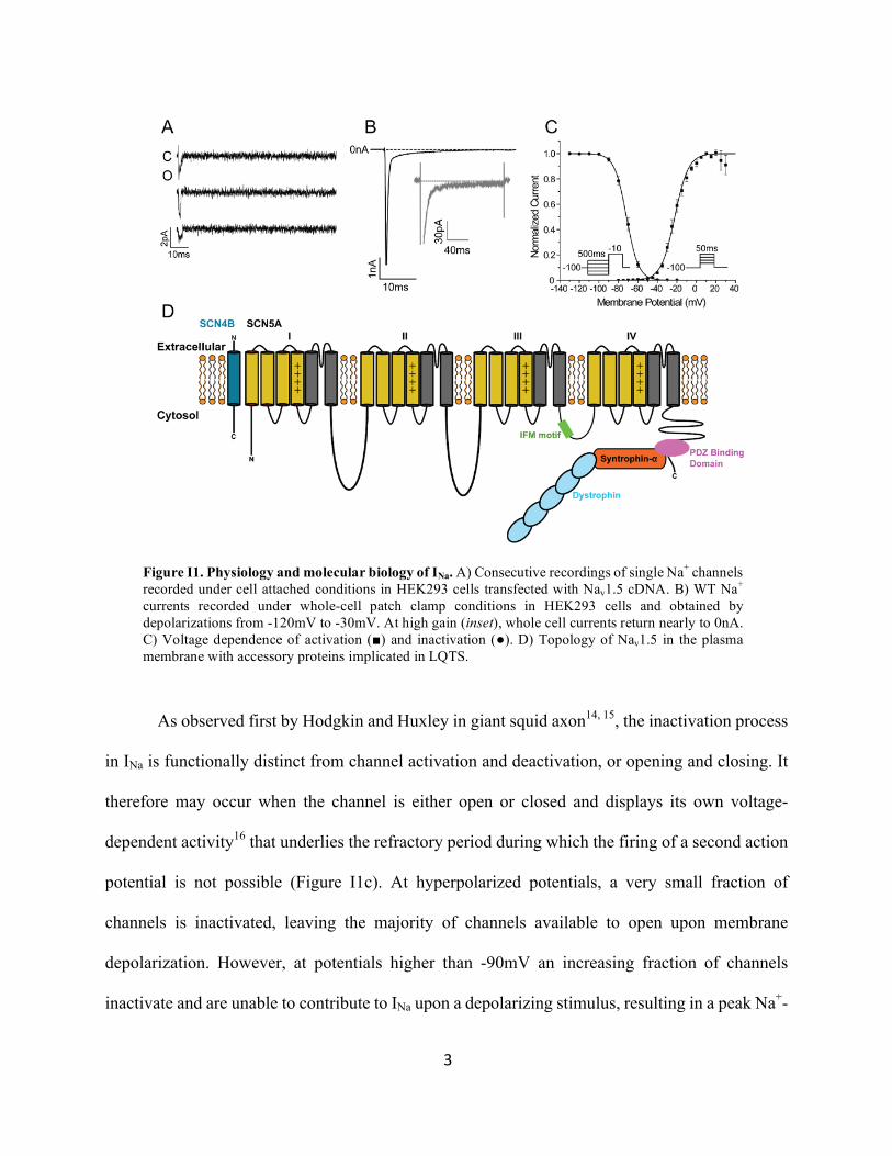
The depolarized membrane has widespread influence on other ionic currents, as well as on
INa itself. In a healthy heart, INa rapidly attenuates with a t1/2 of ~1ms in a process known as
inactivation, and contributes little to the net ionic current in later phases of the action potential
(Figure I1a,b). As INa inactivates, other ionic currents begin to contribute to the action potential
waveform8. A transient outward K+-current 13 creates a brief notch that is followed by a prolonged
plateau period in which inward flux of Ca2+ ions (ICaL) and outward K+ flux through delayed
rectifier K+-channels (IKr and IKs) balance. The influx of Ca2+ during this plateau not only serves
as an electrical signal, but also as a fundamental chemical signal that couples excitation and
contraction: cytoplasmic Ca2+ binds to the Ca2+-sensing machinery of the ryanodine receptor
triggering Ca2+-dependent Ca2+ release from the sarcoplasmic reticulum and removing the troponin
brake on actin-myosin bridging. Voltage- and Ca2+-dependent inactivation of ICaL and continued
activation of IKr and IKs cause a gradual repolarization of the membrane to its resting potential after
approximately 400ms, and Ca2+ extrusion by the Na+/Ca2+ exchanger (NCX) and sarcoplasmic
reuptake allow the muscle to relax.
3
As observed first by Hodgkin and Huxley in giant squid axon14, 15, the inactivation process
in INa is functionally distinct from channel activation and deactivation, or opening and closing. It
therefore may occur when the channel is either open or closed and displays its own voltage-
dependent activity16 that underlies the refractory period during which the firing of a second action
potential is not possible (Figure I1c). At hyperpolarized potentials, a very small fraction of
channels is inactivated, leaving the majority of channels available to open upon membrane
depolarization. However, at potentials higher than -90mV an increasing fraction of channels
inactivate and are unable to contribute to INa upon a depolarizing stimulus, resulting in a peak Na+-
Figure I1. Physiology and molecular biology of INa. A) Consecutive recordings of single Na+ channels recorded under cell attached conditions in HEK293 cells transfected with Nav1.5 cDNA. B) WT Na+ currents recorded under whole-cell patch clamp conditions in HEK293 cells and obtained by depolarizations from -120mV to -30mV. At high gain (inset), whole cell currents return nearly to 0nA. C) Voltage dependence of activation (■) and inactivation (●). D) Topology of Nav1.5 in the plasma membrane with accessory proteins implicated in LQTS.
4
transient that decreases as the resting membrane potential increases. This sub-activation threshold
inactivation, known as steady-state inactivation, results in half of the channels inactivating at
around -70mV. During depolarizing stimuli above the activation threshold (-50mV) (Figure I1c)
activation occurs faster than inactivation, resulting in the large inward Na+-transient that decays
rapidly soon after activation. Upon membrane repolarization channels recover from inactivation
following a bi-exponential time course, suggesting the presence of at least two functionally and
structurally distinct inactivated states of the channel.
Structure and Function of Voltage-Gated Na+-Channels
In the heart, the primary cardiac isoform of the voltage-gated Na+-channel responsible for
carrying INa is Nav1.5, encoded by the gene SCN5A17. The relationship between channel gating and
the structural features of Nav1.5 has been studied extensively, but many questions regarding this
link remain unanswered. Without the benefit of an atomic-scale structure for a eukaryotic Na+-
channel, the majority of our understanding of channel structure is derived from biochemical and
functional work, with recent contributions from a series of crystal structures from a
homotetrameric prokaryotic Na+-channel18, 19.
Nav1.5 is composed of a single polypeptide with 4 homologous domains (DI-DIV) that
resemble the canonical subunit structures observed in homotetrameric K+-channels9 (Figure I1d).
These 4 domains arrange around a single asymmetrical conduction pathway. Each domain contains
6 transmembrane helices (S1-S6) and a reentrant loop between S5 and S6 that serves as the
selectivity filter. The large aqueous inner cavity is lined by S5 and S6, which also converge at the
intracellular face of the channel to form the activation gate. The C-terminal ends of S6 cross in the
closed state of the channel, preventing the flow of ions through the channel.
5
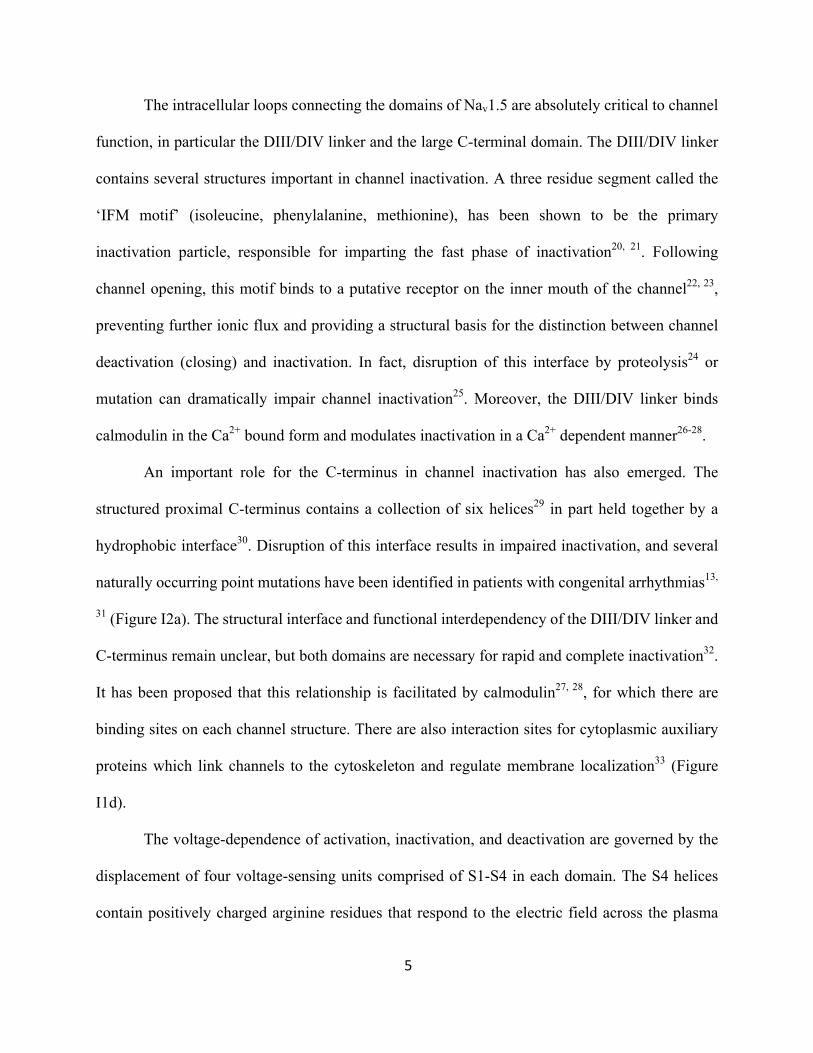
The intracellular loops connecting the domains of Nav1.5 are absolutely critical to channel
function, in particular the DIII/DIV linker and the large C-terminal domain. The DIII/DIV linker
contains several structures important in channel inactivation. A three residue segment called the
‘IFM motif’ (isoleucine, phenylalanine, methionine), has been shown to be the primary
inactivation particle, responsible for imparting the fast phase of inactivation20, 21. Following
channel opening, this motif binds to a putative receptor on the inner mouth of the channel22, 23,
preventing further ionic flux and providing a structural basis for the distinction between channel
deactivation (closing) and inactivation. In fact, disruption of this interface by proteolysis24 or
mutation can dramatically impair channel inactivation25. Moreover, the DIII/DIV linker binds
calmodulin in the Ca2+ bound form and modulates inactivation in a Ca2+ dependent manner26-28.
An important role for the C-terminus in channel inactivation has also emerged. The
structured proximal C-terminus contains a collection of six helices29 in part held together by a
hydrophobic interface30. Disruption of this interface results in impaired inactivation, and several
naturally occurring point mutations have been identified in patients with congenital arrhythmias13,
31 (Figure I2a). The structural interface and functional interdependency of the DIII/DIV linker and
C-terminus remain unclear, but both domains are necessary for rapid and complete inactivation32.
It has been proposed that this relationship is facilitated by calmodulin27, 28, for which there are
binding sites on each channel structure. There are also interaction sites for cytoplasmic auxiliary
proteins which link channels to the cytoskeleton and regulate membrane localization33 (Figure
I1d).
The voltage-dependence of activation, inactivation, and deactivation are governed by the
displacement of four voltage-sensing units comprised of S1-S4 in each domain. The S4 helices
contain positively charged arginine residues that respond to the electric field across the plasma

6
membrane9. At hyperpolarized membrane potentials, the S4 helix sits toward the inner leaflet of
the membrane in the resting position. Upon depolarization, the helices are driven upward to an
activated position, resulting in conformational changes that are transmitted to the channel gating
machinery. Unlike eukaryotic voltage-gated K+ channels, Nav1.5 is not symmetrical and the four
voltage-sensing units activate with different voltage-dependencies to control different aspects of
channel gating34. Mutagenesis35, 36, cysteine accessibility37, and fluorometric38 studies have
identified contributions from the DIII and DIV voltage sensors as important in the voltage-
dependent properties of inactivation.
Pathophysiology of Late Na+ Current
The plateau phase of the ventricular action potential is a period of relatively high input
resistance, so small changes in ionic currents can have a substantial impact on membrane potential.
As discussed above, in healthy hearts this isoelectric period is maintained by a balance of outward
K+ flux and inward Ca2+ flux. Loss of this K+ current, gain of Ca2+-current, or gain of another
depolarizing current can lead to prolongation of the plateau phase, delayed repolarization, and
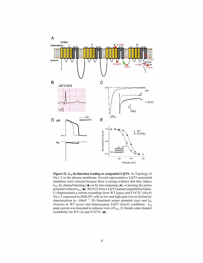
prolongation of the QT-interval on the ECG8(Figure I2b). These changes may arise from channel
mutations, post-translational modifications, or pharmacological targeting of specific currents
(especially the IKr current). In the case where this depolarizing current is due to aberrant INa, this
condition is called Long QT Syndrome Type 3 (LQT3)10.
7
Figure I2. INa dysfunction leading to congenital LQTS. A) Topology of Nav1.5 in the plasma membrane. Several representative LQT3-associated mutations were selected because there is strong evidence that they induce INaL by channel bursting (▲) or by late reopening (●), or prolong the action potential without INaL (■). B) ECG from a LQT3 patient (unpublished data). C) Representative current recordings from WT (gray) and F1473C (black) Nav1.5 expressed in HEK293 cells at low and high gain (inset) elicited by depolarization to -10mV 1. D) Simulated action potential (top) and INa (bottom) in WT (gray) and heterozygous LQT3 (black) conditions. INa peak current was truncated to enhance view of INaL. E) Steady-state channel availability for WT (Δ) and F1473C (■).
8
When INa fails to inactivate or inactivates too slowly the result is a sustained late current
(INaL) Figure I2c). INaL has been identified in many LQT3-associated mutant channels and often
amounts to less than 1% of the amplitude of peak INa10. Despite its limited size, sustained INaL has
been shown sufficient to prolong the ventricular action potential in animal models39, stem-cell
derived cardiomyocytes40, and computational models41, 42 (Figure I2d). Because the ECG is a body
surface representation of changes in the electrical field around the heart, action potential
prolongation is well correlated with prolongation of the QT interval and elevated risk of ventricular
arrhythmia and sudden cardiac death. Unlike patients with K+-channel associated LQT1 and
LQT2, patients with LQT3 tend to have an increased risk of arrhythmic events during periods of
rest43 because, as discussed below, in some LQT3 variants INaL increases with slower pacing
rates41. However, even within the umbrella of LQT3 there is a moderate degree of phenotypic
heterogeneity43. The mechanisms underlying this heterogeneity have been studied in depth, and
several modes of INaL generation have been identified with unique rate-dependent properties13, 44.
Mechanisms of INaL
Channel Bursting
The first LQT3 mutation that was identified in Nav1.5 was the ΔKPQ (1505-1507del)
deletion in the intracellular loop connecting DIII and DIV45. ΔKPQ results in a marked and time-
independent INaL that prolongs the action potential in isolated mouse ventricular myocytes46, 47 and
Purkinje cells46 and in in silico models of human cardiomyocytes41, 42, 48, 49. While in the majority
of depolarizing pulses these channels activate and inactivate normally, in less than 1% of cases a
channel will enter a slow “burst” mode of gating characterized by prolonged periods of channel
opening that often persist for multiple seconds and across multiple sequential sweeps50. Transitions
9
between the background WT-like gating mode and the burst mode are orders of magnitude slower
than the conformational changes that govern normal channel gating. In heterologous expression
systems INaL from ΔKPQ channels displays an inverse rate-dependence with a larger INaL at slower
pacing frequencies51. This results in pause-induced EADs observed in isolated ΔKPQ mouse
cardiomyocytes39 and in silico models41, 42. Consequently, LQT3 patients with the ΔKPQ mutation
are at a higher risk of arrhythmia and sudden cardiac death during periods of rest43, 52.
Our limited knowledge of the structure of the DIII/DIV linker and inactivation machinery
provides little structural explanation for the inverse rate-dependence of ΔKPQ, but functional and
computational work has provided some clues about the gating transitions important in the modal
shifts. At faster pacing frequencies channels tend to spend more time in inactivated states and less
time in closed states. Computational analysis revealed that slow transitions into the bursting mode
from channel closed states can account for the inverse rate-dependence of INaL, as well as the
frequency of occurrence of bursting events at the single-channel level41.
Late Reopenings
Several mutations have been identified that cause INaL not due to channel bursting (Figure
I2a). These mutations, such as R1644H, N1325S, and S1904L, are typically associated with a
deficiency in fast or slow phases of inactivation. The R1644H and N1325S mutations were
identified soon after the ΔKPQ mutation and demonstrated unique effects on the kinetics and
voltage-dependence of inactivation, suggesting that the etiology of INaL in these mutations was
different53. Single channel analysis revealed dispersed channel reopenings that occurred two orders
of magnitude more often than the bursts of ΔKPQ and with a duration that was similar to WT
channel openings54.

10
The S1904L mutation was discovered in a patient that experienced palpitations during
periods of exercise - an uncharacteristic clinical presentation for a patient with LQT313. Whole cell
experiments revealed a time course of fast inactivation that was slower than WT and INaL that
decayed gradually over time, unlike the time-independent INaL of ΔKPQ. At the single channel
level, these channels yielded dispersed late reopenings with a longer mean open duration than WT
channels, consistent with the delayed time to half inactivation of whole cell INa. These findings
contributed to our understanding of the role of the C-terminal domain as an important determinant
of inactivation, and transition metal ion FRET showed that the mutation disrupts an important
hydrophobic interface in this region55.
Computational models have been used to characterize the gating defects that give rise to
these pathological channel properties1. Channel reopening arises due to a deficiency in slow
inactivation that prolongs the mean dwell time of channels in the fast inactivated state. At
physiologically relevant voltages there exists an equilibrium between open and fast inactivated
states the greatly favors inactivation, though not completely. The increased dwell time in this state
therefore results in an increased probability of S1904L channels reopening. It should be noted,
however, that there is a small amount of reopening that does occur even from WT channels.
Eventually the slow inactivated states become fully absorbing, giving rise to the time-dependent
INaL that decays with prolonged depolarizations. One consequence of this deficiency in
inactivation, unlike the mode shifts that give rise to channel bursting, is that the reopenings are not
rate dependent. In fact, reopenings contribute more to action potential prolongation at faster pacing
frequencies where the slow-decaying INaL is still large during the repolarization phase. Similarly,
late reopenings may have a larger effect in certain tissue types, such as the Purkinje fiber, where a

11
lower plateau potential further slows the already impaired voltage-dependent inactivation and
provides a stronger driving force for inward Na+ flux42.
Window Current and Non-Equilibrium Gating
There exist a number of LQT3-associated mutations which do not produce INaL under
standard whole cell patch clamp protocols. The near-instantaneous voltage steps used in square-
pulse experiments fail to account for voltage-dependent gating transitions that occur at
intermediate voltages experienced during gradual membrane repolarization. In WT channels the
voltage-dependence of conductance and steady-state fast inactivation overlap only very slightly,
producing a very narrow “window” of current around -70mV where a small fraction of channels
can activate without inactivating56. Mutations which shift the voltage dependence of inactivation
in the depolarized direction (Figure I2e), or conductance in the hyperpolarized direction, will
increase the area under these two curves, resulting in a wider range over which channels may
activate without inactivating. As the action potential repolarizes back through this range a
“window current” emerges, providing an aberrant depolarizing Na+-flux that slows repolarization.
It is also possible that a similar emergence of INa may occur at prolonged times without any
change in steady-state inactivation properties. For example, the I1768V mutation was identified in
a patient with LQT3, and functional studies in heterologous expression systems revealed no INaL
or shift in steady-state inactivation57. However, voltage ramp protocols showed a significant
increase in late current compared to WT channels. Additional experiments revealed a speeding of
recovery from inactivation over WT that permits channels to recover prematurely and open again
before the membrane has fully repolarized.
12
Na+-Channel Blockers as Antiarrhythmics
Clinical Utility in Long QT Syndrome Type 3
Since the discovery of the role of INaL in the pathology of LQT3 and other acquired
arrhythmia conditions, the pharmacology of INaL has garnered a lot of interest3, 4, 58, 59. However,
the importance of peak INa in the upstroke velocity of the action potential and in the rapid
conduction of electrical impulses through the heart makes the selective inhibition of INaL critically
important. In fact, the widely cited CAST study reported that the use of the Na+-channel inhibitor
flecainide to prevent fatal arrhythmias following myocardial infarction resulted in an increase in
deaths12. Moreover, the pharmacological promiscuity among cardiac ion channels raises the risk
of off-target effects, such as inhibition of the IKr current, that can be counterproductive in efforts
to normalize the action potential. LQT3 patients are therefore the most likely of all LQT patients
to receive invasive implantable cardioverter-defibrillators (ICDs) as a precaution6. There is
therefore a growing demand for unraveling the mechanisms that contribute to preferential
inhibition of INaL and for the application of these lessons to the continued development of novel
and selective drugs60. The majority of Nav-inhibiting drugs are local anesthetics (LAs) or LA-like
drugs that are useful clinically because of their ability to target INaL (Figure I3). Not long after the
discovery of the ΔKPQ mutation, experiments in heterologous expression systems demonstrated
that the commonly used Class 1b antiarrhythmic Lidocaine inhibited INaL with greater potency than
it inhibited peak INa61. Similar observations have been made in heterologous systems with a large
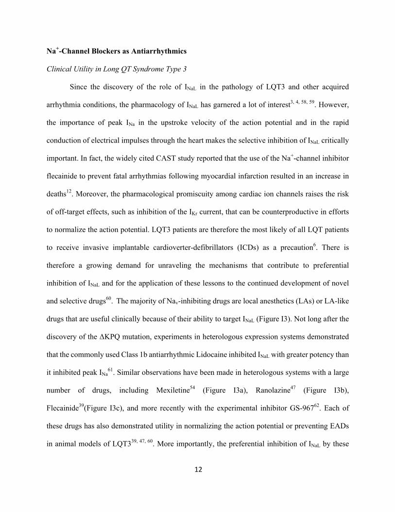
number of drugs, including Mexiletine54 (Figure I3a), Ranolazine47 (Figure I3b),
Flecainide39(Figure I3c), and more recently with the experimental inhibitor GS-96762. Each of
these drugs has also demonstrated utility in normalizing the action potential or preventing EADs
in animal models of LQT339, 47, 60. More importantly, the preferential inhibition of INaL by these
13
drugs in humans has been shown to shorten the QT interval63, 64 and reduce the frequency of
arrhythmic events in patients with LQT365. The suggestion has also been made that preferential
inhibition of the small fraction of INaL present in WT cardiomyocytes may help to offset the
proarrhythmic effects of drug-induced QT-prolongation66.
Drug-Channel Interactions
Local anesthetic drugs bind to a series of conserved residues in the central pore of Nav
channels. In Nav1.5 the residue that contributes the most to channel inhibition is F1760, located
just below the selectivity filter on S6 of DIV67. Mutation of this residue to alanine ablates UDB by
Lidocaine68, Mexiletine69, Ranolazine47, Flecainide68, GS-96762 and others. These inhibitors vary
in their size, charge, and hydrophobicity, and have therefore contributed to our understanding of
Figure I3. Representative recordings at low and high (insets) gain of F1473C inhibition by A) 50µM Mexiletine, B) 50µM Ranolazine, and C) 10µM Flecainide. D) Mexiletine, Ranolazine, and Flecainide inhibit INa with preference for INaL. Figure reproduced from Bankston, et al.1

14
the mechanisms important in INaL block. There are two factors important in their function: state-
dependence of drug binding and access to the occluded binding site.
On the basis of use-dependent block (UDB) - where channel inhibition accumulates with
repetitive pulsing – and shifts in the voltage-dependence of inactivation, Hille proposed the
Modulated Receptor Hypothesis70. In simple terms this hypothesis posits that local anesthetics
bind to a common receptor site with selective affinity to certain channel states and exerts an
allosteric stabilization of those states. High affinity block of open or inactivated channels allows
for the accumulation of non-conducting blocked channels during faster pacing frequencies and
recovery from channel block during periods of rest71. Biochemical37, 72, 73 and fluorometric74
analysis of channel conformations has provided additional evidence that the binding of local
anesthetics alters the distribution of channel states to favor slow inactivation. In the presence of
Lidocaine, the voltage sensors that are responsible for voltage-dependent inactivation accumulate
in their activated position, linking drug binding with allosteric modulation of channel gating.
The cationic form of these molecules imparts stronger UDB than the neutral forms75,
raising the question of how binding of a charged species relies so heavily on the aromatic F1760.
Using a series of unnatural derivatives of F1760 that monotonically diminish the density of
aromatic π-electrons, Ahern and Horn demonstrated that a cation-π interaction is responsible for
imparting UDB, but not tonic block, by Lidocaine76. It is likely, therefore, that conformational
changes in the pore during activation and inactivation create favorable conditions for the formation
of this interaction, in support of the notion of state-dependent drug-channel interaction.
Interestingly, Pless and Ahern discovered that while the cation-π interaction governs UDB by some
channel inhibitors, it does not play a role in the activity of all inhibitors77. For example, Ranolazine
does not require the cation-π for UDB, but is still dependent on the presence of F1760, suggesting
15
that there are multiple channel conformations or drug configurations within the pore that can
impart UDB.
Though not mutually exclusive, the alternative Guarded Receptor Hypothesis proposes that
perceived state-dependent binding affinity is in fact the result of transient occlusion of a static
receptor site that can only be accessed or escaped from certain channel states78. It is curious that
the cationic form of local anesthetics confers UDB but would be unable to access the binding site
when applied extracellularly. However, even drugs with high pKa exist as a small fraction of
neutral drug that can reside in or diffuse across the membrane. Hille proposed that resting-state
access of lipophilic drugs, which reside for a longer time in the lipid membrane, likely proceeds
through an intramembrane access pathway70. Larger, less lipophilic molecules likely pass through
the membrane and require channel openings prior to accessing the binding site75, 79. The time
constant of UDB is therefore highly dependent on pH80. A series of crystal structures of the
prokaryotic Nav homolog NavAB revealed for the first time the existence of intramembrane
fenestrations permeated by membrane lipids and large enough to be traversed by some lipophilic
drugs19. Interestingly, the size and shape of these fenestrations varied between the structures of
putatively inactivated and pre-open states, providing a structural basis for state-dependent drug
access to a central binding site18.
Objective of This Work
The contents of this work will build on the themes discussed above, and present novel
observations and models that contribute to our understanding of preferential inhibition of INaL. In
doing so, this analysis will provide a foundation for patient-specific management of LQT3 and for
the continued development of novel selective inhibitors of INaL. Chapter 1 identifies the single-
16
channel phenotype contributing to INaL in a LQT3 patient with a unique pharmacological response
to Na+-channel blockade. Our experiments reveal a late reopening phenotype that provides a
unique opportunity to study mechanisms of selective INaL inhibition in these channels, revealing
important differences between the pharmacology of Lidocaine and Ranolazine. This chapter also
demonstrate that these differences result in markedly altered selectivity for INaL in a mutation-
dependent manner. Chapter 2 presents in silico models of drug-channel interactions that
recapitulate a series of experimental findings and identify important state-dependent binding
properties that enhance inhibition of INaL by Lidocaine and Ranolazine. In doing so, these models
demonstrate that the gating kinetics of two different LQT3 phenotypes can account for potentially
clinically relevant mutation-specific pharmacology. Lastly, Chapter 3 presents the use of unnatural
amino acids to understand the physical chemical changes that arise during channel inactivation
and in the presence of LQT3 mutations that allows drugs to more effectively inhibit INaL than peak
INa.

17
Chapter 1: Mutations in Nav1.5 cause INaL via diverse mechanisms with
altered pharmacology

18
Summary
The Long QT Syndrome Type 3 arises in patients with mutations to the cardiac voltage-
gated Na+ channel, Nav1.5. These mutations often result in a significant non-inactivating late
current during the plateau phase of the ventricular action potential sufficient to delay
repolarization, prolong the QT interval on an ECG, and increase the risk of ventricular arrhythmia
and sudden cardiac death. Selective inhibitors of the late current have proven useful in decreasing
the risk of arrhythmia, however pharmacological intervention often remains unsatisfactory. We
previously reported the discovery of the F1473C mutation in a patient with severe QT prolongation
and arrhythmias that were incompletely managed by pharmacology alone. Here we present a
detailed characterization of the F1473C mutation that reveals abundant channel reopenings as the
primary mechanism underlying the late current. These reopenings occur frequently enough to
permit an exploration for the first time of the single channel mechanisms by which two Na+ channel
inhibitors, Lidocaine and Ranolazine, preferentially inhibit this channel phenotype. Lidocaine and
Ranolazine reduce the frequency of channel reopenings, while only Ranolazine shortens their
duration. The differential effect on channel reopenings suggests altered state-dependent drug
binding, resulting in dramatic mutation-specific pharmacology compared to that of ΔKPQ, a
naturally occurring mutation with late current caused by rare but prolonged channel bursts. These
findings reveal an important connection between channel phenotype and susceptibility to
preferential inhibition of late current that is an important consideration in the continuation of
development of novel, potent, and selective inhibitors.
19
Introduction
In the heart, the voltage-gated Na+ current, INa, is responsible for rapid membrane
depolarization that leads to the generation of an action potential and the conduction of electrical
signals to adjacent cells. The activation and inactivation of this current are exquisitely timed to
enable a transient flux of Na+ ions that contribute most substantially to the first few milliseconds
of the action potential in a healthy heart9, 81. These currents, carried by the cardiac isoform of the
voltage-gated Na+-current, Nav1.5, inactivate quickly and almost completely82, thereby
contributing very little to the membrane potential during the plateau phase of the action potential8.
However, there exist mutations83, disease states4, 5, and toxins84 that disrupt this inactivation
process, leading to a marked increase in the sustained depolarizing late current (INaL) that is
sufficient to prolong the ventricular action potential and increase the risk of ventricular arrhythmia
and sudden cardiac death8. Consequently, INaL has emerged as an important arrhythmogenic and
disease-potentiating factor in cardiac conditions such as heart failure and ischemic heart disease49,
85, 86. However, many questions still remain regarding the molecular etiology of INaL and
mechanisms by which clinically relevant antiarrhythmic drugs inhibit it.
The Long QT Syndrome Type 3 (LQT3) is a congenital disorder of ventricular
repolarization that is associated with aberrant Na+ flux during the plateau phase of the action
potential 5, 8, 9. Mutations in SCN5A, the gene encoding Nav1.5, have been shown to cause INaL,
prolong the ventricular action potential, prolong the QT interval on the clinical ECG, and enhance
the risk of ventricular arrhythmias and sudden cardiac death in LQT343. These mutations have
therefore served as a useful experimental model of INaL in heterologous systems1, 13, animal
models46, 47, and human induced pluripotent stem cell derived cardiomyocytes13, 40, 87 (iPS-CMs)
and have provided insight into the link between increased Na+ entry and arrhythmia.
20
Several mechanisms of INaL have been characterized in the literature, and involve channel
gating events with vastly different kinetics. Channel bursting, as best exemplified and first
identified in the ΔKPQ (1505-1507del) variant, involves the infrequent transition of channels into
a non-inactivating channel burst state41, 50. These bursts occur in fewer than 0.5% of channels
during a single depolarization, but often persist for multiple seconds. Importantly, channel bursts
occur more frequently during bradycardia, resulting in elevated risk of arrhythmia at rest41, 46, 51.
More recently, a mutation was identified in a patient after arrhythmia occurred during exercise,
suggesting a non-bursting channel phenotype13. Single-channel analysis revealed a propensity for
frequent channel reopenings during prolonged depolarizations that were best described as a
deficiency in channel slow inactivation. Finally, overlap between the voltage-dependent activation
and inactivation curves can create a window over which channels activate but do not inactivation.
Mutations which shift inactivation in the depolarizing direction can increase the size of this
“window current” and the possibility of channels reopening prematurely as the membrane potential
passes through this range during repolarization.
Our group previously reported a mutation in a patient with QT-prolongation and poor
management of arrhythmia with pharmacological intervention, resulting in implantation of an ICD
at 33 days after birth1, 88. Whole-cell patch clamp experiments in a heterologous expression system1
and in iPS-CMs87 revealed a range of biophysical changes in channel gating, included marked INaL,
a depolarizing shift in steady-state inactivation, and a speeding of recovery from inactivation.
Taken together, the clinical and macroscopic experimental data suggest a unique mechanism of
INaL generation with altered pharmacological properties. Here we present single-channel evidence
in support of this hypothesis, and demonstrate that the F1473C mutation results in a mixed
phenotype of channel bursting and late reopening. Moreover, we find that these reopenings occur
21
more frequently than in previously reported mutations, affording the opportunity to study the
precise mechanism of preferential inhibition of INaL by two clinically relevant Na+-channel
inhibitors, Lidocaine and Ranolazine. These inhibitors have different effects on the frequency and
duration of late reopenings that result in strikingly altered selectivity for inhibition of INaL. Lastly,
we show that these mechanisms also result in unique mutation-specific differences across channel
phenotypes, demonstrating that a phenotype-specific approach to pharmacological management is
important in patients with LQT3.
Methods
Expression of recombinant Nav1.5
WT or mutant Nav1.5 cDNA and b-subunit hb1 cDNA were separately sub-cloned into the
pcDNA3.1(+) expression vector (Invitrogen). The hb1 vector additionally included the gene for
CD8, a common and commercially available reporter gene. Equal amounts of a-subunit and b-
subunit totaling 3mg were simultaneously transfected in HEK 293 cells using the Lipofectamine
2000 reagent (Invitrogen). CD8-positive cells were identified using Dynabeads (Thermo Fisher
Scientific), and were selected for patching 48 hours after transfection.
Electrophysiology
Membrane currents were measured using whole-cell and cell-attached patch clamp
configurations. All currents were recorded using the Axon 200B amplifier (Axon Instruments) and
were obtained at room temperature (24°C). Whole cell currents were recorded with pipette
solutions (mmol): 50 Aspartic acid, 5 ATP-Na2, 60 CsCl,11 EGTA, 10 HEPES, 4.27 CaCl2,
resulting in a [Ca2+] of 100 nM 1 MgCl2, pH 7.4 adjusted with CsOH. External solution contained
22
(mmol): 130 NaCl, 5 CsCl, 10 HEPES, 5 glucose, 2 MgCl2. 1.2 CaCl2, pH 7.4 After maintaining
>1GΩ seal and rupturing the membrane, capacitive currents and series resistance were
compensated with analog techniques. For all pharmacology experiments, drug trials are control-
matched in the same cell. Following control and drug-exposed experimental trials cells were
exposed to 50 mM tetrodotoxin (TTX; Abcam) and all whole cell patch clamp results are presented
as TTX-sensitive currents.
Cell-attached patch clamp was performed to measure the activity of single ion channels.
Borosilicate glass pipettes were pulled to a resistance of 5-9 MΩ, and coated with Sylgard (Dow
Corning) to isolate ambient electrical noise. Pipette solution contained (mmol): 110 NaCl 1 CaCl2,
5 HEPES, pH 7.4. Bath solution contained (mmol): 140 KCl, 5 HEPES, 1 MGCl2, pH 7.4. After
obtaining membrane seals of resistance >10GΩ, the number of channels in the patch was estimated
from the maximal current elicited from a depolarization to -30mV from -120mV. Patches
containing 1-12 channels were selected for further experimentation. Data was captured at 20kHz
sampling frequency and filtered online at 5kHz. All experiments were performed using 100ms
depolarizations to -30mV from -120mV at 1Hz pacing. To test the effects of Na+-channel blocking
drugs on single channels, we performed control-matched experiments. Channel openings were
stimulated with 400 consecutive depolarizations under constant perfusion of control bath solution.
While still applying stimulating pulses, vehicle, 100mM Ranolazine, or 200mM Lidocaine was
added to the bath perfusion. Following a 2min equilibration period, an additional 400 traces were
captured as the drug-positive sample under continuous drug perfusion. By comparing control and
experimental recordings in the same patch we were able to control for inter-patch differences in
the number of channels in the patch.
23
Data Analysis
Whole cell currents were analyzed in Clampfit 10 (Molecular Devices, LLC) and are
reported here as TTX-sensitive currents.
Single-channel currents were analyzed using Clampfit 10. Baseline correction was
performed by subtracting the averaged current from at least 10 null sweeps containing zero channel
openings. As we are primarily interested in the effects of drugs in blocking INaL and overlapping
channel openings make early events difficult to interpret, single channel event detection was
performed on the last 90ms of each trace. Single channel openings were identified using Clampfit’s
Event Detection application, with a minimum event duration of 200µs and a half-amplitude
detection threshold with unitary current amplitude set to 1.7pA. Mean open duration was
determined by compiling all channel openings from across patches into an open time histogram,
which was fit to a single exponential distribution.
Statistics
Data are presented at mean ± standard error. Statistical significance was determined by a
Student’s T-test with significance level p<0.05. For comparisons across multiple groups statistical
significance was determined with a one-way ANOVA with significance level p<0.05, and
individual comparisons made using a Tukey post-test. Comparisons of channel mean open times
were tested using Levene’s test for equality of variances. For exponentially distributed data sets,
the mean and standard deviation are equivalent. A significant difference in variance is therefore
indicative of changes in the mean.
24
Drugs
Ranolazine (lot 114M4743V) and Lidocaine (lot MKBR6002V) were purchased from
Sigma-Aldrich. Stock solutions were prepared in water at a concentration of 10mM and 100mM,
respectively. TTX was purchased from Abcam and used at a final concentration of 50µM in water.
Results
F1473C Causes a Mixed INaL Phenotype.
As reported previously1, 87, in whole cell patch clamp F1473C results in a large sustained
INaL during prolonged membrane depolarizations, 0.6% of peak at 200ms, that is significantly
greater than in WT (Figure 1.1a,b) and a 9mV depolarizing shift in steady-state inactivation1. We
reasoned that these macroscopic differences were due to mechanistically unique single-channel
phenotypes and performed cell-attached patch to test this hypothesis. Using trains of 100ms
depolarizations to -30mV from a holding potential of -120mV at 1Hz pacing frequency, we were
able to identify two distinct gating abnormalities contributing to INaL. Figures 1.1c and 1.1d depict
consecutive representative traces from cell-attached patches containing one WT or two F1473C
channels, respectively. While the WT recordings display very rare ionic currents after the first
10ms of the pulse, the F1473C recordings display both frequent channel late openings and a
prolonged burst of channel activity. We only examined the opening events occurring after the first
10ms of each test pulse and do not differentiate between channel reopenings and primary openings
that occur after 10ms, as any opening during prolonged times will contribute to INaL. The F1473C
mutation caused a significant increase in channel reopening per sweep (0.203 ± 0.032 vs 0.012 ±
0.003; p<0.001) (Figure 1.1E) and a trend towards enhanced channel bursting, as measured by the
number of bursts per 10,000 channel sweeps (4.93 ± 1.40 vs 1.49± 0.76; p=0.055) (Figure 1.1F).

25
We next asked if the reopenings
caused by the F1473C mutation were due to
a transient failure of fast inactivation by
comparing their mean open duration to that
of WT. Fits of open dwell time histograms
to a single exponential distribution revealed
similar channel mean open times (0.43ms vs
0.42ms), suggesting that the mutation
affects only the propensity for channel
reopening and not the duration of those
openings (Figure 1.1G and H). These
findings are consistent with a deficiency in
slow inactivation but no effect on the fast
inactivation processes that directly regulate
channel open duration.
Lidocaine and Ranolazine Inhibit Late
Reopenings with Differential Effects on
Open Duration
Lidocaine and Ranolazine are two
clinically important Na+-channel inhibitors
that have been characterized extensively in
the literature47, 49, 75. Computational models
Figure 1.1 Single-channel characterization of F1473C. A and B) Representative whole cell recordings of WT and F1473C Nav1.5 currents. High gain images (insets) depict a substantial sustained INaL from F1473C channels. C and D) Consecutive representative recordings of cell-attached patches of WT (1 channel) and F1473C (2 channels) Nav1.5 currents. E) Quantification of the number of reopenings per channel per sweep after the first 10ms (n=6 patches for WT; n=7 patches for F1473C). F) Quantification of the number of sweeps containing channel bursts (n=6 patches for WT; n=7 patches for F1473C). G and H) Open duration histograms constructed from channel openings after the first 10ms of each sweep. Fits to a single exponential distribution provided a mean open duration of 0.42ms for WT and 0.43ms for F1473C.
26
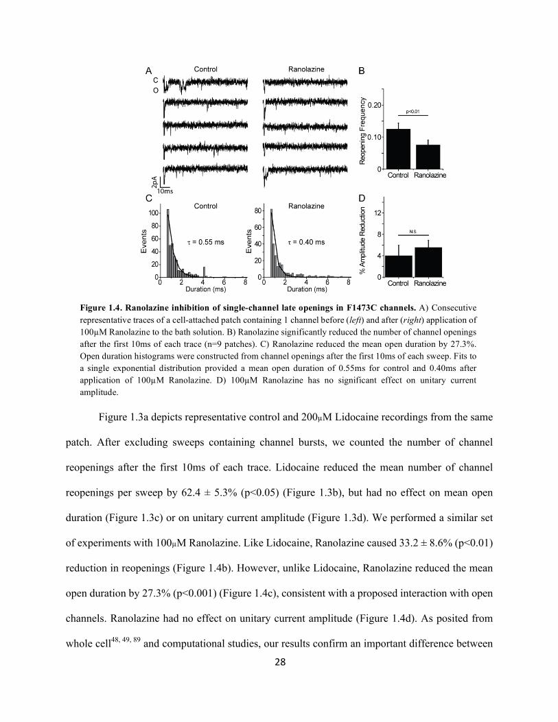
of state-dependent channel affinity based on whole cell experimental data have revealed that
Lidocaine interacts preferentially with inactivated channels48, 89, while Ranolazine interacts
preferentially with open channels49, and experiments with unnatural amino acids have revealed
differences in the chemistry of interaction with the local anesthetic binding site, F176077. Both of
these mechanisms have proven effective in the selective inhibition of INaL in bursting ΔKPQ mutant
channels47-49, 90. The robust reopening phenotype of F1473C channels affords the opportunity to
study the effects of these drugs specifically on those events at the single channel level and to
determine whether pharmacological differences emerge between bursting and late-reopening
channels.
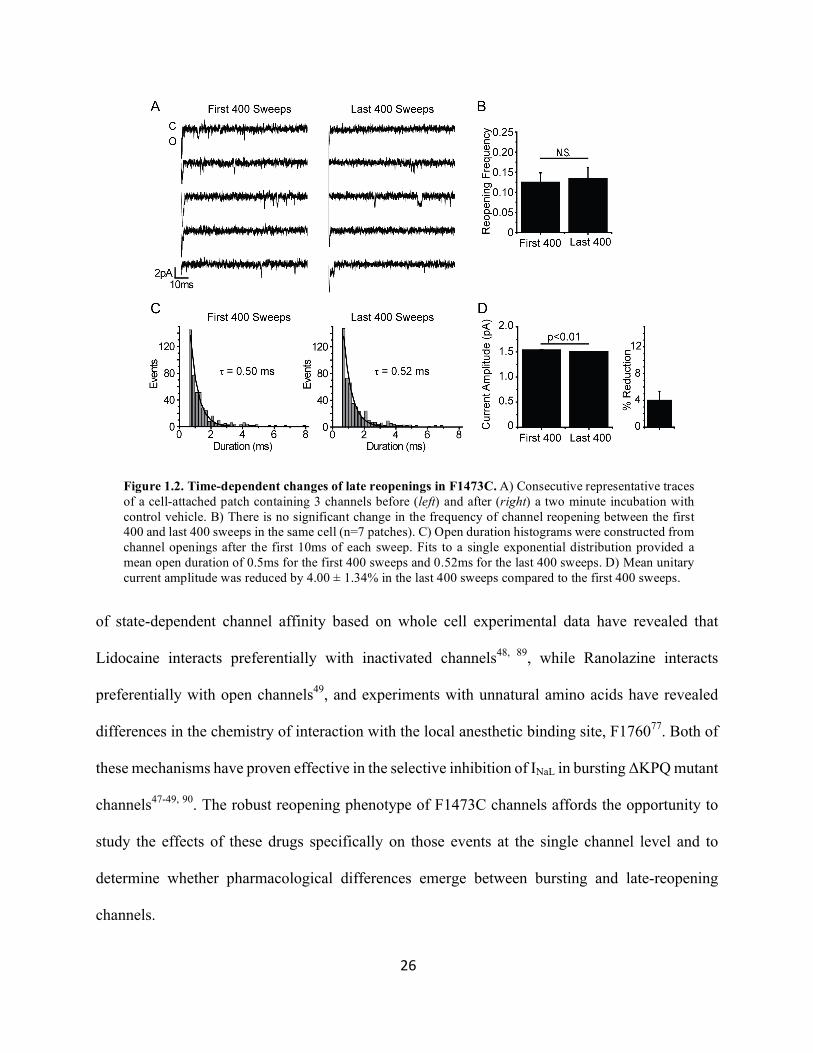
Figure 1.2. Time-dependent changes of late reopenings in F1473C. A) Consecutive representative traces of a cell-attached patch containing 3 channels before (left) and after (right) a two minute incubation with control vehicle. B) There is no significant change in the frequency of channel reopening between the first 400 and last 400 sweeps in the same cell (n=7 patches). C) Open duration histograms were constructed from channel openings after the first 10ms of each sweep. Fits to a single exponential distribution provided a mean open duration of 0.5ms for the first 400 sweeps and 0.52ms for the last 400 sweeps. D) Mean unitary current amplitude was reduced by 4.00 ± 1.34% in the last 400 sweeps compared to the first 400 sweeps.

27
We recorded 400 control and 400 drug-containing sweeps from cell attached patches of
cells exposed to either vehicle (Figure 1.2), 200µM Lidocaine (Figure 1.3) or 100µM Ranolazine
(Figure 1.4) in the bath solution. In a series of control experiments we tested for time-dependent
changes in channel reopening frequency, mean open duration, and unitary current amplitude over
the course of the 15-minute experiment. We observed no time-dependent changes in reopening
frequency (Figure 1.2b) or duration (Figure 1.2c) between the first 400 and second 400 sweeps.
We did observe a small 4.00% ± 1.34% (p<0.01) reduction in unitary current amplitude, likely as
the result of depletion of pipette Na+ and a reduction in the current driving force over the course
of the experiment (Figure 1.2d). Any drug-induced change in unitary current amplitude in future
experiments was compared to this 4% time-dependent change.
Figure 1.3. Lidocaine inhibition of single-channel late openings in F1473C channels. A) Consecutive representative traces of a cell-attached patch containing 1 channel before (left) and after (right) application of 200µM Lidocaine to the bath solution. B) Lidocaine significantly reduced the number of channel openings after the first 10ms of each trace (n=3 patches). C) Open duration histograms were constructed from channel openings after the first 10ms of each sweep. Fits to a single exponential distribution provided a mean open duration of 0.47ms for control and 0.49ms after application of 200µM Lidocaine. D) 200µM Lidocaine has no significant effect on unitary current amplitude.
28
Figure 1.3a depicts representative control and 200µM Lidocaine recordings from the same
patch. After excluding sweeps containing channel bursts, we counted the number of channel
reopenings after the first 10ms of each trace. Lidocaine reduced the mean number of channel
reopenings per sweep by 62.4 ± 5.3% (p<0.05) (Figure 1.3b), but had no effect on mean open
duration (Figure 1.3c) or on unitary current amplitude (Figure 1.3d). We performed a similar set
of experiments with 100µM Ranolazine. Like Lidocaine, Ranolazine caused 33.2 ± 8.6% (p<0.01)
reduction in reopenings (Figure 1.4b). However, unlike Lidocaine, Ranolazine reduced the mean
open duration by 27.3% (p<0.001) (Figure 1.4c), consistent with a proposed interaction with open
channels. Ranolazine had no effect on unitary current amplitude (Figure 1.4d). As posited from
whole cell48, 49, 89 and computational studies, our results confirm an important difference between
Figure 1.4. Ranolazine inhibition of single-channel late openings in F1473C channels. A) Consecutive representative traces of a cell-attached patch containing 1 channel before (left) and after (right) application of 100µM Ranolazine to the bath solution. B) Ranolazine significantly reduced the number of channel openings after the first 10ms of each trace (n=9 patches). C) Ranolazine reduced the mean open duration by 27.3%. Open duration histograms were constructed from channel openings after the first 10ms of each sweep. Fits to a single exponential distribution provided a mean open duration of 0.55ms for control and 0.40ms after application of 100µM Ranolazine. D) 100µM Ranolazine has no significant effect on unitary current amplitude.
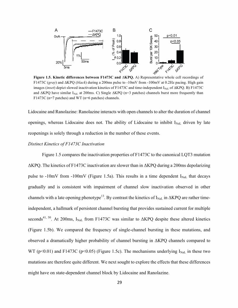
29
Lidocaine and Ranolazine: Ranolazine interacts with open channels to alter the duration of channel
openings, whereas Lidocaine does not. The ability of Lidocaine to inhibit INaL driven by late
reopenings is solely through a reduction in the number of these events.
Distinct Kinetics of F1473C Inactivation
Figure 1.5 compares the inactivation properties of F1473C to the canonical LQT3 mutation
ΔKPQ. The kinetics of F1473C inactivation are slower than in ΔKPQ during a 200ms depolarizing
pulse to -10mV from -100mV (Figure 1.5a). This results in a time dependent INaL that decays
gradually and is consistent with impairment of channel slow inactivation observed in other
channels with a late opening phenotype13. By contrast the kinetics of INaL in ΔKPQ are rather time-
independent, a hallmark of persistent channel bursting that provides sustained current for multiple
seconds41, 50. At 200ms, INaL from F1473C was similar to ΔKPQ despite these altered kinetics
(Figure 1.5b). We compared the frequency of single-channel bursting in these mutations, and
observed a dramatically higher probability of channel bursting in ΔKPQ channels compared to
WT (p<0.01) and F1473C (p<0.05) (Figure 1.5c). The mechanisms underlying INaL in these two
mutations are therefore quite different. We next sought to explore the effects that these differences
might have on state-dependent channel block by Lidocaine and Ranolazine.
Figure 1.5. Kinetic differences between F1473C and ∆KPQ. A) Representative whole cell recordings of F1473C (gray) and ∆KPQ (black) during a 200ms pulse to -10mV from -100mV at 0.2Hz pacing. High gain images (inset) depict slowed inactivation kinetics of F1473C and time-independent INaL of ∆KPQ. B) F1473C and ∆KPQ have similar INaL at 200ms. C) Single ∆KPQ (n=3 patches) channels burst more frequently than F1473C (n=7 patches) and WT (n=6 patches) channels.
30
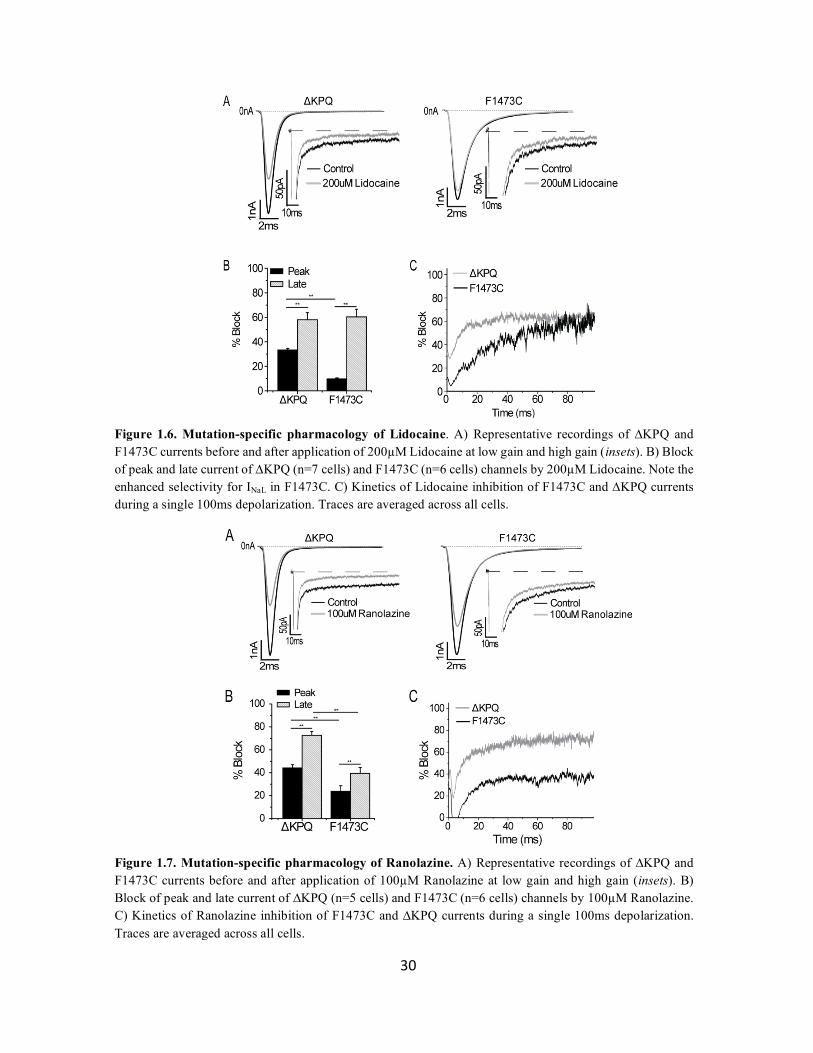
Figure 1.6. Mutation-specific pharmacology of Lidocaine. A) Representative recordings of ∆KPQ and F1473C currents before and after application of 200µM Lidocaine at low gain and high gain (insets). B) Block of peak and late current of ∆KPQ (n=7 cells) and F1473C (n=6 cells) channels by 200µM Lidocaine. Note the enhanced selectivity for INaL in F1473C. C) Kinetics of Lidocaine inhibition of F1473C and ∆KPQ currents during a single 100ms depolarization. Traces are averaged across all cells.
Figure 1.7. Mutation-specific pharmacology of Ranolazine. A) Representative recordings of ∆KPQ and F1473C currents before and after application of 100µM Ranolazine at low gain and high gain (insets). B) Block of peak and late current of ∆KPQ (n=5 cells) and F1473C (n=6 cells) channels by 100µM Ranolazine. C) Kinetics of Ranolazine inhibition of F1473C and ∆KPQ currents during a single 100ms depolarization. Traces are averaged across all cells.
31
Single-Channel Phenotypes Underlie Mutation-Specific Pharmacology
Figures 1.6 and 1.7 illustrate the effects of 200µM Lidocaine and 100µM Ranolazine on
the two LQT3 mutant channels at the whole cell level. Both drugs preferentially inhibit INaL in both
mutations, but the degree of selectivity varies widely in a mutation- and drug-dependent manner.
Lidocaine is a far less potent inhibitor of peak INa in F1473C compared to ΔKPQ (9.70 ± 0.81%
vs 33.81± 1.15%, p<0.001), while the effect on INaL is similar (60.35 ± 6.30% vs 63.62 ± 5.55%,
p=0.70) (Figure 6b). Conversely, Ranolazine is a more potent inhibitor of both peak INa (23.85 ±
1.28% vs 44.28 ± 2.74%, p<0.001) and INaL (39.38 ± 5.16% vs 72.58 ± 3.37%, p<0.001) in ΔKPQ
channels (Figure 1.7b).
We reasoned that differences in block of INa and INaL were likely reflective of differences
in the kinetics of current inhibition that are not captured in our analysis of peak and 100ms time
points. To investigate this hypothesis, we analyzed the percentage of current blocked by drug over
the course of a full 100ms depolarization. 200µM Lidocaine showed dramatically different kinetic
profiles for inhibition of the two mutant channels (Figure 1.6c). Block of F1473C developed
continually over the course of a depolarizing step, whereas block of ΔKPQ developed rapidly and
reached steady-state in approximately 20ms. These kinetic profiles reflect the time courses of
channel inactivation, where F1473C INaL decays slowly and ΔKPQ INaL reaches a plateau more
quickly.
The ability of channel inhibition to reflect the underlying current kinetics is absent in block
of INaL by 100µM Ranolazine, which blocked with similar kinetics but altered amplitude in F1473C
and ΔKPQ channels (Figure 1.7c). Ranolazine binding therefore reflects a kinetic scheme that is
insensitive to changes in slow inactivation. These findings are consistent with our analysis of
32
Lidocaine and Ranolazine inhibition of single-channels, and confirm that these two inhibitors
interact with the channel in unique ways.
Discussion
F1473C: A Complex LQT3 phenotype
The seminal study reporting the macroscopic function of the LQT3-associated mutation
F1473C identified several pathological changes in channel gating, including a depolarizing shift
in steady-state inactivation, a speeding of recovery from inactivation, and a substantial INaL1. Here
we present for the first time a detailed single-channel characterization of INaL in this mutation.
F1473C causes a trending increase in the probability of channel bursting and a more dramatic
increase in the probability of late reopening. In our experiments, a single channel will open after
the first 10ms of a 100ms pulse in 20% of the depolarizing sweeps, 20-fold more often than WT
channels. We found that these reopenings have a mean duration that is very similar to the rare
reopenings in WT channels in our experiments and to the WT mean open duration reported by
others13, 54, 91. The ability of F1473C channels to reopen without a change in open duration suggests
that the mutation alters channel slow inactivation to allow for reopenings, but has no effect on the
kinetics of fast inactivation13. This is a striking finding given the location of F1473C in the linker
between domains III and IV of Nav1.5, which is a known molecular determinant of fast
inactivation20, 21.
While F1473C channel bursting is slightly enhanced compared to WT, it represents only a
fraction of the bursting activity observed in ΔKPQ channels. However, this small amount of
bursting is sufficient to account for the previously reported preferential reduction in INaL with faster
pacing frequency87 – a hallmark of channel bursting41. Taken together, our experiments provide a
33
potential connection between the single-channel behavior, the experimentally observed rate-
dependence, and the severity of disease caused by the F1473C mutation. Increased pacing relieves
the burden of a small degree of channel bursting but does not remedy, and may even unmask, the
effects of channel reopening.
The Molecular Determinants of INaL Inhibition by Lidocaine and Ranolazine
Several studies have used experimental and computational approaches to investigate the
effects of local anesthetic drugs on bursting channels46-49, 90. Lidocaine and Ranolazine, in
particular, have served as model cases for two general mechanisms of channel interaction. Based
on its ability to dramatically shift channel availability in the hyperpolarizing direction, slow
recovery from inactivation, and allosterically modulate a voltage-sensor important in channel
inactivation74,92, Lidocaine is the archetypical local anesthetic with selective affinity for
inactivated channels. More recently, the effects of Ranolazine on ΔKPQ have been interpreted as
preferential inhibition of open channels because of its small effect on channel availability49, 93.
We sought to understand the effects that these putative mechanisms would have on channel
late reopening. The frequent reopenings caused by the F1473C mutation provided an opportunity
to study the single channel block of these events in a way not previously possible. We found that
while both drugs were able to reduce the frequency with which late openings occur, only
Ranolazine had an effect on channel mean open time. Coupled with a smaller impact on steady-
state inactivation and unique kinetics of inhibition, these results imply important differences in the
interaction of these drugs with the local anesthetic binding site that influence selective inhibition
of INaL in these channels.
34
Both Lidocaine and Ranolazine rely heavily on F1760, the local anesthetic binding site, for
use-dependent block and INaL inhibition47, 67, 68. Access to the local anesthetic binding site, which
resides within the central conducting pore67, 68, is limited by channel state and by the chemistry of
the ligand. Hydrophilic or charged drugs must access this site through the open aqueous pore.
Lipophilic drugs, which reside for longer times within the plasma membrane, may access the
binding site via a lipophilic route, predicted first by Bertil Hille70 and identified more recently in
crystal structures of the prokaryotic voltage-gated Na+-channel homolog NavAB18, 19. Interestingly,
Ranolazine is a derivative of Lidocaine and contains a Lidocaine moiety, similar pKa, and similar
partition coefficient77, 94. It is therefore surprising that the effects on channel reopening are so
different. However, Ranolazine is larger in size, likely limiting its ability to access the lipophilic
pathway in non-conducting channels and altering its orientation in the pore with respect to F1760.
In fact, Pless et al identified a critical cation-𝛑 interaction that governs use-dependent block by
Lidocaine but not Ranolazine, indicating important differences in the chemistry of drug binding.
Taken together, these experiments demonstrate that derivatives of classical local anesthetic drugs
may display markedly different pharmacological activity that alter the kinetics and magnitude of
channel inhibition. Understanding the chemical underpinnings of drug access and binding is a
critical step in the design of therapeutic programs for patients with INaL-associated cardiac disease.
Impact of Channel Phenotype on Preferential Inhibition of INaL
We reasoned that because F1473C and ΔKPQ mutations alter channel gating in unique
ways, their susceptibility to state-dependent channel inhibition would also vary. Lidocaine reduced
INaL in both mutations by a similar amount. Our single channel experiments revealed that Lidocaine
inhibits INaL solely by reducing the frequency of late events. In agreement with this notion, a
35
previous study of the inhibition of single ΔKPQ channels revealed that Lidocaine reduced the
frequency of channel bursts without any effect on their duration90. Because the F1473C reopenings
result in a time-varying INaL and the ΔKPQ bursts cause a time-independent INaL, normalization of
currents by elimination of these phenotypes would produce unique kinetics of block. In fact, we
observed a slower time course of inhibition of F1473C currents than ΔKPQ. The depolarized
channel availability and more rapid recovery from inactivation of F1473C channels results in
diminished peak current block and enhanced selectivity for INaL. While the qualitative effect on
late reopenings and bursts is similar, the properties of the channels themselves influence the
kinetics and selectivity of block.
Ranolazine, on the other hand, does not display mutation-dependent kinetics. The time
course of block in the two mutations is similar, but the magnitude of inhibition of ΔKPQ channels
is greater. The reduction in mean open duration caused by Ranolazine requires that the drug binds
very rapidly to open channels. It is therefore likely that the kinetics of INaL inhibition are governed
predominantly by the rapid binding of drug to open channels, with less influence from the kinetics
of inactivation. However, the bursting that causes INaL in ΔKPQ and contributes a fraction of INaL
in F1473C has longer mean open duration than late reopenings or early transient openings,
providing more time for drug to bind and an increased magnitude and selectivity for INaL derived
from bursts. These findings suggest that a shortening of mean open time by some local anesthetics
is detrimental to selective inhibition of reopenings, but effective at targeting channel bursts.
Pharmacological intervention in patients with LQT3 is already complicated by the potential
for off-target effects on other ion channels, which are influenced by genetic diversity among
patients1, 87. Our results highlighting additional heterogeneity within INaL itself enhance the need
for an understanding of specific mechanisms of selective inhibition. The findings presented here
36
imply that the effect of inhibitors on the frequency and duration of late events can contribute to
their selectivity for INaL in a mutation-dependent manner. By examining these mutations and drugs,
which serve as general models for the diverse pathology and pharmacology of LQT3, we can begin
to understand the specific drug-channel interactions that impart preferential inhibition of INaL. In
Chapter 2 we present a series of computational models of LQT3 channel function that extend the
quantitative connection between impaired channel gating, state-dependent drug binding, and
mutation-specific pharmacology of local anesthetic drugs with unique chemical properties. These
findings have important implications in the patient-specific management of LQT3, and an
understanding of the molecular etiology of INaL for a given mutation or disease state may inform
the selection of Na+-channel inhibitors used clinically.
37
Chapter 2: Computational modeling of state-dependent drug binding in mutation-specific
pharmacology: Implications for patient-specific therapy
38
Summary
The late sodium current has emerged as an important factor in the pathogenesis of several
cardiovascular ailments, sparking a renewed interest in the antiarrhythmic mechanisms of local
anesthetic inhibition of the cardiac voltage-gated Na+ channel, Nav1.5. Patients with heritable INaL
arising from mutations in Nav1.5 and the congenital Long QT Syndrome Type 3 (LQT3), have
demonstrated some clinical benefit to pharmacological intervention, but incomplete INaL selectivity
raises the risk of impaired cardiac conduction and drug-induced arrhythmia. A more detailed
understanding of the properties that impart selective inhibition of INaL by local anesthetics is
therefore desirable. Kinetic models of ion channel function are useful in developing testable
hypotheses from dynamic systems, such as channel gating and state-dependent drug binding. This
chapter presents a computational approach to unraveling the mechanisms that underlie mutation-
specific inhibition of different varieties of INaL. First, we develop an updated kinetic model of WT
channel gating based on a new series of kinetic constrains that define transitions important in the
population of high affinity channel states. Only slight deviations from the WT model generate
virtual LQT3 mutant channels with distinct gating phenotypes, such as the F1473C and ΔKPQ
mutations characterized in Chapter 1. The disease models then serve as the foundation for a
characterization of the state-dependent drug-channel interactions that impart mutation-specific
inhibition of INaL by Lidocaine and Ranolazine. The models suggest that much of the mutation-
specific pharmacology can be attributed to differences in channel gating kinetics that result in
unique distributions of high affinity channel states. Finally, this chapter presents proof of concept
for the in silico screening of patient-specific responses to local anesthetics with different
pharmacological signatures that paves the way for personalized management of LQT3 and other
INaL-associated disorders.
39
Introduction
In silico pharmacology holds enormous value in providing a platform for examining
competing theories for mechanisms of drug binding and in predicting clinically relevant outputs
from complex biological systems. Cardiology therapeutics is a particularly well-suited area for the
employment of virtual drug discovery because rhythmic contraction is driven by the integration of
numerous ionic currents and chemical signals. One particular aspect of cardiac physiology that has
gained a growing interest is the pathological late Na+-current (INaL) that has been observed in
multiple disease states to contribute to action potential prolongation and arrhythmogenesis4, 5, 59,
95. Computational analysis has been used repeatedly to predict the pathological effects of INaL and
to demonstrate the potential clinical benefit of selective INaL inhibition across a range of cardiac
disorders11, 41, 42, 46, 48, 49, 85, 86.
To explore altered Na+ channel gating, kinetic models have evolved from simple Hodgkin-
Huxley models to more complex multi-state models that attempt to capture the functional
properties of the channel15, 41, 49, 96, 97. In these newer models, physiologically relevant
conformational changes and functional properties are represented by voltage-dependent transitions
between states in a hidden Markov chain98. By altering the rates of these transitions the models are
capable of reproducing many aspects of the pathophysiology associated with the Long QT
Syndrome Type 3 (LQT3)13, 41, Brugada Syndrome97, 99, and heart failure11. Additionally, these
models have proven useful in describing the state-dependent binding mechanisms of clinically
relevant Na+-channel inhibitors and extrapolating those properties into effects on action potentials,
tissue segments, and ultimately simulated ECGs11, 49, 85. This chapter expands upon that body of
work to describe the fundamental underpinnings of mutation-specific pharmacology in LQT3.
40
LQT3 arises from mutations in the cardiac isoform of the voltage-gated Na+-channel a-
subunit, Nav1.5, that impair channel inactivation resulting in aberrant late Na+ current (INaL),
prolonging the ventricular action potential and consequently prolonging the QT-interval on the
surface ECG10. Data presented in Chapter 1 characterized the LQT3-associated mutation F1473C
at the single channel level. This mutation was discovered in a patient with QT prolongation that
was incompletely controlled by pharmacological intervention1, 88. Our experiments demonstrate
that INaL from this mutation is functionally distinct from INaL in the canonical LQT3 mutation
ΔKPQ, and displays a markedly different response to inhibition by Lidocaine and Ranolazine, two
drugs that have been used clinically to treat LQT3. There exist two possible explanations for these
differences in pharmacology. First, F1473C and ΔKPQ alter channel gating in unique ways, and
Lidocaine and Ranolazine are known to interact with Nav1.5 in a highly state-dependent manner.
It is therefore possible that the altered distribution of channel states in these mutations will result
in differential drug-channel affinity when averaged across all states. Alternatively, it is possible
that these mutations, which reside in the DIII/DIV linker, part of the channel inactivation
machinery21, cause an allosteric disruption of the drug binding site and mutation-specific
modulation of binding affinity. Such a conjecture is difficult to test with our current level of
structural understanding in these channels, but in silico drug screening allows for a prediction of
the pharmacological properties important in inhibition of INaL.
This Chapter first presents an updated model of cardiac INa gating that accounts for
important inactivation properties not reproduced in existing models. These inactivation properties
are impaired in the presence of LQT3-associated channel mutations in a mutation-dependent
manner, and can play a role in preferential inhibition of INaL. We use this model of channel gating
as the foundation for the simulation of INaL generated by the LQT3 mutations F1473C and ΔKPQ.
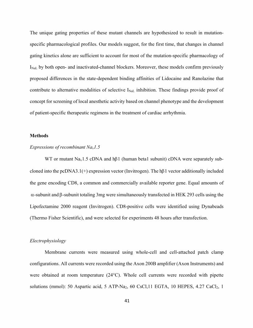
41
The unique gating properties of these mutant channels are hypothesized to result in mutation-
specific pharmacological profiles. Our models suggest, for the first time, that changes in channel
gating kinetics alone are sufficient to account for most of the mutation-specific pharmacology of
INaL by both open- and inactivated-channel blockers. Moreover, these models confirm previously
proposed differences in the state-dependent binding affinities of Lidocaine and Ranolazine that
contribute to alternative modalities of selective INaL inhibition. These findings provide proof of
concept for screening of local anesthetic activity based on channel phenotype and the development
of patient-specific therapeutic regimens in the treatment of cardiac arrhythmia.
Methods
Expressions of recombinant Nav1.5
WT or mutant Nav1.5 cDNA and hb1 (human beta1 subunit) cDNA were separately sub-
cloned into the pcDNA3.1(+) expression vector (Invitrogen). The hb1 vector additionally included
the gene encoding CD8, a common and commercially available reporter gene. Equal amounts of
a-subunit and b-subunit totaling 3mg were simultaneously transfected in HEK 293 cells using the
Lipofectamine 2000 reagent (Invitrogen). CD8-positive cells were identified using Dynabeads
(Thermo Fisher Scientific), and were selected for experiments 48 hours after transfection.
Electrophysiology
Membrane currents were measured using whole-cell and cell-attached patch clamp
configurations. All currents were recorded using the Axon 200B amplifier (Axon Instruments) and
were obtained at room temperature (24°C). Whole cell currents were recorded with pipette
solutions (mmol): 50 Aspartic acid, 5 ATP-Na2, 60 CsCl,11 EGTA, 10 HEPES, 4.27 CaCl2, 1
42
MgCl2. External solution contained: 130 NaCl, 5 CsCl, 10 HEPES, 5 glucose, 2 MgCl2. 1.2 CaCl2.
After maintaining >1GΩ seal and rupturing the membrane, capacitive currents and series
resistance were compensated with analog techniques. For all pharmacology experiments, drug
trials are control-matched in the same cell. Following control and drug-exposed experimental trials
cells were exposed to 50 mM tetrodotoxin (TTX) and all whole cell patch clamp results are
presented as TTX-sensitive currents.
Cell-attached experiments used to examine the function and pharmacology of single ion
channels are presented in Chapter 1.
Computational Modeling of WT and Mutant Nav1.5
We developed a Markov model for Wild Type Nav1.5 gating that is based on the Clancy-
Rudy model41. Our model contains 14 distinct drug-free channel states and 14 parallel drug-bound
states with time-varying occupancy represented as a system of ordinary differential equations
(Figure 2.1a). The model contains several closed states (Cx), closed-inactivated states (ICx), one
open state (O), one fast inactivated state (IF), two sequential slow inactivated states (IS1 and IS2),
burst mode closed states (BCx), and one burst mode open state (BO) (Figure 2.1). Voltage-
dependent rate constants are described by Eyring Rate Theory:
𝑘 = 𝑘$𝑒&'(/*+
where k is the rate constant, k0 is the rate constant at 0mV, q is the charge movement across the
membrane, V is the membrane potential, R is the gas constant, and T is temperature16. k0 and q
were optimized using a least squares error minimization96 with iterative parameter perturbations
performed using a simulated annealing algorithm100. Constraining data included voltage-
dependent conductance, steady state inactivation, fast-inactivation kinetics, recovery from
43
inactivation, and closed-inactivation kinetics. Parameter optimization and simulations were
encoded in MATLAB. Optimized voltage-dependent rate constants are described in Table 2.1.
Time-varying state occupancies were simulated by integrating the system of ordinary
differential equations over time, and INa was calculated as:
𝐼-. = 𝐺-. ∗ 𝑃 𝑂 + 𝑃 𝐵𝑂 ∗ (𝑉 − 𝐸-.)
where GNa is the Na+ conductance, P(O) and P(BO) are the fractional occupancies of burst and
burst open states, V is membrane potential, and ENa is Nernst equilibrium potential for Na+.
Single channel simulations are performed stochastically by calculating a dwell time, T, as:
𝑇 = − ln 𝑟/ 𝛼@A
where r is a random number on the open interval (0,1), and 𝛼@Aare the transitions rates from the
current state (j) into neighboring states (k)13. Following the dwell time in state j, the transition to
an adjacent channel state k has probability:
𝛼@A𝛼@A
LQT3 mutant channel function was simulated after manual modification of transitions rates, which
are summarized in Table 2.2. Three classes of rate adjustments were performed to capture the
range of observed mutant phenotypes: shifts in steady-state availability, channel bursting, and
channel late reopening (Figure 2.1). F1473C rates were modified based on single channel and
whole cell data reported in Chapter 1 and in the literature1. Channel bursting rates for ΔKPQ were
extracted from the literature49.
44
Lidocaine and Ranolazine Models
Models of Lidocaine and Ranolazine binding to LQT3 mutant channels were constructed
by the addition of 14 parallel channel states to the drug-free model (Figure 2.2a). Each drug-free
channel state is capable of interacting with drug and, with the exception of accommodations for
microscopic reversibility (Figure 2.2b), transitions between drug-bound states occur with the same
rate constants as drug-free channels. Drug-channel interactions are defined by a second order rate
constant (kdiff) and binding affinity (Kd), where the on-rate (kon) and off-rate (koff) constants are
calculated as follows:
kon = kdiff*[Drug]
koff = kdiff*Kd
The models presented here were constructed with six free parameters: kdiff-Lipophilic, kdiff-Hydrophilic,
Kd-closed, Kd-inactivated, Kd-slow inactivated, and Kd-open. Hydrophilic and lipophilic rate constants allow for
Figure 2.1 WT and Mutant Nav1.5 Channel Gating. WT channel gating is represented as the transition between closed states (C), inactivated states (Ix), burst states (Bx) and an open state (O). The three mutation-induced changes in channel gating – shifts in stead state inactivation (green), increases in channel bursting (red), and increases in late reopening (blue) - are reproduced by modification of transition rate constants.
Reopening Bursting
SSI Shift

45
the distinction between drug reaching its binding site through the open hydrophilic pore or through
intramembrane pathways putatively identified in crystal structures of homologous prokaryotic
channels18, 19, 70. These rate constants are reflective of state-dependent binding site access, and
capture effects of pKa, charge, size, and partition coefficient.
State-dependent binding affinities allow for the allosteric regulation of channel states by
drug, as posited by the Modulated Receptor Hypothesis70 and detailed in Figure 2.2b. Parameters
were optimized using a least squares error minimization with iterative parameter perturbations
calculated by a simulated annealing algorithm as described above.
Action Potential Clamp Simulations
In addition to simulating currents elicited by square voltage pulses and standard
experimental voltage protocols, we also simulated the effect of mutations on INa during voltage
waveforms of ventricular myocytes. To produce the voltage waveform, current clamp simulations
of the O’Hara-Rudy epicardial myocyte model101 were performed at 1Hz pacing to steady-state
(>100 beats). The resulting voltage output was then used as the input in whole cell voltage-clamp
simulations of our Nav1.5 channel model. State transitions and ionic current were calculated from
the integrated system of ODEs, as above.
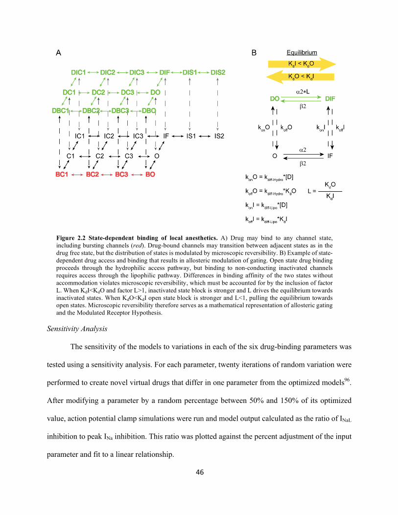
46
Sensitivity Analysis
The sensitivity of the models to variations in each of the six drug-binding parameters was
tested using a sensitivity analysis. For each parameter, twenty iterations of random variation were
performed to create novel virtual drugs that differ in one parameter from the optimized models96.
After modifying a parameter by a random percentage between 50% and 150% of its optimized
value, action potential clamp simulations were run and model output calculated as the ratio of INaL
inhibition to peak INa inhibition. This ratio was plotted against the percent adjustment of the input
parameter and fit to a linear relationship.
Figure 2.2 State-dependent binding of local anesthetics. A) Drug may bind to any channel state, including bursting channels (red). Drug-bound channels may transition between adjacent states as in the drug free state, but the distribution of states is modulated by microscopic reversibility. B) Example of state-dependent drug access and binding that results in allosteric modulation of gating. Open state drug binding proceeds through the hydrophilic access pathway, but binding to non-conducting inactivated channels requires access through the lipophilic pathway. Differences in binding affinity of the two states without accommodation violates microscopic reversibility, which must be accounted for by the inclusion of factor L. When KdI<KdO and factor L>1, inactivated state block is stronger and L drives the equilibrium towards inactivated states. When KdO<KdI open state block is stronger and L<1, pulling the equilibrium towards open states. Microscopic reversibility therefore serves as a mathematical representation of allosteric gating and the Modulated Receptor Hypothesis.
47
Results
Refinement of closed-inactivation kinetics in a model of Nav1.5 gating.
To begin to understand the kinetics of pharmacological inhibition of INaL, there are a series
of properties that models of channel gating must approximate. Certain gating transitions, such as
the kinetics of closed channel inactivation and the kinetics of slow inactivation, regulate the
population of channel states postulated to have high affinity for local anesthetic drugs75, 89, 102. We
developed a new experimental protocol to assess the kinetics and voltage-dependence of these
transitions, and found that previous models which were not constrained with similar data
inadequately reproduced our findings. We therefore expanded on this previous work and
parameterized a new model of Nav1.5 gating using these experimental protocols as additional
constraints.
The structural scheme of the model is based on that of the Clancy-Rudy model41 (Figure
2.3a), developed previously to described the voltage-dependent properties of WT and some LQT3
mutant channels. We assumed that the voltage-dependent transitions between states are defined by
Eyring Rate Theory, as described above, resulting in voltage-dependent currents depicted in Figure
2.3b. This formulation allowed each voltage-dependent rate constant to have a unique value for k0
and q, which we optimized with a series of experimental data sets including the single-channel
mean open duration (0.5ms), the voltage dependence of activation and steady-state inactivation
(Figure 2.3C), the kinetics of fast inactivation (Figure 2.3D), and recovery from 50 and 500ms
depolarizations to -10mV (Figure 2.3E), as described previously96. Including recovery from
inactivation after conditioning pulses of two durations allows the calibration of the transition
between IS1 and IS2 – a transition not readily detectable in other protocols - based on the relative
contribution of the slow phase of recovery, which increases with longer conditioning times.

48
Figure 2.3. Model of WT Nav1.5 Channel Gating. A) Schematic of model structure. The Markov channel model contains three closed (Cx), an open (O), three closed inactivated (ICx), a fast (IF) and two slow (ISx) inactivated states. Additionally, the low probability Burst Mode (BCx, BO) transitions are permitted from Cx and O states. B) Simulated currents obtained from a holding potential of -100mV to test potentials ranging from -60mV to +10mV. C) Voltage dependent activation (n=5) and steady state inactivation (n=12) experiments (symbol) and simulations (lines). D) The voltage-dependence of fast inactivation is measured as the t1/2 of decay from peak (n=4). E) Recovery from inactivation following a 50ms (n=6) and 500ms (n=4) conditioning pulse to -10mV. F) The voltage-dependent kinetics of closed-inactivation (n=9).
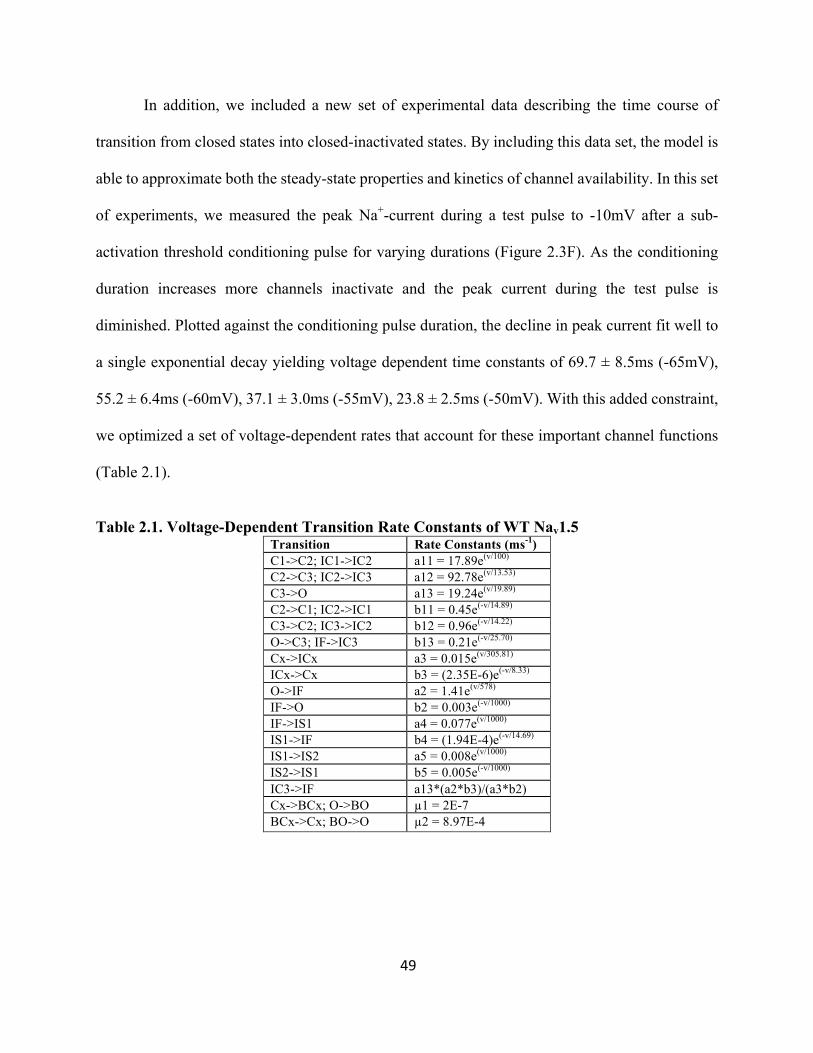
49
In addition, we included a new set of experimental data describing the time course of
transition from closed states into closed-inactivated states. By including this data set, the model is
able to approximate both the steady-state properties and kinetics of channel availability. In this set
of experiments, we measured the peak Na+-current during a test pulse to -10mV after a sub-
activation threshold conditioning pulse for varying durations (Figure 2.3F). As the conditioning
duration increases more channels inactivate and the peak current during the test pulse is
diminished. Plotted against the conditioning pulse duration, the decline in peak current fit well to
a single exponential decay yielding voltage dependent time constants of 69.7 ± 8.5ms (-65mV),
55.2 ± 6.4ms (-60mV), 37.1 ± 3.0ms (-55mV), 23.8 ± 2.5ms (-50mV). With this added constraint,
we optimized a set of voltage-dependent rates that account for these important channel functions
(Table 2.1).
Table 2.1. Voltage-Dependent Transition Rate Constants of WT Nav1.5
Transition Rate Constants (ms-1) C1->C2; IC1->IC2 a11 = 17.89e(v/100) C2->C3; IC2->IC3 a12 = 92.78e(v/13.53) C3->O a13 = 19.24e(v/19.89) C2->C1; IC2->IC1 b11 = 0.45e(-v/14.89) C3->C2; IC3->IC2 b12 = 0.96e(-v/14.22) O->C3; IF->IC3 b13 = 0.21e(-v/25.70) Cx->ICx a3 = 0.015e(v/305.81) ICx->Cx b3 = (2.35E-6)e(-v/8.33) O->IF a2 = 1.41e(v/578) IF->O b2 = 0.003e(-v/1000) IF->IS1 a4 = 0.077e(v/1000) IS1->IF b4 = (1.94E-4)e(-v/14.69) IS1->IS2 a5 = 0.008e(v/1000) IS2->IS1 b5 = 0.005e(-v/1000) IC3->IF a13*(a2*b3)/(a3*b2) Cx->BCx; O->BO µ1 = 2E-7 BCx->Cx; BO->O µ2 = 8.97E-4

50
Effect of mutations on channel gating
Chapter 1 presented experimental evidence that the gain of function pathology that results
from the F1473C and DKPQ mutations are the result of unique current signatures. DKPQ causes
infrequent but prolonged periods of channel bursting50 and a time-independent INaL. F1473C, on
the other hand, caused a nominal increase in channel bursting compared to WT, but a significant
increase in the frequency of dispersed late reopenings, time-varying INaL, and a depolarizing shift
in steady-state inactivation. We hypothesize that these gating deficiencies produce distinct
responses to INaL inhibition, and therefore sought to construct models of channel gating that
reproduce the main features of these mutations to serve as the basis for models of selective INaL
inhibition.
Table 2.2. Rate Modifications of Mutant Nav1.5 Modification Effect
F1473C
b3 = b3*10 Depolarizing shift in SSI; faster recovery a4 = a4/30 Increase in late reopening frequency and INaL µ1 = µ1*2 Slight increase in burst frequency and INaL
DKPQ µ1 = 1.84*10-6 Increase burst frequency and INaL
To model the effects of F1473C and DKPQ mutations on channel gating we manually
manipulated individual rate constants based on experimental data obtained by us and others (Table
2.2)1, 87. Figures 2.4a and 2.4b display simulated whole cell currents from DKPQ and F1473C
models, respectively. Note the varying kinetics of INaL in these two models (inset), where the INaL
continues to decay at prolonged times in the F1474C model, but is relatively time-independent in
DKPQ. Our experiments analyzing the single channel properties of F1473C revealed that the
mutation did not significantly alter the channel mean open duration from WT, so this time-
dependent deficiency in inactivation is likely the result of impaired slow inactivation. We therefore
simulated F1473C INaL by reducing a4, the rate governing the transition from fast inactivation to
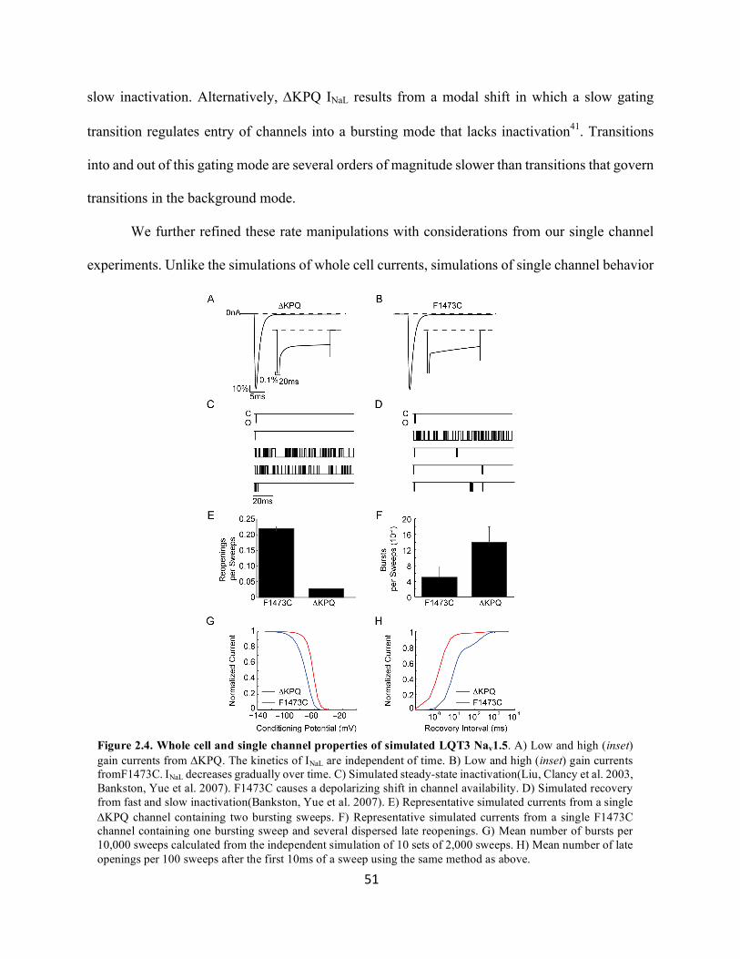
51
slow inactivation. Alternatively, DKPQ INaL results from a modal shift in which a slow gating
transition regulates entry of channels into a bursting mode that lacks inactivation41. Transitions
into and out of this gating mode are several orders of magnitude slower than transitions that govern
transitions in the background mode.
We further refined these rate manipulations with considerations from our single channel
experiments. Unlike the simulations of whole cell currents, simulations of single channel behavior
Figure 2.4. Whole cell and single channel properties of simulated LQT3 Nav1.5. A) Low and high (inset) gain currents from DKPQ. The kinetics of INaL are independent of time. B) Low and high (inset) gain currents fromF1473C. INaL decreases gradually over time. C) Simulated steady-state inactivation(Liu, Clancy et al. 2003, Bankston, Yue et al. 2007). F1473C causes a depolarizing shift in channel availability. D) Simulated recovery from fast and slow inactivation(Bankston, Yue et al. 2007). E) Representative simulated currents from a single DKPQ channel containing two bursting sweeps. F) Representative simulated currents from a single F1473C channel containing one bursting sweep and several dispersed late reopenings. G) Mean number of bursts per 10,000 sweeps calculated from the independent simulation of 10 sets of 2,000 sweeps. H) Mean number of late openings per 100 sweeps after the first 10ms of a sweep using the same method as above.

52
are stochastic and non-deterministic. We therefore approximated our experimental conditions by
simulating ten sets of 2,000 sweeps from a holding potential of -120mV to a test pulse voltage of
-30mV for 100ms at 1Hz. Figure 2.4c and d display simulated single channel recordings of DKPQ
and F1473C channels. The modification of a4 that represented a deficiency in slow inactivation
resulted in a prolongation of the mean occupancy duration of IF, and therefore a prolonged period
over which channels may reopen from this state. The slow decay of INaL in this mutation is therefore
consistent with an increased contribution from late reopenings (Figure 2.4e).
In addition to reopenings, we accounted for a trend in increased F1473C channel bursting
by doubling the rate of transition from the background mode to the burst gating mode (µ1),
resulting in a slight increase in both the frequency of channel bursting and INaL in line with our
experiments (Figure 2.4f). The DKPQ mutation has classically been modeled with an increase in
burst frequency as the sole source of increased INaL42, 46, 48, 49. With only a very slight modification
in the rates of DKPQ bursting reported previously49, we were also able to recapitulate the single
channel and whole cell pathological gating in these channels. We increased the previously reported
rate constant of transition into the burst gating mode from 1.44*10-6ms-1 to 1.84*10-6ms-1 because
the peak Popen of our model was slightly higher than in previous models. Compared to WT (2*10-
7ms-1) and F1473C (4*10-7ms-1), these rates suggest a 9.2 and 4.6-fold greater frequency of
bursting, respectively, in line with our experimental observations (Chapter 1).
The seminal study describing the whole cell properties of F1473C revealed a dramatic
depolarizing shift in steady-state inactivation and a speeding of recovery from inactivation1. While
slowing a4 contributed some speeding of recovery, the transition from ICx to Cx is rate-limiting
in our model and controls steady-state channel availability. We therefore accounted for both the
53
shift in channel availability (Figure 2.4g) and the speeding of recovery from inactivation (Figure
2.4h) by increasing b3 in the F1473C model.
Effect of channel gating on inhibition of INaL
The magnitude and kinetics of inhibition of F1473C and DKPQ currents by the clinically
relevant Na+-channel inhibitors Lidocaine and Ranolazine occur in a highly mutation-dependent
manner (Chapter 1). There exist two possible explanations for these differences that are difficult
to test experimentally: the mutations alter the physical chemical association of drug and channel
to alter binding affinity, or alternatively, the mutation-induced changes in channel gating result in
an altered distribution of high affinity channel states. Our kinetic models of channel bursting and
reopening pathologies afford a convenient setting to test if the latter hypothesis is sufficient to
explain mutation-specific pharmacology by these drugs. By assuming that drugs bind to specific
channel states with the same affinity regardless of channel genotype, we developed models of
Lidocaine and Ranolazine binding that attribute the majority of our experimental findings to
mutation-induced differences in channel kinetics.
Models of Lidocaine and Ranolazine inhibition were constructed with accommodations for
both the Modulated Receptor Hypothesis (state-dependent binding affinity)70, 71, 103, 104 and the
Guarded Receptor Hypothesis (state-dependent binding site access)78 (Figure 2.2). Drugs are
allowed to bind to any channel state (Figure 2.2a), but the ‘on’ rate of binding is limited by
diffusion through differential access routes70. The binding site, which resides in the central cavity
of the channel68, may be accessed through the aqueous pore when channels are open (states O and
BO). All non-conducting channel states must be accessed by a lipophilic diffusion route, which
selectively permits the passage of small, neutral drug species but not charged species75. Channels

54
are then permitted to transition between drug-bound states with considerations for microscopic
reversibility favoring the population of states which bind tightest to the drug (Figure 2.2b). We
constrained the models using whole cell and single channel data sets including block of peak and
late INa, the kinetics of current inhibition, shifts in the voltage-dependence of channel availability,
voltage-independent resting state block, and changes in F1473C single channel mean open time.
We validated these models by comparing the frequency of F1473C reopening in experimental and
simulated trials.
Figure 2.5 illustrates the concordance between the model of Lidocaine activity and the
experimental data, and the rate and state-dependent binding constants are presented in Table 2.3
(“Original” Model). The simulated F1473C and ∆KPQ currents depicted in Figure 2.5a are
inhibited by Lidocaine, with preference for inhibition of INaL (insets). The degree of selectivity for
INaL is greater in the F1473C simulations compared to ∆KPQ, as in our experiments (Chapter 1)
(Figure 2.45). This model was also constrained by and captures a dramatic hyperpolarizing shift
in steady-state inactivation that is greater in the ∆KPQ variant than in F1473C (Figure 2.5c). We
used the voltage-independent reduction in current at -130mV in our steady-state inactivation
protocol as a measure of resting state block. The Lidocaine model contains sufficient closed-state
affinity to capture block of F1473C at this voltage, however the model lacks the mutation-specific
resting state block of ∆KPQ. In addition to steady-state properties of channel inhibition, we
included as a constraint the kinetics of channel block during a single depolarizing pulse from -
120mV to -30mV (Figure 2.5d). This experimental protocol revealed important differences in the
kinetics of block in the two mutations, and the mutation-specific kinetics are also present in the
model. Inhibition of F1473C by Lidocaine develops slowly over time, but inhibition of ∆KPQ
currents reach a plateau within approximately 20ms. Lastly, this model produces a negligible effect
55
on single channel mean open time (Figure 2.5f) and produces a mean reduction in the frequency
of channel reopening (Figure 2.5g) that is similar to our experimental findings, despite not being
used as a constraint in the optimization of model rates.
Simulations of Ranolazine activity in F1473C and ∆KPQ are presented in Figure 2.6. While
the rate and binding constants for Ranolazine (Table 2.4) are quite different than for Lidocaine,
this model’s strengths and limitations are quite similar to the Lidocaine model. Using the same
experimental protocols as those used to constrain the Lidocaine model, plus an added constraint
for an observed 27% reduction in mean open time, the differences in mutant channel gating alone
are sufficient to account for the magnitude of peak and late INa block (Figure 2.6b), the small but
significant shift in channel availability (Figure 2.6c), the similar kinetics but differential magnitude
of F1473C and ∆KPQ inhibition (Figure 2.6d), and a reduction in single channel mean open
duration (Figure 2.6e). Importantly, this model also predicts a reduction in the frequency of
F1473C reopening that was similar to our experimental observations (Figure 2.6f), and less
dramatic than the effect on reopenings by Lidocaine.
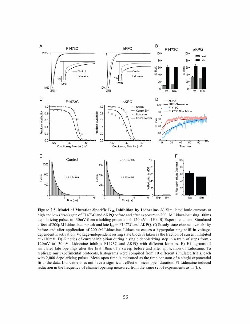
56
Figure 2.5. Model of Mutation-Specific INaL Inhibition by Lidocaine. A) Simulated ionic currents at high and low (inset) gain of F1473C and ∆KPQ before and after exposure to 200µM Lidocaine using 100ms depolarizing pulses to -30mV from a holding potential of -120mV at 1Hz. B) Experimental and Simulated effect of 200µM Lidocaine on peak and late INa in F1473C and ∆KPQ. C) Steady-state channel availability before and after application of 200µM Lidocaine. Lidocaine causes a hyperpolarizing shift in voltage-dependent inactivation. Voltage-independent resting state block is taken as the fraction of current inhibited at -130mV. D) Kinetics of current inhibition during a single depolarizing step in a train of steps from -120mV to -30mV. Lidocaine inhibits F1473C and ∆KPQ with different kinetics. E) Histograms of simulated late openings after the first 10ms of a sweep before and after application of Lidocaine. To replicate our experimental protocols, histograms were compiled from 10 different simulated trials, each with 2,000 depolarizing pulses. Mean open time is measured as the time constant of a single exponential fit to the data. Lidocaine does not have a significant effect on mean open duration. F) Lidocaine-induced reduction in the frequency of channel opening measured from the same set of experiments as in (E).
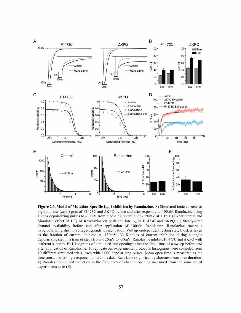
57
Figure 2.6. Model of Mutation-Specific INaL Inhibition by Ranolazine. A) Simulated ionic currents at high and low (inset) gain of F1473C and ∆KPQ before and after exposure to 100µM Ranolazine using 100ms depolarizing pulses to -30mV from a holding potential of -120mV at 1Hz. B) Experimental and Simulated effect of 100µM Ranolazine on peak and late INa in F1473C and ∆KPQ. C) Steady-state channel availability before and after application of 100µM Ranolazine. Ranolazine causes a hyperpolarizing shift in voltage-dependent inactivation. Voltage-independent resting state block is taken as the fraction of current inhibited at -130mV. D) Kinetics of current inhibition during a single depolarizing step in a train of steps from -120mV to -30mV. Ranolazine inhibits F1473C and ∆KPQ with different kinetics. E) Histograms of simulated late openings after the first 10ms of a sweep before and after application of Ranolazine. To replicate our experimental protocols, histograms were compiled from 10 different simulated trials, each with 2,000 depolarizing pulses. Mean open time is measured as the time constant of a single exponential fit to the data. Ranolazine significantly shortens mean open duration. F) Ranolazine-induced reduction in the frequency of channel opening measured from the same set of experiments as in (E).

58
Table 2.3. Combinatorial Analysis of Lidocaine-Channel Affinity.
Parameter Original KdC KdIx KdIS2 KdO kon-lipo
F1473C
KdC (µM) 2282.1 3134.6 2141.3 2100.0 2264.1 1896.0 KdIx (µM) 46.4 55.8 104.3 46.7 46.9 46.3 KdIS2 (µM) 6.8 13.5 15.3 9.3 6.3 6.9 KdO (µM) 225.3 162.1 228.3 236.4 439.3 234.2 kdiff-lipo 0.056 0.052 0.092 0.060 0.064 0.064 kdiff-hydro 0.15 0.19 0.12 0.14 0.14 0.17
∆KPQ
KdC (µM) - 738.5 - - - - KdIx (µM) - - 27.2 - - - KdIS2 (µM) - - - 5.7 - - KdO (µM) - - - - 226.4 - kdiff-lipo (µM-1 s-1) - - - - 0.45 kdiff-hydro (µM-1 s-1) - - - - - -
KF1473C/K∆KPQ - 4.2 3.8 1.6 1.9 0.1 Global Error 218.3 130.2 140.3 188.8 216.5 190.8
Table 2.4. Combinatorial Analysis of Ranolazine-Channel Affinity
Parameter Original KdC KdIx KdIS2 KdO kon-lipo
F1473C
KdC (µM) 537.0 712.8 517.4 534.0 545.9 549.7 KdIx (µM) 208.3 194.9 204.0 199.0 214.3 209.3 KdIS2 (µM) 13.5 14.8 16.5 25.9 13.5 13.4 KdO (µM) 66.9 64.8 63.3 67.5 74.7 69.5 kdiff-lipo (µM-1 s-1) 0.022 0.020 0.020 0.019 0.020 0.092 kdiff-hydro (µM-1 s-1) 6.90 6.90 7.00 7.10 7.00 7.02
∆KPQ
KdC (µM) - 364.1 - - - KdIx (µM) - - 164.2 - - KdIS2 (µM) - - - 12.3 - KdO (µM) - - - - 64.7 kdiff-lipo (µM-1 s-1) - - - - 0.093 kdiff-hydro (µM-1 s-1) - - - - -
KF1473C/K∆KPQ - 2.0 1.2 2.1 1.2 0.98 Global Error 420.3 392.9 398.5 385.7 413.2 401.1
Prediction of Mutation-Induced Modifications to Binding Affinity
The models of Lidocaine and Ranolazine presented above attribute the majority of
mutation-specific pharmacology to differences in channel gating that alter the distribution of high-
affinity channel states. However, there are some aspects of the pharmacology that are not
completely recapitulated by these models, particularly the simulation of voltage-independent
resting state block (Figures 2.5c and 2.6c). It is possible that these discrepancies between the
59
experimental and computational results are due to a limitation in the model topology, or
alternatively that LQT3 mutations do have an indirect effect on the Kd of drug binding to certain
states. We therefore employed a combinatorial approach to examine the incremental improvement
to the model fit by allowing individual binding constants to vary in a mutation-dependent manner.
Tables 2.3 and 2.4 present the parameters and global error derived after re-optimization of
all rates while allowing only one to express mutation-dependence. Not surprisingly, this method
results in an improvement in the error function because the approach requires the inclusion of an
additional degree of freedom. However, the variability in error function and in the divergence of
parameters from the original model suggests that some states are more sensitive to mutation-
dependent binding than others.
The original models for both Lidocaine and Ranolazine were unable to distinguish between
a difference in resting state block at -130mV. In both cases, allowing KdC to vary between the
mutations produced a closed-state affinity that was stronger for ∆KPQ channels than for F1473C
and enabled mutation-specific resting state block (Figures 2.7c and 2.8c) without dramatic changes
in peak and late block (Figures 2.7b and 2.8b), steady-state inactivation shifts (Figures 2.7c and
2.8c), or changes in channel mean open time (Figures 2.7e and 2.8e). No other parameters were
capable of correcting this property when allowed to vary in a mutation-dependent manner.
Modification of KdC also improved the global error of the minimized objective function more
effectively than other modifications, suggesting that these models of drug inhibition are
particularly sensitive to changes in resting state block. It is therefore unlikely that the improved fit
is solely the result of the introduction of an additional degree of freedom.

60
Figure 2.7. Mutation-specific Closed State Binding by Lidocaine. A) Simulated ionic currents at high and low (inset) gain of F1473C and ∆KPQ before and after exposure to 200µM Lidocaine using 100ms depolarizing pulse to -30mV from a holding potential of -120mV at 1Hz. B) Experimental and Simulated effect of 200µM Lidocaine on peak and late INa in F1473C and ∆KPQ. C) Steady-state channel availability before and after application of 200µM Lidocaine. Lidocaine causes a hyperpolarizing shift in voltage-dependent inactivation. Voltage-independent resting state block is taken as the fraction of current inhibited at -130mV, and is improved in this model compared to a model without mutation-varying KdC. D) Kinetics of current inhibition during a single depolarizing step in a train of steps from -120mV to -30mV. Lidocaine inhibits F1473C and ∆KPQ with different kinetics. E) Histograms of simulated late openings after the first 10ms of a sweep before and after application of Lidocaine. To replicate our experimental protocols, histograms were compiled from 10 different simulated trials, each with 2,000 depolarizing pulses. Mean open time is measured as the time constant of a single exponential fit to the data. Lidocaine does not have a significant effect on mean open duration. F) Lidocaine-induced reduction in the frequency of channel opening measured from the same set of experiments as in (E).
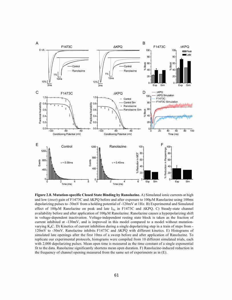
61
Figure 2.8. Mutation-specific Closed State Binding by Ranolazine. A) Simulated ionic currents at high and low (inset) gain of F1473C and ∆KPQ before and after exposure to 100µM Ranolazine using 100ms depolarizing pulses to -30mV from a holding potential of -120mV at 1Hz. B) Experimental and Simulated effect of 100µM Ranolazine on peak and late INa in F1473C and ∆KPQ. C) Steady-state channel availability before and after application of 100µM Ranolazine. Ranolazine causes a hyperpolarizing shift in voltage-dependent inactivation. Voltage-independent resting state block is taken as the fraction of current inhibited at -130mV, and is improved in this model compared to a model without mutation-varying KdC. D) Kinetics of current inhibition during a single depolarizing step in a train of steps from -120mV to -30mV. Ranolazine inhibits F1473C and ∆KPQ with different kinetics. E) Histograms of simulated late openings after the first 10ms of a sweep before and after application of Ranolazine. To replicate our experimental protocols, histograms were compiled from 10 different simulated trials, each with 2,000 depolarizing pulses. Mean open time is measured as the time constant of a single exponential fit to the data. Ranolazine significantly shortens mean open duration. F) Ranolazine-induced reduction in the frequency of channel opening measured from the same set of experiments as in (E).
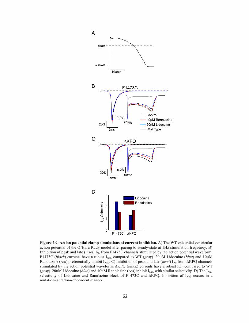
62
Figure 2.9. Action potential clamp simulations of current inhibition. A) The WT epicardial ventricular action potential of the O’Hara Rudy model after pacing to steady-state at 1Hz stimulation frequency. B) Inhibition of peak and late (inset) INa from F1473C channels stimulated by the action potential waveform. F1473C (black) currents have a robust INaL compared to WT (gray). 20uM Lidocaine (blue) and 10uM Ranolazine (red) preferentially inhibit INaL. C) Inhibition of peak and late (inset) INa from ∆KPQ channels stimulated by the action potential waveform. ∆KPQ (black) currents have a robust INaL compared to WT (gray). 20uM Lidocaine (blue) and 10uM Ranolazine (red) inhibit INaL with similar selectivity. D) The INaL selectivity of Lidocaine and Ranolazine block of F1473C and ∆KPQ. Inhibition of INaL occurs in a mutation- and drug-dependent manner.
63
Mutation-specific Inhibition of INaL in Ventricular Action Potentials
One benefit of computational modeling is the ability to extrapolate fundamental models of
pathophysiology and disease into a physiologically relevant setting. We applied a WT ventricular
epicardium action potential waveform (Figure 2.9a) in voltage clamp simulations to examine the
effects of INaL inhibition in this setting. After 100 beats at cycle length 1s, we analyzed block of
INa and INaL at the APD50 of homozygous F1473C (Figure 2.9b) and ∆KPQ (Figure 2.9c) cells.
Both 20µM Lidocaine and 10µM Ranolazine resulted in robust inhibition of peak INa and INaL for
both mutations, but the preference for inhibition of INaL varied in a mutation- and drug-dependent
manner. We quantitatively defined INaL selectivity as the ratio of INaL inhibition to peak INa
inhibition (Figure 2.9d). Peak block of F1473C was larger by Ranolazine, but INaL block was
smaller, resulting in INaL selectivity that was smaller for Ranolazine than for Lidocaine.
Conversely, Ranolazine blocks peak and INaL less than Lidocaine in ∆KPQ at these concentrations.
This resulted in selectivity that was similar for Ranolazine and Lidocaine but pointed to Ranolazine
as the slightly more effective inhibitor of INaL. These results therefore suggest that the
pathophysiology of INaL in patients with LQT3 may contribute to heterogeneous responses to local
anesthetic therapy based on the state-dependent properties of channel inhibition.
If inactivated channel block by Lidocaine is more effective for selective INaL inhibition in
F1473C, and open channel block by Ranolazine is more effective in ∆KPQ, then novel theoretical
inhibitors which emphasize these properties are likely to enhance selectivity. To test this
hypothesis, we employed a sensitivity analysis which estimates the relative role of each binding
parameter in conferring preferential inhibition of INaL. For each of the six parameters we performed
20 action potential clamp simulations in which the parameter in question was perturbed from the
best fit of the Lidocaine or Ranolazine models by a random percentage between 50% and 150%.
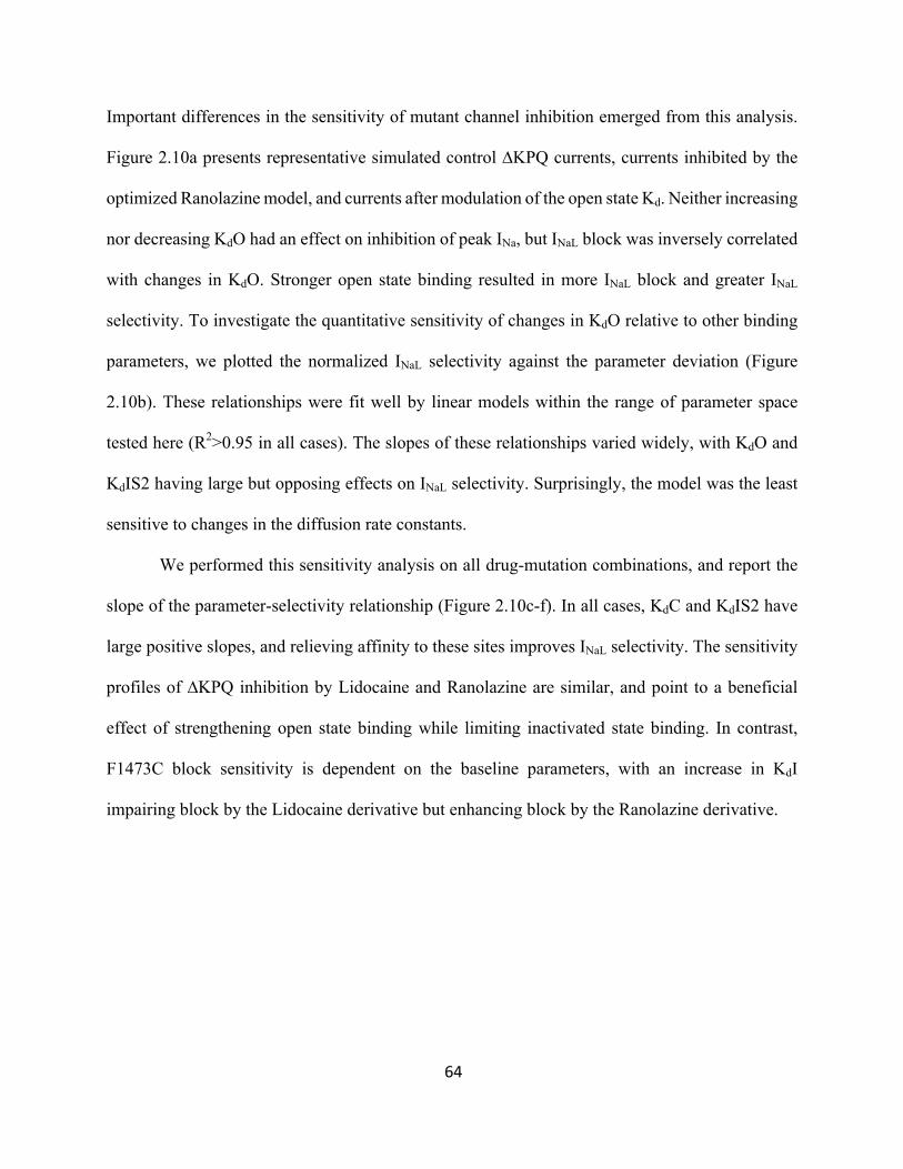
64
Important differences in the sensitivity of mutant channel inhibition emerged from this analysis.
Figure 2.10a presents representative simulated control ∆KPQ currents, currents inhibited by the
optimized Ranolazine model, and currents after modulation of the open state Kd. Neither increasing
nor decreasing KdO had an effect on inhibition of peak INa, but INaL block was inversely correlated
with changes in KdO. Stronger open state binding resulted in more INaL block and greater INaL
selectivity. To investigate the quantitative sensitivity of changes in KdO relative to other binding
parameters, we plotted the normalized INaL selectivity against the parameter deviation (Figure
2.10b). These relationships were fit well by linear models within the range of parameter space
tested here (R2>0.95 in all cases). The slopes of these relationships varied widely, with KdO and
KdIS2 having large but opposing effects on INaL selectivity. Surprisingly, the model was the least
sensitive to changes in the diffusion rate constants.
We performed this sensitivity analysis on all drug-mutation combinations, and report the
slope of the parameter-selectivity relationship (Figure 2.10c-f). In all cases, KdC and KdIS2 have
large positive slopes, and relieving affinity to these sites improves INaL selectivity. The sensitivity
profiles of ∆KPQ inhibition by Lidocaine and Ranolazine are similar, and point to a beneficial
effect of strengthening open state binding while limiting inactivated state binding. In contrast,
F1473C block sensitivity is dependent on the baseline parameters, with an increase in KdI
impairing block by the Lidocaine derivative but enhancing block by the Ranolazine derivative.
65
Figure 2.10. Sensitivity analysis of state-dependent drug binding. A) Representative simulated currents from ∆KPQ during action potential clamp simulations. Currents before and after application of 100µM Ranolazine are compared to currents exposed to derivatives of Ranolazine with modified open channel affinity. B) Sensitivity of INaL selectivity of DKPQ inhibition by Ranolazine to modifications in each of the six model parameters. Data is well described by a linear relationship (R2>0.95). The model is most sensitive to changes in open channel and slow-inactivated channel affinity with opposing effects. C to E) Extracted slopes of sensitivity analysis of F1473C and DKPQ inhibition by 200µM Lidocaine and 100µM Ranolazine. Negative slopes (red) indicate a decrease in selectivity with increased parameter value. Positive slopes (blue) indicate an increase in selectivity with increased parameter value.

66
Discussion
State-dependent Access and Binding of Local Anesthetics
Since the proposal of the Modulated Receptor Hypothesis by Hille in 197770, state-
dependent drug-channel affinity has been used to explain shifts in the voltage-dependence of
channel availability, use-dependent block, and preferential inhibition of INaL. On the basis of whole
cell and single channel observations, Lidocaine has long been understood to interact preferentially
with inactivated channels and to access the local anesthetic binding site through a lipophilic
pathway75. Ranolazine on the other hand, has been modeled to interact preferentially with open
channels49. Our simulations presented here provide a quantitative description of the differential
activity of Lidocaine and Ranolazine that confirms these earlier findings. Lidocaine interacts
selectively with inactivated channels (KdILidocaine = 46.4µM; KdIRanolazine = 208.3µM), while
Ranolazine binds more tightly to open channels (KdOLidocaine = 225.3µM; KdORanolazine = 66.9µM).
These differences in state-dependent binding are reflected in the nature of their interaction with
F1760, the local anesthetic binding site. Lidocaine has been shown to bind via a specific cation-π
interaction with this residue that was absent in Ranolazine binding77. It is therefore likely that these
molecules reside in the central pore of the channel in a manner which alters the geometry, and
state-dependence, of interaction at this site.
Interestingly, though, these binding affinities themselves have little bearing on the effect
of drugs on channel mean open time. The Kds presented here are an estimate of equilibrium drug
binding, but Na+ channel gating is highly dynamic and rarely near equilibrium. The relative
diffusion rate constants therefore play a very important role in the kinetics of channel inhibition
and the effect on channel mean open time. Here we present evidence that the on-rate of Ranolazine
binding through a lipophilic access pathway (kdiff-lipophilic) is slower than for Lidocaine, while
67
the on-rate for hydrophilic binding (kdiff-hydrophilic) to the open state is faster. Ranolazine is larger
than Lidocaine with more hydrogen bond acceptors (6 vs 2), a wider polar surface area (75.5 Å2
vs 33.5 Å2) but similar pKa and partition coefficient94. It is therefore likely that its size and polarity
impinge its ability to penetrate the small, intramembrane lipophilic pathway to the binding site
predicted by Hille70 and identified in crystal structures of the bacterial channel NavAB18, 19.
Mechanisms of Mutation-specific Inhibition of INaL
Peak INa is critically important to rapid cardiac conduction, and reductions in the early
transient by drugs11 or mutations5, 105 can itself be proarrhythmic. Local anesthetics have long been
used in the treatment of LQT3 because of their ability to preferentially inhibit INaL with a smaller
effect on peak INa. The emerging interest in precision medicine requires a more detailed
understanding of phenotypic differences in the etiology of INaL that impact the efficacy and safety
of these drugs. Chapter 1 presented a series of experiments documenting important
pharmacological differences between Lidocaine and Ranolazine inhibition of F1473C and ∆KPQ.
We asked if the differences in mutant channel gating were sufficient to account for unique
responses to blockade by constructing models of INaL inhibition under the assumption that mutant
channels have equivalent state-dependent binding rates and affinities. Models of both drugs
captured several of the mutation-specific signatures we identified experimentally, and the
underlying distribution of channel states therefore has a large impact on the action of state-
dependent channel blockers.
Both the rapid recovery from inactivation and late reopenings of the F1473C mutation are
signatures of impaired channel inactivation. It is therefore not surprising that Lidocaine, a drug
shown here and previously to associate preferentially with inactivated channels74, 89, 90, 102, 106, is

68
more effective at stabilizing these faulty processes. Because late reopenings have the same
duration as the channel openings that give rise to the peak Na+ transient, open channel block by
Ranolazine is unable to differentiate peak INa from INaL and is less effective at correcting the
underlying gating dysfunction. On the other hand, the openings during channel bursting in ∆KPQ
have a prolonged duration compared to early openings, allowing for more time over which
Ranolazine54 may bind to its high affinity state and exert a preferential inhibition of the
pathological current. Lidocaine, with its relatively slow and low affinity open channel binding,
does not have this effect.
The cases of Lidocaine and Ranolazine presented here serve as foundation for a general
examination of local anesthetics with different state-dependent properties. A sensitivity analysis
allowed us to generate a virtual library of local anesthetic derivatives to ask if specific qualities of
these drugs can be accentuated to improve INaL selectivity. Our analysis points to slow inactivated
state and closed state binding as generally detrimental to selective INaL block in all drug-channel
combinations. Only enhancement of open state binding provided consistent improvement in
selectivity, with a greater impact on ∆KPQ where a larger fraction of INaL is derived from bursting.
Interestingly, manipulation of inactivated state binding responded in a mutation- and drug-
dependent manner. This is because, in our model, inactivated state binding not only stabilizes fast
inactivation but also induces a hyperpolarizing shift in channel availability which results in adverse
effects on peak INa. ∆KPQ, with a V1/2 of inactivation near the resting membrane potential,
responds dramatically to hyperpolarizing shifts in availability. F1473C, on the other hand, causes
a depolarizing shift that allows for more leeway. The highest degree of F1473C INaL selectivity in
this mutation would therefore be a drug which differentiates IF and ICx binding, a property not
currently attainable given the simplifying assumptions of our model.
69
Despite the fact that our models capture most of the important aspects of mutation specific
pharmacology through differences in channel gating, some remaining discordance between the
model and data suggest that this simplifying assumption may not be wholly accurate. In fact, a
combinatorial approach to examining the role of mutation-specific Kds revealed a potentially
important modification to closed-state binding affinity by some LQT3 mutations. The suggestion
that LQT3 mutations alter the affinity of closed channels is novel, and is interesting given the
location of these mutations in the DIII/DIV linker near the inactivation gate, a region not
previously thought to interact with the pore domains in the closed state. Alternatively, it is possible
that the 0.1Hz stimulation frequency in these experiments is insufficiently long to allow the system
to reach steady-state, resulting in apparent differences in resting state tonic block that are actually
due to mutation-specific recovery from inactivation. These findings highlight the need for a more
detailed structural understanding of the local anesthetic binding site, its allosteric interactions with
the inactivation machinery, and the geometrical arrangement of channel inhibitors within the pore
in different channel states.
In silico Screening of Patient-Specific Outcomes
Our growing understanding of the genetic basis for cardiac arrhythmia and the patient-
specific response to pharmacological intervention brings with it the need for novel experimental
models of disease. Unfortunately, reliable experimental models of INaL that account for human
physiology and the genetic heterogeneity of LQT3 patients are hard to come by; primary human
tissue is rare, animal models are low throughput, and induced pluripotent stem cell derived
cardiomyocytes remain under-developed for detailed systems analysis. Computational modeling
has emerged as a useful tool in investigating ion channel function and the systems pharmacology
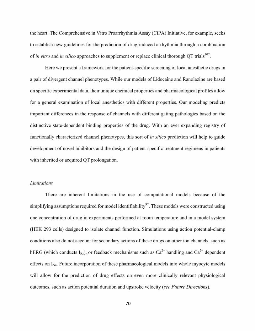
70
the heart. The Comprehensive in Vitro Proarrhythmia Assay (CiPA) Initiative, for example, seeks
to establish new guidelines for the prediction of drug-induced arrhythmia through a combination
of in vitro and in silico approaches to supplement or replace clinical thorough QT trials107.
Here we present a framework for the patient-specific screening of local anesthetic drugs in
a pair of divergent channel phenotypes. While our models of Lidocaine and Ranolazine are based
on specific experimental data, their unique chemical properties and pharmacological profiles allow
for a general examination of local anesthetics with different properties. Our modeling predicts
important differences in the response of channels with different gating pathologies based on the
distinctive state-dependent binding properties of the drug. With an ever expanding registry of
functionally characterized channel phenotypes, this sort of in silico prediction will help to guide
development of novel inhibitors and the design of patient-specific treatment regimens in patients
with inherited or acquired QT prolongation.
Limitations
There are inherent limitations in the use of computational models because of the
simplifying assumptions required for model identifiability97. These models were constructed using
one concentration of drug in experiments performed at room temperature and in a model system
(HEK 293 cells) designed to isolate channel function. Simulations using action potential-clamp
conditions also do not account for secondary actions of these drugs on other ion channels, such as
hERG (which conducts IKr), or feedback mechanisms such as Ca2+ handling and Ca2+ dependent
effects on INa. Future incorporation of these pharmacological models into whole myocyte models
will allow for the prediction of drug effects on even more clinically relevant physiological
outcomes, such as action potential duration and upstroke velocity (see Future Directions).

71
Chapter 3: Utilization of unnatural amino acids to study the chemistry of
pathological late current
72
Summary
State-dependent channel inhibition can help to explain many of the important properties of
local anesthetic pharmacology, including preferential inhibition of late Na+ current (INaL). We
previously established that Lidocaine and Ranolazine exhibit distinct mutation-specific effects on
two Long QT 3 associated mutations, F1473C and ΔKPQ (Chapter 1). Simulations of mutant
channel gating and state-dependent drug binding revealed that differences in channel kinetics are
sufficient to explain the majority of these effects (Chapter 2). However, the ability of our model
to recapitulate resting state pharmacological properties of local anesthetics suggest that there are
important structural effects of F1473 and ΔKPQ mutations at the drug binding site that alter
binding affinity in a way not captured by our models. The dynamic nature of voltage-gated Na+
channel gating and the relative lack of structural information about the channel make any chemical
understanding of selective INaL inhibition difficult. Recently, unnatural amino acids were employed
to make direct measurements of a specific cation-𝛑 interaction important in use-dependent block
of cardiac Na+ channels. This chapter details the use of unnatural amino acids to investigate the
role of this interaction in state-dependent drug binding and selective inhibition of INaL, lending a
structural perspective to the quantitative predictions of our kinetic models. However, we present
here evidence that the unnatural amino acids themselves disrupt inactivation gating, limiting the
ability to distinguish their effects on pharmacology as a modulation of drug-channel affinity or as
a modulation of channel gating that influences state-dependent binding.

73
Introduction
During the plateau phase of the cardiac ventricular action potential INa has nearly
completely inactivated and contributes little to the balance of ionic fluxes governing the membrane
potential. However, there exist several disease states and mutations for which a failure to inactivate
results in a sustained inward late current (INaL) that results in prolongation of the action potential,
prolongation of the QT interval, and increased risk of ventricular arrhythmia and sudden cardiac
death5, 10. INaL has therefore become an important therapeutic target, and efforts are underway to
develop novel, potent, and selective inhibitors4, 58, 95. We previously reported that two such INaL
inhibitors, Lidocaine and Ranolazine, exhibit unique state-dependent binding properties. These
properties, when applied to two mutations with unique molecular etiologies of INaL, result in strong
mutation-specific differences in block of peak INa and in selectivity for INaL. Computational
analysis revealed that most of these mutation-specific effects can be attributed to differences in
channel gating that alter the kinetics and distribution of high-affinity channel states. However,
some lingering discordance between our models and experimental data raises the possibility that
mutations alter the thermodynamics of the interaction between the drug and the channel. Moreover,
our models predict state-dependent drug-channel interactions that are important in preferential
inhibition of INaL, so a refined understanding of the precise chemical interactions governing these
properties would guide the rational development of more selective molecules.
The local anesthetic binding site resides in the central conducting pathway just below the
selectivity filter9. Mutational analysis has revealed several residues important in tonic and use-
dependent block (UDB) of INa, however one mutation, F1760A, results in a particularly severe
abolishment of current block67. Mutation of F1760, located on S6 of domain IV, impairs UDB and
INaL inhibition by many common channel blockers including Lidocaine68, Mexiletine69, and
74
Ranolazine47. These findings raised an interesting question: how does an aromatic residue
contribute significantly to the binding of organic cations? To answer this question a more precise
chemical tool is needed.
Unnatural amino acid (UAA) technology allows one to expand the genetic code with
selective incorporation of a UAA at any site of interest108. After enzymatically ligating a synthetic
dinucleotide-fused UAA to a tRNA with anticodon CUA, this ‘charged’ tRNA and ion channel
Figure 3.1. Unnatural Amino Acid Incorporation into Functional Nav1.5. A) Nonsense suppression workflow. Synthetic amino acids are chemically conjugated to a dinucleotide, which is ligated to tRNA. UAA-tRNA and mRNA containing the TAG ‘stop’ codon are co-injected into Xenopus oocytes for two-electrode voltage clamp experiments. B) UAAs are incorporated at the LA binding site, F1760, which resides in the central cavity on S6 of D4. C) Fluorophenylalanine derivatives with monotonically reduced electron density of the π-electrons in the aromatic ring. D) Chemical structure (top), energy minimized 3-D structure (middle), and electrostatic potential map (bottom) of Mexiletine. The cationic head group is clearly visible in blue.
75
mRNA are co-injected into Xenopus oocytes for two electrode voltage clamp recording. The
mRNA is engineered with a UAG ‘Stop’ codon at the position of interest, so that any emergent
peptide that fails to incorporate a UAA is truncated and non-functional. Successful incorporation
results in a full-length peptide in a process known as Nonsense Suppression109, 110. This procedure
is detailed in Figure 3.1a.
Ahern and Horn first used Nonsense Suppression in voltage-gated Na+-channels in 2008
to incorporate unnatural derivatives of phenylalanine at site F1579 of Nav1.4, a muscle isoform of
the voltage-gated Na+-channel76. This residue is highly conserved among Navs and is homologous
to F1760 in Nav1.5 (Figure 3.1b). The UAAs substituted one, two, or three fluorine atoms in place
of the hydrogen atoms of the aromatic ring (Figure 3.1c). With the addition of each new
electronegative fluorine the π-electrons are drawn away from the face of the aromatic ring,
decreasing the strength of the partial negative charge without altering the residue’s size, shape,
polarizability, or hydrophobicity. Mexiletine, a closely related derivative of Lidocaine, contains a
cationic head group that is attracted to these π-electrons (Figure 3.1d). As the number of fluorine
substituents on F1760 is increased there is a monotonic reduction in the potency of UDB by
Mexiletine, but not in the potency of tonic block, reflecting important conformational changes that
occur during channel opening and/or inactivating that unmask the binding site76. The authors
propose that drug binding via the cation-π interaction stabilizes these states, providing a structural
basis for the state-dependent binding predictions of the Modulated Receptor Hypothesis70. We
reasoned that the allosteric stabilization of inactivation that contributes to UDB likely also
contributes to preferential inhibition of INaL, and therefore that the cation-π is an important
chemical determinant of INaL block, as well.
76
In this chapter, we test the hypothesis that the elevated block of INaL compared to peak INa
is associated with a concomitant increase in the strength of the cation-π interaction during a single
depolarizing pulse. This finding would suggest that the conformational states populated during
prolonged membrane depolarization promote favorable conditions for the formation of this
interaction, providing an actionable chemical target for the future development of selective INaL
inhibitors. In concert with the kinetic studies of channel gating and inhibition in Chapter 2, these
findings would lend a chemical perspective to the state-dependence of drug binding, and enhance
our understanding of the structural relationship between the inactivation gating machinery and the
local anesthetic binding site.
Methods
Molecular Biology and Unnatural Amino Acids
In vivo incorporation of UAAs was performed by the nonsense suppression method as
previously reported76, 77. Mutations in SCN5A, the gene encoding Nav1.5, were engineered into the
WT gene in pcDNA3.1 using overlapping primers and the QuikChange site-directed mutagenesis
kit (Qiagen). In addition to the F1473S and F1760TAG mutations, we also introduced the C373Y
mutation which confers TTX-sensitivity111. mRNA was transcribed using the mMessage
mMachine T7 RNA Transcription Kit (Ambion). cDNA for Tetrahymena thermophile tRNA
containing the CUA anticodon was provided in pcDNA3.1 by the Dr. Chris Ahern (University of
Illinois). tRNA was transcribed using the mMessage mMachine T7 RNA Transcription Kit, ligated
to aminoacyl dinucleotide using T4 RNA Ligase (Ambion) and purified with the RNAeasy Mini
Kit (Qiagen). Aminoacyl dinucleotides were generously provided by the Ahern Lab as a precipitate
and dissolved prior to use to 3mM in H2O.
77
To enhance the yield of aminoacyl dinucleotide synthesis the amino acid is protected with
nitroveratryl oxycarbonyl. Immediately prior to injection, aminoacyl-tRNAs were deprotected by
UV irradiation for 6 minutes. 25ng aminoacyl-tRNA, 25ng F1473S/F1760TAG mRNA, and 5ng
mRNA encoding the hb1 subunit were mixed and injected into Xenopus oocytes. Na+-currents
were recorded 18-24 hours after injection. Negative control experiments performed without co-
injection of aminoacyl-tRNA showed no TTX-sensitive currents over this time range.
TTX was purchased from Abcam and used at a final concentration of 50nM. Mexiletine
was purchased from Sigma and 100mM stocks were made in H2O.
Two Electrode Voltage Clamp Recording
Voltage-gated Na+-currents were recorded using two electrode voltage clamp at room
temperature (~22°C). Currents were amplified using a Axopatch 200A (Axon Instruments) and
digitized with a Digidata 144A (Warner Instrument Corp). The extracellular bath solution
contained (mM): 116 NaCl, 2 KCl, 1 MgCl2, 0.5 CaCl2, 5 HEPES, pH7.4 adjusted with CsOH.
Glass microelectrodes were pulled to a resistance between 0.1 and 1.0 MW and backfilled with 3M
KCl prior to use. Cell membranes were held at -100mV. Currents were elicited by depolarizations
to -20mV for 400ms at 0.2Hz. Recordings were analyzed in Clampfit 8.0 (Molecular Devices
LLC). TTX-subtraction was performed on control and Mexiletine traces.
Statistics
Data are presented at mean ± standard error. Statistical significance was determined by a
Student’s T-test with significance level p<0.05. For comparisons across multiple groups statistical

78
significance was determined with a one-way ANOVA with significance level p<0.05, and
individual comparisons made using a Tukey’s post hoc test.
79
Results
Expression of LQT3 Mutant Nav1.5 with Unnatural Amino Acids
In order to test the role of the cation-π interaction in preferential inhibition of INaL, we
incorporated a spectrum of UAAs into mutant F1473S Nav1.5 at site F1760 in Xenopus oocytes.
We chose the naturally occurring F1473S mutation as a model because its robust INaL provided
ample resolution to detect small changes in channel block in the presence of UAAs112. In the
absence of charged aminoacyl-tRNA, F1473S/F1760TAG expressing cells did not display any
Na+-current in the time frame tested. Co-injection of F1473S/F1760TAG with Phe-, monofluoro-
(F1), difluoro- (F2), or trifluoro-tRNA (F3) resulted in nonsense suppression rescued currents
(Figure 2a). The incorporation of fluoride atoms reduced the efficiency of peak INa rescue
(Figure2b), but the rescued F1473S currents provided ample INaL that is resolvable even at low
gain.
We examined INaL in these cells with depolarizing pulses from -100mV to -20mV for
400ms at 0.2Hz. We used long pulse durations and a slow frequency to ensure that any drug block
observed during prolonged times was the result of conformational changes arising during that
pulse, with no contribution from the accumulation of channels in drug-bound states that arises
during faster pacing rates. There are significant differences in the magnitude of INaL relative to
peak INa across the spectrum of UAA-incorporating channels (Phe: 6.89±0.96%, F: 6.06±0.38%,
F2: 10.74±0.67%, F3: 9.25±1.33%, p<0.01). It is possible that the physiologic concentrations of
Na+ used in these experiments resulted in poorly controlled voltage clamp, improper measurement
of large Na+ transients and, consequently, miscalculation of INaL as a fraction of peak INa. We
therefore repeated experiments with Phe and F2 F1473S/F1760TAG using lower 30mM
extracellular Na+ (Figure 2d). These conditions resulted in smaller currents, yet the F2-induced

80
increase in INaL (5.03±0.30% vs 10.19±0.98%, p<0.001) was still observed. In addition, the
improved voltage-control obtained by decreasing extracellular Na+ allowed us to measure the time
course of fast inactivation. F2 caused a slowing of the time to half-peak (1.55±0.08ms vs
2.23±0.13ms, p<0.01). These results suggest that even small modifications to F1760 such as
fluorine substitutions can result in altered fast inactivation kinetics and substantial INaL.
Figure 3.2. Nonsense Suppression of Mutant Nav1.5 with Fluorophenylalanine. A) Representative TTX-sensitive traces of F1473S/F1760TAG Nav1.5 containing Phe (n=3), fluoro-Phe (n=9), difluoro-Phe (n=9), or Tripler-Phe (n=6) at position 1760.Note the substantial INaL that is resolved in these low gain recordings. INaL is resolvable even at low gain. B) Average peak INa current in rescued F1473S/F1760TAG is diminished in the presence of fluorinated derivatives. C) Current measured at 400ms as a percentage of peak INa varies in the presence of fluorphenylalanines. D) Current measured at 400ms as a percentage of peak from experiments with low 30mM extracellular Na+ in Phe (n=7) and F2 (n=5) expression oocytes. F2 induces an increase in INaL. E) Time to half-inactivation from experiments with low 30mM extracellular Na+. F2 impairs fast inactivation.
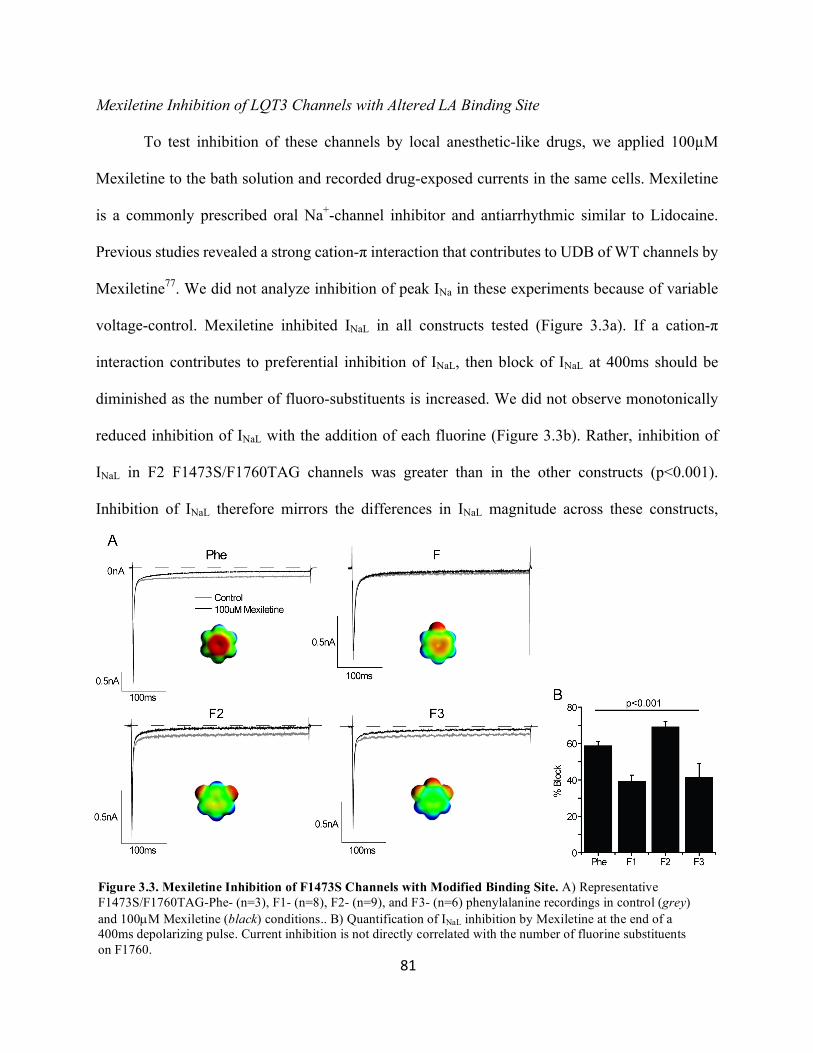
81
Mexiletine Inhibition of LQT3 Channels with Altered LA Binding Site
To test inhibition of these channels by local anesthetic-like drugs, we applied 100µM
Mexiletine to the bath solution and recorded drug-exposed currents in the same cells. Mexiletine
is a commonly prescribed oral Na+-channel inhibitor and antiarrhythmic similar to Lidocaine.
Previous studies revealed a strong cation-π interaction that contributes to UDB of WT channels by
Mexiletine77. We did not analyze inhibition of peak INa in these experiments because of variable
voltage-control. Mexiletine inhibited INaL in all constructs tested (Figure 3.3a). If a cation-π
interaction contributes to preferential inhibition of INaL, then block of INaL at 400ms should be
diminished as the number of fluoro-substituents is increased. We did not observe monotonically
reduced inhibition of INaL with the addition of each fluorine (Figure 3.3b). Rather, inhibition of
INaL in F2 F1473S/F1760TAG channels was greater than in the other constructs (p<0.001).
Inhibition of INaL therefore mirrors the differences in INaL magnitude across these constructs,
Figure 3.3. Mexiletine Inhibition of F1473S Channels with Modified Binding Site. A) Representative F1473S/F1760TAG-Phe- (n=3), F1- (n=8), F2- (n=9), and F3- (n=6) phenylalanine recordings in control (grey) and 100µM Mexiletine (black) conditions.. B) Quantification of INaL inhibition by Mexiletine at the end of a 400ms depolarizing pulse. Current inhibition is not directly correlated with the number of fluorine substituents on F1760.
82
raising the possibility that the unique and unpredictable changes to channel gating introduced by
the UAAs contributes to this pharmacological profile, limiting our ability to interpret the results
as due to changes in the cation-π interaction.
Discussion
Here for the first time we incorporated UAAs into Nav1.5 in the presence of LQT3-
associated mutations. We sought to correlate depolarization-time-dependent changes in a critical
cation-π interaction with enhancements in channel inhibition that lead to preferential block of INaL.
This cation-π interaction is critical in UDB by Mexiletine and Lidocaine77. We reasoned that the
gating transitions underlying UDB - namely, stabilization of inactivated states and a subsequent
accumulation of refractory channels - are also likely to underlie inhibition of INaL. If a cation-π
interaction is important in preferential block of INaL, then with the addition of each sequential
fluorine atom to the aromatic ring the potency of INaL inhibition will decrease. However, UAA-
induced changes in inactivation gating (t1/2 and INaL) limited our ability to interpret these results as
a change to the cation-π interaction itself.
Chapter 2 presented a computational model of channel inhibition with the assumption that
mutations do not influence the physical chemical interaction of drugs and channels or, in other
words, that the putative cation-π interaction is not impacted by channel mutations. With this basic
assumption the mutation-specific pharmacology recapitulated by these models can be attributed to
differences in channel gating that alter the distribution of high-affinity channel states. The
modification of INaL by fluoro-Phe substituents, particularly by di-fluoro-Phe, indicates modulation
of inactivation gating that could translate into altered pharmacological responses, similar to the
phenotypic and pharmacological differences between the F1473C and ΔKPQ mutations. It is
83
therefore possible that the observed profile of INaL inhibition in fluoro-Phe-containing mutant
Nav1.5 is due not to modulation of the cation-π interaction, but to kinetic differences between
experimental conditions that enhance or diminish channel block. While our experiments do not
rule out a role for the cation-π in preferential inhibition of INaL, they are unable to directly detect
its role or magnitude.
It is not surprising that modulation of the chemistry of F1760 destabilizes channel
inactivation and promotes INaL. Despite the limitations of this study to determine the role of a
cation-π interaction in inhibition of INaL, important lessons can still be gleaned regarding the broad
conformational changes associated with inactivation gating and the local anesthetic binding site.
Our work and the work of others has shown that drug binding to inactivated channels produces a
stabilizing effect on channel inactivation113. Previous work examined the accessibility for MTS
reagents to an engineered cysteine at site 1760, and demonstrated that covalent modification of
F1760 enhanced and stabilized slow inactivation and UDB89, 102, 106, 113. Similarly, fluorometry74,
92, cysteine accessibility72, 73, and gating charge114, 115 analysis of voltage sensor movement
indicated that activation of S4 in DIII and, in part, DIV is closely coupled to inactivation gating
and is stabilized in the presence of Lidocaine. This sort of allosteric modulation of channel
behavior is likely to be bi-directional: destabilization of the drug binding site should have an effect
on the closely coupled processes of channel inactivation. In fact, several studies have reported
changes in steady-state and slow inactivation when F1760 is mutated to other residues113 or
modified by MTS reagents37. Even our small fluorine substituents at F1760 are sufficient to perturb
the structure of this site and impair channel inactivation.
Taken together with the in vitro experiments of Chapter 1 and explanatory models of
Chapter 2, these findings provide a framework for the development of novel mutation-specific
84
therapeutics. Lidocaine was a more selective inhibitor of INaL than Ranolazine in F1473C channels,
for which the primary channel pathology was a deficiency in slow inactivation. Lidocaine interacts
primarily with inactivated channels and exhibited a powerful cation-π interaction for UDB76, 77.
Conversely Ranolazine77, which is established above and by others as an open-channel blocker,
did not demonstrate a role for cation-π in UDB. The cation-π therefore appears to be a chemical
signature of high affinity inactivated state block. This suggests that rational targeting of the cation-
π interaction in patients with deficient slow inactivation may provide the optimal therapeutic
benefit.
85
Conclusion
The marriage of experimental and in silico approaches is gaining traction as a valuable tool
in drug discovery, particularly in dynamic systems such as ion channel function and pharmacology.
One emergent use of such an approach is in the study of patient heterogeneity and prediction of
safety and efficacy in patient subpopulations. This thesis presents a proof of concept that
computational modeling based on detailed experimental characterization of ion channel
pharmacology can generate novel insights into patient-specific therapy.
In Chapter 1, we characterized a recently discovered mutation, F1473C, which varied quite
significantly in its gating properties from the canonical ∆KPQ mutation. In simple terms, ∆KPQ
causes channel bursts that occur relatively rarely but with very long duration. F1473C, on the other
hand, results in a gating abnormality that occurs 100-fold more often than channel bursts, but with
a much shorter duration. While this phenotype has been reported previously in other mutations,
the F1473C mutation causes a particularly severe case of reopening that enabled its use as a tool
in the study of pharmacology. The two main drugs used in this study, Lidocaine and Ranolazine,
impacted reopenings in distinct ways. The distinguishing feature of these two drugs is the
prominent effect on mean open time by Ranolazine.
Not surprisingly, the single channel properties of our two mutations and two drugs
translated into mutation- and drug-specific pharmacology at the whole cell level. Lidocaine proved
to inhibit F1473C with dramatically higher selectivity than ∆KPQ, and did so with kinetics that
were reflective of the relative kinetics of INaL. Ranolazine did not distinguish between the kinetics
of INaL in the same way, and only resulted in elevated block of peak and late ∆KPQ current. These
experiments clearly laid out the heterogeneity in channel gating and local anesthetic activity, but

86
the causes and implications of such variability are difficult to decipher without a more quantitative
analysis.
Chapter 2 focused on the construction of versatile models that provide a mechanistic
framework for our experimental studies and allow for exploration of local anesthetic
pharmacology to drive additional hypothesis. The history of in silico pharmacology in LQT3 is
one of continual refinement and iterative growth. We built on the careful work of others to devise
a model for WT channel gating based on data from a new experimental protocol that more
completely defines important transitions. Next, using only small modifications to this model, we
simulated virtual LQT3 mutant currents with properties similar to F1473C and ∆KPQ. From these
models it is quite clear how modest changes in channel gating can cause dramatic differences in
the distribution of channel states – an interesting finding considering the co-localization of these
two mutations to the same region of the channel. Using our experimental data as a guide, we
demonstrated that it is this distinction in the distribution of states that causes most of the mutation-
specific effects of Lidocaine and Ranolazine. Such a finding implies that models of state-
dependent local anesthetic binding are generalizable, and predictions about the selectivity for INaL
in any drug-channel combination can be made if the properties of the mutant channel and drug are
known independently. Moreover, the ease with which model parameters can be varied establishes
the setting for high throughput screening of novel virtual therapeutics.
In Chapter 3, we present an effort to extend observations about the role of state-dependent
binding affinity in block of INaL, and gain a detailed chemical understanding of drug-channel
interactions. The dynamics and structural complexity of Nav1.5 gating have made traditional
ligand binding assays difficult. The application of unnatural amino acid technology to voltage-
gated ion channels has helped to overcome this challenge. We reasoned that a cation-π interaction
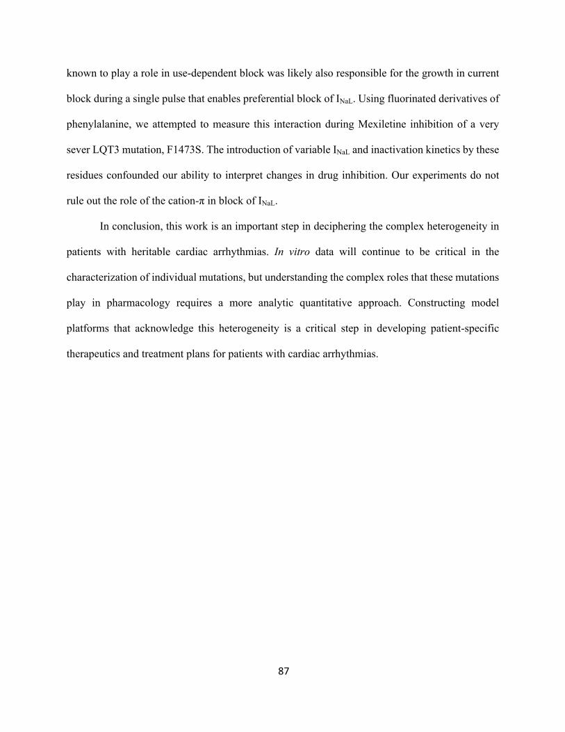
87
known to play a role in use-dependent block was likely also responsible for the growth in current
block during a single pulse that enables preferential block of INaL. Using fluorinated derivatives of
phenylalanine, we attempted to measure this interaction during Mexiletine inhibition of a very
sever LQT3 mutation, F1473S. The introduction of variable INaL and inactivation kinetics by these
residues confounded our ability to interpret changes in drug inhibition. Our experiments do not
rule out the role of the cation-π in block of INaL.
In conclusion, this work is an important step in deciphering the complex heterogeneity in
patients with heritable cardiac arrhythmias. In vitro data will continue to be critical in the
characterization of individual mutations, but understanding the complex roles that these mutations
play in pharmacology requires a more analytic quantitative approach. Constructing model
platforms that acknowledge this heterogeneity is a critical step in developing patient-specific
therapeutics and treatment plans for patients with cardiac arrhythmias.

88
This page intentionally left blank.

89
Future Directions
Our models of the pathophysiology and pharmacology of INaL have taught us valuable
lessons about the molecular etiology of mutation-specific responses to local anesthetic inhibition.
However, models often raise more questions than they answer and serve as a useful tool in the
development of new experimental hypotheses. This section will address certain strengths and
limitations of the work presented here, and propose several experiments and simulations to
advance the field.
Systems Pharmacology of INaL
The models presented in Chapter 2 treat INa as an isolated current, such as one measured
experimentally in heterologous expression systems. Even our voltage-clamp simulations using
action-potential waveforms treat INa explicitly as a model output. In reality, INa is only one of
several ionic currents from various channels, pumps, and transporters in the ventricular myocyte,
each with a distinct localization to subcellular compartments8. The ventricular action potential
waveform is derived from the integration of each of these ionic currents, with constant feedback
between current and voltage. Several models have been developed to capture the systems biology
of the ventricular myocyte and use current-clamp configurations to simulate ionic fluxes and
membrane potential98. The most widely used model of the human ventricular action potential is
the O’Hara-Rudy model101. Incorporation of our revised Nav1.5 Markov model, LQT3 mutant
currents, and local anesthetic activity would allow predictions of mutation-specific pharmacology
on the clinically relevant measures of upstroke velocity and action potential duration. Such a model
would also facilitate an exploration of the systems biology of different flavors of INaL. For example,
the resultant increased flux of Na+ is known to alter the properties of the Na+/Ca2+ exchanger,
90
CaMKII signaling, and Ca2+ dynamics, which may serve as the trigger for early
afterdepolarizations and arrhythmia60, 116, 117.
We replaced the INa and INaL calculations in the O’Hara-Rudy model with two copies of
our WT model to allow for the virtual expression of heterozygous mutant INa. Our experiments
and simulations were performed at room temperature (~24ºC), but the ventricular myocyte model
is simulated at 37ºC. We therefore adjusted all of the transition rates in our model for temperature
Figure 4.1. Temperature-dependence of simulated INaL. A) Simulated epicardial ventricular action potentials of the O’Hara Rudy model at 1Hz steady-state pacing. Replacement of the Hodgkin-Huxley formulation INa in the published model (gray) with the Markov channel model (black) resulted in action potentials with similar morphology and APD. B) Calibration of GNa after replacement of INa in the O’Hara-Rudy model resulted in similar upstroke velocity and peak membrane potential. C) Virtual expression of heterozygous F1473C (red) and DKPQ (blue) caused substantial action potential prolongation at 1Hz steady-state pacing. DKPQ caused a more dramatic effect than F1473C. D) Simulated INaL during the action potentials in (C). Heterozygous DKPQ expression resulted in a larger INaL. E and F) Voltage-clamp simulations of F1473C and DKPQ currents using the ventricular action potential waveform at 24oC and 37oC using Q10=3. INaL in F1473C was reduced by increasing temperature, while INaL in KPQ was not.

91
(Q10=3)49. After calibrating the relative conductance of INa (GNa = 3.4) to match the peak INa from
the OHR model, we simulated steady-state action potentials using 1Hz current-clamp pacing and
measured APD90 (Figure 4.1a) and upstroke velocity (Figure 4.1b) that were similar to the original
model.
Virtual expression of heterozygous F1473C and ∆KPQ currents resulted in action potential
prolongation (Figure 4.1c) and INaL (Figure 4.1d). Surprisingly, F1473C prolonged the action
potential less severely, contrary to the larger INaL that we observed experimentally and reproduced
in simulated square pulse experiments at room temperature. This suggests that, in our models,
these two mutations may respond differently to changes in temperature. To test this possibility, we
returned to action potential voltage-clamp simulations and compared INaL at 24ºC and 37ºC. INaL
in the F1473C model reduced dramatically with increased temperature (Figure 4.1e), but ∆KPQ
did not (Figure 4.1f).
These mutations, while both in the DIII/DIV linker, alter channel gating in unique ways
and likely cause different structural deformities and allosteric effects on distant channel regions.
There currently exists no data to suggest that all channel conformational changes respond the same
to temperature. Our modeling suggests that applying the same temperature-dependence to all rates
may be an over simplification that must be addressed prior to extrapolating mutant channel effects
on the action potential. A straightforward approach to addressing this question is to perform single-
channel recording on both mutant channels at room and physiologic temperature and comparing
the relative changes in channel reopening and channel bursting.

92
Tissue-specificity of LQT3 Pathophysiology and Pharmacology
The heart is not only comprised of epicardial ventricular tissue, but also of endo- and mid-
myocardial cells, atrial myocytes, and specialized conduction tissues such as the sinoatrial node,
atrioventricular node, and His-Purkinje system. Each of these cell types express a different profile
of channels, transporters, pumps, and Ca2+ handling machinery that result in tissue-specific action
potential waveforms. While it is the ventricular action potential prolongation that gives rise to QT
prolongation, recent work has demonstrated that Purkinje cells from the mouse heart are
Figure 4.2. Tissue-specificity of INaL in LQT3. A) Epicardial action potential waveform from the O’Hara-Rudy ventricular myocyte model during steady-state pacing at 1Hz. B) Purkinje cell action potential waveform from the Iyer-Sampson-Kass model during steady-state pacing at 1Hz. C) Peak INa in WT, F1473C and DKPQ in action potential-clamp simulations using the epicardial waveform. D) Peak INa in WT, F1473C and DKPQ in action potential-clamp simulations using the Purkinje waveform. E) Stimulation by the epicardial waveform results in INaL of similar magnitude from F1473C and DKPQ. F) Stimulation by the Purkinje waveform results in INaL of that is substantially larger in F1473C than in DKPQ.
93
particularly susceptible to prolongation and EAD formation by INaL and may be a primary source
of arrhythmogenesis46.
Additional simulations using models of bursting and late reopening channels suggested
that the tissue-specific effects of INaL are also mutation-dependent42. Experiments and models
presented in Chapters 1 and 2 demonstrate that F1473C and ∆KPQ mutations result in different
effects on channel gating that alter the time- and voltage-dependent distribution of channel states.
Compared to the ventricular action potential (Figure 4.2a), the Purkinje action potential (Figure
4.2b) has a higher resting membrane potential, lower plateau potential, slower repolarization
phase, and longer APD90. The voltage-dependent properties of mutant channels are therefore
likely to respond in a tissue-specific manner. We used these action potential waveforms in voltage-
clamp simulations of F1473C and ∆KPQ currents at room temperature. Both voltage waveforms
elicited larger peak currents in F1473C channels than from WT or ∆KPQ because the depolarizing
shift in steady state inactivation results in a larger fraction of available channels at the resting
membrane potential (Figures 4.2c and d). In simulations using the epicardial action potential, INaL
from the two mutant channels was similar (Figure 4.2e). When a Purkinje action potential
waveform is applied, INaL from F1473C is dramatically larger than INaL from ∆KPQ (Figure 4.2f),
consistent with previous models of late reopening channels42. The lower plateau potential results
in slower voltage-dependent inactivation accentuating the effects of the late reopening pathology,
and the slow repolarization phase allows channels to recover prematurely in F1473C but not
∆KPQ.
If our mutations exhibit differential responses to ventricular and Purkinje action potentials
because the voltage waveforms alter the distribution of channel states, then it stands to reason that
the pharmacology of state-dependent channel inhibitors will also exhibit tissue-specific inhibition
94
of peak and late INa. Moreover, the expression of other ion channels with off-target effects may
vary between tissue types, such as the additional INaL provided by expression of Nav1.1 in the
Purkinje118, 119. Integration of mutant and drug models into whole ventricular and Purkinje models
will allow for the prediction of antiarrhythmic – or for that matter, proarrhythmic – effects of INa
inhibitors that may not manifest as clinically measurable changes in QT-interval. Multi-scale and
multi-tissue in silico screening can therefore serve an important role in drug discovery and safety
pharmacology in the future.
95
Bibliography 1. Bankston JR, Yue M, Chung W, Spyres M, Pass RH, Silver E, Sampson KJ and Kass RS. A novel and lethal de novo LQT-3 mutation in a newborn with distinct molecular pharmacology and therapeutic response. PLoS One. 2007;2:e1258. 2. Xu J, Murphy SL, Kochanek KD and Bastian BA. Deaths: Final Data for 2013. Natl Vital Stat Rep. 2016;64:1-119.
3. Maier LS and Sossalla S. The late Na current as a therapeutic target: where are we? J Mol Cell Cardiol. 2013;61:44-50.
4. Belardinelli L, Giles WR, Rajamani S, Karagueuzian HS and Shryock JC. Cardiac late Na(+) current: proarrhythmic effects, roles in long QT syndromes, and pathological relationship to CaMKII and oxidative stress. Heart Rhythm. 2015;12:440-8.
5. Clancy CE, Chen-Izu Y, Bers DM, Belardinelli L, Boyden PA, Csernoch L, Despa S, Fermini B, Hool LC, Izu L, Kass RS, Lederer WJ, Louch WE, Maack C, Matiazzi A, Qu Z, Rajamani S, Rippinger CM, Sejersted OM, O'Rourke B, Weiss JN, Varro A and Zaza A. Deranged sodium to sudden death. J Physiol. 2015;593:1331-45.
6. Schwartz PJ, Spazzolini C, Priori SG, Crotti L, Vicentini A, Landolina M, Gasparini M, Wilde AA, Knops RE, Denjoy I, Toivonen L, Monnig G, Al-Fayyadh M, Jordaens L, Borggrefe M, Holmgren C, Brugada P, De Roy L, Hohnloser SH and Brink PA. Who are the long-QT syndrome patients who receive an implantable cardioverter-defibrillator and what happens to them?: data from the European Long-QT Syndrome Implantable Cardioverter-Defibrillator (LQTS ICD) Registry. Circulation. 2010;122:1272-82.
7. Kennedy A, Finlay DD, Guldenring D, Bond R, Moran K and McLaughlin J. The Cardiac Conduction System: Generation and Conduction of the Cardiac Impulse. Crit Care Nurs Clin North Am. 2016;28:269-79.
8. Nerbonne JM and Kass RS. Molecular physiology of cardiac repolarization. Physiol Rev. 2005;85:1205-53.
9. Catterall WA. Voltage-gated sodium channels at 60: structure, function and pathophysiology. J Physiol. 2012;590:2577-89.
10. Liu H, Clancy C, Cormier J and Kass R. Mutations in cardiac sodium channels: clinical implications. Am J Pharmacogenomics. 2003;3:173-9.
96
11. Moreno JD, Zhu ZI, Yang PC, Bankston JR, Jeng MT, Kang C, Wang L, Bayer JD, Christini DJ, Trayanova NA, Ripplinger CM, Kass RS and Clancy CE. A computational model to predict the effects of class I anti-arrhythmic drugs on ventricular rhythms. Sci Transl Med. 2011;3:98ra83.
12. Echt DS, Liebson PR, Mitchell LB, Peters RW, Obias-Manno D, Barker AH, Arensberg D, Baker A, Friedman L, Greene HL and et al. Mortality and morbidity in patients receiving encainide, flecainide, or placebo. The Cardiac Arrhythmia Suppression Trial. N Engl J Med. 1991;324:781-8.
13. Bankston JR, Sampson KJ, Kateriya S, Glaaser IW, Malito DL, Chung WK and Kass RS. A novel LQT-3 mutation disrupts an inactivation gate complex with distinct rate-dependent phenotypic consequences. Channels (Austin). 2007;1:273-80.
14. Hodgkin AL and Huxley AF. The dual effect of membrane potential on sodium conductance in the giant axon of Loligo. J Physiol. 1952;116:497-506.
15. Hodgkin AL and Huxley AF. A quantitative description of membrane current and its application to conduction and excitation in nerve. J Physiol. 1952;117:500-44.
16. Yue DT, Lawrence JH and Marban E. Two molecular transitions influence cardiac sodium channel gating. Science. 1989;244:349-52.
17. Wang Q, Li Z, Shen J and Keating MT. Genomic organization of the human SCN5A gene encoding the cardiac sodium channel. Genomics. 1996;34:9-16.
18. Payandeh J, Gamal El-Din TM, Scheuer T, Zheng N and Catterall WA. Crystal structure of a voltage-gated sodium channel in two potentially inactivated states. Nature. 2012;486:135-9.
19. Payandeh J, Scheuer T, Zheng N and Catterall WA. The crystal structure of a voltage-gated sodium channel. Nature. 2011;475:353-8.
20. Patton DE, West JW, Catterall WA and Goldin AL. Amino acid residues required for fast Na(+)-channel inactivation: charge neutralizations and deletions in the III-IV linker. Proc Natl Acad Sci U S A. 1992;89:10905-9.
21. West JW, Patton DE, Scheuer T, Wang Y, Goldin AL and Catterall WA. A cluster of hydrophobic amino acid residues required for fast Na(+)-channel inactivation. Proc Natl Acad Sci U S A. 1992;89:10910-4.
97
22. Miyamoto K, Nakagawa T and Kuroda Y. Solution structures of the cytoplasmic linkers between segments S4 and S5 (S4-S5) in domains III and IV of human brain sodium channels in SDS micelles. J Pept Res. 2001;58:193-203.
23. Tang L, Kallen RG and Horn R. Role of an S4-S5 linker in sodium channel inactivation probed by mutagenesis and a peptide blocker. J Gen Physiol. 1996;108:89-104.
24. Valenzuela C and Bennett PB, Jr. Gating of cardiac Na+ channels in excised membrane patches after modification by alpha-chymotrypsin. Biophys J. 1994;67:161-71.
25. Tang L, Chehab N, Wieland SJ and Kallen RG. Glutamine substitution at alanine1649 in the S4-S5 cytoplasmic loop of domain 4 removes the voltage sensitivity of fast inactivation in the human heart sodium channel. J Gen Physiol. 1998;111:639-52.
26. Sarhan MF, Tung CC, Van Petegem F and Ahern CA. Crystallographic basis for calcium regulation of sodium channels. Proc Natl Acad Sci U S A. 2012;109:3558-63.
27. Sarhan MF, Van Petegem F and Ahern CA. A double tyrosine motif in the cardiac sodium channel domain III-IV linker couples calcium-dependent calmodulin binding to inactivation gating. J Biol Chem. 2009;284:33265-74.
28. Van Petegem F, Lobo PA and Ahern CA. Seeing the forest through the trees: towards a unified view on physiological calcium regulation of voltage-gated sodium channels. Biophys J. 2012;103:2243-51.
29. Cormier JW, Rivolta I, Tateyama M, Yang AS and Kass RS. Secondary structure of the human cardiac Na+ channel C terminus: evidence for a role of helical structures in modulation of channel inactivation. J Biol Chem. 2002;277:9233-41.
30. Glaaser IW, Bankston JR, Liu H, Tateyama M and Kass RS. A carboxyl-terminal hydrophobic interface is critical to sodium channel function. Relevance to inherited disorders. J Biol Chem. 2006;281:24015-23.
31. Tateyama M, Liu H, Yang AS, Cormier JW and Kass RS. Structural effects of an LQT-3 mutation on heart Na+ channel gating. Biophys J. 2004;86:1843-51.
32. Motoike HK, Liu H, Glaaser IW, Yang AS, Tateyama M and Kass RS. The Na+ channel inactivation gate is a molecular complex: a novel role of the COOH-terminal domain. J Gen Physiol. 2004;123:155-65.

98
33. Gavillet B, Rougier JS, Domenighetti AA, Behar R, Boixel C, Ruchat P, Lehr HA, Pedrazzini T and Abriel H. Cardiac sodium channel Nav1.5 is regulated by a multiprotein complex composed of syntrophins and dystrophin. Circ Res. 2006;99:407-14.
34. Chanda B and Bezanilla F. Tracking voltage-dependent conformational changes in skeletal muscle sodium channel during activation. J Gen Physiol. 2002;120:629-45.
35. Sheets MF, Kyle JW and Hanck DA. The role of the putative inactivation lid in sodium channel gating current immobilization. J Gen Physiol. 2000;115:609-20.
36. Sheets MF, Kyle JW, Kallen RG and Hanck DA. The Na channel voltage sensor associated with inactivation is localized to the external charged residues of domain IV, S4. Biophys J. 1999;77:747-57.
37. O'Leary ME and Chahine M. MTSET modification of D4S6 cysteines stabilize the fast inactivated state of Nav1.5 sodium channels. Front Pharmacol. 2015;6:118.
38. Capes DL, Goldschen-Ohm MP, Arcisio-Miranda M, Bezanilla F and Chanda B. Domain IV voltage-sensor movement is both sufficient and rate limiting for fast inactivation in sodium channels. J Gen Physiol. 2013;142:101-12.
39. Fredj S, Lindegger N, Sampson KJ, Carmeliet P and Kass RS. Altered Na+ channels promote pause-induced spontaneous diastolic activity in long QT syndrome type 3 myocytes. Circ Res. 2006;99:1225-32.
40. Malan D, Zhang M, Stallmeyer B, Muller J, Fleischmann BK, Schulze-Bahr E, Sasse P and Greber B. Human iPS cell model of type 3 long QT syndrome recapitulates drug-based phenotype correction. Basic Res Cardiol. 2016;111:14.
41. Clancy CE and Rudy Y. Linking a genetic defect to its cellular phenotype in a cardiac arrhythmia. Nature. 1999;400:566-9.
42. Iyer V, Sampson KJ and Kass RS. Modeling tissue- and mutation- specific electrophysiological effects in the long QT syndrome: role of the Purkinje fiber. PLoS One. 2014;9:e97720.
43. Schwartz PJ, Priori SG, Spazzolini C, Moss AJ, Vincent GM, Napolitano C, Denjoy I, Guicheney P, Breithardt G, Keating MT, Towbin JA, Beggs AH, Brink P, Wilde AA, Toivonen L, Zareba W, Robinson JL, Timothy KW, Corfield V, Wattanasirichaigoon D, Corbett C,
99
Haverkamp W, Schulze-Bahr E, Lehmann MH, Schwartz K, Coumel P and Bloise R. Genotype-phenotype correlation in the long-QT syndrome: gene-specific triggers for life-threatening arrhythmias. Circulation. 2001;103:89-95.
44. Clancy CE, Tateyama M, Liu H, Wehrens XH and Kass RS. Non-equilibrium gating in cardiac Na+ channels: an original mechanism of arrhythmia. Circulation. 2003;107:2233-7.
45. Wang Q, Shen J, Splawski I, Atkinson D, Li Z, Robinson JL, Moss AJ, Towbin JA and Keating MT. SCN5A mutations associated with an inherited cardiac arrhythmia, long QT syndrome. Cell. 1995;80:805-11.
46. Iyer V, Roman-Campos D, Sampson KJ, Kang G, Fishman GI and Kass RS. Purkinje Cells as Sources of Arrhythmias in Long QT Syndrome Type 3. Sci Rep. 2015;5:13287.
47. Fredj S, Sampson KJ, Liu H and Kass RS. Molecular basis of ranolazine block of LQT-3 mutant sodium channels: evidence for site of action. Br J Pharmacol. 2006;148:16-24.
48. Clancy CE, Zhu ZI and Rudy Y. Pharmacogenetics and anti-arrhythmic drug therapy: a theoretical investigation. Am J Physiol Heart Circ Physiol. 2007;292:H66-75.
49. Moreno JD, Yang PC, Bankston JR, Grandi E, Bers DM, Kass RS and Clancy CE. Ranolazine for congenital and acquired late INa-linked arrhythmias: in silico pharmacological screening. Circ Res. 2013;113:e50-61.
50. Bennett PB, Yazawa K, Makita N and George AL, Jr. Molecular mechanism for an inherited cardiac arrhythmia. Nature. 1995;376:683-5.
51. Nagatomo T, January CT, Ye B, Abe H, Nakashima Y and Makielski JC. Rate-dependent QT shortening mechanism for the LQT3 deltaKPQ mutant. Cardiovasc Res. 2002;54:624-9.
52. Schwartz PJ, Priori SG, Locati EH, Napolitano C, Cantu F, Towbin JA, Keating MT, Hammoude H, Brown AM, Chen LS and Colatsky TJ. Long QT syndrome patients with mutations of the SCN5A and HERG genes have differential responses to Na+ channel blockade and to increases in heart rate. Implications for gene-specific therapy. Circulation. 1995;92:3381-6.
53. Wang DW, Yazawa K, George AL, Jr. and Bennett PB. Characterization of human cardiac Na+ channel mutations in the congenital long QT syndrome. Proc Natl Acad Sci U S A. 1996;93:13200-5.
100
54. Dumaine R, Wang Q, Keating MT, Hartmann HA, Schwartz PJ, Brown AM and Kirsch GE. Multiple mechanisms of Na+ channel--linked long-QT syndrome. Circ Res. 1996;78:916-24.
55. Glaaser IW, Osteen JD, Puckerin A, Sampson KJ, Jin X and Kass RS. Perturbation of sodium channel structure by an inherited Long QT Syndrome mutation. Nat Commun. 2012;3:706.
56. Wehrens XH, Rossenbacker T, Jongbloed RJ, Gewillig M, Heidbuchel H, Doevendans PA, Vos MA, Wellens HJ and Kass RS. A novel mutation L619F in the cardiac Na+ channel SCN5A associated with long-QT syndrome (LQT3): a role for the I-II linker in inactivation gating. Hum Mutat. 2003;21:552.
57. Rivolta I, Clancy CE, Tateyama M, Liu H, Priori SG and Kass RS. A novel SCN5A mutation associated with long QT-3: altered inactivation kinetics and channel dysfunction. Physiol Genomics. 2002;10:191-7.
58. Makielski JC. Late sodium current: A mechanism for angina, heart failure, and arrhythmia. Trends Cardiovasc Med. 2016;26:115-22.
59. Shryock JC, Song Y, Rajamani S, Antzelevitch C and Belardinelli L. The arrhythmogenic consequences of increasing late INa in the cardiomyocyte. Cardiovasc Res. 2013;99:600-11.
60. Moreno JD and Clancy CE. Pathophysiology of the cardiac late Na current and its potential as a drug target. J Mol Cell Cardiol. 2012;52:608-19.
61. An RH, Bangalore R, Rosero SZ and Kass RS. Lidocaine block of LQT-3 mutant human Na+ channels. Circ Res. 1996;79:103-8.
62. Potet F, Vanoye CG and George AL, Jr. Use-Dependent Block of Human Cardiac Sodium Channels by GS967. Mol Pharmacol. 2016;90:52-60.
63. Moss AJ, Zareba W, Schwarz KQ, Rosero S, McNitt S and Robinson JL. Ranolazine shortens repolarization in patients with sustained inward sodium current due to type-3 long-QT syndrome. J Cardiovasc Electrophysiol. 2008;19:1289-93.
64. Moss AJ, Windle JR, Hall WJ, Zareba W, Robinson JL, McNitt S, Severski P, Rosero S, Daubert JP, Qi M, Cieciorka M and Manalan AS. Safety and efficacy of flecainide in subjects
101
with Long QT-3 syndrome (DeltaKPQ mutation): a randomized, double-blind, placebo-controlled clinical trial. Ann Noninvasive Electrocardiol. 2005;10:59-66.
65. Funasako M, Aiba T, Ishibashi K, Nakajima I, Miyamoto K, Inoue Y, Okamura H, Noda T, Kamakura S, Anzai T, Noguchi T, Yasuda S, Miyamoto Y, Fukushima Kusano K, Ogawa H and Shimizu W. Pronounced Shortening of QT Interval With Mexiletine Infusion Test in Patients With Type 3 Congenital Long QT Syndrome. Circ J. 2016;80:340-5.
66. Yang PC, El-Bizri N, Romero L, Giles WR, Rajamani S, Belardinelli L and Clancy CE. A computational model predicts adjunctive pharmacotherapy for cardiac safety via selective inhibition of the late cardiac Na current. J Mol Cell Cardiol. 2016;99:151-161.
67. Ragsdale DS, McPhee JC, Scheuer T and Catterall WA. Molecular determinants of state-dependent block of Na+ channels by local anesthetics. Science. 1994;265:1724-8.
68. Ragsdale DS, McPhee JC, Scheuer T and Catterall WA. Common molecular determinants of local anesthetic, antiarrhythmic, and anticonvulsant block of voltage-gated Na+ channels. Proc Natl Acad Sci U S A. 1996;93:9270-5.
69. O'Reilly AO, Eberhardt E, Weidner C, Alzheimer C, Wallace BA and Lampert A. Bisphenol A binds to the local anesthetic receptor site to block the human cardiac sodium channel. PLoS One. 2012;7:e41667.
70. Hille B. Local anesthetics: hydrophilic and hydrophobic pathways for the drug-receptor reaction. J Gen Physiol. 1977;69:497-515.
71. Hondeghem LM. Antiarrhythmic agents: modulated receptor applications. Circulation. 1987;75:514-20.
72. Sheets MF, Chen T and Hanck DA. Lidocaine partially depolarizes the S4 segment in domain IV of the sodium channel. Pflugers Arch. 2011;461:91-7.
73. Sheets MF and Hanck DA. Outward stabilization of the S4 segments in domains III and IV enhances lidocaine block of sodium channels. J Physiol. 2007;582:317-34.
74. Arcisio-Miranda M, Muroi Y, Chowdhury S and Chanda B. Molecular mechanism of allosteric modification of voltage-dependent sodium channels by local anesthetics. J Gen Physiol. 2010;136:541-54.
102
75. Liu H, Atkins J and Kass RS. Common molecular determinants of flecainide and lidocaine block of heart Na+ channels: evidence from experiments with neutral and quaternary flecainide analogues. J Gen Physiol. 2003;121:199-214.
76. Ahern CA, Eastwood AL, Dougherty DA and Horn R. Electrostatic contributions of aromatic residues in the local anesthetic receptor of voltage-gated sodium channels. Circ Res. 2008;102:86-94.
77. Pless SA, Galpin JD, Frankel A and Ahern CA. Molecular basis for class Ib anti-arrhythmic inhibition of cardiac sodium channels. Nat Commun. 2011;2:351.
78. Starmer CF, Grant AO and Strauss HC. Mechanisms of use-dependent block of sodium channels in excitable membranes by local anesthetics. Biophys J. 1984;46:15-27.
79. Liu H, Tateyama M, Clancy CE, Abriel H and Kass RS. Channel openings are necessary but not sufficient for use-dependent block of cardiac Na(+) channels by flecainide: evidence from the analysis of disease-linked mutations. J Gen Physiol. 2002;120:39-51.
80. Schwarz W, Palade PT and Hille B. Local anesthetics. Effect of pH on use-dependent block of sodium channels in frog muscle. Biophys J. 1977;20:343-68.
81. DeMarco KR and Clancy CE. Cardiac Na Channels: Structure to Function. Curr Top Membr. 2016;78:287-311.
82. Noble D and Noble PJ. Late sodium current in the pathophysiology of cardiovascular disease: consequences of sodium-calcium overload. Heart. 2006;92 Suppl 4:iv1-iv5.
83. Zimmer T and Surber R. SCN5A channelopathies--an update on mutations and mechanisms. Prog Biophys Mol Biol. 2008;98:120-36.
84. Wanke E, Zaharenko AJ, Redaelli E and Schiavon E. Actions of sea anemone type 1 neurotoxins on voltage-gated sodium channel isoforms. Toxicon. 2009;54:1102-11.
85. Yang PC, El-Bizri N, Romero L, Giles WR, Rajamani S, Belardinelli L and Clancy CE. A computational model predicts adjunctive pharmacotherapy for cardiac safety via selective inhibition of the late cardiac Na current. J Mol Cell Cardiol. 2016.
103
86. Yang PC, Song Y, Giles WR, Horvath B, Chen-Izu Y, Belardinelli L, Rajamani S and Clancy CE. A computational modelling approach combined with cellular electrophysiology data provides insights into the therapeutic benefit of targeting the late Na+ current. J Physiol. 2015;593:1429-42.
87. Terrenoire C, Wang K, Tung KW, Chung WK, Pass RH, Lu JT, Jean JC, Omari A, Sampson KJ, Kotton DN, Keller G and Kass RS. Induced pluripotent stem cells used to reveal drug actions in a long QT syndrome family with complex genetics. J Gen Physiol. 2013;141:61-72.
88. Silver ES, Liberman L, Chung WK, Spotnitz HM, Chen JM, Ackerman MJ, Moir C, Hordof AJ and Pass RH. Long QT syndrome due to a novel mutation in SCN5A: treatment with ICD placement at 1 month and left cardiac sympathetic denervation at 3 months of age. J Interv Card Electrophysiol. 2009;26:41-5.
89. Chen Z, Ong BH, Kambouris NG, Marban E, Tomaselli GF and Balser JR. Lidocaine induces a slow inactivated state in rat skeletal muscle sodium channels. J Physiol. 2000;524 Pt 1:37-49.
90. Dumaine R and Kirsch GE. Mechanism of lidocaine block of late current in long Q-T mutant Na+ channels. Am J Physiol. 1998;274:H477-87.
91. Wang DW, Crotti L, Shimizu W, Pedrazzini M, Cantu F, De Filippo P, Kishiki K, Miyazaki A, Ikeda T, Schwartz PJ and George AL, Jr. Malignant perinatal variant of long-QT syndrome caused by a profoundly dysfunctional cardiac sodium channel. Circ Arrhythm Electrophysiol. 2008;1:370-8.
92. Muroi Y and Chanda B. Local anesthetics disrupt energetic coupling between the voltage-sensing segments of a sodium channel. J Gen Physiol. 2009;133:1-15.
93. Nesterenko VV, Zygmunt AC, Rajamani S, Belardinelli L and Antzelevitch C. Mechanisms of atrial-selective block of Na(+) channels by ranolazine: II. Insights from a mathematical model. Am J Physiol Heart Circ Physiol. 2011;301:H1615-24.
94. Lazar A, Lenkey N, Pesti K, Fodor L and Mike A. Different pH-sensitivity patterns of 30 sodium channel inhibitors suggest chemically different pools along the access pathway. Front Pharmacol. 2015;6:210.
95. Zaza A, Belardinelli L and Shryock JC. Pathophysiology and pharmacology of the cardiac "late sodium current.". Pharmacol Ther. 2008;119:326-39.

104
96. Moreno JD, Lewis TJ and Clancy CE. Parameterization for In-Silico Modeling of Ion Channel Interactions with Drugs. PLoS One. 2016;11:e0150761.
97. Fink M and Noble D. Markov models for ion channels: versatility versus identifiability and speed. Philos Trans A Math Phys Eng Sci. 2009;367:2161-79.
98. Rudy Y and Silva JR. Computational biology in the study of cardiac ion channels and cell electrophysiology. Q Rev Biophys. 2006;39:57-116.
99. Clancy CE and Rudy Y. Na(+) channel mutation that causes both Brugada and long-QT syndrome phenotypes: a simulation study of mechanism. Circulation. 2002;105:1208-13.
100. Kirkpatrick S, Gelatt CD, Jr. and Vecchi MP. Optimization by simulated annealing. Science. 1983;220:671-80.
101. O'Hara T, Virag L, Varro A and Rudy Y. Simulation of the undiseased human cardiac ventricular action potential: model formulation and experimental validation. PLoS Comput Biol. 2011;7:e1002061.
102. Gawali VS, Lukacs P, Cervenka R, Koenig X, Rubi L, Hilber K, Sandtner W and Todt H. Mechanism of Modification, by Lidocaine, of Fast and Slow Recovery from Inactivation of Voltage-Gated Na(+) Channels. Mol Pharmacol. 2015;88:866-79.
103. Hondeghem L and Katzung BG. Test of a model of antiarrhythmic drug action. Effects of quinidine and lidocaine on myocardial conduction. Circulation. 1980;61:1217-24.
104. Hondeghem LM and Katzung BG. Antiarrhythmic agents: the modulated receptor mechanism of action of sodium and calcium channel-blocking drugs. Annu Rev Pharmacol Toxicol. 1984;24:387-423.
105. Sieira J, Dendramis G and Brugada P. Pathogenesis and management of Brugada syndrome. Nat Rev Cardiol. 2016.
106. Balser JR, Nuss HB, Orias DW, Johns DC, Marban E, Tomaselli GF and Lawrence JH. Local anesthetics as effectors of allosteric gating. Lidocaine effects on inactivation-deficient rat skeletal muscle Na channels. J Clin Invest. 1996;98:2874-86.
105
107. Fermini B, Hancox JC, Abi-Gerges N, Bridgland-Taylor M, Chaudhary KW, Colatsky T, Correll K, Crumb W, Damiano B, Erdemli G, Gintant G, Imredy J, Koerner J, Kramer J, Levesque P, Li Z, Lindqvist A, Obejero-Paz CA, Rampe D, Sawada K, Strauss DG and Vandenberg JI. A New Perspective in the Field of Cardiac Safety Testing through the Comprehensive In Vitro Proarrhythmia Assay Paradigm. J Biomol Screen. 2016;21:1-11.
108. Pless SA, Kim RY, Ahern CA and Kurata HT. Atom-by-atom engineering of voltage-gated ion channels: magnified insights into function and pharmacology. J Physiol. 2015;593:2627-34.
109. Ahern CA and Kobertz WR. Chemical tools for K(+) channel biology. Biochemistry. 2009;48:517-26.
110. Beene DL, Dougherty DA and Lester HA. Unnatural amino acid mutagenesis in mapping ion channel function. Curr Opin Neurobiol. 2003;13:264-70.
111. Satin J, Kyle JW, Chen M, Bell P, Cribbs LL, Fozzard HA and Rogart RB. A mutant of TTX-resistant cardiac sodium channels with TTX-sensitive properties. Science. 1992;256:1202-5.
112. Ruan Y, Denegri M, Liu N, Bachetti T, Seregni M, Morotti S, Severi S, Napolitano C and Priori SG. Trafficking defects and gating abnormalities of a novel SCN5A mutation question gene-specific therapy in long QT syndrome type 3. Circ Res. 2010;106:1374-83.
113. Carboni M, Zhang ZS, Neplioueva V, Starmer CF and Grant AO. Slow sodium channel inactivation and use-dependent block modulated by the same domain IV S6 residue. J Membr Biol. 2005;207:107-17.
114. Hanck DA, Makielski JC and Sheets MF. Kinetic effects of quaternary lidocaine block of cardiac sodium channels: a gating current study. J Gen Physiol. 1994;103:19-43.
115. Sheets MF and Hanck DA. Molecular action of lidocaine on the voltage sensors of sodium channels. J Gen Physiol. 2003;121:163-75.
116. Kornyeyev D, El-Bizri N, Hirakawa R, Nguyen S, Viatchenko-Karpinski S, Yao L, Rajamani S and Belardinelli L. Contribution of the late sodium current to intracellular sodium and calcium overload in rabbit ventricular myocytes treated by anemone toxin. Am J Physiol Heart Circ Physiol. 2016;310:H426-35.
106
117. Sag CM, Mallwitz A, Wagner S, Hartmann N, Schotola H, Fischer TH, Ungeheuer N, Herting J, Shah AM, Maier LS, Sossalla S and Unsold B. Enhanced late INa induces proarrhythmogenic SR Ca leak in a CaMKII-dependent manner. J Mol Cell Cardiol. 2014;76:94-105.
118. Sampson KJ, Iyer V, Marks AR and Kass RS. A computational model of Purkinje fibre single cell electrophysiology: implications for the long QT syndrome. J Physiol. 2010;588:2643-55.
119. Haufe V, Cordeiro JM, Zimmer T, Wu YS, Schiccitano S, Benndorf K and Dumaine R. Contribution of neuronal sodium channels to the cardiac fast sodium current INa is greater in dog heart Purkinje fibers than in ventricles. Cardiovasc Res. 2005;65:117-27.