Embed Size (px)
Citation preview

Current and Future Directions in MS Management: Key Considerations for Managed Care Pharmacists
Robert J. Lipsy, PharmD, BCPS, FASHP
Randall T. Schapiro, MD, FAAN
Chris R. Prostko, PhD
SupplementNovember/December 2009
Vol. 15, No. 9-a
Continuing Education Activity

Robert J. Lipsy, PharmD, BCPS, FASHP, was formerly Manager of Clinical Pharmacy Services for Health Net of Arizona and is currently Clinical Assistant Professor in the Department of Pharmacy Practice and Sciences at the University of Arizona College of Pharmacy, both in Tucson, Arizona. He received his bachelor of science degree in phar-macy and his doctor of pharmacy degree from the University of Arizona. His clinical residency was completed at the Veterans Administration Medical Center in Tucson, and he completed a specialized residency in drug information at University Medical Center in Tucson. He is nationally board certified in pharmacotherapy and is a Fellow of the American Society of Health-System Pharmacists.
Randall T. Schapiro, MD, FAAN, is President of the Schapiro Multiple Sclerosis Advisory Group in Eagle, Colorado, and the founder of the Schapiro Center for Multiple Sclerosis at the Minneapolis Clinic of Neurology. He also is Clinical Professor of Neurology at the University of Minnesota in Minneapolis. He received his bachelor of arts degree in biology from Occidental College in Los Angeles, California, and his medi-cal degree from the University of Minnesota Medical School. Dr. Schapiro completed an internship and residency in internal medicine at the Wadsworth Veterans Affairs Hospital at the University of California, Los Angeles, and a residency in neurology at the University of Minnesota. In 1977 he founded the first comprehensive multiple sclerosis center, The Fairview MS Center, which was renamed The Schapiro Center for Multiple Sclerosis in 2004. He was the first elected President of the Consortium of MS Centers, an organization he helped found. Dr. Schapiro is a Fellow of the American Academy of Neurology and a member of the executive committee of the National MS Society’s National Clinical Advisory Board.
Chris R. Prostko, PhD, is Scientific Program Director at PRIME®, an accredited provider of continuing medical education based in Tamarac, Florida. He received his bachelor of arts degree in chemistry from Washington and Jefferson College in Washington, Pennsylvania, and his doctoral degree in biochemistry from the West Virginia University School of Medicine in Morgantown, West Virginia. He subsequently held the posi-tions of Instructor of Pharmacology at the University of Medicine and Dentistry of New Jersey-Robert Wood Johnson School of Medicine in Piscataway, New Jersey, and Research Associate at the Cancer Institute of New Jersey in New Brunswick, New Jersey. He then worked for medical education organizations in San Francisco and Philadelphia before joining PRIME® in 2008. Dr. Prostko also serves on the Board of Directors for ALERTHealth, a nonprofit medical clinic in North Miami, Florida, and as a Preceptor for the University of Florida College of Pharmacy Department of Experiential Education in Gainesville, Florida.
Faculty Disclosures
This supplement was sponsored by PRIME® through an independent educational grant from Teva Neuroscience.
Robert J. Lipsy, PharmD, BCPS, FASHP, discloses that he is a speaker/consultant for Teva Neuroscience.
Randall T. Schapiro, MD, FAAN, discloses that he is a consultant for EMD Serono, Pfizer, Acorda, and Questcor.
Chris R. Prostko, PhD, discloses that he is a full-time employee of PRIME®, the accredi-tor of this activity.
Off-Label Use
This educational activity discusses several medications that are in phase III clinical tri-als and have not been approved by the FDA for treatment or management of multiple sclerosis at the time of publication. This list of medications includes cladribine, dimethyl fumarate, fingolimod, laquinimod, teriflunomide, fampridine, alemtuzumab, dacli-zumab, and rituximab.
Acknowledgements
The authors wish to thank Frank L. Urbano, MD, FACP, of PRIME® for his assistance with the development of this publication.
Fac u lt y
Editor-in-ChiefFrederic R. Curtiss, PhD, RPh, CEBS 830.935.4319, [email protected]
Associate EditorKathleen A. Fairman, MA 602.867.1343, [email protected]
Peer Review AdministratorJennifer A. Booker, 703.317.0725 [email protected]
Graphic DesignerMargie C. Hunter 703.297.9319, [email protected]
November/December Supplement EditorJoshua J. Spooner, PharmD, MS [email protected]
Account ManagerBob Heiman, 856.673.4000 [email protected]
PublisherJudith A. Cahill, CEBS Executive Director Academy of Managed Care Pharmacy
This supplement to the Journal of Managed Care Pharmacy (ISSN 1083–4087) is a publication of the Academy of Managed Care Pharmacy, 100 North Pitt St., Suite 400, Alexandria, VA 22314; 703.683.8416; 703.683.8417 (fax).
Copyright © 2009, Academy of Managed Care Pharmacy. All rights reserved. No part of this publication may be reproduced or transmitted in any form or by any means, electronic or mechanical, without written permission from the Academy of Managed Care Pharmacy.
POSTMASTER: Send address changes to JMCP, 100 North Pitt St., Suite 400, Alexandria, VA 22314.
Supplement Policy StatementStandards for Supplements to the
Journal of Managed Care Pharmacy
Supplements to the Journal of Managed Care Pharmacy are intended to support medical education and research in areas of clinical practice, health care quality improvement, or efficient administration and delivery of health benefits. The following standards are applied to all JMCP supplements to ensure quality and assist readers in evaluating potential bias and determining alternate explanations for findings and results.1. Disclose the principal sources of funding in a manner that permits easy recognition by the reader.2. Disclose the existence of all potential conflicts of interest among supplement contributors, including financial or per-sonal bias.3. Describe all drugs by generic name unless the use of the brand name is necessary to reduce the opportunity for confusion among readers.4. Identify any off-label (unapproved) use by drug name and specific off-label indication.5. Strive to report subjects of current interest to managed care pharmacists and other managed care professionals.6. Seek and publish content that does not duplicate content in the Journal of Managed Care Pharmacy.7. Subject all supplements to expert peer review.

Table of ContentsCurrent and Future Directions in MS Management: Key Considerations for Managed Care Pharmacists
Robert J. Lipsy, PharmD, BCPS, FASHP; Randall T. Schapiro, MD, FAAN; Chris R. Prostko, PhD
S1 Introduction
S3 Results of Educational Needs Assessment
S3 Immunopathology of MS
S4 Diagnosis of MS
S4 Clinically Isolated Syndrome
S5 Is the Current Definition of CIS Adequate?
S6 Imaging Modalities in MS
S7 Treatment of Patients with CIS: Overview of Clinical Trials
S8 Key Implications of Clinical Trials for Managed Care Pharmacists
S9 Emerging Therapies for MS
S9 Oral Agents
S12 Monoclonal Antibodies
S12 Key Implications of Emerging Therapies for Managed Care Pharmacists
S13 Summary
S16 Continuing Education: CE/CME Submission Instructions and Posttest Questions
Target AudienceThis activity is intended for managed care pharmacists and managed care physicians. This is an application-based learning activity.
Learning ObjectivesUpon completion of this program, participants will be able to:
1. Evaluate data submitted by managed care pharmacists pertaining to current pharmacy practice trends in multiple sclerosis (MS) management.
2. Assess newest evidence in MS diagnosis and treatment impacting pharmacy practice.
3. Identify paradigm shifts in MS treatment and management and the rapidly evolving role of the managed care pharmacist.
Funding and Original Presentation of This Learning ActivityThis supplement was sponsored by PRIME® through an independent education grant from Teva Neuroscience. This supplement was developed based on a needs assessment conducted at a satellite symposium held on October 17, 2008, at the Kansas City Convention Center in Kansas City, Missouri, in conjunction with the Academy of Managed Care Pharmacy’s 2008 Educational Conference, which was supported by an independent grant from Teva Neuroscience.
Release date: November 1, 2009Expiration date: October 31, 2010

S2 Supplement to Journal of Managed Care Pharmacy JMCP November/December 2009 Vol. 15, No. 9-a www.amcp.org
Current and Future Directions in MS Management: Key Considerations for Managed Care Pharmacists
Robert J. lipsy, PharmD, BcPS, FaSHP; Randall t. Schapiro, MD, FaaN; chris R. Prostko, PhD
aBStRact
BACKGROUND: The management paradigm for multiple sclerosis (MS) con-tinues to evolve and is shifting toward earlier diagnosis, differentiation of patients with varying clinical prognoses, and earlier initiation of treatment in selected individuals. Based on surveys conducted at the 2008 annual conference of the Academy of Managed Care Pharmacy (AMCP) and at regional meetings held in 2009, several topics were identified for which pharmacists indicated a need for new and updated information.
OBJECTIVE: To review (a) recent insights into the pathophysiology underly-ing MS, (b) the improvements in identification of patients with a clinically isolated syndrome (CIS) who will progress to clinically definite MS (CDMS), (c) the current role of magnetic resonance imaging (MRI) and other tech-nologies in the diagnosis and ongoing management of MS, (d) the optimal time to initiate treatment in patients with CIS or MS, and (e) the potential utility of new and emerging therapies in MS management.
METHODS: The medical education company PRIME conducted an educa-tional needs assessment regarding knowledge of recent developments and future directions in MS management at a symposium held at the Academy of Managed Care Pharmacy Educational Conference in Kansas City, Missouri, on October 17, 2008. This was augmented by an ongoing educational needs assessment initiative that involved a national series of regional dinner meetings for managed care pharmacists on the topic of MS in the first 3 quarters of 2009. Collectively, these needs assessments were designed to determine educational gaps that existed after participants attended the symposia on MS, in an effort to plan a follow-up enduring educational activity that addressed those gaps. Measures of learners’ post-program intent were collected, as well as specific topic areas recom-mended for a follow-up activity.
SUMMARY: Advances have been made in the understanding of CIS sub-types and refinement of MS diagnostic criteria. Early initiation of treatment in patients with a CIS has been shown to prolong the time to progression to CDMS, delay the development of disability, and may also decrease long-term health care costs. In addition, a number of novel therapies for patients with MS are in late stages of clinical development, including several oral medications that are of particular interest to managed care pharmacists. These will provide potentially attractive treatment alternatives for patients with MS, who currently must choose from a selection of injectable drugs.
J Manag Care Pharm. 2009;15(9-a)(Suppl):S2-S15
Copyright © 2009, Academy of Managed Care Pharmacy. All rights reserved.
BENEFIT = Betaseron (IFNβ-1b) in Newly Emerging MS for Initial Treatment
CDMS = clinically definite multiple sclerosis
CHAMPS = Controlled High-Risk Subjects (IFNβ-1a) Avonex Multiple Sclerosis Prevention Study
CIS = clinically isolated syndrome
CLARITY = CLAdRIbine tablets Treating MS orallY
CMSC = Consortium of MS Centers
CNS = central nervous system
CSF = cerebrospinal fluid
DIT = dissemination in time
DIS = dissemination in space
DMT = disease-modifying therapy
EDSS = Expanded Disability Status Scale
ETOMS = Early Treatment of MS Study
FLAIR = fluid attenuated reversion recovery
Gd = gadolinium
ICER = incremental cost-effectiveness ratio
IFNβ = interferon beta
MCST = Modified Card Sorting Test
MRI = magnetic resonance imaging
MS = multiple sclerosis
MTM = medication therapy management
ORACLE = ORAl CLadribine for Early MS
PML = progressive multifocal leukoencephalopathy
PreCISe = Study to Evaluate the Effect of Early Glatiramer Acetate (Copaxone) Treatment in Delaying the Conversion to CDMS of Subjects with a CIS
REFLEX = Rebif (IFNβ-1a) FLEXible Dosing in Early MS
RIS = radiologically isolated syndrome
RRMS = relapsing-remitting multiple sclerosis
SPMS = secondary-progressive multiple sclerosis
T = Tesla
VEP = visual evoked potential
Acronyms used in this supplement
ROBERT J. LIPSY, PharmD, BCPS, FASHP, is Clinical Assistant Professor, University of Arizona College of Pharmacy, 8530 E Green Acres Dr., Tucson, AZ 85715. Tel.: 520.271.4533; E-mail: [email protected].
RANDALL T. SCHAPIRO, MD, FAAN, is Clinical Professor of Neurology, University of Minnesota, Minneapolis, MN, and President, Schapiro Multiple Sclerosis Advisory Group, P.O. Box 4295, Eagle, CO 81631. Tel.: 612.750.3874; E-mail: [email protected].
CHRIS R. PROSTKO, PhD, is Scientific Program Director, PRIME®, Inc., 8201 W. McNab Rd., Tamarac, FL 33321. Tel.: 954.718.6055 (ext 27); Fax: 954.718.6013; E-mail: [email protected].
Authors
DISCLOSURES
This supplement was sponsored by PRIME through an independent educa-tional grant from Teva Neuroscience.
Robert J. Lipsy, PharmD, BCPS, FASHP, discloses that he is a speaker/consul-tant for Teva Neuroscience.
Randall T. Schapiro, MD, FAAN, discloses that he is a consultant for EMD Serono, Pfizer, Acorda, and Questcor.
Chris R. Prostko, PhD, discloses that he is a full-time employee of PRIME, the accreditor of this activity.

www.amcp.org Vol. 15, No. 9-a November/December 2009 JMCP Supplement to Journal of Managed Care Pharmacy S3
Managed Care Pharmacy (AMCP) educational conference in Kansas City, Missouri, on October 17, 2008. This was augmented by an ongoing educational needs assessment initiative conducted via a national series of regional dinner meetings for managed care pharmacists on the topic of MS in the first 3 quarters of 2009. Collectively, these needs assessments were designed to deter-mine educational gaps that existed after participants attended the symposia on MS in an effort to plan a follow-up enduring edu-cational activity that addressed those gaps. Measures of learners’ post-program intent were collected, as well as specific topic areas recommended for a follow-up activity.
Results of Educational Needs AssessmentMore than 100 pharmacists attended the AMCP symposium on MS in October 2008, and to date, more than 350 pharmacists have attended follow-up sessions held throughout the United States. Through PRIME’s continuous assessment process, all attendees were surveyed regarding competence and performance in managing MS patients. Data collected during this assessment process showed that approximately 80% of attendees stated that they had a better understanding of current therapeutic options for MS following the AMCP symposium or regional meetings, and 65% of attendees stated that they would seek educational opportunities to enhance their overall knowledge of MS. Topics on which pharmacists requested more education included recent insights into the pathophysiology of MS, the role of imaging in the diagnosis and ongoing management of MS, the optimal time to initiate treatment for MS, and new and emerging MS therapies. As a result of this needs assessment, content for this enduring article was planned with these specific topics in mind.
Immunopathology of MS Traditionally thought to be primarily an inflammatory demy-elinating disease, MS is now recognized to have a significant neurodegenerative component. While heterogeneity exists, most patients with MS exhibit a dual-phase pattern of disease activity, with the early phase dominated by central nervous system (CNS) inflammation caused by infiltration of activated T cells, B cells, and macrophages (Figure 1). Selective demyelination then begins as neurons are attacked by macrophages, T cells, and antibodies secreted from B cells that have differentiated into plasma cells.5,6
Although Figure 1 shows only Th1 cells entering the CNS, it has recently been recognized that other T-lymphocyte subtypes (par-ticularly Th17 cells that secrete interleukin-17) are important in the pathogenesis of MS.7,8
Some axonal loss also occurs early in the MS disease course, perhaps even in pre-clinical stages.9 Patients with MS gradually transition to a later stage disease that is dominated by neurode-generation and widespread axonal loss, even though inflamma-tion tends to subside. Because of advances in the understanding of the pathophysiology of MS, the management paradigm has evolved to include both earlier diagnosis and early treatment using
Multiple sclerosis (MS) is a degenerative neurologic disorder that produces a variety of symptoms, with some patients progressing to significant degrees of
disability. Over the past 2 decades, advances in therapies for MS have transformed it from a disease of relative despair with few treatment options to one of therapeutic promise. With the introduction and utilization of immunomodulatory therapy in the 1990s, therapeutic goals in MS moved from palliation to disease control. With the advent of newer therapies, disease modification and the potential for a cure may be on the hori-zon. Advances in MS treatments have led to a wider variety of health care practitioners becoming involved in managing MS, including the introduction of multidisciplinary care coordina-tion teams. The addition of these new MS care providers has brought with it the need to educate these professionals on the topic of MS, its therapeutic options, and future directions.
MS management involves (a) disease management, (b) symp-tomatic treatment, and (c) patient management. Disease man-agement is of primary importance; as such, disease-modifying therapies (DMTs) are a mainstay of MS management. However, many would advocate that symptomatic treatment is equally important from the standpoint of quality of life.1 Often forgot-ten is the fact that there is a person behind the disease and that person management (e.g., psychological, vocational, and marital issues) is essential to any effective MS management program. The pharmacist has an opportunity to be involved in many aspects of MS management.
As with many diseases, advocates of multidisciplinary care for patients with MS believe that such an approach is beneficial in improving patient outcomes.2 While overall data are inconsistent, studies have suggested that multidisciplinary treatment programs that include a rehabilitation component may result in improved patient experiences.2 Multidisciplinary treatment programs must include the pharmacist as a core member of the team, especially with the recent upsurge in medication therapy management (MTM) programs following implementation of the Medicare Modernization Act of 2003.3 While there have been no studies investigating the benefit of MTM programs for patients with MS or their role in the provision of multidisciplinary care for patients with MS, the components of such a program — medication therapy review, personal medication record, medication-related action plan, intervention and/or referral, and documentation and follow-up4 — are all appropriate methods for the pharmacist to use when interfacing with the MS patient in an ongoing fashion. Based upon this, education of the pharmacist is vital to optimiz-ing outcomes for patients with MS because pharmacists may improve therapy adherence and provide patients with valuable education on disease information and treatment modalities.
MethodsThe medical education company PRIME conducted an educational needs assessment on MS at a symposium held at the Academy of
Current and Future Directions in MS Management: Key Considerations for Managed Care Pharmacists

S4 Supplement to Journal of Managed Care Pharmacy JMCP November/December 2009 Vol. 15, No. 9-a www.amcp.org
In 2008, an international consensus on the differential diag-nosis of MS was published, which provides an algorithm to help guide the clinical, laboratory, and MRI evaluations of patients with suspected MS.16 These recommendations also consider nearly 80 clinical and paraclinical “red flags” of various “strength” classifications that suggest alternative diagnoses.16,18
Clinically Isolated SyndromeCurrent McDonald criteria emphasize the importance of early diagnosis of patients with clinically isolated syndrome (CIS).14,16
CIS is defined as a single, symptomatic neurologic episode that is consistent with MS. The most common symptoms on initial presentation are optic neuritis, ocular motor syndromes (internuclear ophthalmoparesis, nystagmus), ataxia, dysarthria, sensory or motor signs, partial myelitis, and bladder or bowel dysfunction.19 Many patients with a CIS will have already have lesions on an MRI scan.19 To improve diagnosis of CIS, the 2005 revisions to the McDonald criteria placed greater significance on the presence of spinal cord lesions on MRI.14
Approximately 60% to 80% of individuals with a CIS and demyelinating lesions on an MRI will eventually develop
immunomodulatory agents that target the underlying inflamma-tory components.10,11 As a result, managed care pharmacists are likely to encounter increasing requests for evaluating and approv-ing treatment options for patients with very early MS.
Diagnosis of MSThe McDonald criteria are currently considered the standard for diagnosing MS and were recently reviewed in the Journal of Managed Care Pharmacy.12 The McDonald criteria require the dissemination in space (DIS) and time (DIT) of MS lesions determined by either clinical, paraclinical, or laboratory analyses (including evaluation of cerebrospinal fluid [CSF] and visual evoked potential [VEP]) and integrate magnetic resonance imag-ing (MRI) into the diagnosis.13,14
One of the challenges in diagnosing MS is the overlap of other medical conditions (either demyelinating or nondemyelinating) with respect to both neurological symptoms on patient presenta-tion and the presence of similar appearing lesions on MRI scans. Some of the disorders that can confound the diagnosis of MS are listed in Table 1.15 However, the underlying pathophysiology of these conditions differs significantly from that observed in patients with MS.16 A recent study by Marrie et al. (2009) also found that certain comorbidities (e.g., vascular, musculoskeletal, and visual or mental symptoms) may delay the diagnosis of MS for as long as 10 years.17
Current and Future Directions in MS Management: Key Considerations for Managed Care Pharmacists
FIGuRE 1 Immunopathogenesis of MSa
Blood BBB CNS
MMP
MMP
MMP
Th1+
Th1+ Th1+
Th1+
APC
Resting T cell
Activated (+) T cells
Th1 APC
TNF-αINF-γ
IL-2
B cell
aAccording to current models, proinflammatory T lymphocytes in the periphery are activated by antigen-presenting cells (APCs). These activated T cells migrate to and penetrate the blood-brain barrier (BBB). Once in the central nervous system (CNS), the T cells are reactivated by APCs and secrete proinflammatory cytokines, inducing CNS inflammation via activation of macrophages, other T cells, and B cells. Macrophages and T cells subsequently attack the myelin sheath via cytotoxic mediators, including tumor necrosis factor (TNF)–α, oxygen radicals, and nitric oxide. In addition, B cells differentiate into plasma cells that secrete demyelinating antibodies.5-7
IFN = interferon-gamma; IL-2 = interleukin-2; MMP = matrix metalloprotease; Th1 = T-helper 1 cells.
taBlE 1 Conditions with Potential Clinical or Symptomatic Overlap with Multiple Sclerosis
Autoimmune Myasthenia gravisInfectious Lyme disease
NeurosyphilisListeria
Human T-lymphotropic virus (HTLV) or human immunodeficiency virus (HIV)
West Nile virusHerpes encephalitis
Progressive multifocal leukoencephalopathy (PML)Cysticercosis
Inflammatory Systemic lupus erythematosus (SLE)Sjögren’s syndrome
Neuromyelitis opticaBehcet’s disease
SarcoidosisMetabolic Vitamin B12 deficiency
Wilson’s diseaseMitochondrial diseases
LeukodystrophiesNeurodegenerative Motor neuron diseaseOncologic CNS lymphoma
Brain tumors or metastasesVascular CADASIL
CNS vasculitisAntiphospholipid syndrome
Susac’s syndromeStructural Cervical spondylosis
Spinal stenosis
Derived from Cohen J, Rensel M.15
CADASIL = cerebral autosomal dominant arteriopathy with subcortical infarcts and leukoencephalopathy; CNS = central nervous system.

www.amcp.org Vol. 15, No. 9-a November/December 2009 JMCP Supplement to Journal of Managed Care Pharmacy S5
was higher than that of the McDonald criteria (72% vs. 60%; no P value provided). For both MRI criteria, a higher risk of conversion from CIS to CDMS was seen in patients with both DIS and DIT, compared with those with either DIS or DIT. The DIT component of both criteria was also more specific than DIS alone. While acknowledging some of the study’s limitations, the authors con-cluded that their revised MRI criteria simplified the McDonald criteria without compromising specificity or accuracy.25
Korteweg et al. (2009) recently attempted to identify simpler and more sensitive criteria for DIS based on CNS lesion char-acteristics at the onset of CIS based on single MRI scans.26 The authors were unable to improve the performance of the current diagnostic criteria for DIS alone using a single unenhanced MRI and concluded that follow-up scans with and without contrast are needed to improve the diagnostic algorithm.26
Diagnostic criteria for MS will continue to be updated and refined. This is particularly true regarding the role of MRI; tech-nical advancements will need to be incorporated into clinical practice. Other imaging technologies are likely to play an increas-ing role in the future for both the diagnosis and longitudinal monitoring of patients with CIS and MS.
Is the Current Definition of CIS Adequate?Several criticisms have been noted regarding current use of the term ”clinically isolated syndrome.” For example, although CIS is the clinical presentation of relapsing-remitting MS (RRMS), the current definition of CIS does not consider initial presentations that may not be clinical but might be detected by paraclinical and laboratory results.27 In addition, the current CIS definition does not differentiate between 2 patient populations with different prognoses: individuals who have a single clinical presentation with or without additional symptomatic MRI lesions.20-22
clinically definite multiple sclerosis (CDMS), as will about 20% of CIS patients with normal MRI scans.20-22 The McDonald cri-teria recognize a new T2 MRI lesion occurring any time after 30 days from CIS onset as evidence of DIT. This facilitates earlier diagnosis of MS by allowing for 2 separate MRI scans (instead of 3) to evaluate disease progression, and 1 clinical attack (instead of 2) if MRI lesions demonstrate DIT or DIS.14 By eliminating the requirement for DIT and/or a second clinical event, the McDonald criteria speed the diagnosis and more quickly resolve the period for patients when uncertainty exists regarding a defini-tive diagnosis.
One of the ongoing management challenges is to further improve the identification of patients with CIS who are at high risk for developing CDMS. To address this challenge, 2 modi-fications or simplifications to the McDonald MRI criteria were proposed in 2006 by Swanton et al: (a) that DIS require at least 1 T2 lesion in at least 2 of 4 CNS locations; and (b) that DIT require a new T2 lesion on a follow-up scan regardless of timing of the baseline scan.23 Recent data support the second recommenda-tion, with Tur et al. (2008) reporting that the presence of new T2 lesions in patients with CIS increased the risk of relapse regard-less of the timing of the reference MRI scan.24
In a subsequent publication, Swanton et al. (2007) conducted a comparison of McDonald MRI criteria with their proposed changes for predicting progression of CIS to CDMS.25 This was a retrospective analysis of 208 patients from 4 centers in Europe who had 2 MRI scans within 12 months of CIS onset in whom the specificity and sensitivity of MRI criteria for CDMS were assessed after 3 years. Table 2 provides a more detailed summary of the 2 MRI criteria for DIT and DIS, as well as estimates of their specificity and sensitivity. While the specificity of both criteria was roughly equivalent, the sensitivity of the Swanton criteria
Current and Future Directions in MS Management: Key Considerations for Managed Care Pharmacists
taBlE 2 Comparison of MRI Criteria for Diagnosing CDMS in Patients with CIS
Parameter McDonald14 Swanton25
Criteria DIS DIT DIS DIT≥ 3 of the following: 9 T2 lesions or 1 Gd-enhancing lesion; ≥ 3 periventricular lesions; ≥ 1 juxtacortical lesion; ≥ 1 posterior fossa lesion or spinal-cord lesion
A spinal-cord lesion can replace an infratentorial lesion
Any number of spinal-cord lesions can be included in total lesion count
A Gd-enhancing lesion ≥ 3 months after CIS onset
A new T2 lesion with reference to a baseline scan obtained ≥ 30 days after CIS onset
≥ 1 lesion in each of ≥ 2 characteristic locations: periventricular, juxtacortical, posterior fossa, spinal cord
All lesions in symptomatic region excluded in brainstem and spinal-cord syndromes
A new T2 lesion on follow-up MRI regardless of timing of baseline scan
Specificity 88% 87%Sensitivity 60% 72%
Adapted from Swanton et al.25 CDMS = clinically definite multiple sclerosis; CIS = clinically isolated syndrome; DIS = dissemination in space; DIT-dissemination in time; Gd = gadolinium; MRI = magnetic resonance imaging.

S6 Supplement to Journal of Managed Care Pharmacy JMCP November/December 2009 Vol. 15, No. 9-a www.amcp.org
Okuda et al. (2009) published an analysis of a cohort of asymptomatic patients (n = 44) with incidental white matter abnormalities in the CNS suggestive of MS detected in an initial MRI.28 Neurological examination at baseline was normal in nearly all cases, and every attempt was made to rule out other diseases as responsible for the radiologic abnormalities. Longitudinal clinical and MRI follow-up were conducted in 30 and 41 patients, respectively. Evidence of radiologic progression was seen on an MRI (defined as presence of new foci, gadolinium (Gd) enhance-ment, or enlargement of pre-existing lesions) in 59% (24/41) of the patients. However, only 33% (10/30) of the individuals con-verted to CIS or CDMS at a median of 5.4 years using McDonald criteria. In patients who had lumbar puncture, CSF profiles were positive (i.e., presence of unique oligoclonal bands or an elevated IgG index) in 67% (18/27) of the patients; 8 of these patients (44%) developed clinical symptoms of MS. The authors proposed additional diagnostic criteria for RIS (i.e., type 5 CIS) that attempt to eliminate other potential etiologies and improve identification of clinical situations that are actual precursors to MS. It should be noted that 7 patients in this study had already been prescribed a DMT for MS despite the lack of clinical symptoms at the time of presentation.28
In an editorial in the same Neurology issue that published the Okuda et al. analysis, Bourdette and Simon acknowledged the value of the Okuda study for establishing a framework for which patients with RIS or type 5 CIS might be evaluated and monitored to provide useful insight on the risk of conversion to CDMS but emphasized restraint in using MS medications in these patients in the absence of clinical symptoms, since this is inconsistent with current McDonald guidelines.30 Notably, there are several different medical conditions in which similar lesions can appear on MRI scans, such as Behcets disease or CNS malignancies.31 In addition, some individuals may exhibit neurological changes on an MRI that are consistent with MS but remain asymptomatic.32
Imaging Modalities in MSMRI plays a major role in the diagnosis and ongoing manage-ment of patients with MS. In addition, MRI results have been used as outcome measures in many clinical trials. In 2006, the Consortium of MS Centers (CMSC) published a consensus MRI protocol for imaging of the brain and spinal cord during baseline evaluations and follow-up in patients with MS.33 The CMSC has recently proposed revisions to their MRI guidelines.34
According to the 2009 proposed revisions, MRI sequences to be used include sagittal and axial FLAIR (fluid attenuated inversion recovery), axial T2-weighted scans, and axial T1 scans pre- and post-Gd-enhancement. For patients with a CIS (and suspected MS), a baseline brain MRI with Gd is recommended. Spinal cord MRI is recommended when (a) persisting uncertainty exists regarding the diagnosis or the brain MRI is equivocal; and (b) presenting symptoms are at the level of the spinal cord. In
To improve description of clinical and radiologic findings at the earliest stages of MS, an international consensus panel recently proposed the division of CIS into more specific subcat-egories (Table 3).16 Patients with 1 or more asymptomatic MRI lesions consistent with demyelination (types 1 and 2 CIS) have a higher probability of meeting MS criteria at a later date, and this prognosis correlates with the number and anatomical sites of the lesions.20 Conversely, patients with monofocal clinical presenta-tion and no MRI lesions (type 3 CIS) have a relatively low risk of developing MS.22 It was also noted that type 4 CIS (multifocal presentation with no MRI lesions) is probably rare and highlights the importance of a comprehensive differential diagnosis.16
Type 5 CIS is of particular interest. This classification describes individuals who have no obvious clinical signs and symptoms but have incidental MRI scans showing abnormalities that are highly suggestive of MS lesions. The terms “radiologically isolated syndrome” (RIS) and “subclinical MS” have also recently been used to describe this situation, and identification of patients who fit into this category has increased with expanding use of MRI.28,29
In a short report describing 4 individuals whose “accidental MRI findings” suggested the presence of MS-like lesions but who were asymptomatic or had nonspecific clinical symptoms, all patients had some neurological deficits (measured either at a baseline evaluation or at a longer follow-up) that were consis-tent with those reported in patients with MS.29 This small study utilized several validated neuropsychological instruments (e.g., Modified Card Sorting Test [MCST]) to evaluate attention/con-centration and or executive function. In addition, 2 of 3 patients analyzed had oligoclonal bands detected in their CSF, 2 had an elevated immunoglobulin G (IgG) index, and 2 had VEP latency in 1 eye. Four years after the initial MRI scans, 1 of these individ-uals developed double vision with internuclear ophthalmoplegia and initiated therapy with interferon beta (IFNβ) after meeting the McDonald criteria for diagnosis of MS. The authors concluded that despite the absence of typical symptoms of MS at the time of the abnormal MRIs, neuropsychological assessment detected cognitive impairments consistent with those seen in MS, and at least 1 person developed CDMS.29
Current and Future Directions in MS Management: Key Considerations for Managed Care Pharmacists
taBlE 3 Suggested Classification of Clinically Isolated Syndromes (CIS)
Type 1 CIS Clinically monofocal, at least 1 asymptomatic MRI lesionType 2 CIS Clinically multifocal, at least 1 asymptomatic MRI lesionType 3 CIS Clinically monofocal, MRI may appear normal, no
symptomatic MRI lesionsType 4 CIS Clinically multifocal, MRI may appear normal, no
symptomatic MRI lesionsType 5 CIS No clinical presentation to suggest demyelinating disease, but
MRI is suggestive
Derived from Miller et al.16
MRI = magnetic resonance imaging.
www.amcp.org Vol. 15, No. 9-a November/December 2009 JMCP Supplement to Journal of Managed Care Pharmacy S7
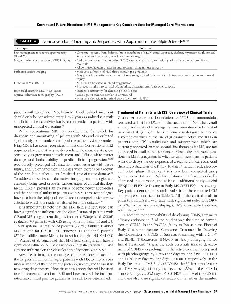
Treatment of Patients with CIS: Overview of Clinical TrialsGlatiramer acetate and formulations of IFNβ are immunodula-tors used as first-line DMTs for the treatment of MS. The overall efficacy and safety of these agents have been described in detail in Ryan et al. (2009).12 This supplement is designed to provide a specific overview of the use of glatiramer acetate and IFNβ in patients with CIS. Natalizumab and mitoxantrone, which are currently approved only as second-line therapies for MS, are not addressed in detail in this supplement. One of the important ques-tions in MS management is whether early treatment in patients with CIS delays the development of a second clinical event (and therefore a diagnosis of CDMS). To date, 4 randomized, placebo-controlled, phase III clinical trials have been completed using glatiramer acetate or IFNβ formulations that have specifically addressed this question, and at least 1 additional study — Rebif (IFNβ-1a) FLEXible Dosing in Early MS (REFLEX) — is ongoing. Key patient demographics and results from the completed CIS studies are summarized in Table 5. All of the clinical trials in patients with CIS showed statistically significant reductions (39% to 50%) in the risk of developing CDMS when early treatment was initiated.42-45
In addition to the probability of developing CDMS, a primary efficacy endpoint in 3 of the studies was the time to conver-sion to CDMS. In the PreCISe (Study to Evaluate the Effect of Early Glatiramer Acetate [Copaxone] Treatment in Delaying the Conversion to CDMS of Subjects Presenting with a CIS)42 and BENEFIT (Betaseron [IFNβ-1b] in Newly Emerging MS for Initial Treatment)43 trials, the 25th percentile time to develop-ment of CDMS was prolonged in the active-treatment compared with placebo groups by 115% (722 days vs. 336 days, P < 0.001) and 142% (618 days vs. 255 days, P < 0.001), respectively. In the Early Treatment of MS Study (ETOMS), the 30th percentile time to CDMS was significantly increased by 122% in the IFNβ-1a arm (569 days vs. 252 days, P = 0.034).45 In all 4 of the CIS tri-als, there also were significant reductions in either the number
patients with established MS, brain MRI with Gd-enhancement should only be considered every 1 to 2 years in individuals with subclinical disease activity but is recommended in patients with unexpected clinical worsening.34
While conventional MRI has provided the framework for diagnosis and monitoring of patients with MS and contributed significantly to our understanding of the pathophysiology under-lying MS, it has some recognized limitations. Conventional MRI sequences have a relatively weak correlation to clinical status, low sensitivity to grey matter involvement and diffuse white matter damage, and limited ability to predict clinical progression.35,36
Additionally, prolonged T2 relaxation identifies areas with tissue injury, and Gd-enhancement indicates when there is breakdown of the BBB, but neither quantifies the degree of tissue damage.37
To address these issues, alternative imaging methodologies are currently being used or are in various stages of clinical develop-ment. Table 4 provides an overview of some newer approaches and their potential utility in patients with MS. These technologies have also been the subject of several recent comprehensive review articles to which the reader is referred for more details.35-40
It is important to note that the MRI field strength used can have a significant influence on the classification of patients with CIS and MS using current diagnostic criteria. Wattjes et al. (2006) evaluated 40 patients with CIS using both 1.5 Tesla (T) and 3.0 T MRI systems. A total of 29 patients (72.5%) fulfilled Barkhof MRI criteria for CIS at 1.5T. However, 11 additional patients (27.5%) fulfilled more MRI criteria with the high-field MRI (3.0 T). Wattjes et al. concluded that MRI field strength can have a significant influence on the classification of patients with CIS and a minor influence on the classification of patients with MS.41
Advances in imaging technologies can be expected to facilitate the diagnosis and monitoring of patients with MS, to improve our understanding of the underlying pathophysiology, and to assist in new drug development. How these new approaches will be used to complement conventional MRI and how they will be incorpo-rated into clinical practice guidelines are still to be determined.
Current and Future Directions in MS Management: Key Considerations for Managed Care Pharmacists
taBlE 4 Nonconventional Imaging and Sequences with Applications in Multiple Sclerosis35-40
Technique Overview
Proton magnetic resonance spectroscopy (1H-MRS)
• Generates spectra from different brain metabolites (e.g., N-acetylaspartate, choline, myoinositol, glutamate) associated with various types of neuronal damage
Magnetization transfer ratio (MTR) imaging • Radiofrequency saturation pulse (RFSP) used to create magnetization gradient in protons from different molecules
• Allows visualization of myelin and axolemmal membrane integrityDiffusion tensor imaging • Measures diffusion of H2O through multiple gradients
• May provide for better evaluation of tissue integrity and differentiation between demyelination and axonal injury
Functional MRI (fMRI) • Measures alterations in blood oxygenation• Provides insight into cortical adaptability, plasticity, and functional capacity
High field strength MRI (> 1.5 Tesla) • Increases sensitivity for detecting brain lesionsOptical coherence tomography (OCT) • Uses light in manner similar to ultrasound
• Measures alterations in retinal nerve fiber layer (RNFL)
S8 Supplement to Journal of Managed Care Pharmacy JMCP November/December 2009 Vol. 15, No. 9-a www.amcp.org
individuals with a first clinical event suggestive of MS. More than 500 patients with a recent isolated demyelinating event (e.g., optic neuritis, myelopathy, or brainstem syndrome) and who had brain MRI scans consistent with early MS have been randomized to receive IFNβ-1a (as subcutaneous (SC) injections in one of two dosing schedules — 44 mcg 3 times a week or once weekly) or placebo. Patients will be treated for 24 months, or up to the time when they experience a second attack leading to a diagnosis of CDMS. The REFLEX trial completed enrollment in 2008; result data are not yet available. Long-term follow-up of the REFLEX study will be conducted in an extension trial known as RELEXION (www.clinicaltrials.gov).
Key Implications of Clinical Trials for Managed Care PharmacistsThe results of all completed CIS trials and extension analyses pro-vide strong clinical evidence that early treatment with glatiramer acetate or IFNβ significantly prolongs the time to conversion or progression to CDMS and also delays the development of physical disability. Thus, it is generally recommended that patients with CIS be treated at the time of diagnosis or first attack.10,49,50 This recommendation is supported by the National Clinical Advisory Board of the National MS Society, which states: “Initiation of treatment with an interferon-beta medication or glatiramer acetate should be considered as soon as possible following a definite diagnosis of MS with active, relapsing disease, and may also be considered for selected patients with a first attack who are at high risk of MS.”51 The U.S. Food and Drug Administration approved labels for 3 of the available IFNβ-1 formulations (Avonex, Betaseron, Extavia), and glatiramer acetate (Copaxone) also include indications for patients with CIS (i.e., a first clinical episode and MRI consistent with MS).
Disability in patients with MS is associated with increased health care costs. Turpcu and Yu (2008) conducted an analysis of
and/or volume of brain lesions seen on MRI.42-45 Because of favor-able results at interim analyses, the placebo arms of both the PreCISe and CHAMPS (Controlled High-Risk Subjects [IFNβ-1a] Avonex Multiple Sclerosis Prevention Study) trials were closed early, and all patients were then offered open-label treatment with the active drug.42,45
Since the studies described above were only 2 to 3 years in duration, the effects of continued DMT on the long-term risk for developing CDMS remained unclear. Two of the initial CIS studies, CHAMPS and BENEFIT were extended for longer time periods to address this question. In both the CHAMPIONS (CHAMPS extension) and BENEFIT extension studies, patients originally randomized to active treatment were continued on the same protocol (“early or immediate” treatment groups), and those who received placebo after the original randomization were then switched to open-label active drug (“delayed” treatment groups). In both extension analyses, early treatment resulted in signifi-cantly fewer patients developing CDMS. In CHAMPIONS, the risk of developing CDMS at 5 years was reduced by 43% in the immediate treatment compared with delayed treatment group (P = 0.030).46
In the BENEFIT extension, a 41% risk reduction for develop-ing CDMS was observed in the immediate treatment versus the delayed treatment group at 3 years (P = 0.001).47 Early treatment also significantly reduced the risk for progression of disability by 40% as measured by the Expanded Disability Status Scale (EDSS) (P = 0.022).47 The 5-year data from BENEFIT were recently pub-lished (September 2009): early treatment with IFNβ-1b delayed the onset of CDMS by 37% (P = 0.003), confirming continuous benefit at 5 years after treatment initiation in patients with CIS.48
The international REFLEX study is evaluating the effect of a new IFNβ-1a formulation on the time to conversion to MS in
Current and Future Directions in MS Management: Key Considerations for Managed Care Pharmacists
Trial PreCISe42 BENEFIT43 CHAMPS44 ETOMS45
Agent Glatiramer IFNß-1b IFNß-1a IFNß-1a
Dose 20 mg per day 250 mcg every other day 30 mcg per week 22 mcg per week
Administration SC SC IM SC
Study duration (yrs.) 3 (stopped early) 2 3 (stopped early) 2
Patients (N)Active treatment (n)Placebo (n)
481243238
468292176
383193190
308154154
Age (mean yrs. ± range) 31 ± 7 30 33 ± 7 28 ± 6
Reduction in CDMS risk 45% 50% 44% 39%
P value < 0.001 < 0.001 0.002 0.047aTable should not be used for comparison between independently conducted trials or estimation of the relative efficacy of individual agents.BENEFIT = Betaseron (IFNb-1b) in Newly Emerging MS for Initial Treatment; CDMS = clinically definite multiple sclerosis; CHAMPS = Controlled High-Risk Subjects (IFNb-1a) Avonex Multiple Sclerosis Prevention Study; ETOMS = Early Treatment of MS Study (Rebif); IM = intramuscular; PreCISe = Study to Evaluate the Effect of Early Glatiramer Acetate (Copaxone) Treatment in Delaying the Conversion to CDMS of Subjects Presenting with a CIS; SC = subcutaneous.
taBlE 5 Summary of Completed Clinically Isolated Syndrome Clinical Trialsa

www.amcp.org Vol. 15, No. 9-a November/December 2009 JMCP Supplement to Journal of Managed Care Pharmacy S9
cardiotoxicity, so cumulative dose limits are specified.57 In addi-tion, all currently approved agents for MS are administered parenterally, and tolerability and adherence are important con-siderations in patients using injectable medications. Thus, the need exists for development of new drugs for MS with different mechanisms of action, routes of administration, improved clini-cal efficacy, and more favorable safety profiles.58-60
More than 100 clinical trials of novel strategies in patients with MS are listed on the National MS Society website (www.NMSS.org). The majority of medical therapies in advanced stages of clinical development target peripheral immune mechanisms that predominate in early stage disease — these therapies are oral agents or monoclonal antibodies. In addition, neuroprotection and/or neuroregeneration, as well as improved management of comorbid symptoms, remain as desired therapeutic goals.58-60 A list of selected investigational agents for MS and their mechanism of action is provided in Table 6; these are discussed in greater detail below.
Oral AgentsThere has been considerable interest in developing oral medica-tions for MS. All of the oral agents discussed have recently com-pleted or are currently in phase III evaluation.
Cladribine. Cladribine (2-chloro-2’-deoxyadenosine) is an ade-nosine-deaminase resistant purine nucleoside analogue that pref-erentially depletes lymphocytes. This agent currently has several approved indications in oncology for lymphoid malignancies.61
A number of cladribine studies have been conducted in patients with MS that have been reviewed.62 Three major MS studies used
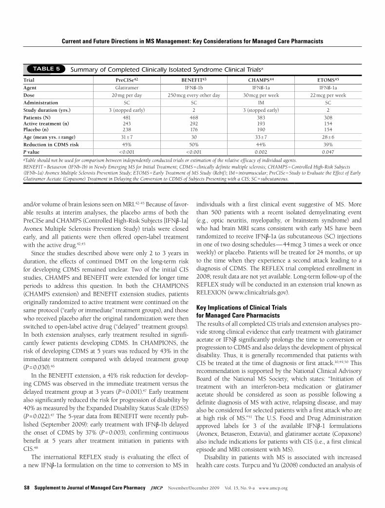
the relationship between total cost of care and the level of disabil-ity based on a meta-analysis of MS pharmacoeconomic studies published from 1966 to 1997 (Figure 2).52 Both direct and indi-rect costs of care increased with increasing levels of disability as measured by EDSS scores. With the introduction of DMTs, direct costs now make up a greater percentage of total costs of MS, but indirect costs still remain relatively high. The authors noted that DMTs may help avoid the high costs associated with greater levels of disability.52
Based on cost data published by Kobelt et al. (2006),53 Burks (2008) has suggested that early initiation of DMTs in patients with CIS or MS may reduce the overall cost of care by delay-ing the development of disability (Figure 3).11 Data from a study recently conducted in Italy are consistent with this finding. These data showed that early use of a DMT in patients with CIS that delayed conversion to CDMS provided a positive incremental cost-effectiveness ratio (ICER) per patient-year compared with no treatment.54
Emerging Therapies for MSAlthough several immunomodulatory agents are available for treatment of MS, these agents reduce relapses only by about 30%.55 Responses may be influenced by a number of factors, including patient-specific disease characteristics and/or genet-ics. The immunosuppressive agents natalizumab and mitox-antrone are very effective for reducing MS relapses but can be associated with significant toxicities. For example, natalizumab is associated with an increased risk of developing progressive multifocal leukoencephalopathy (PML).56 Mitoxantrone can cause
Current and Future Directions in MS Management: Key Considerations for Managed Care Pharmacists
FIGuRE 2 The Cost of MS Increases with Disability Level
EDSS 8EDSS 5
Direct Costs Indirect Costs
05,000
10,00015,00020,00025,00030,00035,00040,00045,000
EDSS 2
U.S
. Dol
lars
a
Without DMT
EDSS 8EDSS 50
5,00010,00015,00020,00025,00030,00035,00040,00045,000
EDSS 2
U.S
. Dol
lars
a
With DMT
aAverage cost across all studies. All costs converted to 2007 U.S. dollars using CPI for U.S. medical care. Turpcu A., et al. Presented at: AAN 60th Annual Meeting; April 12-19, 2008; Chicago, IL. P06.156.52
DMT = disease-modifying therapy; EDSS = Expanded Disability Status Scale.

S10 Supplement to Journal of Managed Care Pharmacy JMCP November/December 2009 Vol. 15, No. 9-a www.amcp.org
more clinically silent lesions on MRI. Subjects will be random-ized (1:1:1) to receive 3.5 mg per kg or 5.25 mg per kg cladribine tablets (or matching placebo) administered as 2 or 4 short courses (0.875 mg per kg per course) in 2 or 4 consecutive months dur-ing the first year. In year 2, subjects receive 2 additional short treatment courses. In participants who develop CDMS according to McDonald criteria, blinded medication will be discontinued, and maintenance therapy initiated with SC IFNβ-1a. Following the initial 2-year treatment period, CDMS-free patients can enroll in a 2-year maintenance follow-up period where they can receive open-label cladribine if they present with MRI activity. This clinical trial will provide insight into the sustained effects of early cladribine treatment of patients with CIS.65
Fumarate. BG000012 is an oral formulation of dimethyl fumarate that appears to have multiple novel effects on the immune system in patients with MS. This agent may have both anti-inflammatory and neuroprotective effects.66-68 In a phase IIb study, 257 patients with RRMS were randomized (1:1:1) to receive 1 of 3 oral doses of BG00012 (120 mg once daily, 120 mg 3 times daily, or 240 mg 3 times daily) or placebo for 24 weeks.69 The primary efficacy outcome was the number of new Gd-enhancing MRI lesions from weeks 12 to 24. Additional endpoints included new or enlarg-
an injectable form of the drug, which was found to reduce (a) the number and volume of T1 Gd-enhancing lesions, (b) T2 lesion volume, (c) relapse rates, and (d) progression of disability. An oral formulation of cladribine has also been developed that has been evaluated in patients with relapsing forms of MS, both as monotherapy or in combination with IFNβ-1a.62
Data from the phase III CLARITY study (CLAdRIbine tablets Treating MS orallY) were released in 2009. Patients with RRMS (n = 1,326) were randomized (1:1:1) to receive 1 of 2 doses of oral cladribine or placebo for 2 years. For the cladribine-tablet groups, subjects received either 2 or 4 courses of 0.875 mg per kg given over 4-5 consecutive days in the first year, then 2 treatment courses in the second year. For the primary efficacy endpoint of relapse rates at 96 weeks, 58% and 55% reductions were seen in the lower dose and higher dose cladribine groups versus placebo, respectively (P < 0.001). The frequency of adverse events was similar in all 3 treatment groups, except for lymphopenia, which was expected based on cladribine’s mechanism of action. The most common adverse events in all 3 groups were headaches and nasopharyngitis.63,64
Cladribine is also being evaluated in a 4-year phase III study in patients with CIS known as ORACLE (ORAl CLadribine for Early MS).65 For enrollment eligibility, patients must have 2 or
Current and Future Directions in MS Management: Key Considerations for Managed Care Pharmacists
Predicted Cost Early Interventiona MRI lesion activity
0
10,000
20,000
30,000
40,000
50,000
60,000
Mild EDSS < 4 Moderate EDSS 4-6 Severe EDSS > 6
Pre-clinical
Clinical Threshold
Atrophy and Axonal Degradation
CIS RRMS SPMS
70,000
U.S
. Dol
lars
per
Yea
r
FIGuRE 3 Natural History of MS and Cost of MS
aThe curve is based on an estimation of the decrease in cost for early treatment of about 40% at each range of EDSS.Derived from: Burks J. J Manag Care Med. 2008;12(1):26-31. [Exhibit 8];11 and Kobelt G., et al. Neurology. 2006;66(11):1696-702.53
CIS = clinically isolated syndrome; EDSS = Expanded Status Scale; RRMS = relapsing remitting multiple sclerosis; SPMS = secondary progressive multiple sclerosis; SPMS = secondary progressive multiple sclerosis.

www.amcp.org Vol. 15, No. 9-a November/December 2009 JMCP Supplement to Journal of Managed Care Pharmacy S11
Th1 and Th2 lymphocytes and cytokine production toward a Th2-pattern and inhibiting migration of lymphocytes into the CNS.73 Laquinimod is pharmacologically distinct from and has a more favorable benefit/risk profile than linomide, a structurally related compound that had previously reached phase III studies in patients with MS. In a 24-week phase II study of patients with relapsing MS (n = 209), the group treated with oral laquinimod at 0.3 mg per day had 44% fewer active lesions on MRI compared with placebo (P = 0.050). Subgroup analysis showed that this reduction was more pronounced in patients with at least 1 active lesion at baseline (52% fewer active lesions; P = 0.005).74
However, in a subsequent 36-week phase IIb trial comparing laquinimod at 0.3 and 0.6 mg per day with placebo in patients with RRMS (n = 306), only the 0.6 mg dose group had a signifi-cant reduction (40.4%) in the number of Gd-enhancing lesions on MRI scans (P = 0.005). One of the key safety considerations with this agent was transient and dose-dependent increases in hepatic enzymes.75
Teriflunomide. Teriflunomide is the active metabolite of lefluno-mide, an oral anti-inflammatory drug that is widely used in rheumatoid arthritis. This agent binds to dihydro-orotate dehydrogenase and inhibits de novo pyrimidine synthesis in T lymphocytes and other rapidly dividing cells.76 In a 36-week study of 179 patients with RRMS or secondary progressive MS (SPMS), subjects received oral teriflunomide at either of 2 doses (7 mg or 14 mg per day) or placebo.77 For the primary efficacy endpoint of the number of combined active unique lesions per MRI scan, significantly reduced mean numbers of lesions compared with placebo were seen in both the 7 mg (1.04 vs. 2.68, P < 0.030) and 14 mg teriflunomide groups (1.06 vs. 2.68, P < 0.010). Teriflunomide-treated patients also had significantly fewer T1-enhancing lesions per MRI scan (0.87 [7 mg] and 0.86 [14 mg] vs. 2.25 for placebo, P < 0.040), as well as fewer new T2 lesions (0.17 [7 mg] and 0.17 [14 mg] vs. 0.3 for placebo,
ing T2-hyperintense lesions, new T1-hypointense lesions, and relapse rates. In the 240 mg 3 times daily BG00012 dose group, significant reductions compared with placebo were reported for (a) the mean number of Gd-enhancing lesions (P < 0.001), (b) new or enlarging T2-hyperintense lesions (P < 0.001), and (c) new T1-hypointense lesions (P = 0.014). A 32% relative reduction in the annualized relapse rate was seen in the high dose BG00012 group, although the study was insufficiently powered to detect a significant difference for this outcome. Adverse events more frequently observed in BG00012 treatment groups included abdominal pain and flushing. Notably, 25% of patients in the 240 mg 3 times daily BG00012 dose group withdrew from the study or discontinued treatment primarily because of adverse events or lack of tolerance.69
Fingolimod. Fingolimod (FTY720) has a unique mechanism of action. By binding to sphingosine-1 phosphate type 1 receptors on lymphocytes, this agent stops the cells from sensing signals to egress from lymphoid tissue.70 In a proof-of-concept trial, 281 patients with relapsing MS were randomized (1:1:1) to receive oral fingolimod at 1.25 or 5.0 mg per day or placebo for 6 months.71
For the 255 patients who completed the study, the total number of Gd-enhancing lesions on MRI and the annualized relapse rate were significantly lower in both fingolimod dose groups com-pared with placebo (P ≤ 0.010). Dyspnea, diarrhea, nausea, and asymptomatic elevations of alanine aminotransferase were more frequent in the fingolimod treatment groups.71 In a 24-month phase II extension analysis, reductions in the relapse rate and lesion counts were seen in the patient group that switched from placebo to fingolimod (compared with the placebo phase of the trial), while these parameters remained low in patients who had received continuous fingolimod.72
Laquinimod. The mechanism of action of laquinimod is incom-pletely understood, but it is thought that this agent exerts its anti-inflammatory activity in MS by shifting the balance of
Current and Future Directions in MS Management: Key Considerations for Managed Care Pharmacists
taBlE 6 Therapies in Later Stages of Clinical Development for Multiple Sclerosis
Mechanism(s) of Action Reference(s)
Oral agentsDMTsCladribine Purine nucleoside analog; preferentially depletes lymphocytes 61-65Dimethyl fumarate (BG00012) Fumaric acid ester; may have both antiinflammatory and neuroprotective properties 66-69Fingolimod (FTY720) Sphingosine-1P (S-1P) receptor agonist; blocks lympocyte migration 70-72Laquinimod (ABR-215062) Believed to alter balance of Th1 and Th2 lymphocytes and cytokine profiles 72-75Teriflunomide Dihydro-orotate dehydrogenase inhibitor; blocks pyrimidine synthesis 76,77Symptom ManagementFampridine Blocks voltage-dependent K+-channels; may restore conduction in poorly myelinated nerve fibers 78-83
Monoclonal antibodies (mAb)Alemtuzumab Anti-CD52 mAb; depletes T and B lymphocytes 84,85Daclizumab Anti-CD25 (IL-2R α chain) mAb; inhibits T lymphocyte activation and expansion 86-89Rituximab (Rituxan) Anti-CD20 mAb; depletes B lymphocytes 90
CNS = central nervous system; DMT = disease-modifying therapy.

S12 Supplement to Journal of Managed Care Pharmacy JMCP November/December 2009 Vol. 15, No. 9-a www.amcp.org
first 5.5 months, then as monotherapy.87,88 In a small study of 9 patients who were evaluated at 3-month intervals, daclizumab was reported to be significantly effective in reducing total and new contrast-enhancing MRI lesions (P < 0.001) when comparing pre-treatment scans (baseline) with scans taken at all treatment intervals over 27.5 months. For secondary outcomes, daclizumab also significantly reduced relapse rates (P < 0.001) and improved both timed ambulation (P < 0.050) and EDSS scores (P < 0.050) over the course of the trial. Adverse events in the daclizumab group included respiratory tract infections, rash, and transient thrombocytopenia.87
Results from the larger phase II CHOICE trial have only been presented in abstract form.88 In this study, patients with active MS despite IFNβ therapy (n = 230) were randomized to receive daclizumab 2 mg per kg every 2 weeks (n = 75) or 1 mg every 4 weeks (n = 78) or placebo (n = 77) in addition to IFNβ. At 24 weeks, the number of new or enlarging Gd-enhancing lesions significantly decreased by a mean of 72% in the high-dose dacli-zumab group (P = 0.004), but only by 25% in the low-dose group (nonsignificant; P = 0.501), compared with placebo plus IFNβ. Adverse events more commonly observed in daclizumab-treated groups included serious infections.88,89
Rituximab. Approved for more than a decade for the treatment of B-cell lymphoma, rituximab is also being evaluated in patients with RRMS. In a 48-week phase II trial, subjects were random-ized to receive rituximab (1,000 mg on days 1 and 15) or placebo. Patients in the rituximab group had significantly fewer total and new Gd-enhancing lesions on MRI (primary endpoint) from weeks 12 to 24 compared with placebo, and these differences were sustained at 48 weeks (P < 0.001 for all time points). The relapse rate was also significantly lower with rituximab treatment at week 24 (14.5% vs. 34.3%, P = 0.020) and week 48 (23.0% vs. 40.0%, P = 0.040). Adverse events that were more common in the rituximab group included infusion-associated events within the 24 hours after the first infusion (78.3% vs. 40.0%), but these were predominantly mild to moderate in severity and decreased on the second infusion. Urinary tract infections (14.5% vs. 8.6%) and sinusitis (13.0% vs. 8.6%) were more frequent in the rituximab group compared with placebo.90
Key Implications of Emerging Therapies for Managed Care Pharmacists All medications currently approved for the treatment of MS are administered by SC or IM injection or IV infusion. Many patients have needle phobia or injection anxiety, particularly at the initia-tion of therapy.91 Injection-site reactions are also relatively com-mon with SC formulations, although the introduction of auto-injector technologies has made injections easier and improved patient satisfaction.92 However, the requirement for being on long-term injections places a burden on patients with MS and may lead to reduced adherence that can compromise treatment efficacy.
Oral medications for MS offer the potential of increasing
P < 0.040). Fewer patients in the 14 mg teriflunomide group had increases in disability, and a trend was reported for a lower annualized relapse rate and numbers of relapsing patients in the high-dose group versus placebo. Adverse events that were more common in the teriflunomide treatment groups included nausea, paresthesias, and limb pain.77
Fampridine. Fampridine (4-aminopyridine) is a voltage-depen-dent potassium channel blocker.78 This agent is not a DMT but instead targets specific symptoms in MS. Fampridine may restore action potential conduction in poorly myelinated nerve fibers79
and influence synaptic transmission and neuronal excitability.80 Fampridine use has been associated with improvements in strength, ambulation, fatigue, and endurance in patients with MS.81,82
In a randomized phase III study in patients with any type of MS, subjects who received sustained-release fampridine (10 mg twice daily; n = 229) had improved motor function measures com-pared with the placebo group (n = 72).83 For example, the number of responders based on a timed-walk measurement was signifi-cantly higher after 14 weeks of treatment with fampridine (35% vs. 8% for placebo, P < 0.001). In addition, timed-walk responders showed greater improvement in 12-item MS walking scale scores compared with timed-walk nonresponders (P < 0.001).83
Monoclonal AntibodiesAlemtuzumab. Alemtuzumab is a humanized monoclonal that targets CD52 on T cells, B cells, and monocytes and leads to rapid immune depletion.84 This agent was recently compared with IFNβ-1a in a 36-month phase II study of 334 patients with RRMS.85 Alemtuzumab was given at doses of either 12 mg or 24 mg per day by intravenous (IV) infusion on 5 consecutive days during month 1 and on 3 consecutive days at months 12 and 24. IFNβ-1a was administered at 44 mcg 3 times weekly SC after dose escalation. For the combined group analysis, alemtuzumab was significantly more effective than IFNβ-1a for (a) reducing the rate of sustained accumulation of disability (9.0% vs. 26.2%, P < 0.001, (b) reducing the annualized relapse rate (0.10 vs. 0.36, P < 0.001), (c) decreasing lesion burden based on T2 imaging (P = 0.005), and (d) increasing brain volume (P = 0.020). In addition, the mean EDSS score improved by 0.39 points in alemtuzumab-treated patients and worsened by 0.38 in the IFNβ-1a group (P < 0.001). Adverse events that were more common in the alemtuzumab group included infections (66% vs. 47%), thyroid disorders (23% vs. 3%), and thrombocytopenic purpura (3% vs. 1%).85
Daclizumab. Daclizumab is a humanized monoclonal that targets the α-subunit (CD25) of the high affinity interleukin 2 receptor (IL-2R), which is expressed on activated lymphocytes.86
This agent inhibits CD4+ and CD8+ T-cell activation and is cur-rently approved for the prevention of renal transplant rejection. It is being evaluated in patients with RRMS who have had an inadequate response to IFNβ. In 2 phase II studies, daclizumab (1 mg per kg intravenously every 2 weeks for the first 2 doses, then every 4 weeks) was given in combination with IFNβ for the
Current and Future Directions in MS Management: Key Considerations for Managed Care Pharmacists

www.amcp.org Vol. 15, No. 9-a November/December 2009 JMCP Supplement to Journal of Managed Care Pharmacy S13
convenience, comfort, and adherence to therapy. The oral treat-ments have different mechanisms of action, potentially signifi-cant adverse effects, and a relative lack of long-term safety data. Thus, the risk-benefit ratio may be not as favorable as that for the existing treatments. An ongoing question is how will formulary decisions be made regarding the new oral medications for MS if they are approved. In addition, consider the theoretical patient with MS who is currently using an injectable medication whose disease is well controlled with no new symptoms or relapses. What if this patient becomes aware of new oral treatment options and wants to switch therapies? Physicians and other health care prescribers will have to perform due diligence in discussing these issues with such patients. How will the decision be made regard-ing whether a switch is therapeutically appropriate, and should the new medication be covered by insurance? Managed care organizations can also benefit by addressing these critical issues in advance of anticipated new drug approvals.
■■ SummaryOne of the ongoing goals in MS management is to improve iden-tification of patients with CIS who will progress to CDMS and are the best candidates for early treatment with a DMT. Several recent advances have been made in the understanding of CIS subtypes and the refinement of MS diagnostic criteria. Imaging technologies other than traditional MRI are also likely to play an increasing role in MS management. Randomized clinical tri-als have shown that early treatment with glatiramer acetate and IFNβ formulations in patients with a CIS can prolong the time to conversion or progression to CDMS and delay the development of disability. Early treatment of CIS or MS may also decrease long-term health care costs. In addition, a number of new therapies for patients with MS are in late stages of clinical development, including several oral medications, offering promise for the near future but also potential challenges to managed care.
REFERENCES
1. Boissy AR, Cohen JA. Multiple sclerosis symptom management. Expert Rev Neurother. 2007;7(9):1213-22.
2. Khan F, Turner-Stokes L, Ng L, Kilpatrick T. Multidisciplinary reha-bilitation for adults with multiple sclerosis. Cochrane Database Syst Rev. 2007;(2):CD006036.
3. DaVanzo J, Dobson A, Koenig K, Book R. Medication therapy manage-ment services: a critical review. American College of Clinical Pharmacy. May 17, 2005. Available at: http://www.accp.com/docs/positions/commentaries/mtms.pdf. Accessed October 9, 2009.
4. American Pharmacists Association and National Association of Chain Drug Stores Foundation. Medication therapy management in pharmacy practice: core elements of an MTM service model, version 2.0. March 2008. Available at: http://www.nacdsfoundation.org/user-assets/Documents/PDF/2009/MTM/FinalMTMServiceModel_WEB.pdf. Accessed October 11, 2009.
5. Neuhaus O, Archelos JJ, Hartung HP. Immunomodulation in multiple sclerosis: from immunosuppression to neuroprotection. Trends Pharmacol Sci. 2003;24(3):131-38.
6. Bar-Or A. The immunology of multiple sclerosis. Semin Neurol. 2008;28(1):29-45.
7. Compston A, Coles A. Multiple sclerosis. Lancet. 2008;372(9648):1502-17.
8. Korn T. Pathophysiology of multiple sclerosis. J Neurol. 2008;255(Suppl 6):2-6.
9. Trapp BD, Peterson J, Ransohoff RM, Rudick R, Mörk S, Bö L. Axonal transection in the lesions of multiple sclerosis. N Engl J Med. 1998;338(5):278-85. Available at: http://content.nejm.org/cgi/reprint/338/5/278.pdf. Accessed October 9, 2009.
10. Tintoré, M. Rationale for early intervention with immunomodulatory agents. J Neurol. 2008;255(Suppl 1):37-43.
11. Burks J. MS: the treatment paradigm, a pathway to success for improved patient outcomes. J Manag Care Med. 2008;12(1):26-31. Available at: http://www.namcp.org/journals/jmcm/articles/12-1/JMCM%20MS%20TX%20Paradigm.pdf. Accessed October 9, 2009.
12. Ryan M, Deno S, Zwibel H. Review of the clinical debate regarding inter-ventions for multiple sclerosis. J Manag Care Pharm. 2009;15(Suppl 1-b):S1-S17. Available at: http://www.amcp.org/data/jmcp/Feb%20B%20supplement.pdf.
13. McDonald WI, Compston A, Edan G, et al. Recommended diagnostic criteria for multiple sclerosis: guidelines from the International Panel on the diagnosis of multiple sclerosis. Ann Neurol. 2001;50(1):121-27.
14. Polman CH, Reingold SC, Edan G, et al. Diagnostic criteria for mul-tiple sclerosis: 2005 revisions to the “McDonald Criteria.” Ann Neurol. 2005;58(6):840-46.
15. Cohen J, Rensel M. In: Burks J, Johnson K, eds. Multiple Sclerosis: Diagnosis, Medical Management, and Rehabilitation. New York: Demos; 2000:127-38.
16. Miller DH, Weinshenker BG, Filippi M, et al. Differential diag-nosis of suspected multiple sclerosis: a consensus approach. Mult Scler. 2008;14(9):1157-74. Available at: http://msj.sagepub.com/cgi/reprint/14/9/1157. Accessed October 9, 2009.
17. Marrie RA, Horwitz R, Cutter G, Tyry T, Campagnolo D, Vollmer T. Comorbidity delays diagnosis and increases disability at diagnosis in MS. Neurology. 2009;72(2):117-24.
18. Charil A, Yousry TA, Rovaris M, et al. MRI and the diagnosis of multiple sclerosis: expanding the concept of “no better explanation.” Lancet Neurol. 2006;5(10):841-52.
19. Frohman EM, Goodin DS, Calabresi PA, et al. The utility of MRI in suspected MS: report of the Therapeutics and Technology Assessment Subcommittee of the American Academy of Neurology. Neurology. 2003;61(5):602-11. Available at: http://www.neurology.org/cgi/reprint/61/5/602. Accessed October 9, 2009.
20. Brex PA, Ciccarelli O, O’Riordan JI, Sailer M, Thompson AJ, Miller DH. A longitudinal study of abnormalities on MRI and disability from multiple sclerosis. N Engl J Med. 2002;346(3):158-64. Available at: http://content.nejm.org/cgi/reprint/346/3/158.pdf. Accessed October 9, 2009.
21. Minneboo A, Barkhof F, Polman CH, Uitdehaag BM, Knol DL, Castelijns JA. Infratentorial lesions predict long-term disability in patients with initial findings suggestive of multiple sclerosis. Arch Neurol. 2004;61(2):217-21. Available at: http://archneur.ama-assn.org/cgi/reprint/61/2/217. Accessed October 9, 2009.
22. Tintoré M, Rovira A, Rio J, et al. Baseline MRI predicts future attacks and disability in clinically isolated syndromes. Neurology. 2006;67(6):968-72.
23. Swanton JK, Fernando K, Dalton CM, et al. Modification of MRI criteria for multiple sclerosis in patients with clinically isolated syndromes. J Neurol Neurosurg Psychiatry. 2006;77(7):830-33. Available at: http://www.pub-medcentral.nih.gov/picrender.fcgi?artid=2117493&blobtype=pdf. Accessed October 9, 2009.
Current and Future Directions in MS Management: Key Considerations for Managed Care Pharmacists

S14 Supplement to Journal of Managed Care Pharmacy JMCP November/December 2009 Vol. 15, No. 9-a www.amcp.org
43. Kappos L, Polman CH, Freedman MS, et al. (for the BENEFIT study group). Treatment with interferon beta-1b delays conversion to clinically definite and McDonald MS in patients with clinically isolated syndromes. Neurology. 2006;67(7):1242-49.
44. Jacobs L, Beck R, Simon J, et al. Intramuscular interferon beta-1a therapy initiated during a first demyelinating event in multiple sclerosis. CHAMPS Study Group. N Engl J Med. 2000;343(13):898-904. Available at: http://con-tent.nejm.org/cgi/reprint/343/13/898.pdf. Accessed October 9, 2009.
45. Comi G, Filippi M, Barkhof F, et al. Effect of early interferon treatment on conversion to definite multiple sclerosis: a randomised study. Lancet. 2001;357(9268):1576-82.
46. Kinkel RP, Kollman C, O’Connor P, et al. IM interferon beta-1a delays definite multiple sclerosis 5 years after a first demyelinating event. Neurology. 2006;66(5):678-84.
47. Kappos L, Freedman MS, Polman CH, et al. Effect of early versus delayed interferon beta-1b treatment on disability after a first clinical event sugges-tive of multiple sclerosis: a 3-year follow-up analysis of the BENEFIT study. Lancet. 2007;370(9585):389-97.
48. Kappos L, Freedman MS, Polman CH, et al. Long-term effect of early treatment with interferon beta-1b after a first clinical event suggestive of multiple sclerosis: 5-year active treatment extension of the phase 3 BENEFIT trial. Lancet Neurol. 2009; Sep 10 (E pub ahead of print), PMID 19748319.
49. Frohman EM, Havrdova E, Lublin F, et al. Most patients with multiple sclerosis or a clinically isolated demyelinating syndrome should be treated at the time of diagnosis. Arch Neurol. 2006;63(4):614-19.
50. Coyle P. Early treatment of multiple sclerosis to prevent neurologic dam-age. Neurology. 2008;71(24 Suppl 3):S3-S7.
51. National Clinical Advisory Board of the National Multiple Sclerosis Society (NMSS). Disease management consensus statement (2007). Available at: http://www.nationalmssociety.org/for-professionals/healthcare-profes-sionals/publications/expert-opinion-papers/index.aspx. Accessed October 9, 2009.
52. Turpcu A, Yu E. The cost of multiple sclerosis: an updated review of the literature. Poster presented at: American Academy of Neurology 60th Annual Meeting; April 12-19, 2008; Chicago, Illinois.
53. Kobelt G, Berg J, Atherly D, Hadjimichael O. Costs and quality of life in multiple sclerosis: a cross-sectional study in the United States. Neurology. 2006;66(11):1696-702.
54. Lazzaro C, Bianchi C, Peracino L, Zacchetti P, Uccelli A. Economic evaluation of treating clinically isolated syndrome and subsequent multiple sclerosis with interferon beta 1-b. Neurol Sci. 2009;30(1):21-31. Available at: http://www.springerlink.com/content/027631173013g8w4/fulltext.pdf. Accessed October 9, 2009.
55. Goodin DS, Frohman EM, Garmany GP, et al. Disease modifying therapies in multiple sclerosis: report of the Therapeutics and Technology Assessment Subcommittee of the American Academy of Neurology and the MS Council for Clinical Practice Guidelines. Neurology. 2002;58(2):169-78.
56. Goodin DS, Cohen BA, O’Connor P, Kappos L, Stevens JC. Assessment: the use of natalizumab (Tysabri) for the treatment of multiple sclerosis (an evidence-based review): report of the Therapeutics and Technology Assessment Subcommittee of the American Academy of Neurology. Neurology. 2008;71(10):766-73.
57. Fox EJ. Management of worsening multiple sclerosis with mitoxantrone: a review. Clin Ther. 2006;28(4):461-74.
58. Greenberg BM, Calbresi PA. Future research directions in multiple scle-rosis therapies. Semin Neurol. 2008;28(1):121-27.
59. Menge T, Weber MS, Hemmer B, et al. Disease-modifying agents for multiple sclerosis: recent advances and future prospects. Drugs. 2008;68(17):2445-68.
24. Tur C, Tintoré M, Rovira A, et al. Very early scans for dissemination in time in multiple sclerosis. Mult Scler. 2008;14(5):631-35.
25. Swanton JK, Rovira A, Tintoré M, et al. MRI criteria for multiple sclero-sis in patients presenting with clinically isolated syndromes: a multicentre retrospective study. Lancet Neurol. 2007;6(8):677-86.
26. Korteweg T, Tintoré M, Uitdehaag BM, et al. A search for new MRI criteria for dissemination in space in subjects with a clinically isolated syndrome. Eur Radiol. 2009;19(9):2244-48. Available at: http://www.spring-erlink.com/content/t41n323233g40323/fulltext.pdf. Accessed October 9, 2009.
27. Mastronardo G, Rocca MA, Iannucci G, Pereira C, Filippi M. A longi-tudinal MR study of the presymptomatic phase in a patient with clinically definite multiple sclerosis. AJNR Am J Neuroradiol. 1999;20(7):1268-72. Available at: http://www.ajnr.org/cgi/reprint/20/7/1268. Accessed September 15, 2009.
28. Okuda DT, Mowry EM, Beheshtian A, et al. Incidental MRI anoma-lies suggestive of multiple sclerosis: the radiologically isolated syndrome. Neurology. 2009;72(9):800-05.
29. Hakiki B, Goretti B, Portaccio E, Zipoli V, Amato MP. ”Subclinical MS”: follow-up of four cases. Eur J Neurol. 2008;15(8):858-61.
30. Bourdette D, Simon J. The radiologically isolated syndrome: is it very early multiple sclerosis? Neurology. 2009;72(9):780-81.
31. Traboulsee A, Li DK. The role of MRI in the diagnosis of multiple sclero-sis. Adv Neurol. 2006;98:125-46.
32. Engell T. A clinical patho-anatomical study of clinically silent multiple sclerosis. Acta Neurol Scand. 1989;79(5):428-30.
33. Simon JH, Li D, Traboulsee A, et al. Standardized MR imaging protocol for multiple sclerosis: Consortium of MS Centers consensus guidelines. AJNR Am J Neuroradiol. 2006;27(2):455-61. Available at: http://www.ajnr.org/cgi/reprint/27/2/455. Accessed October 9, 2009.
34. Consortium of Multiple Sclerosis Centers. MRI protocol for the diagnosis and follow-up of MS. Proposed 2009 revised guidelines. Available at: http://www.mscare.org/cmsc/images/pdf/mriprotocol2009.pdf. Accessed October 9, 2009.
35. Neema M, Stankiewicz J, Arora A, Guss ZD, Bakshi R. MRI in multiple sclerosis; what’s inside the toolbox? Neurotherapeutics. 2007;4(4):602-17. Available at: http://download.journals.elsevierhealth.com/pdfs/jour-nals/1933-7213/PIIS1933721307001468.pdf. Accessed October 9, 2009.
36. Rovira A, León, A. MR in the diagnosis and monitoring of multiple sclerosis:an overview. Eur J Radiol. 2008;67(3):409-14.
37. Fox RJ. Picturing multiple sclerosis: conventional and diffusion tensor imaging. Semin Neurol. 2008;28(4):453-66.
38. Bakshi R, Thompson AJ, Rocca MA, et al. MRI in multiple sclerosis: cur-rent status and future prospects. Lancet Neurol. 2008;7(7):615-25.
39. Khan O. Introduction to nonconventional sequences and imaging. Int J MS Care. 2007;9(Suppl 3):6-9. Available at: http://www.mscare.org/cmsc/images/pdf/supplements-november-2007.pdf. Accessed October 11, 2009.
40. Rashid W, Miller DH. Recent advances in neuroimaging of multiple scle-rosis. Semin Neurol. 2008;28(1):46-55.
41. Wattjes MP, Harzheim M, Kuhl CK, et al. Does high-field MR imaging have an influence on the classification of patients with clinically isolated syndromes according to current diagnostic MR imaging criteria for multiple sclerosis? AJNR Am J Neuroradiol. 2006;7(8):1794-98. Available at: http://www.ajnr.org/cgi/reprint/27/8/1794. Accessed October 9, 2009.
42. Comi G, Martinelli V, Rodegher M, et al. Effect of glatiramer acetate on conversion to clinically definite multiple sclerosis in patients with clini-cally isolated syndrome (PreCISe study): a randomised, double-blind, placebo-controlled trial. Lancet. 2009; October 6 epub ahead of print, PMID 19815268.
Current and Future Directions in MS Management: Key Considerations for Managed Care Pharmacists

www.amcp.org Vol. 15, No. 9-a November/December 2009 JMCP Supplement to Journal of Managed Care Pharmacy S15
75. Comi G, Pulizzi A, Rovaris M, et al. Effect of laquinimod on MRI-monitored disease activity in patients with relapsing-remitting multiple scle-rosis: a multicentre, randomised, double-blind, placebo-controlled phase IIb study. Lancet. 2008;371(9630):2085-92.
76. Cherwinski HM, Cohn RG, Cheung P, et al. The immunosuppressant leflunomide inhibits lymphocyte proliferation by inhibiting pyrimidine bio-synthesis. J Pharmacol Exp Ther. 1995;275(2):1043-49.
77. O’Connor PW, Li D, Freedman MS, et al. A Phase II study of the safety and efficacy of teriflunomide in multiple sclerosis with relapses. Neurology. 2006;66(6):894-900.
78. Judge SI, Bever CT Jr. Potassium channel blockers in multiple sclerosis: neuronal Kv channels and effects of symptomatic treatment. Pharmacol Ther. 2006;111(1):224-59.
79. Shi R, Blight AR. Differential effects of low and high concentrations of 4-aminopyridine on axonal conduction in normal and injured spinal cord. Neuroscience. 1997;77(2):553-62.
80. Mainero C, Inghilleri M, Pantano P, et al. Enhanced brain motor activity in patients with MS after a single dose of 3,4-diaminopyridine. Neurology. 2004;62(11):2044-50.
81. Schwid SR, Petrie MD, McDermott MP, Tierney DS, Mason DH, Goodman AD. Quantitative assessment of sustained-release 4-amin-opyridine for symptomatic treatment of multiple sclerosis. Neurology. 1997;48(4):817-21.
82. Goodman AD, Brown TR, Cophen JA, et al. Dose comparison trial of sustained-release fampridine in multiple sclerosis. Neurology. 2008;71(15):1134-41.
83. Goodman AD, Brown TR, Krupp LB, et al. Sustained-release oral fam-pridine in multiple sclerosis: a randomised, double-blind controlled trial. Lancet. 2009;373(9665):732-38.
84. Coles AJ, Cox A, Le Page E, et al. The window of therapeutic opportu-nity in multiple sclerosis: evidence from monoclonal antibody therapy. J Neurol. 2006;253(1):98-108.
85. The CAMMS223 trial investigators, Coles AJ, Compston DA, et al. Alemtuzumab vs. interferon beta-1a in early multiple sclerosis. N Engl J Med. 2008;359(17):1786-801. Available at: http://content.nejm.org/cgi/reprint/359/17/1786.pdf. Accessed October 11, 2009.
86. Rose JW, Burns JB, Bjorklund J, Klein J, Watt HE, Carlson NG. Daclizumab phase II trial in relapsing and remitting multiple sclerosis: MRI and clinical results. Neurology. 2007;69(8):785-89.
87. Bielekova B, Howard T, Packer AN, et al. Effect of anti-CD25 antibody daclizumab in the inhibition of inflammation and stabilization of disease progression in multiple sclerosis. Arch Neurol. 2009;66(4):483-89.
88. Montalban X, Wynn D. Kaufman M, Wang M, Fong A. Preliminary CHOICE results: a phase II, randomised, double-blind, placebo-con-trolled multicentre study of subcutaneous daclizumab in patients with active, relapsing forms of multiple sclerosis on interferon beta. Mult Scler. 2007;13(suppl 2):S18. Abstract presented at the 23rd Congress of the European Committee for Treatment and Research in Multiple Sclerosis. Prague, Czech Republic, October 11-14, 2007.
89. Kim SE. Daclizumab treatment for multiple sclerosis. Pharmacotherapy. 2009;29(2):227-35.
90. Hauser SL, Waubant E, Arnold DL, et al. B-cell depletion with rituximab in relapsing-remitting multiple sclerosis. N Engl J Med. 2008;358(7):676-88. Available at: http://content.nejm.org/cgi/reprint/358/7/676.pdf. Accessed October 9, 2009.
91. Cox D, Stone J. Managing self-injection difficulties in patients with relapsing-remitting multiple sclerosis. J Neurosci Nurs. 2006;38(3):167-71.
92. Cramer JA, Cuffel BJ, Divan V, Al-Sabbagh A, Glassman M. Patient satis-faction with an injection device for multiple sclerosis treatment. Acta Neurol Scand. 2006;113(3):156-62.
60. Giacomini PS, Darlington PJ. Bar-Or A. Emerging multiple sclerosis disease-modifying therapies. Curr Opin Neurol. 2009;22(3):226-32.
61. Sipe JC. Cladribine for multiple sclerosis: review and current status. Expert Rev Neurother. 2005;5(6):721-27.
62. Leist TP, Vermersch P. The potential role for cladribine in the treatment of multiple sclerosis: clinical experience and development of an oral tablet formulation. Curr Med Res Opin. 2007;23(11):2667-76.
63. Rieckmann P, Giovannoni G, Comi G, et al. The CLARITY study: effi-cacy of cladribine tablets for relapsing-remitting multiple sclerosis. Int J MS Care. 2009;11(Suppl 2):84. Abstract W32. Available at: http://www.mscare.org/cmsc/images/pdf/IJMSC_Abstracts_2009.pdf. Accessed October 9, 2009.
64. Jeffrey S. CLARITY trial of oral cladribine in MS meets primary end-point. Medscape Medical News, January 26, 2009. Available at: http://www.medscape.com/viewarticle/587314. Accessed October 11, 2009.
65. Coyle P, Leist T, Comi G, et al. Study design of the ORACLE multiple sclerosis study: cladribine tablets in early multiple sclerosis. Int J MS Care. 2009;11(Suppl 2):84. Abstract W31. Available at: http://www.mscare.org/cmsc/images/pdf/IJMSC_Abstracts_2009.pdf. Accessed October 9, 2009.
66. Lukashev M, Zeng W, Ryan S, et al. Activation of Nrf2 and modula-tion of disease progression in EAE models by BG00012 (dimethyl fumar-ate) suggests a novel mechanism of action combining anti-inflammatory and neuroprotective modalities. Mult Scler. 2007;13(suppl 2):S149. Abstract presented at the 23rd Congress of the European Committee for Treatment and Research in Multiple Sclerosis. Prague, Czech Republic, October 11-14, 2007.
67. Chen XL, Dodd G, Thomas S, et al. Activation of Nrf2/ARE pathway pro-tects endothelial cells from oxidant injury and inhibits inflammatory gene expression. Am J Physiol Heart Circ Physiol. 2006;290(5):H1862-70. Available at: http://ajpheart.physiology.org/cgi/reprint/290/5/H1862. Accessed October 9, 2009.
68. Li J, Johnson D, Calkins M, Wright L, Svendsen C, Johnson J. Stabilization of Nrf2 by tBHQ confers protection against oxidative stress-induced cell death in human neural stem cells. Toxicol Sci. 2005;83(2):313-28. Available at: http://toxsci.oxfordjournals.org/cgi/reprint/83/2/313. Accessed October 9, 2009.
69. Kappos L, Gold R, Miller DH, et al. Efficacy and safety of oral fumar-ate in patients with relapsing-remitting multiple sclerosis: a multicentre, randomised, double-blind, placebo-controlled phase IIb study. Lancet. 2008;372(9648):1463-72.
70. Brinkmann V, Cyster JG, Hla T. FTY720: sphingosine 1-phosphate receptor-1 in the control of lymphocyte egress and endothelial barrier func-tion. Am J Transplant. 2004;4(7):1019-25. Available at: http://www3.inter-science.wiley.com/cgi-bin/fulltext/118820664/PDFSTART. Accessed October 9, 2009.
71. Kappos L, Antel J, Comi G, et al. Oral fingolimod (FTY720) for relapsing multiple sclerosis. N Engl J Med. 2006;355(11):1124-40. Available at: http://content.nejm.org/cgi/reprint/355/11/1124.pdf. Accessed October 9, 2009.
72. O’Connor P, Comi G, Montalban X, et al. Oral fingolimod (FTY720) in multiple sclerosis: two-year results of a phase II extension study. Neurology. 2009;72(1):73-79.
73. Zou LP, Abbas N, Volkmann I, et al. Suppression of experimental auto-immune neuritis by ABR-215062 is associated with altered Th1/Th2 balance and inhibited migration of inflammatory cells into the peripheral nerve tis-sue. Neuropharmacology. 2002;42(5):731-39.
74. Polman C, Barkhof F, Sandberg-Wollheim M, et al. Treatment with laquinimod reduces development of active MRI lesions in relapsing MS. Neurology. 2005;64(6):987-91.
Current and Future Directions in MS Management: Key Considerations for Managed Care Pharmacists

S16 Supplement to Journal of Managed Care Pharmacy JMCP November/December 2009 Vol. 15, No. 9-a www.amcp.org
4. A 29-year-old female presented with blurred vision when she looked to her left and numbness in her arm. She was referred to an ophthalmologist who diagnosed with optic neuritis. Her primary care provider also sent her for a battery of tests including blood analysis, MRI, and lumbar puncture. Based on the 2005 revisions to the McDonald criteria for the diag-nosis of MS, which of the following findings would have greater significance?a. The severity of the symptoms associated with the initial
attackb. An elevated IgG index in the cerebrospinal fluid (CSF)c. The presence of autoantibodies in the serumd. The presence of spinal cord lesions on MRI
5. Which of the following represents a key modification to MRI criteria proposed by Swanton et al.?a. Requiring the use of gadolinium to enhance MRI
sensitivityb. Increasing the number of required T2 lesions c. Removing the time requirement for appearance of a new
T2 lesiond. Reducing size requirements for infratentorial lesions
6. A breakdown of the current definition of clinically isolated syndrome (CIS) into additional subcategories was recently proposed. In addition to assisting in development of a ratio-nal monitoring and treatment plan, these subcategories may also predict:
1. All of the following statements regarding the current under-standing of the pathophysiology underlying MS are correct EXCEPT:a. The early phase of MS is characterized by CNS
inflammation and infiltration of T and B cells.b. Axonal loss is not seen in pre-clinical or early stages of
MS.c. Demyelination leads to neurodegeneration in later stages
of MS.d. Both Th1 and Th17 lymphocytes may be important in the
pathogenesis of MS.2. Which of the following conditions would be most likely to
present similarly to MS?a. Hunter syndromeb. Alzheimer’s diseasec. Viral encephalitisd. Brain aneurysm
3. Your organization has asked you to prepare a presentation on the differential diagnosis of MS. All of the following state-ments would be correct EXCEPT:a. Nondemyelinating CNS disorders have minimal overlap
with MS with respect to initial physical symptoms.b. The presence of comorbidities can significantly delay the
time that MS is accurately diagnosed.c. The diagnosis of MS typically involves the exclusion of
other medical conditions with similar symptoms or MRI results.
d. There is no single definitive clinical test for MS.
cONtINuING EDucatION
Current and Future Directions in MS Management: Key Considerations for Managed Care Pharmacists
PRIME Education, Inc. (PRIME®) is accredited by the Accreditation Council for Pharmacy Education (ACPE) as a provider of continu-ing pharmaceutical education. A total of 0.150 CEUs (1.5 contact hours) will be awarded and a continuing education statement will be sent to pharmacists for successful completion of this continuing education program, which is defined as receiving a minimum score of 70% on the posttest and completion of the Program Evaluation form. ACPE Universal Program Number. 255-000-09-021-H01-P.
Release date: November 1, 2009 Expiration date: October 31, 2010
Continuing Education for this program is processed either through AMCP at www.amcp.org (CME/CE Center) or through PRIME at www.primeinc.org/jmcp/nov. No mailed forms will be accepted.
The posttest worksheet is provided to assist you in marking your answers prior to entering the online CE center for submission; these pages cannot be submitted for CE credits.
In order to receive CE credit for this program, you must complete the following forms online:1. The posttest form for this program, “Current and Future Directions in MS Management: Key Considerations for Managed Care
Pharmacists,” is accessible at www.amcp.org (CME/CE Center) or at PRIME® at www.primeinc.org/jmcp/nov. To receive credit, you must receive a score of 70%. You will have 2 opportunities to pass the posttest.
2. Program Evaluation form.
Upon successful completion of this program, you will automatically receive your CE statement. Your CE credits will be automatically archived and tracked for you on the AMCP.org (CME/CE Center) or at www.primeinc.org. All information is kept confidential.
There is no fee for this learning activity.
Posttest Worksheet: Current and Future Directions in MS Management: Key Considerations for Managed Care Pharmacists

www.amcp.org Vol. 15, No. 9-a November/December 2009 JMCP Supplement to Journal of Managed Care Pharmacy S17
a. Teriflunomide: inhibits purine synthesisb. Cladribine: pyrimidine nucleoside analoguec. Laquinimod: may inhibit lymphocyte entry into the CNSd. Fampridine: central-acting calcium-channel blocker
12. Based on clinical trial data, which of the following therapies may improve ambulation in patients with MS by increasing axonal transmission of action potentials?a. Fampridineb. Fingolimodc. Laquinimodd. Fumarate
13. Which of the following adverse events has been seen more often in active treatment groups compared with placebo across all clinical trials of experimental antibodies in patients with MS?a. Cardiotoxicityb. Infections c. Anaphylaxisd. Hepatic enzyme elevation
14. Your organization has asked you to prepare a presentation on emerging therapies for MS. All the following statements would be correct EXCEPT:a. Some oral agents in clinical development will most likely
require multiple doses every day.b. Cladribine will likely be administered in several short
intensive treatment courses every year, similar to chemotherapy.
c. The majority of therapies in late-stage clinical development specifically target lymphocytes that have entered the CNS.
d. Protection or regeneration of nerve cells remain as therapeutic goals.
15. Which of the following statements regarding clinical trials of alemtuzumab and daclizumab in combination with IFNβ for the treatment of MS is correct?a. Both alemtuzumab and daclizumab reduced brain lesion
volume but not the number of lesions. b. Alemtuzumab plus IFNβ had roughly equivalent efficacy
in reducing the rate of sustained disability compared with IFNβ alone.
c. Daclizumab dose groups had similar efficacy for reducing the number of Gd-enhancing lesions.
d. Addition of alemtuzumab and daclizumab to IFNβ significantly reduced relapse rates compared with IFNβ alone.
a. The rate of disease progressionb. The risk that a patient will develop MSc. The level of disability a patient will ultimately reachd. The efficacy of potential treatments
7. Which of the following statement regarding conventional MRI technology is most accurate?a. MRI allows for accurate quantification of the amount of
brain tissue damage in patients with MS.b. MRI has relatively high sensitivity to identify grey matter
lesions.c. MRI results strongly correlate with the clinical status of
patients with MS and assist with determining treatment protocols.
d. MRI field strength can influence classification of patients with a CIS.
8. A 37-year-old female was diagnosed with a CIS based on clinical symptoms and MRI scanning and is considered a good candidate for initiation of a disease-modifying therapy (DMT). In discussing with her the rationale for beginning therapy now, all of the following statements regarding clini-cal studies of early treatment initiation in patients with CIS would be correct EXCEPT:a. Early treatment with a DMT has a good chance of
delaying the patient’s progression to clinically definite multiple sclerosis (CDMS).
b. Early treatment with a DMT will probably increase the overall cost of care.
c. The occurrence of relapses may be reduced using a DMT early in the disease course.
d. Early treatment with a DMT may delay the development of physical disability.
9. The patient described in the previous question and her provider make the decision to initiate a DMT. Which of the following agents is NOT currently approved for treatment of patients with a CIS and MRI suggestive of MS?a. Glatiramer acetateb. Interferon (IFN) β-1ac. Mitoxantroned. IFNβ-1b
10. Which of the following statements regarding approved MS agents is INCORRECT?a. Currently available agents for MS only reduce relapses by
about 30%.b. Natalizumab is associated with progressive multifocal
leukoencephalopathy. c. All drugs approved for MS in 2009 are administered by
injection or infusion.d. Extension phases of clinical trials have generally failed to
show long-term efficacy. 11. Which of the following correctly matches an experimental
oral agent for MS with its proposed mechanism of action?
Posttest Worksheet: Current and Future Directions in MS Management: Key Considerations for Managed Care Pharmacists
To complete this activity, go to www.amcp.org (CE/CME Center) or http://primeinc.org/jmcp/nov to access the posttest and evaluation form.

Supplement