Embed Size (px)
Citation preview
JOURNAL OF MODERN BIOTECHNOLOGY, VOL. 2, NO. 2, pp 40–45, March 2013 Copyright © 2013, by Madras Institute of Biotechnology. All Right Reserved. www.thebiotech.org
40 JOURNAL OF MODERN BIOTECHNOLOGY VOLUME 2· NUMBER 2· MARCH 2013
Research Article
Culturing Marine Green Microalgae Dunaliella salina Teod. and Dunaliella tertiolecta Masjuk in Dewalne’s Medium for Valuable Feeds Stock
Subramaniyan Venkatesan1, Munuswamy Senthil Swamy2, Chinnasamy Senthil3, Sailendra Bhaskar3 and Ramasamy Rengasamy1*
1Centre for Advanced Studies in Botany, University of Madras, Guindy Campus, Chennai 600 025, Tamil Nadu, India. 2EID Parry India Ltd, Parry Nutraceuticals Division, Research and Development Centre, Chennai 600 019, Tamil Nadu, India.
3Aban Infrastructure Pvt. Ltd., Biotechnology Division, Thandalam, Chennai 602 102, Tamil Nadu, India.
*Correspondence Author e-mail: [email protected]
Received 20 February 2013; Revised 27 February 2013; Accepted 28 February 2013
Abstracts
The single cell, marine, halo tolerant carotenogenic species of Dunaliella salina have the ability to accumulate secondary carotenoids, β-carotene as they grow in high salinity and high irradiance. Commercially β-carotene has many applications such as nutritional supplements to chemotherapeutic agent in cancer therapy and antioxidant. Pro vitamin-A is a natural colorant; aqua feed and prevents night blindness, enhances immunity and reduces the risk of heart diseases. Whereas, non-carotenogenic species have the ability of fast growth and lipid production and can be used for biofuel production. In the present study, the carotenogenic Dunaliella salina and non-carotenogenic Dunaliella tertiolecta obtained from the Centre for Advanced Studies in Botany, University of Madras and were cultured using Dewalne’s medium for a period of 30 days under laboratory conditions. Maximum cell number in Dunaliella salina of 6.83 x10⁶ cells/ml was recorded on 16th day and the maximum amount of total carotenoid was produced as 55 mg/L on 26th day, while the maximum cell number recorded in Dunaliella tertiolecta was 8.48 x10⁶ cells/ml on 20th day and maximum lipid content of 295 mg/L on 30th day.
Key words: Dunaliella salina, Dunaliella tertiolecta, growth rate, total carotenoids, Dewalne’s medium.
___________________________________________________________________________________________________
INTRODUCTION
Microalgae culture is one of the commercially important aspects in modern biotechnology. The first unialgal culture was achieved by Beijerinck (1890), on the green alga Chlorella vulgaris and the use of such culture for studying plant physiology was developed by Warburg in the early 1900 (Warburg, 1919). Certain microalgae, namely Chlorella, Spirulina and Dunaliella are grown commercially for nutraceuticals and other algal products such as hydrocarbons, fatty acids, phycocolloids, bioactive molecules and fine chemicals.
The genus Dunaliella, includes about 30 species of which 25 are found in brackish water and 5 in freshwater (Melkonian and Preisig, 1984; Leonard and Cascers, 1993). The optimal conditions for carotenogenesis are those that limit growth and include exposure to high light intensities and other stress factors, especially nutrient deprivation.
The biflagellate microalga, Dunaliella growing profusely in salt marshes was first discovered by a French Scientist Dunal in 1837. This naked allay of Chlamydomonas, responsible for the red coloration characteristic of salt
Venkatesan et al Culturing marine green microalgae Dunaliella salina
41 VOLUME 2· NUMBER 2· MARCH 2013 JOURNAL OF MODERN BIOTECHNOLOGY
marshes was later described by Teodoresco in 1905 and is now named as Dunaliella Teod. The unique morphological feature of Dunaliella is it lacks a cell wall. The cell is enclosed by a thin plasma membrane or periplast, which permits rapid changes in cell shape and volume in response to osmotic changes. The genus Dunaliella of the order Volvocales includes a variety of well-defined species of unicellular, ovoid and biflagellate green algae. Dunaliella demonstrates a remarkable degree of environmental adaptation to salt and is widely distributed in natural habitats. The cells are motile and have two equal, long, smooth, whiplash flagella. The cells have one large cup shaped chloroplast, which occupies about half the volume and it is oriented around the nucleus (Ben-Amotz et al., 1982; Loeblich, 1982; Garcia Gonzalez et al., 2003; Gomez et al., 2003; Gomez and Gonzalez et al., 2005).
Species of Dunaliella are very well adopted to propagate in media ranging from less than seawater concentration (0.1M sodium chloride or 0.5844 g/1000 ml) to saturated salt solution (5M sodium chloride or 292.2 g/1000 ml). This extremely halotolerant organism is one of the eukaryotic organisms found under such extreme conditions. Dunaliella salina collected from the lakes without any addition of chemicals is a potent source of natural mixed carotenoids.
Dunaliella salina has 90% of β-carotene and 10% of other carotenoids. Carotenoids are made up of α-carotene and xanthophylls like lutein, zeaxanthin, and cryptoxanthis similar to the ones found in food and vegetables (Gouveia and Emphis, 2003). These xanthophylls have a wide spread application in the pharmaceutical and cosmetics as well as in animal feed. Approximately 500 different carotenoids have been identified so far. Dunaliella salina is a mixture of two stereoisomer all-trans carotene and 9-cis carotene (synthetic β-carotene has only the all trans isomer). 9-cis β-carotene is a powerful antioxidant and it is well known that it contributes significantly in maintaining the health. In the present study carotenogenic strains of Dunaliella salina and non carotenogenic strains of Dunaliella tertiolecta obtained from Centre for Advanced Studies in Botany, University of Madras were cultured in Dewalne’s medium under laboratory condition. The growth, pigment composition, total protein, carbohydrate and lipid content were estimated at every three days interval up to 30 days.
MATERIAL AND METHODS
Growth study of Dunaliella salina and D. tertiolecta
Experiments were carried out in 1000 ml Erlenmeyer’s flasks containing 500 ml sterilized Dewalne’s medium inoculated with 50 ml of optimally grown cultures of Dunaliella salina and Dunaliella tertiolecta and kept under 25±1oC at 30µE m-2 s-1 light intensity, 12/12 light/dark photoperiod for a period of 30 days. At every alternate day interval, 5 ml of samples were drawn from the culture and recorded for the following parameters. (i) cell count using Haemocytometer (ii) concentration of photosynthetic pigments viz., Chlorophyll a, Chlorophyll b and total carotenoid (Lichtenthaler, 1987) (iii) Total protein (Bradford et al.,1976) (iv) Total carbohydrate (Dubois et al., 1956) (v) Total lipid (Folch et al., 1956). Growth curve was plotted against time and log10 of cell number/ml was calculated by the formula given by Guillard (1973). The concentration of pigments i.e., Chl a, Chl b and total carotenoids were expressed as µg/106 cells and total protein, carbohydrate and total lipid were expressed as µg/ml. All the experiments were carried out in triplicates and means are presented.
RESULTS AND DISCUSSION
An industrially important unicellular green alga, Dunaliella which is recognized for the production of β-Carotene was screened for its growth and carotenogenesis. Besides, the concentrations of total protein, carbohydrate and lipid contents were also estimated in the present study. Up to date, around 30 different species of Dunaliella were reported (Melkonian and Preisig, 1984; Leonard and Cascers, 1993). Among the two different strains chosen in the present study, one of them was identified as carotenogenic strain Dunaliella salina (Teodoresco, 1905) and the other as non-carotenogenic Dunaliella tertiolecta (Masjuk, 1973) based on their morphological characteristics. Dewalne’s medium was used to grow the organism since Dunaliella is reported to perform best for the growth, biomass and β-carotene production in Dunaliella.
Five different types of media were tested for their effects on the growth of Dunaliella sp.: M1 (BG-11 medium, Stanier et al., 1971), M2 (f2 medium, Jeffrey and LeRoi 1997), M3 (f/2 medium, Guillard 1962), M4 medium of Ben-Amotz et al. (1989), and M5 of Sallal et al. (1987).

Culturing marine green microalgae Dunaliella salina Venkatesan et al
42 JOURNAL OF MODERN BIOTECHNOLOGY VOLUME 2· NUMBER 2· MARCH 2013
Under constant illumination at room temperature 25 ± 2 °C after 14 days, the maximum growth of Chlorophyll-a was 7.5 mg/l while β-carotene was 5.2 mg/l and the maximum number of Dunaliella cells was found to be 6.5x10⁶
cells/ml in M1 medium (Nader Fareid Abu Sara, et al., 2011). A comparative study was made on D. salina with respect to its growth characteristics in Dewalne’s medium (control) and modified Dewalne’s medium. The algae grown in the modified Dewalne’s medium showed a maximum number of 6.71 log10 cells/ml whereas in Dewalne’s medium (Control) was 6.63 log10 cells/ml. Similarly, the concentration of Chl-a and Chl-b were found to be maximum in the modified Dewalne’s medium and their increment were 8.7% and 7.0%, respectively, on 12th day. A maximum amount of 6.64 mg/L β-carotene was recorded in the modified Dewalne’s medium on 18th day, which was more than 7.5% to that of control. The division rate (µ) of Dunaliella salina in the Modified Dewalne’s medium was 0.43 when compared to 0.40 in the control (Raja et al., 2004).
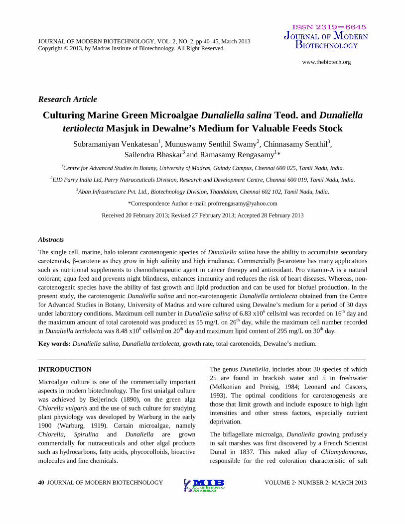
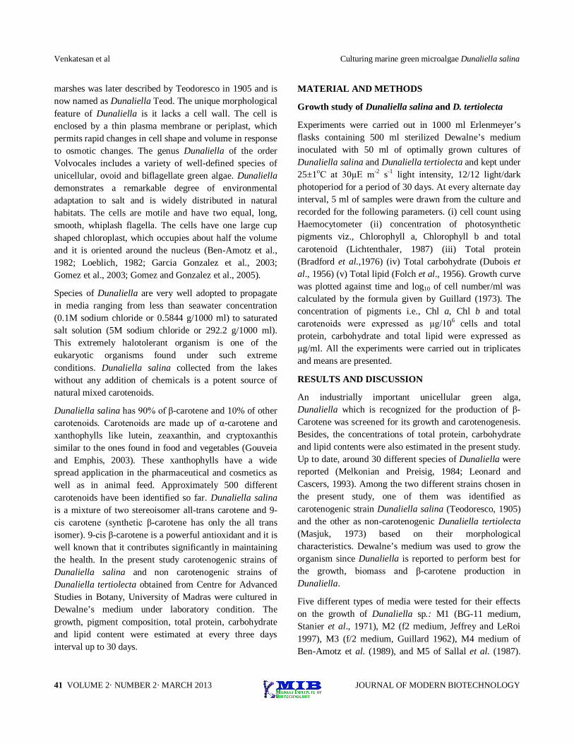
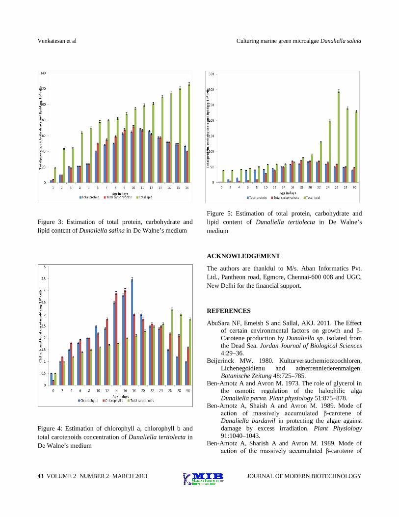
In the present study, Dunaliella salina showed a maximum accumulation of total carotenoids of 55 µg/106 cells in Dewalne’s medium on 26th day. The amount of Chlorophyll-a and Chlorophyll-b were 8.64 and 6.51 µg/106 cells on 16th and 14th day respectively. The maximum of cell number Dunaliella salina was 6.83 x 10⁶ cells/ml on 16th day. The maximum amount of total protein, carbohydrate and lipid for Dunaliella salina were 68.6, 72 and 126 µg/ml on 18th, 20th and 30th day respectively (Figure 1, 2 and 3). While, the maximum accumulation of total catotenoids in Dunaliella tertiolecta was 3.22 µg/106 cells on 26th day. The amounts of Chlorophyll-a and Chlorophyll-b recorded were 4.48 µg/106 cells and 4.03 µg/106 cells on 18th and 16th day respectively. The maximum of cell number recorded in Dunaliella tertiolecta was 8.48 x 10⁶ cells/ml 20th day in Dewalne’s medium. The maximum concentration of total protein, carbohydrate and lipid recorded in Dunaliella tertiolecta were 79, 101 and 295 µg/ml on 20th, 18th and 30th day, respectively (Figure 1, 4 and 5).
CONCLUSION
The growth and lipid content were found to be maximum in non-carotenogenic strain of Dunaliella tertiolecta when compared to carotenogenic strain Dunaliella salina. Whereas, Chlorophyll-a, Chlorophyll-b and β-carotene
were maximum in Dunaliella salina when compared to the pigment of the non carotenogenic strain. Therefore it is concluded that the carotenogenic D. salina can be mass cultured for β-carotene production, while the non- carotenogenic strain D. tertiolecta can be used for achieving the maximum biomass and for biofuel production.
Figure 1: Growth curve of Dunaliella salina and Dunaliella tertiolecta in De Walne’s medium
Figure 2: Estimation of chlorophyll-a, chlorophyll-b concentration of Dunaliella salina in De Walne’s medium
Venkatesan et al Culturing marine green microalgae Dunaliella salina
43 VOLUME 2· NUMBER 2· MARCH 2013 JOURNAL OF MODERN BIOTECHNOLOGY
Figure 3: Estimation of total protein, carbohydrate and lipid content of Dunaliella salina in De Walne’s medium
Figure 4: Estimation of chlorophyll a, chlorophyll b and total carotenoids concentration of Dunaliella tertiolecta in De Walne’s medium
Figure 5: Estimation of total protein, carbohydrate and lipid content of Dunaliella tertiolecta in De Walne’s medium
ACKNOWLEDGEMENT
The authors are thankful to M/s. Aban Informatics Pvt. Ltd., Pantheon road, Egmore, Chennai-600 008 and UGC, New Delhi for the financial support.
REFERENCES
AbuSara NF, Emeish S and Sallal, AKJ. 2011. The Effect of certain environmental factors on growth and β-Carotene production by Dunaliella sp. isolated from the Dead Sea. Jordan Journal of Biological Sciences 4:29–36.
Beijerinck MW. 1980. Kulturversuchemiotzoochloren, Lichenegoidienu and adnerrenniederenmalgen. Botanische Zeitung 48:725–785.
Ben-Amotz A and Avron M. 1973. The role of glycerol in the osmotic regulation of the halophilic alga Dunaliella parva. Plant physiology 51:875–878.
Ben-Amotz A, Shaish A and Avron M. 1989. Mode of action of massively accumulated β-carotene of Dunaliella bardawil in protecting the algae against damage by excess irradiation. Plant Physiology 91:1040–1043.
Ben-Amotz A, Sharish A and Avron M. 1989. Mode of action of the massively accumulated β-carotene of
Culturing marine green microalgae Dunaliella salina Venkatesan et al
44 JOURNAL OF MODERN BIOTECHNOLOGY VOLUME 2· NUMBER 2· MARCH 2013
Dunaliella bardawil in protecting the alga against damage by excess irradiation. Plant Physiology 91:479–487.
Borowitzka, L. J. and Brown, A. D. 1974. The salt relations of marine and halophilic species of the unicellular green alga Dunaliella. Arch. Microbiol., 96: 37–52.
Bradford MM. 1979. A rapid and sensitive method for the quantification of microgram quantities of protein utilizing the principle of protein dye binding. Analytical Biochemistry 72:248–254.
Craigie JS and McLachlan J. 1964. Glycerol as photosynthetic product in Dunaliella tertiolecta Butcher. Canadian Journal of Botany 173:370–376.
Dubois M, Gilles KA, Hamilton TK, Rebers PA and Smith F. 1956. Colorimetric method for determination of sugars and related substances. Analytical Chemistry 28:350–356.
Dunal F. 1838. Extrait de un mémoire sur les algues qui colorent en rouge certains eaux des marais salants méditerranéens. Annals Sc Nat Bot 2 Series 9:172.
Dunal MF. 1837. Note surtesalguesauicolorentem rouge certainseaux des maraisselantsmediterraneens. Comptesrendus de la Academic de science, Paris, 15:585–587.
Fazeli MR, Tofighi H, Samadi N, Jamalifar H, Fazeli A. 2006. Carotenoids accumulation by Dunaliella tertiolecta (Lake Urmia Isolate) and Dunaliella Salina (Ccap 19/18 & Wt) under stress conditions. DARU Journal of Pharmaceutical Sciences 14:146–150.
Garcia-Gonzalez M, Moreno J, Moreno J, Canavate P, Anguis V, Prieto A, Manzano C, Florencia FJ and Guerrero MG. 2003. Conditions for open-air outdoor culture of Dunaliella salina in southern spain. Journal of Applied Phycology 15:177–184.
Ginzburg M. 1978. Dunaliella: a green alga adapted to salt. Advances in Botanical Research 14:93–183.
Ginzburg M. 1987. Dunaliella: a green alga adapted to salt. Advances in Botanical Research 14:93–183.
Gomez PI and Gonzalez MA. 2005. The effect of temperature and irradiance on the growth and carotenoigenic capacity of seven strains of Dunaliella salina (Chlorophyta) cultivated under laboratory conditions. Journal of Biological Research 38:151–162.
Gomez PIA, Barriga AS, Cifuentes M and Gonzalez A. 2003. Effect of salinity on the quantity and quality of carotenoids accumulated Dunaliella salina (strain CONC-007) and Dunaliella bardawil (strain ATCC-30861) Chlorophyta. Journal of Biological Research 36:185–192.
Gouveia L and Emphis J. 2003. Relative stability of microalgal carotenoids in microalgal extracts, biomass and fish feed: effect of storage conditions. Innovative food science and Emerging Technologies 4:227–233.
Guillard RRL and Ryther JH. 1962. Studies on the marine planktonic diatoms. L. Cyclotella nana Hustedt and Detonula confervaceae (Clev) Gran. Canadian Journal of Microbiology 8:229–239.
Guillard RRL. 1973. Division rates. In: Handbook of phycological methods: culture methods and growth measurements. (ed.) Stein JR, Cambridge University Press, London, pp 289–311.
Jeffrey SW and LeRoi JM. 1997. Simple procedures for growing SCOR reference microalgal cultures, In: Jeffrey SW Mantoura RFC and Wright SW (Eds) Phytoplankton pigments in oceanography. UNESCO Publishing, France pp.181–205.
Leonard PI and Caceres EJ. 1994. Comparative analysis of fine structure of young and adult individuals of Dunaliella salina (Polyblepharidaceae, Chlorophyceae) with emphasis on the flagellar apparatus. Journal of Phycology 30:642–653.
Lichtenthaler HK. 1987. Chlorophylls and carotenoids: pigments of photosynthetic membranes. Methods in Enzymology 148:350–382.
Loeblich LA. 1982. Photosynthesis and pigments influenced by light intensity and salinity in the halophile Dunaliella salina (Chlorophyta). Journal of the Marine Biological Association of the United Kingdom 62:493–508.
Orset S and Young AJ. 1999. Low temperature induced synthesis of α-carotene in the microalga Dunaliella salina (Chlorophyta). Journal of Phycology 35:520–527.
Raja R, Anbazhagan C, Senthilswamy M, Lakshmi D and Rengasamy R. 2004. Nutritional studies on Dunaliella salina (Volvocales, Chlorophyta) under laboratory condition. Seaweed Research Utilization 26:127–146.
Sallal AKJ, Al-Hasan RH and Nimer NA. 1987. Localization of glycollate dehydrogenase in Dunaliella salina. Planta 171:429–432.
Stanier RY, Kunisawa R Mandel M and Cohen-Bazire G. 1971. Purification and properties of unicellular blue-green algae (order Chroococcales). Archive of "Bacteriological Reviews 35:171–205.
Teodoresco EC. 1905. Organization et development du Dunaliella, nouveau genre de volvocalae Polyblepharidae. Beihefte zum Botanischen Centralblatt 18:230–239.
Teodoresco, EC. 1905. Organisation et développement du Dunaliella, nouveau genre de Volvocacée-
Venkatesan et al Culturing marine green microalgae Dunaliella salina
45 VOLUME 2· NUMBER 2· MARCH 2013 JOURNAL OF MODERN BIOTECHNOLOGY
Polyblepharidée. Beih z Bot Centralbl., Bd. XVIII: 215–232.
Warburg O. 1919. Uber die Greschwindiling Keit der Koti; ensarezusammensetzung in Lebendenzellen. Biochemischezeitschrift 100:230–270.


![Chloroplast phylogenomic analysis of chlorophyte green algae ......nas reinhardtii [29], Volvox carteri f. nagariensis [30], Chlamydomonas moewusii [5], Dunaliella salina [31] and](https://img.pdfslide.us/doc/110x75/60eef2527f1f704a697716f6/chloroplast-phylogenomic-analysis-of-chlorophyte-green-algae-nas-reinhardtii.jpg)