Embed Size (px)
Citation preview
Culturing Embryonic and Adult-Derived Stem Cells:
Introduction and Key Applications
Amy Laws, Ph.D.November 2008

Outline
• Introduction to stem cells• Embryonic stem cells• Alternatives to embryonic stem cells• Introduction to human embryonic stem cell culture• Adult stem cells• Introduction to adult stem cell culture and
differentiation

What Does It Mean To Be a Stem Cell?
Stem cells are the foundation for every
organ, tissue, and cell in the human body.
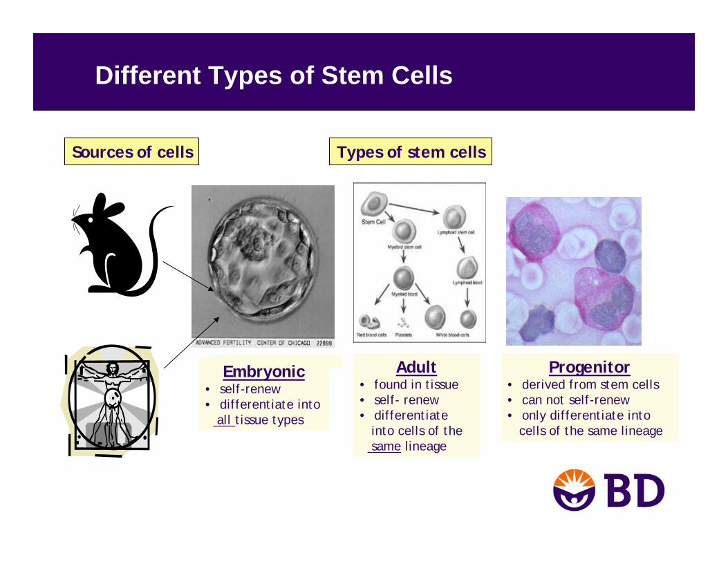
Different Types of Stem Cells
Embryonic• self-renew • differentiate into
all tissue types
Progenitor• derived from stem cells• can not self-renew• only differentiate into
cells of the same lineage
Adult• found in tissue • self- renew • differentiate
into cells of the same lineage
Sources of cells Types of stem cells
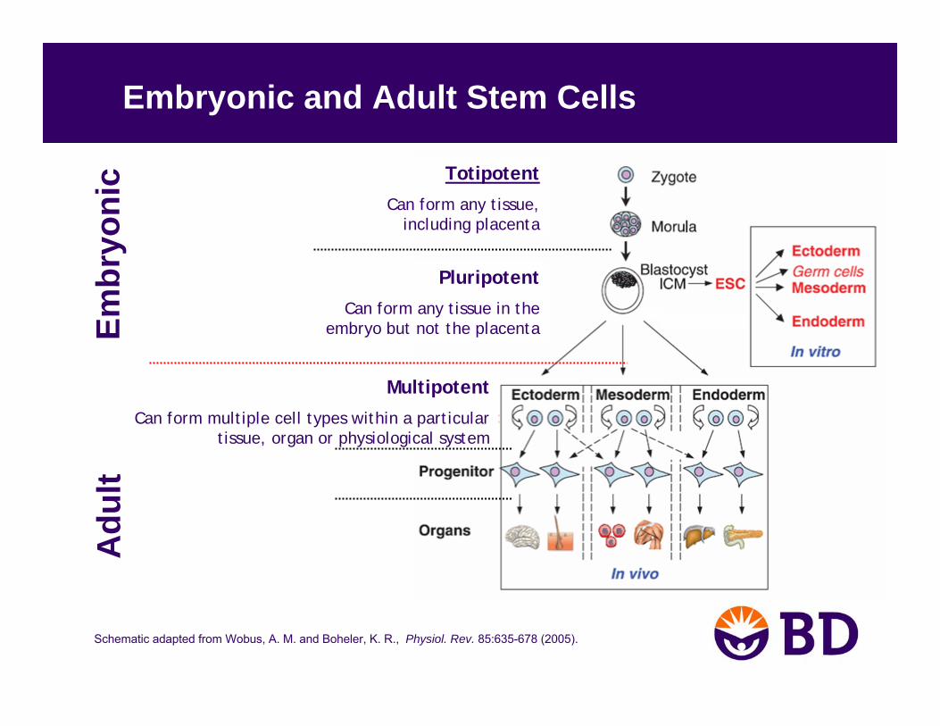
Embryonic and Adult Stem Cells
Schematic adapted from Wobus, A. M. and Boheler, K. R., Physiol. Rev. 85:635-678 (2005).
Embr
yoni
cA
dult
Totipotent
Can form any tissue, including placenta
Pluripotent
Can form any tissue in the embryo but not the placenta
Multipotent
Can form multiple cell types within a particular tissue, organ or physiological system
Schematic adapted from http://stemcells.nih.gov/index.asp
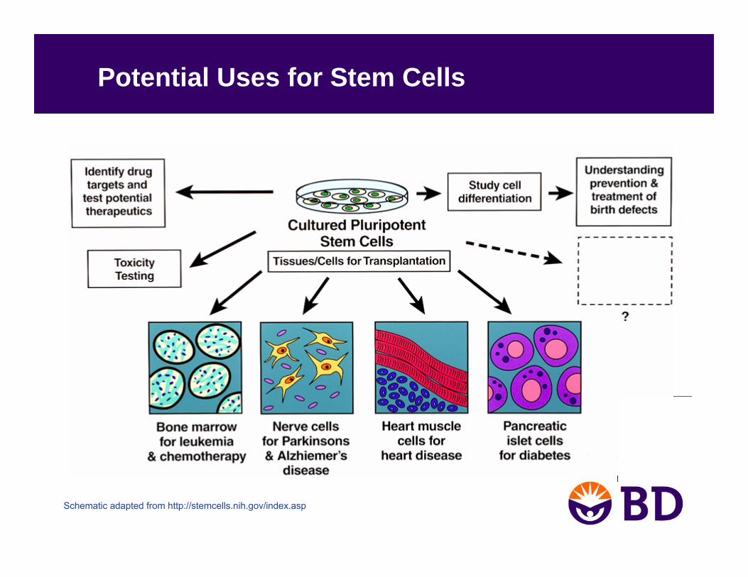
Potential Uses for Stem Cells
Current and Future Clinical Stem Cell Applications
• Blood disease– Hematopoietic stem cells have been used for bone
marrow transplantation for over 20 years.
• Spinal cord injury– Geron is awaiting FDA approval to begin clinical trial
with hESC-derived oligodendrocyte progenitor cells.
• Type II diabetes treatment– Restore glucose-responsive insulin-secreting cells
either by transplantation of stem cell-derived cells or reprogramming of existing cells.

Schematic adapted from Fischbach and Fischbach, J. Clin. Invest. 114:1364-1370 (2004).
• Undifferentiated/non-committed
• Self renewal
• Pluripotency
Characteristics of Embryonic Stem (ES) Cells

• ES cells were first derived in 1981 from a MOUSE embryo– Nature 292:54-156 (1981); Proc Natl Acad Sci USA 78:7634–7638 (1981)
• Isolation of human ES cells by James Thomson, et al. in 1998– Science 282:1145–1147 (1998)
• Protocols for hES cell culture were optimized from mouse ES cells and other embryonic stem cells’ culturing practices.
• hES cells were isolated by transferring the inner cell mass of a 3-5 day old embryo onto a mouse fibroblast feeder layer.
How Were ES cells First Isolated?
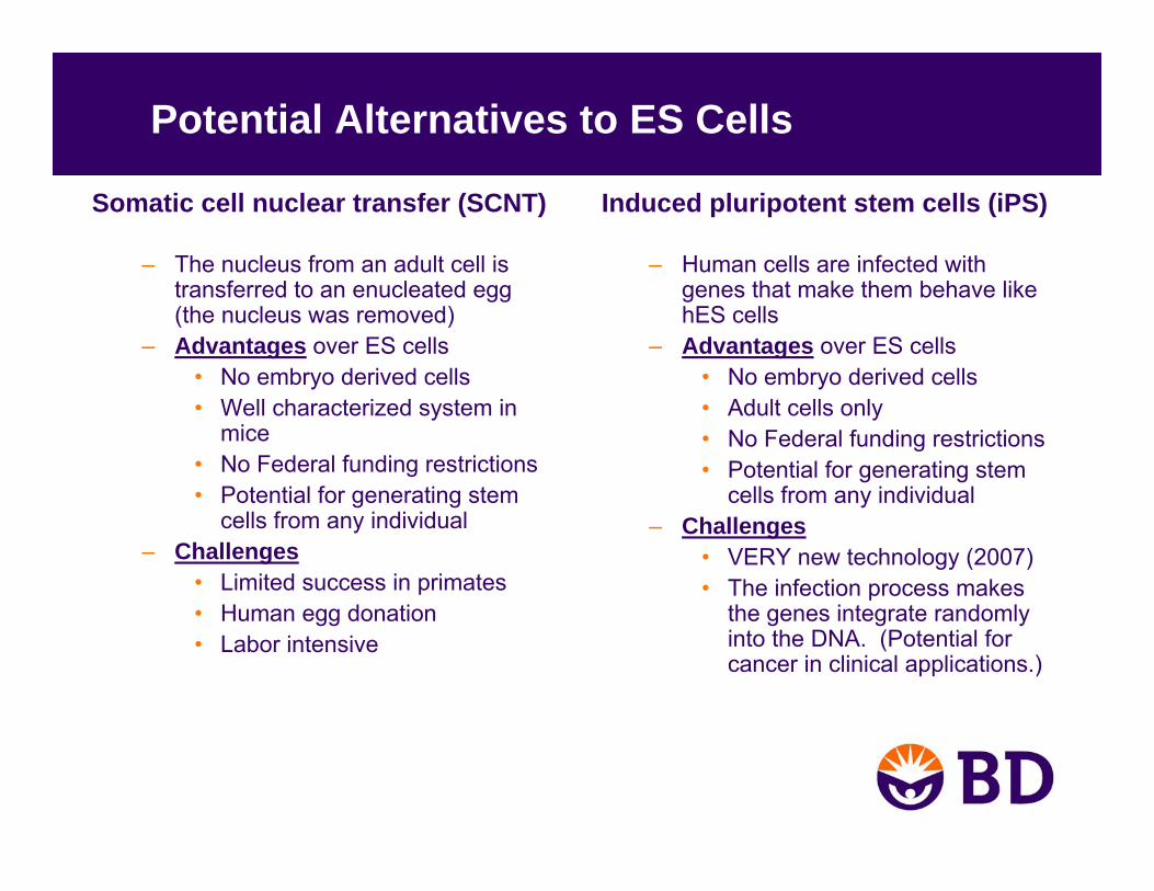
Potential Alternatives to ES Cells
Induced pluripotent stem cells (iPS)
– Human cells are infected with genes that make them behave like hES cells
– Advantages over ES cells• No embryo derived cells• Adult cells only• No Federal funding restrictions• Potential for generating stem
cells from any individual– Challenges
• VERY new technology (2007)• The infection process makes
the genes integrate randomly into the DNA. (Potential for cancer in clinical applications.)
Somatic cell nuclear transfer (SCNT)
– The nucleus from an adult cell is transferred to an enucleated egg (the nucleus was removed)
– Advantages over ES cells• No embryo derived cells• Well characterized system in
mice• No Federal funding restrictions• Potential for generating stem
cells from any individual– Challenges
• Limited success in primates• Human egg donation• Labor intensive
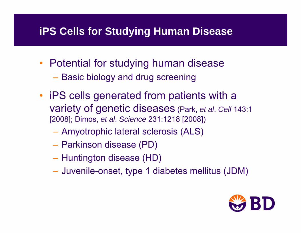
iPS Cells for Studying Human Disease
• Potential for studying human disease– Basic biology and drug screening
• iPS cells generated from patients with a variety of genetic diseases (Park, et al. Cell 143:1 [2008]; Dimos, et al. Science 231:1218 [2008])
– Amyotrophic lateral sclerosis (ALS)– Parkinson disease (PD)– Huntington disease (HD)– Juvenile-onset, type 1 diabetes mellitus (JDM)
Key Challenges with hES Cell Culture
• Lack of defined culturing environment with a standard protocol
• Spontaneous differentiation• Scaling up cultures• Efficient transfection of hES cells• Simulating physiological conditions in vitro• Time required for full characterization of new
culturing condition: in vitro and in vivo• Difficulty in comparing data from different laboratories
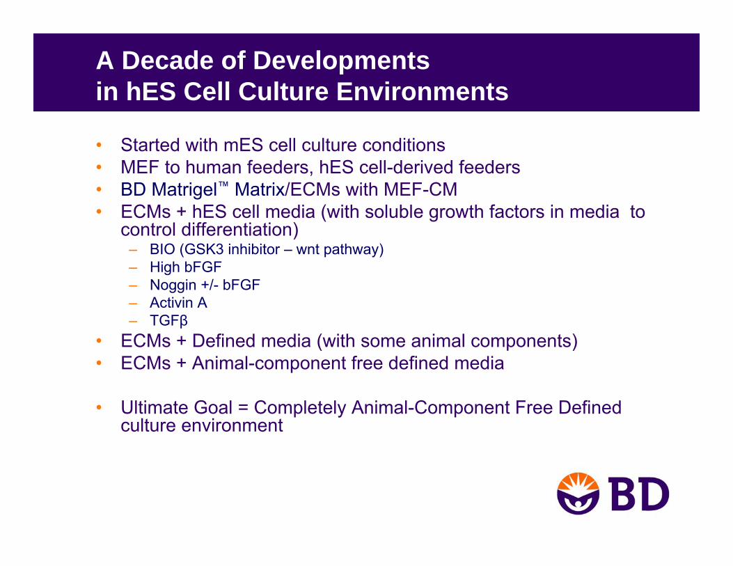
A Decade of Developmentsin hES Cell Culture Environments
• Started with mES cell culture conditions• MEF to human feeders, hES cell-derived feeders• BD Matrigel™ Matrix/ECMs with MEF-CM• ECMs + hES cell media (with soluble growth factors in media to
control differentiation)– BIO (GSK3 inhibitor – wnt pathway)– High bFGF– Noggin +/- bFGF– Activin A– TGFβ
• ECMs + Defined media (with some animal components)• ECMs + Animal-component free defined media
• Ultimate Goal = Completely Animal-Component Free Defined culture environment
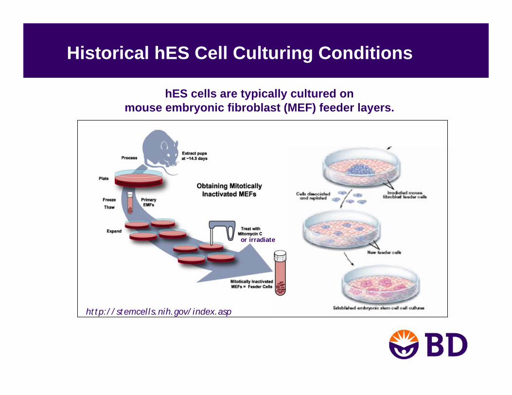
Historical hES Cell Culturing Conditions
hES cells are typically cultured on mouse embryonic fibroblast (MEF) feeder layers.
or irradiate
http://stemcells.nih.gov/index.asp

Limitations of Mouse Embryonic Fibroblast (MEF) Feeder Layer Systems
• MEF Issues– Labor intensive to maintain two cell types– Variation of different MEF lots
• Contamination– Potential contamination of animal pathogens from mouse feeders
is a major concern when trying to move into therapeutic applications
• Downstream Manipulation– Colonies that form on feeder layers are compact and difficult to
genetically manipulate and transfect– Transfection efficiency of hESCs is low. Quenching can occur from
the feeder layers – Difficult to isolate DNA / RNA due to potential cross-contamination
from feeder layers
Limitations of Mouse Embryonic Fibroblast (MEF) Feeder Layer Systems (Cont’d)
• Standardization– There is no widely accepted standard protocol which could result in
major issues when moving into the therapeutic arena – Difficulty in comparing data from different laboratories exists due to
the absence of standard hESC culture practices– Cells undergo spontaneous differentiation on a MEF feeder layer
which makes comparisons from culture to culture difficult
Feeder-free hES Cell Culture
• First documented in 2001 (Xu, et al. Nature Biotech. 24:185)
• BD Matrigel™ Matrix-coated surface used with mouse embryonic fibroblast feeder layer conditioned media (MEF-CM)
• Multiple media conditions and defined mediahave been used successfully in combination with BD Matrigel Matrix-coated surface for culturing hES cells
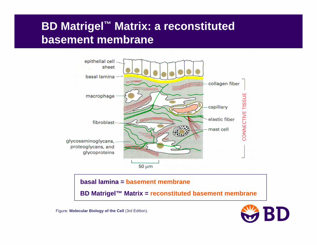
BD Matrigel™ Matrix: a reconstitutedbasement membrane
Figure: Molecular Biology of the Cell (3rd Edition).
basal lamina = basement membrane
BD Matrigel™ Matrix = reconstituted basement membrane
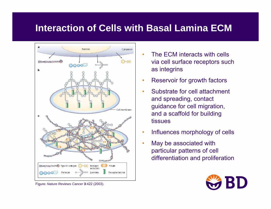
Interaction of Cells with Basal Lamina ECM
• The ECM interacts with cells via cell surface receptors such as integrins
• Reservoir for growth factors
• Substrate for cell attachment and spreading, contact guidance for cell migration, and a scaffold for building tissues
• Influences morphology of cells
• May be associated with particular patterns of cell differentiation and proliferation
Figure: Nature Reviews Cancer 3:422 (2003).
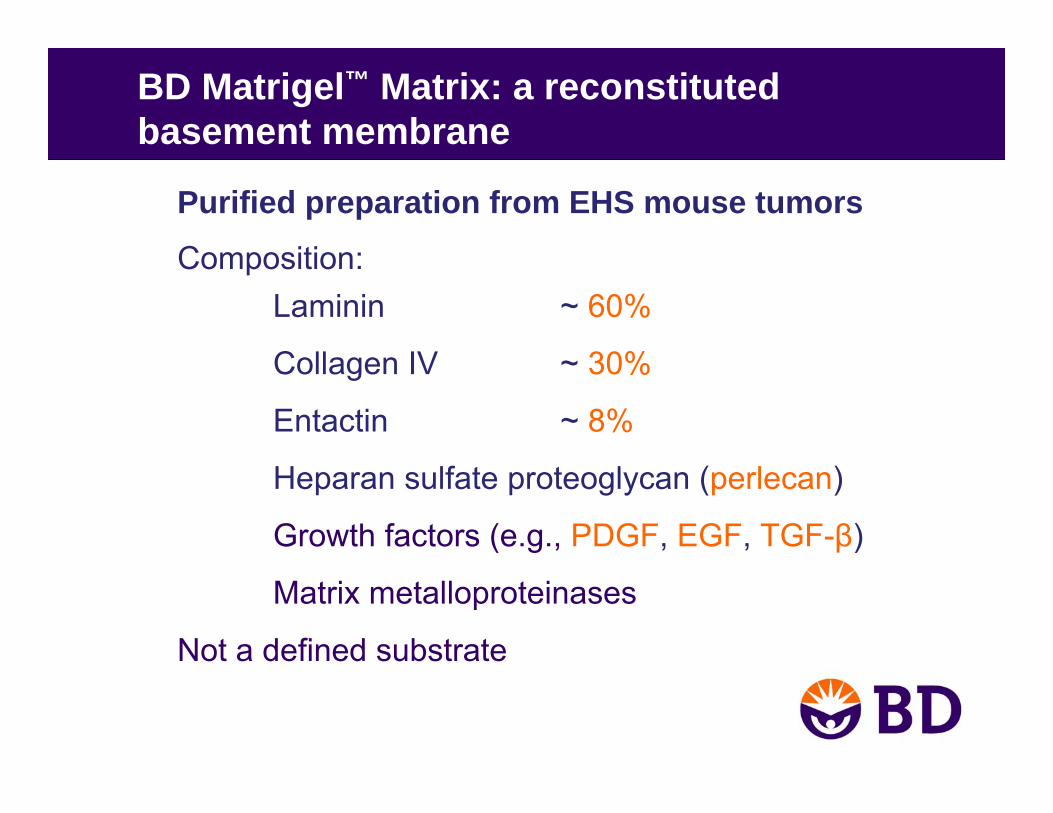
BD Matrigel™ Matrix: a reconstitutedbasement membrane
Purified preparation from EHS mouse tumors
Composition:Laminin ~ 60%
Collagen IV ~ 30%
Entactin ~ 8%
Heparan sulfate proteoglycan (perlecan)
Growth factors (e.g., PDGF, EGF, TGF-β)
Matrix metalloproteinases
Not a defined substrate
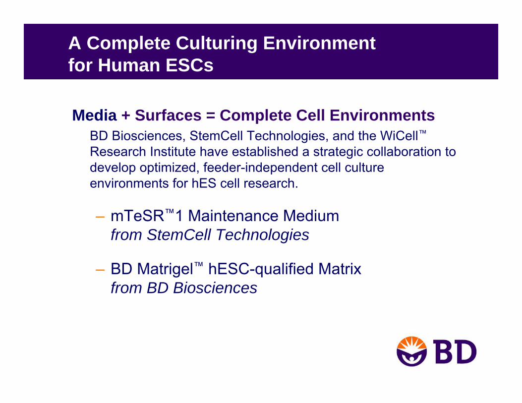
A Complete Culturing Environmentfor Human ESCs
Media + Surfaces = Complete Cell EnvironmentsBD Biosciences, StemCell Technologies, and the WiCell™Research Institute have established a strategic collaboration todevelop optimized, feeder-independent cell culture environments for hES cell research.
– mTeSR™1 Maintenance Medium from StemCell Technologies
– BD Matrigel™ hESC-qualified Matrixfrom BD Biosciences
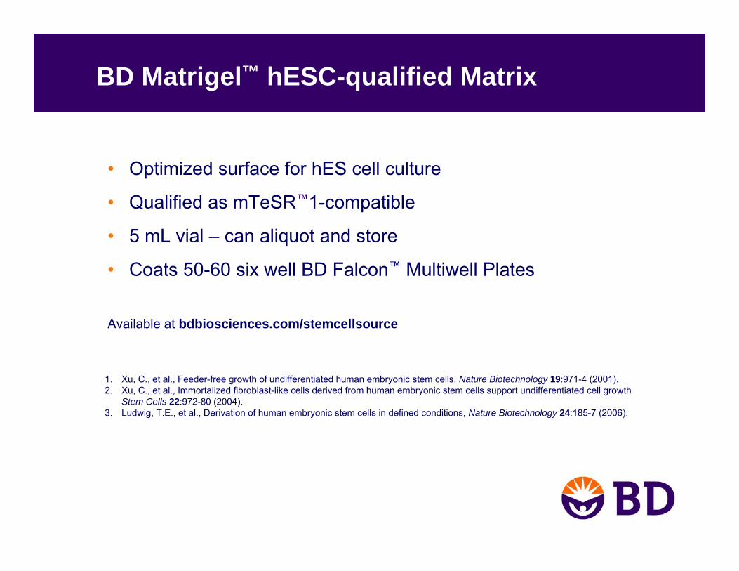
BD Matrigel™ hESC-qualified Matrix
• Optimized surface for hES cell culture
• Qualified as mTeSR™1-compatible
• 5 mL vial – can aliquot and store
• Coats 50-60 six well BD Falcon™ Multiwell Plates
Available at bdbiosciences.com/stemcellsource
1. Xu, C., et al., Feeder-free growth of undifferentiated human embryonic stem cells, Nature Biotechnology 19:971-4 (2001).2. Xu, C., et al., Immortalized fibroblast-like cells derived from human embryonic stem cells support undifferentiated cell growth
Stem Cells 22:972-80 (2004).3. Ludwig, T.E., et al., Derivation of human embryonic stem cells in defined conditions, Nature Biotechnology 24:185-7 (2006).
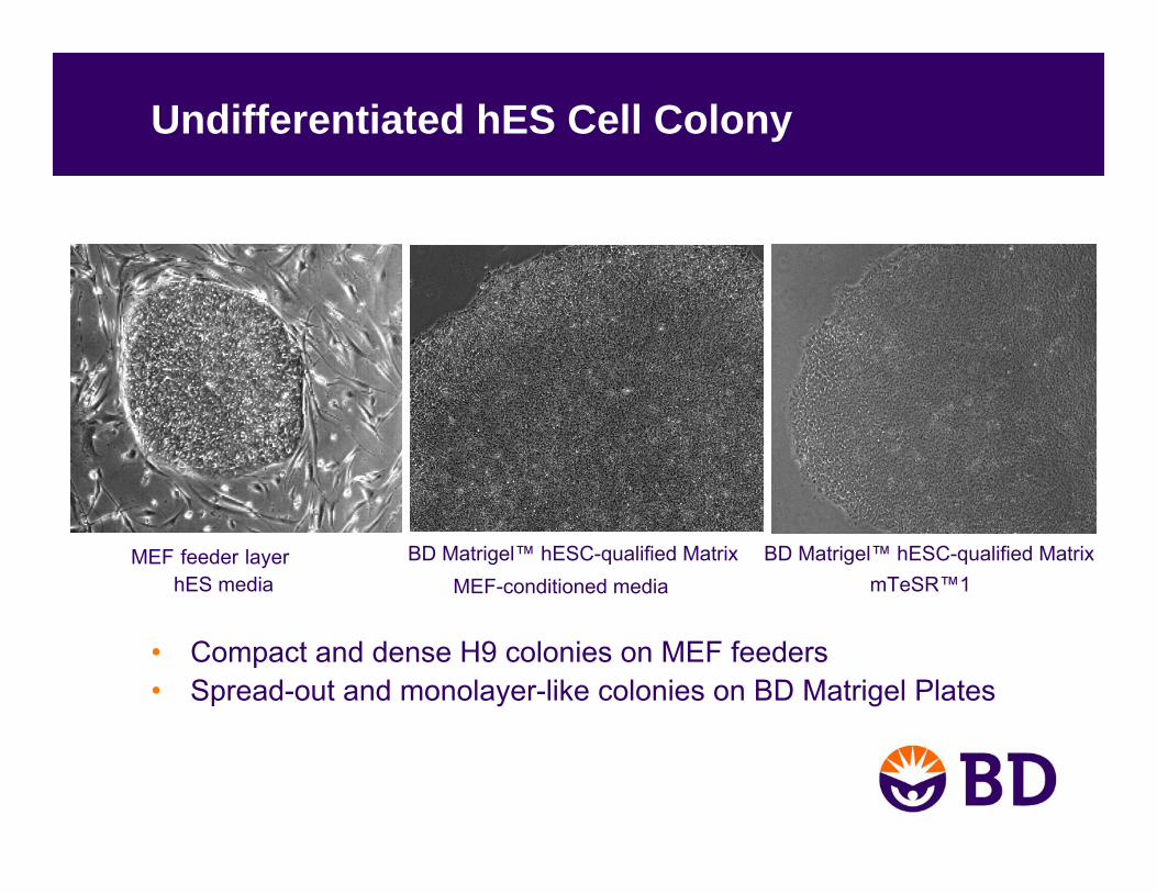
Undifferentiated hES Cell Colony
• Compact and dense H9 colonies on MEF feeders• Spread-out and monolayer-like colonies on BD Matrigel Plates
MEF-conditioned mediahES mediaMEF feeder layer BD Matrigel™ hESC-qualified Matrix BD Matrigel™ hESC-qualified Matrix
mTeSR™1
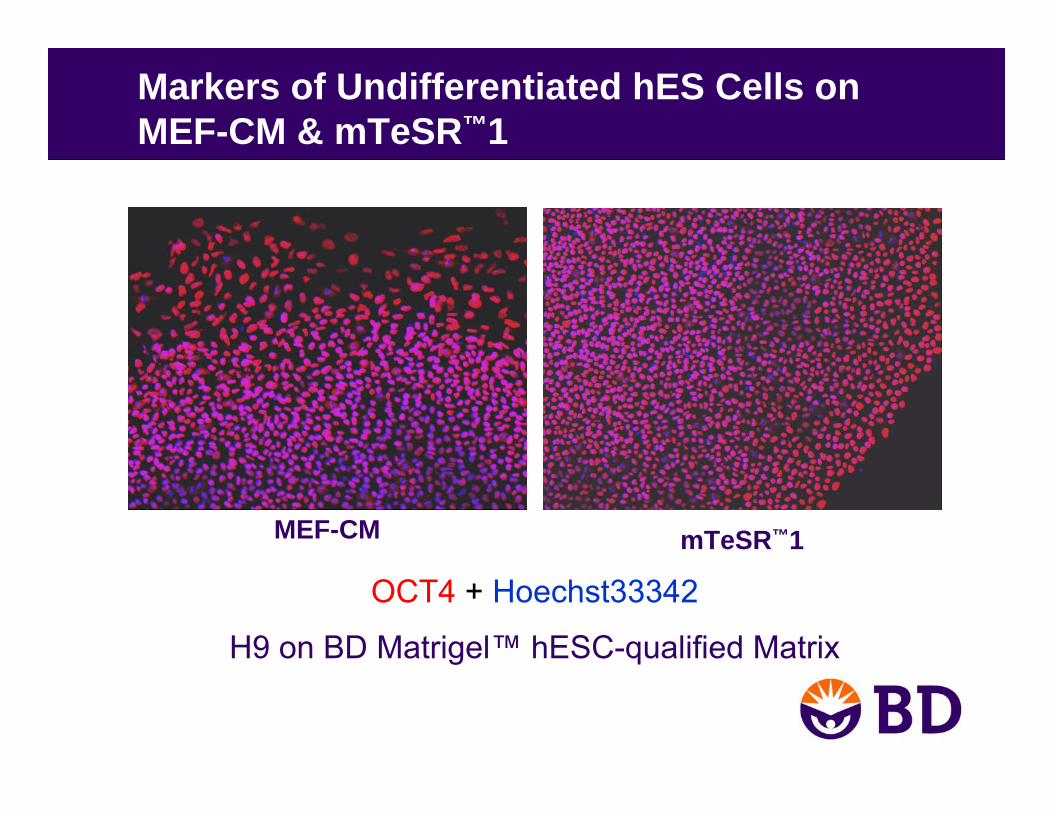
Markers of Undifferentiated hES Cells on MEF-CM & mTeSR™1
H9 on BD Matrigel™ hESC-qualified Matrix
OCT4 + Hoechst33342
mTeSR™1MEF-CM

Undifferentiated Marker Expression FACS Analysis
H9 cells grown on BD Matrigel™ hESC-qualified Matrix in mTeSR™1 for 5 passages
98% OCT4 positive cells
Isotype control
Oct-3/4
Isotype control
SSEA-4
94% SSEA4 positive cells
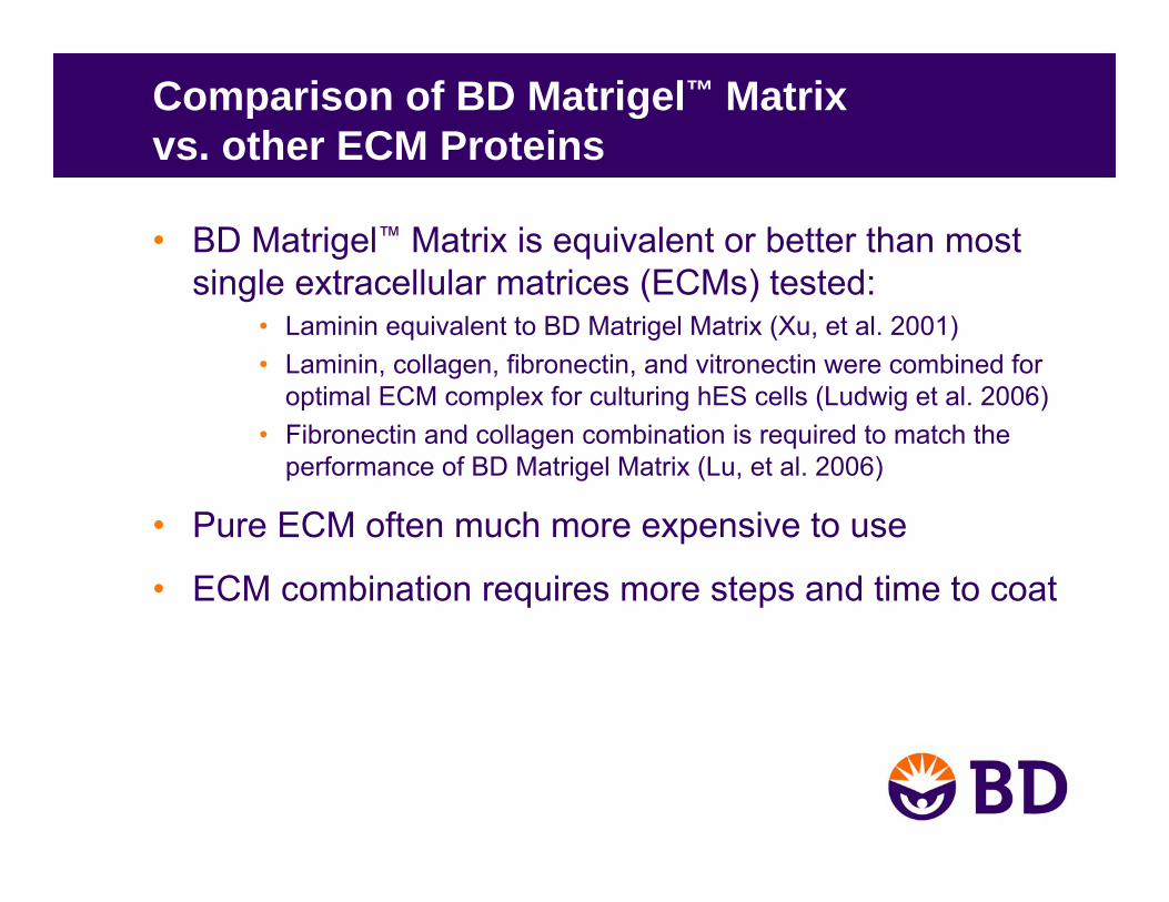
Comparison of BD Matrigel™ Matrixvs. other ECM Proteins
• BD Matrigel™ Matrix is equivalent or better than most single extracellular matrices (ECMs) tested:
• Laminin equivalent to BD Matrigel Matrix (Xu, et al. 2001)• Laminin, collagen, fibronectin, and vitronectin were combined for
optimal ECM complex for culturing hES cells (Ludwig et al. 2006)• Fibronectin and collagen combination is required to match the
performance of BD Matrigel Matrix (Lu, et al. 2006)
• Pure ECM often much more expensive to use
• ECM combination requires more steps and time to coat
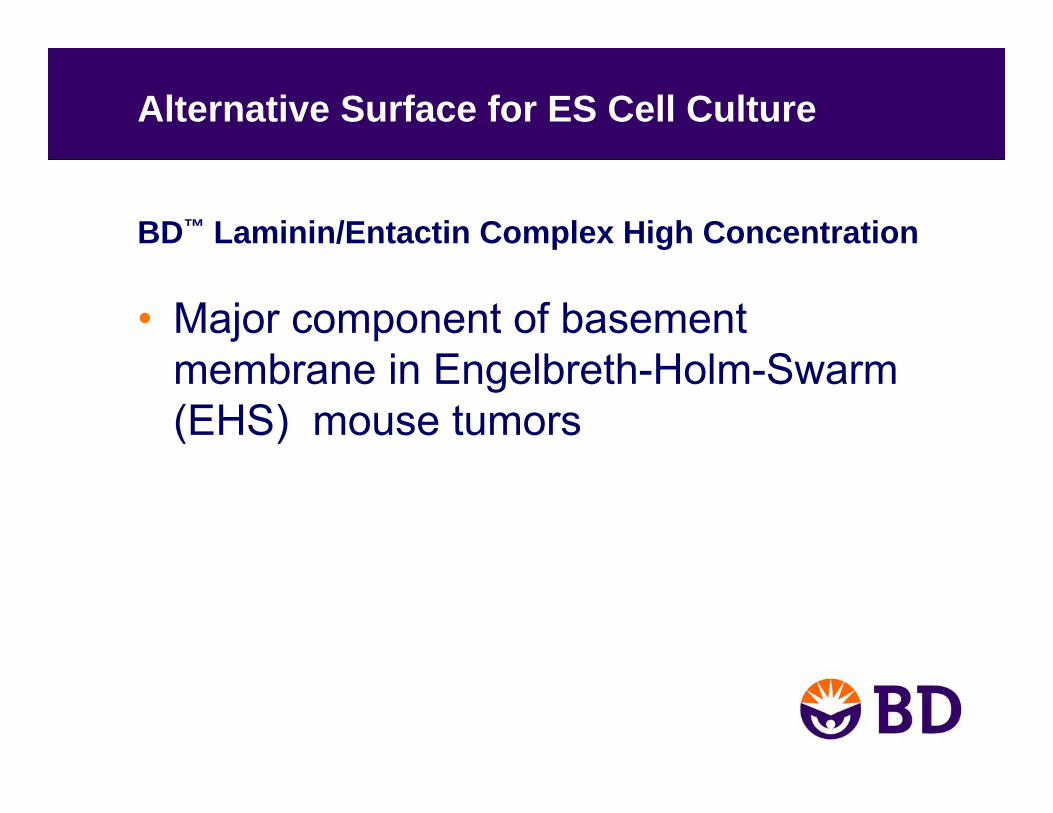
Alternative Surface for ES Cell Culture
BD™ Laminin/Entactin Complex High Concentration
• Major component of basement membrane in Engelbreth-Holm-Swarm (EHS) mouse tumors
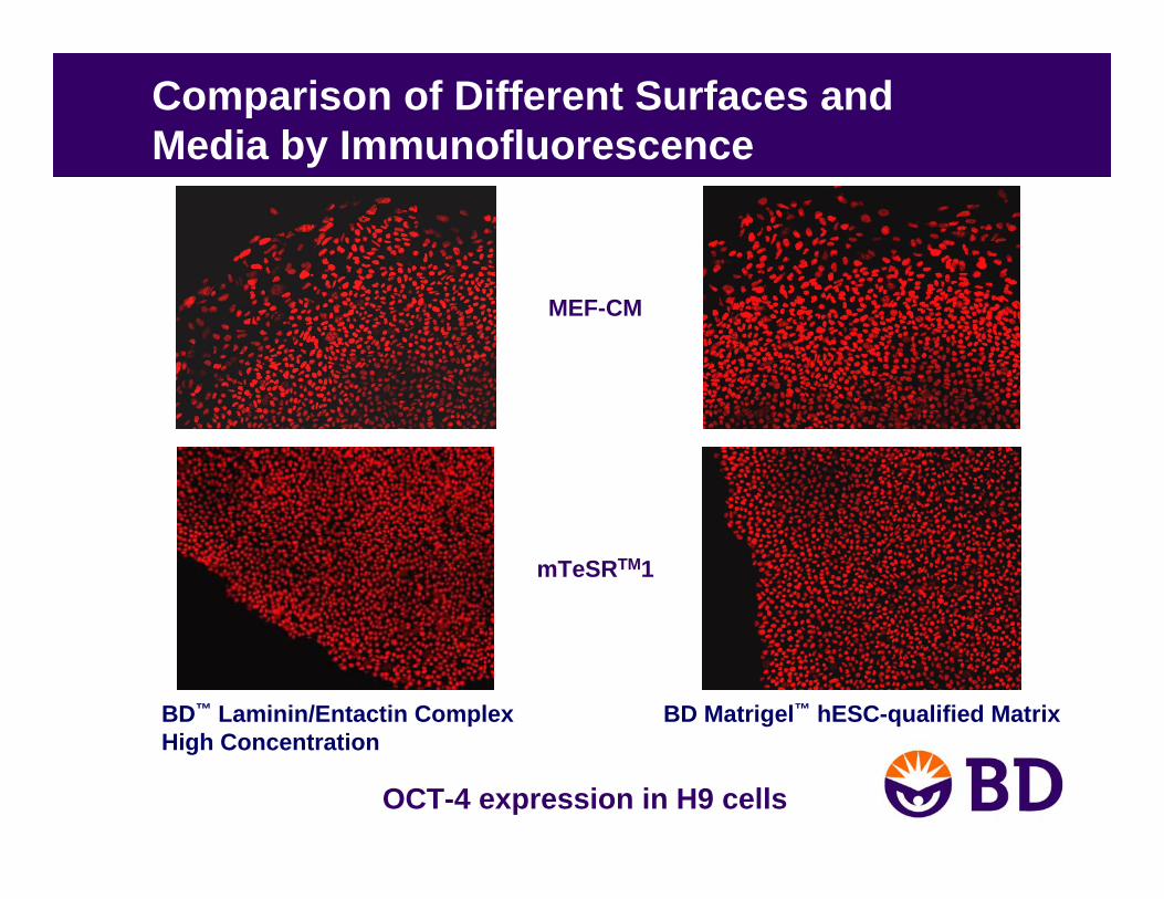
Comparison of Different Surfaces and Media by Immunofluorescence
BD™ Laminin/Entactin Complex High Concentration
BD Matrigel™ hESC-qualified Matrix
mTeSRTM1
MEF-CM
OCT-4 expression in H9 cells
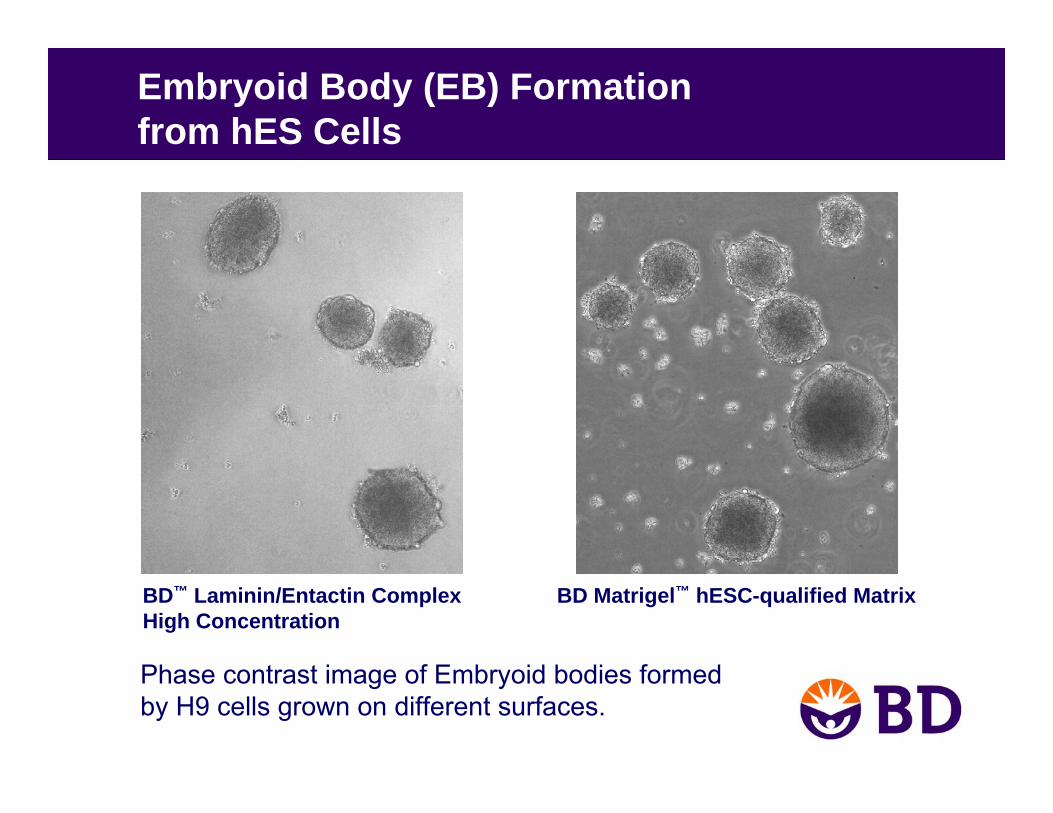
Embryoid Body (EB) Formationfrom hES Cells
Phase contrast image of Embryoid bodies formedby H9 cells grown on different surfaces.
BD Matrigel™ hESC-qualified MatrixBD™ Laminin/Entactin ComplexHigh Concentration

Neurons and Cardiomyocytesfrom H9-derived Embryoid Bodies
EBs derived from H9 cells cultured on BD™ Laminin/Entactin Complex High Concentration for 32 passages in mTeSR™1 media
Cardiomyocytes
Neurons Nestin
GATA-4
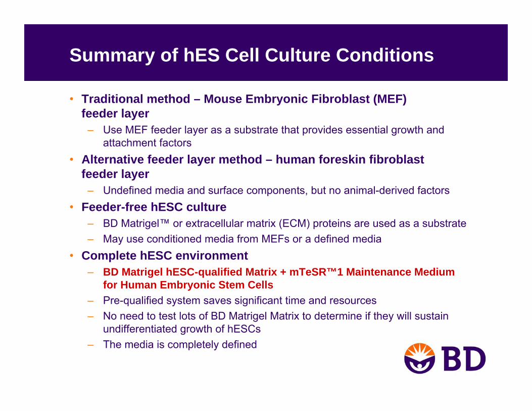
Summary of hES Cell Culture Conditions
• Traditional method – Mouse Embryonic Fibroblast (MEF)feeder layer
– Use MEF feeder layer as a substrate that provides essential growth and attachment factors
• Alternative feeder layer method – human foreskin fibroblastfeeder layer
– Undefined media and surface components, but no animal-derived factors• Feeder-free hESC culture
– BD Matrigel™ or extracellular matrix (ECM) proteins are used as a substrate– May use conditioned media from MEFs or a defined media
• Complete hESC environment– BD Matrigel hESC-qualified Matrix + mTeSR™1 Maintenance Medium
for Human Embryonic Stem Cells– Pre-qualified system saves significant time and resources– No need to test lots of BD Matrigel Matrix to determine if they will sustain
undifferentiated growth of hESCs– The media is completely defined

Embryonic vs. Adult Stem Cells
• Embryonic Stem Cells– Pluripotent– Relatively easy to grow in culture
• Adult Stem Cells– Multipotent– Difficult to isolate, purify and maintain in
the undifferentiated state
Characteristics of Adult Stem Cells
• Adult stem cells are found in many tissues– They are undifferentiated cells found among differentiated cells.
• Their primary role in the body is to maintain and repair the tissue in which they are found.
• Adult stem cells are multipotent, not pluripotent– Pluripotent: can differentiate into any cell type in the embryo– Multipotent: can differentiate into a subset of cell types, but NOT
a complete organism
• Adult stem cells may exhibit plasticity
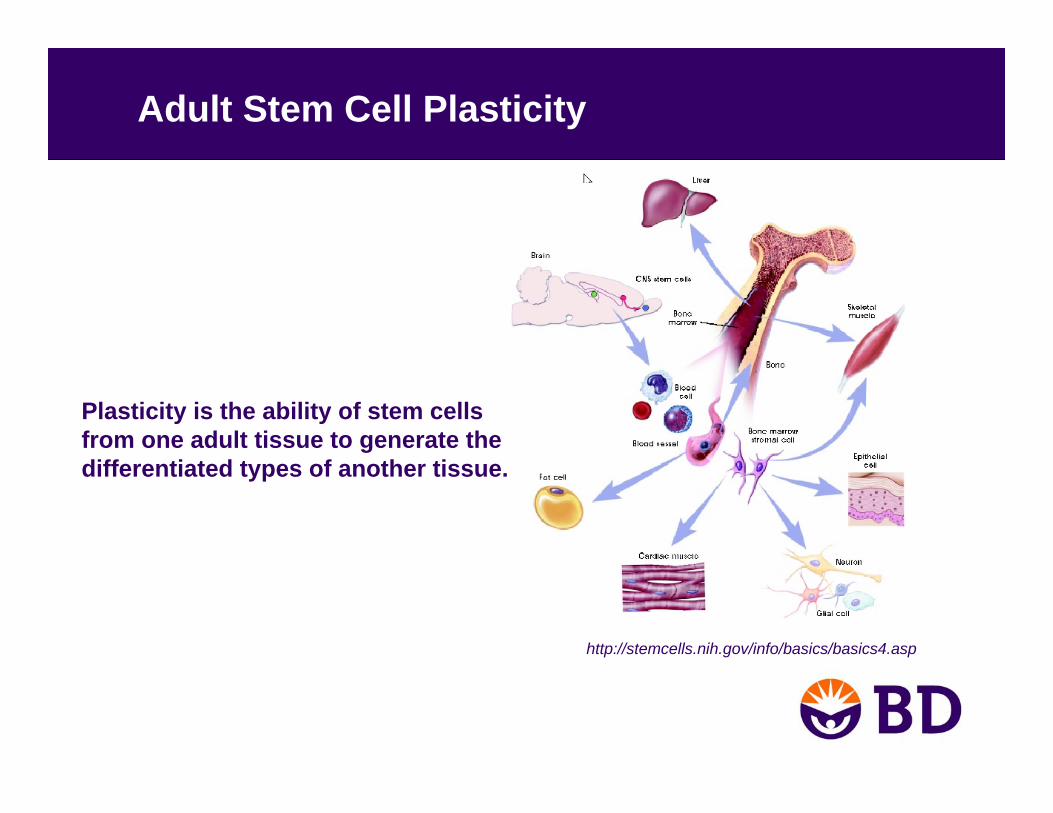
Adult Stem Cell Plasticity
Plasticity is the ability of stem cells from one adult tissue to generate the differentiated types of another tissue.
http://stemcells.nih.gov/info/basics/basics4.asp
Adult stem cells
• Hematopoietic• Mesenchymal• Neuronal

Hematopoietic Stem Cells (HSCs)
• Source: bone marrow
• Differentiation pathways– Ex. T cell, B cell, Erythrocyte
• Culture conditions– Maintenance of undifferentiated HSCs
• Ex. stem cell factor (SCF), IL-3, IL-6– Differentiation
• Ex. EPO, G-CSF
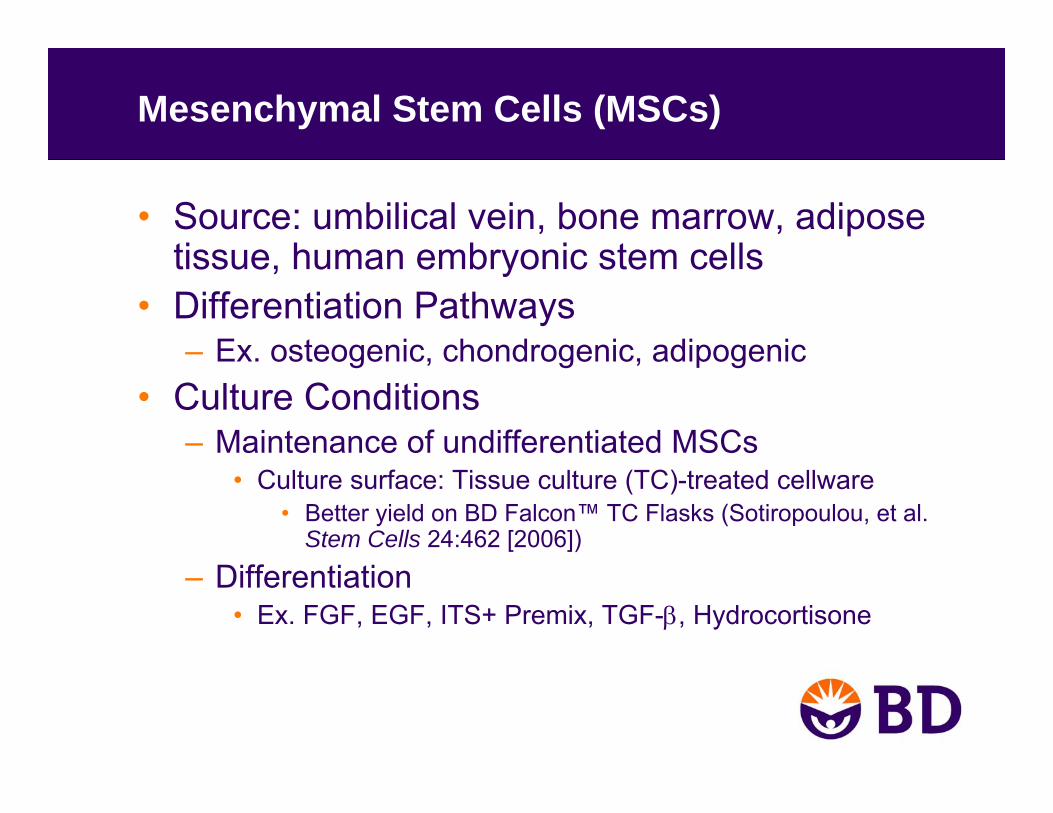
Mesenchymal Stem Cells (MSCs)
• Source: umbilical vein, bone marrow, adipose tissue, human embryonic stem cells
• Differentiation Pathways– Ex. osteogenic, chondrogenic, adipogenic
• Culture Conditions– Maintenance of undifferentiated MSCs
• Culture surface: Tissue culture (TC)-treated cellware• Better yield on BD Falcon™ TC Flasks (Sotiropoulou, et al.
Stem Cells 24:462 [2006])
– Differentiation• Ex. FGF, EGF, ITS+ Premix, TGF-β, Hydrocortisone
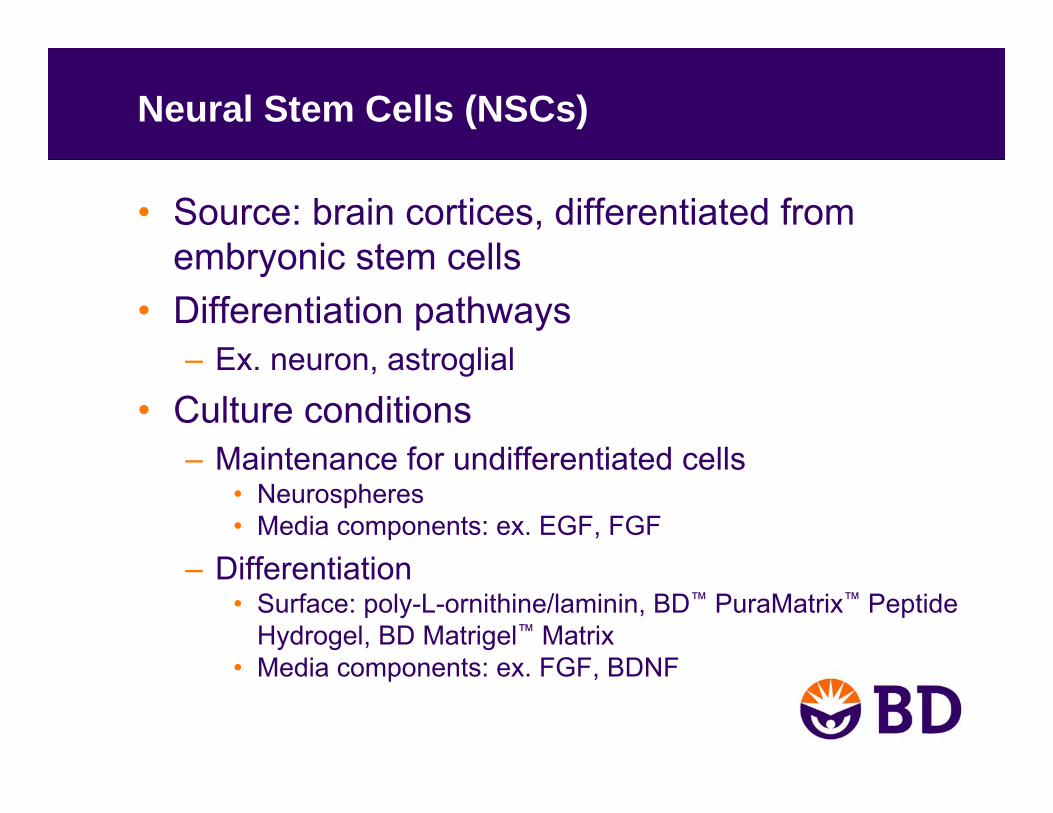
Neural Stem Cells (NSCs)
• Source: brain cortices, differentiated from embryonic stem cells
• Differentiation pathways– Ex. neuron, astroglial
• Culture conditions– Maintenance for undifferentiated cells
• Neurospheres• Media components: ex. EGF, FGF
– Differentiation• Surface: poly-L-ornithine/laminin, BD™ PuraMatrix™ Peptide
Hydrogel, BD Matrigel™ Matrix• Media components: ex. FGF, BDNF
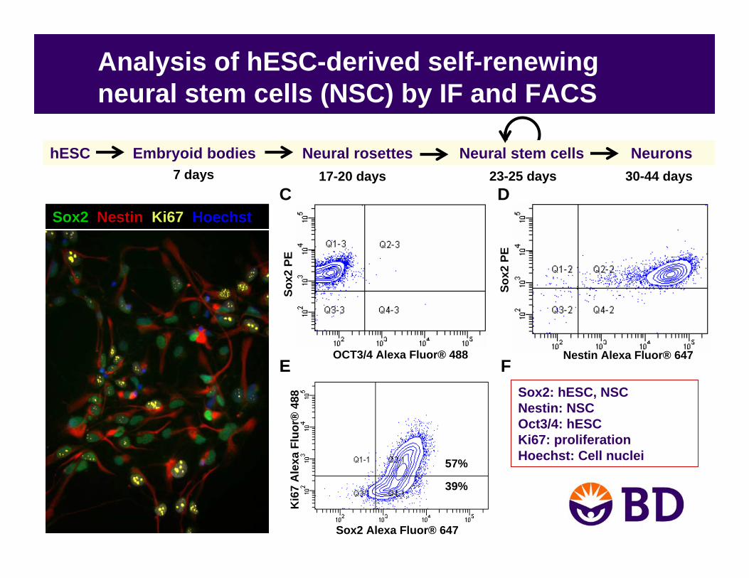
Analysis of hESC-derived self-renewing neural stem cells (NSC) by IF and FACS
Sox2
PE
OCT3/4 Alexa Fluor® 488
Sox2
PE
Nestin Alexa Fluor® 647
Sox2 Nestin Ki67 Hoechst
Ki6
7 A
lexa
Fluo
r®48
8
Sox2 Alexa Fluor® 647
57%
39%
Sox2: hESC, NSCNestin: NSCOct3/4: hESCKi67: proliferationHoechst: Cell nuclei
C D
E F
hESC Embryoid bodies Neural rosettes Neural stem cells Neurons 7 days 17-20 days 23-25 days 30-44 days

Summary
• Embryonic stem cells– Pluripotent– Alternatives: SCNT and iPS cells– Multiple culture methods
• Feeder layer• BD Matrigel™ Matrix• Defined ECM
• Adult stem cells– Multipotent– Culture environment (surface and media) specific
to cell type and differentiation pathway
References – hES and iPS Cells
hES and iPS cells– Xu, et al. Feeder-free growth of undifferentiated human embryonic stem
cells. Nature 19:971 (2001).– Ludwig, et al. Feeder-independent culture of human embryonic stem cells.
Nat. Methods 3(8):637 (2006).– Ludwig, et al. Derivation of human embryonic stem cells in defined
conditions. Nat. Biotechnology 24(2):185 (2006).– Takahashi, et al. Induction of pluripotent stem cells from adult human
fibroblasts by defined factors. Cell 131:1 (2007).– Yu, et al. Induced pluripotent stem cell lines derived from human somatic
cells. Science 318:1917 (2007).– Dimos, et al. Induced pluripotent stem cells generated from patients with
ALS can be differentiated into motor neurons. Science 321:1218 (2008).– Park, et al. Disease-specific induced pluripotent stem cells. Cell 134:1
(2008).
References – Adult Stem CellsHSCs
– Petzer, et al. Self renewal of primitive human hematopoietic cells (long-term-culture-initiating cells) in vitro and their expansion in defined medium. PNAS 93:1470 (1996).
– Bhatia, et al. Quantitative analysis reveals expansion of human hematopoietic repopulating cells after short-term ex vivo culture. J. Exp. Med. 186:619 (1997).
– Kang, et al. A novel function of interleukin-10 promoting self-renewal of hematopoietic stem cells. Stem Cells25:1814 (2007).
– Wilson & Trumpp Bone-marrow haematopoietic-stem-cell niches. Nat. Rev. Immunol. 6:93– Staal, et al (2008) WNT signalling in the immune system: WNT is spreading its wings. Nat. Rev. Immunol. 8:581
(2006).– O’Connell, et al. Sustained expression of microRNA-155 in hematopoietic stem cells causes a myeloproliferative
disorder. J. Exp. Med. 205:585 (2008).MSCs
– Pittenger, et al. Multilineage potential of adult human mesenchymal stem cells. Science 284:143 (1999).– Lee, et al. Isolation of multipotent mesenchymal stem cells from umbilical cord blood. Blood 103:1669 (2004).– Sotiropoulou, et al. Characterization of the optimal culture conditions for clinical scale production of human
mesenchymal stem cells. Stem Cells 24:462 (2006).– Uccelli et al. Mesenchymal stem cells in health and disease. Nat. Rev. Immunol. 8:726 (2008).
NSCs– Flanagan, et al. Regulation of human neural precursor cells by laminin and integrins. J. Neurosci. Res. 83:845 (2006).– Malaterre, et al. c-Myb is required for neural progenitor cell proliferation and maintenance of neural stem cell niche
in adult brain. Stem Cells 26:173 (2008).– Thornhoff, et al. Compatibility of human fetal neural stem cells with hydrogel biomaterials in vitro. Brain Res.
1187:42 (2008).

Acknowledgements
Susan QianSuparna SanyalDeepa SaxenaJeff PartridgeJennifer BrownChristian Carson

Contact Information
Amy Laws, Ph.D.Technical Support Representative tel: 978-901-7213 [email protected]
PuraMatrix is a registered trademark of 3DM Inc.StemCell Technologies and all other StemCell trademarks are the property of StemCell Technologies Inc.WiCell™ logo, mTeSR,TeSR and all other WiCell trademarks are the property of WiCell Research Institute.BD, BD logo, and BD Matrigel are trademarks of Becton, Dickinson and Company. © 2008