Embed Size (px)
Citation preview
CTV, MRV, Venography and
IVUS for CVD
Peter Neglén, MD, PhDVascular Surgeon
Cyprus

Disclosure
Peter Neglen, M.D., PhD, FACS
I disclose the following financial relationship(s):
•Ownership Interest: Veniti Ltd;
•Consultant/Advisory Board: Angiodynamics
Stockholder of Veniti, Inc.
Member of SAB, AngioDynamics
Wallstents and nitenol stents are used “off-label,” e.g.,
the use for iliac venous stenting is not described on the
product’s label.
Faculty DisclosurePeter Neglén, M.D., Ph.D
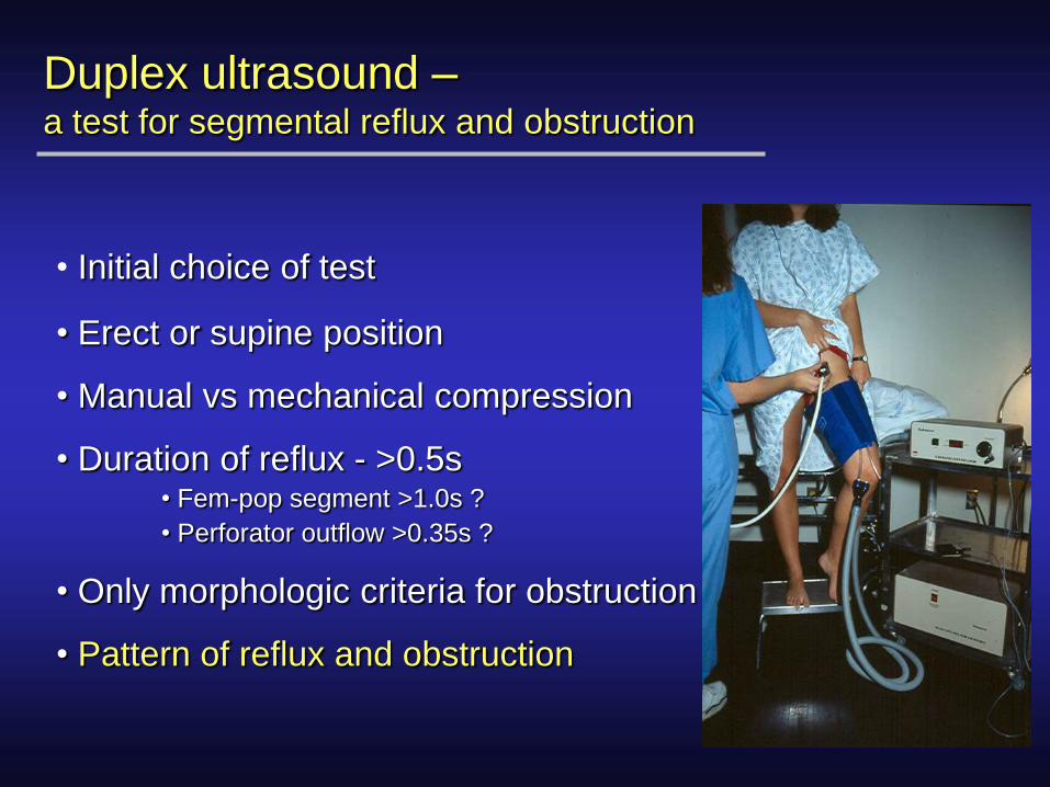
Duplex ultrasound –a test for segmental reflux and obstruction
• Initial choice of test
• Erect or supine position
• Manual vs mechanical compression
• Duration of reflux - >0.5s• Fem-pop segment >1.0s ?
• Perforator outflow >0.35s ?
• Only morphologic criteria for obstruction
• Pattern of reflux and obstruction
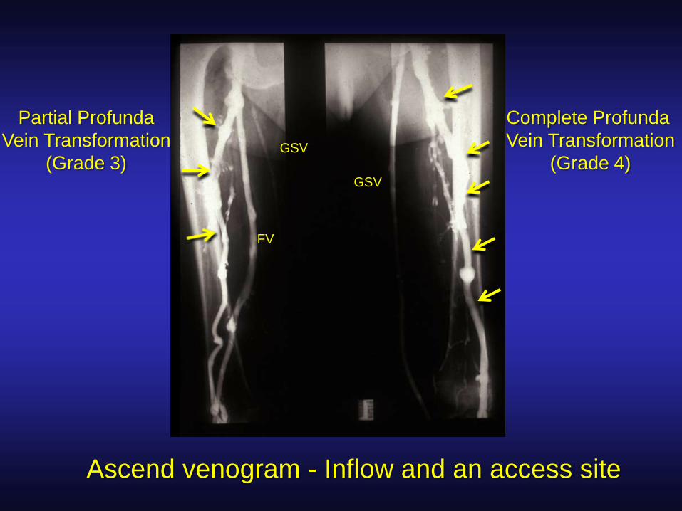
Complete Profunda
Vein Transformation
(Grade 4)
Partial Profunda
Vein Transformation
(Grade 3)
FV
GSV
GSV
Ascend venogram - Inflow and an access site
How do I find patients with femoro-ilio-
caval obstruction?
• At what degree a venous obstruction is hemodynamically significant is not defined!
• Not possible to hemodynamically quantify venous outflow obstruction!
• No reliable non-invasive study is available!
• Invasive pressure tests are insensitive!• hand/foot pressure differential
• reactive hyperemia pressure increase
• femoral vein pressure
• femoro-caval vein gradient
Morphologic Diagnosis
• Dx is morphologic, not hemodynamic
• Ultrasound scanning of the lower extremity has to
be complemented by transfemoral venography,
MRV, CT-V or IVUS in C3-6 cases
• IVUS is the standard for all other imaging
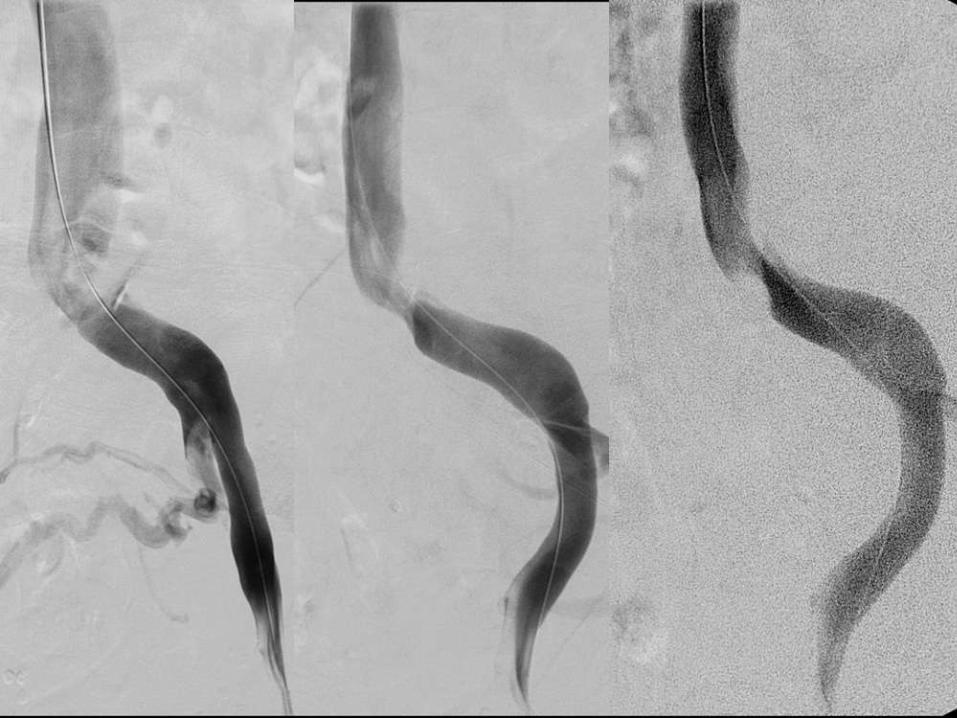
Transfemoral venogram
Multi-plane venograms
MRV
CTV
IVUS-verified area stenosis of >50% is considered
significant
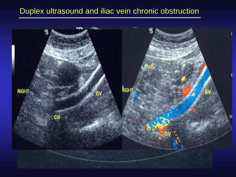
Duplex ultrasound and iliac vein chronic obstruction
>50% stenosis = post/pre stenotic peak velocity ratio >2.5[Labropoulos et al, J Vasc Surg 2007;46:101-7]
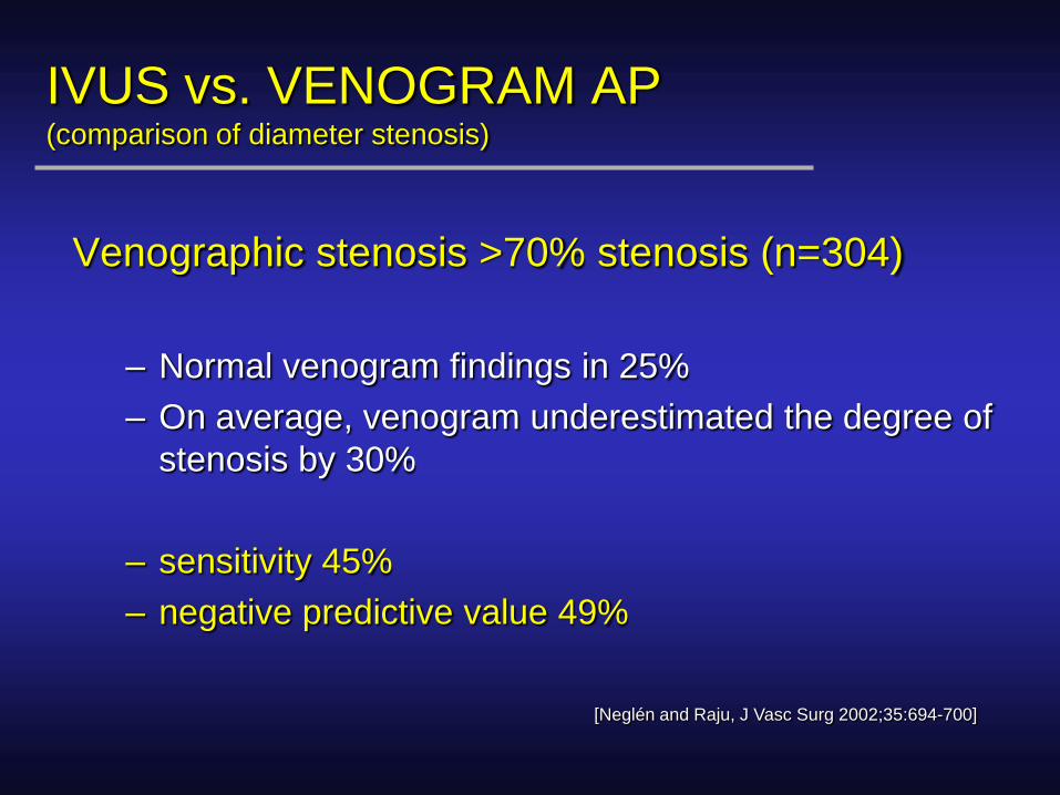
IVUS vs. VENOGRAM AP(comparison of diameter stenosis)
Venographic stenosis >70% stenosis (n=304)
– Normal venogram findings in 25%
– On average, venogram underestimated the degree of
stenosis by 30%
– sensitivity 45%
– negative predictive value 49%
[Neglén and Raju, J Vasc Surg 2002;35:694-700]
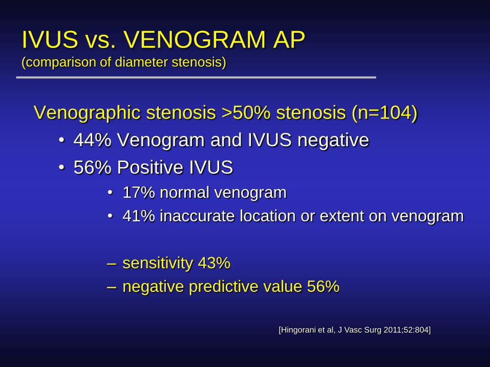
IVUS vs. VENOGRAM AP(comparison of diameter stenosis)
Venographic stenosis >50% stenosis (n=104)
• 44% Venogram and IVUS negative
• 56% Positive IVUS
• 17% normal venogram
• 41% inaccurate location or extent on venogram
– sensitivity 43%
– negative predictive value 56%
[Hingorani et al, J Vasc Surg 2011;52:804]
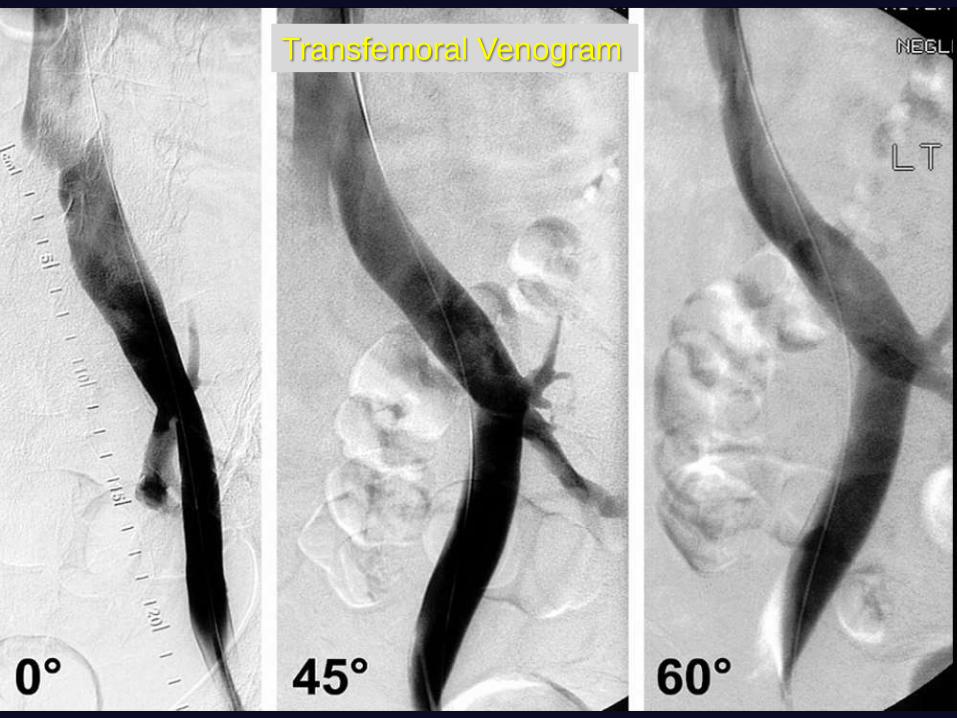
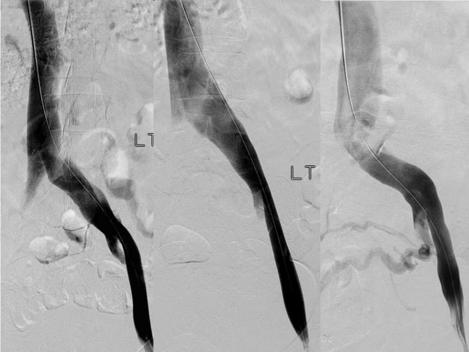
Transfemoral Venogram
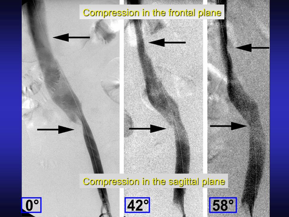
Compression in the frontal plane
Compression in the sagittal plane
AV
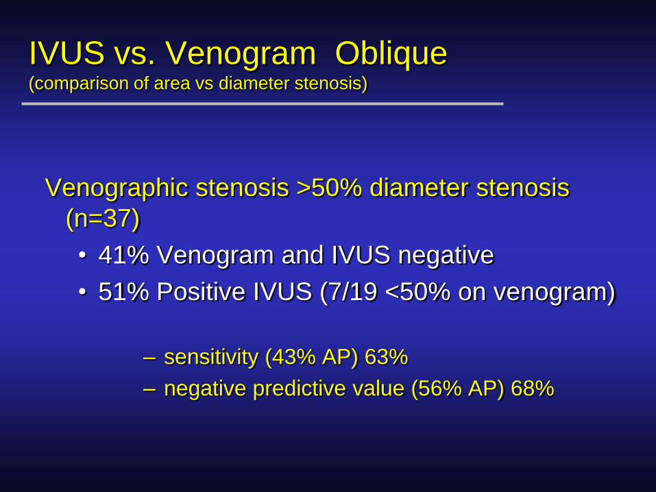
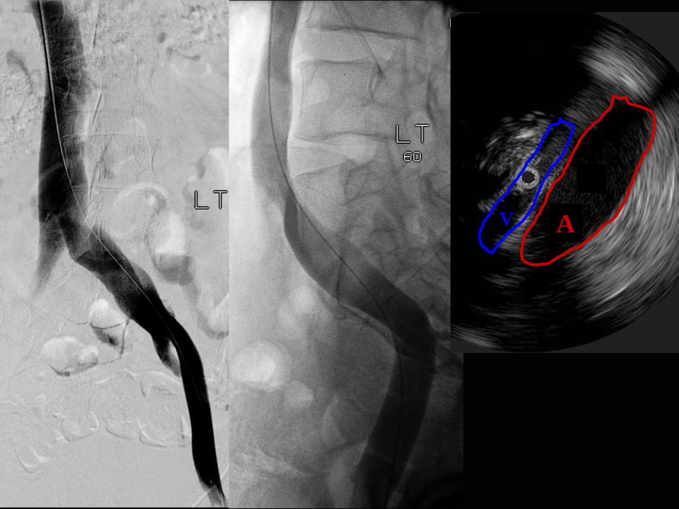
IVUS vs. Venogram Oblique(comparison of area vs diameter stenosis)
Venographic stenosis >50% diameter stenosis
(n=37)
• 41% Venogram and IVUS negative
• 51% Positive IVUS (7/19 <50% on venogram)
– sensitivity (43% AP) 63%
– negative predictive value (56% AP) 68%
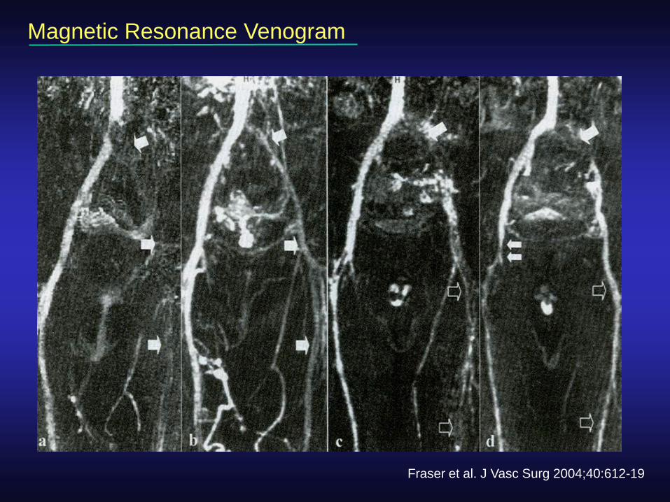
Magnetic Resonance Venogram
Fraser et al. J Vasc Surg 2004;40:612-19
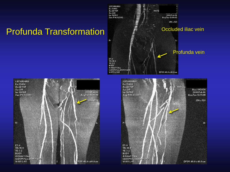
Occluded iliac vein
Profunda vein
Profunda Transformation
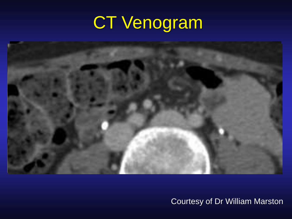
CT Venogram
Courtesy of Dr William Marston
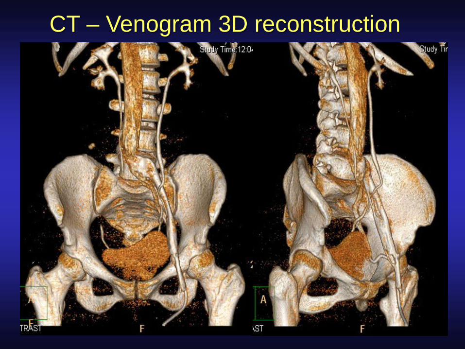
CT – Venogram 3D reconstruction
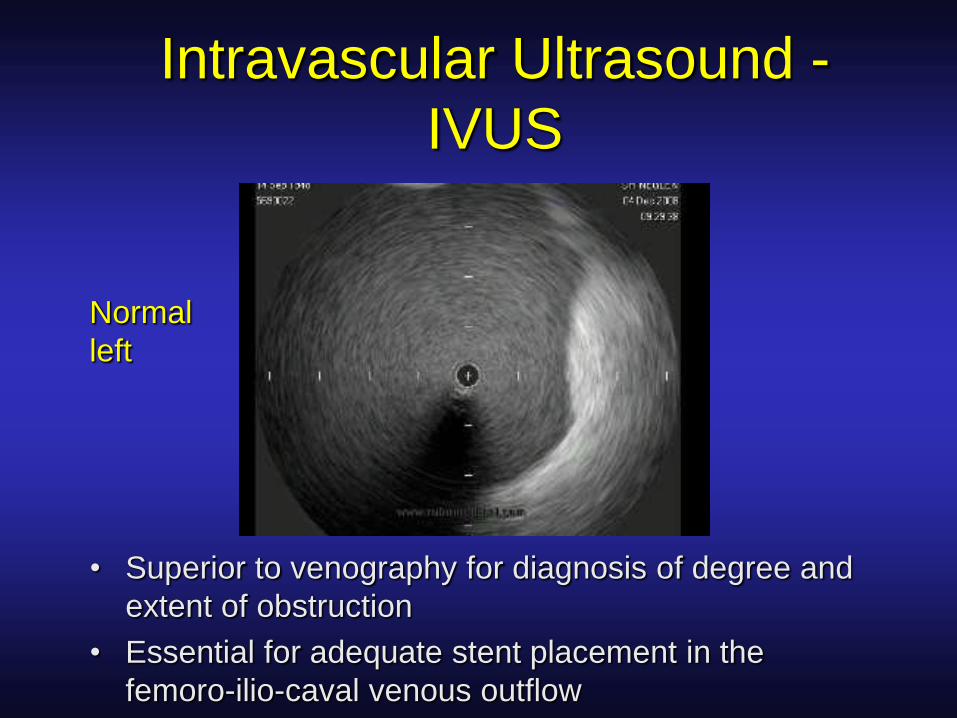
Intravascular Ultrasound -
IVUS
• Superior to venography for diagnosis of degree and
extent of obstruction
• Essential for adequate stent placement in the
femoro-ilio-caval venous outflow
Normal
left
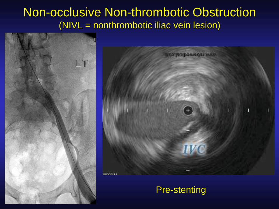
Non-occlusive Non-thrombotic Obstruction(NIVL = nonthrombotic iliac vein lesion)
Pre-stenting
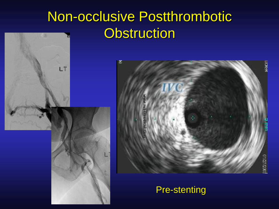
Non-occlusive Postthrombotic
Obstruction
Pre-stenting
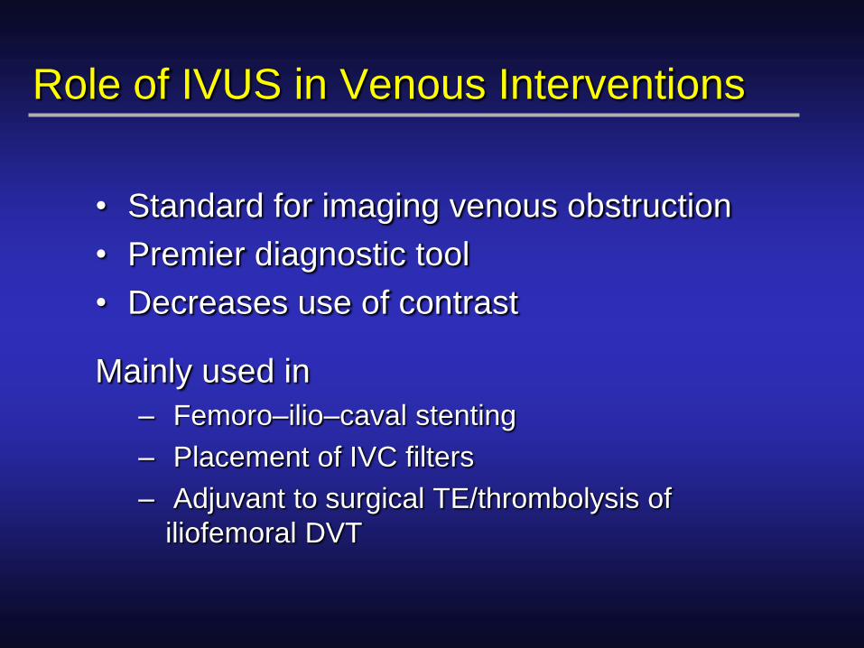
Role of IVUS in Venous Interventions
• Standard for imaging venous obstruction
• Premier diagnostic tool
• Decreases use of contrast
Mainly used in
– Femoro–ilio–caval stenting
– Placement of IVC filters
– Adjuvant to surgical TE/thrombolysis of
iliofemoral DVT
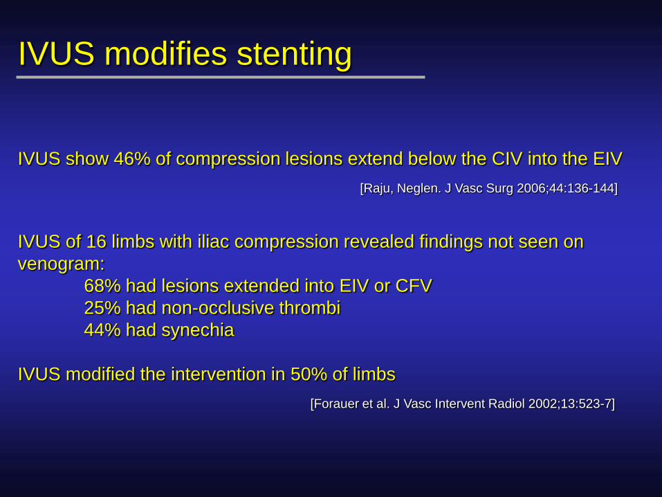
IVUS modifies stenting
IVUS show 46% of compression lesions extend below the CIV into the EIV
[Raju, Neglen. J Vasc Surg 2006;44:136-144]
IVUS of 16 limbs with iliac compression revealed findings not seen on
venogram:
68% had lesions extended into EIV or CFV
25% had non-occlusive thrombi
44% had synechia
IVUS modified the intervention in 50% of limbs
[Forauer et al. J Vasc Intervent Radiol 2002;13:523-7]
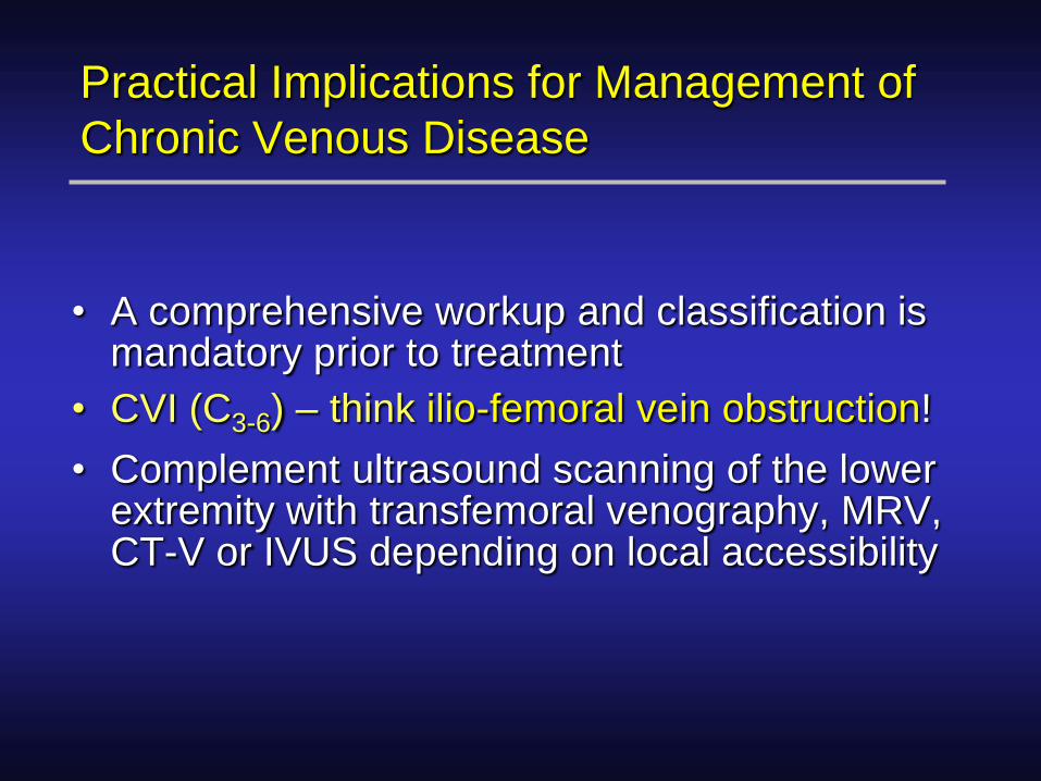
Practical Implications for Management of
Chronic Venous Disease
• A comprehensive workup and classification is mandatory prior to treatment
• CVI (C3-6) – think ilio-femoral vein obstruction!
• Complement ultrasound scanning of the lower extremity with transfemoral venography, MRV, CT-V or IVUS depending on local accessibility

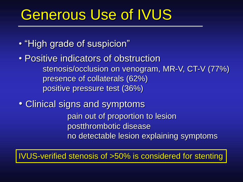
Generous Use of IVUS
• “High grade of suspicion”
• Positive indicators of obstructionstenosis/occlusion on venogram, MR-V, CT-V (77%)
presence of collaterals (62%)
positive pressure test (36%)
• Clinical signs and symptoms
pain out of proportion to lesion
postthrombotic disease
no detectable lesion explaining symptoms
IVUS-verified stenosis of >50% is considered for stenting
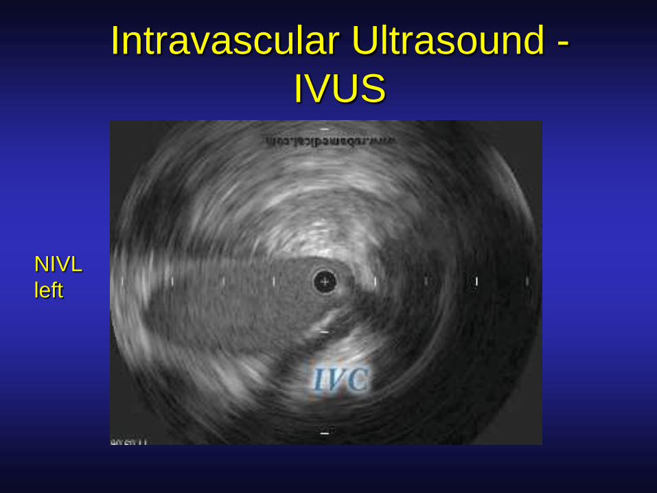
Intravascular Ultrasound -
IVUS
NIVL
left
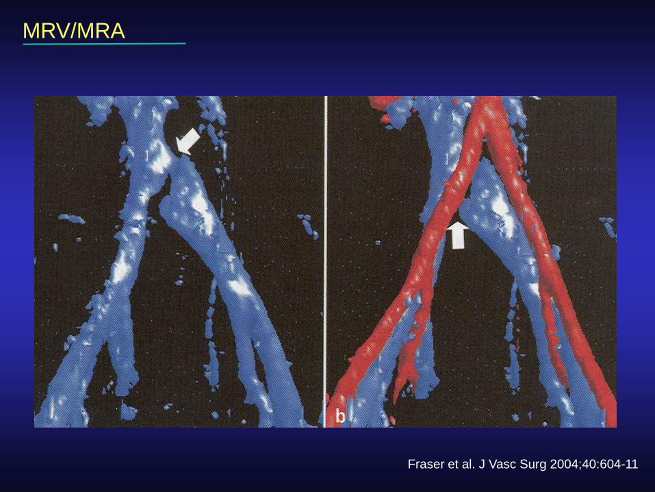
MRV/MRA
Fraser et al. J Vasc Surg 2004;40:604-11