Embed Size (px)
Citation preview

Technology Assessment Institute: Summit on CT Dose
CT Perfusion: How to do it right
Rajiv Gupta, PhD, MD
Neuroradiology
Massachusetts General Hospital
Harvard Medical School
Technology Assessment Institute: Summit on CT Dose
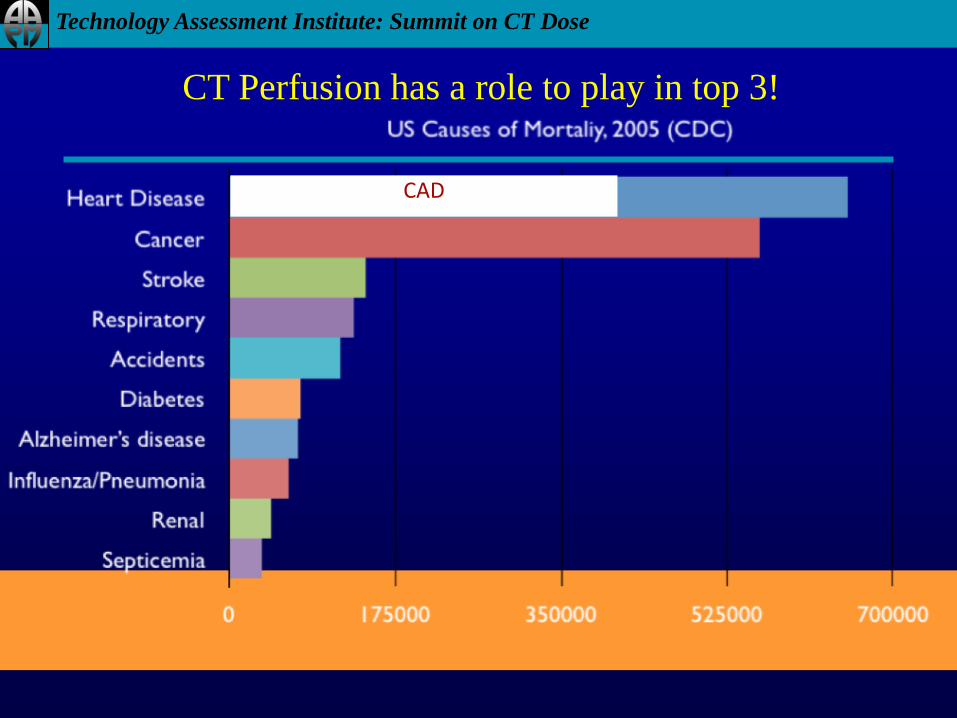
CAD
CT Perfusion has a role to play in top 3!

Technology Assessment Institute: Summit on CT Dose
Outline
• Application domains
– Stroke imaging
– Vasospasm
– Myocardial Imaging
– Tumor Imaging
• Basic CT Perfusion
Paradigm
• Neuro Perfusion
– Motivation
– Technique
– Artifacts and Pitfalls
– Dose Issues
• Myocardial Perfusion
– Motivation
– Technique
– Artifacts and Pitfalls
– Dose Issues
Technology Assessment Institute: Summit on CT Dose
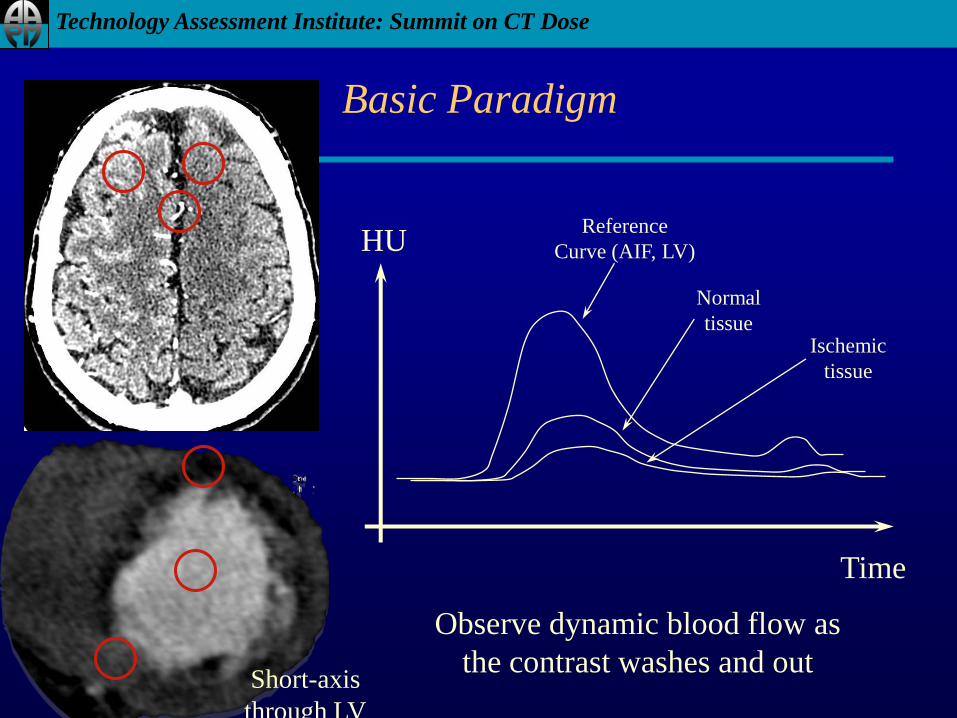
Basic Paradigm
HU
Time
Reference
Curve (AIF, LV)
Normal
tissueIschemic
tissue
Observe dynamic blood flow as
the contrast washes and out Short-axis
through LV
Technology Assessment Institute: Summit on CT Dose
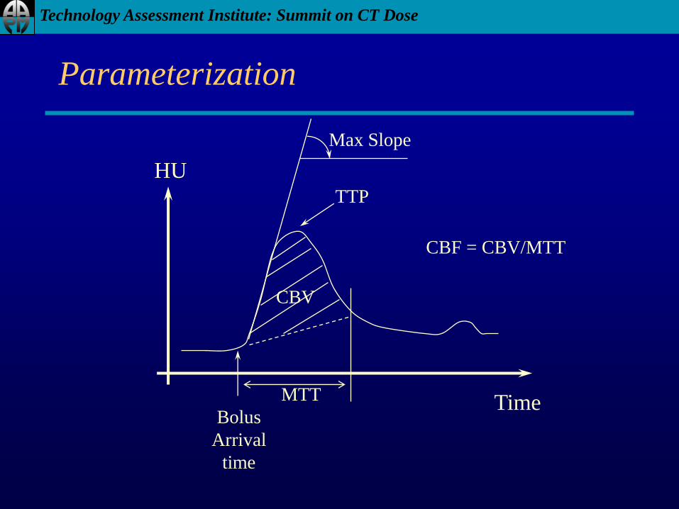
Parameterization
HU
Time
TTP
CBV
Bolus
Arrival
time
Max Slope
MTT
CBF = CBV/MTT

Technology Assessment Institute: Summit on CT Dose
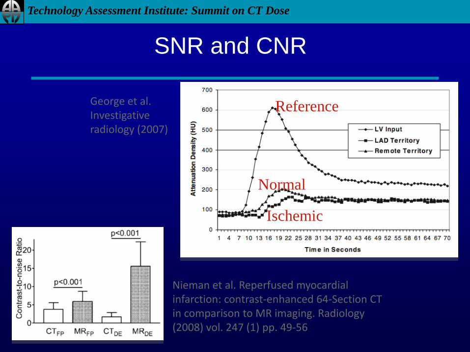
George et al. Quantification of myocardial perfusion using dynamic 64-detector computed tomography. Investigative radiology (2007) vol. 42 (12) pp. 815-22
Density = [Iodine] = Blood Flow
Technology Assessment Institute: Summit on CT Dose
George et al. Investigative radiology (2007)
SNR and CNR
Nieman et al. Reperfused myocardial infarction: contrast-enhanced 64-Section CT in comparison to MR imaging. Radiology (2008) vol. 247 (1) pp. 49-56
Reference
Normal
Ischemic
Technology Assessment Institute: Summit on CT Dose
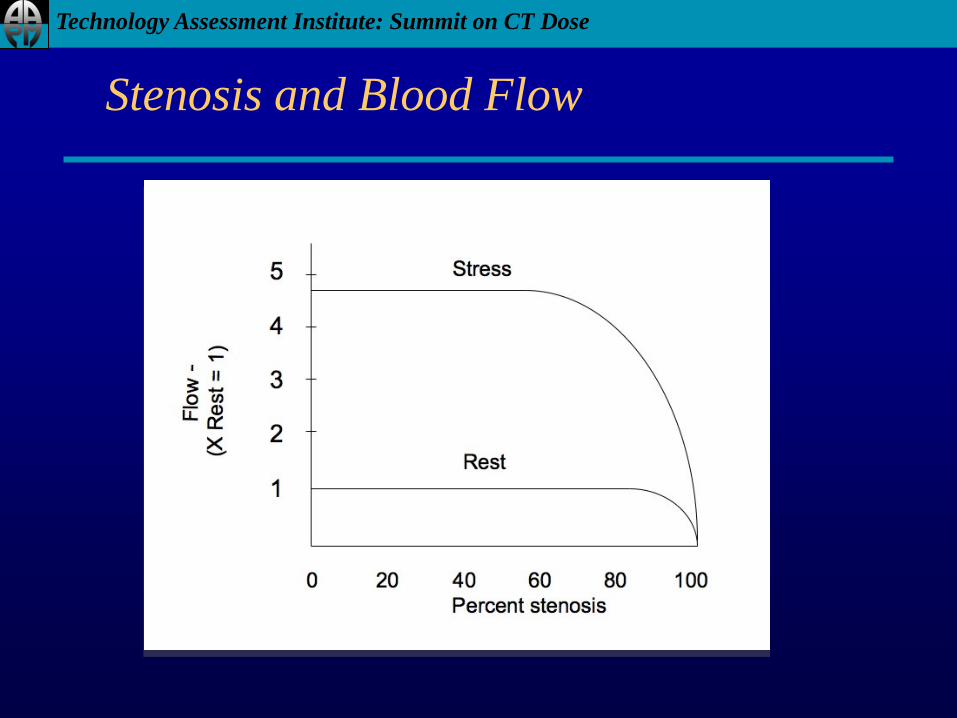
Stenosis and Blood Flow
Technology Assessment Institute: Summit on CT Dose
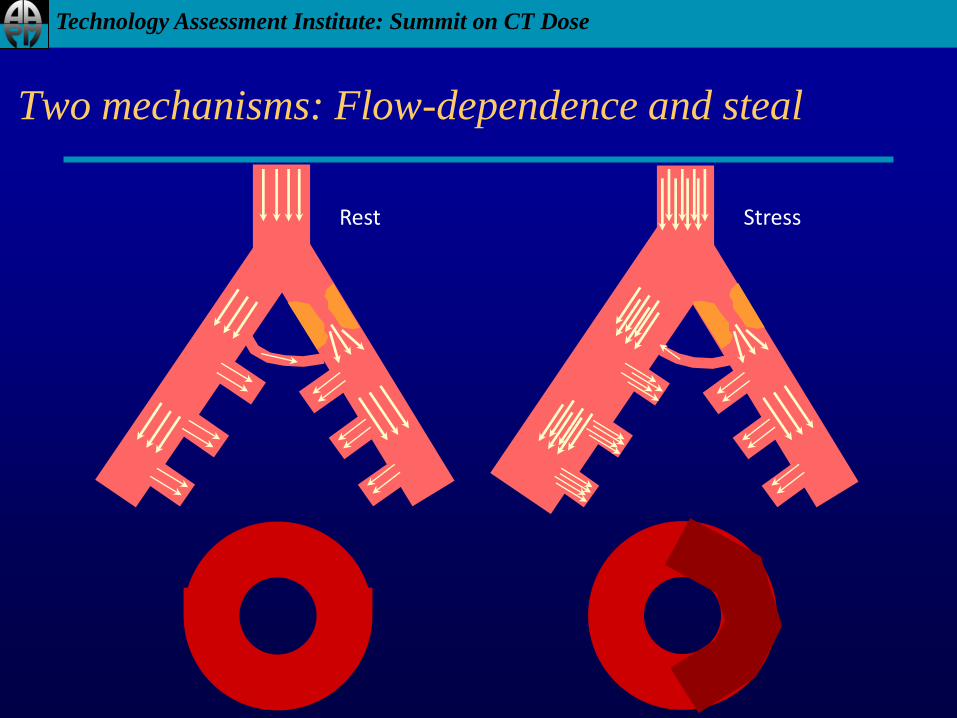
Two mechanisms: Flow-dependence and steal
Rest Stress

Technology Assessment Institute: Summit on CT Dose
Main Challenges
• Too many technologies
– CT scanners
– Processing algorithms
• CNR and SNR are low
• Dose can be very high
• Clinical applications are still being worked out
Other than that, life is good!
Technology Assessment Institute: Summit on CT Dose
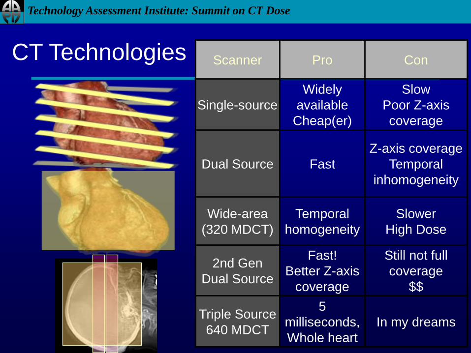
Scanner Pro Con
Single-source
Widely
available
Cheap(er)
Slow
Poor Z-axis
coverage
Dual Source Fast
Z-axis coverage
Temporal
inhomogeneity
Wide-area
(320 MDCT)
Temporal
homogeneity
Slower
High Dose
2nd Gen
Dual Source
Fast!
Better Z-axis
coverage
Still not full
coverage
$$
Triple Source
640 MDCT
5
milliseconds,
Whole heart
In my dreams
CT Technologies
Technology Assessment Institute: Summit on CT Dose
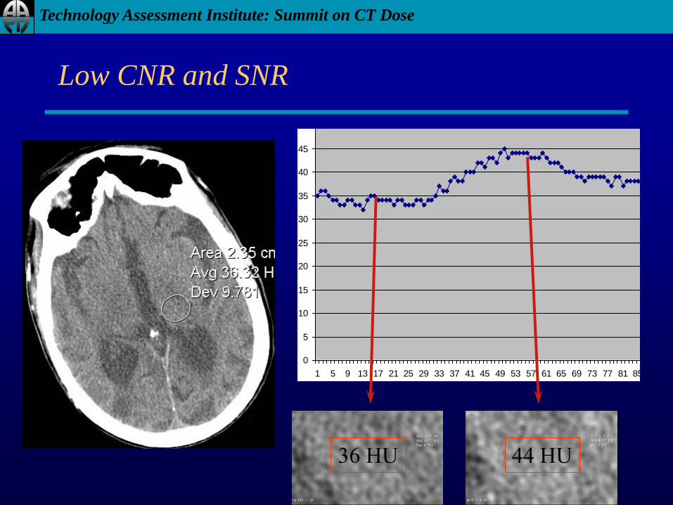
Low CNR and SNR
36 HU 44 HU
0
5
10
15
20
25
30
35
40
45
50
1 5 9 13 17 21 25 29 33 37 41 45 49 53 57 61 65 69 73 77 81 85
Series1
Technology Assessment Institute: Summit on CT Dose
time time
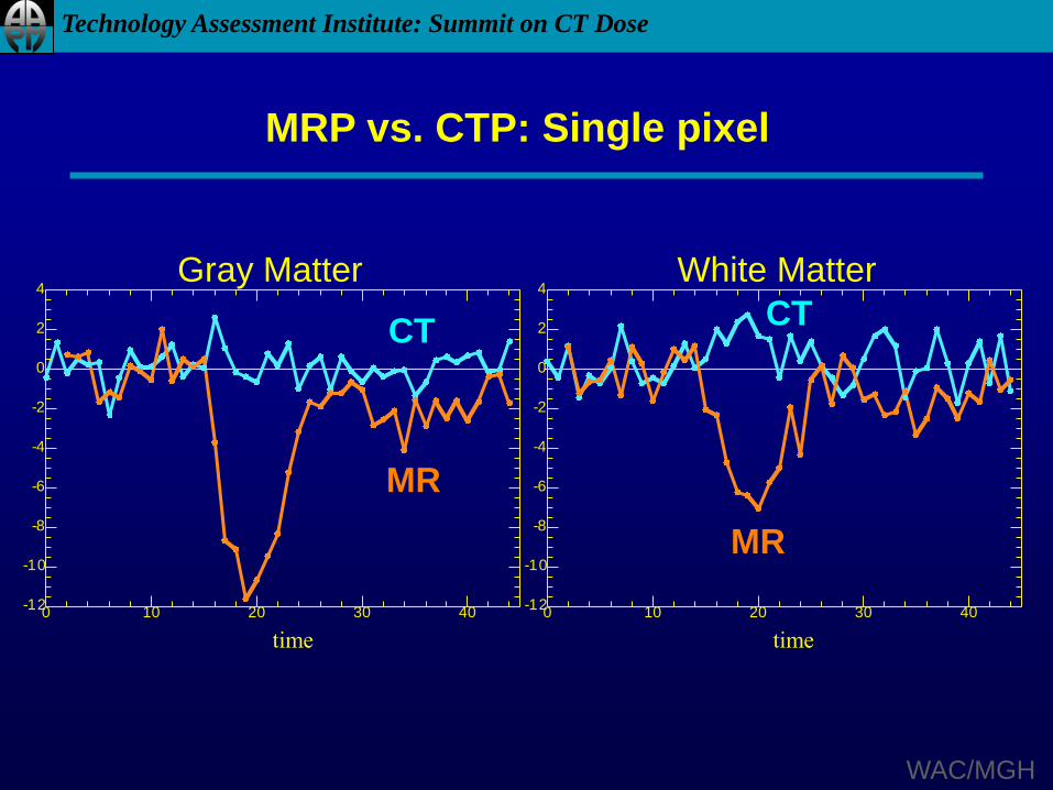
MRP vs. CTP: Single pixel
-12
-10
-8
-6
-4
-2
0
2
4
0 10 20 30 40
CT
MR
CT
MR
-12
-10
-8
-6
-4
-2
0
2
4
0 10 20 30 40
Gray Matter White Matter
WAC/MGH
Technology Assessment Institute: Summit on CT Dose
time time
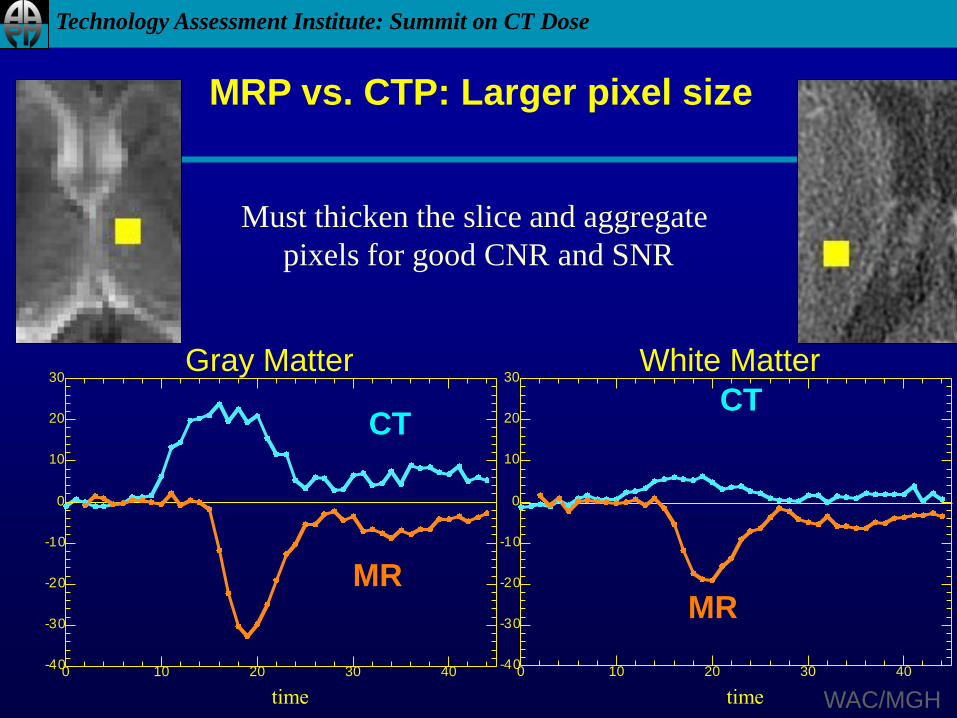
MRP vs. CTP: Larger pixel size
CT
MR
CT
MR
Gray Matter White Matter
-40
-30
-20
-10
0
10
20
30
0 10 20 30 40-40
-30
-20
-10
0
10
20
30
0 10 20 30 40
WAC/MGH
Must thicken the slice and aggregate
pixels for good CNR and SNR

Technology Assessment Institute: Summit on CT Dose
Neuro Perfusion CT
Technology Assessment Institute: Summit on CT Dose
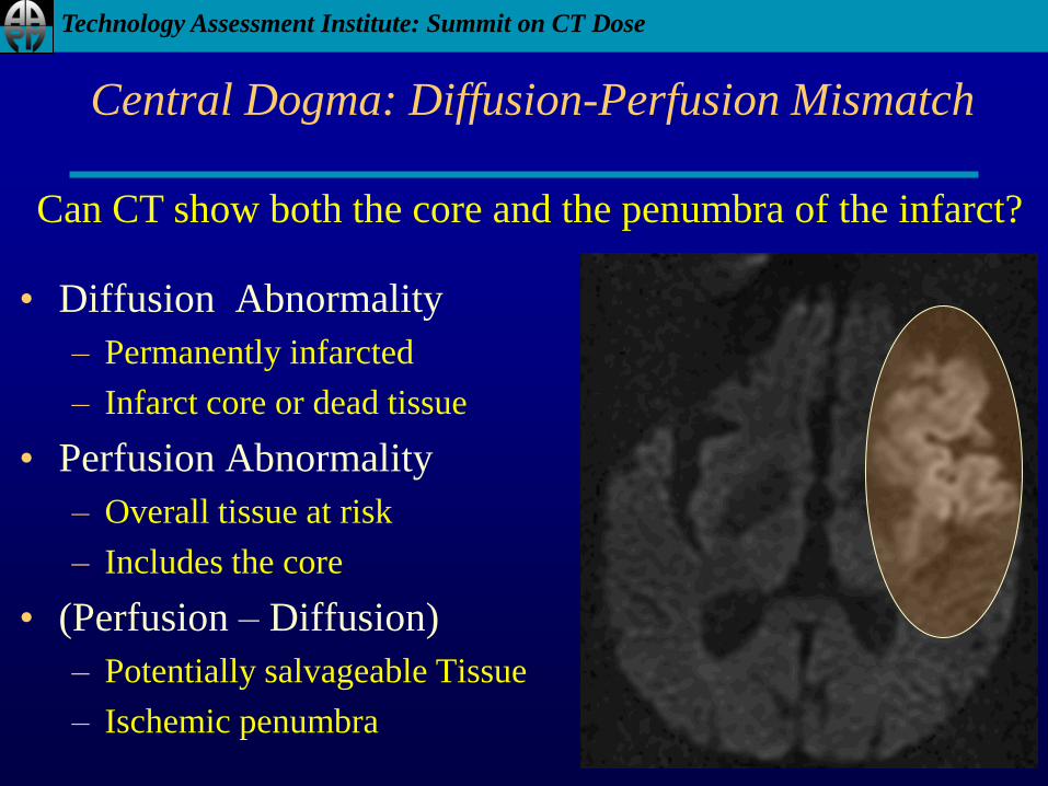
Central Dogma: Diffusion-Perfusion Mismatch
• Diffusion Abnormality
– Permanently infarcted
– Infarct core or dead tissue
• Perfusion Abnormality
– Overall tissue at risk
– Includes the core
• (Perfusion – Diffusion)
– Potentially salvageable Tissue
– Ischemic penumbra
Can CT show both the core and the penumbra of the infarct?
Technology Assessment Institute: Summit on CT Dose
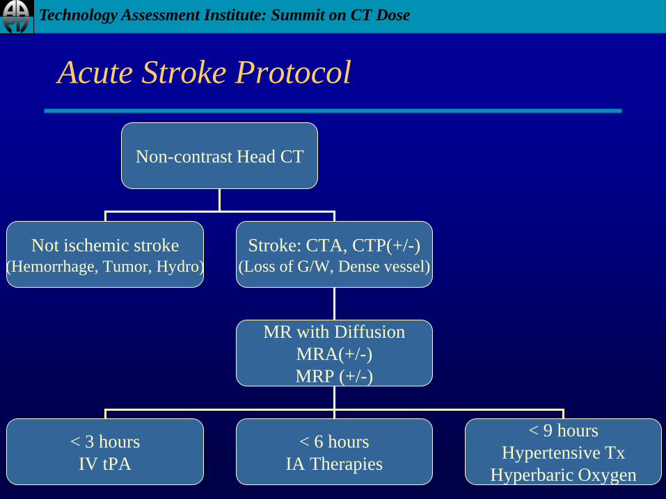
Acute Stroke Protocol
Non-contrast Head CT
Not ischemic stroke(Hemorrhage, Tumor, Hydro)
Stroke: CTA, CTP(+/-)(Loss of G/W, Dense vessel)
MR with Diffusion
MRA(+/-)
MRP (+/-)
< 3 hours
IV tPA
< 6 hours
IA Therapies
< 9 hours
Hypertensive Tx
Hyperbaric Oxygen

Technology Assessment Institute: Summit on CT Dose
MGH Single Slab Perfusion Protocol
• Perfusion (single slab, cine)
– 80 kVp 200 mA, 1 second rotation, 8 x 5 mm slices
– Phase I (cine): 1 image every second for 40s (0.5s recon interval)
– Phase II (axial): 1 image every 3 seconds for 27 s
– Total duration = 67 s
– Total X-ray exposure = 49 s
• CTDIvol=470 mGy
• DLP = 1890 mGy-cm
• CTP protocol well within the 0.5 Gy CTDI (vol)
• Further 25% reduction with 150mA
Technology Assessment Institute: Summit on CT Dose
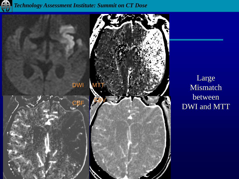
DWI MTT
CBFCBV
Large
Mismatch
between
DWI and MTT
Technology Assessment Institute: Summit on CT Dose
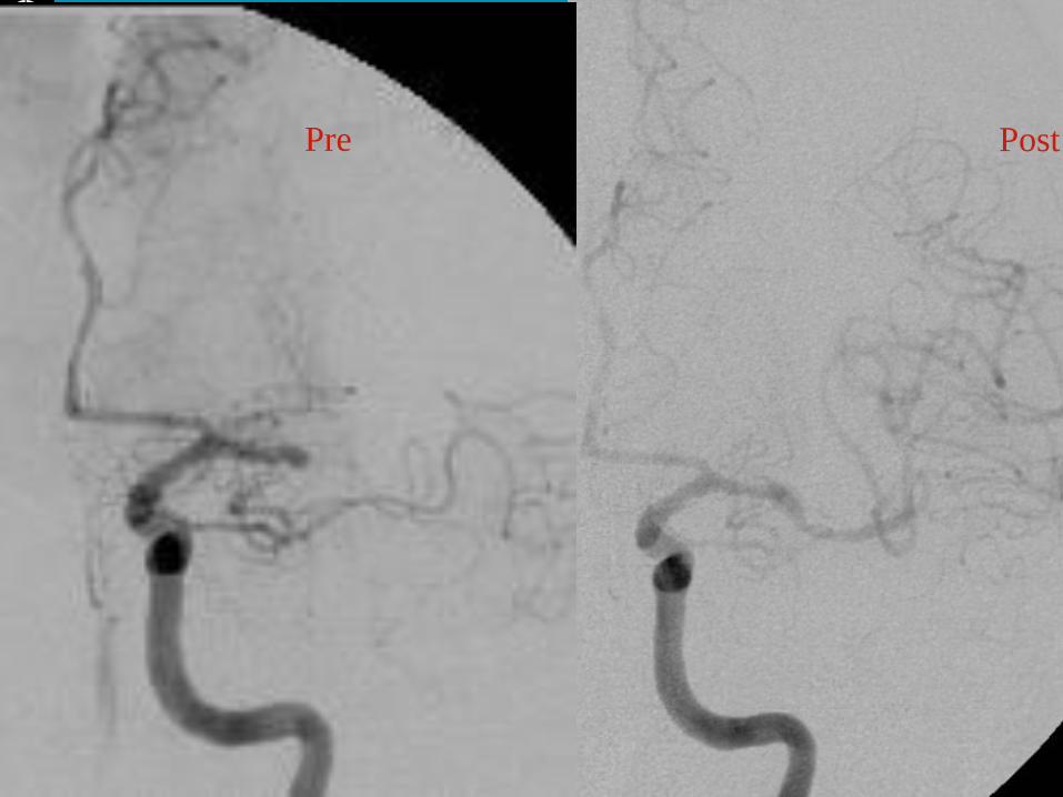
Pre Post
Technology Assessment Institute: Summit on CT Dose
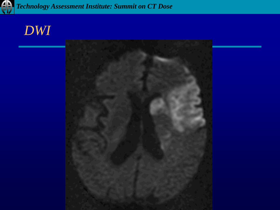
DWI
Technology Assessment Institute: Summit on CT Dose
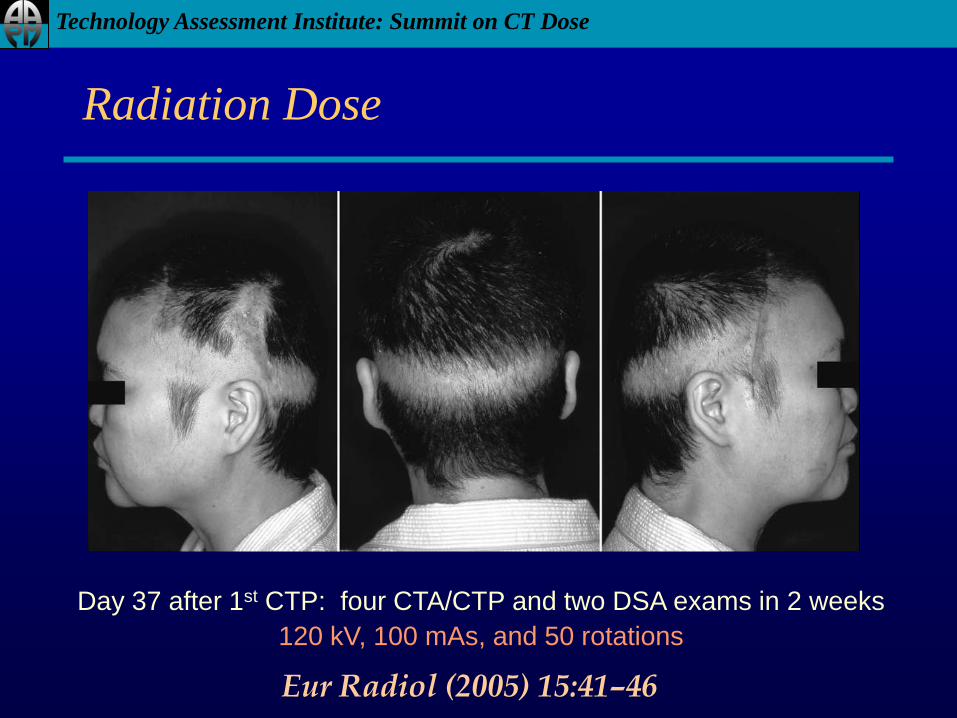
Radiation Dose
Eur Radiol (2005) 15:41–46
Day 37 after 1st CTP: four CTA/CTP and two DSA exams in 2 weeks
120 kV, 100 mAs, and 50 rotations
Technology Assessment Institute: Summit on CT Dose
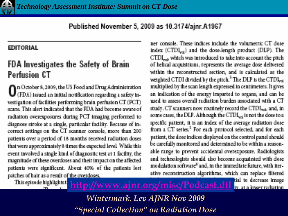
Wintermark, Lev AJNR Nov 2009
“Special Collection” on Radiation Dose
http://www.ajnr.org/misc/Podcast.dtl
Technology Assessment Institute: Summit on CT Dose
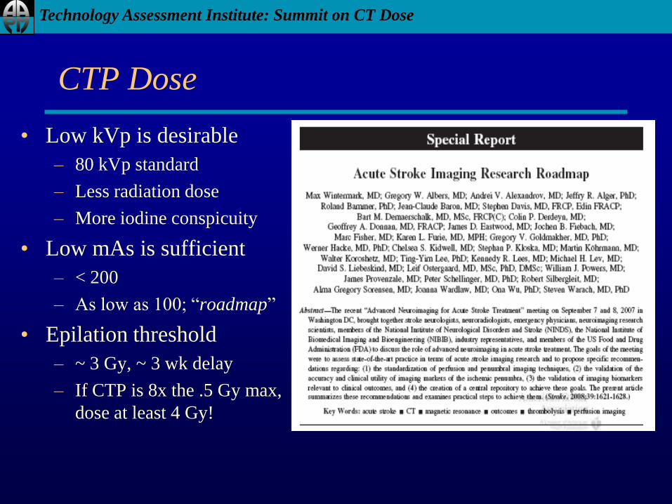
CTP Dose
• Low kVp is desirable
– 80 kVp standard
– Less radiation dose
– More iodine conspicuity
• Low mAs is sufficient
– < 200
– As low as 100; “roadmap”
• Epilation threshold
– ~ 3 Gy, ~ 3 wk delay
– If CTP is 8x the .5 Gy max,
dose at least 4 Gy!

Technology Assessment Institute: Summit on CT Dose
CT Perfusion Dose vs kVp
kVp mA t CTDI
Effective
dose
mSv n Rot
Total organ
dose
(mGy)
Total Effective
dose (mSv)
80 200 1 16.1 0.19 40 644 7.6
100 200 1 28.6 0.35 40 1144 14
120 200 1 43.4 0.55 40 1736 22
140 200 1 59.6 0.67 40 2384 26.8

Technology Assessment Institute: Summit on CT Dose
Cardiac Perfusion CT

Technology Assessment Institute: Summit on CT Dose
Nesto RW, Kowalchuk GJ. The ischemic cascade: temporal sequence ofhemodynamic, electrocardiographic and symptomatic expressions of ischemia.Am J Cardiol. 1987;59:23C–30C.
Perfusion defect
Metabolic disorders
Diastolic dysfunction
Systolic dysfunction
EKG changes
Chest pain
Myocardial infarction
Isch
em
ia
Time
The Ischemic
Cascade
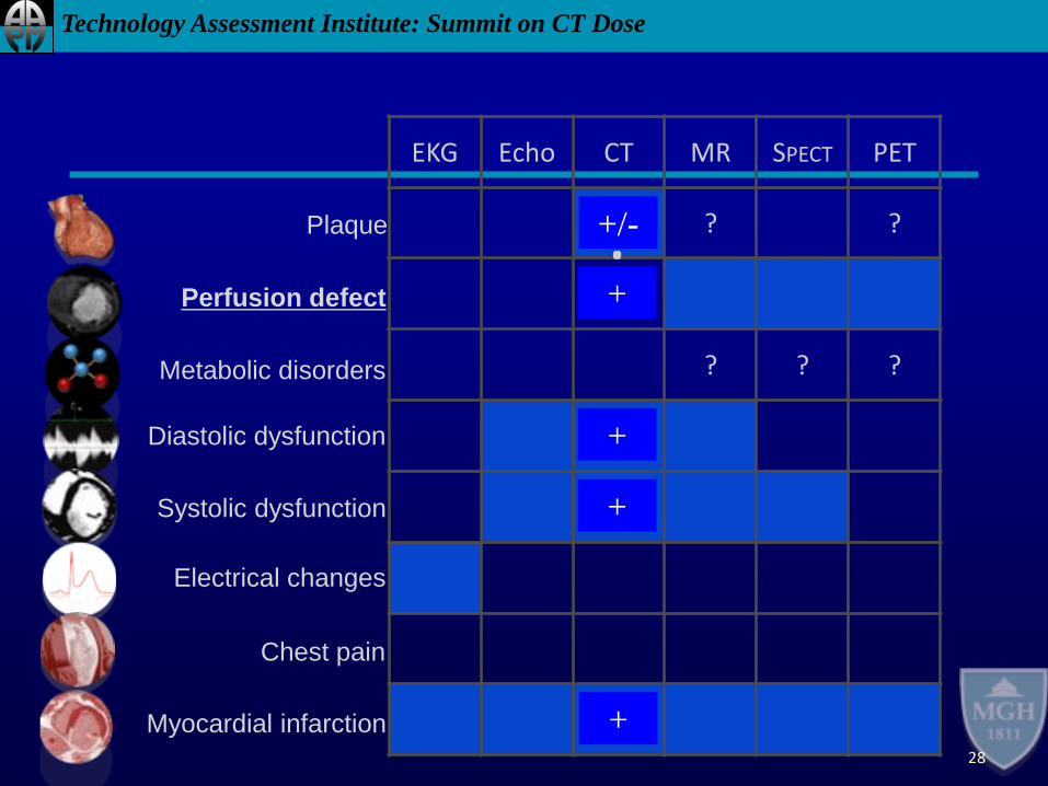
Technology Assessment Institute: Summit on CT Dose
28
Plaque
Perfusion defect
Metabolic disorders
Diastolic dysfunction
Systolic dysfunction
Electrical changes
Chest pain
Myocardial infarction
EKG Echo CT MR SPECT PET
? ??
? ? ?
+/-
+
+
+
+
Technology Assessment Institute: Summit on CT Dose
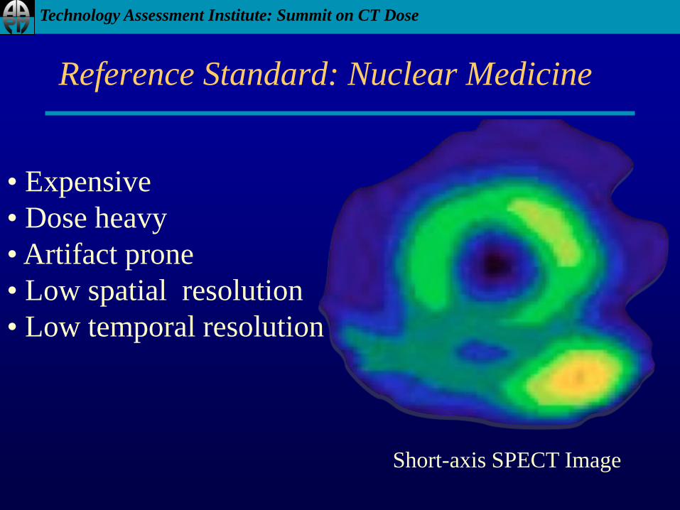
Reference Standard: Nuclear Medicine
• Expensive
• Dose heavy
• Artifact prone
• Low spatial resolution
• Low temporal resolution
Short-axis SPECT Image
Technology Assessment Institute: Summit on CT Dose
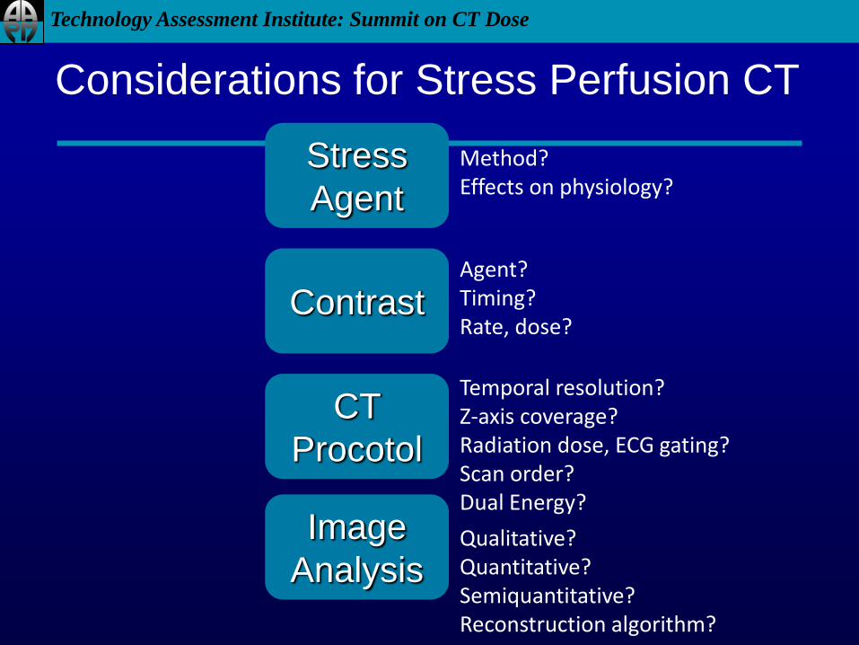
Stress
Agent
Contrast
CT
Procotol
Image
Analysis
Method?Effects on physiology?
Agent?Timing?Rate, dose?
Temporal resolution?Z-axis coverage?Radiation dose, ECG gating?Scan order?Dual Energy?
Qualitative?Quantitative?Semiquantitative?Reconstruction algorithm?
Considerations for Stress Perfusion CT
Technology Assessment Institute: Summit on CT Dose
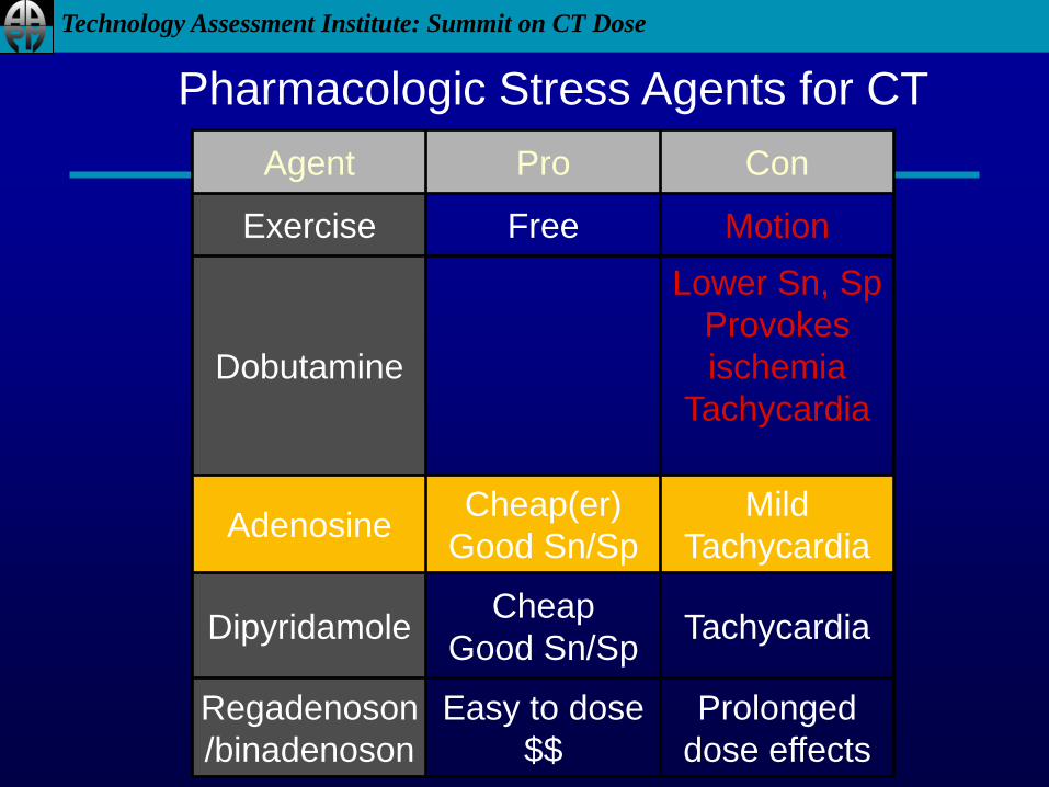
Agent Pro Con
Exercise Free Motion
Dobutamine
Lower Sn, Sp
Provokes
ischemia
Tachycardia
AdenosineCheap(er)
Good Sn/Sp
Mild
Tachycardia
DipyridamoleCheap
Good Sn/SpTachycardia
Regadenoson
/binadenoson
Easy to dose
$$
Prolonged
dose effects
Pharmacologic Stress Agents for CT

Technology Assessment Institute: Summit on CT Dose
Contrast bolus60-80 cc
@ 4 cc/sec
AdenosinePerfusion CT Scan
~5 minuteRecovery period
Contrast bolus60-80 cc
@ 4 cc/sec
Resting CTA
~10 minuteDelay
Delayed
CT
MGH Scan Protocol
Multiple variations possible
Retrospectively
Gated
Prospectively
Gated
Prospectively
Gated
Technology Assessment Institute: Summit on CT Dose
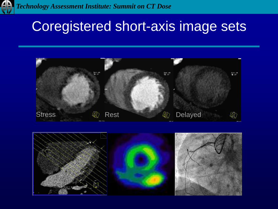
Stress Rest Delayed
Coregistered short-axis image sets
Technology Assessment Institute: Summit on CT Dose
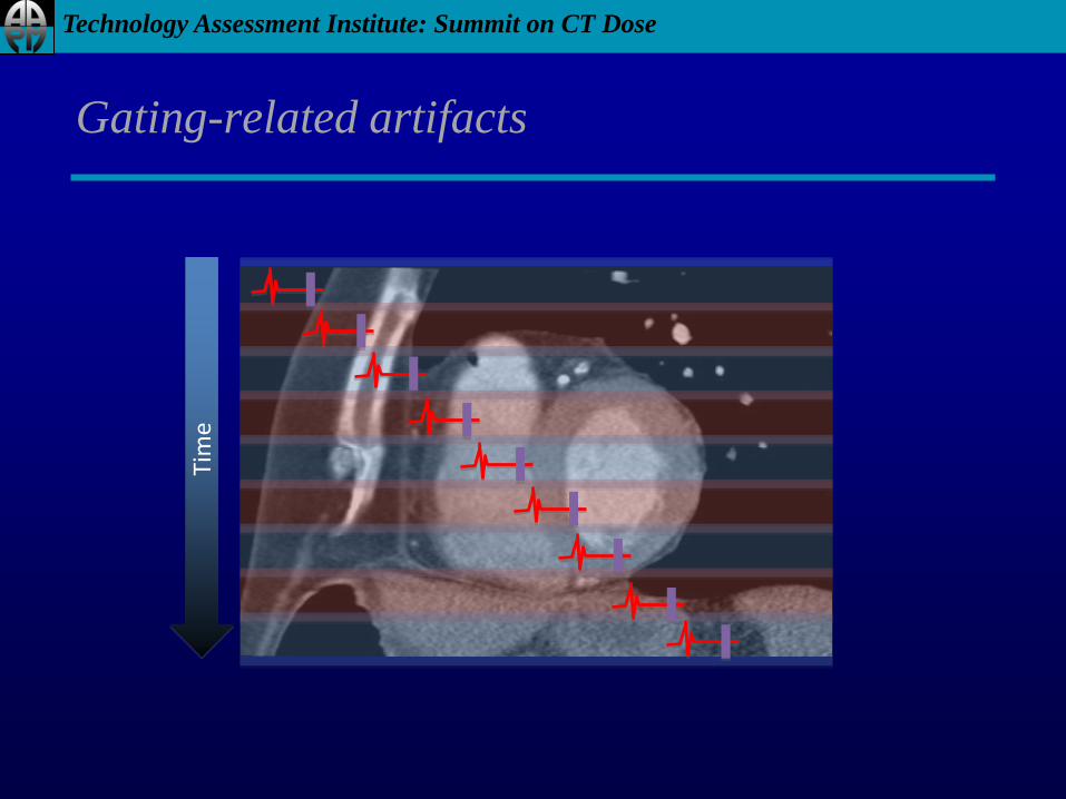
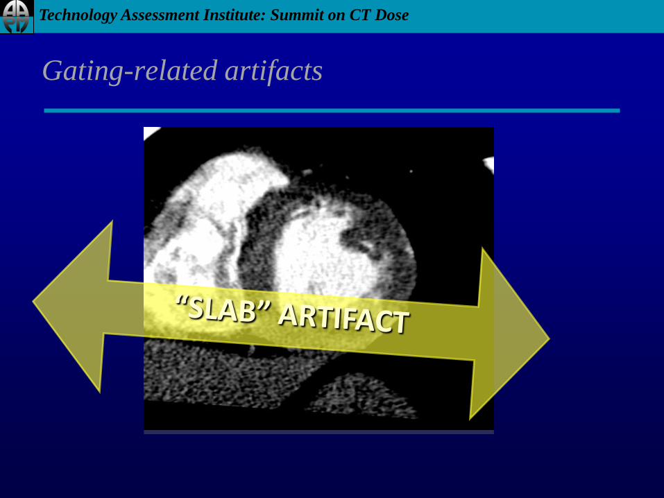
Gating-related artifacts
Tim
e
Technology Assessment Institute: Summit on CT Dose
Gating-related artifacts
Technology Assessment Institute: Summit on CT Dose
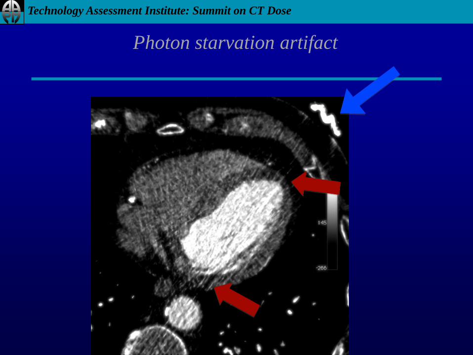
Photon starvation artifact
Technology Assessment Institute: Summit on CT Dose
Future Directions in CTP
Novel reconstruction techniques: Iterative
Dual-energy imaging
Technology Assessment Institute: Summit on CT Dose
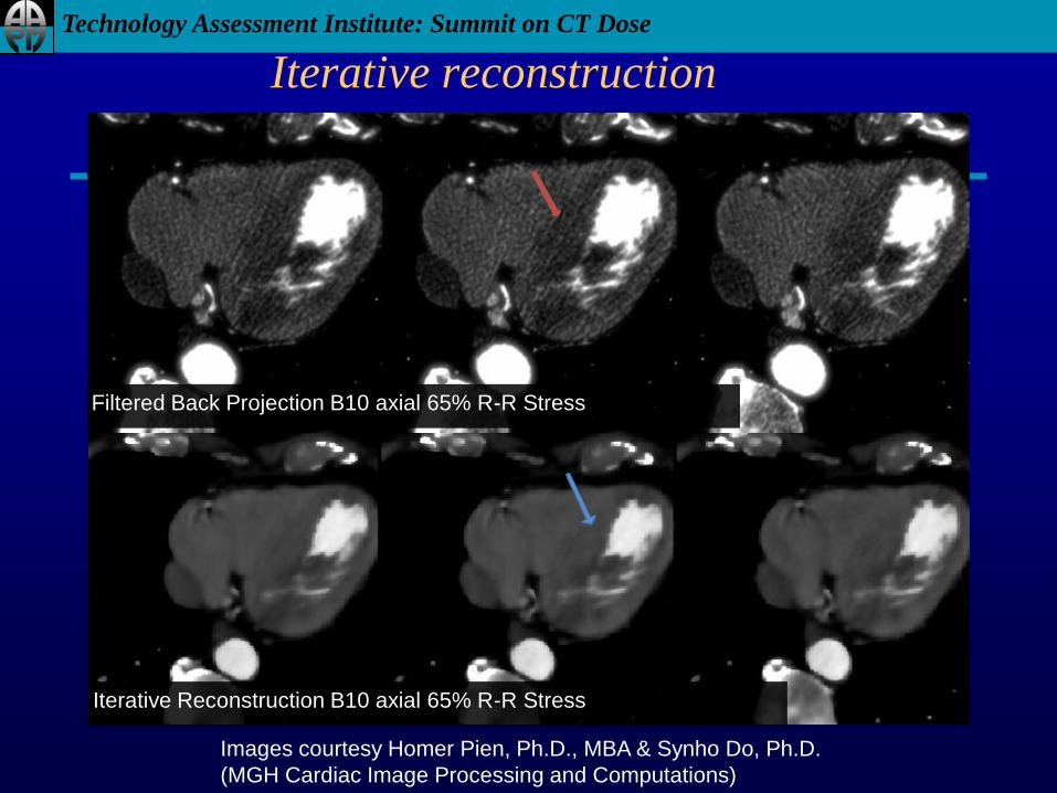
Iterative reconstruction
Filtered Back Projection B10 axial 65% R-R Stress
Iterative Reconstruction B10 axial 65% R-R Stress
Images courtesy Homer Pien, Ph.D., MBA & Synho Do, Ph.D.
(MGH Cardiac Image Processing and Computations)
Technology Assessment Institute: Summit on CT Dose
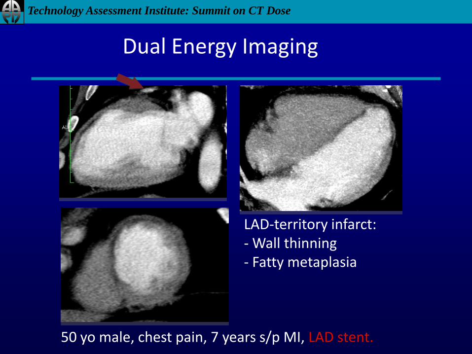
50 yo male, chest pain, 7 years s/p MI, LAD stent.
Dual Energy Imaging
LAD-territory infarct:- Wall thinning- Fatty metaplasia

Technology Assessment Institute: Summit on CT Dose
100 kVImage
140 kVImage
“Iodineonly”
Image
“Iodine Map” Delayed Enhanced

Technology Assessment Institute: Summit on CT Dose
Conclusion
• CTP is exciting
– “Time is muscle”
– “Time is brain”
– “Mismatch is brain”
• CTP is challenging
– Many technologies
– Low CNR and SNR
– Potentially high dose
• The complexity can be
managed
– Use low kVP
– Use sufficient temporal
resolution
– Don’t truncate the time
opcification curve
• Many new promising
developments