Embed Size (px)
Citation preview
© Turkish Society of Radiology 2012
111
M ultiple myeloma (MM) is a hematologic malignancy character-ized by lytic bone lesions that are most frequently located in the spine. Vertebral involvement is present in approximately
60% of the patients at diagnosis (1). The consequences of bone disso-lution are back pain caused by microinstability, altered biomechanics and tumor infiltration, compression fractures due to vertebral structural weakness, spinal deformity, potential spinal cord or nerve root compres-sion resulting from epidural tumor extension, and vertebral height loss.
Non-invasive treatment options for pain management in myeloma patients include bed rest, bracing, bisphosphonates, analgesic drugs, and radiotherapy. However, these therapeutic regimens are often not sufficient to control the acute and chronic spinal pain accompanying pathologic fractures and progressive spinal deformity. Surgical options, such as open decompression, internal fixation, and bone grafting, may be limited due to a high comorbidity and reduced bone mass (2). The consequences of insufficient surgical treatment options are physical de-terioration, reduced psychosocial skills, and a seriously affected general quality of life for the patients.
Percutaneous vertebroplasty has been established with wide clinical acceptance for the treatment of symptomatic osteoporotic vertebral compression fractures (VCFs) (3, 4). The indications for this procedure have meanwhile been extended to metastatic disease and MM of the spine, which, compared to osteoporotic VCFs, are characterized by a higher risk of symptomatic cement leakage, mainly due to osteolytic destruction of cortical bone (5, 6).
Mono or biplane fluoroscopy is common for guiding percutaneous vertebroplasty in the USA (7). Various European centers use computed tomography (CT) fluoroscopy combined with a mobile C-arm device that provides fluoroscopy for needle insertion and cement injection (8–10). This combination allows for exact needle positioning through visu-alization of the vertebral cross-sectional area and adjacent soft tissues in the CT fluoroscopic cross-sectional image and of the needle inclination and cement extravasation into the paravertebral veins in the lateral fluo-roscopic view. Few authors have reported successfully performing needle placement (11, 12) or the combination of needle placement and cement injection for sacroplasty under CT fluoroscopic guidance only (13, 14). In comparison to the lateral fluoroscopic view, cement injection is mon-itored by CT fluoroscopic acquisitions obtained in table positions along the z-axis, superior or inferior to the tip of the vertebroplasty needle, to follow the leading edge of the accumulated cement (14).
The aim of this retrospective, single-center study was to assess the technical safety with respect to the frequency and clinical impact of ce-ment leakage occurring during CT fluoroscopy-guided vertebroplasty in myeloma patients suffering from spinal osteolyses and malignant
INTERVENTIONAL RADIOLOGYORIGINAL ARTICLE
CT fluoroscopy-guided percutaneous vertebroplasty in patients with multiple myeloma: analysis of technical results from 44 sessions with 67 vertebrae treated
Christoph Trumm, Tobias Jakobs, Anne Pahl, Robert Stahl, Thomas Helmberger, Philipp Paprottka, Maximilian Reiser, Ralf-Thorsten Hoffmann
From the Department of Clinical Radiology (C.T. [email protected], A.P., R.S., P.P., M.R.), Ludwig-Maximilians-Universität München, Munich, Germany; the Department of Diagnostic and Interventional Radiology (T.J.), Krankenhaus Barmherzige Brüder München, Munich, Germany; the Department of Diagnostic and Interventional Radiology and Nuclear Medicine (T.H.), Städtisches Klinikum München-Bogenhausen, Munich, Germany; and the Department and Policlinics of Diagnostic Radiology (R.T.H.), Universitätsklinikum Carl Gustav Carus, Dresden, Germany.
Received 24 January 2011; revision requested 19 March 2011; revision received 4 April 2011; accepted 26 April 2011.
Published online 17 October 2011DOI 10.4261/1305-3825.DIR.4226-11.1
PURPOSEThis study aimed to assess the results of computed tomogra-phy (CT) fluoroscopy-guided vertebroplasty in patients with multiple myeloma, focusing on the frequency and clinical im-pact of polymethylmethacrylate (PMMA) leaks.
MATERIALS AND METHODSFrom December 2001 to August 2008, 39 patients (17 fe-males, 22 males; mean age, 65±7 years) with multiple my-eloma suffering from painful spinal osteolyses underwent ver-tebroplasty. A total of 67 vertebrae were treated in 44 sessions under CT fluoroscopy (single-slice, 4-row CT, and 16-row CT). In the planning CT scan, osteolytic destruction (i.e., none, ≤25%, ≤50%, ≤75%, or ≤100%) was assessed regarding the vertebral cross-sectional area, the cortical border of the spi-nal canal, and the outer circumference. CT performed after vertebroplasty was used to detect local PMMA leaks. Patient charts were retrospectively reviewed with special respect to peri and postinterventional adverse events. Clinical outcomes were assessed on a visual analog scale (VAS) 24 hours before, 24 hours after, and 6 months after vertebroplasty.
RESULTSOverall, 37.3%, 12.0%, and 6.0% of vertebrae showed at least 50% osteolytic involvement of the cross-sectional area, spinal canal, and outer vertebral cortex, respectively. Intradis-cal, intraspinal, paravertebral, and intercostovertebral/poste-rolateral leaks were seen in 21.6%, 35.1%, 43.3%, and 0% of vertebrae, respectively. The ratio of basivertebral to segmental venous leaks was 16.2%/40.5%. No major complications oc-curred. The mean VAS score decreased significantly (P < 0.05) from 6.4 at 24 hours before vertebroplasty to 3.2 at a mean follow-up of 9.0 months.
CONCLUSIONVertebroplasty in multiple myeloma can be performed safely under CT fluoroscopy, even with substantial destruction of the vertebral cross-sectional area or cortical bone. A high clinical success rate was achieved, regardless of whether PMMA leaks were present.
Key words: • multiple myeloma • vertebroplasy • fluoroscopy • computed tomography
Diagn Interv Radiol 2012; 18:111–120
Trumm et al.112 • January 2012 • Diagnostic and Interventional Radiology
percutaneous vertebroplasty (which are in accordance with the Cardiovascular and Interventional Radiological Society of Europe quality assurance guidelines for percutaneous vertebro-plasty) (16), radiculopathy in excess of vertebral pain (caused by a compres-sive syndrome unrelated to vertebral collapse), asymptomatic retropulsion of a fracture fragment causing signifi-cant spinal canal compromise, and asymptomatic tumor extension into the epidural space were regarded as relative contraindications to vertebro-plasty (17). Absolute contraindications were patients improving on analgesic therapy, myelopathy in patients with spinal canal compromise due to retro-pulsion of bone fragments or a tumor, active local or systemic infections, un-correctable coagulopathy, and an al-lergy to bone cement or opacification agents (17).
Imaging workup and guidanceBefore vertebroplasty, previous cross-
sectional images not older than two weeks, such as those from CT, magnet-ic resonance imaging (MRI) or positron emission tomography (PET)/CT, were checked for all patients by a team of in-terventional radiologists. Additionally, all patients underwent in-house MRI (T1-weighted sequence ± contrast, short tau inversion recovery sequence, axial and sagittal orientation) prior to verte-broplasty to assess the pattern of bone marrow involvement and vertebral and paravertebral tumor involvement and to detect the level(s) of vertebral edema in cases of multiple VCFs detected on CT (18).
Each patient underwent a pre and postinterventional CT scan (Siemens Somatom Plus 4, Sensation 4 or 16 MDCT, Siemens Medical Solutions, Forchheim, Germany) of the involved spine level, including at least two ver-tebral body heights above and below the osteolytic/fractured vertebral body. The CT scan involved 3-mm slices and coronal and sagittal multiplanar re-constructions (MPRs) to visualize the extent of vertebral compression and bone destruction, to detect possible in-volvement of the posterior wall of the vertebral body, and to plan the needle trajectory for vertebroplasty. Using the postinterventional CT scan, the dis-tribution of polymethylmethacrylate (PMMA) in the vertebral body and ce-ment leaks were analyzed.
For CT guidance, a single-detector Siemens Somatom Plus 4 scanner (120 kV, 15 to 25 mA, 4×2.5 mm collima-tion, 3-mm slices), a Siemens Sensation 4 (120 kV, 15 to 25 mA, 4×1 mm colli-mation, 3-mm slices) or a Sensation 16 MDCT (120 kV, 15 to 25 mA, 12×0.75 mm collimation, 3-mm slices) with CT fluoroscopy was used (CARE Vision CT, Siemens Medical Solutions). CT fluoroscopy was performed with an-gular beam modulation (HandCare™, Siemens Medical Solutions), i.e., the X-ray beam was turned off within a 120° angle sector above the patient to decrease radiation exposure to the interventional radiologist and patient (19). The entire vertebroplasty pro-cedure, including needle placement and PMMA injection, was performed under CT fluoroscopic guidance only. The needle was inserted under inter-mittent single-shot CT fluoroscopic acquisitions. Continuous CT fluoros-copy was only used during the PMMA injection. The needle tip and the ad-jacent cement distribution within the vertebral body were monitored in real time on a ceiling-mounted, in-room monitor (256×256 imaging matrix in-terpolated to 1024×1024 for display) by moving the table along the z-axis in incremental steps of 1.0 mm using a control panel attached to the CT ta-ble. Beginning in February 2006, CARE View, with a synchronous display of three adjacent slices on the in-room monitor, was employed. Precautions with respect to the radiation protection of the operator during CT fluoroscopy included aprons, thyroid shields and eyeglasses of 0.5-mm lead equivalent. An additional shield was put onto the lower half of the patient before sterile draping to reduce the amount of scat-tered radiation.
ProcedureAll procedures were performed by
one of the board-certified authors or by residents under their supervision who were experienced in CT-guided inter-ventions. The vertebroplasty needles (OptiMed Medical Devices, Ettlingen, Germany) utilized were 15, 13, or 10 gauge and had a length of 10 or 15 cm. In the cervical spine, an anterolateral approach (patient in the supine posi-tion) was generally used, while an in-tercostovertebral approach (patient in the prone position) was used in the thoracic spine. A transpedicular access
compression fractures. Moreover, we wanted to assess the clinical success rate of vertebroplasty with regard to pain reduction, patient comfort, and the ability to reduce the use of analge-sic agents.
Materials and methodsPatient selection
Percutaneous vertebroplasty is an ac-cepted and widely used method to sta-bilize fractured vertebral bodies. Our in-stitutional ethics board did not demand approval of this retrospective study re-garding the review of patient charts and the assessment of clinical outcomes af-ter vertebroplasty using a standardized questionnaire. The principles of the Declaration of Helsinki were followed. All patients were referred by the local oncology department. The diagnosis of myeloma had been made using stand-ard clinical criteria (15).
Clinical indications for vertebro-plasty were regularly confirmed by an interdisciplinary team of oncolo-gists, radiation oncologists, spinal sur-geons, and interventional radiologists prior to the intervention. A biopsy of the spinal segment to be treated with vertebroplasty had not generally been performed prior to vertebroplasty, and biopsy results were not analyzed separately.
Each patient underwent a physical examination by the referring clinician to identify the painful or fractured ver-tebral level(s). Informed consent by the patient or his/her legal guardian to undergo vertebroplasty was obtained 24 hours before and directly prior to the intervention after an extensive ex-planation of the method and its poten-tial complications as well as alternative treatments.
Between December 2001 and August 2008, 224 patients with tumoral in-volvement of the spine underwent ver-tebroplasty, 39 of whom (17 females, 22 males) suffered from painful spinal osteolyses and compression fractures due to MM. The mean age of the pa-tients was 65±7 years, with a range from 51 to 82 years. All patients re-ferred for vertebroplasty suffered from severe back pain refractory to conserv-ative analgesic therapy, either alone or in combination with chemotherapy and/or radiation therapy.
According to the Society of Interventional Radiology (SIR) qual-ity improvement guidelines for
CT fluoroscopy-guided percutaneous vertebroplasty in patients with multiple myeloma • 113Volume 18 • Issue 1
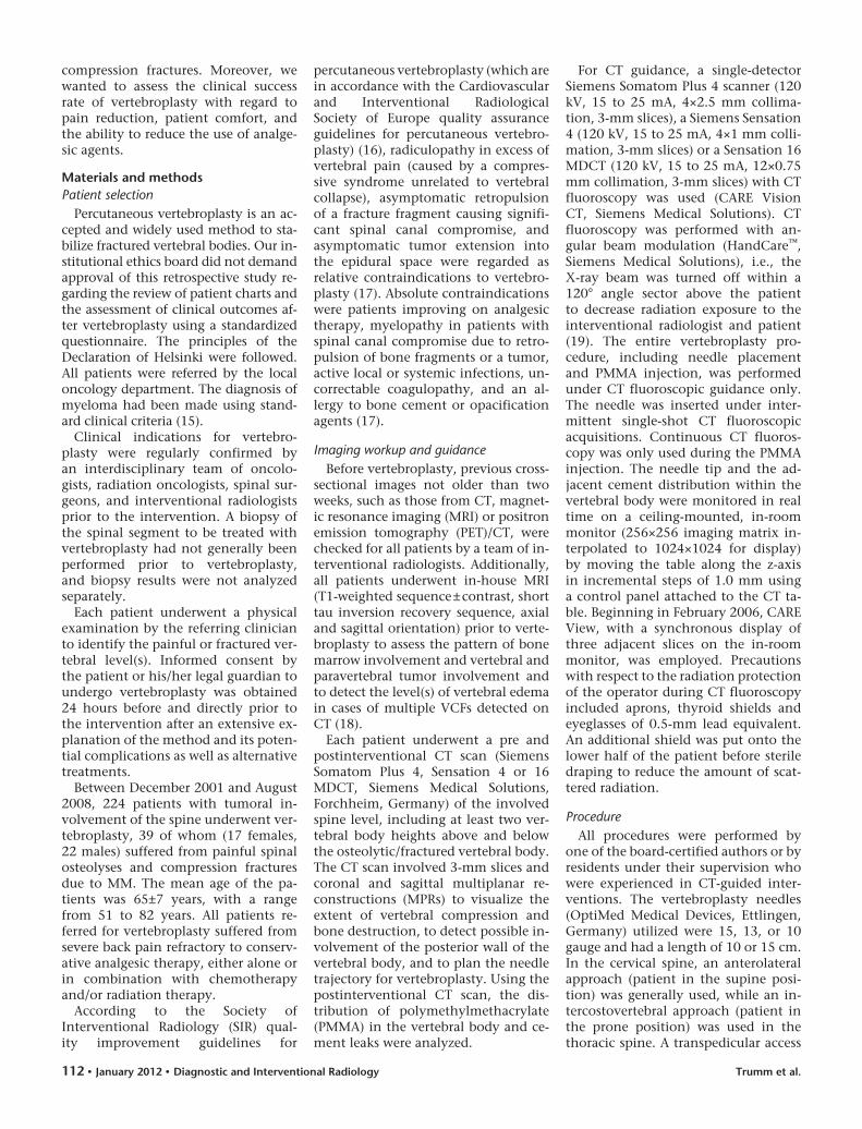
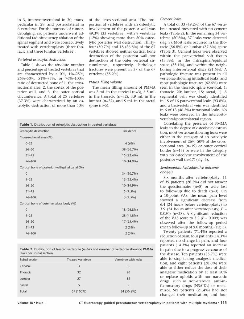
Figure 1. Axial CT image of a thoracic vertebra shows the three evaluated vertebral zones of osteolytic destruction: (1) cross-sectional area including vertebral body and pedicles (shaded area), (2) cortical border of posterior wall and spinal canal (dashed line), and (3) outer cortical border of vertebral body (solid line).
was chosen in the thoracic spine only when the pedicular diameter was large enough. In the lumbar and sacral spine, a 10 G needle with transpedicular or posterolateral (in cases of insufficient pedicle diameter or pedicle osteolysis) access was applied in most cases.
All patients received preinterven-tional antibiosis with 2 g of mezlocil-lin (Baypen®, Bayer AG, Leverkusen, Germany). During the procedure, each patient was monitored with pulse oxymetry. After sterile draping and disinfection of the skin overlying the treated vertebral body, local anesthesia with 10–20 mL of 2% mepivacaine hy-drochloride (Scandicain®, AstraZeneca GmbH, Wedel, Germany) was applied to the soft tissue and the periosteum along the access route. Additional conscious sedation with midazolam hydrochloride (5 mg of intravenous Dormicum®, Ratiopharm GmbH, Ulm, Germany) and piritramide (15 mg of intravenous Dipidolor®, Janssen-Cilag GmbH, Neuss, Germany) was adminis-tered in six cases of additional radiofre-quency ablation before vertebroplasty.
A small skin incision was made, and the vertebroplasty needle was ad-vanced with a sterile hammer through the cortical bone under intermittent single-shot CT fluoroscopic control. The direction of the needle was ad-ditionally guided by turning the bev-eled tip in 90° steps. The insertion was stopped when the needle tip reached the anterior third of the vertebral body or the center of the osteolysis. Three types of bone cement were used dur-ing the study period: Osteopal 40, Osteopal V, and Biomet Bone Cement V (Biomet Deutschland GmbH, Berlin, Germany). After the needle position-ing, the PMMA was mixed in a sterile bowl according to the manufacturer’s guidelines. To reduce the risk of extra-vertebral leakage and venous extravasa-tion, the cement injection was started with the application device (Optimed Gangi Cemento-Re Gun, OptiMed Medical Devices) after another 60–90 s when the PMMA was in its pasty po-lymerization phase. The PMMA was slowly injected by the operator under intermittent CT fluoroscopy using se-quential withdrawing of the needle and incremental table movement along the z-axis to assess the localiza-tion and direction of cement distri-bution within the vertebral body. In cases of initial cement extravasation
to the adjacent disc spaces or to the soft tissue structures on the real-time sectional CT fluoroscopic images, the injection was immediately stopped by depressurizing the application screw. If the leakage persisted, the needle tip was repositioned by turning the han-dle bar in 90° steps or by sequentially withdrawing the needle. The cement injection was stopped when at least one- to two-thirds of the osteolytic le-sion was filled. In cases of two or three vertebrae treated in one session, the vertebra with major cortical destruc-tion was treated last to ensure a pasty consistency of the cement. The stylet of the needle was finally inserted again to empty the needle cavity completely, and then both were removed by slowly turning the handle bar. Then, a CT scan of the treated vertebral level was immediately performed for documen-tation of the cement distribution. After the intervention, all patients were sent back to the ward for further clinical monitoring.
Technical outcomesIn a retrospective analysis, two in-
terventional radiologists who were experienced in performing CT-guided vertebroplasty (RTH and CT)
retrospectively evaluated pre and pos-tinterventional CT scans, as well as the periinterventional CT fluoroscopic da-tasets acquired in the 44 vertebroplasty sessions for the purpose of obtaining a consensus regarding the degree of ver-tebral osteolytic involvement, and the types of cement leakage.
Vertebral osteolytic destructionThe degree of osteolytic destruction
was quantified in the preintervention-al CT dataset by visual estimation (Fig. 1) of the following: 1: The osteolytic destruction of the
vertebral cross-sectional area, including the vertebral body and the pedicles (a. 0%–25%, b. 26%–50%, c. 51%–75%, d. 76%–100%)
2: The cortical border of the posterior wall and spinal canal (a. 0%, b. 1%–25%, c. 26%–50%, d. 51%–75%, e. 76%–100%)
3: The cortical border of the outer vertebral body (a. 0%, b. 1%–25%, c. 26%–50%, d. 51%–75%, e. 76%–100%)
4: The presence of pathologic fractures was evaluated on both axial images and multiplanar reconstructions.
Trumm et al.114 • January 2012 • Diagnostic and Interventional Radiology
Vertebroplasty procedureThe type of needle access to the ver-
tebral body (anterolateral, intercos-tovertebral, transpedicular, or postero-lateral) was evaluated in the periinter-ventionally acquired CT fluoroscopic datasets. The amount of PMMA (mL) applied was taken from the report that documented the procedure.
Types of cement leakage According to the compartment in-
volved, the following types of cement leakage were differentiated: - intradiscal (upper or lower
intervertebral space), - intraspinal (including non-
vascular soft-tissue leaks through the cortical bone and vascular leaks into the basivertebral vein),
- paravertebral (including non-vascular leaks through cortical defects into the surrounding soft
tissue and vascular leaks into the segmental veins), and
- intercostovertebral/posterolateral (along the needle access path).
Vascular leaks were assessed sepa-rately if a basivertebral or segmental vein could be clearly identified.
In cases of several leakage types oc-curring at the same vertebral level, every leak location was counted as a separate event.
Clinical outcomes and adverse eventsTwenty-four hours before and after
the procedure, all patients were asked to answer a standardized questionnaire assessing the following items: 1. Pain level on a 10-point
(0 [no pain]–10 [maximum pain intensity]) visual analog scale (VAS)
2. Type and dose of analgesic agents.
Six months after the procedure, all pa-tients were sent the same questionnaire to assess any changes in analgesic agent use, subjective changes in the severity of pain (better, no change, or worse) and quantitative VAS scores. Clinical complications that occurred during the procedure or that had been reported by the referring clinicians within 24 hours after the procedure were documented in the final report. Additionally, clini-cal patient charts were retrospectively reviewed with respect to adverse ad-vents potentially related to the proce-dure that occurred more than 24 hours after the vertebroplasty.
Statistical analysisThe data analysis was performed
using a commercially available soft-ware (Statistical Package for Social Sciences, version 18, SPSS Inc., Chicago, Illinois, USA). P values of less than 0.05 were considered statistically significant. A Wilcoxon rank sum test was used for comparisons of VAS pain scores 24 hours before vertebroplasty and after the follow-up period.
ResultsThe total number of differently treat-
ed vertebrae in 44 sessions was 67 (one level, 25; two levels, 15; three levels, 4 sessions), including 3 cervical, 32 thoracic, 27 lumbar and 5 sacral ver-tebrae. All 67 vertebrae were treated successfully in the first session (Fig. 2). The access route was anterolateral
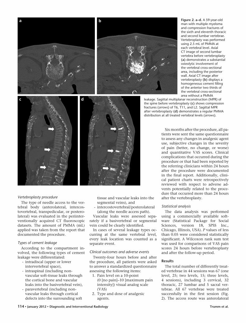
Figure 2. a–d. A 59-year-old man with multiple myeloma and compression fractures of the sixth and eleventh thoracic and second lumbar vertebrae. Vertebroplasty was performed using 2.5 mL of PMMA at each vertebral level. Axial CT image of second lumbar vertebra before vertebroplasty (a) demonstrates a substantial osteolytic involvement of the vertebral cross-sectional area, including the posterior wall. Axial CT image after vertebroplasty (b) displays a homogeneous cement filling of the anterior two thirds of the vertebral cross-sectional area without a PMMA
b
d
a
c leakage. Sagittal multiplanar reconstruction (MPR) of the spine before vertebroplasty (c) shows compression fractures (arrows) of T6, T11, and L2. Sagittal MPR after vertebroplasty (d) demonstrates a regular PMMA distribution at all treated vertebral levels (arrows).
CT fluoroscopy-guided percutaneous vertebroplasty in patients with multiple myeloma • 115Volume 18 • Issue 1
in 3, intercostovertebral in 30, trans-pedicular in 28, and posterolateral in 6 vertebrae. For the purpose of tumor-debulging, six patients underwent ad-ditional radiofrequency ablation of the spinal segment and were consecutively treated with vertebroplasty (three tho-racic and three lumbar vertebrae).
Vertebral osteolytic destructionTable 1 shows the absolute number
and percentage of treated vertebrae that are characterized by a 0%, 1%–25%, 26%–50%, 51%–75%, or 76%–100% ratio of destructed bone of 1. the cross-sectional area, 2. the cortex of the pos-terior wall, and 3. the outer cortical circumference. A total of 25 vertebrae (37.3%) were characterized by an os-teolytic destruction of more than 50%
of the cross-sectional area. The pro-portion of vertebrae with an osteolytic involvement of the posterior wall was 49.3% (33 vertebrae), with 8 vertebrae (12%) showing more than 50% osteo-lytic posterior wall destruction. Thirty-four (50.7%) and 18 (26.8%) of the 67 vertebrae showed neither cortical bone destruction of the posterior wall nor destruction of the outer vertebral cir-cumference, respectively. Pathologic fractures were present in 37 of the 67 vertebrae (55.2%).
PMMA filling volumeThe mean filling amount of PMMA
was 2 mL in the cervical (n=3), 3.5 mL in the thoracic (n=32), 3.9 mL in the lumbar (n=27), and 5 mL in the sacral spine (n=5).
Cement leaks A total of 33 (49.2%) of the 67 verte-
brae treated presented with no cement leaks (Table 2). In the remaining 34 ver-tebrae (50.8%), 37 leaks were detected (Fig. 3). Most leaks occurred in the tho-racic (56.8%) or lumbar (37.8%) spine (Table 3). Cement leaks were observed within the paravertebral soft tissues (43.3%), in the intraspinal/epidural space (35.1%), and within the neigh-boring intervertebral discs (21.6%). A pathologic fracture was present in all vertebrae showing intradiscal leaks, and most pathologic fractures (62.5%) were seen in the thoracic spine (cervical, 1; thoracic, 20; lumbar, 15; sacral, 1). A segmental vein was clearly identified in 15 of 16 paravertebral leaks (93.8%), and a basivertebral vein was identified in 6 of 13 (46.2%) intraspinal leaks. No leaks were observed in the intercosto-vertebral/posterolateral region.
Correlating the presence of PMMA leaks to the degree of osteolytic destruc-tion, most vertebrae showing leaks were either in the category of an osteolytic involvement of 26%–50% of the cross-sectional area (n=19) or outer cortical border (n=15) or were in the category with no osteolytic involvement of the posterior wall (n=17) (Fig. 4).
Semiquantitative/subjective outcome analysis
Six months after vertebroplasty, 11 of 39 patients (28.2%) did not answer the questionnaire (n=8) or were lost to follow-up due to death (n=3). On a 10-point VAS, the mean pain level showed a significant decrease from 6.4 (24 hours before vertebroplasty) to 3.9 (24 hours after vertebroplasty; P = 0.030) (n=28). A significant reduction of the VAS score to 3.2 (P = 0.009) was observed after the follow-up period (mean follow-up of 9.0 months) (Fig. 5).
Twenty patients (71.4%) reported a reduction of pain, four patients (14.3%) reported no change in pain, and four patients (14.3%) reported an increase in pain due to a progressive course of the disease. Ten patients (35.7%) were able to stop taking analgesic medica-tion, and eight patients (28.6%) were able to either reduce the dose of their analgesic medication by at least 50% or replace opioids with non-narcotic drugs, such as non-steroidal anti-in-flammatory drugs (NSAIDs) or meta-mizol. Six patients (21.4%) had not changed their medication, and four
Table 1. Distribution of osteolytic destruction in treated vertebrae
Osteolytic destruction Incidence
Cross-sectional area (%)
0–25 4 (6%)
26–50 38 (56.7%)
51–75 15 (22.4%)
76–100 10 (14.9%)
Cortical bone of posterior wall/spinal canal (%)
0 34 (50.7%)
1–25 15 (22.4%)
26–50 10 (14.9%)
51–75 5 (7.5%)
76–100 3 (4.5%)
Cortical bone of outer vertebral body (%)
0 18 (26.8%)
1–25 28 (41.8%)
26–50 17 (25.4%)
51–75 2 (3%)
76–100 2 (3%)
Table 2. Distribution of treated vertebrae (n=67) and number of vertebrae showing PMMA leaks per spinal section
Spinal section Treated vertebrae Vertebrae with leaks
Cervical 3 0
Thoracic 32 20
Lumbar 27 12
Sacral 5 2
Total 67 (100%) 34 (50.8%)
Trumm et al.116 • January 2012 • Diagnostic and Interventional Radiology
patients (14.3%) had increased their use of analgesic drugs or had changed to opioids at the time of the survey.
Most patients (n=18; 64.3%) were satisfied with the procedure, while the remaining patients (n=10; 35.7%)
complained about discomfort during the treatment under local anesthesia. The main points of criticism were be-ing placed in the prone position, the duration of the procedure, and the dis-comfort during needle insertion.
Adverse eventsNo major adverse events (i.e., neu-
rological symptoms requiring surgi-cal decompression, pulmonary em-bolism, or death) according to the SIR definition (minor hospitalization
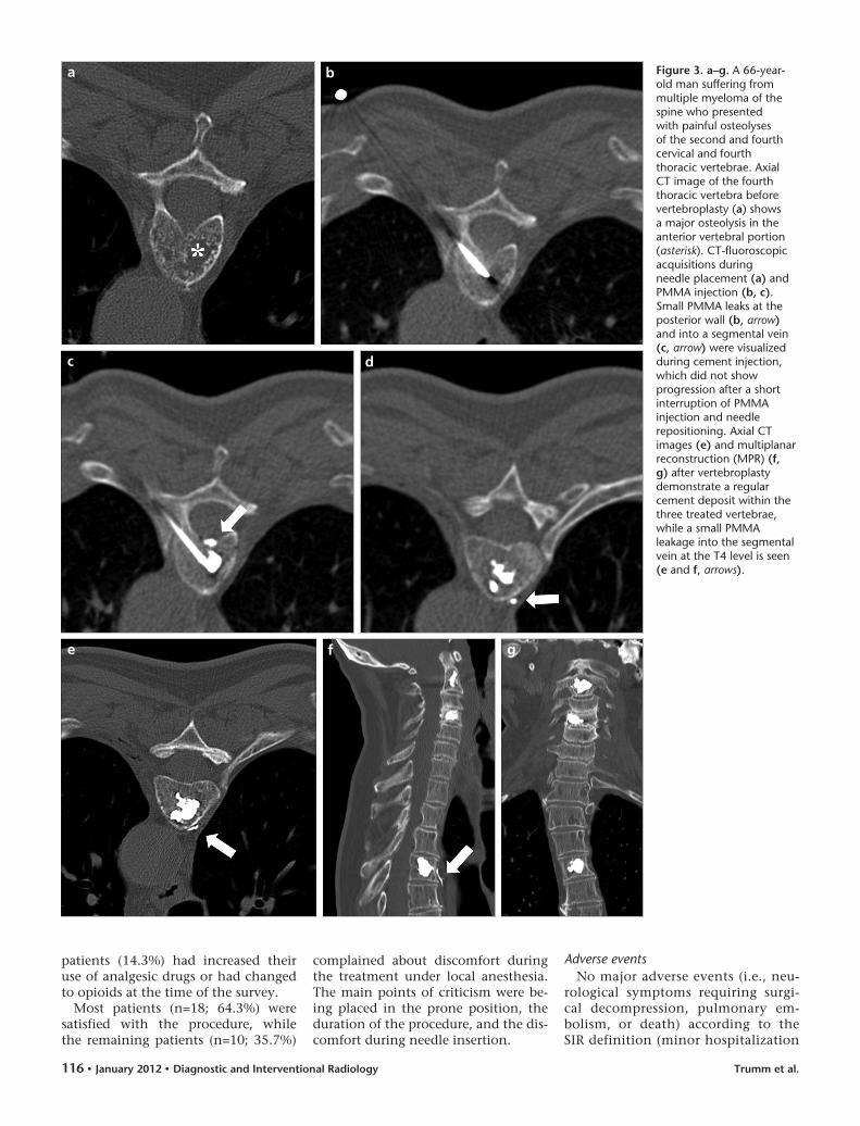
Figure 3. a–g. A 66-year-old man suffering from multiple myeloma of the spine who presented with painful osteolyses of the second and fourth cervical and fourth thoracic vertebrae. Axial CT image of the fourth thoracic vertebra before vertebroplasty (a) shows a major osteolysis in the anterior vertebral portion (asterisk). CT-fluoroscopic acquisitions during needle placement (a) and PMMA injection (b, c). Small PMMA leaks at the posterior wall (b, arrow) and into a segmental vein (c, arrow) were visualized during cement injection, which did not show progression after a short interruption of PMMA injection and needle repositioning. Axial CT images (e) and multiplanar reconstruction (MPR) (f, g) after vertebroplasty demonstrate a regular cement deposit within the three treated vertebrae, while a small PMMA leakage into the segmental vein at the T4 level is seen (e and f, arrows).
b
d
f
a
c
e g
CT fluoroscopy-guided percutaneous vertebroplasty in patients with multiple myeloma • 117Volume 18 • Issue 1
less than 48 hours, prolonged hospi-talizations longer than 48 hours for major therapy, permanent adverse sequelae, or deaths) were observed after dismissal of the patients to the ward or were documented in the clinical records of the referring clini-cians 6 months after the procedures (17). Moreover, no cases of infection within the treated spinal segments were found after the follow-up pe-riod. In patients with obvious local cement extravasation detected on the postinterventional CT scan, no chest pain, dyspnea or significant decrease of blood oxygen saturation was ob-served by the operators periinter-ventionally or immediately after the procedure.
After the application of the local an-esthetic drug, one patient developed a generalized seizure that was success-fully managed by the anesthesiologist on call. The vertebroplasty was success-fully performed one week later under general anesthesia.
One patient showed a pronounced drop in oxygen saturation after admin-istration of midazolam, requiring an immediate oxygen supply and admin-istration of flumazenil.
Two patients treated at the first lumbar and first sacral vertebral level, respectively, showed prolonged bleed-ing at the site of the skin incision and needle entry; they were successfully treated with manual compression and an additional single-knot suture.
DiscussionPercutaneous vertebroplasty has been
described as a safe and effective mini-mally invasive method for the treat-ment of tumoral involvement of the spine, resulting in rapid and continu-ing clinical improvements in pain and patient mobility (5, 6, 20, 21). Several reports have focused on the use of ver-tebroplasty, particularly in the treat-ment of osteolyses and compression fractures in myeloma patients (22–30). Conservative pain management with bed rest, bracing, bisphosphonates and analgesic drugs is often not sufficient to control the acute and chronic spinal pain accompanying pathologic frac-tures in myeloma patients. While the stabilizing effect of radiotherapy with
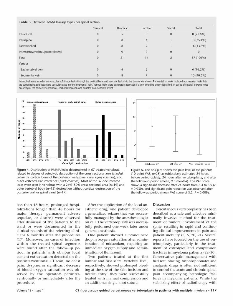
Table 3. Different PMMA leakage types per spinal section
Cervical Thoracic Lumbar Sacral Total
Intradiscal 0 5 3 0 8 (21.6%)
Intraspinal 0 8 4 1 13 (35.1%)
Paravertebral 0 8 7 1 16 (43.3%)
Intercostovertebral/posterolateral 0 0 0 0 0
Total 0 21 14 2 37 (100%)
Venous
Basivertebral vein 0 4 2 0 6 (16.2%)
Segmental vein 0 8 7 0 15 (40.5%)
Intraspinal leaks included nonvascular soft-tissue leaks through the cortical bone and vascular leaks into the basivertebral vein. Paravertebral leaks included nonvascular leaks into the surrounding soft tissue and vascular leaks into the segmental vein. Venous leaks were separately assessed if a vein could be clearly identified. In cases of several leakage types occurring at the same vertebral level, each leak location was counted as a separate event.
Figure 4. Distribution of PMMA leaks documented in 67 treated vertebrae, related to degree of osteolytic destruction of the cross-sectional area (shaded columns), cortical bone of the posterior wall/spinal canal (gray columns), and outer vertebral circumference (black columns). Most of the 37 documented leaks were seen in vertebrae with a 26%–50% cross-sectional area (n=19) and outer vertebral body (n=15) destruction without cortical destruction of the posterior wall or spinal canal (n=17).
Figure 5. The box plot shows the pain level of the patients (10-point VAS, n=28) as subjectively estimated 24 hours before vertebroplasty, 24 hours after vertebroplasty, and after the follow-up period (mean, 9.0 months). The VAS score shows a significant decrease after 24 hours from 6.4 to 3.9 (P = 0.030), and significant pain reduction was observed after the follow-up period (mean VAS score of 3.2, P = 0.009).
Trumm et al.118 • January 2012 • Diagnostic and Interventional Radiology
respect to progressive spinal deformity is minimal and delayed, major spinal surgeries, such as open decompression, internal fixation and bone grafting, may be limited due to comorbidities and reduced bone mass (2).
Our retrospective study aimed to assess the technical success in terms of the frequency and clinical impact of cement leaks occurring during CT fluoroscopy-guided vertebroplasty in patients suffering from MM of the spine. The primary intentions of the procedures were the relief of back pain refractory to NSAIDs, opioid medica-tion, and radiation therapy, as well as the stabilization of malignant VCFs or extensive osteolyses.
Chew et al. (31) recently published a meta-analysis of 30 studies that in-cluded 987 patients who underwent vertebroplasty due to spinal metasta-ses and myeloma. Several studies have demonstrated that an applied cement volume of greater than 4 mL correlates with an increased rate of complica-tions (4.2%–13.5%) (32, 33). All au-thors reported small-volume local, in-tradiscal or paravertebral cement leaks, but none of them was symptomatic. In a patient group with tumoral posterior wall involvement of the treated verte-brae, no increase in the complication rate was observed.
In a further meta-analysis of 69 studies by Hulme et al. (34), PMMA leaks without clinical consequences were also frequently observed (27 vertebroplasty studies; 41% vertebral leakage rate), while only 2.6% of the treated vertebrae (3.9% of patients) among 31 studies resulted in clinical complications.
Tancioni et al. (28) reviewed 28 ver-tebral levels in a cohort of MM patients treated at their institution. In 10 cases, vertebroplasty was performed at the time of diagnosis (six patients after chemotherapy and four before sys-temic therapy), and vertebroplasty was performed following disease progres-sion in four patients (two patients were treated after radiotherapy received for vertebral body fracture). Fracture or substitution of part of the posterior wall or pedicular tract was seen in 28%, and epidural disease was present in 14% of the treated vertebrae. Given an overall leakage rate of 21% and a me-dian amount of 4 mL PMMA injected, the aforementioned cases did not show any cement leaks.
Bartolozzi et al. (24) reported 19 procedures (10 kyphoplasties, 9 ver-tebroplasties) performed in 14 pa-tients (13 patients in Durie-Salmon stage IIIA, 1 patient in stage IIA) with MM. Vertebroplasty was chosen in collapsed vertebral bodies in cases of wide destruction of the posterior wall or in cases of increased bone density. Kyphoplasty was chosen in cases of a high risk of cement leakage due to vertebral instability. No complications were observed in either patient group. The authors concluded that both tech-niques can be safely and effectively performed in every phase of the dis-ease, independent of concomitant chemotherapy.
Ramos et al. (26) performed 19 verte-broplasty procedures in 12 consecutive myeloma patients. Despite a leakage rate of 84%, no patient developed neu-rological or other clinical symptoms.
It is commonly known that the rate of cement leakage detected depends on the utilized imaging modality. Therefore, when comparing leakage rates, the method of leak detection should always be mentioned. Schmidt et al. (35) compared the sensitivity of intraoperative fluoroscopy and post-operative radiography in the detection of cement leakage on postoperatively-acquired CT scans. With only lateral or biplane (anteroposterior and lateral) radiograms, the authors could only de-tect 34% or 48% of the leaks verified on CT, respectively. Due to the concav-ity and overprojection of the posterior wall in the thoracolumbar spine, the majority of leaks into the basivertebral vein (50% to 60%) were not detected with biplane radiography. Using intra-operative fluoroscopy, the detection rate was even lower (21%) due to the inferior image resolution.
In our patient group, local PMMA leaks were detected in 34 of 67 treated vertebrae (50.8% leakage rate). Given an injected cement volume ranging between 2 mL in the cervical and 5 mL in the sacral spine, 37.3% of the treated vertebrae were characterized by osteolytic destruction of more than 50% of the cross-sectional area and by osteolytic involvement of the posterior wall in 49.3% of the treated vertebrae. Aside from the aforementioned periin-terventional adverse events that could be successfully managed, there were no major adverse events observed in our patient group. As our results and those
of other authors showed, the number and volume of extraosseous cement leakages alone did not correlate with clinical complications (10). In our study, the amount of PMMA leakage was not calculated but was very small in most cases.
While osteolytic destruction of the posterior vertebral body wall and sig-nificant epidural tumor extension have been considered as relative con-traindications for vertebroplasty (16), CT fluoroscopy enables the operator to detect initial cement extravasation through the posterior wall; there-fore, this imaging modality might in-crease the safety of the vertebroplasty procedure, especially in those cases. According to our experience, with the use of a tube current-time product for CT fluoroscopic acquisitions between 15 to 25 mAs, a sufficient visualization of the posterior wall during needle in-sertion and cement injection is pos-sible, and relevant cement extravasa-tion or dislocation of fragments can be avoided. In combination with careful needle repositioning, initial leaks can be stopped if the injection is paused for at least 20–30 s, until the viscosity of the PMMA has increased
The mechanism of pain reduction following PMMA injection during ver-tebroplasty or kyphoplasty is still un-clear. The analgesic effect is mainly at-tributed to stabilization that prevents both microscopic movements and macroscopic collapse due to osteolytic destruction (36). Kaufmann et al. (37) and Cotten et al. (38) could not find a significant association between the amount of injected PMMA and the analgesic effect because even small ce-ment volumes lead to partial or com-plete pain relief. Other hypotheses regarding the underlying mechanism include the destruction of nerve end-ings through the exothermic reaction of PMMA during polymerization and its additional cytotoxic effects (39). These results are in accordance with our observations because even patients with a small vertebral filling volume of 1–2 mL of PMMA (13 of 67 vertebrae; 19.4%) in the thoracic and lumbar spine have reported substantial pain relief.
McDonald et al. (22) presented clini-cal outcome data from 67 MM pa-tients undergoing vertebroplasty over a 7-year period. They reported a statis-tically significant improvement on the
CT fluoroscopy-guided percutaneous vertebroplasty in patients with multiple myeloma • 119Volume 18 • Issue 1
Roland-Morris Disability Questionnaire, as well as in the VAS values that their patients documented immediately after the procedure and during the follow-up period of up to 1 year. In particular, the authors observed a similar short- and long-term reduction in scores when comparing their results in myeloma pa-tients to earlier analog measures of pain at rest and during activity in patients with osteoporotic fractures (40), sug-gesting a similar prognostic outcome in both patient groups. Our retrospective assessment of pain levels showed com-parable, significant mid-term (mean fol-low-up of 9.0 months) clinical success in the majority of the myeloma patients regarding both the semiquantitative (VAS score) and subjective (reduction of pain; use of analgesic agents) param-eters that were assessed.
In addition to evaluating the mid-term analgesic effect after vertebro-plasty (as attributed by the patients to the procedure) and the changes in the use of analgesic medication with respect to dose or strength, we did not separately assess the presence or dura-tion of chemo- or radiation therapy administered prior to or after the ver-tebroplasty procedure. The patients were usually referred to our depart-ment due to intractable back pain, despite the above-mentioned, non-invasive, palliative treatment regi-mens, or due to painful, pathologic compression fractures. After the treat-ment, systemic chemotherapy and/or local radiotherapy—if conformable with the general patient condition—were continued. As a consequence, an interference with the analgesic effect of vertebroplasty cannot be excluded in most cases.
Regarding the assessment of the clin-ical outcomes with the questionnaire, another drawback of this study is the moderate response rate of only 28 of 39 patients who completed the last questionnaire after 6 months.
In our patient group with osteolyses and VCFs due to MM of the spine, pri-mary technical success, in terms of the PMMA injection during the first ses-sion, was reached in all vertebrae, and significant clinical success was main-tained after a 9-month follow-up pe-riod. No clinical or neurological seque-lae were evident during the follow-up period. Given an experienced opera-tor, CT fluoroscopy provides excellent visualization of the posterior wall and
paravertebral soft tissues during needle placement and PMMA injection. This process enables palliative treatment to be given to myeloma patients, even with substantial osteolytic vertebral destruction.
Conflict of interest disclosureThe authors declared no conflicts of interest.
References 1. Lecouvet FE, Vande Berg BC, Maldague
BE, et al. Vertebral compression fractures in multiple myeloma. Part I. Distribution and appearance at MR imaging. Radiology 1997; 204:195–199.
2. Lieberman I, Reinhardt MK. Vertebroplasty and kyphoplasty for osteolytic vertebral collapse. Clin Orthop Relat Res 2003; 176–186.
3. Cortet B, Cotten A, Boutry N, et al. Percutaneous vertebroplasty in the treat-ment of osteoporotic vertebral compres-sion fractures: an open prospective study. J Rheumatol 1999; 26:2222–2228.
4. Deramond H, Depriester C, Galibert P, Le Gars D. Percutaneous vertebroplasty with polymethylmethacrylate. Technique, indi-cations, and results. Radiol Clin North Am 1998; 36:533–546.
5. Jensen ME, Kallmes DE. Percutaneous ver-tebroplasty in the treatment of malignant spine disease. Cancer J 2002; 8:194–206.
6. Weill A, Chiras J, Simon JM, Rose M, Sola-Martinez T, Enkaoua E. Spinal metastases: indications for and results of percutane-ous injection of acrylic surgical cement. Radiology 1996; 199:241–247.
7. Morrison WB, Parker L, Frangos AJ, Carrino JA. Vertebroplasty in the United States: guidance method and provider distribu-tion, 2001-2003. Radiology 2007; 243:166-170.
8. Vogl TJ, Proschek D, Schwarz W, Mack M, Hochmuth K. CT-guided percutaneous vertebroplasty in the therapy of vertebral compression fractures. Eur Radiol 2006; 16:797–803.
9. Gangi A, Kastler BA, Dietemann JL. Percutaneous vertebroplasty guided by a combination of CT and fluoroscopy. AJNR Am J Neuroradiol 1994; 15:83–86.
10. Pitton MB, Herber S, Koch U, Oberholzer K, Drees P, Duber C. CT-guided vertebro-plasty: analysis of technical results, ex-traosseous cement leakages, and complica-tions in 500 procedures. Eur Radiol 2008; 18:2568–2578.
11. Masala S, Konda D, Massari F, Simonetti G. Sacroplasty and iliac osteoplasty under combined CT and fluoroscopic guidance. Spine 2006; 31:667–669.
12. Strub WM, Hoffmann M, Ernst RJ, Bulas RV. Sacroplasty by CT and fluoroscopic guidance: is the procedure right for your patient? AJNR Am J Neuroradiol 2007; 28:38–41.
13. Brook AL, Mirsky DM, Bello JA. Com-puterized tomography guided sacroplasty: a practical treatment for sacral insuffi-ciency fracture: case report. Spine 2005; 30:450–454.
14. Layton KF, Thielen KR, Wald JT. Per-cutaneous sacroplasty using CT fluorosco-py. AJNR Am J Neuroradiol 2006; 27:356–358.
15. Criteria for the classification of mono-clonal gammopathies, multiple myelo-ma and related disorders: a report of the International Myeloma Working Group. Br J Haematol 2003; 121:749–757.
16. Gangi A, Sabharwal T, Irani FG, Buy X, Morales JP, Adam A. Quality assurance guidelines for percutaneous vertebro-plasty. Cardiovasc Intervent Radiol 2006; 29:173–178.
17. McGraw JK, Cardella J, Barr JD, et al. Society of Interventional Radiology quality improvement guidelines for percutaneous vertebroplasty. J Vasc Interv Radiol 2003; 14:311–315.
18. Baur A, Stabler A, Bruning R, et al. Dif-fusion-weighted MR imaging of bone marrow: differentiation of benign ver-sus pathologic compression fractures. Radiology 1998; 207:349–356.
19. Hohl C, Suess C, Wildberger JE, et al. Dose reduction during CT fluoroscopy: phan-tom study of angular beam modulation. Radiology 2008; 246:519–525.
20. Alvarez L, Perez-Higueras A, Quinones D, Calvo E, Rossi RE. Vertebroplasty in the treatment of vertebral tumors: postpro-cedural outcome and quality of life. Eur Spine J 2003; 12:356–360.
21. Jakobs TF, Trumm C, Reiser M, Hoffmann RT. Percutaneous vertebroplasty in tumoral osteolysis. Eur Radiol 2007; 17:2166–2175.
22. McDonald RJ, Trout AT, Gray LA, Dispenzieri A, Thielen KR, Kallmes DF. Vertebroplasty in multiple myeloma: out-comes in a large patient series. AJNR Am J Neuroradiol 2008; 29:642–648.
23. Pflugmacher R, Schleicher P, Schroder RJ, Melcher I, Klostermann CK. Maintained pain reduction in five patients with mul-tiple myeloma 12 months after treatment of the involved cervical vertebrae with ver-tebroplasty. Acta Radiol 2006; 47:823–829.
24. Bartolozzi B, Nozzoli C, Pandolfo C, et al. Percutaneous vertebroplasty and kypho-plasty in patients with multiple myeloma. Eur J Haematol 2006; 76:180–181.
25. Masala S, Anselmetti GC, Marcia S, Massari F, Manca A, Simonetti G. Percutaneous vertebroplasty in multiple myeloma ver-tebral involvement. J Spinal Disord Tech 2008; 21:344–348.
26. Ramos L, de Las Heras JA, Sanchez S, et al. Medium-term results of percutaneous vertebroplasty in multiple myeloma. Eur J Haematol 2006; 77:7–13.
27. Diamond TH, Hartwell T, Clarke W, Manoharan A. Percutaneous vertebroplas-ty for acute vertebral body fracture and deformity in multiple myeloma: a short report. Br J Haematol 2004; 124:485–487.
28. Tancioni F, Lorenzetti M, Navarria P, et al. Vertebroplasty for pain relief and spinal stabilization in multiple myeloma. Neurol Sci 2010; 31:151–157.
29. Anselmetti GC, Manca A, Montemurro F, et al. Percutaneous vertebroplasty in multiple myeloma: prospective long-term follow-up in 106 consecutive patients. Cardiovasc Intervent Radiol 2011 Feb 9 [Epub ahead of print 2011]

Trumm et al.120 • January 2012 • Diagnostic and Interventional Radiology
30. Saliou G, Kocheida el M, Lehmann P, et al. Percutaneous vertebroplasty for pain management in malignant fractures of the spine with epidural involvement. Radiology 2010; 254:882–890.
31. Chew C, Craig L, Edwards R, Moss J, O’Dwyer PJ. Safety and efficacy of percu-taneous vertebroplasty in malignancy: a systematic review. Clin Radiol 66:63–72.
32. Calmels V, Vallee JN, Rose M, Chiras J. Osteoblastic and mixed spinal metasta-ses: evaluation of the analgesic efficacy of percutaneous vertebroplasty. AJNR Am J Neuroradiol 2007; 28:570–574.
33. Barragan-Campos HM, Vallee JN, Lo D, et al. Percutaneous vertebroplasty for spi-nal metastases: complications. Radiology 2006; 238:354–362.
34. Hulme PA, Krebs J, Ferguson SJ, Berlemann U. Vertebroplasty and kyphoplasty: a sys-tematic review of 69 clinical studies. Spine 2006; 31:1983–2001.
35. Schmidt R, Cakir B, Mattes T, Wegener M, Puhl W, Richter M. Cement leakage during vertebroplasty: an underestimated prob-lem? Eur Spine J 2005; 14:466–473.
36. Evans AJ, Jensen ME, Kip KE, et al. Vertebral compression fractures: pain reduction and improvement in functional mobility after percutaneous polymethylmethacrylate vertebroplasty retrospective report of 245 cases. Radiology 2003; 226:366–372.
37. Kaufmann TJ, Trout AT, Kallmes DF. The effects of cement volume on clinical out-comes of percutaneous vertebroplasty. AJNR Am J Neuroradiol 2006; 27:1933–1937.
38. Cotten A, Dewatre F, Cortet B, et al. Percutaneous vertebroplasty for osteolytic metastases and myeloma: effects of the percentage of lesion filling and the leakage of methyl methacrylate at clinical follow-up. Radiology 1996; 200:525–530.
39. San Millan Ruiz D, Burkhardt K, Jean B, et al. Pathology findings with acrylic im-plants. Bone 1999; 25:85–90.
40. Trout AT, Kallmes DF, Gray LA, et al. Evaluation of vertebroplasty with a vali-dated outcome measure: the Roland-Morris Disability Questionnaire. AJNR Am J Neuroradiol 2005; 26:2652–2657.