Embed Size (px)
Citation preview
Hani Haykal1
Amir Zamani Ay-Ming Wang
John Barsotti
Received March 31, 1986; accepted after revision September 16, 1986
1 All authors: Department of Radiology, Harvard Medical School, Brigham and Women's Hospital, 75 Francis St. , Boston, MA 02115. Address reprint requests to H. Haykal.
AJNR 8:279-282, March/April 1987 0195-6108/87/0802-0279 © American Society of Neuroradiology
279
CT Features of Early Listeria monocytogenes Cerebritis
Listeria monocytogenes is a relatively uncommon pathogen affecting infants or adults with predisposing conditions, such as cirrhosis, diabetes mellitus, autoimmune disease, renal transplants, and solid and Iymphoreticular malignancies. Cerebral parenchymal involvement is rare and consists of focal cerebritis, which may progress to abscess formation. This article presents three cases of early Listeria monocytogenes cerebritis, two of which demonstrated ill-defined superficial areas of low attenuation with curvilinear gyral enhancement and one of which demonstrated a deep, low-attenuation lesion with faint surrounding enhancement. Although these findings are nonspecific, their early recognition in the proper clinical setting may help institute early antibiotic therapy, which appears to be successful without surgical intervention.
Listeria monocytogenes rarely involves the cerebral parenchyma. We report the cranial CT features of three cases in which such involvement was present, two of which demonstrate superficial lesions with low attenuation and curvilinear gyral enhancement. These findings have not been noted previously in the early phase of the infection. The third case shows a deep, low-density lesion with faint ring enhancement, although no suppurative lesion was later seen on necropsy.
Case Reports
Case 1
A 23-year-old man was admitted with a severe headache of 5 days' duration. He had a 2-month history of idiopathic thrombocytopenic purpura, for which he had been receiving Prednisone, 80 mg/day. At the time of admission , his steroid dose had been tapered to 10 mg/day. Five days before admission , he developed dizziness, diplopia, severe headache, and emotional lability. Lumbar puncture, performed the next day, was normal except for an opening pressure of 300 mm H20 . On admission , the patient was febrile (T 103.8°F) and showed signs of meningeal irritation. CT showed an ill-defined, superficial area of low attenuation in the left parietal lobe with curvilinear gyral enhancement (Fig . 1). A repeat lumbar puncture showed a pressure of 225 mm H20 and 300 WBC/hpf (14% P, 77% L, 7% M), with a protein value of 36 mg/dl and a glucose level of 68 mg/dl. CFS and blood cultures were positive for Listeria monocytogenes , and the patient was treated successfully with a 4-week course of intravenous penicillin .
Case 2
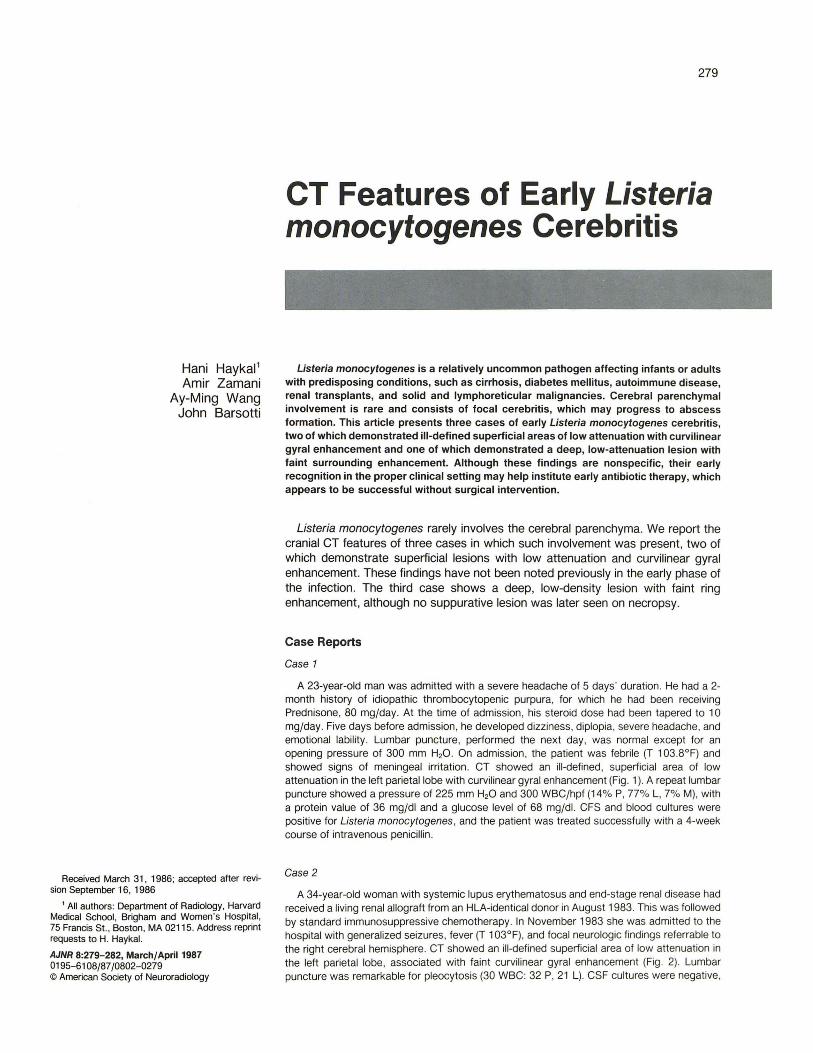
A 34-year-old woman with systemic lupus erythematosus and end-stage renal disease had received a living renal allograft from an HLA-identical donor in August 1983. This was followed by standard immunosuppressive chemotherapy. In November 1983 she was admitted to the hospital with generalized seizures, fever (T 103°F), and focal neurologic findings referrable to the right cerebral hemisphere. CT showed an ill-defined superficial area of low attenuation in the left parietal lobe, associated with faint curvilinear gyral enhancement (Fig. 2). Lumbar puncture was remarkable for pleocytosis (30 WBC: 32 P, 21 L). CSF cultures were negative,
280 HA YKAL ET AL. AJNR:8, March/April 1987
A B
A B
whereas the blood cultures were positive for Listeria monocytogenes. The patient was treated with intravenous penicillin for 4 weeks; her fever subsided and CT scans showed a decrease in the size of the right parietal lesion. Her neurologic symptoms resolved except for mild residual slurring of speech. In March 1984 she was readmitted with a 5-day history of severe right-sided headache and fever (T 101 OF). Her neurologic examination showed no interval change since her previous admission. Liver function tests showed a dramatic elevation. Two days later, the patient became confused and somnolent. At this time, serologic examination became newly positive for HBsAg. (Antibodies to HBsAg were negative.) She continued to show clinical deterioration and died 18 days after her final admission.
Case 3
A 19-year-old woman had been diagnosed as having acute myelogenous leukemia. She relapsed after chemotherapy and twice
Fig. 1.-Nonenhanced CT (A) shows iiI-defined region of low attenuation in left parietal area involving white matter, with curvilinear gyral enhancement (8).
Fig. 2.-Nonenhanced CT (A) shows iII-defined area of low attenuation in right frontoparietal area with mass effect. Enhanced CT (8) shows faint curvilinear gyral enhancement superficially and deep, ill-defined ring enhancement.
underwent bone marrow transplantation. Three days after receiving her second transplant, she became febrile and blood cultures were positive for Listeria monocytogenes . She was started on ampicillin intravenously. Two days later she developed generalized seizures. Lumbar puncture showed pleocytosis (60 WBC: 95% Pl. CSF cultures were positive for Listeria monocytogenes . The patient remained asymptomatic for 2 weeks, at which time she developed generalized seizures and lethargy and was readmitted.
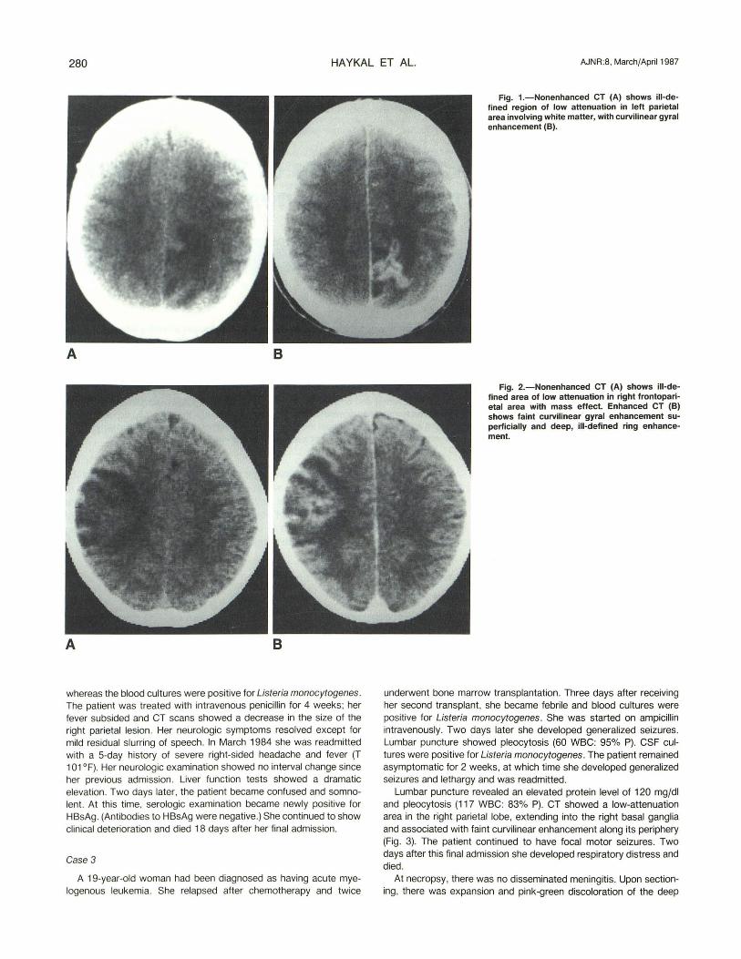
Lumbar puncture revealed an elevated protein level of 120 mg/dl and pleocytosis (117 WBC: 83% Pl. CT showed a low-attenuation area in the right parietal lobe, extending into the right basal ganglia and associated with faint curvilinear enhancement along its periphery (Fig. 3). The patient continued to have focal motor seizures. Two days after this final admission she developed respiratory distress and died.
At necropsy, there was no disseminated meningitis. Upon sectioning, there was expansion and pink-green discoloration of the deep
AJNR:8, March/April 1987 LISTERIA MONOCYTOGENES CEREBRITIS 281
Fig. 3.-Nonenhanced CT (A) shows deep ganglionic area of low attenuation with mass effect. Enhanced CT (8) shows faint curvilinear enhancement around periphery of zone of low attenuation (arrowhead) as well as vague gyral enhancement (arrow).
hemispheric structures of the right cerebral hemisphere extending from the anterior margin of the putamen to the thalamus, with involvement of the intervening lentiform nucleus and the internal capsule. Microscopically, the lesion consisted of multifocal perivascular and parenchymal foci of inflammatory cells with some focal necrosis. Organisms were seen with both Hand E and Gram stains, and with the latter they appeared as Gram-positive rods and were mostly present in the cytoplasm of the macrophages. Within the lesions there were necrotic small vessels, many containing fibrin thrombi, and there was fibrin leakage into the damaged parenchyma and diffuse edema.
Discussion
Listeria monocytogenes was first implicated as a cause of human disease by Nyfeldt in 1929 [1]. Since then , there have been numerous reports of this infection. In 1967, an association between Listeria and malignant disease was emphasized [2]. Since then, a number of factors have led to an increase in human Listeria infections. Chemotherapy and radiation therapy have prolonged the survival times of individuals with Iympho-proliferative [ 3, 4] and solid [5, 6] malignancies; unfortunately, these patients have suffered a concomitant increase in opportunistic infections. Renal transplantation with iatrogenic immunosuppression has also exposed a large number of patients to infectious complications. Other susceptible groups include pregnant women, diabetic patients, and patients with liver cirrhosis [7, 8].
Listeria monocytogenes is a facultative intracellular Grampositive pathogen. The organism is usually of low virulence but depends on its ability to survive and replicate within host phagocytes. Resistance to this organism is generally assumed to be associated with the appearance of cellular immunity, consisting of immunologically committed lymphocytes and the effector cells: the activated macrophage that possesses an enhanced ability to phagocytose and destroy intracellular pathogens [9, 10]. Animal experiments have
shown that corticosteroids suppress the production of the immunologically committed lymphocyte in the spleen, thereby enhancing the susceptibility to Listeria infection [11]. In mice, resistance to Listeria may be reduced by cytotoxic agents such as azathioprine and cyclophosphamide [12].
In a recent review of 102 cases [13], the major manifestation of listeriosis was meningitis in 50% of the patients, parenchymal disease of the CNS in 9%, and primary bacteremia in 30%, with an overall mortality rate of 25%. Prominent clinical features of meningitis include ataxia, tremors, seizures, and altered consciousness. CSF findings are variable, and the result of Gram stain is usually negative. Nonmeningitic infections of the CNS are characterized by an acute onset of hemiparesis or cranial nerve palsies. Blood cultures, in this form, almost invariably yield Listeria monocytogenes [14].
CT scan abnormalities have not been reported in meningeal Listeria infections, but might conceivably be similar to the findings seen in acute bacterial meningitis in the immunocompetent host. Since the first report of a brain abscess by Buchner and Schneierson in 1968 [7], there have been reports of seven patients with Listeria infections who had distinct macroscopic abscesses [15-20]. CT findings were reported in two cases [21 , 22] and were described as low-density areas with faint ring enhancement. To our knowledge, the CT features seen in the first two cases we are reporting-namely , superficial low-attenuation regions with gyral, curvilinear enhancement seen early in the clinical course of events-have not been previously reported . The third case we report demonstrates a faint ring-enhancing lesion involving the parietal lobe and extending into the basal ganglia, with features similar to the case reported by Lechtenberg et al. [21 ].
Summary
Infections due to Listeria monocytogenes are relatively uncommon and are usually restricted to infants or adults with
282 HA YKAL ET AL. AJNR:8, March/April 1987
predisposing conditions, such as cirrhosis , diabetes mellitus, autoimmune disease, renal transplants, and solid and Iymphoreticular malignancies. Cytoxic and corticosteroid chemotherapy also appear to be predisposing factors. The infection most commonly affets the meninges, and no CT abnormalities have been described to date. Parenchymal involvement is rare and consists of focal cerebritis, which may progress to macroscopic focal suppuration . The first two cases we report demonstrate the CT findings seen in the early cerebritis form. To our knowledge, these have not been described to date. They consist of ill-defined areas of low attenuation with curvil inear gyral enhancement. Although these findings are nonspecific, their early recognition in the proper clinical setting should suggest Listeria infections. Early antibiotic therapy appears to be successful without surgical intervention [23].
REFERENCES
1. Nyf Idt A. Etiologie de la mononucleose inlecteuse. CR Soc Bioi (PariS) 1929; 101 : 590- 591
2, Louri DB, Hensle T, Armstrong 0 , et al. Listerosis complicating malignant dis as . A new association. Ann Intern Med 1967; 67:260-281
3. Aisenb rg AC. Current concepts in cancer. The staging and treatment 01 Hodgkin' disease. N Engl J Med 1978; 299 :1228-1231
4. Ge rg L. Aur RJA, Mauer AM. Simone JV. A reappraisal of the results of stopping th rspy in childllood leukemia. N Engl J Med 1979; 300 :269-273
5. B n donna G, Bru molino E, Valagussa P, et al. Combination chemotherapy a an adjuv nt treatment in operable breast cancer. N Engl J Med 1976;294 :405- 410 Einhorn LH, Donohue J. Cis-diammine dichlaro-platinum, Vinblastine and bl mycin combination chemotherapy in disseminated testicular cancer. Arm/nt rn Moo 1979; 87 :293-298
7. Buchner LH, Schneierson SS. Clinical and laboratory aspects of Listeria monocytogenes infection. Am J Moo 1968; 45 :904-921
8. Halkin H, Shacked IJ , Altmann G. Brain abscess due to Listeria monocytogenes in a patient with cirrhosis of the liver. Br J Med Sci 1971 ; 7 : 11 92-1195
9. Mackaness GB. Resistance to intracellular infections. J Infect Dis 1971 ; 123 :439- 445.
10. Krahenbuhl JC, Rosenberg L T, Remington JS. The role of thymus-derived lymphocytes in the in-vitro activation of macro phages to kill Listeria monocytogenes. J Immunol 1973; 111 : 992-995
11 . Miller JK, Hedberg M. Effects of cortisone on susceptibility of mice to Listeria monocytogenes. Am J Clin Patho/1965 ; 43:248-250
12. Tripathy SP, Mackaness GB. The effects 01 cytotoxic agents on the primary immune response to Listeria monocytogenes. J Exp Med 1969; 130: 1-16
13. Stamm AM , Dismukes WE, Simmons BP, et al. listeriosis in renal transplant recipients: report of an outbreak and review of 102 cases. Rev Inf Dis 1982; 4 :665-682
14. Nieman RE , Lorber B. Listeriosis in adults: a changing pattern . Report of eight cases and review of the literature, 1968-1 978. Rev Inf Dis 1980;
2:207-225 15. Touraine JL, Toussaint C, Blanc N, Traeger J. Listeriose apres transplan
tation renale. Nouv Presse Med 1972; 1 :2813-2817 16. Crocker EF, Leicester J. Cerebral abscess due to Listeria monocytogenes.
Med J Aust 1976; 1: 90-92 17. Niklaisson PM, Hambraeus A, Lundgren G, Magnusson G, Sundelin P,
Groth CG. Listeria encephalitis in live renal transplant recipients. Acta Med Scand 1978; 203 :181- 185
18. Watson GW, Fuller TJ , Elms J, Kluge RM. Listeria cerebrit is: relapse of infection in renal transplant patients . Arch Intern Med 1978;138 :83-87
19. Buset M, Dupont E, Vereerstraeten P, et al. Listeriose apres transplantation renale: dix observations. Nouv Presse Med 1979; 8 : 3221-3224
20. Hudgins LB, Acchiareo SR. Diagnosis and treatment of Listeria monocytogenes infection in renal transplant recipients. Dialysis Transplantation 1978; 7: 1 023- 1 027
21 . Lechtenberg MD, Sierra MF, Prugle GF, Shucar WA, Butt KMH. Listeria monocytogenes: brain abscess or meningoencephalitis? Neurology 1979; 29:86-90
22. Dykes A, Baraff LJ, Herzog P. Listeria brain abscess in an immunosuppressed child. J Pediatr 1979; 94 :72-74
23. Chow AW, Alexander E, Montogomerie JZ, Guze LB. Successful treatment of non-meningitic Listerial brain abscess without operation. West J Med 1975; 122: 167-171