Embed Size (px)
Citation preview
CT DIAGNOSTICS W. B. Henry Jr. DVM, DACVS
CT DIAGNOSTICS W. B. HENRY, JR. DVM, DACVS •
BUZZARDS BAY
•DEN
NIS
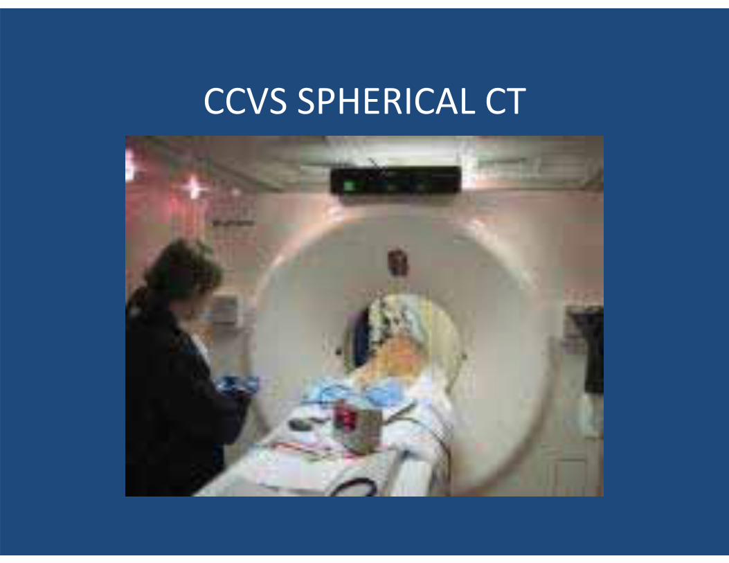
CCVS SPHERICAL CT
• CT = Computed Tomography ( CAT Scan ) • A Radiographic Image that uses computer processing
to generate an image of tissue density in slices through the patient’s body.
• CT imaging offers superior diagnostic images over plain radiography.
• Radiographs are flat projections in two dimensions of tri-dimensional (3D) structures With a CT tissues are examined in thin slices and can be examined in three dimensions thus eliminating superimposition of different tissues and organs.
• Organs and other structures can then be differentiated more easily by reformatting (by computers) in any imaging plane, or as 3D projections.
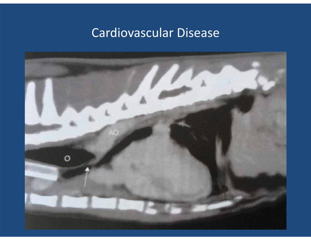
Cardiovascular Disease
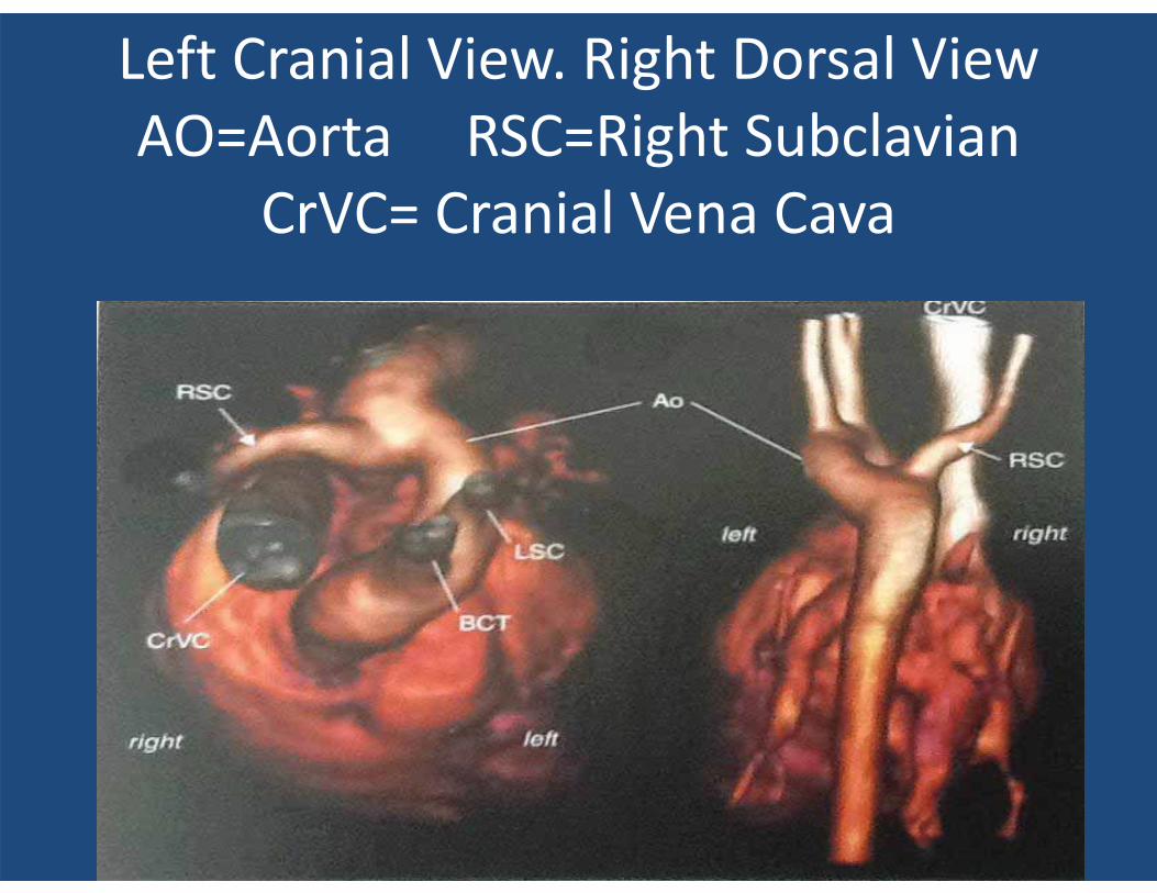
Left Cranial View. Right Dorsal View AO=Aorta RSC=Right Subclavian
CrVC= Cranial Vena Cava
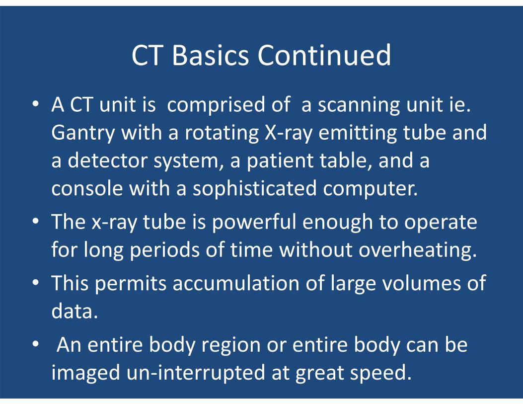
CT Basics Continued • A CT unit is comprised of a scanning unit ie.
Gantry with a rotating X-ray emitting tube and a detector system, a patient table, and a console with a sophisticated computer.
• The x-ray tube is powerful enough to operate for long periods of time without overheating.
• This permits accumulation of large volumes of data.
• An entire body region or entire body can be imaged un-interrupted at great speed.
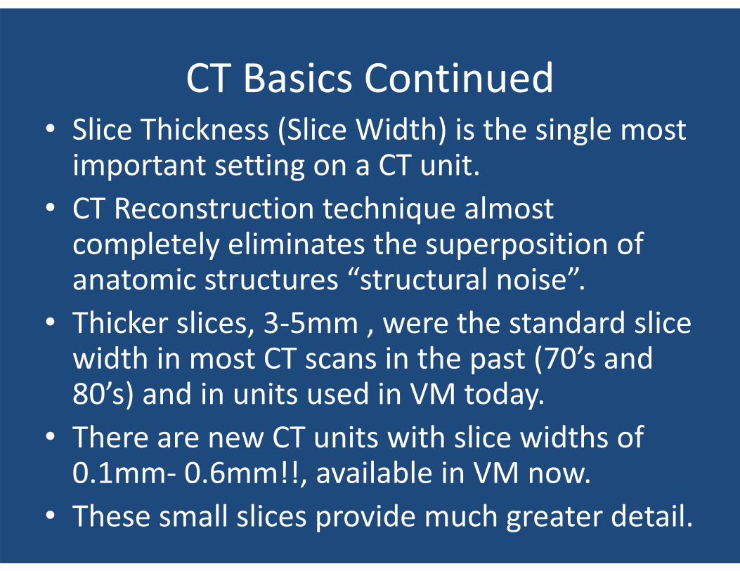
CT Basics Continued • Slice Thickness (Slice Width) is the single most
important setting on a CT unit. • CT Reconstruction technique almost
completely eliminates the superposition of anatomic structures “structural noise”.
• Thicker slices, 3-5mm , were the standard slice width in most CT scans in the past (70’s and 80’s) and in units used in VM today.
• There are new CT units with slice widths of 0.1mm- 0.6mm!!, available in VM now.
• These small slices provide much greater detail.
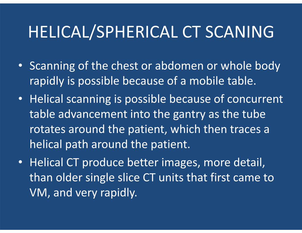
HELICAL/SPHERICAL CT SCANING
• Scanning of the chest or abdomen or whole body rapidly is possible because of a mobile table.
• Helical scanning is possible because of concurrent table advancement into the gantry as the tube rotates around the patient, which then traces a helical path around the patient.
• Helical CT produce better images, more detail, than older single slice CT units that first came to VM, and very rapidly.
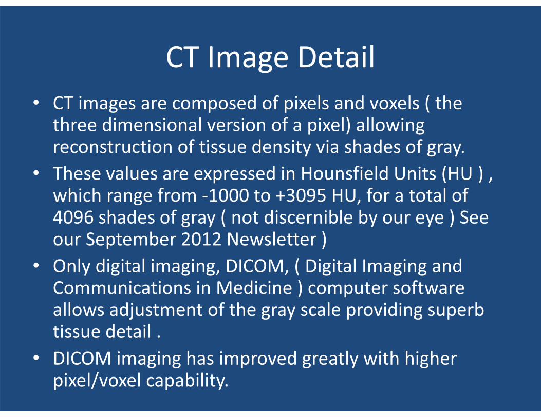
CT Image Detail • CT images are composed of pixels and voxels ( the
three dimensional version of a pixel) allowing reconstruction of tissue density via shades of gray.
• These values are expressed in Hounsfield Units (HU ) , which range from -1000 to +3095 HU, for a total of 4096 shades of gray ( not discernible by our eye ) See our September 2012 Newsletter )
• Only digital imaging, DICOM, ( Digital Imaging and Communications in Medicine ) computer software allows adjustment of the gray scale providing superb tissue detail .
• DICOM imaging has improved greatly with higher pixel/voxel capability.
Interpretation of CT Images
• It necessitates an understanding of the range of HU found in different types of normal and abnormal tissues.
• Soft tissues share the same opacity on radiographs, mild differences can be better detected with a CT, thanks to it’s superior contrast resolution.
• Contrast enhancing solutions help define tissues better, again also enhanced by the number of image pixels available now.
Iodinated Contrast Enhancement
• Requires a high pressure/high speed injection pump providing IV bolus injection.
• The distribution of this “hyperattenuating substance” (dye) can be tracked throughout the body.
• This provides perfusion information of tissues.
• The HU of a tissue will increase in proportion to the concentration of the contrast medium. (Vascular soft tissue tumor vs. minimally vascular tumors ie benign lipoma.)
CT Angiography
• Angiography represents another important application of the CT in veterinary medicine.
• Must have helical scanner and automated contrast injectors.
• Scans are performed rapidly after the bolus injections and timed for the vascular phase examined , examples cardiac anomaly (PRS) vs. a liver anomaly (PSS)
• Most dye enhanced scans need not be timed.
Principal Application of CT • Whole body imaging for tumor staging ( most
accurate method of detecting lung metastasis ).
• Evaluation of disease affecting the head ( including the nasal and middle ears ).
• Bone, Joint, and Spinal Disease. • Thoracic Disease and Abdominal Disease • Surprisingly, helpful in diagnosing brain
disease!! As you will soon see….
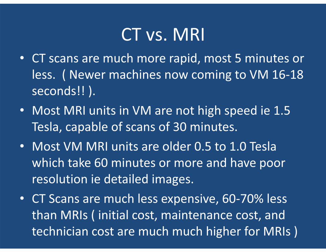
CT vs. MRI • CT scans are much more rapid, most 5 minutes or
less. ( Newer machines now coming to VM 16-18 seconds!! ).
• Most MRI units in VM are not high speed ie 1.5 Tesla, capable of scans of 30 minutes.
• Most VM MRI units are older 0.5 to 1.0 Tesla which take 60 minutes or more and have poor resolution ie detailed images.
• CT Scans are much less expensive, 60-70% less than MRIs ( initial cost, maintenance cost, and technician cost are much much higher for MRIs )
CT Scans of the Head
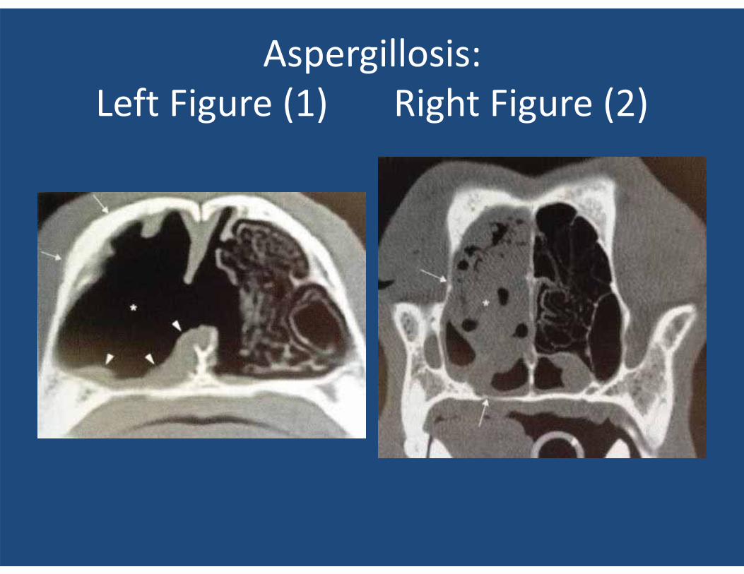
Aspergillosis: Left Figure (1) Right Figure (2)
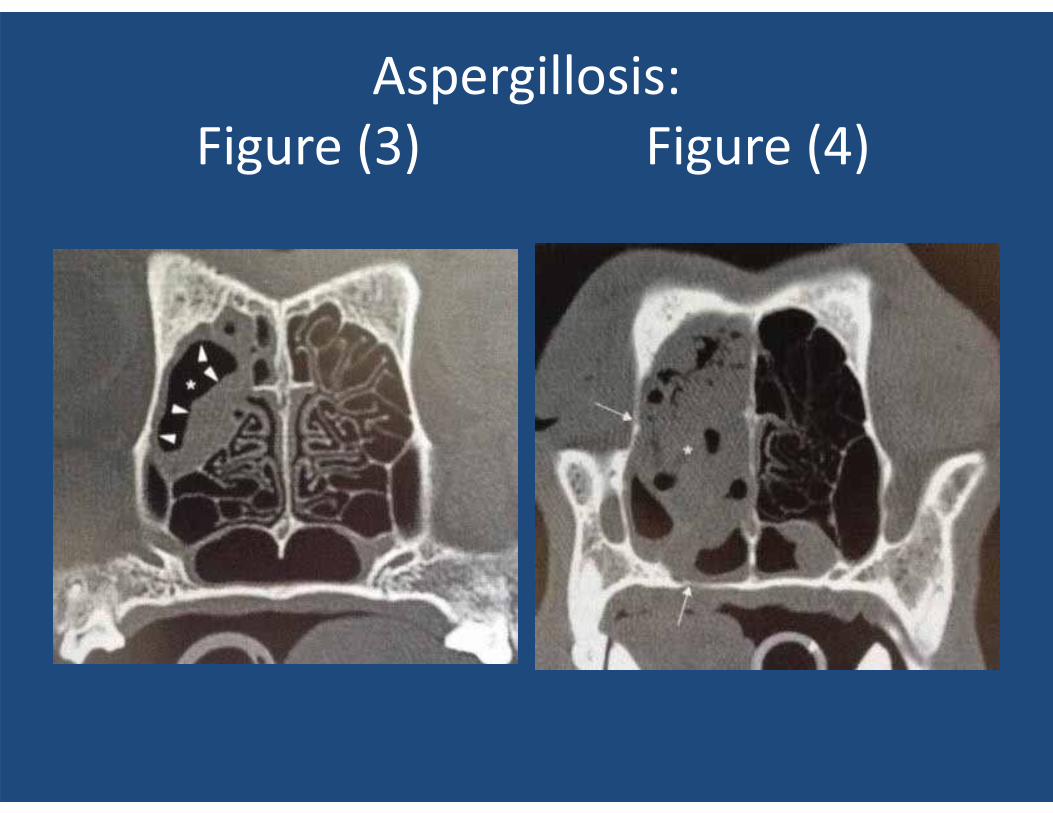
Aspergillosis: Figure (3) Figure (4)
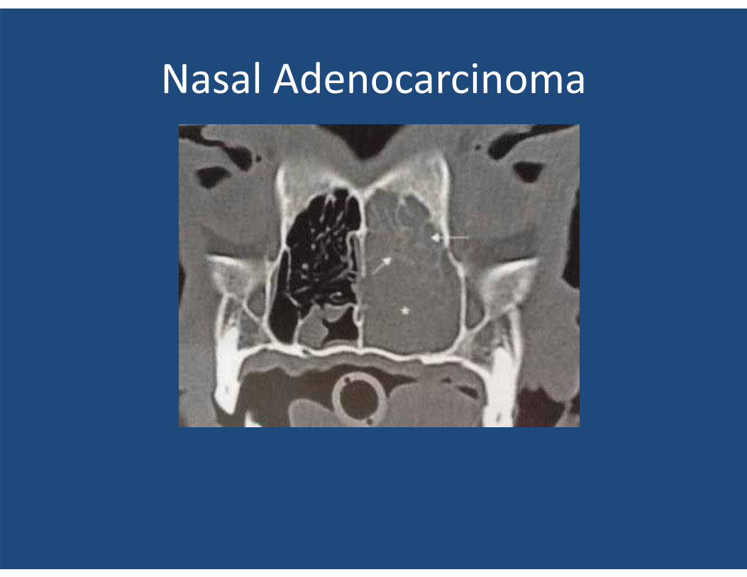
Nasal Adenocarcinoma
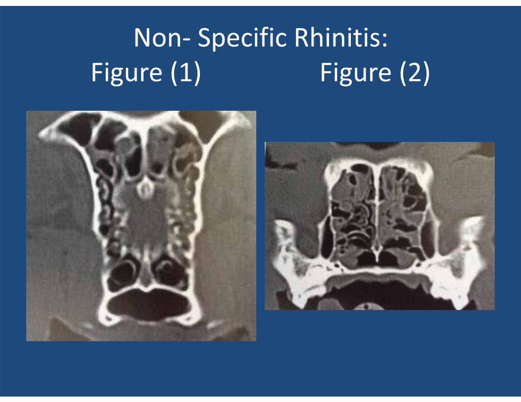
Non- Specific Rhinitis: Figure (1) Figure (2)
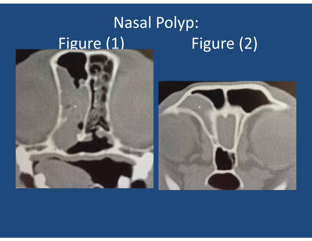
Nasal Polyp: Figure (1) Figure (2)
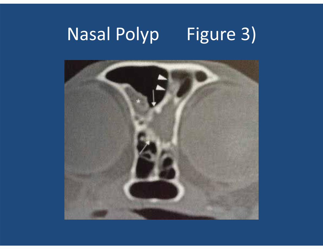
Nasal Polyp Figure 3)
Temporomandibular Joint Disease (TMJ )
• TMJ joints are difficult to visualize with two dimensioal radiographs.
• The CT of TMJ joints demonstrates the detail of this joint and “OMG”, look what I could not see until you have a CT allowing isolation and three dimensional views of a TMJ!!
• There can be dislocations, fractures, DJD secondary to trauma, benign bone cysts , even OCD lesions we NEVER saw before or knew existed.!!
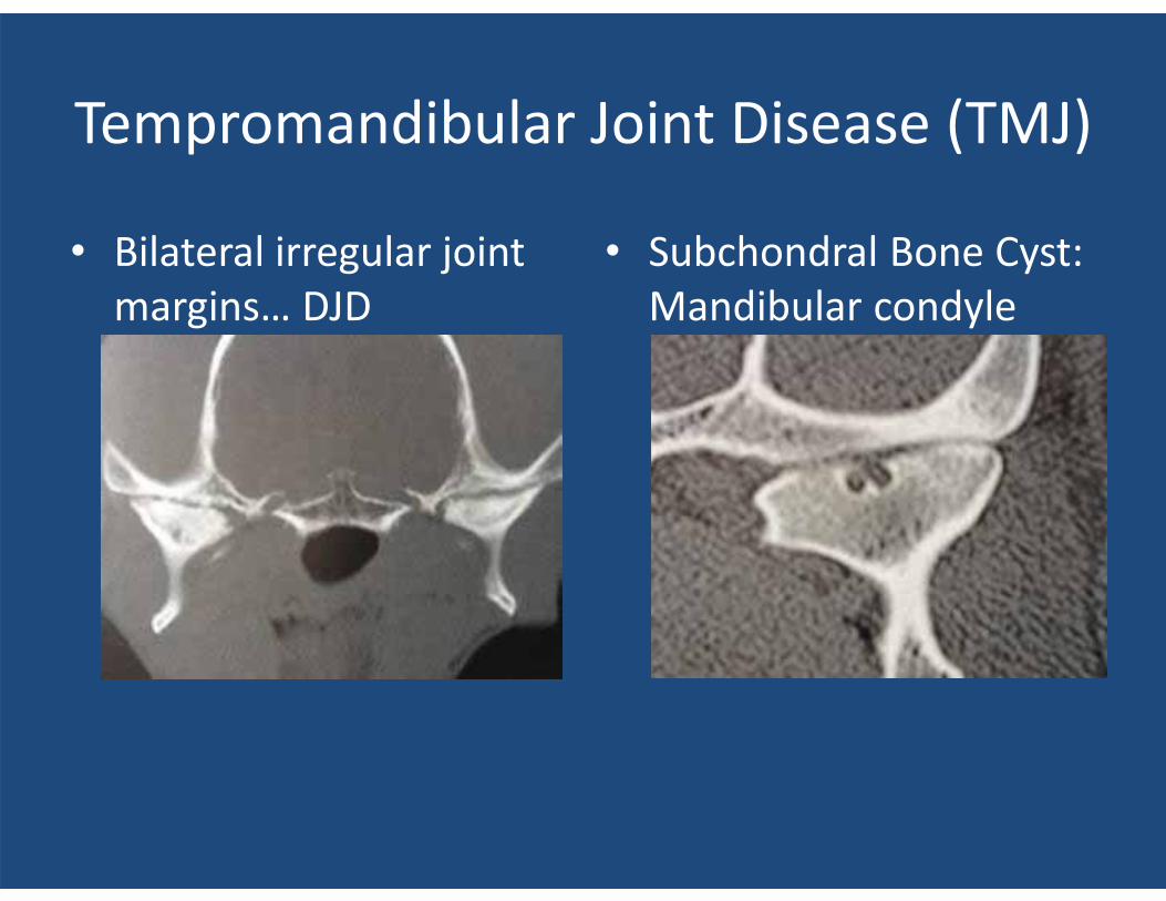
Tempromandibular Joint Disease (TMJ)
• Bilateral irregular joint margins… DJD
• Subchondral Bone Cyst: Mandibular condyle
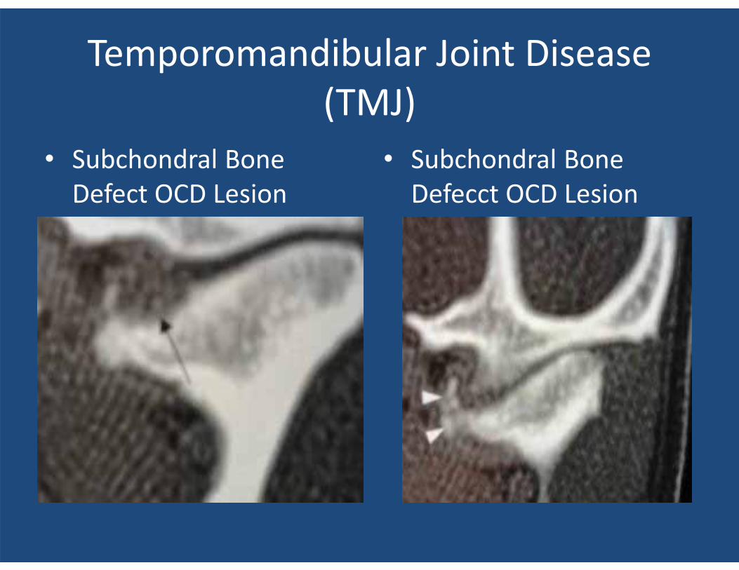
Temporomandibular Joint Disease (TMJ)
• Subchondral Bone Defect OCD Lesion
• Subchondral Bone Defecct OCD Lesion
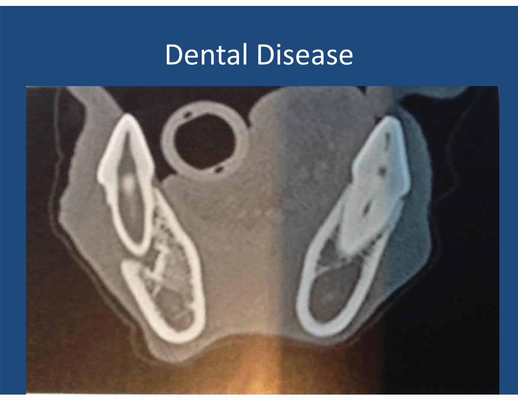
Dental Disease
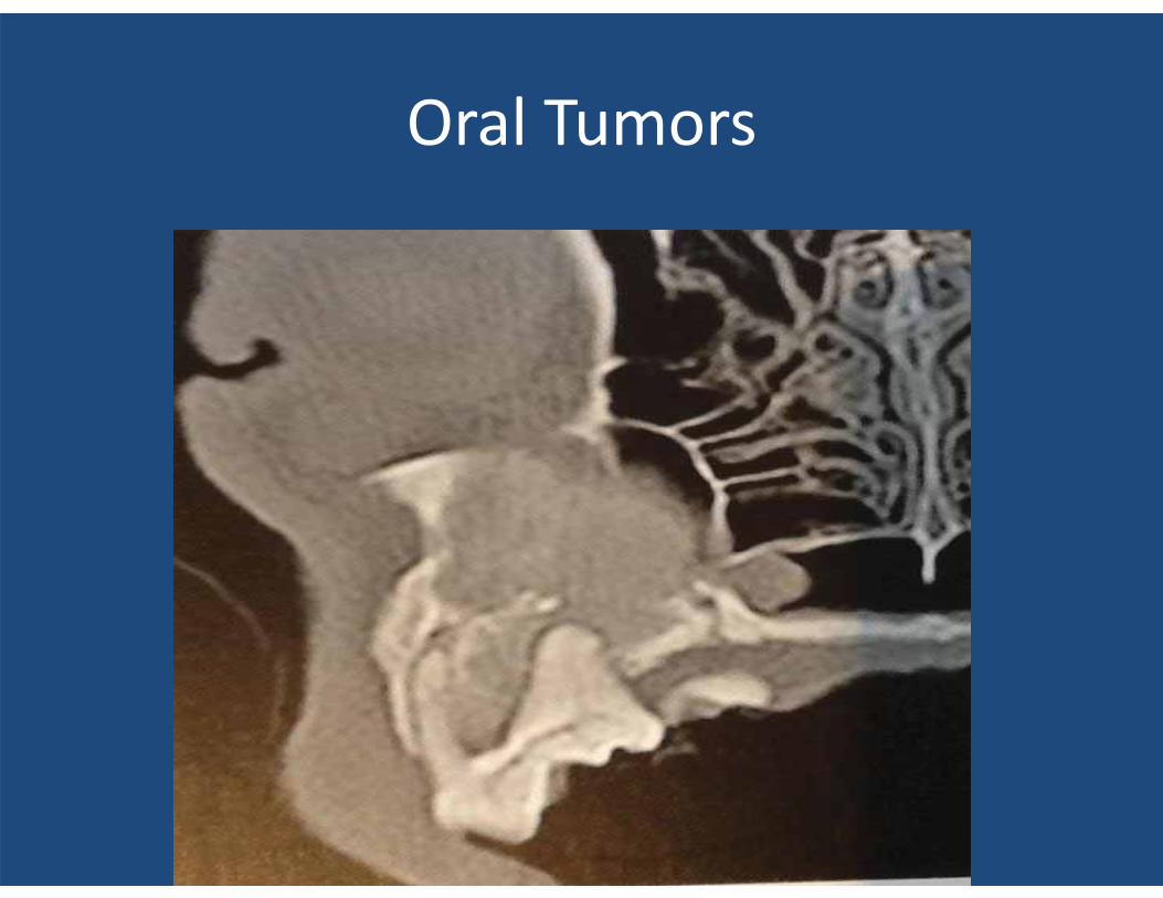
Oral Tumors
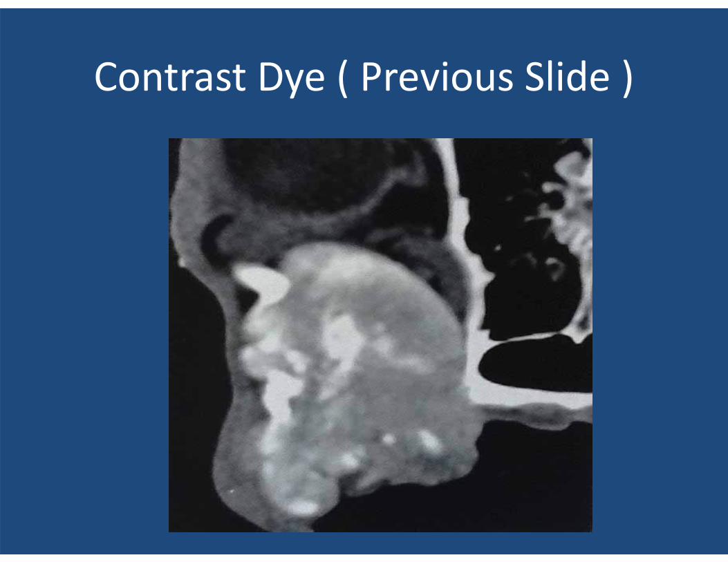
Contrast Dye ( Previous Slide )
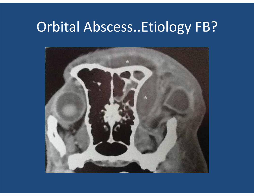
Orbital Abscess..Etiology FB?
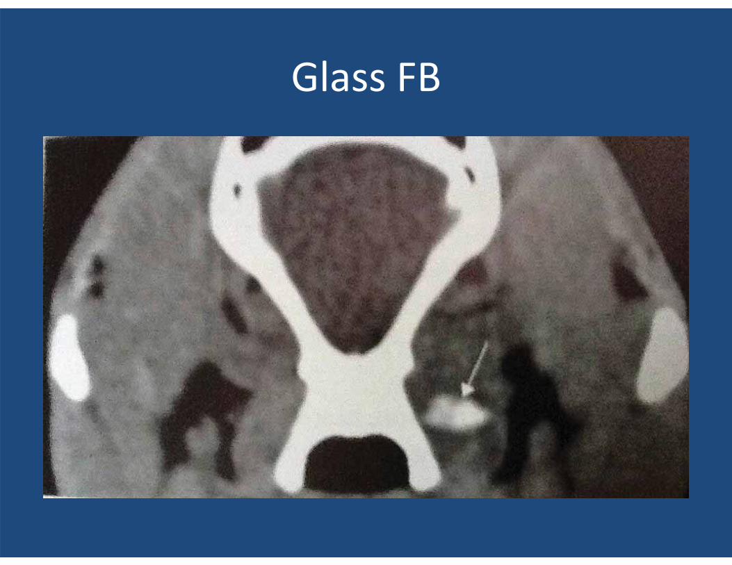
Glass FB
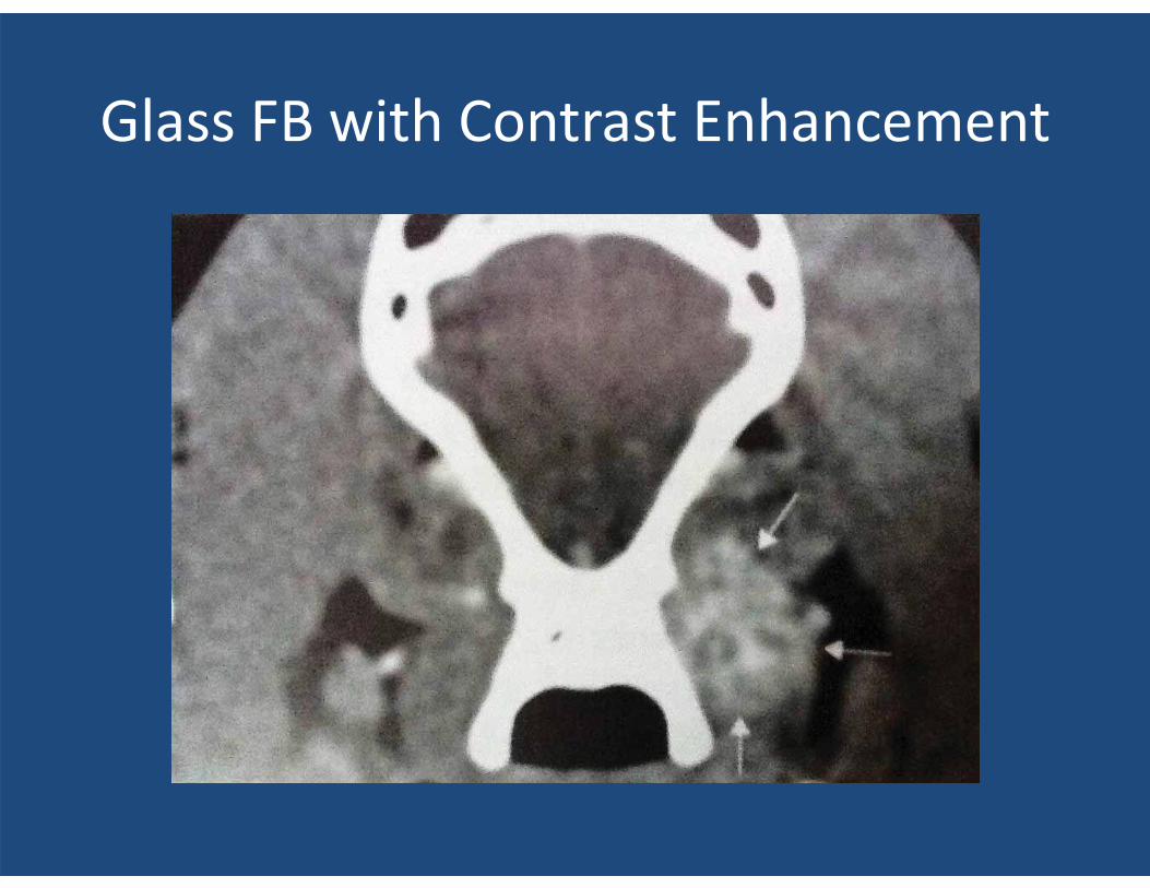
Glass FB with Contrast Enhancement
BRAIN CTs
• Brain CTs are more diagnostic for brain disease than most realize. MRIs get all the press, why?
• Brain disease is more treatable in human medicine and MRIs provide more finite detail.
• The CT scans to follow will demonstrate their effectiveness in diagnosing primary and metastatic brain tumors, meningioencephalitis/leukoencephalitis, bacterial meningoencephalitis, hemorrhagic infarcts, and hydrocephalus.
Advantage of CT vs. MRI
• CTs are 60-70% less expensive than MRIs. • The anesthetic time is 90% less! • The CT scan times are 5 minutes or less ( now
available in VM, 15-60 seconds!! ) MORE LATER ON THIS!!
• MRI scan times are 30 minutes at a minimum if you have a 1.5 Tesla unit. ( only a few of these in VM ). Thus many MRI units in VM take 60-90 minutes scan time and have poor resolution. Reason$$, Initial cost and maintenance cost for high field MRs, 1.5 Tesla.
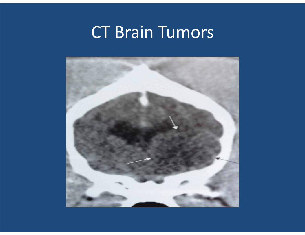
CT Brain Tumors
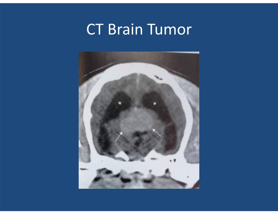
CT Brain Tumor
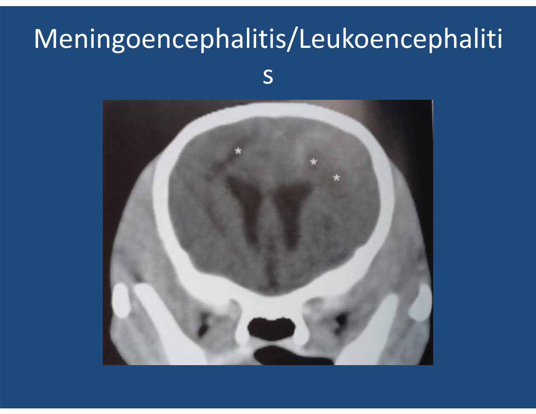
Meningoencephalitis/Leukoencephalitis
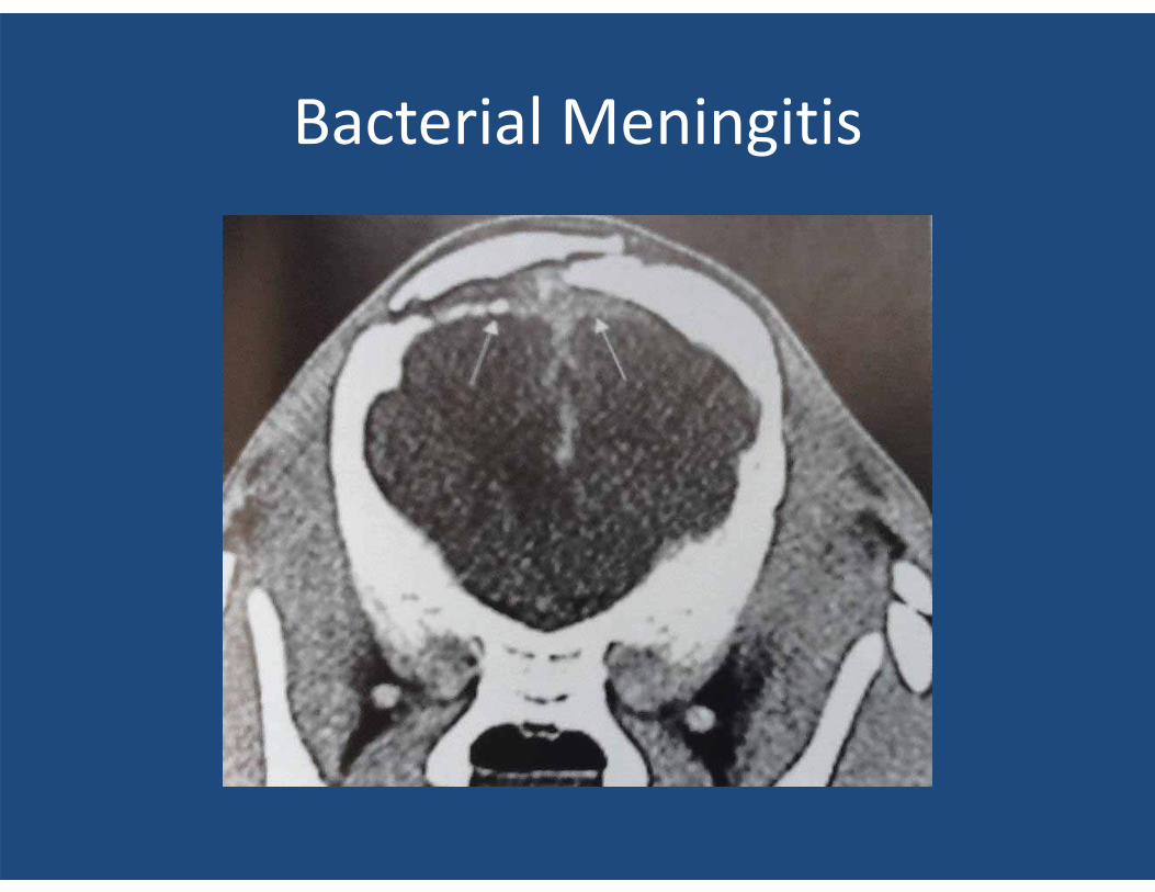
Bacterial Meningitis
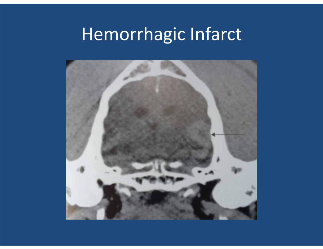
Hemorrhagic Infarct
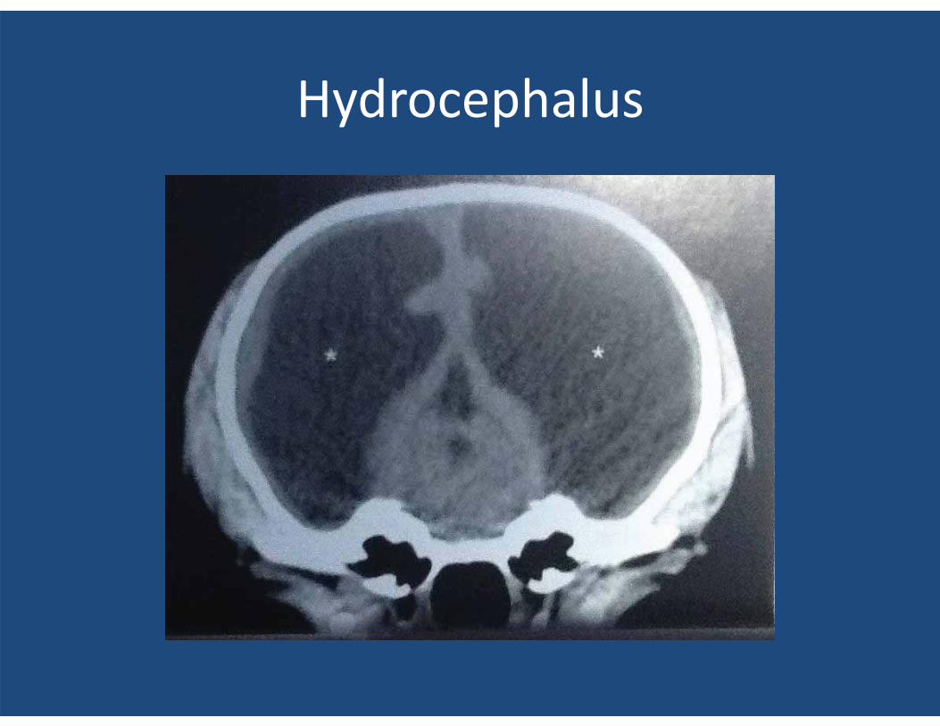
Hydrocephalus
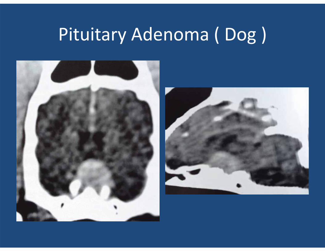
Pituitary Adenoma ( Dog )
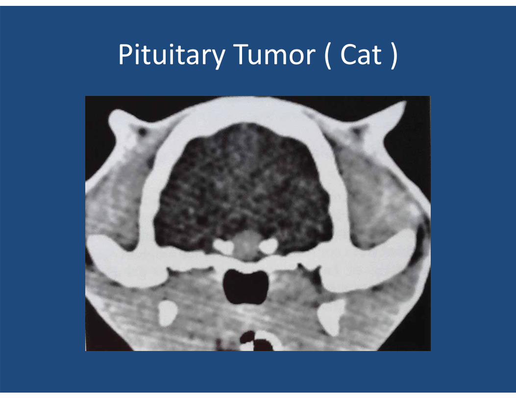
Pituitary Tumor ( Cat )
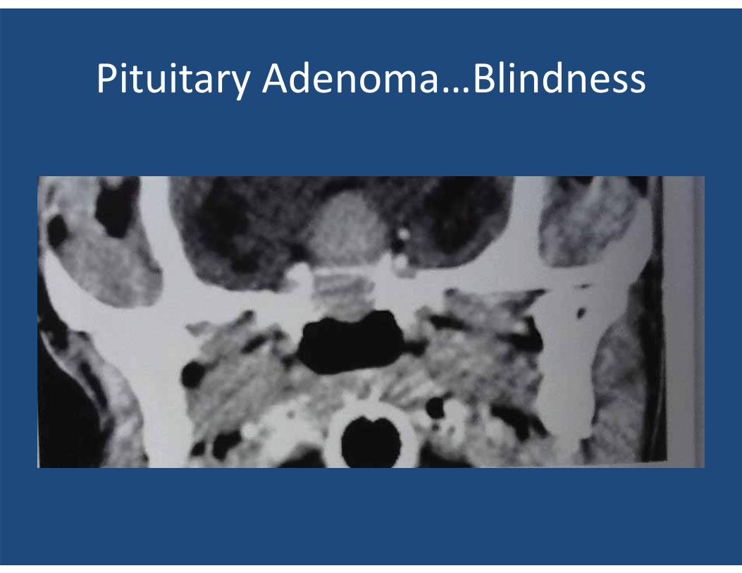
Pituitary Adenoma…Blindness
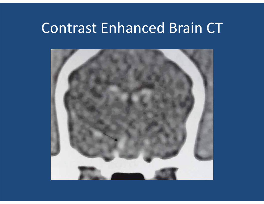
Contrast Enhanced Brain CT
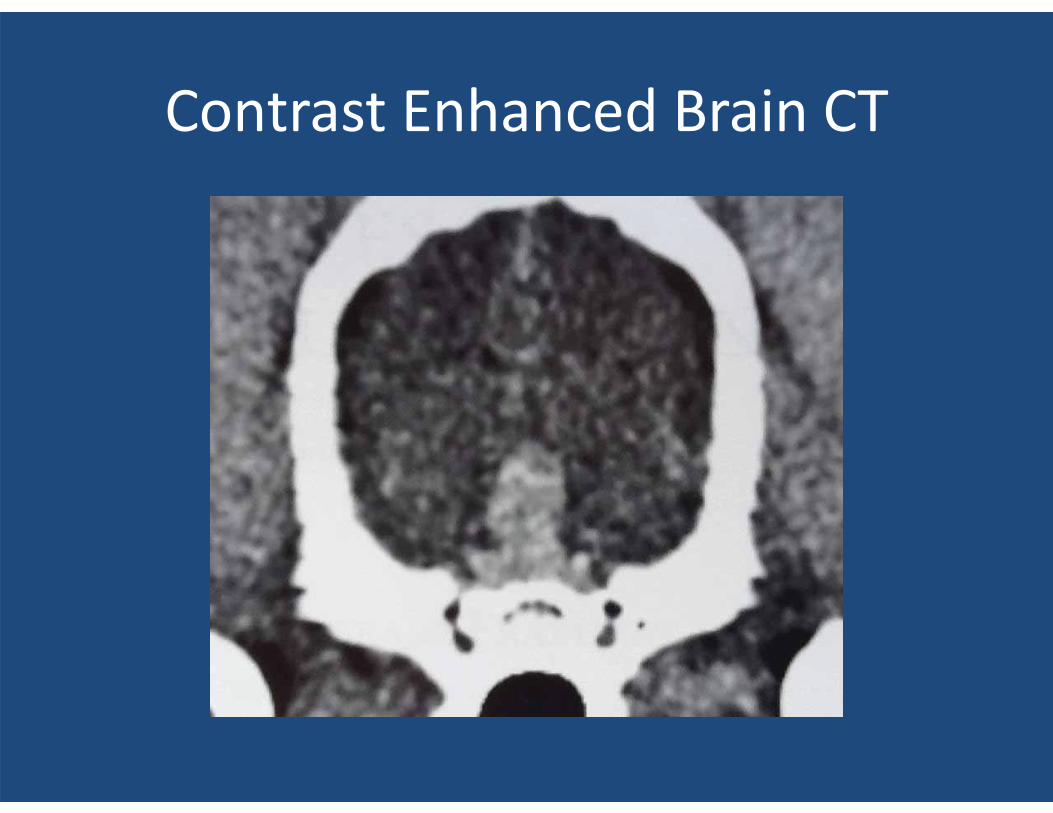
Contrast Enhanced Brain CT
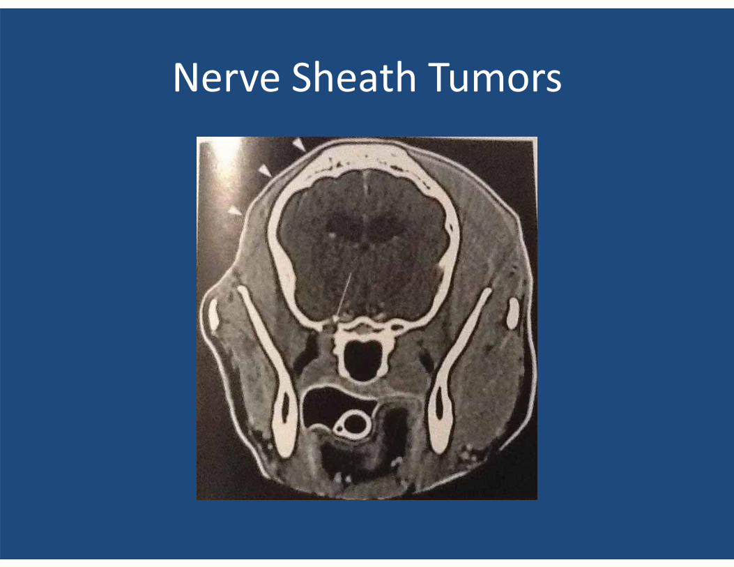
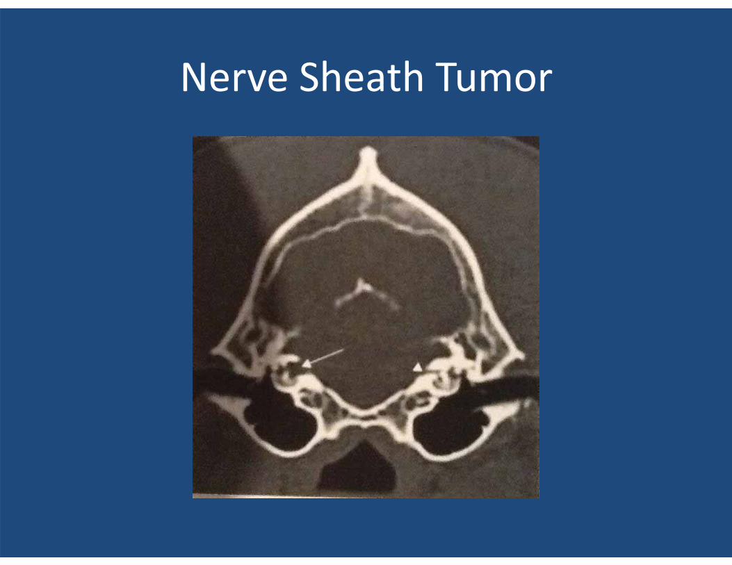
Nerve Sheath Tumors
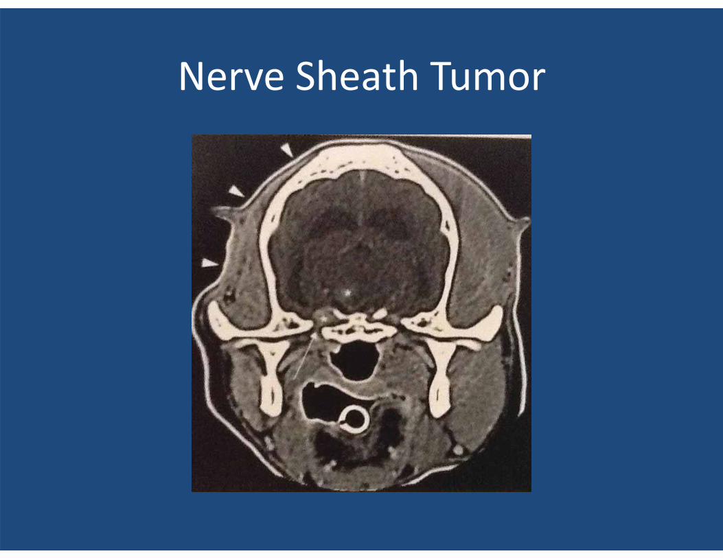
Nerve Sheath Tumor
Nerve Sheath Tumor
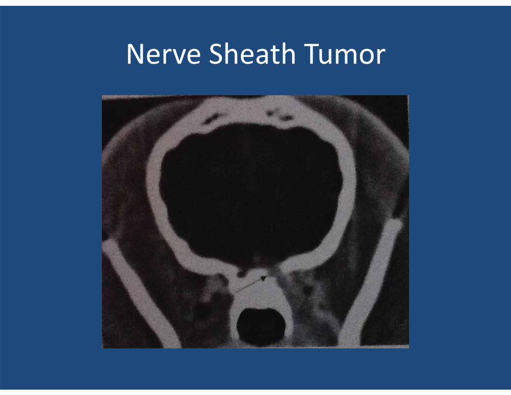
Nerve Sheath Tumor
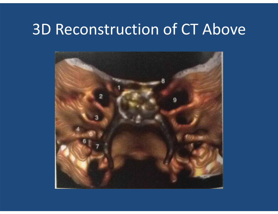
3D Reconstruction of CT Above
CT Scans of Fatty Tumors??
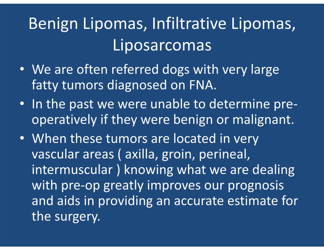
Benign Lipomas, Infiltrative Lipomas, Liposarcomas
• We are often referred dogs with very large fatty tumors diagnosed on FNA.
• In the past we were unable to determine pre-operatively if they were benign or malignant.
• When these tumors are located in very vascular areas ( axilla, groin, perineal, intermuscular ) knowing what we are dealing with pre-op greatly improves our prognosis and aids in providing an accurate estimate for the surgery.
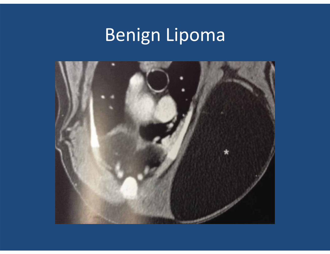
Benign Lipoma
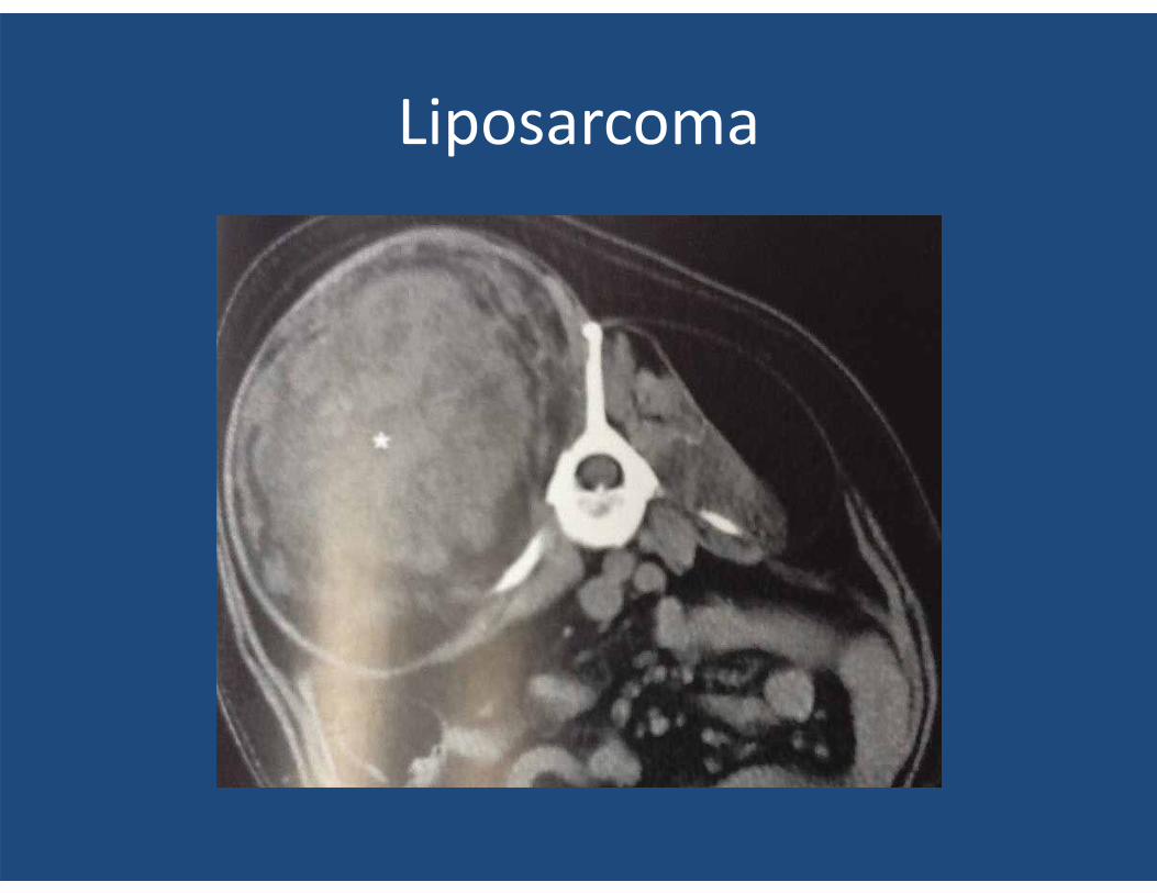
Liposarcoma
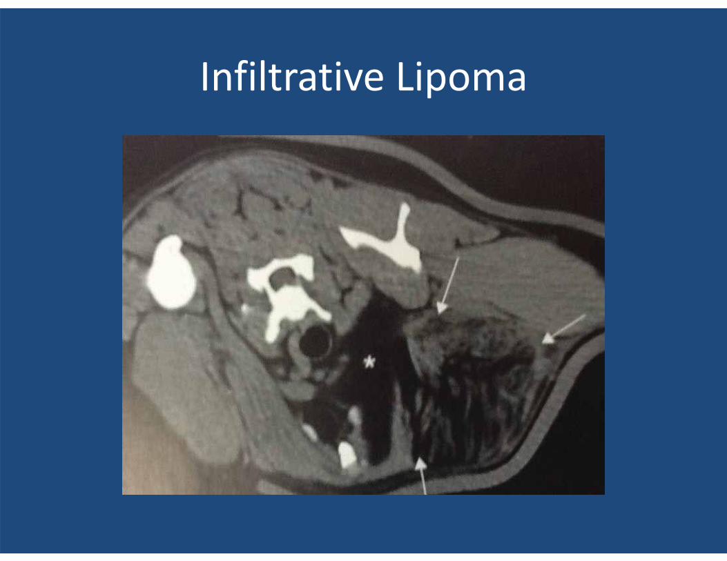
Infiltrative Lipoma
SPINAL DISEASE
• Malformations: Atlantoaxial subluxation, Syringomyelia, Cervical spondylomyelopathy, Trauma ( dural tears, fractures/luxation, hemorrhage, myelomalacia ), IVDD, Neoplasia, Discospondylitis, Enlarge articular processes and dorsal ligament hypertrophy causing spinal cord compression, L/S OCD lesions, Subarchnoid cysts. ALL OF WHICH I AM NOT GOING TO SHOW EXAMPLES DUE TO TIME!!
Lumbosacral Instability
• A definitive diagnosis is best accomplished by dynamic imaging of the L/S.
• Only necessary if contemplating Sx. • CT’s provide excellent bone resolution. • Because CT images can be obtained in minutes
axial flexion and extension positioning are less challenging than for MRI that requires much more scan time for each L/S position.
• Transverse CT images provide the best view the nerve roots as they exits thru the spinal foramena.
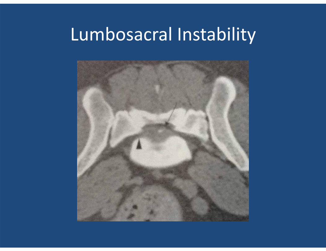
Lumbosacral Instability
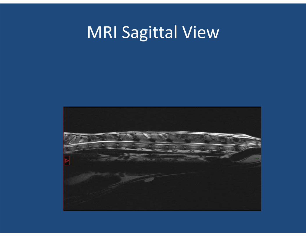
MRI Sagittal View
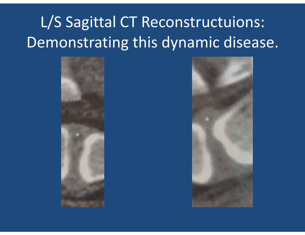
L/S Sagittal CT Reconstructuions: Demonstrating this dynamic disease.
THORACIC DISEASE
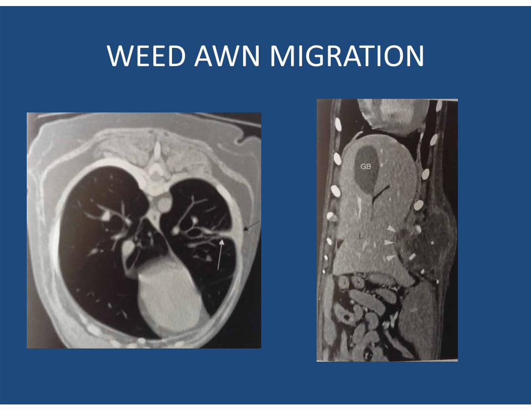
WEED AWN MIGRATION
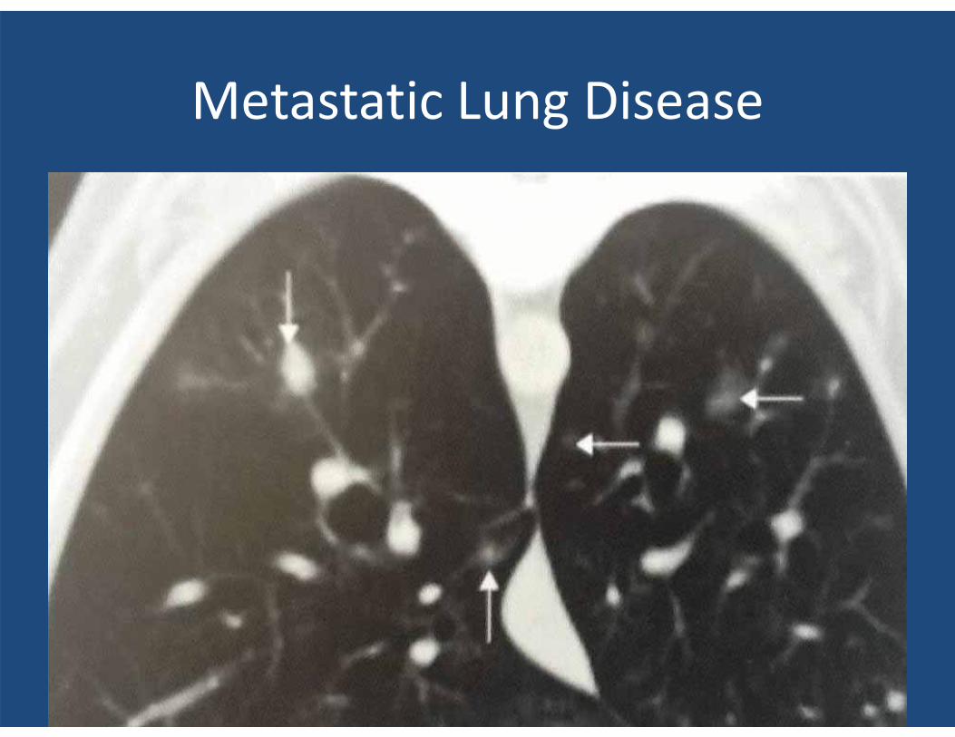
Metastatic Lung Disease
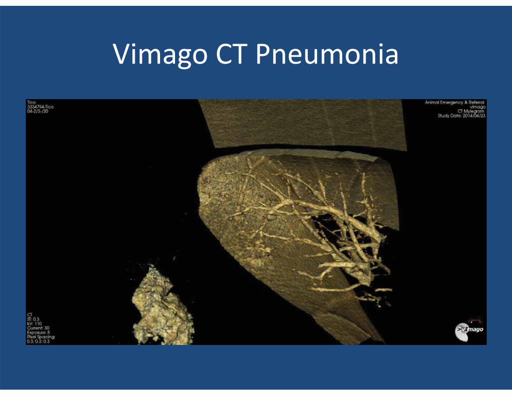
Vimago CT Pneumonia
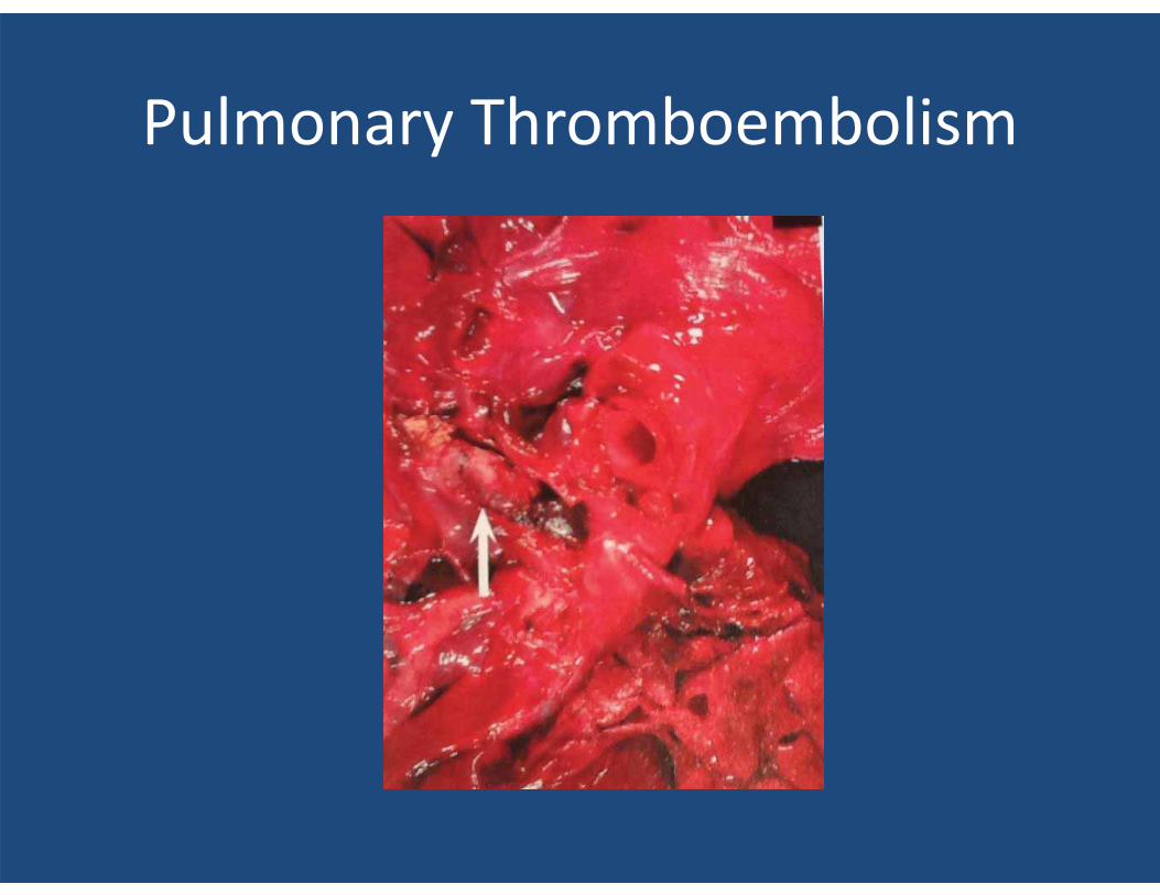
Pulmonary Thromboembolism
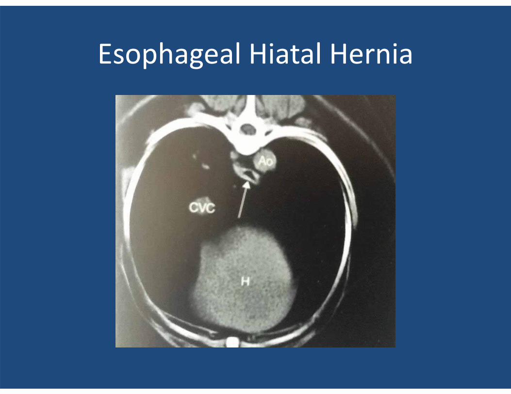
Esophageal Hiatal Hernia
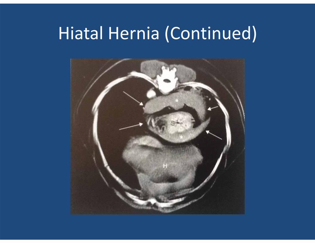
Hiatal Hernia (Continued)
ABDOMINAL DISEASE
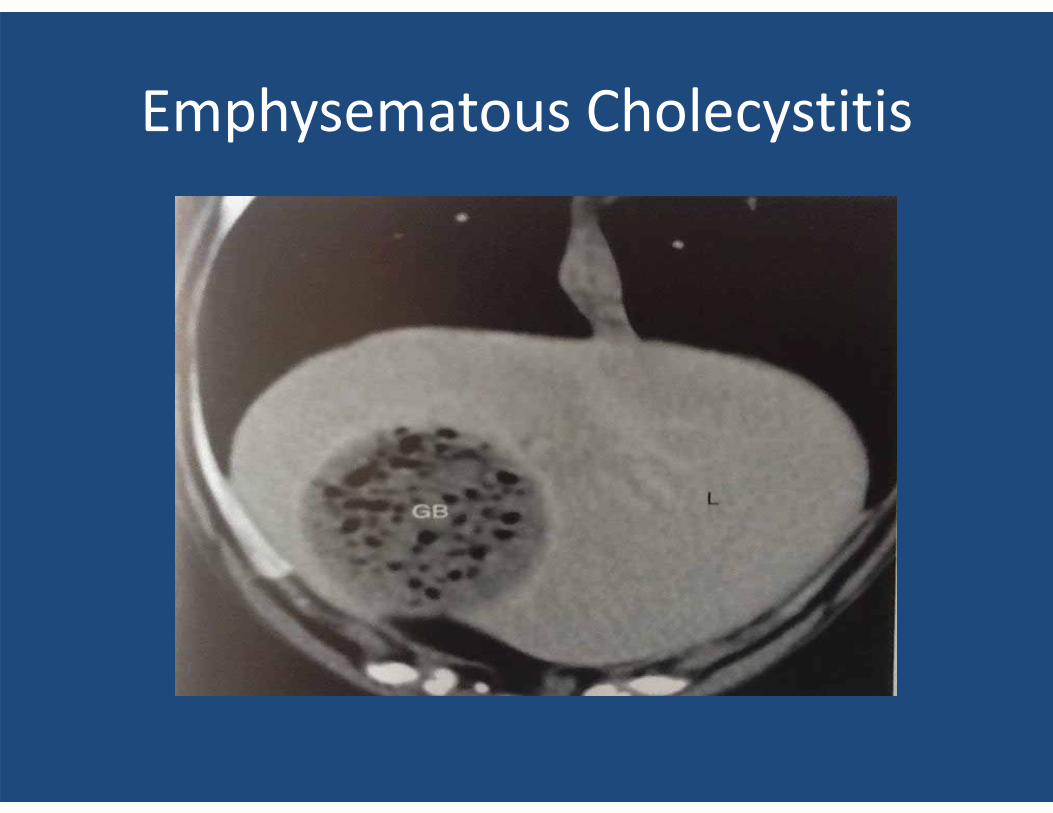
Emphysematous Cholecystitis
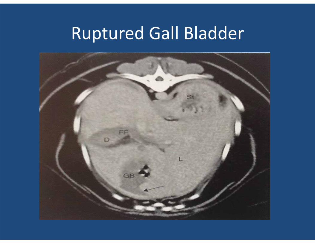
Ruptured Gall Bladder
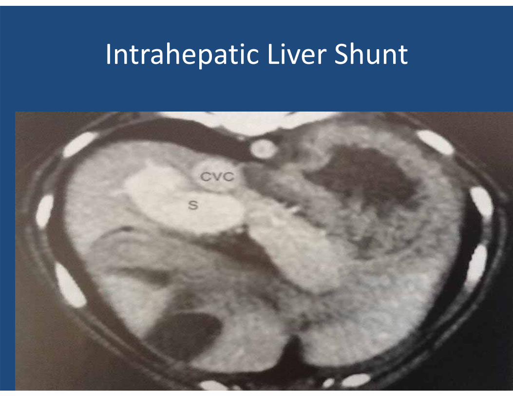
Intrahepatic Liver Shunt
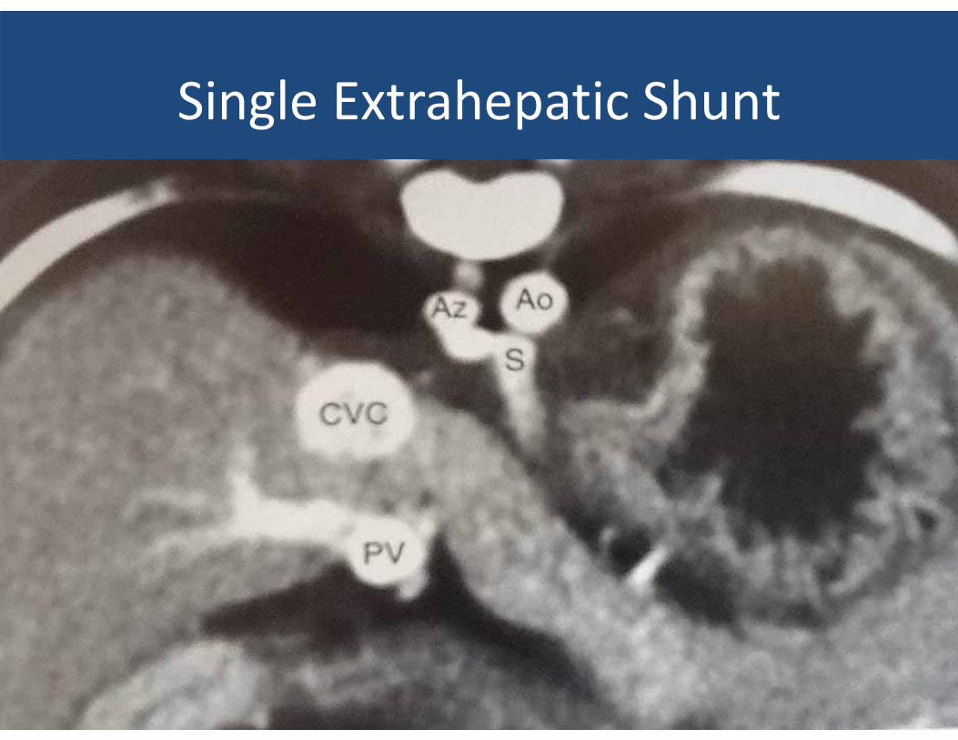
Single Extrahepatic Shunt
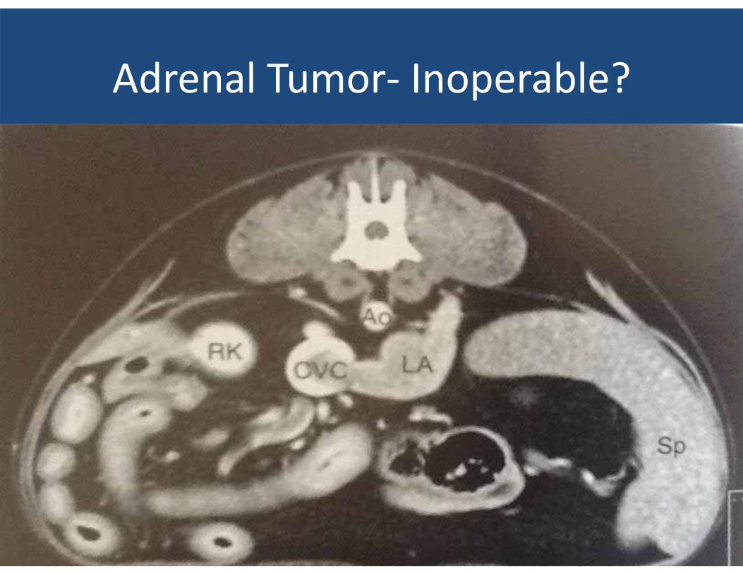
Adrenal Tumor- Inoperable?
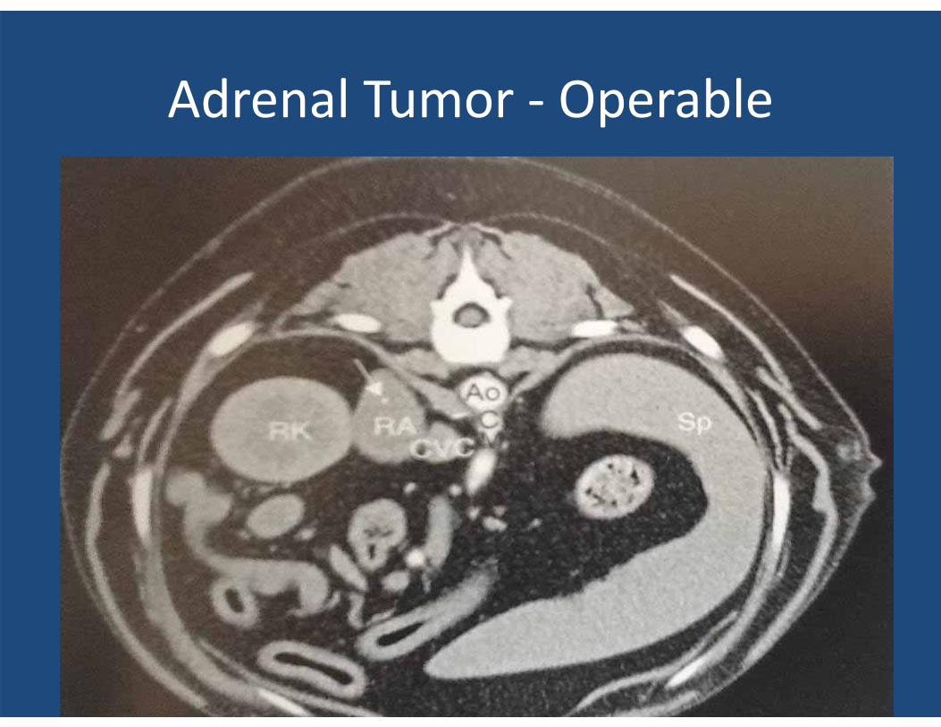
Adrenal Tumor - Operable
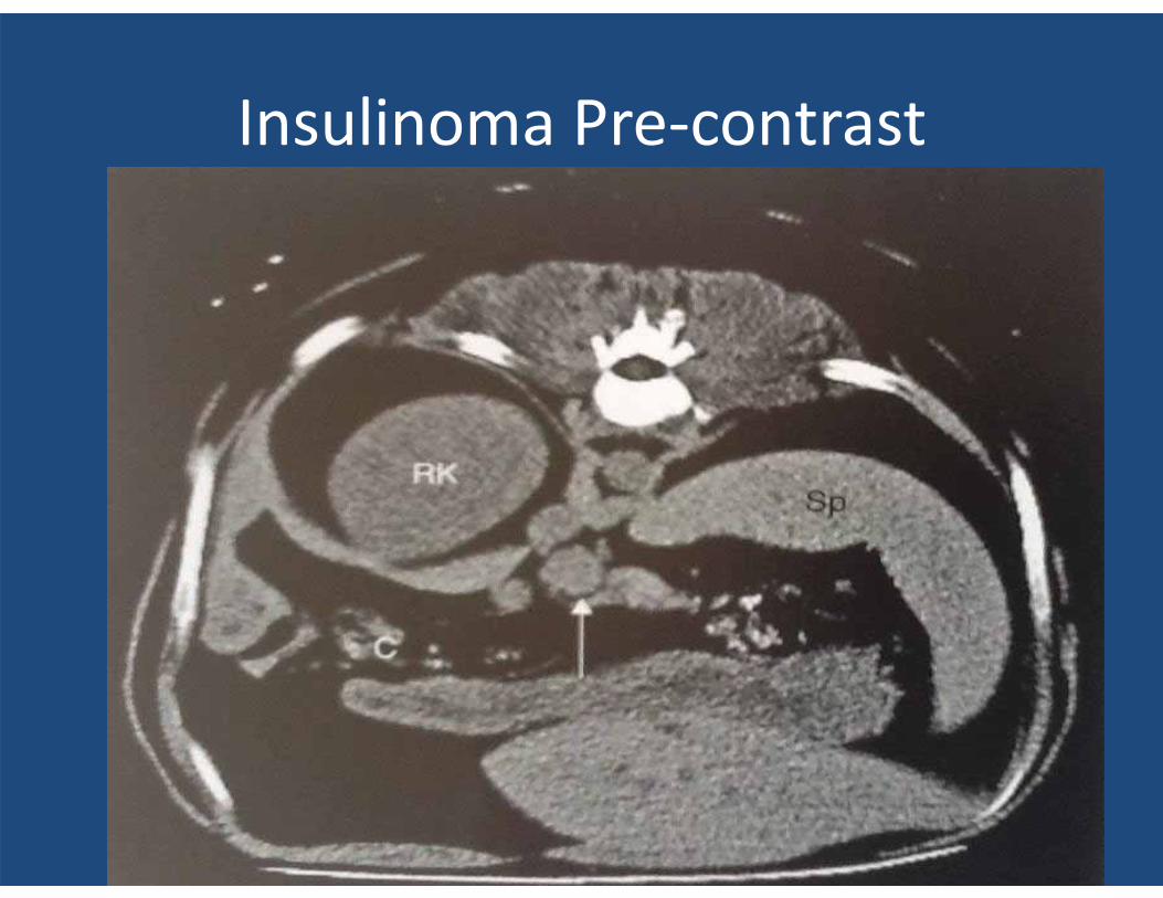
Insulinoma Pre-contrast
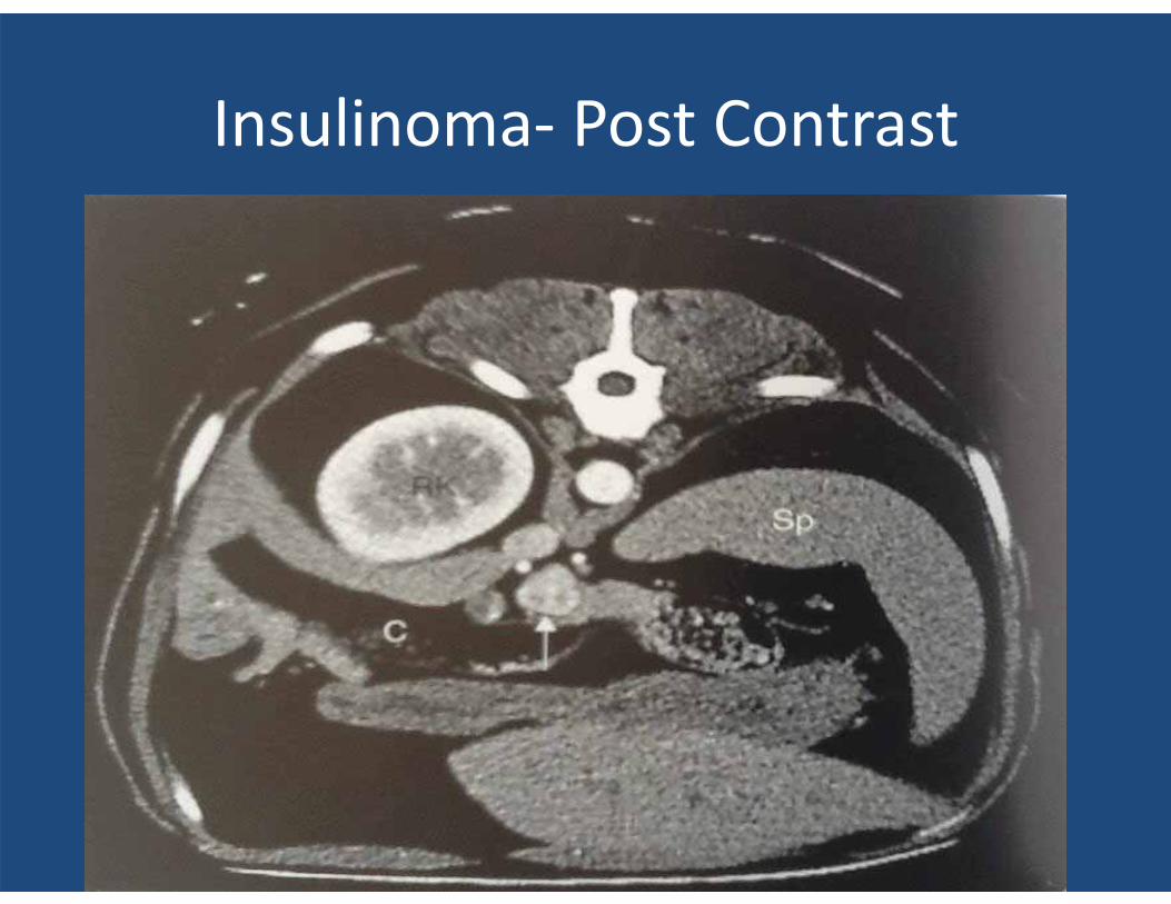
Insulinoma- Post Contrast
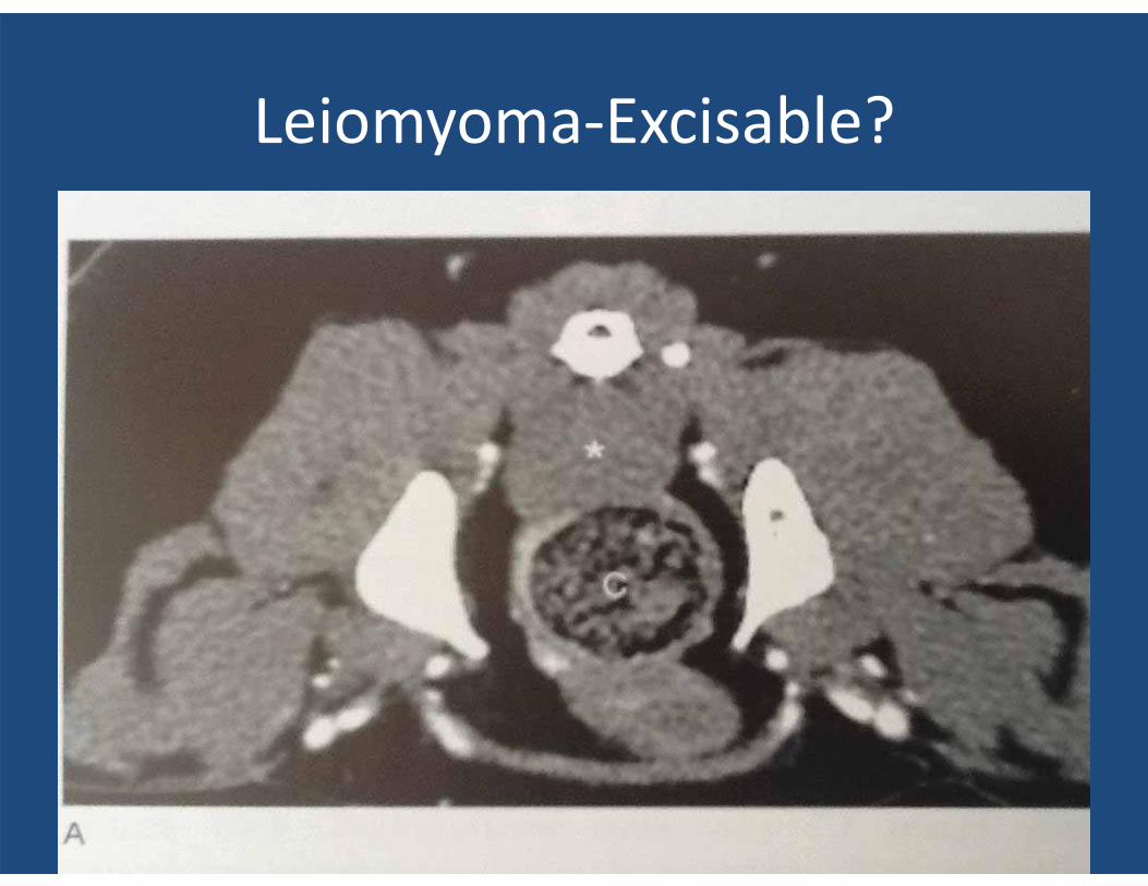
Leiomyoma-Excisable?
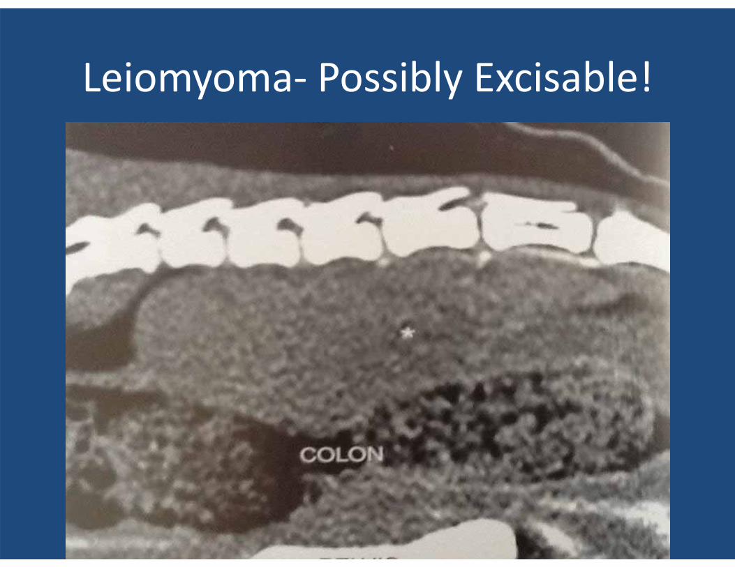
Leiomyoma- Possibly Excisable!
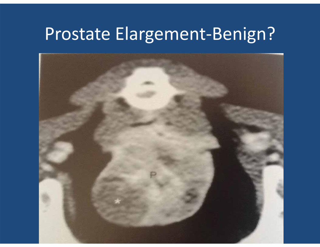
Prostate Elargement-Benign?
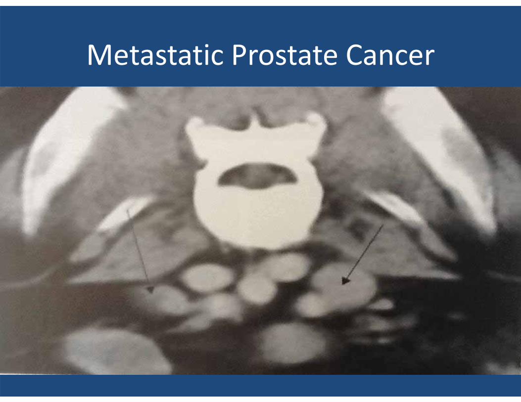
Metastatic Prostate Cancer

Ureteral Calculi
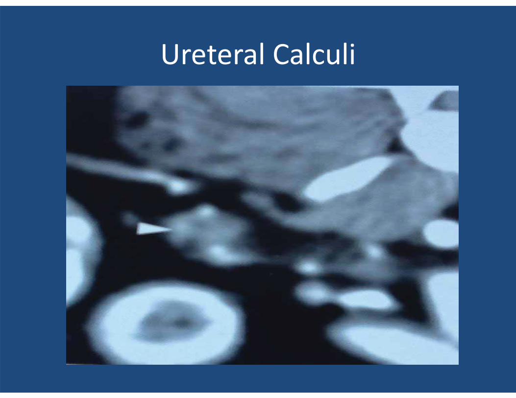
Ureteral Calculi
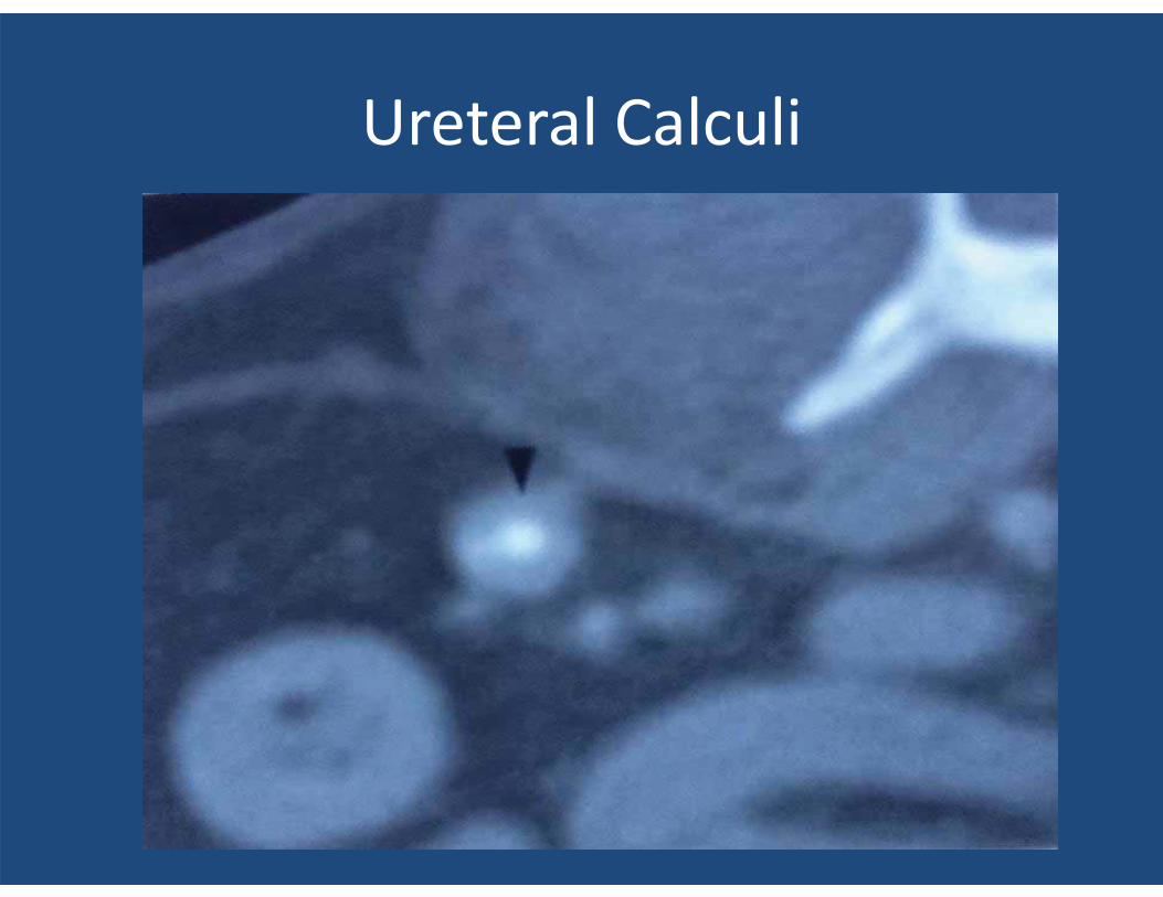
Ureteral Calculi
ORTHOPEDIC DISEASE
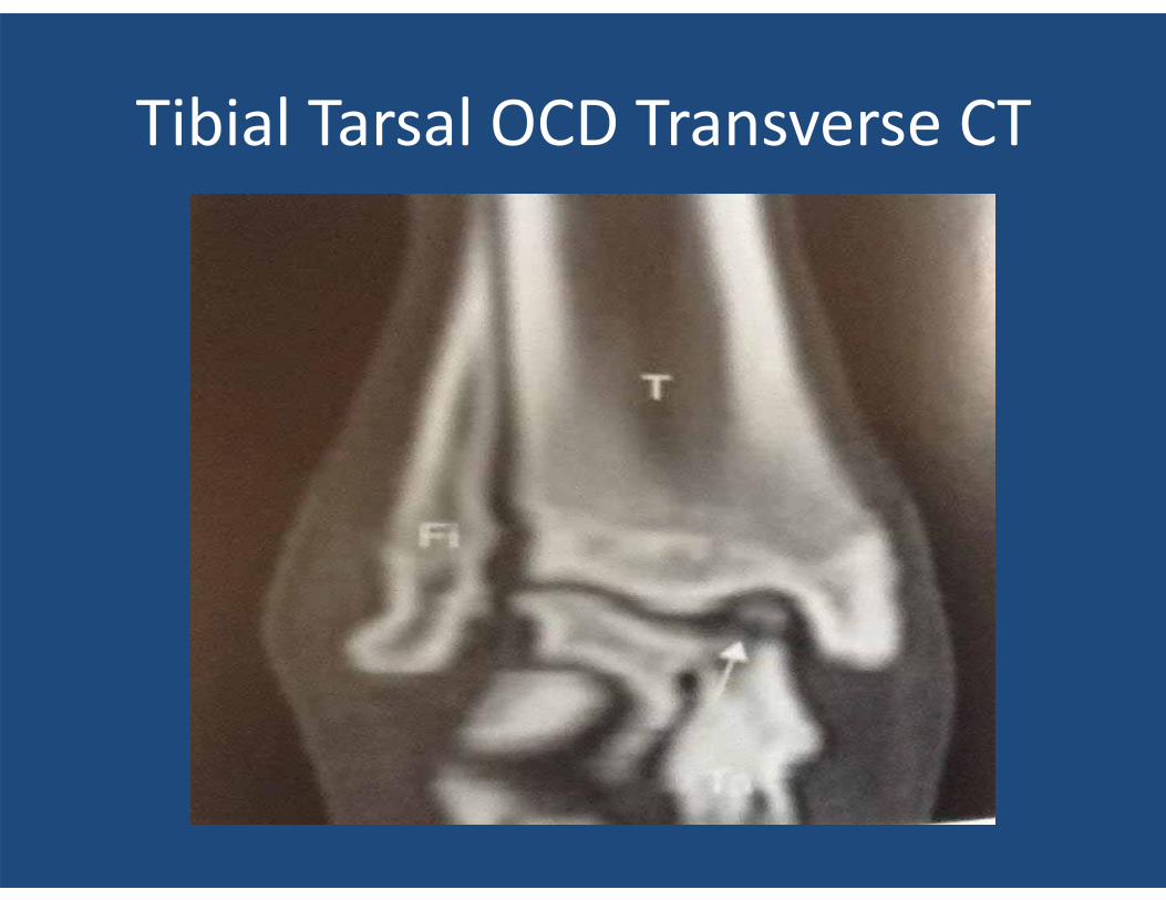
Tibial Tarsal OCD Transverse CT
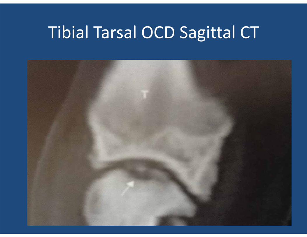
Tibial Tarsal OCD Sagittal CT
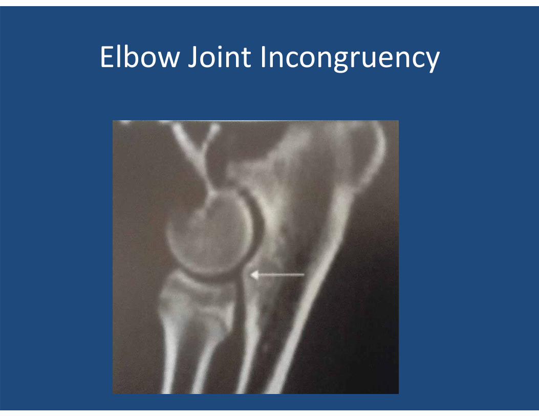
Elbow Joint Incongruency
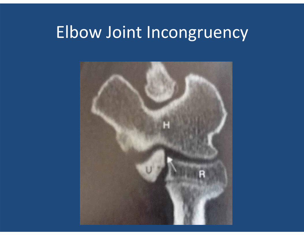
Elbow Joint Incongruency
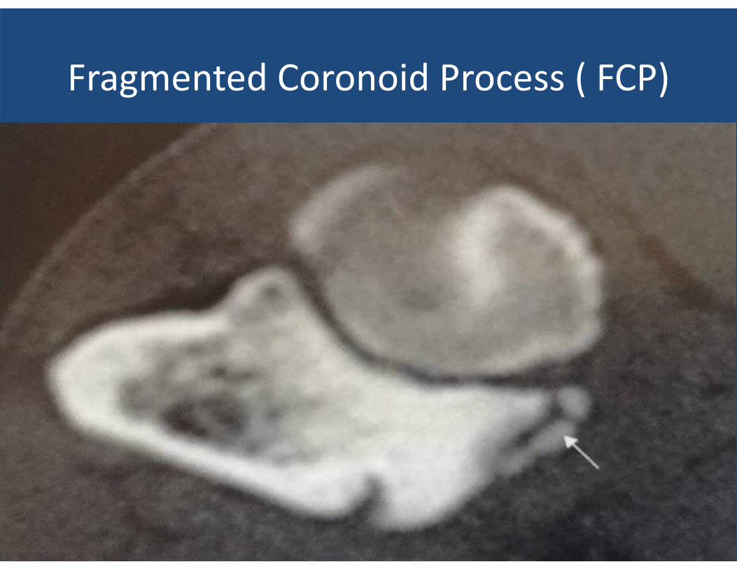
Fragmented Coronoid Process ( FCP)
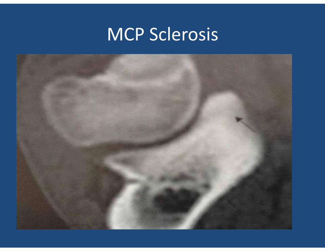
MCP Sclerosis
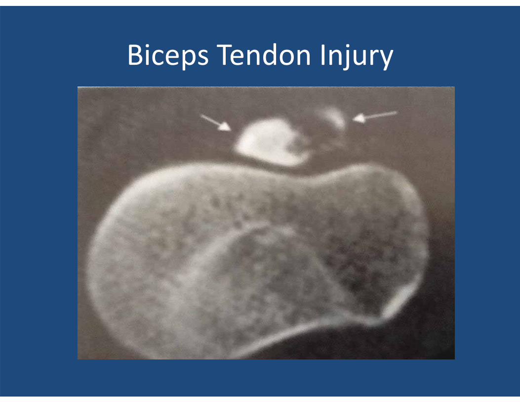
Biceps Tendon Injury
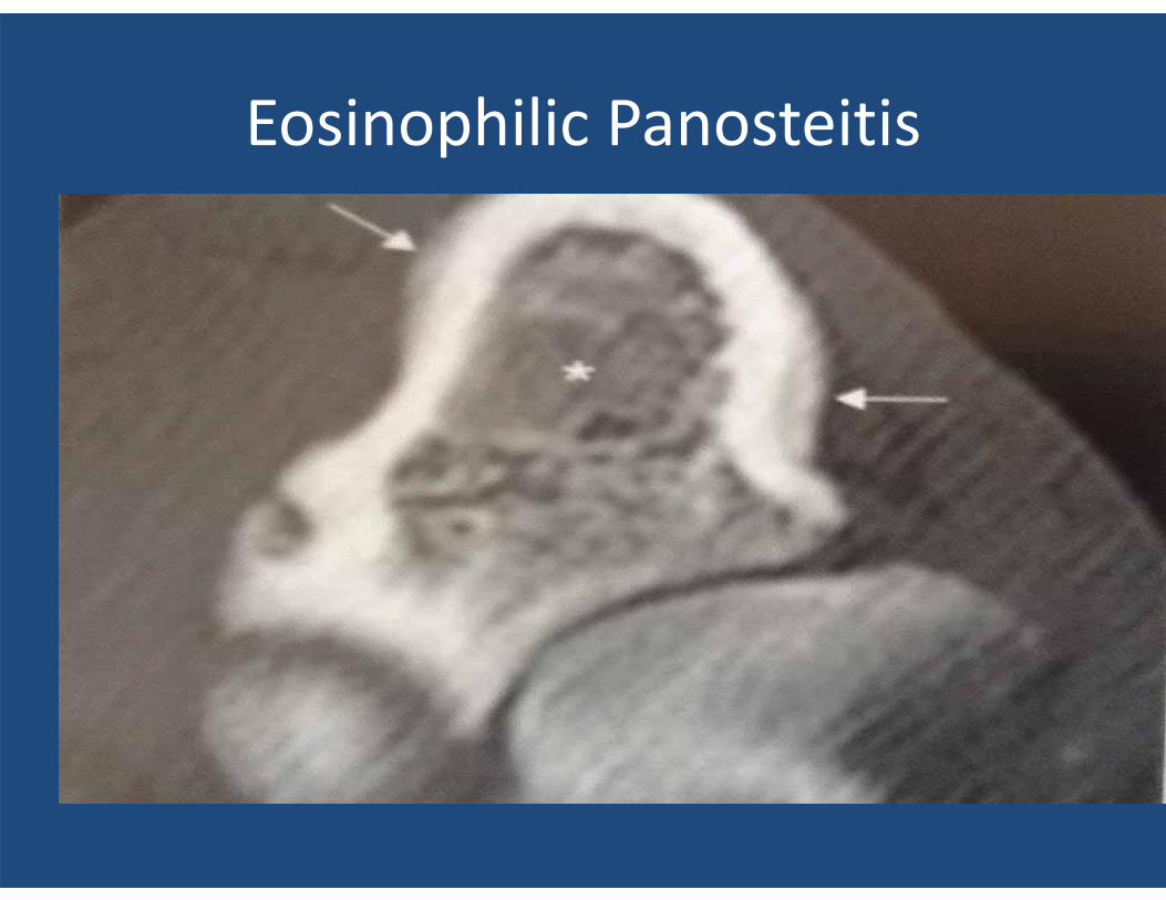
Eosinophilic Panosteitis
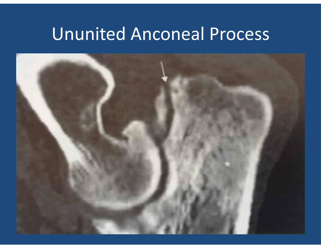
Ununited Anconeal Process
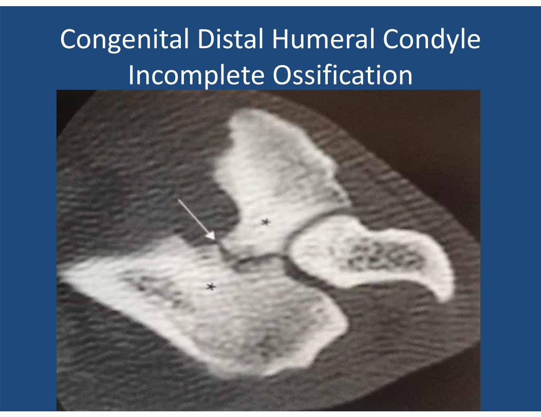
Congenital Distal Humeral Condyle Incomplete Ossification
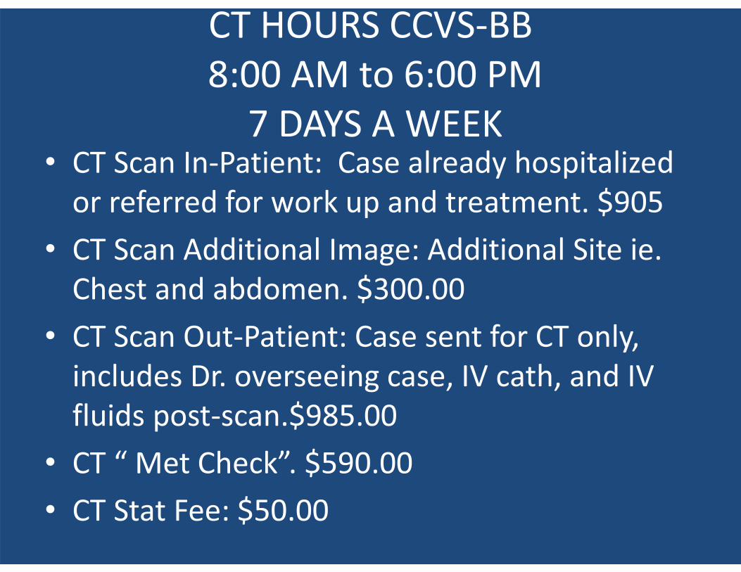
CT HOURS CCVS-BB 8:00 AM to 6:00 PM
7 DAYS A WEEK • CT Scan In-Patient: Case already hospitalized
or referred for work up and treatment. $905 • CT Scan Additional Image: Additional Site ie.
Chest and abdomen. $300.00 • CT Scan Out-Patient: Case sent for CT only,
includes Dr. overseeing case, IV cath, and IV fluids post-scan.$985.00
• CT “ Met Check”. $590.00 • CT Stat Fee: $50.00
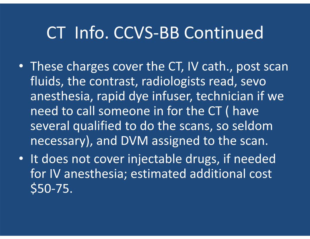
CT Info. CCVS-BB Continued
• These charges cover the CT, IV cath., post scan fluids, the contrast, radiologists read, sevo anesthesia, rapid dye infuser, technician if we need to call someone in for the CT ( have several qualified to do the scans, so seldom necessary), and DVM assigned to the scan.
• It does not cover injectable drugs, if needed for IV anesthesia; estimated additional cost $50-75.
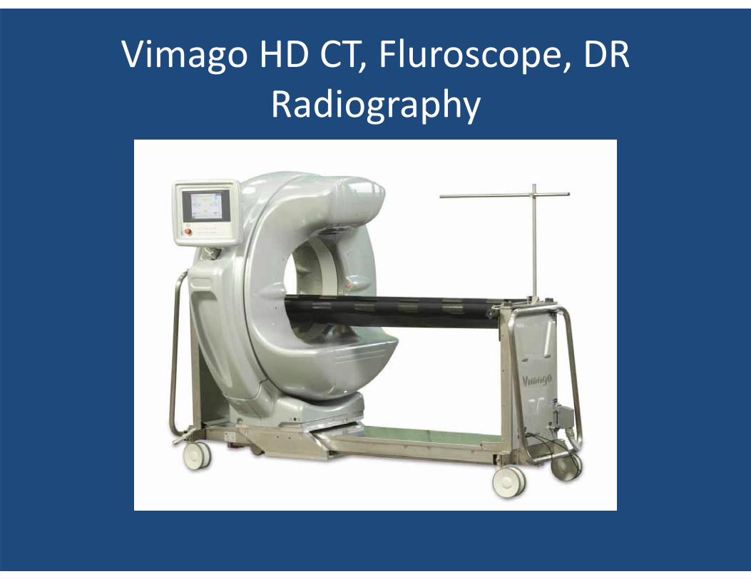
Vimago HD CT, Fluroscope, DR Radiography
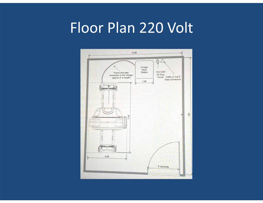
Floor Plan 220 Volt
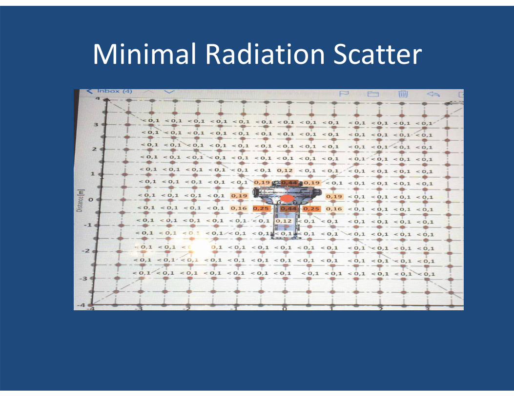
Minimal Radiation Scatter
Scan Times • Standard resolution < 50 #’s: Abdominal,
Thorax, Extremities, Spine ( Cervical and T-L ). 20 Seconds!!
• Standard Resolution > 50#’s-100#’s: Abdominal, Thorax, Extremities, Spine ( Cervical and T-L ) 60 Seconds!!
• High Resolution: < 50#’s 24 Seconds!! 50-100#’s 72 Seconds!!
• Standard Resolution= 300 images/rotation • High Resolution= 720/images/rotation
Sedation vs. Anesthesia
• All MRIs require anesthesia because of the long scan times and adds additional cost and risk.
• CT Scans with the newer units entering veterinary referral practices allows for sedation ( dexdomitor ) or ultra short intravenous ( propofol ) ( sevoflorane ) because of the ultra short scan times.
• CT cost and safety better vs. MRI scans
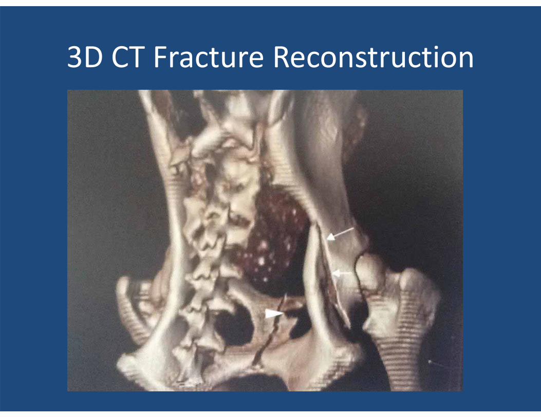
3D CT Fracture Reconstruction
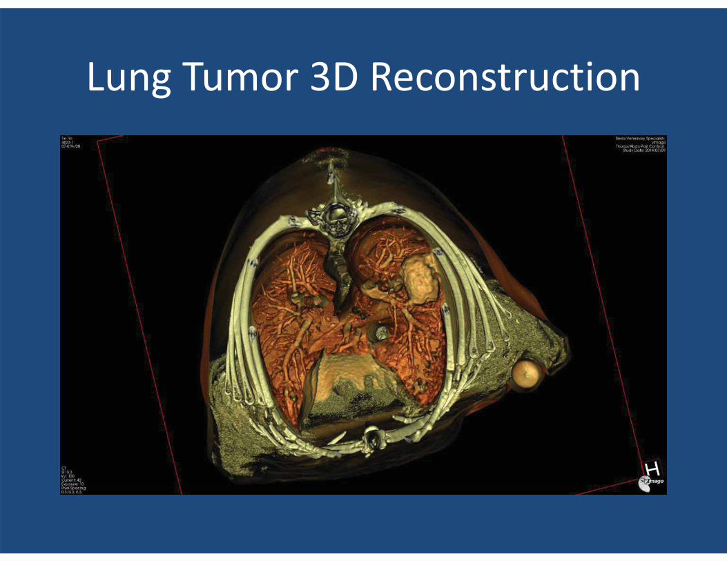
Lung Tumor 3D Reconstruction

Vimago Youtube Video
• http://www.youtube.com/watch?v=BT3Lib1rNtc
Where to learn lots more!!
QUESTIONS? THANK YOU FOR COMING!
TRUPANION VIDEO • Admission: Recently recruited to their board
of directors. NO PAY! Small stock option if they go public.
• TP sold 62% of ALL pet insurance in the US last year!
• 70% of all their payments go to clients of GENERAL PRACTIONERS!! 30% to REFERRAL PRACTICES!!
• TRUPANION EXPRESS: We Beta tested it in our practices. Now available to all veterinary practices. VIDEO TO FOLLOW
Trupanion Video
![Synovial Chondromatosis of the TMJ: MR and CT Findings · Synovial Chondromatosis of the TMJ: ... often in larger joints, such as the ... and hip [1 , 2]. Synovial chondromatosis](https://img.pdfslide.us/doc/110x75/5adafbfc7f8b9a86378e15b7/synovial-chondromatosis-of-the-tmj-mr-and-ct-chondromatosis-of-the-tmj-often.jpg)