Embed Size (px)
Citation preview
RESEARCH ARTICLE Open Access
E4BP4 facilitates glucocorticoid-evoked apoptosisof human leukemic CEM cells via upregulation ofBimJessica A Beach†, Laura J Nary†, Yasuko Hirakawa, Eli Holland, Rebeka Hovanessian and Rheem D Medh*
Abstract
Background: Synthetic GCs serve as therapeutic agents for some lymphoid leukemias because of their ability toinduce transcriptional changes via the GC receptor (GR) and trigger apoptosis. Upregulation of the BH3-onlymember of Bcl-2 family proteins, Bim, has been shown to be essential for GC-evoked apoptosis of leukemiclymphoblasts. Using human T cell leukemic sister clones CEM-C7-14 and CEM-C1-15, we have previously shownthat the bZIP transcriptional repressor, E4BP4, is preferentially upregulated by GCs in CEM-C7-14 cells that aresusceptible to GC-evoked apoptosis, but not in refractory CEM-C1-15 cells. E4BP4 is an evolutionarily conservedmember of the PAR family of bZIP transcription factors related to the C. elegans death specification gene ces2.
Results: Mouse E4BP4 was ectopically expressed in CEM-C1-15 cells, resulting in sensitization to GC-evokedapoptosis in correlation with restoration of E4BP4 and Bim upregulation. shRNA mediated modest knockdown ofE4BP4 in CEM-C7-14 cells resulted in concomitant reduction in Bim expression, although GC-evoked fold-inductionand sensitivity to apoptosis was similar to parental cells.
Conclusion: Data presented here suggest that GC-mediated upregulation of E4BP4 facilitates Bim upregulation andapoptosis of CEM cells. Since the Bim promoter does not contain any consensus GRE or EBPRE sequences,induction of Bim may be a secondary response.
BackgroundGlucocorticoids (GCs) are known to evoke human lym-phoid cell apoptosis [1-3] primarily by binding to andmodulating the transcriptional activity of the GC recep-tor (GR) [4]. GCs possess immunosuppressive and anti-inflammatory properties and serve as effective therapeu-tic agents for different forms of leukemia [5], asthma,rheumatoid arthritis, and irritable bowel syndrome [6].In order to exploit the full therapeutic potential of GCs,GC/GR-mediated gene regulation and its impact on var-ious cellular processes needs to be better understood.To this end, we and others have studied GR-dependentgene regulation by microarray-based transcriptional pro-filing [7-9]. A subset of genes were identified as thosebeing upregulated selectively in human leukemic CEMcells susceptible to, but not in cells refractory to, GC-
evoked apoptosis [7]. In this report, one of those genes,E4BP4, was evaluated for its role GC-evoked apoptosis.E4BP4 (adenovirus E4 binding protein 4), also called
NFIL3 (nuclear factor, interleukin 3 regulated) is classi-fied as a mammalian basic leucine zipper (bZIP) tran-scription factor and is closely related to the PAR(proline and acid rich) sub-family of bZIP transcriptionfactors, although it lacks a PAR domain [10]. VertebratePAR family transcription factors include hepatic leuke-mia factor (HLF), D-box binding protein (DBP), andthyrotroph embryonic factor (TEF) [11]. While otherPAR family members activate transcription, E4BP4represses transcription by binding to the same DNAsequence (E4BP4 response element; EBPRE), whose con-sensus sequence is (G/A)T(G/T)A(C/T) GTAA (C/T)[10]. The repressing activity of E4BP4 has been attribu-ted to a small 65 amino acid C-terminal repressiondomain that is rich in charged residues [10,12]. Thereare instances where it activates transcription of targetgenes as well [11].
* Correspondence: [email protected]† Contributed equallyDepartment of Biology, California State University Northridge, Northridge, CA91330-8303, USA
Beach et al. Journal of Molecular Signaling 2011, 6:13http://www.jmolecularsignaling.com/content/6/1/13
© 2011 Beach et al; licensee BioMed Central Ltd. This is an Open Access article distributed under the terms of the Creative CommonsAttribution License (http://creativecommons.org/licenses/by/2.0), which permits unrestricted use, distribution, and reproduction inany medium, provided the original work is properly cited.
Orthologs of PAR family proteins include C. elegansCes-2 [13], D. melanogaster Vrille [14], and X. laevisGene8 and Gene9 [15], which are known to have crucialfunctions in apoptosis, morphogenesis, and tail resorp-tion. E4BP4 has been implicated in diverse functions,including regulation of circadian rhythms [16], osteo-blast function [17], motoneuron survival [18], protectionof B cells from apoptosis induced by IL-3 deprivation[19], IgE class switching [20], and NK cell development[21]. Interestingly, E4BP4 has been shown to exhibitboth pro-apoptotic and pro-survival functions in a cell-and stimulus-specific fashion. For example, IL-3-mediated survival of pro-B cells is facilitated by theupregulation of E4BP4 [19], while the antitumor proper-ties of cantharidin have been attributed to its ability toupregulate E4BP4 and inhibit the antiapoptotic proper-ties of HLF [22]. Owing to its repressive activity, E4BP4has been suggested to function as an antagonist to otherPAR family transcription factors, which compete to bindto the same DNA sequences [23].E4BP4 has been shown to bind the TBP-binding
repressor protein Dr1 and facilitate its ability to repressboth basal and activated transcription [24]. There is evi-dence that PAR proteins follow a pathway analogous totheir ortholog in C. elegans, Ces-2, which is known todown regulate the survival gene Ces-1, which subse-quently permits the upregulation of the proapoptoticgene Egl-1 [13,25,26]. PAR family proteins, includingE4BP4, have been shown to modulate the activity ofEgl-1 orthologs, the pro-apoptotic BH-3 only membersof the Bcl-2 family, either directly or via Ces-1 orthologsSlug and Snail [27,28].BH3-only proteins of the Bcl-2 family, Bim and Puma,
are required for the initiation of apoptosis by multiplestimuli, including g-radiation, oxidative stress and GCs[29-31]. Bim is required for negative selection of T cellsand B cells, and for termination of T cell immuneresponse [32]. Puma has been identified as a p53-induci-ble gene and is thought to be critical for DNA-damageinduced apoptosis [33]. In CEM cells, induction of Bimis essential for GC-evoked apoptosis, and was one of thegenes identified through microarray-based expressionprofiling, along with E4BP4, as being selectively upregu-lated in response to GCs in the GC-sensitive sister sub-clones of CEM cells [7]. In this report, we presentevidence that E4BP4 plays a crucial role in GC-evokedapoptosis of CEM cells by enabling induction of Bim.
ResultsPrevious work has demonstrated the sensitivity andresistance of CEM-C7-14 and CEM-C1-15 cells, respec-tively, to GC-evoked cell death via apoptosis [7]. Theresistance in CEM-C-15 cells is thought to occurbecause of a blunted GR-dependent transcriptional
response, hence several studies have focused on identify-ing key transcriptional changes that are unique to GC-sensitive CEM cell clones [7-9]. E4BP4 was identified asone of the key genes upregulated in correlation withGC-evoked apoptosis [7] only in GC-sensitive CEMlines through microarray-based expression profiling. Toinvestigate the role of E4BP4 in GC-evoked apoptosis,CEM cells were manipulated either to overexpress(CEM-C1-15) or knockdown (CEM-C7-14) E4BP4.
Creation of a clone of CEM-C1-15 cells expressing mouseE4BP4 transcriptThe mouse and human E4BP4 genes are highly homolo-gous, with 79% nucleotide sequence identity in the cod-ing region. The two proteins are 462 amino acids longand have an 84% sequence identity and 92% sequencehomology, based on a BLASTP pairwise sequence align-ment algorithm. To determine whether a lack of E4BP4upregulation was contributing to GC resistance inCEM-C1-15 cells, a construct expressing M. E4BP4(pCR3.1-mE4) was transfected into CEM-C1-15 cells,allowing for selective amplification of either the humanor mouse transcript using species-specific primers.Although both proteins are almost identical, we cannotrule out the possibility that the mouse protein may haveunique characteristics compared to the human version.For both species, the entire coding sequence of E4BP4 iswithin a single exon. As shown in Figure 1A, regions ofsequence variation within the partial coding sequenceshown were utilized to design PCR primers for species-specific amplification, and mouse (376bp) and human(250bp) specific amplicons were identified based ontheir sizes. As shown in Figure 1B, the mE4BP4 primersfailed to amplify any product from CEM-C7-14 orCEM-C1-15 cells (top panel), whereas the hE4BP4 pri-mers failed to amplify any product when the plasmidpCR3.1-mE4 was used as a template (bottom panel). Asshown in Figure 1C, upper panel, two batches of masscultures of M. E4BP4 transfected CEM-C1-15 cells(mE*a and mE*b) as well as four different clones gener-ated by limiting dilution were tested for presence oftranscript corresponding to mouse E4BP4 sequences.Except for clone 2 (mE#2), the mass cultures and allclones had amplicons corresponding to M. E4BP4 tran-script. Clone #3 (mE#3) was chosen for further analysis,and as shown in Figure 1C, lower panel, it exhibitedboth mouse and human specific E4BP4 transcript byreverse transcription PCR analysis.
Transfection of M. E4BP4 in CEM-C1-15 cells restoressensitivity to Dex-evoked apoptosisParental CEM-C1-15 cells, mass cultures of cells trans-fected with M.E4BP4 (CEM-C1-15mE*), and clone # 3(CEM-C1-15-mE#3) were tested for their sensitivity to 1
Beach et al. Journal of Molecular Signaling 2011, 6:13http://www.jmolecularsignaling.com/content/6/1/13
Page 2 of 13
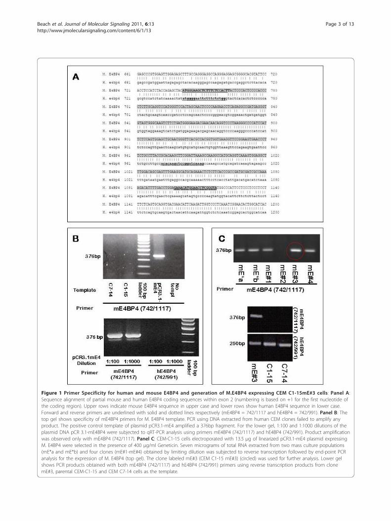
Figure 1 Primer Specificity for human and mouse E4BP4 and generation of M.E4BP4 expressing CEM C1-15mE#3 cells: Panel A:Sequence alignment of partial mouse and human E4BP4 coding sequences within exon 2 (numbering is based on +1 for the first nucleotide ofthe coding region). Upper rows indicate mouse E4BP4 sequence in upper case and lower rows show human E4BP4 sequence in lower case.Forward and reverse primers are underlined with solid and dotted lines respectively (mE4BP4 = 742/1117 and hE4BP4 = 742/991). Panel B: Thetop gel shows specificity of mE4BP4 primers for M. E4BP4 template. PCR using DNA extracted from human CEM clones failed to amplify anyproduct. The positive control template of plasmid pCR3.1-mE4 amplified a 376bp fragment. For the lower gel, 1:100 and 1:1000 dilutions of theplasmid DNA pCR 3.1-mE4BP4 were subjected to qRT-PCR analysis using primers mE4BP4 (742/1117) and hE4BP4 (742/991). Product amplificationwas observed only with mE4BP4 (742/1117). Panel C: CEM-C1-15 cells electroporated with 13.5 μg of linearized pCR3.1-mE4 plasmid expressingM. E4BP4 were selected in the presence of 400 μg/ml Geneticin. Seven micrograms of total RNA extracted from two mass culture populations(mE*a and mE*b) and four clones (mE#1-mE#4) obtained by limiting dilution was subjected to reverse transcription followed by end-point PCRanalysis for the expression of M. E4BP4 (top gel). The clone labeled mE#3 (CEM C1-15 mE#3) (circled) was used for further analysis. Lower gelshows PCR products obtained with both mE4BP4 (742/1117) and hE4BP4 (742/991) primers using reverse transcription products from clonemE#3, parental CEM-C1-15 and CEM C7-14 cells as the template.
Beach et al. Journal of Molecular Signaling 2011, 6:13http://www.jmolecularsignaling.com/content/6/1/13
Page 3 of 13
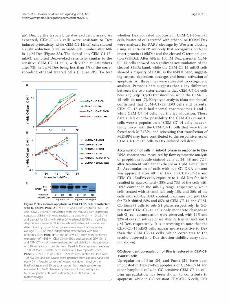
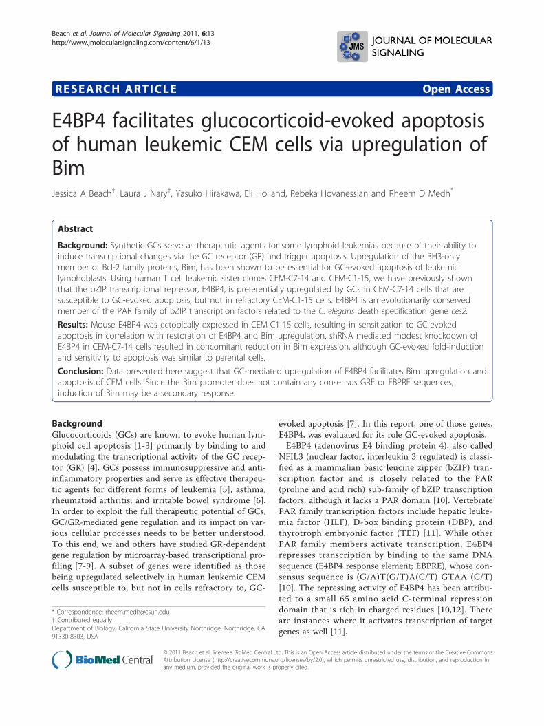
μM Dex by the trypan blue dye exclusion assay. Asexpected, CEM-C1-15 cells were resistant to Dexinduced cytotoxicity, while CEM-C1-15mE* cells showeda slight reduction (18%) in viable cell number after 68hin 1 μM Dex (Figure 2A). The cloned line, CEM-C1-15-mE#3, exhibited Dex-evoked sensitivity similar to thesensitive CEM-C7-14 cells, with viable cell numbersafter 72h in 1 μM Dex being less than 5% of the corre-sponding ethanol treated cells (Figure 2B). To test
whether Dex activated apoptosis in CEM-C1-15-mE#3cells, lyastes of cells treated with ethanol or 100nM Dexwere analyzed for PARP cleavage by Western blottingusing an anti-PARP antibody that recognizes both theintact protein (116kDa) and the cleaved C-terminal por-tion (85kDa). After 48h in 100nM Dex, parental CEM-C1-15 cells showed no significant accumulation of thecleaved 85kDa band, while the CEM-C1-15-mE#3 cellsshowed a majority of PARP as the 85kDa band, suggest-ing caspase-dependent cleavage, and hence activation ofapoptosis. All three lines were subjected to cytogeneticanalysis. Previous data suggests that a key differencebetween the two sister clones is that CEM-C7-14 cellsbear a t(1;2)(p13;q21) translocation, while the CEM-C1-15 cells do not [7]. Karyotype analysis (data not shown)confirmed that CEM-C1-15mE#3 cells and parentalCEM-C1-15 cells had normal chromosomes 1 and 2,while CEM-C7-14 cells had the translocation. Thesedata ruled out the possibility the CEM-C1-15-mE#3cells were a population of CEM-C7-14 cells inadver-tently mixed with the CEM-C1-15 cells that were trans-fected with M.E4BP4, and reiterating that transfection ofM.E4BP4 may have contributed to the responsiveness ofCEM-C1-15mE#3 cells to Dex-induced cell death.
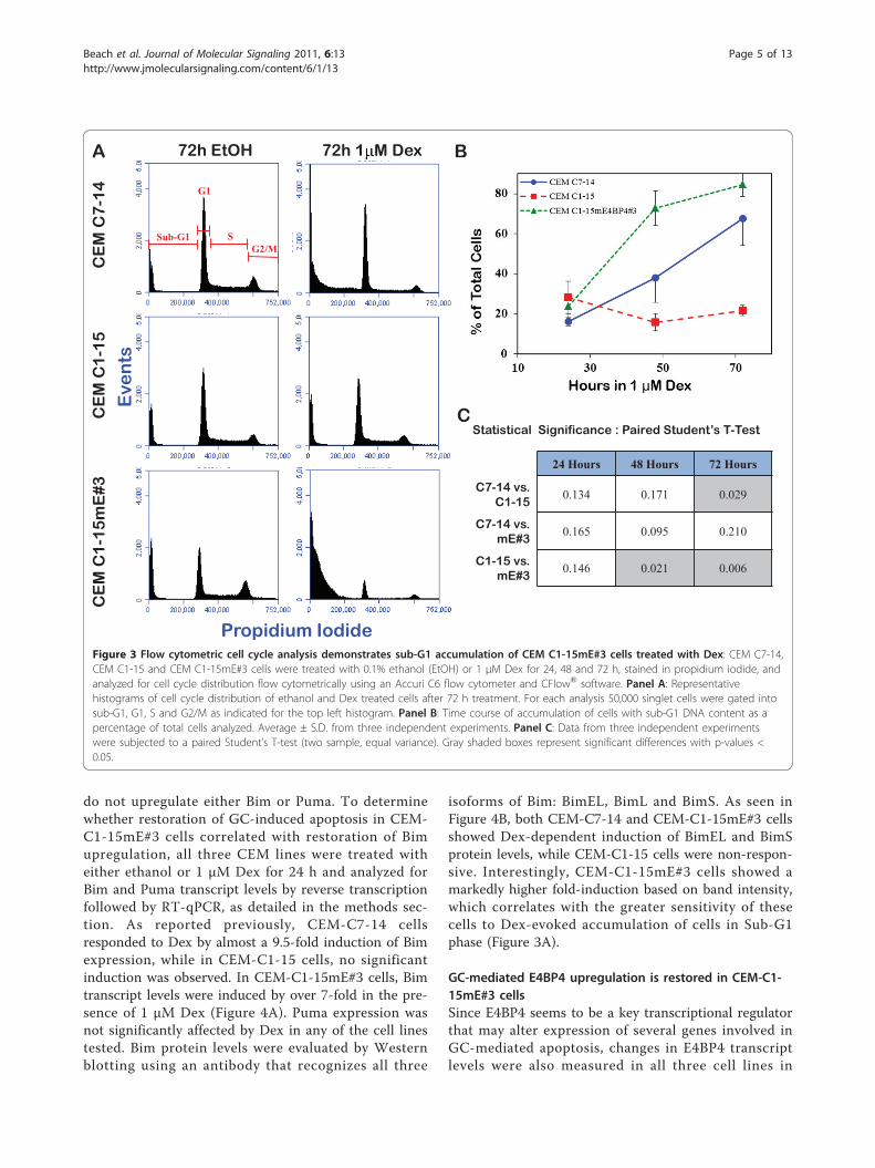
Accumulation of cells in sub-G1 phase in response to DexDNA content was measured by flow cytometric analysisof propidium iodide stained cells at 24, 48 and 72 hafter treatment with either ethanol or 1 μM Dex (Figure3). Accumulation of cells with sub-G1 DNA contentwas apparent after 48 h in Dex. In CEM-C7-14 andCEM-C1-15mE#3 cells, exposure to 1 μM Dex for 48 hresulted in approximately 38% and 73% of the cells withDNA content in the sub-G1 range, respectively, whilecells treated with ethanol had only 15% and 20% of thecells with sub-G1 DNA content. Exposure to 1 μM Dexfor 72 h shifted 68% and 85% of CEM-C7-14 and CEM-C1-15mE#3 cells to sub-G1 phase, respectively. In GC-resistant CEM-C1-15 cells only moderate changes insub-G1 cell accumulation were observed, with 14% and22% of cells in sub G1 phase after 72 h in ethanol and 1μM Dex, respectively. It is interesting to note that theCEM-C1-15mE#3 cells appear more sensitive to Dexthan the CEM-C7-14 cells, which correlates to theresults observed in a Dex titration viability assay (datanot shown).
GC-dependent upregulation of Bim is restored in CEM-C1-15mE#3 cellsUpregulation of Bim [34] and Puma [31] have beenimplicated in Dex-evoked apoptosis of CEM-C7-14 andother lymphoid cells. In GC-sensitive CEM-C7-14 cell,Bim upregulation has been shown to contribute toapoptosis, while in GC-resistant CEM-C1-15 cells, GCs
A
B
Hours in100nM Dex
C116
85
CEM C1-15 CEM C1-15-mE#3
0 024 2448 48
PARP
Figure 2 Dex induces apoptosis in CEM C1-15 cells transfectedwith M. E4BP4: Panel A:CEM C1-15 and a mass culture CEM C1-15cells (CEM C1-15mE*) transfected with the mouse E4BP4 expressingconstruct pCR3.1-mE4 were seeded at a density of 1 × 10⁵cells/mland treated for 72 h with either 0.1% ethanol (EtOH) or 1 μM Dex.Aliquots were taken at 24 h intervals and viable cell number wasdetermined by trypan blue dye exclusion assay. Data representaverage ± S.D. of three independent experiments with tworeplicates each. Panel B:A clone of CEM C1-15mE* with confirmedexpression of mE4BP4 (CEM C1-15mE#3), and parental CEM C1-15and CEM C7-14 cells were analyzed for cell viability in the presenceof 0.1% ethanol or 1 μM Dex as in Panel A. Data represent averages± S.D. of three separate experiments with two replicates each.Panel C: CEM C1-15 or CEM C1-15mE#3 cells treated for 48 h with100 nM Dex and cell lysates were prepared from aliquots harvestedevery 24 h. Protein content of lysates was determined by theBradford assay and 30 μg total protein from each sample wasevaluated for PARP cleavage by Western blotting using a C-terminal-specific anti-PARP antibody (SC-7150, Santa CruzBiotechnology).
Beach et al. Journal of Molecular Signaling 2011, 6:13http://www.jmolecularsignaling.com/content/6/1/13
Page 4 of 13
do not upregulate either Bim or Puma. To determinewhether restoration of GC-induced apoptosis in CEM-C1-15mE#3 cells correlated with restoration of Bimupregulation, all three CEM lines were treated witheither ethanol or 1 μM Dex for 24 h and analyzed forBim and Puma transcript levels by reverse transcriptionfollowed by RT-qPCR, as detailed in the methods sec-tion. As reported previously, CEM-C7-14 cellsresponded to Dex by almost a 9.5-fold induction of Bimexpression, while in CEM-C1-15 cells, no significantinduction was observed. In CEM-C1-15mE#3 cells, Bimtranscript levels were induced by over 7-fold in the pre-sence of 1 μM Dex (Figure 4A). Puma expression wasnot significantly affected by Dex in any of the cell linestested. Bim protein levels were evaluated by Westernblotting using an antibody that recognizes all three
isoforms of Bim: BimEL, BimL and BimS. As seen inFigure 4B, both CEM-C7-14 and CEM-C1-15mE#3 cellsshowed Dex-dependent induction of BimEL and BimSprotein levels, while CEM-C1-15 cells were non-respon-sive. Interestingly, CEM-C1-15mE#3 cells showed amarkedly higher fold-induction based on band intensity,which correlates with the greater sensitivity of thesecells to Dex-evoked accumulation of cells in Sub-G1phase (Figure 3A).
GC-mediated E4BP4 upregulation is restored in CEM-C1-15mE#3 cellsSince E4BP4 seems to be a key transcriptional regulatorthat may alter expression of several genes involved inGC-mediated apoptosis, changes in E4BP4 transcriptlevels were also measured in all three cell lines in
Statistical Significance : Paired Student’s T-Test
24 Hours 48 Hours 72 Hours
C7-14 vs. C1-15
0.134 0.171 0.029
C7-14 vs. mE#3
0.165 0.095 0.210
C1-15 vs. mE#3
0.146 0.021 0.006
B
C
G1
G2/MSSub-G1
CE
M C
7-1
4C
EM
C1
-15
CE
M C
1-1
5m
E#3
72h EtOH 72h 1 M DexA
Propidium Iodide
Eve
nts
Figure 3 Flow cytometric cell cycle analysis demonstrates sub-G1 accumulation of CEM C1-15mE#3 cells treated with Dex: CEM C7-14,CEM C1-15 and CEM C1-15mE#3 cells were treated with 0.1% ethanol (EtOH) or 1 μM Dex for 24, 48 and 72 h, stained in propidium iodide, andanalyzed for cell cycle distribution flow cytometrically using an Accuri C6 flow cytometer and CFlow® software. Panel A: Representativehistograms of cell cycle distribution of ethanol and Dex treated cells after 72 h treatment. For each analysis 50,000 singlet cells were gated intosub-G1, G1, S and G2/M as indicated for the top left histogram. Panel B: Time course of accumulation of cells with sub-G1 DNA content as apercentage of total cells analyzed. Average ± S.D. from three independent experiments. Panel C: Data from three independent experimentswere subjected to a paired Student’s T-test (two sample, equal variance). Gray shaded boxes represent significant differences with p-values <0.05.
Beach et al. Journal of Molecular Signaling 2011, 6:13http://www.jmolecularsignaling.com/content/6/1/13
Page 5 of 13
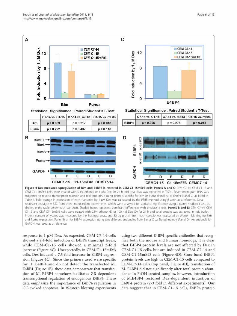
response to 1 μM Dex. As expected, CEM-C7-14 cellsshowed a 8.4-fold induction of E4BP4 transcript levels,while CEM-C1-15 cells showed a minimal 2-foldincrease (Figure 4C). Unexpectedly, in CEM-C1-15mE#3cells, Dex induced a 7.5-fold increase in E4BP4 expres-sion (Figure 4C). Since the primers used were specificfor H. E4BP4 and do not detect the transfected M.E4BP4 (Figure 1B), these data demonstrate that transfec-tion of M. E4BP4 somehow facilitates GR-dependenttranscriptional regulation of endogenous E4BP4. Thesedata emphasize the importance of E4BP4 regulation inGC-evoked apoptosis. In Western blotting experiments
using two different E4BP4-specific antibodies that recog-nize both the mouse and human homologs, it is clearthat E4BP4 protein levels are not affected by Dex inCEM-C1-15 cells, but are induced in CEM-C7-14 andCEM-C1-15mE#3 cells (Figure 4D). Since basal E4BP4protein levels are high in CEM-C1-15 cells compared toCEM-C7-14 cells (top panel, Figure 4D), transfection ofM. E4BP4 did not significantly alter total protein abun-dance in EtOH treated samples, however, introductionof M.E4BP4 restored Dex-dependent induction ofE4BP4 protein (2-3 fold in different experiments). Ourdata suggest that in CEM-C1-15 cells, E4BP4 protein
BimELBimL
BimS
GAPDHE D
CEMC1-15
E D
CEMC7-14
E D
C1-15mE#3
Puma
B
A
sc-282
03
E4
BP
4
sc-955
0
GA
PD
H
E D
CEMC1-15
E D
CEMC7-14
E D
C1-15mE#3
D
C
0
4
8
12
E4BP4
Fold
Ind
ucti
on b
y 1
M D
ex
CEM C7-14CEM C1-15CEM C1-15mE#3
Figure 4 Dex-mediated upregulation of Bim and E4BP4 is restored in CEM C1-15mE#3 cells: Panels A and C: CEM C7-14, CEM C1-15 andCEM C1-15mE#3 cells were treated with 0.1% ethanol or 1 μM Dex for 24 h and total RNA was extracted in TriZol. Seven microgram RNA wassubjected to reverse transcription reaction and real-time qPCR using primers specific for Bim or Puma (Panel A) or E4BP4 (Panel C) as listed inTable 1. Fold change in expression of each transcript by 1 μM Dex was calculated by the Pfaffl method using b-actin as a reference. Datarepresent averages ± S.D. from three independent experiments, which were analyzed for statistical significance using a paired student t-test, asshown in the table below each bar chart. Shaded boxes represent significant differences with p-values ≤ 0.05. Panels B and D: CEM C7-14, CEMC1-15 and CEM C1-15mE#3 cells were treated with 0.1% ethanol (E) or 100 nM Dex (D) for 24 h and total protein was extracted in lysis buffer.Protein content of lysates was measured by the Bradford assay, and 30 μg protein from each sample was evaluated by Western blotting for Bimand Puma expression (Panel B) or for E4BP4 expression using two different antibodies from Santa Cruz Biotechnology (Panel D). An antibody forGAPDH was used as a reference.
Beach et al. Journal of Molecular Signaling 2011, 6:13http://www.jmolecularsignaling.com/content/6/1/13
Page 6 of 13
levels are deregulated, and that regulation is restoredupon M. E4BP4 transfection.
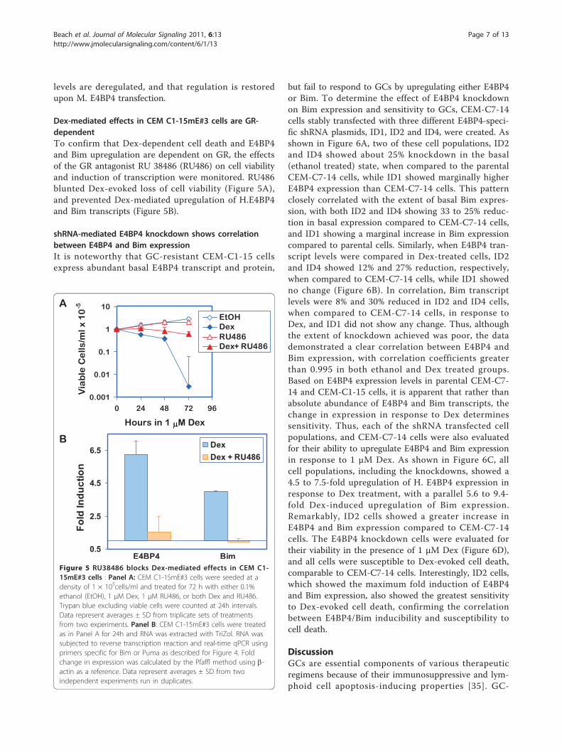
Dex-mediated effects in CEM C1-15mE#3 cells are GR-dependentTo confirm that Dex-dependent cell death and E4BP4and Bim upregulation are dependent on GR, the effectsof the GR antagonist RU 38486 (RU486) on cell viabilityand induction of transcription were monitored. RU486blunted Dex-evoked loss of cell viability (Figure 5A),and prevented Dex-mediated upregulation of H.E4BP4and Bim transcripts (Figure 5B).
shRNA-mediated E4BP4 knockdown shows correlationbetween E4BP4 and Bim expressionIt is noteworthy that GC-resistant CEM-C1-15 cellsexpress abundant basal E4BP4 transcript and protein,
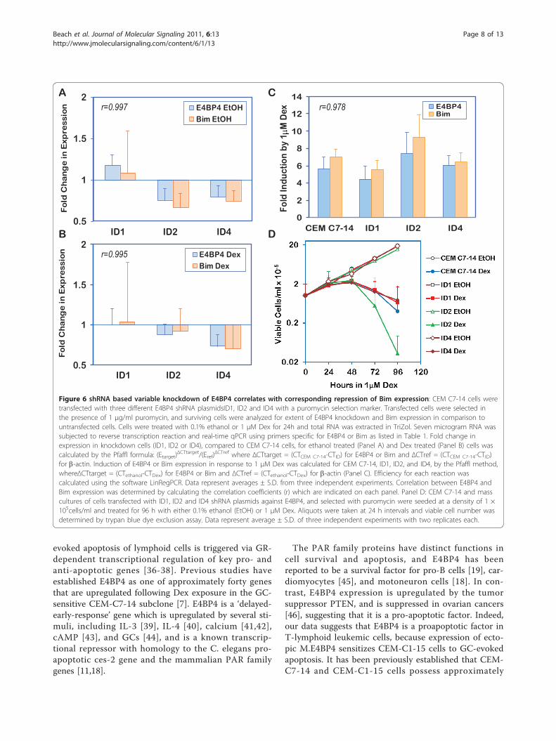
but fail to respond to GCs by upregulating either E4BP4or Bim. To determine the effect of E4BP4 knockdownon Bim expression and sensitivity to GCs, CEM-C7-14cells stably transfected with three different E4BP4-speci-fic shRNA plasmids, ID1, ID2 and ID4, were created. Asshown in Figure 6A, two of these cell populations, ID2and ID4 showed about 25% knockdown in the basal(ethanol treated) state, when compared to the parentalCEM-C7-14 cells, while ID1 showed marginally higherE4BP4 expression than CEM-C7-14 cells. This patternclosely correlated with the extent of basal Bim expres-sion, with both ID2 and ID4 showing 33 to 25% reduc-tion in basal expression compared to CEM-C7-14 cells,and ID1 showing a marginal increase in Bim expressioncompared to parental cells. Similarly, when E4BP4 tran-script levels were compared in Dex-treated cells, ID2and ID4 showed 12% and 27% reduction, respectively,when compared to CEM-C7-14 cells, while ID1 showedno change (Figure 6B). In correlation, Bim transcriptlevels were 8% and 30% reduced in ID2 and ID4 cells,when compared to CEM-C7-14 cells, in response toDex, and ID1 did not show any change. Thus, althoughthe extent of knockdown achieved was poor, the datademonstrated a clear correlation between E4BP4 andBim expression, with correlation coefficients greaterthan 0.995 in both ethanol and Dex treated groups.Based on E4BP4 expression levels in parental CEM-C7-14 and CEM-C1-15 cells, it is apparent that rather thanabsolute abundance of E4BP4 and Bim transcripts, thechange in expression in response to Dex determinessensitivity. Thus, each of the shRNA transfected cellpopulations, and CEM-C7-14 cells were also evaluatedfor their ability to upregulate E4BP4 and Bim expressionin response to 1 μM Dex. As shown in Figure 6C, allcell populations, including the knockdowns, showed a4.5 to 7.5-fold upregulation of H. E4BP4 expression inresponse to Dex treatment, with a parallel 5.6 to 9.4-fold Dex-induced upregulation of Bim expression.Remarkably, ID2 cells showed a greater increase inE4BP4 and Bim expression compared to CEM-C7-14cells. The E4BP4 knockdown cells were evaluated fortheir viability in the presence of 1 μM Dex (Figure 6D),and all cells were susceptible to Dex-evoked cell death,comparable to CEM-C7-14 cells. Interestingly, ID2 cells,which showed the maximum fold induction of E4BP4and Bim expression, also showed the greatest sensitivityto Dex-evoked cell death, confirming the correlationbetween E4BP4/Bim inducibility and susceptibility tocell death.
DiscussionGCs are essential components of various therapeuticregimens because of their immunosuppressive and lym-phoid cell apoptosis-inducing properties [35]. GC-
0.001
0.01
0.1
1
10
0 24 48 72 96
Hours in 1 M Dex
Via
ble
Ce
lls/m
l x 1
0-5
EtOHDexRU486Dex+ RU486
0.5
2.5
4.5
6.5 DexDex + RU486
Fold
Indu
ctio
n
E4BP4 Bim
A
B
Figure 5 RU38486 blocks Dex-mediated effects in CEM C1-15mE#3 cells : Panel A: CEM C1-15mE#3 cells were seeded at adensity of 1 × 10⁵cells/ml and treated for 72 h with either 0.1%ethanol (EtOH), 1 μM Dex, 1 μM RU486, or both Dex and RU486.Trypan blue excluding viable cells were counted at 24h intervals.Data represent averages ± SD from triplicate sets of treatmentsfrom two experiments. Panel B: CEM C1-15mE#3 cells were treatedas in Panel A for 24h and RNA was extracted with TriZol. RNA wassubjected to reverse transcription reaction and real-time qPCR usingprimers specific for Bim or Puma as described for Figure 4. Foldchange in expression was calculated by the Pfaffl method using b-actin as a reference. Data represent averages ± SD from twoindependent experiments run in duplicates.
Beach et al. Journal of Molecular Signaling 2011, 6:13http://www.jmolecularsignaling.com/content/6/1/13
Page 7 of 13
evoked apoptosis of lymphoid cells is triggered via GR-dependent transcriptional regulation of key pro- andanti-apoptotic genes [36-38]. Previous studies haveestablished E4BP4 as one of approximately forty genesthat are upregulated following Dex exposure in the GC-sensitive CEM-C7-14 subclone [7]. E4BP4 is a ‘delayed-early-response’ gene which is upregulated by several sti-muli, including IL-3 [39], IL-4 [40], calcium [41,42],cAMP [43], and GCs [44], and is a known transcrip-tional repressor with homology to the C. elegans pro-apoptotic ces-2 gene and the mammalian PAR familygenes [11,18].
The PAR family proteins have distinct functions incell survival and apoptosis, and E4BP4 has beenreported to be a survival factor for pro-B cells [19], car-diomyocytes [45], and motoneuron cells [18]. In con-trast, E4BP4 expression is upregulated by the tumorsuppressor PTEN, and is suppressed in ovarian cancers[46], suggesting that it is a pro-apoptotic factor. Indeed,our data suggests that E4BP4 is a proapoptotic factor inT-lymphoid leukemic cells, because expression of ecto-pic M.E4BP4 sensitizes CEM-C1-15 cells to GC-evokedapoptosis. It has been previously established that CEM-C7-14 and CEM-C1-15 cells possess approximately
C
DD0.5
1
1.5
2E4BP4 EtOHBim EtOH
ID1 ID2 ID4
Fo
ld C
ha
ng
e in
Exp
ress
ion
0.5
1
1.5
2E4BP4 DexBim Dex
ID1 ID2 ID4
Fo
ld C
ha
ng
e in
Exp
ress
ion
A
B
r=0.995
r=0.997
0
2
4
6
8
10
12
14
Fold
Indu
ctio
n by
1 M
Dex
E4BP4Bim
CEM C7-14 ID1 ID2 ID4
r=0.978
Figure 6 shRNA based variable knockdown of E4BP4 correlates with corresponding repression of Bim expression: CEM C7-14 cells weretransfected with three different E4BP4 shRNA plasmidsID1, ID2 and ID4 with a puromycin selection marker. Transfected cells were selected inthe presence of 1 μg/ml puromycin, and surviving cells were analyzed for extent of E4BP4 knockdown and Bim expression in comparison tountransfected cells. Cells were treated with 0.1% ethanol or 1 μM Dex for 24h and total RNA was extracted in TriZol. Seven microgram RNA wassubjected to reverse transcription reaction and real-time qPCR using primers specific for E4BP4 or Bim as listed in Table 1. Fold change inexpression in knockdown cells (ID1, ID2 or ID4), compared to CEM C7-14 cells, for ethanol treated (Panel A) and Dex treated (Panel B) cells wascalculated by the Pfaffl formula: (Etarget)
ΔCTtarget/(Eref)ΔCTref where ΔCTtarget = (CTCEM C7-14-CTID) for E4BP4 or Bim and ΔCTref = (CTCEM C7-14-CTID)
for b-actin. Induction of E4BP4 or Bim expression in response to 1 μM Dex was calculated for CEM C7-14, ID1, ID2, and ID4, by the Pfaffl method,whereΔCTtarget = (CTethanol-CTDex) for E4BP4 or Bim and ΔCTref = (CTethanol-CTDex) for b-actin (Panel C). Efficiency for each reaction wascalculated using the software LinRegPCR. Data represent averages ± S.D. from three independent experiments. Correlation between E4BP4 andBim expression was determined by calculating the correlation coefficients (r) which are indicated on each panel. Panel D: CEM C7-14 and masscultures of cells transfected with ID1, ID2 and ID4 shRNA plasmids against E4BP4, and selected with puromycin were seeded at a density of 1 ×10⁵cells/ml and treated for 96 h with either 0.1% ethanol (EtOH) or 1 μM Dex. Aliquots were taken at 24 h intervals and viable cell number wasdetermined by trypan blue dye exclusion assay. Data represent average ± S.D. of three independent experiments with two replicates each.
Beach et al. Journal of Molecular Signaling 2011, 6:13http://www.jmolecularsignaling.com/content/6/1/13
Page 8 of 13
equal numbers of GR sites per cell, however, CEM-C1-15 cells possess a lower binding affinity between GCsand the GR than CEM-C7-14 cells, and a blunted GR-dependent transcriptional response [47]. E4BP4 may sta-bilize the GC-GR transcription complex, and ectopicE4BP4 expression may restore GR-dependent transcrip-tion and therefore reestablish GC sensitivity in CEM-C1-15mE#3 cells. Human E4BP4 expression was signifi-cantly induced in CEM-C1-15mE#3 cells following Dexexposure, with levels comparable to the inductionobserved in CEM-C7-14 cells, suggesting that E4BP4may be involved in an autoregulatory loop, with ectopicexpression of the mouse gene restoring regulation of theendogenous gene. When CEM-C7-14 cells were trans-fected with E4BP4 shRNA, upto 25% knockdown ofbasal and Dex-induced E4BP4 expression, over parentalCEM-C7-14 cells was achieved. However, the fold-induction in the presence of Dex (comparing basal andDex-induced expression within each knockdown group)was comparable to parental CEM-C7-14 cells, explainingwhy shRNA-mediated knockdown did not cause CEM-C7-14 cells to become refractory to Dex-evoked apopto-sis. Two lines of evidence indicate that CEM-C1-15mE#3 cells are truly derived from CEM-C1-15 cells:karyotype analysis and PCR-array-based transcriptionalprofiling both suggest these cells to be distinct fromCEM-C7-14 cells and related to CEM-C1-15 cells (datanot shown).Upregulation of Bim is required for Dex-induced cell
death in CCRF-CEM cells. Knockdown of Bim expres-sion by shRNA gene silencing strongly reduced celldeath in response to Dex treatment [48]. In data pre-sented here, Dex-mediated Bim induction was restoredin CEM-C1-15mE#3 cells in conjunction with their sen-sitization to Dex-evoked apoptosis upon expression ofectopic M. E4BP4. These data suggest that Bim expres-sion may be regulated by E4BP4, or that ectopic E4BP4enables GR-dependent transcriptional responses, whichinclude Bim upregulation. In CEM-C7-14 cells trans-fected with E4BP4 shRNA, reduction of basal or Dex-induced E4BP4 expression (when compared to similarlytreated parental CEM-C7-14 cells) by about 25%, corre-lated with a parallel reduction in basal and Dex-inducedBim expression by about 30%, strongly suggesting thatBim is a downstream target of E4BP4-mediated tran-scriptional regulation. Within each knockdown group,the fold-induction in Bim expression after Dex treat-ment (comparing basal to Dex-induced expression) wassimilar to parental CEM-C7-14 cells, hence there wasminimal effect on cell viability. These data suggest thatthe relative change in expression, rather than the abso-lute amount of expression, of E4BP4 and Bim determinesensitivity to GC-evoked apoptosis. Promoter analysis ofBim did not indicate the presence of an EBPRE or GRE,
which suggests E4BP4 or GR do not directly regulateBim expression. However, E4BP4 could regulate Bimexpression through a yet to be determined intermediate,or through the formation of a ternary complex withanother transcription factor on the Bim promoter.Recent studies have proposed that PAR bZIP proteinshave a role in the transcriptional control of BH3-onlyproapoptotic genes. Benito et al. showed that promoterfor bcl-gs (a BH3-only gene) is responsive to TEF activa-tion and is silenced by E4BP4 in human tumor cells[28,49].E4BP4 has been shown to induce differentiation in
monocyte-macrophages [50], to drive natural killer celllineage development [51] and to regulate IgE class-switching [20], all important immunological responses.E4BP4 has also been shown to antagonize the functionof other PAR family proteins, namely, HLF, TEF andDBP, owing to the presence of a repressor domain inE4BP4 competing for the same or similar DNA bindingsequences [23,52]. Studies have implicated HLF andTEF as antiapoptotic factors [53,54], and leukemic stemcells have been shown to consistently overexpress HLF[55,56]. Moreover, microarray profiling has identifiedHLF as a “stemness” gene in hematopoietic stem cells[57,58]. E4BP4, by virtue of its ability to antagonizeHLF, has been suggested to regulate the anti-apoptoticproperties of HLF [23,59]. In fact, cantharidins, a classof potential anti-tumor agents, are thought to induceapoptosis by E4BP4-mediated inhibition of the antiapop-totic activity of HLF [55]. It is possible that HLF andother PAR family proteins play an important role inCEM cell apoptosis, and that the ratio of E4BP4 andother PAR proteins regulates the apoptotic state ofthese cells.
ConclusionsStudies presented here strongly suggest an importantrole for E4BP4 in leukemic T-cell apoptosis throughactivation of the Bim pathway. E4BP4 may modulateGR-dependent transcription, either by binding directlyto GR, or to one of its coregulatory factors. Alterna-tively, E4BP4 may activate a repression pathway, suchthat it may repress a negative regulator of Bim, facilitat-ing enhanced Bim transcription.
MethodsReagentsDexamethasone (Dex) was purchased from EMD Bios-ciences (Madison, WI). The GR antagonist RU38486was a gift from Dr. E. Brad Thompson (UTMB, Galves-ton, TX). Reagents for reverse transcription (RT) andReal-time qPCR, including M-MLV reverse transcrip-tase, oligo(dT)15 primer, RNasin®Ribonuclease inhibitor,dNTP mix, and Taq DNA polymerase were purchased
Beach et al. Journal of Molecular Signaling 2011, 6:13http://www.jmolecularsignaling.com/content/6/1/13
Page 9 of 13
from Promega Life Sciences (Madison, WI). SYBR®-
JumpStart™TaqReadyMix was from Sigma-Aldrich (St.Louis, MO). Other reagent grade chemicals were pur-chased from Fisher Scientific (Pittsburgh, PA) or Sigma-Aldrich.
Cell culture and treatmentsThe CCRF-CEM [60] derived human T-ALL cell linesCEM C7-14 and CEM C1-15 are sensitive and resistantto GCs, respectively, and are generous gifts from Dr. E.B. Thompson, University of Texas Medical Branch, Gal-veston. The cells were cultured in RPMI 1640 (with L-glutamine) from Cellgro(Manassas, VA, Catalog #50-020-PB) supplemented with 5% heat-inactivated fetalbovine serum (FBS) from Atlanta Biologicals (Lawrence-ville, GA, Cat #S11150). Cells were maintained in logphase at 37°C in a 5% CO2 incubator. Cell treatmentswere for 24 to 96 h in RPMI supplemented with 5% FBScontaining either 100 nM or 1 μM Dex (diluted from a1000x stock in ethanol) or 0.1% ethanol as vehicle alone.
Estimation of cell viabilityCells were plated at a density of 1 × 10⁵ cells/ml andtreated for 96 h with 0.1% ethanol or 1 μM Dex dilutedfrom a 1000x stock prepared in ethanol. Aliquots wereremoved at 24 h intervals for cell counts. Viable cellswere counted by the trypan blue exclusion methodusing a Hemocytometer.
Primer designMouse (GenBank accession # NM_017373) and human(accession # NM_005384) E4BP4 cDNA sequences werealigned to identify regions of maximum mismatch (Fig-ure 1A). Species-specific forward primers from the sameregion (starting at nucleotide 742) and staggered reverseprimers were designed to allow for amplicons of 376bpand 250bp for mouse- and human-specific PCR pro-ducts, respectively, as indicated in Figure 1A. Primerspecificity was confirmed as indicated in Figure 1B. Thethree isoforms of human Bim: BimS, BimL, and BimEL,
differ in amino acid length, but all possess the charac-teristic BH3 domain within exon five. The Bim primerswere designed across exons five and six to detect andamplify all three transcripts concurrently. These andadditional primers used for quantitation of Puma and b-actin are listed in Table 1.
Transfection of M. E4BP4The M. E4BP4 expressing construct pCR 3.1-mE4BP4(generous gift from Dr. Sotirios Tetradis, UCLA) waslinearized with Sca I and resuspended 10 mM TrisHCl,pH 8.0. Logarithmic phase CEM C1-15 cells were resus-pended in 400 μl of serum free RPMI to a final densityof 5 × 10⁶ cells/ml, mixed with 13.5 μg of linearizedpCR 3.1-mE4BP4, andelectroporated at 1050 μF and 260volts in the BioRad Gene Pulser II. Following electro-poration, the cells were allowed to recover for 48 h in 2ml of pre-warmed RPMI media, supplemented with 5%FBS. Transfected cells were selected in 400 μg/mlGeneticin, and a preliminary analysis of surviving cellsrevealed presence of mouse E4BP4-specific transcript(Figure 1C). This mass culture was cloned by limitingdilution to establish multiple clonal lines of CEM C1-15cells expressing M. E4BP4. The clonal line CEM C1-15mE#3 was used for further analysis.
Reverse transcription and RT-qPCR (real-time-quantitativePCR) analysisCells were treated at a density of 4 × 10⁵ cells/ml for 24h with either ethanol or Dex, and RNA was extractedfrom approximately 1 × 10⁷ cells using the TRIzolreagent (Invitrogen Life Technologies, La Jolla, CA). Forfirst-strand DNA synthesis, 7 μg of total RNA wasreverse transcribed for 3 h at 42°C in the presence of0.5 μg of oligo(dT)15, 1 μl (~200 U) of M-MLV reversetranscriptase, 0.5 mM dNTP mix, and 100 U of RNaseinhibitor. For RT-qPCR, 1 μl of the reverse transcriptionproduct was mixed with SYBR® Green JumpStart™ TaqReady Mix(Sigma-Aldrich, Cat #4438) and the appropri-ate primers (Table 1) in a final volume of 25 μl, and run
Table 1 Primers used for PCR
Transcript(Primer Name)
Forward Primer Reverse Primer Product Size
H. E4BP4(HUMANE4BP4-742 & 991)
5’ATGGGGAATTCTTTCTCTGG3’ 5’CTTTGATCCGGAGCTTGTGT3’ 250 bp
M. E4BP4(Mouse E4BP4-742 &1117)
5’ATGGGAAGCTCTTTCTCCACT3’ 5’TACCCGAGGTTCCATGTTTC3’ 376 bp
Bim(BIM 5/6 SENSE & ANTI)
5’CAGATATGCGCCCAGAGATA3’ 5’ACCAGGCGGACAATGTAAC3’ 163 bp
Puma(PUMA SENSE & ANTI)
5’AAGAGCAAATGAGCCAAACG3’ 5’GCAGAGCACAGGATTCACAG3’ 181 bp
b-actin(B-ACTIN-130 SENSE & ANTI)
5’AGTCCTCTCCCAAGTCCACA3’ 5’CACGAAGGCTCATCATTCAA3’ 130 bp
Beach et al. Journal of Molecular Signaling 2011, 6:13http://www.jmolecularsignaling.com/content/6/1/13
Page 10 of 13
on a Cepheid SmartCycler (Sunnyvale, CA). To quanti-tate the relative expression levels, the cycle threshold(CT) values for each sample were used to calculate foldinductions using the Pfaffl method [61] formula: (E)ΔCTtarget(control-sample)/(E)ΔCTreference(control-sample), whereb-actin was the reference gene. E represents primer effi-ciency, which was calculated for each sample reactionusing the freeware program LinRegPCR. Statistical ana-lysis was done on Excel using a paired Student’s t-test,where p ≤ 0.05 was considered to be statisticallysignificant.
Analysis of Cell Cycle Distribution by Flow CytometryCells were seeded at a density of 1.0 × 10⁵ cells/mL in5-25 ml of media, and treated with ethanol or 1.0 μMDex for 24, 48, or 72 hours prior to fixing. Cells werewashed twice in PBS, resuspended in 0.5 ml of low saltstain (30 mg/ml PEG 6000, 25 μg/mlpropidium iodide,0.01% Triton-X-100, and 0.01% RNase A in 4 mMsodium citrate), and incubated at 37°C for 20 minutes.Following incubation, 0.5 ml of high salt stain (30 mg/ml PEG 6000, 25 μg/ml propidium iodide, 0.01% Tri-ton-X-100, and 0.01% RNase A in 400 mM sodiumchloride) was added to the samples and mixed by gentlevortexing. After a brief storage at 4°C, samples wereanalyzed for propidium iodide staining on an Accuri C6Flow Cytometer®(Accuri Cytometers Inc., Ann Arbor,MI). Data were processed using the Accuri CFlow® soft-ware, gating to include only single cells. Extent of propi-dium iodide staining of the gated population wasdisplayed in a histogram and the following regions weredefined: sub-G1, G1, S, and G2/M. Percentages of cellsin each region was calculated from three independentexperiments. Standard deviation and probability by Stu-dent T-tests were calculated in Excel.
Western blottingCells plated at a density of 4 × 10⁵ cells/ml were treatedfor 24 h or 48 h with the ethanol or Dex, and approx. 8× 10⁶ cells were harvested, washed and lysed in buffercontaining 50 mM Tris-HCl, pH 7.4, 150 mM NaCl, 1%NP-40, plus a protease inhibitor cocktail. The amount ofprotein in each sample was estimated using the Bradfordassay, and 30 μg of each sample was boiled in SDS-PAGE sample buffer (final composition: 120 mM Tris,4% SDS, 20% glycerol, 5% 2-mercaptoethanol, 0.05%bromophenol blue, pH 6.8) Samples were resolved on a10% or 12.5% polyacrylamide-SDS gel, and electoblottedon to PVDF membranes. Membranes were blocked in10% non-fat dry milk and incubated sequentially withappropriate primary and secondary antibodies. Polyclo-nal antibodies (cat#s sc-9550 and sc-28203) that recog-nize both mouse and human E4BP4, and anti-PARP
antibody (cat# sc-7150) that recognizes both the full-length and truncated versions of the protein, were fromSanta Cruz Biotechnology (Santa Cruz, CA). Polyclonalanti-Bim antibody (cat# 559685) which recognizes BimS,BimL and BimEL was from BD-Pharmingen (San Diego,CA), polyclonal anti-Puma antibody (cat# AP1317a) wasfrom Abgent (San Diego, CA), and polyclonal anti-GAPDH antibody (cat# 2118) was from Cell SignalingTechnology (Beverly, MA). Secondary horseradish per-oxidase (HRP) conjugated anti-rabbit IgG and anti-goatIgG were from Santa Cruz Biotechnology (Santa Cruz,CA). Membranes were developed using an EnhancedChemiluminescence (ECL) kit from Pierce Biotechnol-ogy (Rockford, IL).
shRNA based E4BP4 knockdownFive micrograms each of four pre-designed shRNA plas-mids (ID1 to ID4) from SA Biosciences, each containinga different short hairpin RNA (shRNA) sequence target-ing human E4BP4, and containing a puromycin resis-tance gene, were electroporated separately into2 × 106
CEM-C7-14 cells (in 250 μl) at 1050 μF and 260 volts,using the IngenioTM Electroporation reagent (MirusBio, Madison, WI). Puromycin resistant populationsfrom three transfectants, ID1, ID2 and ID4 could beselected in media containing 1 μg/ml puromycin, whileID3 transfected cells failed to survive. Efficiency ofE4BP4 knockdown in ethanol and 1 μM Dex treatedcells compared to similarly treated parental CEM C7-14cells, and extent of Dex-dependent upregulation ofE4BP4 and Bim was determined by RT-qPCR analysis.Data were processed from three independent experi-ments using the Pfaffl method, as detailed in the legendfor Figure 5. To determine the relationship betweenE4BP4 expression and Bim expression, correlation coef-ficients were calculated for each set of data. Effect ofDex on cell viability in shRNA transfected cells wasdetermined in three independent experiments by trypanblue dye exclusion assay.
AcknowledgementsThis work was supported in part by NIH-AREA (1R15CA122613-01A1) andNIH MBRS-SCORE (5SC3GM 081099) grants awarded to RDM, the CSUNOffice of Graduate Studies, Research and International Programs, and theCSUN College of Science & Mathematics. We thank Dr. E. B. Thompson(UTMB, Galveston, TX) for kindly providing CEM-C7-14 and CEM-C1-15 cells,Dr. Dr. Sotirios Tetradis (UCLA) for the construct pCR 3.1-mE4BP4, and Dr.Nagesh Rao (UCLA) for the cytogenetic analysis. JAB and LJN performedsome of this work as part of their MS thesis projects at CSUN.
Authors’ contributionsJAB, LJN and RDM were involved in experimental design and data analysis. JAB,LJN and RH performed the experiments and helped with preparation of figures.YH and EH participated in critical analyses of data and provided experttechnical advice. RDM prepared the manuscript and served as the principalinvestigator. All authors have read and approved the final manuscript.
Beach et al. Journal of Molecular Signaling 2011, 6:13http://www.jmolecularsignaling.com/content/6/1/13
Page 11 of 13
Competing interestsThe authors declare that they have no competing interests.
Received: 17 June 2011 Accepted: 5 October 2011Published: 5 October 2011
References1. Amsterdam A, Tajima K, Sasson R: Cell-specific regulation of apoptosis by
glucocorticoids: implication to their anti-inflammatory action. BiochemPharmacol 2002, 64(5-6):843-850.
2. Herold MJ, McPherson KG, Reichardt HM: Glucocorticoids in T cellapoptosis and function. Cell Mol Life Sci 2006, 63(1):60-72.
3. Korsmeyer SJ: Regulators of cell death. Trends in Genetics 1995,11(3):101-105.
4. Sohn SJ, Rajpal A, Winoto A: Apoptosis during lymphoid development.Curr Opin Immunol 2003, 15(2):209-216.
5. Schuler D, Szende B: Apoptosis in acute leukemia. Leuk Res 2004,28(7):661-666.
6. Newton R: Molecular mechanisms of glucocorticoid action: what isimportant? Thorax 2000, 55(7):603-613.
7. Medh RD, Webb MS, Miller AL, Johnson BH, Fofanov Y, Li T, Wood TG,Luxon BA, Thompson EB: Gene expression profile of human lymphoidCEM cells sensitive and resistant to glucocorticoid-evoked apoptosis.Genomics 2003, 81(6):543-555.
8. Ploner C, Schmidt S, Presul E, Renner K, Schröcksnadel K, Rainer J, Riml S,Kofler R: Glucocorticoid-induced apoptosis and glucocorticoid resistancein acute lymphoblastic leukemia. J Steroid Biochem Mol Biol 2005, 93(2-5):153-160.
9. Schmidt S, Rainer J, Riml S, Ploner C, Jesacher S, Achmüller C, Presul E,Skvortsov S, Crazzolara R, Fiegl M, et al: Identification of glucocorticoid-response genes in children with acute lymphoblastic leukemia. Blood2006, 107(5):2061-2069.
10. Cowell IG, Skinner A, Hurst HC: Transcriptional repression by a novelmember of the bZIP family of transcription factors. Mol Cell Biol 1992,12(7):3070-3077.
11. Cowell IG: E4BP4/NFIL3, a PAR-related bZIP factor with many roles.BioEssays 2002, 24(11):1023-1029.
12. Cowell IG, Hurst HC: Transcriptional repression by the human bZIP factorE4BP4: definition of a minimal repression domain. Nucleic Acids Res 1994,22(1):59-65.
13. Ellis RE, Horvitz HR: Two C. elegans genes control the programmeddeaths of specific cells in the pharynx. Development 1991, 112(2):591-603.
14. Szuplewski S, Kottler B, Terracol R: The Drosophila bZIP transcriptionfactor Vrille is involved in hair and cell growth. Development 2003,130(16):3651-3662.
15. Brown DD, Wang Z, Furlow JD, Kanamori A, Schwartzman RA, Remo BF,Pinder A: The thyroid hormone-induced tail resorption program duringXenopus laevis metamorphosis. Proc Natl Acad Sci USA 1996,93(5):1924-1929.
16. Doi M, Nakajima Y, Okano T, Fukada Y: Light-induced phase-delay of thechicken pineal circadian clock is associated with the induction ofcE4bp4, a potential transcriptional repressor of cPer2 gene. Proc NatlAcad Sci USA 2001, 98(14):8089-8094.
17. Ozkurt IC, Tetradis S: Parathyroid hormone-induced E4BP4/NFIL3 down-regulates transcription in osteoblasts. J Biol Chem 2003,278(29):26803-26809.
18. Junghans D, Chauvet S, Buhler E, Dudley K, Sykes T, Henderson CE: TheCES-2-related transcription factor E4BP4 is an intrinsic regulator ofmotoneuron growth and survival. Development 2004, 131(18):4425-4434.
19. Ikushima S, Inukai T, Inaba T, Nimer SD, Cleveland JL, Look AT: Pivotal rolefor the NFIL3/E4BP4 transcription factor in interleukin 3-mediatedsurvival of pro-B lymphocytes. Proc Natl Acad Sci USA 1997,94(6):2609-2614.
20. Kashiwada M, Levy DM, McKeag L, Murray K, Schroder AJ, Canfield SM,Traver G, Rothman PB: IL-4-induced transcription factor NFIL3/E4BP4controls IgE class switching. Proc Natl Acad Sci USA 2009, 107(2):821-826.
21. Gascoyne DM, Long E, Veiga-Fernandes H, de Boer J, Williams O, Seddon B,Coles M, Kioussis D, Brady HJM: The basic leucine zipper transcriptionfactor E4BP4 is essential for natural killer cell development. Nat Immunol2009, 10(10):1118-1124.
22. Zhang JP, Ying K, Xiao ZY, Zhou B, Huang QS, Wu HM, Yin M, Xie Y,Mao YM, Rui YC: Analysis of gene expression profiles in human HL-60cell exposed to cantharidin using cDNA microarray. International J Cancer2004, 108(2):212-218.
23. Mitsui S, Yamaguchi S, Matsuo T, Ishida Y, Okamura H: Antagonistic role ofE4BP4 and PAR proteins in the circadian oscillatory mechanism. Genes &development 2001, 15(8):995-1006.
24. Cowell IG, Hurst HC: Protein-protein interaction between thetranscriptional repressor E4BP4 and the TBP-binding protein Dr1. NucleicAcids Res 1996, 24(18):3607-3613.
25. del Peso L, Gonzalez VM, Nunez G: Caenorhabditis elegans EGL-1 disruptsthe interaction of CED-9 with CED-4 and promotes CED-3 activation. JBiol Chem 1998, 273(50):33495-33500.
26. Fraser AG: Programmed cell death in C. elegans. Cancer Metastasis Rev1999, 18(2):285-294.
27. Inukai T, Inoue A, Kurosawa H, Goi K, Shinjyo T, Ozawa K, Mao M, Inaba T,Look AT: SLUG, a ces-1-related zinc finger transcription factor gene withantiapoptotic activity, is a downstream target of the E2A-HLFoncoprotein. Mol Cell 1999, 4(3):343-352.
28. Benito A, Gutierrez O, Pipaon C, Real PJ, Gachon F, Ritchie AE, Fernandez-Luna JL: A novel role for proline- and acid-rich basic region leucinezipper (PAR bZIP) proteins in the transcriptional regulation of a BH3-only proapoptotic gene. J Biol Chem 2006, 281(50):38351-38357.
29. Bouillet P, Strasser A: BH3-only proteins - evolutionarily conservedproapoptotic Bcl-2 family members essential for initiating programmedcell death. J Cell Sci 2002, 115(Pt 8):1567-1574.
30. Wang Z, Malone MH, He H, McColl KS, Distelhorst CW: Microarray analysisuncovers the induction of the proapoptotic BH3-only protein Bim inmultiple models of glucocorticoid-induced apoptosis. J Biol Chem 2003,278(26):23861-23867.
31. Erlacher M, Michalak EM, Kelly PN, Labi V, Niederegger H, Coultas L,Adams JM, Strasser A, Villunger A: BH3-only proteins Puma and Bim arerate-limiting for gamma-radiation- and glucocorticoid-induced apoptosisof lymphoid cells in vivo. Blood 2005, 106(13):4131-4138.
32. Bouillet P, Purton JF, Godfrey DI, Zhang LC, Coultas L, Puthalakath H,Pellegrini M, Cory S, Adams JM, Strasser A: BH3-only Bcl-2 family memberBim is required for apoptosis of autoreactive thymocytes. Nature 2002,415(6874):922-926.
33. Nakano K, Vousden KH: PUMA, a novel proapoptotic gene, is induced byp53. Mol Cell 2001, 7(3):683-694.
34. Zhang L, Insel PA: The pro-apoptotic protein Bim is a convergence pointfor cAMP/protein kinase A- and glucocorticoid-promoted apoptosis oflymphoid cells. J Biol Chem 2004, 279(20):20858-20865.
35. Gaynon PS, Lustig RH: The use of glucocorticoids in acute lymphoblasticleukemia of childhood. Molecular, cellular, and clinical considerations. JPediatric Hematol/Oncol 1995, 17(1):1-12.
36. Thompson EB: Stepping stones in the path of glucocorticoid-drivenapoptosis of lymphoid cells. Acta Biochim Biophys Sin (Shanghai) 2008,40(7):595-600.
37. Webb MS, Miller AL, Johnson BH, Fofanov Y, Li T, Wood TG, Thompson EB:Gene networks in glucocorticoid-evoked apoptosis of leukemic cells. JSteroid Biochem Mol Biol 2003, 85(2-5):183-193.
38. Thompson EB, Johnson BH: Regulation of a distinctive set of genes inglucocorticoid-evoked apoptosis in CEM human lymphoid cells. RecentProg Hormone Res 2003, 58:175-197.
39. Kuribara R, Kinoshita T, Miyajima A, Shinjyo T, Yoshihara T, Inukai T,Ozawa K, Look AT, Inaba T: Two distinct interleukin-3-mediated signalpathways, Ras-NFIL3 (E4BP4) and Bcl-xL, regulate the survival of murinepro-B lymphocytes. Mol Cell Biol 1999, 19(4):2754-2762.
40. Chu CC, Paul WE: Expressed genes in interleukin-4 treated B cellsidentified by cDNA representational difference analysis. Mol Immunol1998, 35(8):487-502.
41. Nishimura Y, Tanaka T: Calcium-dependent activation of nuclear factorregulated by interleukin 3/adenovirus E4 promoter-binding protein geneexpression by calcineurin/nuclear factor of activated T cells and calcium/calmodulin-dependent protein kinase signaling. J Biol Chem 2001,276(23):19921-19928.
42. Priceman SJ, Kirzner JD, Nary LJ, Morris D, Shankar DB, Sakamoto KM,Medh RD: Calcium-dependent upregulation of E4BP4 expressioncorrelates with glucocorticoid-evoked apoptosis of human leukemicCEM cells. Biochem Biophys Res Commun 2006, 344(2):491-499.
Beach et al. Journal of Molecular Signaling 2011, 6:13http://www.jmolecularsignaling.com/content/6/1/13
Page 12 of 13
43. Ozkurt IC, Pirih FQ, Tetradis S: Parathyroid hormone induces E4bp4messenger ribonucleic acid expression primarily through cyclicadenosine 3’,5’-monophosphate signaling in osteoblasts. Endocrinology2004, 145(8):3696-3703.
44. Wallace AD, Wheeler TT, Young DA: Inducibility of E4BP4 suggests a novelmechanism of negative gene regulation by glucocorticoids. BiochemBiophys Res Commun 1997, 232(2):403-406.
45. Weng YJ, Hsieh DJ-Y, Kuo WW, Lai TY, Hsu HH, Tsai CH, Tsai FJ, Lin DY,Lin JA, Huang C-Y, et al: E4BP4 is a cardiac survival factor and essentialfor embryonic heart development. Mol Cell Biochem 340(1-2):187-194.
46. Unoki M, Nakamura Y: Growth-suppressive effects of BPOZ and EGR2,two genes involved in the PTEN signaling pathway. Oncogene 2001,20(33):4457-4465.
47. Medh RD, Saeed MF, Johnson BH, Thompson EB: Resistance of humanleukemic CEM-C1 cells is overcome by synergism betweenglucocorticoid and protein kinase A pathways: correlation with c-Mycsuppression. Cancer Res 1998, 58(16):3684-3693.
48. Lu J, Quearry B, Harada H: p38-MAP kinase activation followed by BIMinduction is essential for glucocorticoid-induced apoptosis inlymphoblastic leukemia cells. FEBS Lett 2006, 580(14):3539-3544.
49. Ritchie A, Gutierrez O, Fernandez-Luna JL: PAR bZIP-bik is a noveltranscriptional pathway that mediates oxidative stress-inducedapoptosis in fibroblasts. Cell Death Differ 2009, 16(6):838-846.
50. Baek YS, Haas S, Hackstein H, Bein G, Hernandez-Santana M, Lehrach H,Sauer S, Seitz H: Identification of novel transcriptional regulators involvedin macrophage differentiation and activation in U937 cells. BMC Immunol2009, 10:18.
51. Kamizono S, Duncan GS, Seidel MG, Morimoto A, Hamada K, Grosveld G,Akashi K, Lind EF, Haight JP, Ohashi PS, et al: Nfil3/E4bp4 is required forthe development and maturation of NK cells in vivo. J Exp Med 2009,206(13):2977-2986.
52. Chen WJ, Lewis KS, Chandra G, Cogswell JP, Stinnett SW, Kadwell SH,Gray JG: Characterization of human E4BP4, a phosphorylated bZIP factor.Biochimica et biophysica acta 1995, 1264(3):388-396.
53. Inukai T, Inaba T, Dang J, Kuribara R, Ozawa K, Miyajima A, Wu W, Look AT,Arinobu Y, Iwasaki H, et al: TEF, an antiapoptotic bZIP transcription factorrelated to the oncogenic E2A-HLF chimera, inhibits cell growth bydown-regulating expression of the common beta chain of cytokinereceptors. Blood 2005, 105(11):4437-4444.
54. Zhang J, Tang Y, Li S, Liao C, Guo X: Targeting of the B-lineage leukemiastem cells and their progeny with norcantharidin encapsulatedliposomes modified with a novel CD19 monoclonal antibody 2E8 invitro. J Drug Target 18(9):675-687.
55. Dorn DC, Kou CA, Png KJ, Moore MAS: The effect of cantharidins onleukemic stem cells. Int J Cancer 2009, 124(9):2186-2199.
56. Chung KY, Morrone G, Schuringa JJ, Wong B, Dorn DC, Moore MAS:Enforced expression of an Flt3 internal tandem duplication in humanCD34+ cells confers properties of self-renewal and enhancederythropoiesis. Blood 2005, 105(1):77-84.
57. Ivanova NB, Dimos JT, Schaniel C, Hackney JA, Moore KA, Lemischka IR: Astem cell molecular signature. Science 2002, 298(5593):601-604.
58. Georgantas RW, Tanadve V, Malehorn M, Heimfeld S, Chen C, Carr L,Martinez-Murillo F, Riggins G, Kowalski J, Civin CI: Microarray and serialanalysis of gene expression analyses identify known and noveltranscripts overexpressed in hematopoietic stem cells. Cancer Res 2004,64(13):4434-4441.
59. Ishida H, Ueda K, Ohkawa K, Kanazawa Y, Hosui A, Nakanishi F, Mita E,Kasahara A, Sasaki Y, Hori M, et al: Identification of multiple transcriptionfactors, HLF, FTF, and E4BP4, controlling hepatitis B virus enhancer II. Jof Virol 2000, 74(3):1241-1251.
60. Foley GE, Lazarus H, Farber S, Uzman BG, Boone BA, McCarthy RE:Continuous culture of human lymphoblasts from peripheral blood of achild with acute leukemia. Cancer 1965, 18:522-529.
61. Pfaffl MW, Institute of Physiology FMLWCoL, Food Sciences TUoMGpwd: Anew mathematical model for relative quantification in real-time RT-PCR.Nucleic Acids Res 2001, 29(9):e45.
doi:10.1186/1750-2187-6-13Cite this article as: Beach et al.: E4BP4 facilitates glucocorticoid-evokedapoptosis of human leukemic CEM cells via upregulation of Bim. Journalof Molecular Signaling 2011 6:13.
Submit your next manuscript to BioMed Centraland take full advantage of:
• Convenient online submission
• Thorough peer review
• No space constraints or color figure charges
• Immediate publication on acceptance
• Inclusion in PubMed, CAS, Scopus and Google Scholar
• Research which is freely available for redistribution
Submit your manuscript at www.biomedcentral.com/submit
Beach et al. Journal of Molecular Signaling 2011, 6:13http://www.jmolecularsignaling.com/content/6/1/13
Page 13 of 13