Embed Size (px)
Citation preview

8/14/2019 CSG_brain_manual.pdf
http://slidepdf.com/reader/full/csgbrainmanualpdf 1/185

8/14/2019 CSG_brain_manual.pdf
http://slidepdf.com/reader/full/csgbrainmanualpdf 2/185
Improving Outcomes for People with Brain and Other CNS TumoursCancer service guidance supports the implementation of The NHS Cancer Plan for England,1 and the NHS Plan forWales Improving Health in Wales.2 The service guidance programme was initiated in 1995 to follow on from theCalman–Hine Report, A Policy Framework for Commissioning Cancer Services.3 The focus of the cancer serviceguidance is to guide the commissioning of services and is therefore different from clinical practice guidelines.Health services in England and Wales have organisational arrangements in place for securing improvements incancer services and those responsible for their operation should take this guidance into account when planning,commissioning and organising services for cancer patients. The recommendations in the guidance concentrate onaspects of services that are likely to have significant impact on health outcomes. Both the objectives and resourceimplications of implementing the recommendations are considered. This guidance can be used to identify gaps inlocal provision and to check the appropriateness of existing services.
References
1. Department of Health (2001) The NHS Cancer Plan. Available from: www.dh.gov.uk
2. National Assembly for Wales (2001) Improving Health in Wales: A Plan for the NHS and its Partners.Available from: www.wales.gov.uk/healthplanonline/health_plan/content/nhsplan-e.pdf
3. A Policy Framework for Commissioning Cancer Services: A Report by the Expert Advisory Group onCancer to the Chief Medical Officers of England and Wales (1995). Available from: www.dh.gov.uk
National Institute forHealth and Clinical Excellence
MidCity Place71 High HolbornLondonWC1V 6NA
Web: www.nice.org.uk
ISBN: 1-84629-219-0
Copies of this document can be obtained from the NHS Response Line by telephoning 0870 1555 455 and quotingreference N1047. Information for the public is also available from the NHS Response Line (reference numberN1048). A CD-ROM with all documentation, including the research evidence on which the guidance is based, isavailable from the NHS Response Line (reference N1049).
Published by the National Institute for Health and Clinical Excellence
June 2006
© National Institute for Health and Clinical Excellence, June 2006. All rights reserved. This material may be freelyreproduced for educational and not-for-profit purposes. No reproduction by or for commercial organisations, or forcommercial purposes, is allowed without the express written permission of the Institute.

8/14/2019 CSG_brain_manual.pdf
http://slidepdf.com/reader/full/csgbrainmanualpdf 3/185
Guidance on Cancer Services
Improving Outcomes for
People with Brain andOther CNS TumoursThe Manual
June 2006
Developed by the National Collaborating Centre for Cancer

8/14/2019 CSG_brain_manual.pdf
http://slidepdf.com/reader/full/csgbrainmanualpdf 4/185

8/14/2019 CSG_brain_manual.pdf
http://slidepdf.com/reader/full/csgbrainmanualpdf 5/185
Guidance on cancer services: brain and other CNS tumours
Contents
Foreword...................................................................................................4
Key recommendations..............................................................................5
1. Background ............................................................................8
Scope of the document ........................................................8
CNS tumours: nature ............................................................8Incidence, prevalence, mortality, and survivalrates and trends ...................................................................10
Classification of CNS tumours .........................................16
Aetiology and risk factors ..................................................16
Familial syndromes with an increased risk oftumours of the CNS .............................................................17
Symptoms, diagnosis and treatment ...............................18
Brain tumours ......................................................................18
Rarer CNS tumours..............................................................19
Spinal tumours.....................................................................19
Skull base tumours ..............................................................20
Pituitary tumours.................................................................20
Other rarer CNS tumours...................................................21
NHS services for patients with CNS tumours ................21
Neurosurgical services........................................................22
Oncology and radiotherapy services...............................23
Specialist neurorehabilitation units ................................24
Stereotactic radiosurgery ...................................................24
References .............................................................................24
1
Improving Outcomes for
People with Brain and
Other CNS Tumours
Contents

8/14/2019 CSG_brain_manual.pdf
http://slidepdf.com/reader/full/csgbrainmanualpdf 6/185
2. Multidisciplinary teams ..............................................27
Designated lead ....................................................................31
Multidisciplinary teams......................................................32
3. Presentation and referral ..........................................45
4. Diagnosis: radiology and pathology .................50
5. Treatment and follow-up: brain tumours.....................................................................58
Treatment ..............................................................................58
Low-grade glioma (WHO grades I and 2) .......................58
High-grade glioma (WHO grades 3 and 4) .....................59
Initial treatment ...................................................................59
Treatment at relapse ...........................................................61
Meningioma ..........................................................................61
Metastases..............................................................................62
Follow-up...............................................................................63
6. Treatment and follow-up: pituitary,spinal cord and skull base tumours..................74
Pituitary and pituitary-related tumours ..........................80
Intradural spinal cord tumours........................................81
Skull base tumours ..............................................................83
7. Treatment and follow-up: primary CNS
lymphoma, medulloblastoma, pinealtumours and optic gliomas ......................................89
Primary central nervous system lymphoma .................90
Medulloblastoma ..................................................................92
Pineal tumours .....................................................................92
Optic pathway glioma .........................................................94
Genetic predispositions .....................................................97
2 National Institute for Health and Clinical Excellence
Improving Outcomes for
People with Brain and
Other CNS Tumours
Contents

8/14/2019 CSG_brain_manual.pdf
http://slidepdf.com/reader/full/csgbrainmanualpdf 7/185
8. Supportive care................................................................100
Communication ..................................................................101
Patient information ...........................................................103
Psychological support services including
neuropsychology and neuropsychiatry ........................107
Rehabilitation services......................................................111
General palliative care ......................................................115
Social support and continuing care ....................................119
9. Specialist palliative care ...........................................120
10. Information management .......................................124
11. Research ................................................................................129
Appendices
Appendix 1:Scope of the guidance .......................................................134
Appendix 2:
List of low-grade glioma (LGG) and high-gradeglioma (HGG) tumour classification ..............................140
Appendix 3:Prognostic factors for survival in adult patients
with cerebral low-grade glioma (EORTC criteria).......142
Appendix 4:Economic implications of the guidance .......................143
Appendix 5:
How this guidance manual was produced ...................149
Appendix 6:People and organisations involved in theproduction of this guidance ............................................152
Appendix 7:Glossary of terms...............................................................165
Appendix 8: Abbreviations .................................................................................179
3Guidance on cancer services: brain and other CNS tumours
Improving Outcomes for
People with Brain and
Other CNS Tumours
Contents

8/14/2019 CSG_brain_manual.pdf
http://slidepdf.com/reader/full/csgbrainmanualpdf 8/185
Foreword
This is the latest guidance document in the Improving outcomes in
cancer series and gives advice on the service arrangements forpatients with brain and other central nervous system (CNS) tumours.
The great majority of patients whose care is covered by this guidance
have brain tumours, and some of the most important recommendedchanges largely apply to them. This is a group of patients whose care
can be fragmented and uncoordinated, and who may face a lengthy period of physical and cognitive decline following their initial
treatment, often without access to appropriate support andrehabilitation. I hope that the recommendations in the guidance willbe seen as a constructive way of trying to improve this situation.
In addition, there is guidance on the management of patients with theless common tumours of the CNS. Some of these patients require
access to highly specialised services, and many would benefit from
more consistent care across the UK. The guidance has some very specific recommendations in this area.
I am very grateful to all the members of the Guidance DevelopmentGroup, especially the chair, Dr Penny Bridger, and the lead clinician,
Dr Sean Elyan, who gave so much of their time to the developmentof the guidance. I hope that all their hard work will be rewarded by
significant improvements in the way that care is organised and
delivered and, eventually, in clinical outcomes for these patients.
Dr Fergus Macbeth
Note: The title of the guidance uses the term ‘brain and other CNS
tumours’. This term is used once in each chapter but thereafter, forthe sake of brevity, the term ‘CNS tumours’ is used, unless a specific
group of patients is being referred to (for example those with
‘pituitary tumours’ or ‘base of skull tumours’).
4 National Institute for Health and Clinical Excellence
Improving Outcomes for
People with Brain and
Other CNS Tumours
Foreword

8/14/2019 CSG_brain_manual.pdf
http://slidepdf.com/reader/full/csgbrainmanualpdf 9/185

8/14/2019 CSG_brain_manual.pdf
http://slidepdf.com/reader/full/csgbrainmanualpdf 10/185
• There should be ready access to a neurosurgical biopsy or
resection service, including image localisation and stereotactictechniques. Preoperative discussions should take place at the
neuroscience brain and other CNS tumours MDT to determinethe optimum approach to surgery and the processing of tissue
specimens, including intraoperative histological evaluation.
• Healthcare professionals should have face-to-face communication
with patients, their relatives and carers at critical points in the
care pathway to discuss diagnosis, prognosis, treatment options(including no treatment), recurrence and end-of-life care. Clear,
high-quality and relevant written information material should be
made available to support patients, their relatives, carers andprofessionals in this process.
• Clinical nurse specialists should be core members of theneuroscience brain and other CNS tumours MDT and the cancer
network brain and other CNS tumours MDT, and may need to work across several geographical sites. They are likely to take on
the role of key worker for many patients, especially during the
early stages of their clinical care, providing supportive care,information and continuity of care with other healthcare
professionals. There should be ready access to specialist
neuropsychology and neuropsychiatry services for assessmentand management of complex cognitive, emotional and
behavioural problems. There should also be access to specialist
healthcare professionals as appropriate for any other problemspatients may experience, such as epilepsy, headaches, and
functional loss, for example speech, language or visual problems.
• Palliative care specialists should be included as members of the
neuroscience brain and other CNS tumours MDT and the cancer
network brain and other CNS tumours MDT. They shouldprovide advice on palliative and supportive care, the
management of symptoms, and contribute to the patient’smanagement plan.
• There should be rapid access to allied health professionalassessment and rehabilitation services, including specialist
neurorehabilitation when appropriate, as a patient’s condition
changes. There should be immediate access to specialistequipment as necessary.
• Data collection systems should be in place that allow entry of information on all patients with a radiologically or
histopathologically confirmed CNS tumour. Consideration should
be given to a web-based information system that will allow easy data sharing between healthcare professionals across services.
6 National Institute for Health and Clinical Excellence
Improving Outcomes for
People with Brain and
Other CNS Tumours
Key recommendations

8/14/2019 CSG_brain_manual.pdf
http://slidepdf.com/reader/full/csgbrainmanualpdf 11/185
Guidance on cancer services: brain and other CNS tumours
• The National Cancer Research Institute Clinical Studies Group on
brain tumours is encouraged to develop an extended portfolioof trials. Cancer networks should be able to demonstrate how
they intend to ensure that trials are supported. Patient entry intothese studies should be actively monitored.
• National tumour groups for rare CNS tumours should beestablished to coordinate the approach to care; this should
include developing protocols for the investigation, management,
registration and clinical research into rare tumours. They shouldalso maintain a national register of all these cases.
7
Improving Outcomes for
People with Brain and
Other CNS Tumours
Key recommendations

8/14/2019 CSG_brain_manual.pdf
http://slidepdf.com/reader/full/csgbrainmanualpdf 12/185
Background
Scope of the document
The purpose of this guidance is to describe key aspects of servicesrequired to achieve the best outcomes for adult patients with tumours
of the brain and central nervous system (CNS). The document
predominantly deals with primary tumours although metastases fromother primary sites that need complex neurological or neurosurgical
interventions are also included. Spinal cord compression as a result of metastatic tumours is not included (see scope in appendix 1); this is
the subject of a separate National Institute for Health and Clinical
Excellence (NICE) clinical guideline ‘Diagnosis and management of patients with metastatic spinal cord compression’, expected
publication in 2008.
This guidance covers all aspects of care for adult patients with CNS
tumours from diagnosis onwards. The interface between services for
adolescents and adults is covered in the recently published NICE
guidance ‘Improving outcomes in children and young people withcancer’. [1]
This cancer service guidance manual is intended to inform the
commissioning and provision of services for people with CNS
tumours. It will not offer the level of detail required to informdecision-making about specific treatments for individual patients.
This background section is intended to inform non-specialist readers
about this group of diseases and their management.
CNS tumours: nature
Primary CNS tumours are uncommon. The most numerous are braintumours, which are said to account for only 1.6% of cancers in
England and Wales. [2] The variety of pathological tumour types is
large. In addition, the following four important characteristics of tumours in the CNS determine why the terms ‘malignant tumour’
(often equated with ‘cancer’) and ‘benign tumour’ lack validity when
applied to this clinical setting.
8 National Institute for Health and Clinical Excellence
Improving Outcomes for
People with Brain and
Other CNS Tumours
Background
1

8/14/2019 CSG_brain_manual.pdf
http://slidepdf.com/reader/full/csgbrainmanualpdf 13/185
Guidance on cancer services: brain and other CNS tumours
• The cranium (skull), which surrounds the brain, is a rigid box,
so that even a small, slowly growing tumour can cause severesymptoms and detrimental (even fatal) effects when it raises
intracranial pressure.
• Slowly growing tumours in the brain can infiltrate extensively
into adjacent normal tissue, which makes excision impossible.
• The vital functions of the brain, in which these tumours arise,
pose a particular challenge for surgical excision.
• A slowly growing tumour may undergo transformation to an
aggressive tumour.
Therefore, the terms ‘high-grade tumour’ and ‘low-grade tumour’ are
preferred to define, respectively, a tumour that grows rapidly and is
aggressive and a tumour that grows slowly, but which may or may not be successfully treated. These terms will be used in this guidance
document. However, as national official registrations are compiled with the terms ‘benign’ and ‘malignant’, there is reference to these
terms in this background chapter. High-grade tumours are grades 3
and 4 and low-grade tumours are grades 1 and 2 in the World HealthOrganization (WHO) classification (see appendix 2). The grade of
CNS tumour correlates with prognosis (1 – best; 4 – worst). At
present the cancer waiting time standards only apply to CNS tumours with a ‘malignant’ diagnosis, and those with a ‘benign’ diagnosis are
not reported in the same manner. For the reasons stated above, it isthe opinion of the Guidance Development Group (GDG) that allintrinsic CNS tumours (grade 1–4) should be reported under the
cancer waiting time standards. This will enable quicker access to
appropriate treatments for patients with these tumours in order toimprove outcomes.
In general, CNS tumours have a poor prognosis. Both their anatomical
position and pathology play an important role in prognosis and
decisions about appropriate investigation and treatment. Sometimes,
the risks of obtaining tissue for histopathological assessment areconsidered clinically unacceptable, and the patient is managed on the
basis of a diagnosis made on neuroradiological features.
The anatomical location influences symptoms that include physical,
cognitive and psychological components. For this reason, adults withCNS tumours pose a unique challenge to healthcare professionals; the
patient may not be the best person to explain his or her symptoms,
and cognitive dysfunction may greatly increase the need forpsychological/psychiatric, social and physical support. In view of the
poor survival of many patients, even with optimal treatment, animportant aspect of improving outcome is maximising quality of life.
9
Improving Outcomes for
People with Brain and
Other CNS Tumours
Background
1

8/14/2019 CSG_brain_manual.pdf
http://slidepdf.com/reader/full/csgbrainmanualpdf 14/185
Incidence, prevalence, mortality, andsurvival rates and trends
Approximately 6500 primary tumours of the CNS in those aged
15 years and over were registered annually in England and Walesbetween 1995 and 2000, of which 58% were classed as ‘malignant’
(Table 1). There is, however, evidence of significant under-registrationof intracranial tumours in the UK, particularly low-grade tumours. Ithas been suggested that almost half of intracranial tumours are not
recorded by cancer registries. [3,4]
The incidence of these tumours rises throughout adulthood (after a
peak in childhood) to reach its highest among the 75–79-year agegroup at 37 registrations per 100,000 population per year over the
above mentioned 6-year period (Figure 1). In the 10 years from 1991 to
2000 the rate of tumour registration increased by 17%. The rise was
particularly marked in older age groups and registrations for thesetumours more than doubled among very elderly people in that decade
(Table 2). The reasons for this are unclear, but the increase may be dueto more intensive investigation of neurological deficit in older patients
in recent years. As the number of tumours registered in England and
Wales has risen, hospital admissions recorded for this group of patientshave also increased (Figure 2).
10 National Institute for Health and Clinical Excellence
Improving Outcomes for
People with Brain and
Other CNS Tumours
Background
1

8/14/2019 CSG_brain_manual.pdf
http://slidepdf.com/reader/full/csgbrainmanualpdf 15/185
Guidance on cancer services: brain and other CNS tumours
Table 1. Registrations and mortality from primary brain and
central nervous system tumours in England and Wales,
1995–2000, people aged 15 and over
Data supplied by the National Cancer Intelligence Centre of the Office for National Statistics
and the Wales Cancer Intelligence and Surveillance Unit. As these data use the International
Classification of Diseases (ICD) coding, CNS lymphomas are not distinguished as a separategroup. Registrations (ICD-10) and mortality (ICD-9) codes may not match exactly.
*Including craniopharyngeal tumours.
11
Registrations Mortality
Number per Crude Male:female Number per Crudeannum rate per ratio (rates) annum rate per
100,000≥
15 million≥
15Brain tumours
Malignant 3550 8.54 1.44 2691.2 64.74
Benign/uncertainbehaviour 520 1.25 1.04 699.2 16.82
Intracranial meningiomas
Malignant 54 0.13 0.85 18.0 0.43
Benign/uncertainbehaviour 758 1.82 0.46 226.5 5.45
Spinal cord tumours
Malignant 69 0.17 1.38 21.5 0.52
Benign/uncertainbehaviour 56 0.13 1.12 0.8 0.02
Spinal meningiomas
Malignant 5 0.01 0.65 0.5 0.01
Benign/uncertainbehaviour 60 0.14 0.27 1.2 0.03
Pituitary tumours*
Malignant 29 0.07 0.99 7.8 0.19
Benign/uncertainbehaviour 661 1.59 1.13 31.0 0.75
Cranial nerve tumours
Malignant 17 0.04 1.27 2.8 0.07
Benign/uncertainbehaviour 412 0.99 1.02 17.8 0.43
Pineal tumours
Malignant 19 0.05 4.29 5.0 0.12
Benign/uncertainbehaviour 13 0.03 0.66 1.7 0.04
Other registered as CNS tumours
Malignant 24 0.06 0.75 9.7 0.23
Benign/uncertainbehaviour 215 0.52 0.58 0.5 0.01
Total malignant 3767 9.06 1.42 2748.8 66.13
Total benign/ uncertain 2695 6.48 0.79 986.3 23.73
Total 6462 15.54 1.11 3735.2 89.86
Improving Outcomes for
People with Brain and
Other CNS Tumours
Background
1

8/14/2019 CSG_brain_manual.pdf
http://slidepdf.com/reader/full/csgbrainmanualpdf 16/185
Figure 1. Age-related rates per 100,000 population for total
primary tumours, subdivided as ‘malignant’/non ‘malignant’,
1995–2000
Table 2. Age-specific registration rates for primary brain/central
nervous system tumours per 100,000 population per year among
those aged 15 years and older; selected ages x year (1991–2000)
12 National Institute for Health and Clinical Excellence
Improving Outcomes for
People with Brain and
Other CNS Tumours
Background
1
I n c i d e n c e
p e r 1 0 0 , 0
0 0
p o p u l a t i o n
40
35
30
25
20
15
10
5
0
Age group
1 5 – 1 9
2 0 – 2 4
2 5 – 2 9
3 0 – 3 4
3 5 – 3 9
4 0 – 4 4
4 5 – 4 9
5 0 – 5 4
5 5 – 5 9
6 0 – 6 4
6 5 – 6 9
7 0 – 7 4
7 5 – 7 9
8 0 – 8 4
8 5 +
15–19 30–34 40–49 55–59 65–69 75–79 85+ All ages
1991 2.7 6.4 14.0 23.1 27.7 31.1 18.4 13.7
1992 3.5 6.9 14.7 22.5 33.1 31.6 20.0 15.1
1993 3.0 7.4 13.6 22.9 32.2 30.1 20.7 14.7
1994 3.4 6.7 13.1 20.9 30.3 35.9 22.6 14.7
1995 2.7 6.9 13.1 23.3 29.7 34.9 23.7 15.2
1996 2.5 6.2 13.6 22.5 32.1 34.3 25.6 15.3
1997 2.8 6.8 14.7 23.9 34.6 37.7 27.9 15.8
1998 2.8 6.1 13.8 20.7 30.8 35.5 28.9 15.4
1999 3.5 5.9 13.2 22.9 32.7 39.0 31.4 15.6
2000 3.2 6.8 13.8 21.3 31.4 41.5 37.3 16.1
▲
▲
▲
▲ ▲
▲
▲
▲ ▲
■ ■
■■• • • •
••
•
•
•
•
•
•
• •
•
•
■■
■
■
■
■
■
■
■■
■
■
▲ ▲
▲ ▲
▲
▲
▲
Data supplied by the National Cancer Intelligence Centre of the Office for National Statisticsand the Wales Cancer Intelligence and Surveillance Unit
Double line indicates transition from ICD-9 to ICD-10; definitions of tumour groups included
do not match exactly across this transition. ‘All ages’ refers to crude rate in those aged 15and over. Data supplied by the National Cancer Intelligence Centre of the Office for NationalStatistics and the Wales Cancer Intelligence and Surveillance Unit.ICD, International Classification of Diseases.
Total
Malignant
Non-malignant

8/14/2019 CSG_brain_manual.pdf
http://slidepdf.com/reader/full/csgbrainmanualpdf 17/185
Guidance on cancer services: brain and other CNS tumours 13
Figure 2. Inpatient bed days and registrations for patients with
brain tumours (‘benign’, ‘malignant’ and ‘uncertain’)
1995–2001
It is not known how many primary lymphomas of the CNS are
registered in England and Wales each year, as the coding system used(‘International classification of diseases’, 10th edition [ICD-10]) does
not readily distinguish these from primary lymphomas occurring in
other sites. They are, however, rare, accounting for approximately
2–3% of biopsied CNS tumours. [5] At a national level a substantialnumber of CNS tumours do not have specific morphology recorded,3
and so reliable data are not available for tumour subtypes defined by their morphology (for example oligodendroglioma).
For approximately 3700 deaths per year in England and Wales amongthose aged 15 years and over, primary tumours of the CNS are given
as the underlying cause. The majority of these are due to brain
tumours. As with registrations, mortality among very elderly people
3 Of brain tumours registered for England for 1995–2000, 37% had non-specific morphology codes (20% ‘neoplasm’ malignant/benign/uncertain whether benign or malignant; 17%‘glioma, malignant’). Data supplied by the National Cancer Intelligence Centre of theOffice for National Statistics.
Improving Outcomes for
People with Brain and
Other CNS Tumours
Background
1
180,000
170,000
160,000
150,000
140,000
130,000
120,000
4500
4250
4000
3750
3500
3250
3000
B e d
d a y s
R e g i s t r a t i o n s
Year
1995 1996 1997 1998 1999 2000 2001
Inpatient bed days
Registrations
◆
◆ ◆
◆
◆
◆
◆
■
■
■
■
■
■
■
Hospital activity data represent any admission for patients with a known diagnosis of thistumour type. Data supplied by the National Cancer Services Analysis Team (HospitalEpisodes Statistics/Patient Episode Database in Wales data; year refers to commencement of financial year (incomplete data available for financial year 1997–98), National CancerIntelligence Centre of the Office for National Statistics and the Wales Cancer Intelligence andSurveillance Unit.

8/14/2019 CSG_brain_manual.pdf
http://slidepdf.com/reader/full/csgbrainmanualpdf 18/185
14
(those aged over 85 years) attributed to tumours of the CNS hasincreased substantially in the decade between 1991 (13 per 100,000)
and 2000 (26 per 100,000). As with incidence, this may be related to
increased investigation (in particular computed tomography [CT] andmagnetic resonance imaging [MRI] scans) among this age group.
Details of the recorded incidence and mortality of CNS tumours for
England and Wales are shown in Table 1.
Survival of those with brain tumours classed as ‘malignant’ (ICD-10
C71) is poor, and is shown in Table 3. Relative survival has decreasedin the past 20 years. The increased incidence in elderly people does
not explain the decrease in relative survival, but an increasedtendency to investigate severe disability in elderly people and hence
diagnose tumours with poor prognosis may be a contributing factor.
The European cancer registries study on cancer patients’ survival andcare (EUROCARE-3),[6] with participating registries from both England
and Wales, showed that the 5-year age-standardised relative survival
rates were similar to those among other participating registries of other countries of Europe, although both England and Wales were
among the lower range for 1-year survival (Table 3).
Table 3. Age-standardised 1-year and 5-year relative survival
(95% confidence intervals) for adults diagnosed (1990–94) with
‘malignant’ brain tumour (ICD-9 191), participating registers for
EUROCARE study, England, Wales and Europe
Improving Outcomes for
People with Brain and
Other CNS Tumours
Background
National Institute for Health and Clinical Excellence
1
Men Women
1-year 5-year 1-year 5-year
England 31.7 15.7 34.2 17.9(30.7 to 32.8) (14.9 to 16.7) (32.9 to 35.4) (16.8 to 19)
Wales 33.8 17.5 33.6 19.7(30.2 to 37.8) (14.3 to 21.4) (29.9 to 37.8) (16.1 to 24.1)
Europe 37 16.4 39 18.5(35.6 to 38.5) (15.2 to 17.6) (37.4 to 40.7) (17.1 to 20)

8/14/2019 CSG_brain_manual.pdf
http://slidepdf.com/reader/full/csgbrainmanualpdf 19/185
Guidance on cancer services: brain and other CNS tumours 15
Improving Outcomes for
People with Brain and
Other CNS Tumours
Background
1
Patients diagnosed Patients diagnosed Difference*1991–1995 1996–1999
1-year 5-year 1-year 5-year 1-year 5-year
Men 30.8 13 32.2 12.5(30.0 to (12.4 to (31.3 to (11.6 to 1.4 –0.531.7) 13.7) 33.2) 13.4)
Women 32.2 15.4 32.1 15.3(31.2 to (14.5 to (31.0 to (14.2 to –0.1 –0.133.2) 16.2) 33.3) 16.4)
Table 4. Age-standardised 1-year and 5-year relative survival
(95% confidence intervals) for men and women aged 15–99
diagnosed with ‘malignant’ brain tumours (ICD-10 C71) in
1991–95 and 1996–99, England and Wales
As the proportion of elderly people in the population increases, the
incidence of CNS tumours is expected to rise. Based on the
Government Actuary’s Department figures [8] and the average number
of registrations for CNS tumours between 1995 and 2000, Figure 3
shows the predicted registrations of tumours of the CNS up to 2041.
The growth in registrations may be greater than predicted if the trend
of increased incidence of CNS tumours among elderly people
continues.
Figure 3. Predicted numbers and crude rates of brain and
central nervous system tumour registrations based on age- and
sex-specific rates, 1995–2000; age ≥ 15 years
P r o j e c t e d n u m b e r o f r e g i s t r a
t i o n s
P r o j e c t e d c r u d e r a t e o f r e g i s t r a t i o n
p e r 1 0 0 , 0
0 0 ≥ 1 5
10000
8000
6000
4000
2000
0
19
18.5
18
17.517
16.5
16
15.5
15
14.5
14
Year
2 0 0 5
2 0 0 6
2 0 0 7
2 0 0 8
2 0 0 9
2 0 1 0
2 0 1 1
2 0 1 2
2 0 1 3
2 0 1 4
2 0 1 5
2 0 1 6
2 0 1 7
2 0 1 8
2 0 1 9
2 0 2 0
2 0 2 1
2 0 2 2
2 0 2 3
2 0 2 4
2 0 2 5
2 0 2 6
2 0 2 7
2 0 3 1
2 0 3 6
2 0 4 1
▲ ▲ ▲
▲ ▲
▲ ▲
▲ ▲
▲ ▲
▲▲ ▲
▲ ▲
▲
▲ ▲
▲ ▲
▲ ▲
▲
▲
▲
▲
■
■
■ ■ ■ ■ ■ ■ ■ ■ ■ ■ ■ ■ ■ ■
■ ■ ■ ■ ■ ■ ■ ■■
■ ■
Number of tumour registrations
Crude rate of tumour registrations
* The difference (in percentage points) between the relative survival rates for patients
diagnosed in 1991–95 and in 1996–99. [7]

8/14/2019 CSG_brain_manual.pdf
http://slidepdf.com/reader/full/csgbrainmanualpdf 20/185
Classification of CNS tumours
The classification of CNS tumours is complex, and various
classifications have been developed since that of Bailey and Cushingin the 1920s. [9] The WHO produced a classification in 1993, most
recently updated in 1999. [10] The use of this classification is now
endorsed by professional bodies (Royal College of Pathologists [11])and the European Network of Cancer Registries. [12] It is also used as
part of the United Kingdom National External Quality AssessmentService [13] for laboratory and diagnostic neuropathology. In this
classification CNS tumours are divided into the basic types of tumours
of neuroepithelial tissue (including what are often described asgliomas), tumours of the peripheral nerves, tumours of the meninges,
lymphomas and haematopoietic neoplasms, tumours of the sellar
region, and metastatic tumours. Each type has further subdivisions.
The usefulness of this WHO classification in epidemiological terms islimited by the fact that tissue from tumours of the CNS is not alwaysavailable for analysis. Also, the system used to collect statistics on
pathology of CNS tumour registrations at a national level does not
conform to this WHO classification.
For simplicity and ease of understanding, this guidance classifiestumours into the categories of brain tumours and rarer CNS tumours,
in particular, intradural spinal cord tumours, skull base tumours,
pituitary tumours, optic tract gliomas, primary CNS lymphomas,
medulloblastomas and pineal tumours.
Aetiology and risk factors
The aetiology of tumours of the CNS is largely unknown. The only
unequivocally identified causative factors are inherited cancersyndromes and, in rare cases, ionising radiation. [14] Unlike a number
of other cancers, currently there is no evidence that brain tumours can
be prevented by lifestyle changes. Immunosuppression, for example as
a result of AIDS, is a well-recognised cause of cerebral lymphoma. [10]
The risk of developing CNS tumours is dependent on age and gender(see above), and also shows an inverse social gradient; tumours of the
brain are more common among more affluent groups, [2] and this is
also true for mortality. The reverse trend is evident for brainmetastases. [4]
16 National Institute for Health and Clinical Excellence
Improving Outcomes for
People with Brain and
Other CNS Tumours
Background
1

8/14/2019 CSG_brain_manual.pdf
http://slidepdf.com/reader/full/csgbrainmanualpdf 21/185
Guidance on cancer services: brain and other CNS tumours
Geographical variation in CNS tumours is less than for most human
neoplasms. Less developed countries have a lower incidence thanmore developed countries. There is also evidence that in multicultural
communities those of African or Asian descent have a lower
incidence than those of Caucasian descent. Japan is a developedcountry with a particularly low level of reported tumours although it
is not clear if this is related to inadequate registration. [14] There is
no consistent regional variation within England and Wales. [2]
Familial syndromes with an increased riskof tumours of the CNS
A number of familial syndromes give rise to an increased risk of
tumours of the CNS. These syndromes are shown in Table 5.
These are, in general, autosomal dominant conditions, [10] and many
have distinctive skin features (phakomatoses). Neurofibromatosistype 1 is the most common of these syndromes with a prevalence of
1 in 3000. [15] Neurofibromatosis type 2 has an incidence of about1 in 40,000. [16] Multiple endocrine neoplasia type I, associated with
pituitary tumours, is sometimes included in this group. [5]
Table 5. Inheritable syndromes carrying an increased risk for
central nervous system neoplasms
17
Improving Outcomes for
People with Brain and
Other CNS Tumours
Background
1
Syndrome Gene Chromosome Nervous system
Neurofibromatosis type 1 NF1 17q11 Neurofibromas, malignant nervesheet tumour, optic nerve gliomas,astrocytoma
Neurofibromatosis type 2 NF2 22q12 Bilateral acoustic schwannomas,multiple meningiomas, astrocytomas,glial hamartomata
von Hippel–Lindau VHL 3p25 HaemangioblastomasSyndrome
Tuberous sclerosis TSC1 9q34 Subependymal giant cellTSC 2 16p13 astrocytoma, cortical tubers
Li–Fraumeni p53 17p13 Astrocytomas/primitiveneuroectodermal tumour
Cowden’s disease PTEN 10q23 Dysplastic gangliocytoma of thecerebellum
Turcot’s syndrome APC 5q21 MedulloblastomaHMLH1 3p21 GlioblastomaHPSM2 7p22
Naevoid basal cell PTCH 9q22 Medulloblastomacarcinoma syndrome(Gorlin syndrome)
Source: adapted from references [5,10] and [17].

8/14/2019 CSG_brain_manual.pdf
http://slidepdf.com/reader/full/csgbrainmanualpdf 22/185
Symptoms, diagnosis and treatment
CNS tumours can result in a wide range of physical, cognitive andpsychological symptoms. The list of differential diagnoses is
considerable, and the incidence of many of these alternatives is
usually far greater than that of brain tumours, such that these may be
exhaustively explored before the diagnosis of a CNS tumour isconsidered. Consequently, for some patients and families there is a
long delay from first symptoms to reaching a diagnosis, causingconsiderable stress and anxiety. [18]
Brain tumours
Brain tumours account for the majority of CNS tumours. In this
document they are taken to include all primary intracranial tumours
apart from rare and unusual tumours considered separately. Inparticular this group includes tumours of the brain substance itself,
many of which arise from the glial or support cells of the brain, forexample, glioblastoma multiforme. Glial tumours (gliomas) may be
considered low grade (grades I and II, less aggressive) or high grade
(grades III and IV, more aggressive) in accordance with the WHOclassification of tumours of the nervous system (see appendix 2).
This group also includes tumours that arise from the tissues aroundthe brain, such as tumours of the meninges and metastases from other
primary sites that require complex neurological or neurosurgical
interventions.
In spite of the variety of brain tumour pathologies, presentation tends
to be related to:
• headache with cognitive or behavioural symptoms
• epilepsy
• progressive focal neurological deficits
or
• headache with raised intracranial pressure.
Headache accompanied by cognitive, memory or behavioural
symptoms is a common presentation. Adult-onset epilepsy is a
common feature of brain tumours, and may present as either focal orgeneralised seizures. It usually presents without other neurological
symptoms or signs.
18 National Institute for Health and Clinical Excellence
Improving Outcomes for
People with Brain and
Other CNS Tumours
Background
1

8/14/2019 CSG_brain_manual.pdf
http://slidepdf.com/reader/full/csgbrainmanualpdf 23/185
Guidance on cancer services: brain and other CNS tumours 19
Focal neurological deficits may result in a large variety of symptoms
depending on the part of the neurological system affected. Gradual-
onset weakness or sensory loss on one side of the body is common,as is difficulty with speech or understanding. Occasionally patients
present with unilateral visual field loss.
Raised intracranial pressure typically causes headaches, which may be
worse in the morning, nausea and vomiting or visual deterioration.More severe raised intracranial pressure may be associated with
altered levels of consciousness, and this may be in the form of
lethargy or somnolence in the early stages. Swelling of the optic disc(papilloedema) is a sign that may be present when there is raised
intracranial pressure.
The diagnosis of a possible brain tumour is first indicated following
imaging of the brain with CT or MRI. The diagnosis is confirmed by
surgical biopsy, which allows histopathological classification, althoughin a few cases biopsy is either not feasible or clinically inappropriate.
Management of these tumours depends on their anatomical position
and their pathological type. Tumours within the skull, but outside the
brain, such as meningiomas, can often be completely excised with a very good prognosis. Tumours within the brain, such as gliomas, can
rarely be completely removed because of their relation to critical
structures and the infiltrating nature of the tumour. Depending on thetype of tumour (for example, high-grade glioma) there may be
benefits associated with treatment by resection, radiotherapy,chemotherapy or a combination. Steroids are often used to reduceintracranial pressure or to try to improve focal signs, for example
weakness on one side. Most patients will also require input from a
variety of healthcare professionals, including allied healthprofessionals, and those providing psychological help and support for
patients, their relatives and carers.
Rarer CNS tumours
In this document, rarer CNS tumours are those considered to besufficiently uncommon to warrant specialist input beyond that
required for those brain tumours included above.
Spinal tumours
Primary tumours of the spinal cord are rare. Patients with thesetumours are likely to present with focal neurological symptoms
relating to compression or invasion of nerve roots or the spinal corditself. Pain along a nerve root is a common initial symptom.
Improving Outcomes for
People with Brain and
Other CNS Tumours
Background
1

8/14/2019 CSG_brain_manual.pdf
http://slidepdf.com/reader/full/csgbrainmanualpdf 24/185
Tumours around the spinal cord, such as meningiomas, and nerve
sheath tumours, often schwannomas, are more likely to grow slowly.Complete excision may lead to a good prognosis, although their
location may pose technical difficulties. This guidance includes
services for nerve sheath tumours that cause compression of the spinalcord and chordomas (an uncommon tumour that may occur in the
sacral or cervical region); it does not cover services for spinal cord
compression due to metastatic tumours.
Skull base tumours
The term ‘skull base tumour’ does not appear in formal classifications
of CNS tumours and does not specify the pathological type. The term
refers to multiple tumour types that occur at this anatomical location,for example, acoustic schwannoma (a cranial nerve tumour), some
meningiomas and invasive tumours from adjacent sites, such as nasaltumours. These tumours may cause specific symptoms because of damage to structures in the region, such as cranial nerves, resulting in
palsies, difficulty with balance or hearing. Patients with acousticschwannoma often present with hearing loss on one side (90%), and
many experience tinnitus (70%). [19] The progress of symptoms is
highly variable depending on tumour type.
Pituitary tumours
Pituitary tumours may be functional and secrete hormones, or non-
functional. Larger tumours (> 1 cm) are usually non-functional. [20]
Symptoms may be the result of either hormone secretion or pressureeffects. Pressure on the pituitary gland/hypothalamus may result in
hormonal imbalance (for example, hypopituitarism) and pressure on the
optic chiasma/optic nerves in visual disturbance. Craniopharyngiomas,more common in children, also arise in the sellar region, and are
grouped with pituitary tumours in this guidance.
Diagnosis of these tumours is primarily by imaging, although hormonal
measurements are also important. Apart from any hormonal or medical
treatment that may be required, the management of these tumoursinvolves surgical resection, which may be undertaken by either the
trans-sphenoidal or the standard craniotomy approaches. [21]
20
Improving Outcomes for
People with Brain and
Other CNS Tumours
Background
National Institute for Health and Clinical Excellence
1

8/14/2019 CSG_brain_manual.pdf
http://slidepdf.com/reader/full/csgbrainmanualpdf 25/185
Guidance on cancer services: brain and other CNS tumours
Other rarer CNS tumours
Some tumour groupings are rare. For example, in England and Wales
there are 0.08 registrations of pineal tumours per 100,000 populationper year (that is, 32 registrations). Some rarer tumours have been
given particular attention in the guidance because of their specific
needs. These include primary CNS lymphomas, which have beenincreasing in incidence globally as a result of the AIDS epidemic. [10]
Immunocompetent patients show treatment response rates of 85%, with 2- and 5-year survival of 40–70% and 25–45%, respectively.
Outlook is much poorer for patients with AIDS, in whom the median
survival is 2–6 months. [10]
Medulloblastoma requires particular attention because of its biological
behaviour, in particular its tendency to spread through the neuraxis.Pineal tumours, and in particular germ cell tumours, may be curable
with appropriate management. Optic pathway gliomas, which usually present with visual disturbance, may be associated withneurofibromatosis type I, which usually occurs in children, and the
uncertainties surrounding their treatment requires specialist input.
NHS services for patients with CNS tumours
Services for patients with tumours of the CNS are provided in allhealthcare sectors. Primary healthcare teams and local acute hospitals
often provide essential services for these patients; however, as thesetumours are rare, the role of specialised services is particularly
important.
The route of care for patients may be complex because catchment
areas for neuroscience centres and oncology/radiotherapy centres
often do not coincide and may not match well with the boundaries of cancer networks or strategic health authorities. An analysis was
undertaken of the catchment areas of the 27 neuroscience centres, as
mapped by the National Cancer Services Analysis Team (NATCANSAT)
for this guidance, and the 37 cancer networks covering England and Wales. Only 11 of the neuroscience centres cover areas contained
within one network, 15 of the other neuroscience centre catchmentareas cover more than one network area4 (see appendix H in the
needs assessment, which is available on the CD-ROM that
accompanies this guidance and on the NICE website[www.nice.org.uk/csgbraincns]). As a result of this incongruity, it has
been necessary in this guidance to distinguish functions that occur ata cancer network level from those that occur at the
neuroscience/neurosurgical unit level.
21
Improving Outcomes for
People with Brain and
Other CNS Tumours
Background
1
4 For one of the London units the relation was unclear from the analysis.
8/14/2019 CSG_brain_manual.pdf
http://slidepdf.com/reader/full/csgbrainmanualpdf 26/185
Patients may present to general practitioners (GPs), accident and
emergency departments or other acute medical services before being
referred on to specialist services. The average GP would seeapproximately one new patient with a primary brain tumour every
7 years and one new patient with any other primary CNS tumour
every 12 years.
The following description of services is based on a survey of neuroscience centres (with the assistance of the Society of British
Neurological Surgeons) and oncology/radiotherapy units, which was
undertaken in England and Wales during early 2004, to inform thisguidance.
Neurosurgical services
Neuroscience centres are often the first specialist service to whichpatients with tumours of the CNS are referred. There are 27 unitsproviding neurosurgical services to adults in England and Wales. Most
are based within a university hospital (78%), and their self-estimated
population ranges from 1 million to 3.5 million (average 2.2 million).Obtaining information on the total number of new patients with CNS
tumours seen by a unit per year is difficult because of the variation in
the way data are (or are not) collected by the units; the estimatesgiven varied widely from 63 to 700 new patients (median 190).
For CNS tumours as a whole there is little evidence of specialisationamong neurosurgeons; in virtually all units in England and Wales all
neurosurgeons perform surgery on CNS tumours. However, for somesubtypes of tumours, in particular pituitary and acoustic nerve/skull
base tumours, there is a high level of specialisation with only one or
two surgeons performing procedures in most units. Most neurosciencecentres have clinical nurse specialists in neuro-oncology (80%)
although the role varies from unit to unit. Often a key function of
clinical nurse specialists in neuro-oncology is the coordination of services for patients.
Increasingly, multidisciplinary teams (MDTs) are being established forCNS tumours. In the report by the Commission for Health
Improvement on NHS cancer care in England and Wales, [22] based
on visits during 2000/2001, about a third of acute trusts providingservices for CNS tumours had defined MDTs. In the survey
undertaken for this guidance, 2003/04, 80% of neuroscience centresreported that they had established MDTs. Almost half of units have
specific MDTs for pituitary tumours, and many have MDTs for skull
base tumours and other specific types of CNS tumours.
22
Improving Outcomes for
People with Brain and
Other CNS Tumours
Background
National Institute for Health and Clinical Excellence
1

8/14/2019 CSG_brain_manual.pdf
http://slidepdf.com/reader/full/csgbrainmanualpdf 27/185
Guidance on cancer services: brain and other CNS tumours
Specialist clinics for patients with CNS tumours were described in 40%
of units. These may be joint clinics with oncologists (22% of units),endocrinologists (19% of units) or otolaryngologists (15% of units). In
some cases there were nurse-led tumour clinics, and an epilepsy
nurse was also present.
The survey showed that allied health professionals were available
on-site in all the neuro-oncology units, but in a third of units nopalliative care consultant or palliative care nurse was available
on-site.
Oncology and radiotherapy services
The survey of the 52 radiotherapy units in England and Wales (92%response rate) found that almost all of these units (94%) undertake
CNS tumour work. Although a number are standalone, approximately 40% are located in university/teaching hospitals and 40% in districtgeneral hospitals. These units each see between 17 and 350 neuro-
oncology patients a year.
Oncology/radiotherapy units may provide treatments such as inpatient
and outpatient chemotherapy and radical or palliative radiotherapy.There appears to be variation among units not only in the types of
treatment provided, [23] but also in the access to these services. Mean
waiting times quoted for a patient with a CNS tumour to start radical
radiotherapy varied from less than 1 week to 8–12 weeks.
All but one unit reported having some degree of specialisation amongoncologists for CNS tumours. More than half the units (56%) have
clinical nurse specialists in neuro-oncology.
Around three quarters of units have access to an MDT, either on-site
or at another site, for their patients. Multiple-site MDTs exist, where
either several units communicate by videoconference or conclusionsare passed on to treating clinicians who may not attend the meetings.
In some cases a local expert team is available to provide support in asmaller unit without an MDT.
Many allied health professionals are available on-site in all or almostall units. Just under half of units have on-site access to
neuropsychological/neuropsychiatric services, and two thirds of units
have on-site access to a neurologist with an interest in epilepsy.
23
Improving Outcomes for
People with Brain and
Other CNS Tumours
Background
1
8/14/2019 CSG_brain_manual.pdf
http://slidepdf.com/reader/full/csgbrainmanualpdf 28/185
Specialist neurorehabilitation units
Almost all neuroscience centres in England and Wales (96%) reported
having access to a specialist neurorehabilitation unit. Access tospecialist neurorehabilitation units was much lower for oncology/
radiotherapy units (60%).
Stereotactic radiosurgery
Stereotactic radiosurgery is a specialised technique designed to focushigh doses of ionising radiation on a tumour in a single fraction while
sparing normal tissue. Much of the stereotactic radiosurgery isundertaken at the national centre in Sheffield. However, there are
eight centres in total in England and Wales to which patients are
referred for stereotactic radiosurgery by neuroscience centres.
References
1. National Institute for Health and Clinical Excellence (2005)
Improving outcomes in children and young people with cancer.
NICE cancer service guidance . London: National Institute of Health and Clinical Excellence. Available from:
www.nice.org.uk/CSGCYP
2. Quinn M, Babb P, Brock A et al. (2001) Brain. Cancer trends in
England and Wales 1950–1999: studies on medical and
population subjects No. 66 . London: The Stationery Office,p34–9.
3. Pobereskin LH, Chadduck JB (2000) Incidence of brain tumoursin two English counties: a population based study. Journal of
Neurosurgical Psychiatry 69: 464–71.
4. Counsell CE, Collie DA, Grant R (1996) Incidence of intracranialtumours in the Lothian region in Scotland: 1989–90. Journal of
Neurology, Neurosurgery, and Psychiatry 61: 143–50.
5. Ellison D, Chimelli L, Harding B et al. (2004) Neuropathology
2nd edition. London: Mosby.
6. EUROCARE. ‘The EUROCARE-3 results’ [online]. Available from: www.eurocare.it [accessed 12 January 2004].
24 National Institute for Health and Clinical Excellence
Improving Outcomes for
People with Brain and
Other CNS Tumours
Background
1

8/14/2019 CSG_brain_manual.pdf
http://slidepdf.com/reader/full/csgbrainmanualpdf 29/185
Guidance on cancer services: brain and other CNS tumours
7. Office for National Statistics (2004) ‘Cancer survival: England and Wales, 1991–2001, twenty major cancers by age group’ [online].
Available from: www.statistics.gov.uk/statbase/ssdataset.
asp?vlnk=7899 [accessed 24 October 2004].
8. Government Actuary’s Department.
http://www.gad.gov.uk/Population/2002/engwal/wew025y.xls[accessed 2 March 2004].
9. Davis FG, Preston-Martin S (1998) Epidemiology. In: Bigner DD,
McLendon RE, Bruner JM, editors Russell and Rubenstein’s
pathology of tumours of the nervous system 6th edition. Volume1. New York: Arnold.
10. Kleihues P, Cavenee WK (2000) Pathology and genetics of
tumours of the nervous system . Lyon: IARC Press.
11. Royal College of Pathologists (2003) ‘Standards and minimumdatasets for recording common cancers: minimum dataset for
the histopathological reporting of tumours of the central
nervous system’ [online]. London: Royal College ofPathologists. Available at:
www.rcpath.org/resources/pdf/CNSminimumDatasetFINAL04.pdf [accessed 13 October 2004].
12. European Network of Cancer Registries (1998)
Recommendations for coding tumours of the brain and central
nervous system . Lyon: ENCR. Available from: www.encr.com.fr/
[accessed 13 October 2004].
13. United Kingdom National External Quality Assessment Service
(2003) ‘Neuropathology’ [online]. Available from: www.ukneqas.org.uk/Directory/HIST/neurpath.htm [accessed 13
October 2004].
14. International Agency for Research on Cancer (2003) Tumours of
the Nervous System. In: Stewart BW, Kleihues P, editors World cancer report . Lyon: IARC Press.
15. Friedman JM (1999) Epidemiology of neurofibromatosis type I.
American Journal of Medical Genetics 89: 1–6.
16. Evans DGR, Sainio M, Baser ME (2000) Neurofibromatosis type 2.
Journal of Medical Genetics 37: 897–904.
17. Pan E, Uyehara-Lock JH, Nicholas MK (2002) Familial brain
tumour syndromes. In: Steele GD, Phillips TL, Chabner BA,editors American Cancer Society Atlas of Clinical Oncology:
Brain Cancer . London: BC Decker, Inc.
25
Improving Outcomes for
People with Brain and
Other CNS Tumours
Background
1

8/14/2019 CSG_brain_manual.pdf
http://slidepdf.com/reader/full/csgbrainmanualpdf 30/185
18. Grant R (2004) Overview: Brain tumour diagnosis and
management/Royal College of Physicians guidelines. Journal of
Neurology, Neurosurgery, and Psychiatry 75 (Suppl. 2): ii18–ii23.
19. British Association of Otorhinolaryngologists – Head and NeckSurgeons (2002) Clinical effectiveness guidelines: acoustic
neuroma (vestibular schwannoma) BAO-HNS Document 5.
London: Royal College of Surgeons of England. Available from: www.sbns.org.uk/members/society_reports/ceg_acousticneuroma
.pdf [accessed 22 October 2004].
20. Chin CT, Dillon WP (2002) Magnetic resonance imaging of
central nervous system tumours. In: Steele GD, Phillips TL,Chabner BA, editors American Cancer Society Atlas of Clinical
Oncology: Brain Cancer . London: BC Decker, Inc.
21. Thorner MO (1996) Anterior pituitary disorders. In: Weatherall
DG, Ledingham JGG, Warrell DA, editors Oxford textbook of
medicine 3rd edition. Volume 3. Oxford: Oxford University Press.
22. Commission for Health Improvement (2001) NHS cancer care in
England and Wales, supporting data 5, multidisciplinary team working. In: National Service Framework Assessments No. 1 NHS
Cancer Care in England and Wales . London: The Stationery
Office. Available from: www.chi.nhs.uk/cancer/data/sd5.pdf [accessed 24 May 2004]. (The Commission for Health
Improvement was replaced by the Healthcare Commission on1 April 2004. The link is now at www.healthcarecommission.org.uk/assetRoot/04/00/00/18/
04000018.pdf.)
23. Gerrard GE, Prestwich RJ, Franks KN et al. (2003) Neuro-
oncology practice in the UK. Clinical Oncology 15: 478–84.
26 National Institute for Health and Clinical Excellence
Improving Outcomes for
People with Brain and
Other CNS Tumours
Background
1

8/14/2019 CSG_brain_manual.pdf
http://slidepdf.com/reader/full/csgbrainmanualpdf 31/185
Guidance on cancer services: brain and other CNS tumours
Multidisciplinary teams
Patients with brain and other central nervous system (CNS) tumourshave very particular clinical needs which means that the arrangements
for their care need to be structured quite differently from those for
people with the more common cancers.
Their healthcare needs are complex for a number of reasons:
• Many patients are severely disabled by their disease.
• Many patients have a poor prognosis.
• The patients present through a variety of specialties.
• There are significant differences in the care needs of patients
with tumours of different histological types and arising indifferent parts of the CNS.
•Many tumour subtypes are extremely rare.
• Many patients experience long-term progressive cognitive,
physical and emotional problems.
Because of this, care often has to be provided at different locations.
This chapter describes the organisation of care for the majority of
patients, that is those with brain gliomas, meningiomas or metastatictumours. Specific specialist care is required for those with spinal cord
and other less common tumours and this is described in more detailChapter 6.
Patients present in a variety of ways. Patients are referred by their
general practitioner (GP) to one of a number of specialties includingneurology, acute services (such as acute medicine), ophthalmology,
endocrinology, radiology or orthopaedics. First-line investigations,
including imaging, are usually carried out at the local hospital.Radiological imaging (computed tomography [CT] or magnetic
resonance imaging [MRI], see Chapter 4) is essential in the diagnosis
of CNS tumours, and it is the first point at which a suspecteddiagnosis of a CNS tumour is made that prompts entry into the
multidisciplinary team (MDT).
27
Improving Outcomes for
People with Brain and
Other CNS Tumours
Multidisciplinary teams
2

8/14/2019 CSG_brain_manual.pdf
http://slidepdf.com/reader/full/csgbrainmanualpdf 32/185
The initial treatment options for these patients can vary greatly, so the
definition and measurement of key time points for the monitoring of ‘cancer waiting times’ may need careful consideration and general
agreement. It is important that a consistent approach is adopted within
and across MDTs. The MDT is pivotal in the management of patients with CNS tumours and attendance of healthcare professionals at the
multidisciplinary meetings is essential. The meetings should form part
of the timetabled activities of core members. All patients, includingthose who have had emergency surgery, need to have their clinical
history and images reviewed by a specialist neuroscience brain and
other CNS tumours MDT, whether or not they go on to have furtheractive treatment.
Once the initial treatment (surgery, radiotherapy, chemotherapy or acombination) is completed, many patients will have a prolonged
period of follow-up, with input from a variety of support services,
either to monitor the effects of treatment or to manage gradualphysical or neuropsychological decline. Many of the continuing
rehabilitation, supportive and palliative care needs are met by alliedhealth professionals (AHPs), and these services need to be provided
as near to the patient’s home as possible. During this time all aspects
of the patient’s care should be properly assessed and addressed, andimmediate access provided to the appropriate services.
This guidance, together with the ‘Manual for cancer services’,5 isexplicit in describing the role of core members of the MDT. The
Society of British Neurosurgeons, currently implementing new
manpower and training needs under the process of ModernisingMedical Careers,6 has recognised that training and service demands
will need to develop to achieve the supply of neurosurgeons
specialising in CNS tumours.
As the management of patients with CNS tumours is often provided indifferent care settings, it is important that their care is, at all stages,
adequately coordinated. This may be best achieved by ensuring that
every patient has a clearly identified key worker.5 The key worker is
the identified point of contact for patients, their relatives and carers; isresponsible for ensuring that the supportive care needs of the patients,
their relatives and carers are met; and is responsible for coordinatingcare across the patient pathway. The role of the key worker is
outlined in the NICE guidance ‘Improving supportive and palliative
care for adults with cancer’7 (Chapter 1 – coordination of care andrecommendation 1.29).
28
2
National Institute for Health and Clinical Excellence
Improving Outcomes for
People with Brain and
Other CNS Tumours
Multidisciplinary teams
5 Department of Health (2004) Manual for cancer services . London: Department of Health. Available from: www.dh.gov.uk
6 More information is available from the Society of British Neurosurgeons website
http://www.sbns.org.uk/site/1092/default.aspx and the NHS Modernising Medical Careers website http://www.mmc.nhs.uk/pages/home
7 National Institute for Clinical Excellence (2004) Improving supportive and palliative carefor adults with cancer. NICE cancer service guidance . Available from:
www.nice.org.uk/csgsp

8/14/2019 CSG_brain_manual.pdf
http://slidepdf.com/reader/full/csgbrainmanualpdf 33/185
Guidance on cancer services: brain and other CNS tumours
The clinical nurse specialist role is well established in oncology and
clinical neuroscience. The emphasis of the role is to providecontinuity throughout the patient pathway. As members of the MDT,
neuro-oncology clinical nurse specialists provide a variety of services:
advice, information and supportive care to patients, their relatives andcarers; assessment of needs of patients, relatives and carers; and
facilitation of appropriate referrals to AHPs, palliative care and
community services. The role also involves the coordination of services, provision of consultative advice to hospital and community
teams and education to healthcare professionals working with this
group of patients.
Some neuro-oncology clinical nurse specialist posts are peripatetic
and will cover both the neuroscience centre and theoncology/radiotherapy centre. Other services cover a large
geographical area, and in these instances there may be clinical nurse
specialists at the neuroscience centre and the oncology/radiotherapy centre. While a patient’s care is being managed at either the
neuroscience centre or the oncology/radiotherapy centre it is likely that the clinical nurse specialist will take on the role of key worker.
Patients with CNS tumours need a multidisciplinary approach to theircare throughout their illness and follow-up, with continuing input
from a variety of rehabilitation and support services. The patient
pathway must be structured to ensure that access to appropriateservices is always safe, easy and equitable. At all stages of the patient
pathway, there may be a need to involve AHPs and supportive andpalliative care services to address the patient’s problems.
Appropriate clinical assessments are essential at three specific times:
• When the diagnosis of a CNS tumour is initially suggested by
radiological investigations and appropriate onward referral isarranged.
• When the diagnosis of a CNS tumour is reviewed, usually in
conjunction with further radiological and pathological diagnostictests, and when the initial management plan (as defined in
responsibilities of MDTs, page 35), which may or may notinclude neurosurgery and/or adjuvant therapy, is determined.
• When any indicated adjuvant therapy is instigated, and whenfurther clinical review and initial and continuing access to
rehabilitation, supportive and palliative care services are
arranged.
29
2
Improving Outcomes for
People with Brain and
Other CNS Tumours
Multidisciplinary teams

8/14/2019 CSG_brain_manual.pdf
http://slidepdf.com/reader/full/csgbrainmanualpdf 34/185
It is possible that some patients may have all their care coordinatedand delivered in one location. But, as is clear from Chapter 1, the
populations served by neuroscience centres and cancer networks are
different in many parts of the country. Where they coincide it shouldbe easier to coordinate the necessary care, but where they differ, clear
arrangements for referral and continuing management are essential to
ensure that patients get the most appropriate care.
Fundamental to ensuring efficient clinical care when patients aremanaged by different teams on different sites is good communication.
Figure 4 summarises the flow of information through the patient
pathway.
Figure 4. Brain and other central nervous system (CNS) tumours
patient pathway
30
2
Improving Outcomes for
People with Brain and
Other CNS Tumours
Multidisciplinary teams
National Institute for Health and Clinical Excellence
MDT, multidisciplinary team; GP, general practitioner.
Localhospital
doctors orGPs
Localdiagnosticimaging
InitialPresentation
Supportive and palliative care(Access throughout)
Acutehospital
Neuro-scienceMDT
Specialistdiagnosticimaging
Neuro-sciencecentre
Cancernetwork
MDT
Networkdiagnosticimaging
Location incancer
network
Key
Patientreferral for
assessment/management
Patientreferral fordiagnostic
imaging

8/14/2019 CSG_brain_manual.pdf
http://slidepdf.com/reader/full/csgbrainmanualpdf 35/185
Guidance on cancer services: brain and other CNS tumours
A. Recommendations
The care of all patients with CNS tumours should be coordinated
through a specific model of multidisciplinary assessment and care:
• A designated lead in every acute trust (see Table 6).
• A neuroscience brain and other CNS tumours MDT, usually
based at a neuroscience centre (see Tables 7 and 8).
• A cancer network brain and other CNS tumours MDT (see
Tables 9 and 10).
• A key worker.
Where the population served by the neuroscience centre and the
cancer network are the same, the neuroscience MDT and cancernetwork MDT may involve many of the same healthcareprofessionals, but their responsibilities should be distinct (see below).
In these circumstances it may be possible for the neuroscience and
cancer network MDT meetings to occur at the same time. However,in many places separate MDTs with close working relationships
should be in place (see Chapter 1).
All bodies that commission services for adults with CNS tumours
within each cancer network should work together to ensure that
these services function in a coordinated way. As many neuroscienceunits cover more than one cancer network area, it is important that
networks collaborate and pool resources to deliver a full range of services. The establishment of teleconferencing facilities should be
considered if geography makes it difficult for core team members to
attend MDT meetings in person.
Neuroscience brain and other CNS tumours MDTs and cancer
network brain and other CNS tumours MDTs should define theirpatient pathways.
Designated lead
In every acute hospital there should be clearly defined mechanisms,
coordinated by a designated lead for the trust (see Table 6), forreferring all patients with suspected primary CNS tumours to the
neuroscience MDT. These should ensure that clinical summaries for
discussion and imaging scans of all patients with suspected primary CNS tumours are sent as soon as possible for review.
31
2
Improving Outcomes for
People with Brain and
Other CNS Tumours
Multidisciplinary teams

8/14/2019 CSG_brain_manual.pdf
http://slidepdf.com/reader/full/csgbrainmanualpdf 36/185
Table 6. Designated lead
Multidisciplinary teams
Designated coordinator
Each neuroscience MDT should have a designated coordinator whose
responsibilities include obtaining the imaging scans of patients with
CNS tumours from radiology departments in their catchment area. Inaddition, the designated coordinator should obtain clinical summaries
requested by the neuroscience MDT lead clinician (see Table 11).
MDT lead clinician
The neuroscience MDT lead clinician should ensure that processes are
in place for obtaining information about patients with CNS tumours
directly from the clinician who arranged the imaging, if this is notforthcoming.
The designated lead should coordinate care at a trust level for allthe hospitals within that trust. It is likely to be the role of the
trust cancer lead clinician at the hospital or delegated to an
appropriate consultant colleague. He or she is NOT clinically responsible for the individual patients, but should ensure that
mechanisms are in place for the following:
• Receipt and management/processing of general practitioner
referrals of patients with suspected CNS tumours
• Direct referral of patients to the neuroscience, spinal cord,
pituitary or skull base MDTs as appropriate
• Availability of imaging scans and reports concerning
suspected CNS tumours to the neuroscience MDT from
radiology departments
• Timely communication between hospital clinicians, theneuroscience MDT and the cancer network MDT where
these exist as separate teams
• Implementing actions within the trust arising from audits
relevant to this component of the patient pathway
32
2
Improving Outcomes for
People with Brain and
Other CNS Tumours
Multidisciplinary teams
National Institute for Health and Clinical Excellence

8/14/2019 CSG_brain_manual.pdf
http://slidepdf.com/reader/full/csgbrainmanualpdf 37/185
Guidance on cancer services: brain and other CNS tumours
Neuroscience brain and other CNS tumours MDT
The responsibilities and membership of the neuroscience MDT
should be as defined in Tables 7 and 8, respectively.
All specialist neurosurgeons treating patients with CNS tumours
should be core members of the neuroscience brain and other CNStumours MDT (see Table 8).
The neuroscience MDT (see Tables 7 and 8) should review the casehistory and images and suggest a management plan which should be
communicated back to the appropriate consultant. This plan might
suggest referral of the patient for neurosurgical or oncologicalmanagement or continuing care locally.
The neuroscience MDT should meet at weekly intervals to review allnew patients and advise on the initial management of their disease in
accordance with national cancer waiting times standards. If emergency intervention is necessary on clinical grounds, particularly
out of hours, this should proceed according to agreed protocols prior
to discussion by the neuroscience MDT. The operating surgeonshould present these patients to the next available neuroscience MDT
for discussion and referral, if necessary, to an appropriate core
member. Patients reviewed and discussed previously should bereferred back to the neuroscience MDT by the cancer network MDT
for advice on further surgery or specialist interventions on relapse,
according to agreed protocols.
The neuroscience MDT, in collaboration with the cancer network
MDT, should develop, regularly review and audit evidence-basedprotocols for the management of patients with CNS tumours.
Following surgery or a decision by the neuroscience MDT thatsurgery would be inappropriate, continuing management and
specialist supportive care should be provided according to protocolunder the supervision of the cancer network MDT.
There may be some frail patients in whom active medicalintervention is not considered appropriate. After review of the
imaging scans and clinical summary at the neuroscience MDT, these
patients should not be seen at the neuroscience centre but referred tothe cancer network MDT to arrange appropriate assessment locally
by a member of the MDT.
33
2
Improving Outcomes for
People with Brain and
Other CNS Tumours
Multidisciplinary teams
8/14/2019 CSG_brain_manual.pdf
http://slidepdf.com/reader/full/csgbrainmanualpdf 38/185
Clinical nurse specialist
Clinical nurse specialists should be core members of the neuroscience
brain and other CNS tumours MDT and the cancer network brain andother CNS tumours MDT, and may need to work across several
geographical sites. They are likely to take on the role of key worker
for many patients, especially during the early stages of their clinicalcare, providing supportive care, information and continuity of care
with other healthcare professionals.
Key worker
All patients should have a clearly identified key worker8 as nominatedby the neuroscience or cancer network MDT.
The key worker should promote continuity of care and managetransitions of care. This is achieved by assessing patients’ needs,
ensuring care plans have been agreed with patients and that findingsfrom assessments and care plans are communicated to others involved
in a patient’s care. Coordination of care across the patient pathway
should also include ensuring referral of patients to the appropriatemultidisciplinary services at any time.
The key worker should ensure that patients, their relatives and carersknow whom to contact when help and advice is needed. The key
worker is likely to be the clinical nurse specialist or AHP most closely
involved with a patient’s care.
The key worker role should be transferred to the most appropriate
healthcare professional, for example a neurosurgeon, neurologist, GP,community nurse, AHP or palliative care team member, as the
patient’s needs change, or at transitional points in the patient
pathway, for example a neuroscience centre, oncology/radiotherapy centre or community.
The patient, their relatives and carers, should be informed of who
their key worker is and how their key worker may be contacted.
Cancer network brain and other CNS tumours MDT
The responsibilities and membership of the cancer network MDTshould be as defined in Tables 9 and 10, respectively.
The cancer network MDT (see Tables 9 and 10) should meet at leastmonthly to coordinate care for 5–15 new patients and to monitor the
ongoing care of approximately 50–100 patients on follow-up.
34
2
Improving Outcomes for
People with Brain and
Other CNS Tumours
Multidisciplinary teams
National Institute for Health and Clinical Excellence
8 Department of Health (2004) Manual for cancer services. Topic 2A The generic
multidisciplinary team . London: Department of Health. Available from: www.dh.gov.uk

8/14/2019 CSG_brain_manual.pdf
http://slidepdf.com/reader/full/csgbrainmanualpdf 39/185
Guidance on cancer services: brain and other CNS tumours
Table 7. Neuroscience brain and other CNS tumours MDT –
responsibilities
The neuroscience MDT is the team responsible for the diagnosisand initial management (both surgical and non-surgical aspects of
care) of most adult patients with CNS tumours. Membership of this team is summarised in Table 8, and its responsibilities
include the following:
• Establish a diagnosis for the optimal clinical management of
the patient
• Develop management plans for patients with CNS tumours
at first presentation, to include initial supportive care needs,
diagnostic and surgical interventions, non-surgical oncology interventions, treatment of symptoms and follow-up
• Nominate and record a key worker to act as point of contact for patients, their relatives and carers. This should
be agreed with the patient, their relatives and carers
• Agree who is responsible for implementing the next stage
of the management plan
• Inform the diagnostic clinician/team at the local referring
hospital and GP of the management plan (seecommunication below)
• Inform the cancer network MDT of the management plan(usually via a representative who is a member of the
neuroscience MDT and also in writing)
• Review and advise on patients referred back from the
cancer network MDT on disease progression or relapse
• Develop MDT protocols, in collaboration with the cancer
network MDT, to define appropriate follow-up imagingrequirements for patients with CNS tumours
• Implement the national management protocols for CNSlymphoma, medulloblastoma, pineal tumours and optic
gliomas (see Chapter 7)
35
2
Improving Outcomes for
People with Brain and
Other CNS Tumours
Multidisciplinary teams

8/14/2019 CSG_brain_manual.pdf
http://slidepdf.com/reader/full/csgbrainmanualpdf 40/185
Table 7. Continued
• Act as an educational resource for local service providers
• Develop and maintain evidence-based local managementprotocols covering all aspects of the patient pathway
• Participate in regular site-specific group meetings to review
care pathways and protocols
• Introduce and maintain systems for data entry across the
area of service provision including links to cancer registries
• Audit practice against this guidance and other national
guidelines as they are published
•Facilitate the entry of patients into appropriate National
Cancer Research Network (NCRN) and local clinical trials
• Liaise with the cancer network MDT
Lead clinician responsibilities
• Ensure that processes are in place for obtaining informationabout patients directly from the clinician who arranged the
imaging, if this is not forthcoming
• Refer onwards patients with spinal cord, pituitary or skull
base tumours if inappropriately referred to this MDT
36
2
Improving Outcomes for
People with Brain and
Other CNS Tumours
Multidisciplinary teams
National Institute for Health and Clinical Excellence

8/14/2019 CSG_brain_manual.pdf
http://slidepdf.com/reader/full/csgbrainmanualpdf 41/185
Guidance on cancer services: brain and other CNS tumours
Table 8. Core membership of the neuroscience brain and other
CNS tumours MDT*
37
2
Improving Outcomes for
People with Brain and
Other CNS Tumours
Multidisciplinary teams
9 Department of Health (2004) Manual for cancer services. Topic 2A The generic
multidisciplinary team 2A-122 to 2A-127 . London: Department of Health. Available from: www.dh.gov.uk
MDT member Description
Neurosurgeon(s)
Neuroradiologist(s)
Neuropathologist(s)
Neurologist(s)
Oncologist(s)
Clinical nurse specialist(s)
Palliative care
Neuropsychologist(s)
Specialist AHP(s)
Coordinator(s)
Others as required(extended MDT members)
A specialist neurosurgeon who spends at least 50%
of his or her clinical programmed activities in neuro-oncological surgery and is regularly involved indedicated specialty clinics caring for these patients
A consultant radiologist in a substantive post with atleast 50% of clinical programmed activities spent inthe practice of neuroradiology
An accredited pathologist who is registered as aneuropathologist or histopathologist, has specialistexpertise in neuro-oncology, and takes part in thenational External Quality Assurance scheme forneuropathology organised by the British
Neuropathological Society A consultant neurologist with expertise inneuro-oncology, epilepsy or neuro-rehabilitation
A clinical oncologist with a special interest intumours of the CNS
A nurse with specialist knowledge of CNS tumoursand skills in communication as defined by the‘Manual for cancer services’9
A healthcare professional (normally a member of the specialist palliative care team) with experience
and expertise in the provision of palliative careservices for patients with CNS tumours
A clinical neuropsychologist with a special interestin tumours of the CNS
Representative(s) of the allied health professions,including occupational therapy, physiotherapy,speech and language therapy, dietetics and othersas appropriate, who have knowledge andexperience of dealing with this patient group, withresponsibility for education and liaison with otherlocal specialist AHPs
An administrative post responsible for coordinatingpatient registration with the neuroscience MDT anddata collection
For example, representatives from ward nursing,community palliative nursing, psychology/psychiatry, neuropsychiatry and epilepsy nursespecialists
*Appropriate cross-cover should be available for all MDT members. AHP, allied health professional.

8/14/2019 CSG_brain_manual.pdf
http://slidepdf.com/reader/full/csgbrainmanualpdf 42/185
Table 9. The cancer network brain and other CNS tumours
MDT – responsibilities
The cancer network MDT is the coordinating team for the non-surgical management of most adult patients with CNS tumours.
Membership of this team is summarised in Table 10, and itsresponsibilities include the following:
• Implement the non-surgical aspects of the management planproduced by the neuroscience MDT
• Nominate and record a key worker to act as point of
contact for patients, their relatives and carers. This should
be agreed with the patient, their relatives and carers
• Agree who is responsible for implementing the next stageof the management plan
• Ensure that there are systems in place for the continuous
assessment of the needs of patients, their relatives andcarers, and provide or ensure provision of appropriatesupport
• Re-refer patients to the neuroscience MDT where
appropriate, as defined in local protocols
• Inform the local referring hospital and general practitioner
of the current management plans
• Involve the local referring hospital or community services in
continuing, palliative and supportive care where
appropriate, and provide specialist advice to localhealthcare professionals when needed
• Develop MDT protocols, in collaboration with the
neuroscience MDT, to define appropriate follow-up imagingrequirements for patients with CNS tumours
• Act as an educational resource for local service providers
• Develop and maintain evidence-based local managementprotocols covering all aspects of the patient pathway
• Participate in regular site-specific group meetings to review pathways of care and protocols
• Maintain data entry across the area of service provision
• Audit practice against this guidance and other national
guidelines as they are published
• Facilitate entry of patients into appropriate NCRN and local
clinical trials
• Liaise with the neuroscience MDT
38
2
Improving Outcomes for
People with Brain and
Other CNS Tumours
Multidisciplinary teams
National Institute for Health and Clinical Excellence

8/14/2019 CSG_brain_manual.pdf
http://slidepdf.com/reader/full/csgbrainmanualpdf 43/185
Guidance on cancer services: brain and other CNS tumours
Table 10. Core membership of the cancer network brain and
other CNS tumours MDT*
39
2
Improving Outcomes for
People with Brain and
Other CNS Tumours
Multidisciplinary teams
10 Department of Health (2004) Manual for cancer services. Topic 2A The generic
multidisciplinary team 2A-122 to 2A-127 . London: Department of Health. Available from: www.dh.gov.uk
MDT member Description
Neurologist(s)
Radiologist(s)
Radiographer(s)
Oncologist(s)
Clinical nurse specialist(s)
Palliative care
Specialist AHP(s)
Coordinator(s)
Others as required(extended MDT members)
A consultant neurologist with expertise inneuro-oncology, epilepsy or neuro-rehabilitation
A radiologist with a specialist interest in CNS imaging
A therapy radiographer with a special interest inpatients with CNS tumours who has dedicated timeallocated to participate in the local MDT
A clinical oncologist with a special interest intumours of the CNS and who is the designatedneuro-oncologist for the cancer network
A nurse with specialist knowledge of CNS tumoursand skills in communication as defined by the‘Manual for cancer services’10
A healthcare professional (usually a member of thespecialist palliative care team) with experience andexpertise in the provision of palliative care servicesfor CNS tumour patients
Representative(s) of the allied health professions,including occupational therapy, physiotherapy,speech and language therapy, dietetics and others asappropriate who have knowledge and experience of dealing with this patient group, with responsibility foreducation and liaison with other local specialist AHPs, who participate in MDT meetings
An administrative post coordinating the MDT meetingand collecting/collating and recording appropriateinformation through clinicians, radiology and theneuroscience and cancer network MDTs
For example, epilepsy nurse specialists, andrepresentatives from ward nursing, community palliative nursing, epilepsy nurse specialist,psychology/psychiatry, neuropsychology/neuropsychiatry
*Appropriate cross-cover should be available for all MDT members.
AHP, allied health professional.

8/14/2019 CSG_brain_manual.pdf
http://slidepdf.com/reader/full/csgbrainmanualpdf 44/185
Communication between MDTs
Good communication is essential for the smooth and effective
provision of services and should be a standard of care.Communication episodes and expected timescales are summarised in
Table 11.
Table 11. Communication framework
B. Anticipated benefits
The establishment of neuroscience MDTs, the appointment of a
designated lead clinician at each trust and the development of clear
mechanisms for review of imaging and referral of patients willincrease the speed of referral and ensure that all patients are
discussed at a specialist MDT, whether or not they need specialistintervention.
40
2
Improving Outcomes for
People with Brain and
Other CNS Tumours
Multidisciplinary teams
National Institute for Health and Clinical Excellence
Communication episode Expected timescale
Logging of patients with a possible diagnosis of CNStumour onto neuroscience MDT data base
Clinical summary from the diagnosing clinician receivedby the neuroscience MDT
Written summary of the proposed management planproduced by the neuroscience MDT sent back to thereferring clinician, cancer network MDT and GP
Informing the patient, their relatives or carers of diagnosis and management plan
Referral to the rehabilitation and supportive care servicesand palliative care team where appropriate
Referral to the cancer network MDT for furthermanagement
Discussion of key worker appointment and their role with patient, their relatives and carers
Referral back to the neuroscience MDT for further
management
Within 1 week of imaging report date
Within 2 workingdays of the imagingreport
Within 1 workingday of the MDT
Within 1 workingday of MDT forinpatients Within 5 workingdays for outpatients
Within 1 workingday of the decision
Within 2 workingdays of dischargefrom neurosurgicalcare
Within 1 workingday of MDT forinpatients Within 5 workingdays for outpatients
Within 1 working
day of decision
CNS, central nervous system; GP, general practitioner; MDT, multidisciplinary team.
8/14/2019 CSG_brain_manual.pdf
http://slidepdf.com/reader/full/csgbrainmanualpdf 45/185
Guidance on cancer services: brain and other CNS tumours
A fully staffed neuroscience MDT that meets regularly and discusses
all relevant patients will improve diagnostic accuracy, as well as thespeed and quality of management decisions.
A fully staffed cancer network MDT will ensure that the ongoingtreatment, care and support for patients are properly coordinated and
that the patients’ needs are regularly assessed.
C. Evidence
The management of patients with CNS tumours requires a range of disciplines (see Chapters 2–9) and the survey carried out to inform
the guidance revealed that many neuroscience centres have
established MDTs. Evidence for the optimal configuration of the MDTfor CNS tumours, however, is limited to expert opinion and
consensus-based guidelines.
There is limited evidence to support MDTs for the management of
patients with other types of cancer; in a review conducted for the
NHS Executive ‘Improving outcomes in breast cancer’,multidisciplinary breast cancer teams were more likely to provide
appropriate diagnosis and treatment.
Full details of the references appraised and evidence tables are
available in the evidence review, which is available on the CD-ROM
that accompanies this guidance.
D. Measurement
Structure
• designated lead clinician
• neuroscience and cancer network MDTs established
• support staff in place for every MDT
• rapid and effective communication systems between local
hospitals and the neuroscience MDT to the time standards
identified (see above)
• establishment of Internet-based database for central datacollection
41
2
Improving Outcomes for
People with Brain and
Other CNS Tumours
Multidisciplinary teams

8/14/2019 CSG_brain_manual.pdf
http://slidepdf.com/reader/full/csgbrainmanualpdf 46/185
Process
• numbers of neurosurgeons, neuropathologists, pituitary surgeons
and neuroradiologists
• referral patterns for CNS tumours
• current information and audit of diagnosis and treatment
• protocols in place and whether they are agreed across thepathway
• relationship between different specialties involved in diagnosisand/or management of patients with CNS tumours, for example,
ophthalmology
•staffing and configuration of existing MDTs
• evidence that every patient with a diagnosis of CNS tumour is
registered with the neuroscience MDT
• evidence that a clinical summary is sent to the neuroscience
MDT for all patients with a radiological diagnosis of CNS tumour
• audit agreed and reviewed over whole network
Outcome
• all patients with a presumptive diagnosis of CNS tumour arereviewed by the neuroscience MDT
• evidence that MDTs audit treatment interventions against MDTdecisions
• record of each member’s attendance at MDT meetings
• record of business carried out (including patients discussed anddecisions made) at MDT meetings
• logging of patients with imaging suspicious of brain tumour
42
2
Improving Outcomes for
People with Brain and
Other CNS Tumours
Multidisciplinary teams
National Institute for Health and Clinical Excellence

8/14/2019 CSG_brain_manual.pdf
http://slidepdf.com/reader/full/csgbrainmanualpdf 47/185
Guidance on cancer services: brain and other CNS tumours
E. Resource implications
Neuroscience MDTs
The estimated annual employment costs per neuroscience centre for
full attendance at weekly neuroscience MDT meetings is between
£140,660 for a 2-hour meeting and £188,084 for a 3-hour meeting.This estimate is based on the employment cost for all core members
plus preparation time for members including lead clinicians, key workers and MDT coordinators. However, with the exception of the
MDT coordinator (who attends 100% of the MDT meetings), if other
MDT members attend only the minimum of 50% of the MDT meetingsthe annual employment cost would be between £73,320 and £97,370
for a 2-hour and 3-hour meeting, respectively.
It is anticipated that for those networks without a neuroscience MDT
in place, there will be an additional annual opportunity cost of between £73,320 for minimum attendance at weekly 2-hourneuroscience MDT meetings and £188,084 for full attendance at
weekly 3-hour MDT meetings. For those neuroscience centre MDTs
that currently meet fortnightly, moving to weekly MDT meetings willhave an estimated annual opportunity cost of between £70,330 and
£94,042 for a 2-hour and 3-hour meeting, respectively. For thosenetworks that currently meet monthly, moving to weekly MDT
meetings will have an estimated annual opportunity cost of between
£108,200 and £144,680 for a 2-hour and 3-hour meeting, respectively.
Not all MDTs have the full complement of consultants, AHPs, nurse
specialists and palliative care specialists attending regular meetings. Theopportunity cost implications for those centres with these additional
staff attending neuroscience MDT meetings where required would vary
from £4160 (palliative care specialist) to £16,224 (consultant),depending on the duration of the meeting and the specialty required.
Additional staffing required to support the guidance, together withminimum staffing for neuroscience centres, is discussed in the
resource implications of Chapter 5 and in more detail in section 5 of the economic analysis, which is available on the CD-ROM that
accompanies this guidance.
Cancer network MDTs
The estimated annual employment cost for each cancer network MDT,for full attendance at monthly MDT meetings, is between £13,332 for
a 1-hour meeting and £18,336 for a 2-hour meeting. This estimate is
for all core members plus preparation time for members including
lead clinicians, key workers and MDT coordinators. However, withthe exception of the MDT coordinator (who attends 100% of the MDT
43
2
Improving Outcomes for
People with Brain and
Other CNS Tumours
Multidisciplinary teams

8/14/2019 CSG_brain_manual.pdf
http://slidepdf.com/reader/full/csgbrainmanualpdf 48/185
meetings), if other MDT members attend only the minimum of 50% of
the MDT meetings the annual employment cost would be between£7266 and £9858 for a 1-hour and 2-hour meeting, respectively.
It is assumed that there are no cancer network MDTs at present,therefore this represents an additional opportunity cost of between
£7266 per year for minimum attendance at monthly 1-hour MDT
meetings and £18,336 per year for full attendance at monthly 2-hourMDT meetings for each cancer network. In practice, some networks
may choose to combine MDT provision across networks, for example
where neuro-oncology patients are referred into a neighbouringcancer network for treatment. Where neuroscience and cancer
network MDT co-locate and membership overlaps, the costs will be
significantly less.
MDT coordinators
It is estimated that each neuroscience MDT will require one full-time
MDT coordinator and each cancer network MDT will require a 0.5full-time equivalent (FTE) MDT coordinator. The employment cost of
a full-time MDT coordinator is estimated to be £22,582 per annum
(Agenda for Change point 16, 2005/06 payscale).
The total number of additional MDT coordinator posts required are
uncertain. If each of the 37 cancer networks in England and Walesrequired 0.5 FTE post for a cancer network MDT, the maximum
additional annual employment cost for MDT coordinators is estimated
to be £417,767. In practice, some networks may choose to worktogether and combine services, for example where neuro-oncology
patients are referred into a neighbouring network, so the actual costs
of additional MDT coordinator posts for cancer network MDTs islikely to be less than this estimate.
There will be uncertainty in our estimates reflecting variation in
staffing levels and actual salaries paid to individuals. Local
commissioners will need to consider this further according to their
existing patterns of work.
Key worker
The resource implications of each patient having a designated key
worker have been included in the cost estimate for minimum corestaffing levels at neuroscience centres to provide a safe and
sustainable service. Further details are discussed in section 5 of the
economic analysis, which is available on the CD-ROM thataccompanies this guidance and on the NICE website
(www.nice.org.uk/csgbraincns).
44
2
Improving Outcomes for
People with Brain and
Other CNS Tumours
Multidisciplinary teams
National Institute for Health and Clinical Excellence

8/14/2019 CSG_brain_manual.pdf
http://slidepdf.com/reader/full/csgbrainmanualpdf 49/185
Guidance on cancer services: brain and other CNS tumours
Presentation andreferral
This chapter primarily refers to the services for patients with primary
brain tumours. This is partly because these patients constitute the
largest proportion of patients with central nervous system (CNS)tumours, and partly because aspects of services for those with other
CNS tumours are dealt with in specific chapters later on in the
guidance. However, the organisational arrangements that arerecommended will also be relevant to many patients with other
CNS tumours.
The symptoms associated with brain tumours include headaches,
seizures, changes of mental, psychological or mood states, unilateral
deafness and progressive neurological deficit. All of these symptomsare common in general practice. On average, a general practitioner
(GP) may expect to see one adult with a primary brain tumour in7 years (data from needs assessment conducted for this guidance).11
The aim of the referral process is to ensure early access to imagingfor people most likely to have brain tumours. The more disabled a
patient with a brain tumour is at presentation, the worse the
prognosis, so early diagnosis is important. However, this does pose adilemma in that diagnostic services are limited and referrals need to
be prioritised. The combination of new neurological symptoms with
new neurological signs is more suggestive of pathology thansymptoms alone. The combination of new neurological symptoms in
any patient with a past history of cancer is suggestive of metastatic
disease and needs to be referred back as a priority to the originalteam treating the patient.
The National Institute for Health and Clinical Excellence (NICE)
‘Referral guidelines for suspected cancer’12 make specific
recommendations for the referral of patients with suspected brain orother CNS tumours. Implementation of these guidelines should help
professionals and agencies providing first contact care to identify
45
3
Improving Outcomes for
People with Brain and
Other CNS Tumours
Presentation and referral
11 Humphreys C (2005) Health needs assessment: adults with tumours of the brain and
central nervous system in England and Wales. National Public Health Service for Wales,Gwasanaeth Iechyd Cyhoeddus Cenedlaethol Cymru. Available on the CD-ROM thataccompanies this guidance and on the NICE website (www.nice.org.uk/csgbraincns).
12 National Institute for Health and Clinical Excellence (2005) Referral guidelines forsuspected cancer. NICE clinical guideline no. 27. Available from: www.nice.org.uk/CG027

8/14/2019 CSG_brain_manual.pdf
http://slidepdf.com/reader/full/csgbrainmanualpdf 50/185
those adult patients more likely to have a high-grade tumour. Such
professionals and agencies include GPs, nurse practitioners,
ophthalmic practitioners, out-of-hours services, NHS Direct andclinicians in accident and emergency departments. The advice for
managing children and young people with these conditions has been
addressed separately in the NICE guidance ‘Improving outcomes inchildren and young people with cancer’.13
It is important that there are explicit local arrangements whereby GPs
can access appropriate advice or diagnostic tests.
Many patients who are eventually diagnosed with a primary brain
tumour will have been referred for investigation of problems not
initially thought to be tumour related or be admitted as an emergency with an acute neurological problem such as epilepsy and stroke.
Hospital staff who care for these patients need to be aware of how
they access the appropriate advice.
A. Recommendations
Primary care trusts/local health boards should ensure appropriate
training is provided for implementation of the NICE clinical guidelines
on ‘Referral guidelines for suspected cancer’14 as they apply to CNStumours. This provision should include the new forms of primary care
contact such as NHS Direct, walk-in centres, nurse practitioners and
health visitors, and the use of relevant information technology (IT)links. The contents of the guidelines should be incorporated into
electronic decision support systems/algorithms used in such settings.
Cancer networks should ensure that the trust lead clinician has set up
clear and well-publicised mechanisms for the receipt andmanagement/processing of GP referrals of patients with suspected
primary brain tumours. There should be similar mechanisms for the
management of internal referrals.
Where a brain tumour is identified on imaging requested by the GP,the trust lead clinician should ensure that processes are in place forthe GP to be informed quickly, allowing early referral through local
arrangements.
46
3
Improving Outcomes for
People with Brain and
Other CNS Tumours
Presentation and referral
National Institute for Health and Clinical Excellence
13
National Institute for Health and Clinical Excellence (2005). Improving outcomes inchildren and young people with cancer. NICE cancer service guidance . Availablefrom: www.nice.org.uk/csgsyp
14 National Institute for Health and Clinical Excellence (2005) Referral guidelines forsuspected cancer. NICE clinical guideline no. 27. Available from: www.nice.org.uk/CG027

8/14/2019 CSG_brain_manual.pdf
http://slidepdf.com/reader/full/csgbrainmanualpdf 51/185
Guidance on cancer services: brain and other CNS tumours
Cancer networks should set up robust local mechanisms to ensurethat every patient with imaging that suggests a diagnosis of CNS
tumour is discussed by the neuroscience MDT without delay (see
Table 11). This is to ensure that radiological diagnosis is confirmedand advice on further management can be given, regardless of the
source of the initial referral or possible need for specialist treatment.
Radiological images sent to the neuroscience MDT should be
supplemented by clinical information provided either by the
consultant responsible for the patient’s care or by a member of theMDT who has seen the patient. There should be prior review of all
images by the participating neuroradiologist(s) who should havesufficient time to provide an unhurried professional opinion for the
MDT meeting. The opinion given should be annotated on the MDT
list and retained for future reference.15
B. Anticipated benefits
Prompt identification of patients whose symptoms are likely to be
due to a primary CNS tumour and subsequent rapid referral to a
neurologist or appropriate imaging services will minimise delays indiagnosis. This will reduce the level of anxiety for patients, their
relatives and carers.
Explicit systems for managing patients with radiological imaging
showing a possible primary CNS tumour should reduce the timetaken to review by the neuroscience MDT and to the start of appropriate management. Overall diagnostic accuracy will also
be increased.
C. Evidence
A review of the evidence on factors influencing delays in referral anddiagnosis in the NICE guidelines ‘Referral guidelines for suspected
cancer’16 showed that there was insufficient evidence to makestatements on the reasons for delay between the onset of symptoms
and referral from primary to secondary care.
Evidence from observational and qualitative studies identified both
patient/carer and physician factors that contribute to diagnostic delays.
47
3
Improving Outcomes for
People with Brain and
Other CNS Tumours
Presentation and referral
15
Board of the Faculty of Clinical Radiology and Royal College of Radiologists (2005)Cancer multidisciplinary team meeting – standards for clinical radiologists . London: RoyalCollege of Radiologists. Available from: www.rcr.ac.uk
16 National Institute for Health and Clinical Excellence (2005) Referral guidelines forsuspected cancer. NICE clinical guideline no. 27. Available from: www.nice.org.uk/CG027

8/14/2019 CSG_brain_manual.pdf
http://slidepdf.com/reader/full/csgbrainmanualpdf 52/185
The evidence confirms that early symptoms of CNS tumours mimic
common and self-limiting conditions in adults.
There is a low volume of observational study evidence on the effect of
delays in diagnosis on outcomes such as survival. One retrospective,population-based study from a UK cancer registry observed that in
patients with CNS tumours, planning radical treatment in patients with
good prognosis took longer than planning palliative treatment inpatients with poor prognosis. In patients with high-grade glioma,
survival was significantly shorter for patients treated within a month of
referral, which was attributed to the observation that patients withmore urgent symptoms and poorer prognosis necessitated urgent
referral and treatment.
Full details of the references appraised and evidence tables are
available in the evidence review, which is available on the CD-ROM
that accompanies this guidance and on the NICE website(www.nice.org.uk/csgbraincns).
D. Measurements
The following should be subject to regular audit.
Structure
• evidence of a designated lead clinician in every acute trust
• evidence that fully staffed MDTs are established in appropriateneurosciences centres and in each cancer network
• local protocols for referring patients to the neuroscience andcancer network MDT, and for image and information transfer
Process
• staff attendance at MDT meetings
• time between first presentation to the GP with possible symptoms
of primary CNS tumour and referral by the GP to secondary careor to the neuroradiologist for imaging (GP delay)
• time between GP referral and first image suggestive of a CNStumour (hospital plus imaging delay)
• for brain imaging, time between request and imaging procedure
(by source of request) (imaging delay)
48
3
Improving Outcomes for
People with Brain and
Other CNS Tumours
Presentation and referral
National Institute for Health and Clinical Excellence

8/14/2019 CSG_brain_manual.pdf
http://slidepdf.com/reader/full/csgbrainmanualpdf 53/185
Guidance on cancer services: brain and other CNS tumours
• time between imaging and referral to the neuroscience MDT
(MDT referral delay)
• time between imaging and pathological diagnosis (surgical delay)
• time between GP referral and first surgical treatment (that is, first
treatment, for example steroids + biopsy or steroids + resection)(Cancer Audit Standard A [surgical])
• time between neuroscience MDT and appropriate feedback toreferrers and to the GP (neuroscience MDT communication delay)
• time between surgery (pathological diagnosis) to start of radiotherapy (radiotherapy delay)
• time between GP referral and first radiotherapy treatment (Cancer Audit Standard B [radiotherapy])
• audit of all communication targets in Table 11
E. Resource implications
No resource implications were identified as a result of these
recommendations.
49
3
Improving Outcomes for
People with Brain and
Other CNS Tumours
Presentation and referral
8/14/2019 CSG_brain_manual.pdf
http://slidepdf.com/reader/full/csgbrainmanualpdf 54/185
Diagnosis: radiology and pathology
The establishment of an accurate diagnosis to inform management
decisions is a key element in the care pathway for patients with brainand other central nervous system (CNS) tumours. This usually
involves neuroradiological imaging and histopathological evaluation
following biopsy or tumour resection. Other laboratory tests, such asfor germ cell tumour markers, occasionally have a role in specific
situations. Molecular analysis will increasingly be used alongside
histopathological evaluation to characterise CNS tumours, providinginformation about prognosis and therapeutic response, and thereby
facilitating patient stratification.
Timely access to the neuroscience brain and other CNS tumours
multidisciplinary team (MDT), defined in Chapter 2, is needed todiscuss potential diagnoses and to ensure that patients are actively
considered for the full range of investigations and treatments.
Neuroradiological imaging is central to the diagnostic process
because this is when the diagnosis of a tumour is raised or
confirmed. Radiology departments are in a unique position to identify the majority of patients with potential CNS tumours. Delays may
occur because of competing demands on imaging services. Despite
an increase in equipment during the past few years there is stillunmet need, partly because of difficulties in funding and recruiting
staff. Even if these are resolved, there will continue to be a
significant number of patients who present with advanced diseaseas an emergency.
Magnetic resonance imaging (MRI) may remain a limited resource.
Therefore, many patients will have computed tomography (CT) as
their primary investigation. A CT scan read by a neuroradiologist, orradiologist experienced in CNS imaging, should reliably exclude a
tumour in the majority of cases. However, CT may miss early
tumours, for example in the temporal lobes. Some patients will havean MRI scan – as an initial investigation or in addition to CT – at the
local hospital before referral to the neuroscience MDT. The
neuroscience MDT may require an additional MRI to obtain a moredefinitive diagnosis, discriminate between areas of infarction and
tumour, and assist in planning for surgery and radiotherapy.
50
4
Improving Outcomes for
People with Brain and
Other CNS Tumours
Diagnosis: radiology and
pathology
National Institute for Health and Clinical Excellence

8/14/2019 CSG_brain_manual.pdf
http://slidepdf.com/reader/full/csgbrainmanualpdf 55/185
Guidance on cancer services: brain and other CNS tumours
Many other imaging techniques, including MR spectroscopy, diffusion
and perfusion imaging, single photon emission computed tomography (SPECT) and positron emission tomography (PET) may help to
identify foci of high-grade tumour prior to surgery, thereby improving
the accuracy of histopathological evaluation. Diffusion tensor analysisand MR spectroscopy may be able to distinguish between primary
and secondary tumours, and they may also show that the lesion is
more extensive than was defined on conventional MRI.
These newer techniques also have the potential to identify specific
biopsy sites, aid the planning of surgery, and provide additionalinformation that will inform preoperative discussions between the
surgeon and patient about prognosis. However, they cannot replace
neurosurgical biopsy and histopathological assessment in making thedefinitive diagnosis. Currently, neuroradiological imaging has a high
sensitivity for identifying a brain tumour, but not for determining the
type or grade of tumour, and it may be difficult to distinguish tumourfrom ischaemic or inflammatory lesions. Thus, histopathological
classification of a CNS tumour is regarded as a prerequisite toeffective patient management. Occasionally, a diagnosis can be
achieved by cytological examination of the cerebrospinal fluid (CSF).
Tissue for histopathology can be obtained either by biopsy or by
tumour resection. The decision about which surgical procedure to
undertake is based on a presumptive diagnosis made on theneuroradiological imaging features. Potential procedures include
stereotactically guided biopsy, neuroradiologically guided needle
biopsy, neuroradiologically guided open biopsy (usually in the formof an operation to remove the tumour), endoscopic biopsy and
electrophysiologically guided resection.
There should be preoperative discussion between the neurosurgeon,
neuropathologist and neuroradiologist regarding the optimumapproach to surgery and the processing of tissue specimens, including
intraoperative histopathological evaluation.
Intraoperative histopathological evaluation by a specialist pathologistis particularly valuable during needle biopsy and helps to ensure
that sufficient and appropriate tissue is obtained for diagnosticpurposes. It can also provide information that will influence the
course of the operation.
51
4
Improving Outcomes for
People with Brain and
Other CNS Tumours
Diagnosis: radiology and
pathology

8/14/2019 CSG_brain_manual.pdf
http://slidepdf.com/reader/full/csgbrainmanualpdf 56/185
The neuroscience MDT meeting (see Chapter 2) provides a setting in which the histopathological findings are compared with clinical
disease features and the results of neuroimaging. The participating
neuropathologist might be in a position to issue a definitive diagnosisbefore the meeting, or to use information provided at this time to
direct further tests or to inform a definitive (final) report. The final
neuropathology report is regarded as the definitive distillation of information from the histopathological evaluation process informed by
discussions held at the neuroscience MDT meeting. Patientmanagement decisions should be made on the basis of the final report
which should form part of the patient’s record. The process for
disseminating the final report should be agreed locally.
Recent translational research on high-grade gliomas has begun to
define novel molecular diagnostic tests with clinicopathological utility,such as the association between a profile of loss on chromosomes 1p
and 19q and chemosensitivity among anaplastic oligodendrogliomas.
Further developments in the use of molecular tests alongsidehistopathological assessment are to be expected in future.
Even with the benefit of modern histopathological techniques, preciseclassification of a CNS tumour is not always feasible. In such cases,
management decisions are helped by discussion of the clinical,
radiological and histopathological disease features at theneuroscience MDT meeting. When a histopathological diagnosis is
difficult to make, a system should be in place to facilitate eitherreferral for a second opinion to a CPA-accredited department or
double reporting, if appropriate.
A. Recommendations
All acute trusts should have adequate CT and MRI facilities so thatinvestigations of patients with suspected CNS tumours meet cancer
waiting time national targets.17,18
An electronic image transfer system should be in place to ensure timely image transfer between the local hospital and neuroscience MDT (see
Chapter 3). A function of the MDT meeting should be to determine whether or not further imaging is necessary prior to surgery.
When initial CT imaging is not diagnostic there should be rapid accessto adequate MRI resources.
52
4
Improving Outcomes for
People with Brain and
Other CNS Tumours
Diagnosis: radiology and
pathology
National Institute for Health and Clinical Excellence
17 Department of Health (2000) The NHS plan . London: Department of Health. Availablefrom: www.dh.gov.uk
18 Welsh Assembly Government (2005) Monthly data collection of cancer waiting times to
start of definitive treatment . Welsh health circular 2005 (027). Cardiff: Welsh Assembly Government. Available from: www.wales.nhs.uk

8/14/2019 CSG_brain_manual.pdf
http://slidepdf.com/reader/full/csgbrainmanualpdf 57/185
Guidance on cancer services: brain and other CNS tumours
Neuropathology and neuroradiology services should be provided to a
level that ensures practitioners in these specialties can deliverappropriate diagnostic investigations in a timely and efficient manner,
complying with national cancer waiting times targets, and such that
they can be involved in preoperative and postoperative managementdecisions and intraoperative diagnosis. Neuropathologists should be
able to report to the standards detailed in the document ‘Minimum
dataset for the histopathological reporting of tumours of the centralnervous system’ (Royal College of Pathologists).19 Neuroradiologists
should be able to report and review examinations to the standards
detailed in the document ‘Cancer multidisciplinary team meeting – standards for clinical radiologists’ (Royal College of Radiologists).20
There should be ready access to a neurosurgical biopsy or resectionservice, including image localisation and stereotactic techniques.
Preoperative discussions should take place at the neuroscience MDT
to determine the optimum approach to surgery and the processing of tissue specimens, including intraoperative histological evaluation.
Molecular diagnostic tests will become increasingly important as
supplementary investigations to the neuropathological assessment of
CNS tumours, informing diagnosis, prognosis and therapeuticdecisions. The evaluation, development and implementation of these
tests should be supported.
There is a need for improved biological research into CNS tumours;
tumour biopsy material, when processed optimally at the time of
operation, should be stored for future scientific research, withappropriate consent, as well as for diagnostic purposes.
B. Anticipated benefits
Better access to modern diagnostic radiological, neuropathological
and neurosurgical services for CNS tumour patients will improve thespeed and accuracy of diagnosis.
Improvements in the nature and scope of radiological,neuropathological and molecular diagnostic tests will result in better-
informed clinical management decisions and the ability to offerpatients, their relatives and carers, clearer information about the
specific diagnosis and prognosis.
53
4
Improving Outcomes for
People with Brain and
Other CNS Tumours
Diagnosis: radiology and
pathology
19 The Royal College of Pathologists (2004) Minimum dataset for the histopathological
reporting of tumours of the central nervous system . London: The Royal College of Pathologists. Available from: www.rcpath.org20 Board of the Faculty of Clinical Radiology and Royal College of Radiologists (2005)
Cancer multidisciplinary team meeting – standards for clinical radiologists . London: RoyalCollege of Radiologists. Available from: www.rcr.ac.uk

8/14/2019 CSG_brain_manual.pdf
http://slidepdf.com/reader/full/csgbrainmanualpdf 58/185
C. Evidence
There is consistent evidence that MRI is more sensitive than CT in the
detection of CNS tumours and gives better definition of tumourextent. Observational studies suggest the specificity of MRI for tumour
histology is not sufficient to allow it to replace biopsy.
Individual case series highlight the potential usefulness of new
diagnostic imaging technologies, such as MR spectroscopy, diffusionand perfusion imaging, SPECT and PET, in the management of CNS
tumours. However, meta-analysis of such studies is problematic
because of small sample sizes, non-standardised techniques anddifferences in study populations.
Two evidence-based technology appraisals of MR spectroscopy forthe evaluation of brain tumours reported that there was insufficient
high-quality evidence to conclude that MR spectroscopy could replacebiopsy in the diagnosis of brain tumours.
There is consensus supporting the usefulness of PET in distinguishing
between brain tumour and radiation necrosis. An evidence-basedtechnology appraisal estimated the sensitivity of PET in this context as
between 76% and 83%, with specificity between 50% and 62%. Thereview also estimated the sensitivity of PET for distinguishing high-
grade from low-grade gliomas as ranging from 69% to 100%, with
specificity from 57% to 100%. However, in the absence of studies
directly comparing the accuracy of PET with that of conventional MRIfor distinguishing low-grade and high-grade gliomas, it is difficult to
estimate whether the addition of PET would improve the preoperativeevaluation of tumour grade.
There is a lack of prospective research designed to compare biopsy techniques, with evidence limited to case series describing the
morbidity and mortality associated with biopsy. These studies suggest
that the risk for additional permanent morbidity from stereotactic orimage-directed biopsy is about 4%, with a less than 0.4% risk of
death. However, these figures relate to centres carrying out largenumbers of biopsies, where more difficult cases may be referred.
Two systematic reviews have compared the outcomes of adults withhigh-grade gliomas following surgical resection or biopsy. Both were
unable to draw firm conclusions, as only one small randomised
controlled trial was identified. Observational case series reportimproved median survival in patients undergoing resection; but these
studies are prone to selection bias as patients with better prognosis
are more likely to undergo resection.
54
4
Improving Outcomes for
People with Brain and
Other CNS Tumours
Diagnosis: radiology and
pathology
National Institute for Health and Clinical Excellence
8/14/2019 CSG_brain_manual.pdf
http://slidepdf.com/reader/full/csgbrainmanualpdf 59/185
Guidance on cancer services: brain and other CNS tumours
Consistent evidence supports the usefulness of intraoperative
neuropathology to confirm the adequacy of biopsy specimens. A UK-based case series described how the diagnostic rate for
stereotactic CT-guided biopsy of intracranial lesions was improved
from 87% to 94% by the introduction of intraoperative cytopathology.Two UK audits both showed increased diagnostic yield following
stereotactic biopsy if an intraoperative histological procedure was
done to confirm that sufficient tissue had been obtained fordiagnostic purposes.
Four observational studies examined the accuracy of theintraoperative histopathological and cytopathological diagnosis of
CNS tumours. Agreement between intraoperative diagnosis based on
frozen sections and the definitive diagnosis was seen in between 87%and 90% of cases. When intraoperative diagnosis was based on both
histopathological and cytopathological techniques, accuracy wasslightly higher, with 91–94% concordance.
Observational studies suggest that characteristic cytogeneticchanges in anaplastic oligodendrogliomas are predictive of
chemosensitivity and prognosis, and useful in differential diagnosis.
A recent trial identified a potential role for molecular diagnostictesting in predicting the response of patients with glioblastoma
to temozolomide.
Full details of the references appraised and evidence tables are
available in the evidence review, which is available on the CD-ROM
that accompanies this guidance and on the NICE website(www.nice.org.uk/csgbraincns).
D. Measurement
Structure
• access to appropriate imaging equipment (both radiological and
pathological) for neuroscience MDTs
• sessional time for neuroradiologists/neuropathologists to attend
neuroscience MDT meetings
• provision of adequate resources and staff or demonstrable
contingency plans to cope with the estimated demand forbiopsy and resectional surgery to reduce the risk of cancellation
and waiting times for diagnostic/therapeutic surgery to
recommended levels
55
4
Improving Outcomes for
People with Brain and
Other CNS Tumours
Diagnosis: radiology and
pathology

8/14/2019 CSG_brain_manual.pdf
http://slidepdf.com/reader/full/csgbrainmanualpdf 60/185
Process
Audit of:
• waiting times for neuroimaging and biopsy/resection
• proportion of patients with potential CNS tumours identified tothe MDT meeting via a referral from the responsible clinician
• percentage of patients being identified to the MDT meeting by review of the imaging reports that identify a potential tumour
• frequency of obtaining a definitive histopathological diagnosis
• turnaround times for neuropathology reports
•number of patients with advanced disease presenting as
emergencies.
Outcome
• morbidity and mortality associated with biopsy procedures
• patients’, their relatives’ and carers’ satisfaction with thediagnostic process
E. Resource implications
Government initiatives are underway to improve access to CT and MRI
facilities. However, not all of the existing facilities have sufficient staff to keep them fully operational. Based on staffing levels for a dedicated
brain and CNS patient scanner, it is estimated that each MRI scanner
requires minimum staffing provision of 1.3 full-time equivalent (FTE)consultant neuroradiologists, 3.0 FTE neuroradiographers and
additional administrative support. The annual employment costs for
this level of staffing per MRI scanner are estimated to be £267,071.There may be some overlap between this estimate and that for the
minimum staffing discussed in the resource implications in Chapter 5(and section 5 of the economic analysis, which is available on the CD-
ROM that accompanies this guidance and on the NICE website
[www.nice.org.uk/csgbraincns]). Local commissioners will need toinvestigate current staffing levels and increase staffing where necessary.
56
4
Improving Outcomes for
People with Brain and
Other CNS Tumours
Diagnosis: radiology and
pathology
National Institute for Health and Clinical Excellence

8/14/2019 CSG_brain_manual.pdf
http://slidepdf.com/reader/full/csgbrainmanualpdf 61/185
Guidance on cancer services: brain and other CNS tumours
Systems to enhance electronic data transfer are being implemented in
England and Wales as a result of the National Programme forInformation Technology in England and Informing Healthcare in
Wales. This will take some time to be fully implemented. Cost
estimates for electronic transfer of data have been included for thosenetworks with inadequate provision. The estimated costs for high-
quality electronic transfer of data and images, including
videoconferencing capability, are around £15,000 (£18,000 inclusive of value-added tax [VAT] and delivery) per centre inclusive of
installation, software and a 3-year maintenance contract.
Molecular diagnosis is a fast-developing area of clinical practice, and
cancer networks will need to plan for expansion in the next 10–20
years. At present, existing services for molecular diagnosis areadequate for the numbers of patients who require testing. However, if
a specific molecular diagnostic test such as the O6-methylguanine-
DNA methyltransferase (MGMT) assay is introduced, then it isanticipated that an additional biomedical scientist per testing
laboratory would be needed. The current annual employment cost of a grade 2 biomedical scientist is around £26,103. The total number of
extra staff across the UK would probably be 30 at maximum. There
would be an additional cost of around £500,000 for consumables forthe estimated 2500 tests that would be expected to be required across
England and Wales. The total additional annual cost for 2500 MGMT
tests including consumables for the tests and staffing to carry out thetests is estimated at £1,283,090.
Cancer networks will need to monitor the situation and if two moresimilar molecular tests were to be adopted for CNS tumours over a
period of 10 years, then the total costs might double. However, it is
not possible to predict the implications of this recommendation; it will be for cancer networks to consider in line with local
requirements.
It has been estimated that an additional 15 FTE neuropathologists will
be needed across England and Wales with an estimated annual
employment cost of around £1,474,208. This is discussed in moredetail in the resource implications of Chapter 5 and section 5 of the
economic analysis, which is available on the CD-ROM thataccompanies this guidance and on the NICE website
(www.nice.org.uk/csgbraincns).
57
4
Improving Outcomes for
People with Brain and
Other CNS Tumours
Diagnosis: radiology and
pathology

8/14/2019 CSG_brain_manual.pdf
http://slidepdf.com/reader/full/csgbrainmanualpdf 62/185
Treatment andfollow-up: brain
tumours
This chapter covers healthcare services for the more common braintumours:
• low-grade gliomas
• high-grade gliomas
• meningiomas
• metastases.
Treatment
A wide variety of treatments is available for these tumours. Choicebetween the various options crucially depends on the diagnosis made,either by histopathological evaluation of specimens from biopsy or
resection, or by review of the radiological imaging.
Low-grade glioma (WHO grades I and 2)
The natural history of low-grade glioma (LGG) and neuronally basedtumours is variable, and the histopathological characteristics of the
tumour are the most important determinant of outcome. Imaging is
not reliable for determining the grade of a glioma and up to 40% of LGGs diagnosed on imaging are found to have features of high-grade
tumour on histopathological evaluation of biopsy material. All patients
need to have a confirmed histopathological diagnosis unless theneuroscience brain and other central nervous system (CNS) tumours
multidisciplinary team (MDT) decides that performing a biopsy wouldbe too high a risk for the patient or is otherwise inappropriate.
58
5
Improving Outcomes for
People with Brain and
Other CNS Tumours
Treatment and follow-up:
brain tumours
National Institute for Health and Clinical Excellence

8/14/2019 CSG_brain_manual.pdf
http://slidepdf.com/reader/full/csgbrainmanualpdf 63/185
Guidance on cancer services: brain and other CNS tumours
Local protocols agreed by the neuroscience and cancer network brain
and other CNS tumours MDTs are required to define those patientsmanaged by watchful waiting or early surgical intervention. The
European Organisation for Research and Treatment of Cancer
(EORTC) criteria (appendix 3) will help identify those at increasedrisk of rapid deterioration who may benefit from early intervention.
Similarly, local protocols that define those patients requiring
radiotherapy should be in place.
The exact role of chemotherapy is uncertain; the research evidence is
not strong and the results of recent trials are awaited.
The requirements for follow-up imaging should be decided by the
neuroscience MDT in collaboration with the cancer network MDTand defined by the MDT protocol.
High-grade glioma (WHO grades 3 and 4)
High-grade glioma (HGG) includes glioblastoma, anaplastic
astrocytomas, anaplastic oligodendrogliomas and anaplasticependymomas (see Chapter 7 on rare tumours). These are usually
associated with a poor prognosis, although some patients withanaplastic oligodendrogliomas and anaplastic astrocytomas have a
better outcome than others. Among the important prognostic factors
are age, performance status, comorbidity, tumour type and grade,
and presence or absence of seizures.
Initial treatment
The main aims of treatment and follow-up are to increase survival
while maximising a patient’s functional capability and quality of life,and to ensure ready and timely access to appropriate supportive care
for patients, their relatives and carers.
The first management decision is to identify whether the patient:
• needs urgent surgical intervention (for example, emergency decompression or shunt insertion for hydrocephalus)
• needs elective surgery to decide the management plan withinprotocols and guidelines agreed by the MDT
• is fit for any intervention.
59
5
Improving Outcomes for
People with Brain and
Other CNS Tumours
Treatment and follow-up:
brain tumours

8/14/2019 CSG_brain_manual.pdf
http://slidepdf.com/reader/full/csgbrainmanualpdf 64/185
The current cancer waiting time targets (in both Wales and England)
from general practitioner (GP) referral to first definitive treatment is 2months, and from diagnosis (‘the decision to treat date’) to first
definitive treatment is 1 month.21,22 It is important to emphasise that
these targets also apply to patients with CNS tumours, although theremay be some patients with rapidly growing tumours and/or worsening
symptoms who will need treatment much more quickly.
Some patients in whom tumour growth is very rapid may deteriorate
quickly with or without surgical intervention.
Radical radiotherapy may be considered following confirmed
histopathological diagnosis. In exceptional circumstances where the
neuroscience MDT considers that any surgical intervention would putthe patient at an unacceptable risk, radical radiotherapy may be given
in the absence of histology.
Adjuvant chemotherapy has been shown to have a small but
significant survival advantage and may be considered in some patientsafter careful discussion according to local protocol.
The use of carmustine implants in combination with surgical resectionand of adjuvant postoperative temozolomide in combination with
radiotherapy in patients with newly diagnosed HGG is the subject of
an ongoing National Institute for Health and Clinical Excellence(NICE) technology appraisal, due for publication in August 2006.23
Implementation of these and other appraisal recommendations will be
considered and used appropriately with the approval of theneuroscience MDT. They will inform the development of a
management plan for each patient.
HGGs encompass the brain tumours for which novel molecular
diagnostic tests have clinicopathological utility. The development of molecular tests for these and future biological markers that can be
used alongside histopathological evaluation has resource implications.
New service developments would require additional resources.
60
5
Improving Outcomes for
People with Brain and
Other CNS Tumours
Treatment and follow-up:
brain tumours
National Institute for Health and Clinical Excellence
21 Department of Health (2000) The NHS plan . London: Department of Health. Availablefrom: www.dh.gov.uk
22 Welsh Assembly Government (2005) Monthly data collection of cancer waiting times to
start of definitive treatment . Welsh health circular 2005 (027). Cardiff: Welsh Assembly Government. Available from: www.wales.nhs.uk
23 See www.nice.org.uk/appraisals.inprogress

8/14/2019 CSG_brain_manual.pdf
http://slidepdf.com/reader/full/csgbrainmanualpdf 65/185
Guidance on cancer services: brain and other CNS tumours
Treatment at relapse
Treatment with chemotherapy may be considered in some patients
with HGG. NICE guidance24 recommends that nitrosourea-basedchemotherapy be considered following first relapse of HGG in patients
who have had surgery and radiotherapy. Temozolomide may then be
considered following failure of nitrosourea-based chemotherapy. Thistechnology appraisal guidance on the use of temozolomide for
recurrent malignant HGG is scheduled for update in 2006 withpublication expected in November 2007.25
Implantable intra-tumoural chemotherapy (carmustine implants) may be useful in some patients at relapse. This is the subject of a NICE
health technology appraisal in 2006 with publication expected in
November 2007.25 The implementation of these and subsequentrecommendations in formulating a management plan for patients with
recurrent tumours is the responsibility of the neurosciences MDT.
The use of new approved treatments for patients with CNS tumours
may result in increased patient survival and hence an increased
demand for supportive care resources, drug monitoring (for exampletemozolomide) and pharmacy costs (for example carmustine
implants). Survival is very poor in elderly, frail patients withcomorbidities. Such patients tolerate active intervention poorly and
are often best managed with corticosteroids and supportive care only.
The role of newer chemotherapies remains uncertain and needs to be
the subject of clinical trials.
Meningioma
Meningiomas are tumours arising from the meninges. They most
commonly arise in the skull vault and are usually low grade (WHOgrade 1) with an indolent course. The commonest presenting symptom
is with focal or generalised epilepsy. The wider use of CNS imaging
has led to an increase in the incidental discovery of meningiomas.
Management depends on signs, symptoms, the patient’s fitness and
the site and size of the tumour. Small, incidental meningiomas can besafely managed conservatively, although there is a lack of consensus
regarding the optimum follow-up periods. Skull base meningiomas
are considered in Chapter 6.
61
5
Improving Outcomes for
People with Brain and
Other CNS Tumours
Treatment and follow-up:
brain tumours
24 National Institute for Clinical Excellence (2001) Guidance on the use of temozolomide forthe treatment of recurrent malignant glioma (brain cancer). NICE technology appraisal
guidance no. 23. London: National Institute for Clinical Excellence. Available from: www.nice.org.uk
25 See www.nice.org.uk/appraisals.inprogress

8/14/2019 CSG_brain_manual.pdf
http://slidepdf.com/reader/full/csgbrainmanualpdf 66/185
Resection may be appropriate for patients with skull vault
meningiomas. It can prevent further disease progression and theassociated deterioration in neurological function although recurrence
may occur. Radiotherapy may be considered following
histopathological confirmation of the diagnosis in patients with thefollowing features:
• a WHO histopathological grade 2/3 tumour
• invasion by tumour of adjacent brain or extensive invasion of
other tissues
• a second or subsequent relapse
• a contraindication to surgery.
Stereotactic radiotherapy and radiosurgery may be useful in selected
patients although the value of these approaches is still uncertain. There
is no clearly established role for chemotherapy, hormonal therapy andradiolabelled treatments in the management of these patients.
Metastases
Metastases in the brain occur in 20–40% of patients with other primary
cancers. Brain metastases are usually associated with a poor prognosis.
Most of these patients require appropriate palliative management which may include radiotherapy, chemotherapy or hormone therapy
depending on the site of the primary tumour. However, there is asmall group of patients needing consideration by a specialist neuro-
oncology team:
• Patients presenting with cerebral metastases as the first sign of
malignant disease where surgery is required to clarify the
diagnosis.
• Patients in whom imaging findings are in doubt followingneuroradiological assessment.
• Patients presenting with solitary metastases, who are otherwisefit, with a prognosis that warrants considering neurosurgical
intervention.
Complete surgical excision should be considered in patients with
single metastases where the risk of unacceptable complications is low.
Postoperative radiotherapy following the resection of solitary
metastases may reduce the likelihood of intracranial relapse inappropriately selected patients.
62
5
Improving Outcomes for
People with Brain and
Other CNS Tumours
Treatment and follow-up:
brain tumours
National Institute for Health and Clinical Excellence

8/14/2019 CSG_brain_manual.pdf
http://slidepdf.com/reader/full/csgbrainmanualpdf 67/185
Guidance on cancer services: brain and other CNS tumours
Stereotactic radiotherapy can be considered as an alternative to
surgery in small lesions (< 3 cm) when the histopathological diagnosisis known. Occasionally it may be considered in patients with more
than one lesion. The role of further treatment with radiotherapy to the
brain following stereotactic radiotherapy is uncertain, although it may prevent intracranial relapse.
Follow-up
There are difficulties in defining the most appropriate methods and
frequency of follow-up in patients with brain tumours. The follow-uprequired varies between tumour types and will involve a combined
approach to symptom management and disease surveillance. The
main reasons for clinical assessment and imaging follow-up are to:
• manage any continuing problems, such as epilepsy, resultingfrom the disease or initial treatment
• diagnose recurrence when symptoms change and refer for
appropriate management
• provide access to appropriate information, support andrehabilitation
• provide symptomatic and palliative care
• provide information to patients on new treatments and
opportunities to participate in research studies.
Imaging is an integral part of follow-up for patients with brain
tumours. There is uncertainty about the value and frequency of follow-up imaging. Ideally it should be reserved for patients in whom
the result of the scan is going to alter management. The frequency of
scans should then be determined by the MDT. The role of the newerimaging techniques such as positron emission tomography (PET) and
single photon emission computed tomography (SPECT) is uncertain.
With regard to patients with LGGs and benign meningiomas, locally
agreed protocols for follow-up need to be in place. It is important toidentify a key worker (see Chapter 2) who is the main point of
contact for the GP, patients, their relatives and carers.
63
5
Improving Outcomes for
People with Brain and
Other CNS Tumours
Treatment and follow-up:
brain tumours
8/14/2019 CSG_brain_manual.pdf
http://slidepdf.com/reader/full/csgbrainmanualpdf 68/185
A. Recommendations
The neuroscience MDT should ensure that there are explicit and widely
known mechanisms for the urgent management of patients developingacute problems that might require neurosurgical intervention.
The neuroscience MDT (Tables 7 and 8) should be adequately resourced to ensure that all patients start their definitive treatment
without delay.
Members of the neuroscience MDT should be responsible for
implementing the surgical aspects of the management plan andadjuvant therapy based on neuropathological diagnosis. All other care
including chemotherapy, radiotherapy and coordination of supportive
care should be the responsibility of the cancer network MDT (Tables9 and 10).
The neuroscience MDT should be responsible for assessing new treatments and relevant NICE technology appraisals and updates. The
patient’s management plan defined by the neuroscience MDT should
take these into consideration. The final recommendations fortreatment should apply the relevant patient factors such as
comorbidity when considering these treatments.
The cancer network MDT and the neuroscience MDT should develop
locally agreed guidelines for follow-up. There should be robust
mechanisms in place to ensure that GPs and community palliativecare teams are able to communicate efficiently with the specialist
teams as the need arises. Patients, their relatives and carers should begiven clear information on whom to contact and how if they are
concerned about their condition.
After initial treatment, patients should have follow-up as close to home
as possible, with a member of either the neuroscience MDT or the
cancer network MDT in a multidisciplinary outpatient clinic setting.
There should be ready access to specialist neuropsychology andneuropsychiatry services for assessment and management of complex
cognitive, emotional and behavioural problems. There should also be
access to specialist healthcare professionals as appropriate for any other problems patients may experience, such as epilepsy, headaches
and functional loss, for example speech, language or visual problems.
The neuroscience MDT should advise on the management of patients
presenting with metastases in the brain in whom:
• biopsy is required to clarify the diagnosis
64
5
Improving Outcomes for
People with Brain and
Other CNS Tumours
Treatment and follow-up:
brain tumours
National Institute for Health and Clinical Excellence

8/14/2019 CSG_brain_manual.pdf
http://slidepdf.com/reader/full/csgbrainmanualpdf 69/185
Guidance on cancer services: brain and other CNS tumours
• there is doubt about the imaging findings following
neuroradiological assessment
• neurosurgical intervention is considered appropriate.
Stereotactic radiotherapy should be available as an alternative to
surgery in patients with a single, small (< 3 cm) metastasis in thebrain, or occasionally two small metastases, when the
histopathological diagnosis is known.
Novel treatments currently under evaluation should not generally be
used outside the context of a clinical trial/research setting.
B. Anticipated benefits
Patients will have faster access to the most appropriate specialist care, whatever the diagnosis. They will also have better access to trained
health professionals for support, rehabilitation and management of
specific problems such as epilepsy.
Follow-up will be provided in the most appropriate place, balancing
the needs for geographical convenience and specialist expertise.
C. Evidence
Full details of the references appraised and evidence tables are
available in the evidence review, which is available on the CD-ROMthat accompanies this guidance and on the NICE website
(www.nice.org.uk/csgbraincns).
Low-grade gliomas
The data supporting the recommendations for surgery are lacking andconstitute expert opinion based on retrospective studies. However,
there is an association between extent of resection (where this can be
done safely) and prolonged survival. There is observational evidencethat radiotherapy improves survival in LGG. There is a single
randomised controlled trial (RCT) indicating that early radiotherapy
delays local relapse compared with radiotherapy at clinical orradiological progression, but does not extend survival. Early
radiotherapy is associated with a higher risk of late radiation damage
compared with delayed radiotherapy.
65
5
Improving Outcomes for
People with Brain and
Other CNS Tumours
Treatment and follow-up:
brain tumours

8/14/2019 CSG_brain_manual.pdf
http://slidepdf.com/reader/full/csgbrainmanualpdf 70/185
High-grade gliomas
One rigorous systematic review concluded that there is insufficientevidence to conclude whether better outcomes arise for patients with
HGG following surgical resection compared with stereotactic biopsy,
as only one underpowered RCT was identified. Evidence fromobservational studies is suggestive of an association between extent of
resection (where this can be done safely) and prolonged survival.
Radiotherapy has been shown to prolong survival in patients with
HGG in four RCTs and one well-conducted systematic review.
In radiotherapy for patients with HGG, evidence to support the use of
a localised treatment to spare as much normal brain tissue as possibleis provided by one RCT. This study compared whole-brain
radiotherapy (WBRT) with low-dose brain radiotherapy plus a boost
local to the tumour and found no significant survival differencebetween the groups.
Dose escalation beyond 60 Gy has not been shown to be beneficialand neither brachytherapy nor stereotactic boost have been shown to
confer benefit. A well-conducted systematic review found that seven
of eight randomised studies of hyperfractionated versus conventionally fractionated radiotherapy demonstrated no significant survival benefit
of hyperfractionated radiotherapy.
Chemotherapy in an adjuvant setting has been the subject of a recent,high-quality meta-analysis. The meta-analysis included 12 RCTs,
representing numerous chemotherapy regimens and demonstrated a2-month median survival advantage for chemotherapy plus
radiotherapy compared with radiotherapy alone (hazard ratio [HR]0.85; 95% confidence interval [CI] 0.78 to 0.91; p = 0.00004) and a 5%
increase in 2-year survival.
Single-agent versus multiple-agent chemotherapy in patients with
high-grade astrocytoma was studied in a meta-analysis of nine RCTs.
The meta-analysis found no significant difference in the odds of survival at 1 year between patients treated with radiotherapy plus
single-drug chemotherapy and patients treated with radiotherapy plus
combination chemotherapy (odds ratio [OR] 1.22; 95% CI 0.99 to 1.36).
A recent, well-conducted RCT demonstrated benefit from concurrent
and adjuvant chemotherapy with temozolomide and radiotherapy,compared with radiotherapy alone, in improving median survival by
2.5 months (HR 0.63; 95% CI 0.52 to 0.75; p < 0.001) and a 16%increase in patients surviving at 2 years.
66
5
Improving Outcomes for
People with Brain and
Other CNS Tumours
Treatment and follow-up:
brain tumours
National Institute for Health and Clinical Excellence

8/14/2019 CSG_brain_manual.pdf
http://slidepdf.com/reader/full/csgbrainmanualpdf 71/185
Guidance on cancer services: brain and other CNS tumours
Three double-blinded RCTs demonstrated improved survival following
the use of surgically implanted carmustine wafers compared withplacebo wafers in patients with HGG. One RCT included 222 patients
with recurrent HGG requiring re-operation. Median survival of
patients in the carmustine group was 31 weeks compared with 23 weeks for the placebo group with a 33% reduction in risk of death in
the carmustine group during the study period (HR 0.67, 95% CI 0.51
to 0.90, p = 0.006). In a second RCT of 32 patients undergoingprimary surgical resection, median overall survival was 58.1 weeks for
the carmustine group compared to 39.9 weeks for the placebo group
(p = 0.012). In a third RCT of 240 patients undergoing primary surgical resection, median survival was 13.9 months in the carmustine
group and 11.6 months in the placebo group (p = 0.03), with a 29%
reduction in risk of death in the carmustine group during the study period (HR 0.71, 95% CI 0.52 to 0.96).
The effect of temozolomide on recurrent malignant HGG was thesubject of NICE technology appraisal guidance no. 23 published in
2001.26 This guidance will be updated in 2006/7. The effect of bothcarmustine implants and temozolomide treatment on patients with
newly diagnosed HGG is the subject of an ongoing NICE health
technology appraisal due for publication in August 2006.27
There are numerous case reports of palliative chemotherapy although
the optimum regimen has not yet been established.
Anaplastic oligodendrogliomas are more frequently responsive to
chemotherapy than astrocytomas. Observational study evidencesuggests that in patients with oligodendroglioma, demonstration of
chromosome 1p/19q loss of heterozygosity not only predicts response
to chemotherapy but also survival.
Meningiomas
No RCTs to guide the management of these tumours were identified.
Evidence from case series describes the population, and can be used
to suggest a safe population for watch and wait and to indicate thesafety of surgery.
The role of radiotherapy has been explored in a number of case
series but none had sufficient follow-up for conclusions to be drawn.
67
5
Improving Outcomes for
People with Brain and
Other CNS Tumours
Treatment and follow-up:
brain tumours
26 National Institute for Clinical Excellence (2001) Guidance on the use of temozolomide forthe treatment of recurrent malignant glioma (brain cancer). NICE technology appraisal
guidance no. 23. London: National Institute for Clinical Excellence. Available from: www.nice.org.uk
27 See www.nice.org.uk/appraisals.inprogress

8/14/2019 CSG_brain_manual.pdf
http://slidepdf.com/reader/full/csgbrainmanualpdf 72/185
Metastases
A systematic review and an evidence-based guideline each compared
surgical resection and WBRT with WBRT alone in selected patients with a single brain metastasis. No significant difference in overall
survival was noted in a meta-analysis of three RCTs. Improved
functionally independent survival was seen in patients receivingsurgical resection and WBRT in the single RCT that included this
outcome.
One of the reviews considered the issue of stereotactic biopsy of
presumed solitary brain metastases before initiation of treatment and
identified two primary studies. In one study of patients with a knownsystemic malignancy and a CT scan reported as being consistent with
a single brain metastasis, 11% of patients were diagnosed as having
either primary brain tumours or non-neoplastic lesions followingbiopsy. The authors concluded that all patients should undergo
biopsy. However, a second study reported the rate of MRImisdiagnosis in patients undergoing surgical resection of presumed
solitary brain metastases as 2%.
An RCT comparing WBRT plus stereotactic radiosurgery boost with
WBRT alone in patients with one to three brain metastases found no
significant difference in the median overall survival or performancestatus of the two treatment groups. However, in patients with a single
metastasis a stereotactic radiosurgery boost was associated with
improved median overall survival on univariate analysis, and thisbenefit approached significance on multivariate analysis.
Evidence comparing surgical resection with stereotactic radiosurgery for patients with a solitary brain metastasis was limited to a
retrospective case series. The study did not observe an overall survival
difference between the treatment groups, but noted improved localcontrol in patients treated using stereotactic radiosurgery.
It remains uncertain whether WBRT is really necessary after resection
of a single brain metastasis. Although WBRT may reduce likelihood of further brain metastases, it may also be associated with radiation-related CNS toxicity.
An evidence-based guideline comparing WBRT with supportive carealone in patients with multiple brain metastases identified a single
RCT. Median survival was 14 weeks in the WBRT compared with 10
weeks in the supportive care group (p value not stated) with similarimprovements in performance status seen in both groups. Patients in
both groups received oral corticosteroids.
68
5
Improving Outcomes for
People with Brain and
Other CNS Tumours
Treatment and follow-up:
brain tumours
National Institute for Health and Clinical Excellence

8/14/2019 CSG_brain_manual.pdf
http://slidepdf.com/reader/full/csgbrainmanualpdf 73/185
Guidance on cancer services: brain and other CNS tumours
Hospital case volume and patient outcome
Consistent observational evidence suggests a positive relationship
between hospital case volume and perioperative outcome followingneurosurgery. A large population-based American study noted
postoperative mortality following craniotomy for CNS tumour resection
was significantly lower in hospitals with high case volumes, and forsurgeons with high case volumes. Similar findings were reported in a
study of mortality in patients undergoing craniotomy for tumour in 33
American acute care hospitals and in a population-based study of craniotomy for the resection of metastatic brain tumours. A positive
association between case volume and patient outcome was reported
in a study of mortality and morbidity following surgical removal of primary brain tumours in Russia, although case mix adjustment was
not made in this study. Two American studies reported that
perioperative mortality after surgery for cerebral aneurysm wassignificantly lower in higher-volume hospitals and for higher-volume
surgeons.
Evidence reviewed, for example in the NICE guideline ‘Improving
outcomes in colorectal cancer’, suggests that for complex or high-riskcancer surgery outcomes are better in higher-volume hospitals.
Place of care
A report by Northern and Yorkshire Cancer Registry compared the
survival of patients with CNS tumours (HGG, LGG or meningioma)treated at three specialist neuroscience centres with those managed
elsewhere, in the period 1986–94. Although in a simple comparison
survival was better for those treated in neuroscience centres, patients with very poor prognosis tended not to be referred for neurosurgery.
When patient age and treatment factors were adjusted for, there was
no significant difference in survival between those referred to theneuroscience centres and those treated elsewhere.
The Glioma Outcomes Project examined patterns of care and survival
in 565 American patients with HGG. In a case-mix-adjusted analysis,patients treated in academic institutions did not have improvedsurvival compared with those treated elsewhere. In a univariate
comparison, survival was better for those treated in academic centres.
The authors concluded that the survival difference reflected theincreased use of chemotherapy and radiotherapy at academic centres,
and the younger age of patients referred to such institutions.
Subspecialisation in neurosurgery
A Scottish observational study compared the survival of a series of 168 patients with HGG treated by a specialist surgical neuro-
oncologist with 68 patients treated by non-specialist neurosurgeons.No survival difference was seen in a case-mix-adjusted comparison.
69
5
Improving Outcomes for
People with Brain and
Other CNS Tumours
Treatment and follow-up:
brain tumours

8/14/2019 CSG_brain_manual.pdf
http://slidepdf.com/reader/full/csgbrainmanualpdf 74/185
A small retrospective audit of surgery for intracranial aneurysm in a
UK neurosurgery department noted that there was less morbidity andmortality and better patient performance status after neurovascular
subspecialisation was established in the unit.
An American observational study analysed the correlation between
neurosurgical subspecialisation and outcome using data from three
clinical trials which included 485 children with medulloblastomas/primitive neuro-ectodermal tumours (PNET) and 247 children with
HGGs. Paediatric neurosurgeons were more likely than general
neurosurgeons to resect more than 90% of the tumour. No difference was observed in the rates of complications of the paediatric and
general neurosurgeons, and survival data were not reported.
Evidence for neurosurgical subspecialisation in the treatment of pituitary and skull base tumours is presented in the Chapter 6.
D. Measurement
Structure
• evidence for the establishment of adequately resourced MDTs
• provision of staff and resources to provide an adequate
neurosurgical service, including emergency procedures
• access to stereotactic radiotherapy equipment
Process
• time intervals from diagnosis to start of definitive treatment (31
days), especially for HGG patients, in whom the total time fromGP referral to definitive treatment should be well within the 62-
day target
• number of operations carried out per annum by specialist
neurosurgeons and number of new referrals for treatment by clinical oncologists
• the proportions of patients having surgery, radiotherapy andchemotherapy
• entry of patients into clinical trials
• delays to start of radiotherapy
• follow-up imaging
70
5
Improving Outcomes for
People with Brain and
Other CNS Tumours
Treatment and follow-up:
brain tumours
National Institute for Health and Clinical Excellence

8/14/2019 CSG_brain_manual.pdf
http://slidepdf.com/reader/full/csgbrainmanualpdf 75/185
Guidance on cancer services: brain and other CNS tumours
Outcome
• neurosurgical complication rates
• 1-year and 5-year survival rates
• quality of life for patients
• patients’, their relatives’ and carers’ satisfaction
E. Resource implications
The resource implications of the neuroscience MDT and cancernetwork MDT are presented in Chapter 2. Additional costs will be
incurred in some neuroscience centres where the recommended
minimum staffing levels to provide a safe and sustainable service arenot met.
Core staffing components for brain and other CNStumour patients at neuroscience centres
To estimate the costs of providing a safe and sustainable service forthe care and treatment of patients with CNS tumours, minimum
staffing levels have been estimated. The costs represent opportunity costs because the staff involved in the treatment centres are already
contracted to the NHS.
The annual employment costs of the core medical, nursing and other
staff caring for 100 new patients per year is estimated to be around
£1,744,006 (± 25% range, £1,308,005 to £2,180,008). The costcalculations are for the core components of the staff who will be
caring for patients with CNS tumours, but it is not inclusive of all
staff who would be involved with the patients’ care.
There is likely to be an additional requirement for some healthcare
professionals, in particular, neuropathologists, clinical nursespecialists, specialist allied health professionals (AHPs),
neuropsychologists and palliative care specialists. Additionaladministrative staff are also required to support the functions of
the MDT.
71
5
Improving Outcomes for
People with Brain and
Other CNS Tumours
Treatment and follow-up:
brain tumours

8/14/2019 CSG_brain_manual.pdf
http://slidepdf.com/reader/full/csgbrainmanualpdf 76/185
There will be uncertainty in these estimates reflecting variation in
staffing levels and actual salaries paid to individuals. Localcommissioners will need to consider this further according to their
existing patterns of work. Staffing is discussed in more detail in
section 5 of the economic analysis, which is available on the CD-ROMthat accompanies this guidance and on the NICE website
(www.nice.org.uk/csgbraincns).
Neuropathologists
The increase in MDT working would increase the workload of neuropathologists. A report for the Royal College of Pathologists from
the British Neuropathological Society has recommended that there
should be one full-time equivalent (FTE) neuropathologist per1,000,000 population. To achieve this level, a further 15 FTE posts are
required across England and Wales with an estimated employment
cost of around £1,474,208. There are currently seven single-handedneuropathologists in England and Wales, and these would need to be
supported to ensure continual cover of the adult neuro-oncology service in these centres.
Clinical nurse specialists
Based on evidence from the needs assessment28 conducted for this
guidance, it has been estimated that an additional 50 clinical nursespecialists will be required across England and Wales with an
estimated annual employment cost of around £1,932,346.
Allied health professionals and speech therapists
It has been estimated by members of the Guidance DevelopmentGroup (GDG) that it is likely an additional 0.5–1 FTE speech and
language therapist, occupational therapist and physiotherapist may beneeded at each neuroscience centre as a result of the guidance. The
employment cost estimate for the three additional AHPs is between
£48,726 (1.5 FTE) and £97,453 (3 FTE) per centre. This estimate is
uncertain, as it may be that some centres would recruit additionaljunior staff to enable more experienced staff to concentrate on
specialist work.
72
5
Improving Outcomes for
People with Brain and
Other CNS Tumours
Treatment and follow-up:
brain tumours
National Institute for Health and Clinical Excellence
28 Humphreys C (2005) Health needs assessment: adults with tumours of the brain and
central nervous system in England and Wales . National Public Health Service for Wales,Gwasanaeth Iechyd Cyhoeddus Cenedlaethol Cymru. Available on the CD-ROM thataccompanies this guidance and on the NICE website (www.nice.org.uk/csgbraincns).

8/14/2019 CSG_brain_manual.pdf
http://slidepdf.com/reader/full/csgbrainmanualpdf 77/185
Guidance on cancer services: brain and other CNS tumours
Palliative care specialists
The needs assessment29 conducted to inform the guidance
development indicated that additional palliative care staff wererequired at neurosurgical units. To ensure that there are a minimum
of 0.4 FTE palliative care specialists available at all neuroscience
centres additional staff will need to be recruited. The estimated costsfor the neurosurgical centres without palliative care support is
estimated to be around £223,848. The employment cost estimate is
based on a 40/60 split between palliative care consultants andpalliative care nurses. Further investigation will be required at a local
level to establish whether palliative care consultants, nurse specialists
or a mixture of both professionals are required. In addition, thosecentres that stated that palliative care was available may require
additional funding to increase their current level of staffing.
73
5
Improving Outcomes for
People with Brain and
Other CNS Tumours
Treatment and follow-up:
brain tumours
29 Humphreys C (2005) Health needs assessment: adults with tumours of the brain and
central nervous system in England and Wales . National Public Health Service for Wales,Gwasanaeth Iechyd Cyhoeddus Cenedlaethol Cymru. Available on the CD-ROM thataccompanies this guidance and on the NICE website (www.nice.org.uk/csgbraincns).

8/14/2019 CSG_brain_manual.pdf
http://slidepdf.com/reader/full/csgbrainmanualpdf 78/185
Treatment andfollow-up: pituitary,
spinal cord and skullbase tumours
This chapter covers the care of those patients with brain and other
central nervous system (CNS) tumours who would benefit from the
advice and management of specialist multidisciplinary teams (MDTs)(different from the neurosciences brain and other CNS tumours MDT
as defined in Chapter 2). These include pituitary, spinal cord andskull base tumours. These MDTs are defined by the specialist
expertise required to manage the patients and will be detailed in the
individual sections that follow.
Patients with tumours of the pituitary, spinal cord or skull base
present in a variety of ways and, as with other CNS tumour patients,the diagnosis will normally first be indicated by imaging. The
designated lead in every trust (see Table 6) will be responsible forensuring clear pathways are in place to refer such patients directly tothe appropriate specialist MDT.
Currently, controversy exists regarding the manpower requirements
for many pituitary tumours. The Society of British Neurological
Surgeons is considering manpower planning for specialist services forthe next 10 years. The service configuration will depend on local
circumstances and in some cases there will be overlap in the
membership between the pituitary MDT and the neurosciences MDT.
However, all patients should benefit from assessment by the specialistmembership of the pituitary MDT.
For spinal cord and skull base tumours the specialist teams are likely
to relate to more that one neuroscience centre. This will be
determined by the access to specialist healthcare professionals andservices. There will need to be clear pathways of referral to these
very specialist teams. The general responsibilities of these specialist
MDTs are described in Table 12.
74
6
Improving Outcomes for
People with Brain and
Other CNS Tumours
Treatment and follow-up:
pituitary, spinal cord and
skull base tumours
National Institute for Health and Clinical Excellence

8/14/2019 CSG_brain_manual.pdf
http://slidepdf.com/reader/full/csgbrainmanualpdf 79/185
Guidance on cancer services: brain and other CNS tumours 75
Table 12. Pituitary, spinal cord and skull base MDTs –
responsibilities
The pituitary, spinal cord and skull base MDTs are responsiblefor the diagnosis and initial management plan (both surgical and
non-surgical aspects of care) of most adult patients with these
tumours. Membership of these is summarised in Tables 13–15 andresponsibilities include the following:
• Establish as complete a diagnosis as possible for the optimal
clinical management of the patient
• Develop management plans at first presentation, to include
initial supportive care needs, diagnostic and surgicalinterventions, non-surgical oncology interventions, treatment
of symptoms and follow-up
• Inform the diagnostic clinician/team at the local referringhospital of the management plan
• Inform the cancer network MDT of the management plan
• Review of and advise on patients referred back for disease
progression or relapse
• Develop MDT protocols, in collaboration with the
neuroscience MDT and the cancer network MDTs, to define
appropriate follow-up imaging requirements for patients
• Act as an educational resource for local service providers
• Organise regular site-specific group meetings to review pathways of care and protocols
• Nominate and record a key worker to act as a point of
contact for patients, their relatives and carers. This shouldbe agreed with the patient, their relatives and carers
• Develop and maintain evidence-based local managementprotocols covering all aspects of the patient pathway
• Introduce and maintain systems for data entry across the
area of service provision including links to cancer registries
• Audit practice at local, cancer network and supra network
levels against national standards of care
• Facilitate the entry of patients into appropriate National
Cancer Research Network (NCRN) and local clinical trials
• Liaise with the cancer network MDT.
6
Improving Outcomes for
People with Brain and
Other CNS Tumours
Treatment and follow-up:
pituitary, spinal cord and
skull base tumours

8/14/2019 CSG_brain_manual.pdf
http://slidepdf.com/reader/full/csgbrainmanualpdf 80/185
Table 13. Core membership of the pituitary MDT*
76
6
Improving Outcomes for
People with Brain and
Other CNS Tumours
Treatment and follow-up:
pituitary, spinal cord and
skull base tumours
National Institute for Health and Clinical Excellence
MDT member Description
30 Department of Health (2004) Manual for cancer services. Topic 2A The generic
multidisciplinary team 2A-122 to 2A-127 . London: Department of Health. Available from: www.dh.gov.uk
Specialist pituitary surgeon
Specialist endocrinologist
Neuroradiologist(s)
Neuropathologist(s)
Clinical oncologist
Clinical nurse specialist
Coordinator(s)
Others as required(extended MDT members)
A neurosurgeon or ear, nose and throat (ENT)surgeon with appropriate training who works in
close association with the specialist endocrinologist,including specialist pituitary clinics and has specialistpituitary surgical responsibility
An endocrinologist with a special interest in tumoursof the pituitary gland and appropriate expertise inthe diagnosis and management of pituitary dysfunction who runs a specialist pituitary clinic
A consultant radiologist in a substantive post with atleast 50% of clinical programmed activity spent inthe practice of neuroradiology
An accredited pathologist who is registered as aneuropathologist or histopathologist, has specialistexpertise in the pathology of tumours in the regionof the pituitary gland, and takes part in the nationalExternal Quality Assurance (EQA) scheme forneuropathology organised by the BritishNeuropathological Society or an equivalent EQAscheme for endocrine pathology
A consultant clinical oncologist with specialistexpertise in the management and irradiation of tumours of the sellar region
A nurse with expertise and experience inneurology/neurosurgery and/or endocrinology working in close association with the specialistendocrinologist and skills in communication asdefined by the ‘Manual for cancer services’30
An administrative post responsible for coordinatingpatient registration with the neuroscience MDT anddata collection
A consultant ophthalmologist with expertise in themanagement of patients with visual disturbanceassociated with a central nervous system tumour.Representative(s) of the allied health professions,including prosthetics, speech and language therapy,dietetics and others as appropriate, who haveknowledge and experience of dealing with thispatient group
*Appropriate cross-cover should be available for all MDT members. AHP, allied health professional.

8/14/2019 CSG_brain_manual.pdf
http://slidepdf.com/reader/full/csgbrainmanualpdf 81/185
Guidance on cancer services: brain and other CNS tumours 77
6
Table 14. Core membership of the spinal cord MDT* Improving Outcomes for
People with Brain and
Other CNS Tumours
Treatment and follow-up:
pituitary, spinal cord and
skull base tumours
MDT member Description
31 Department of Health (2004) Manual for cancer services. Topic 2A The generic
multidisciplinary team 2A-122 to 2A-127 . London: Department of Health. Available from: www.dh.gov.uk
Spinal surgeons(s)
Neuroradiologist(s)
Neuropathologist(s)
Clinical nurse specialist(s)
Specialist AHP(s)
Coordinator(s)
Others as required(extended MDT members)
A specialised spinal surgeon (neurosurgical/orthopaedic) who spends at least 50% of clinical
programmed activities in neuro-oncological spinalsurgery/spinal surgery and is appropriately trainedand participates in a specialist clinic
A consultant radiologist in a substantive post with atleast 50% of clinical programmed activities spent inthe practice of neuroradiology
An accredited pathologist who is registered as aneuropathologist or histopathologist, has specialistexpertise in neuro-oncology, and takes part in thenational External Quality Assurance scheme forneuropathology organised by the British
Neuropathological Society
A nurse with specialist knowledge of central nervoussystem tumours and skills in communication asdefined by the ‘Manual for cancer services’31
Representative(s) of the allied health professions,including occupational therapy, physiotherapy,dietetics and others as appropriate, who haveknowledge and experience of dealing with thispatient group
An administrative post responsible for coordinatingpatient registration with the neuroscience MDT anddata collection
• A named clinical oncologist
• Palliative care specialist
*Appropriate cross-cover should be available for all MDT members. AHP, allied health professional.

8/14/2019 CSG_brain_manual.pdf
http://slidepdf.com/reader/full/csgbrainmanualpdf 82/185
Table 15. Core membership of the skull base MDT*
78
6
Improving Outcomes for
People with Brain and
Other CNS Tumours
Treatment and follow-up:
pituitary, spinal cord and
skull base tumours
National Institute for Health and Clinical Excellence
MDT member Description
32 Department of Health (2004) Manual for cancer services. Topic 2A The generic
multidisciplinary team 2A-122 to 2A-127 . London: Department of Health. Available from: www.dh.gov.uk
Surgeons
Neuroradiologist(s)
Neuropathologist(s)
Oncologist(s)
Clinical nurse specialist(s)
Specialist AHP(s)
Coordinator(s)
Others as required(extended MDT members)
Neurosurgical, ear, nose and throat (ENT),maxillofacial, ophthalmic or plastic surgeons. Theskull base team should include or have access tosurgeons proficient in reconstruction, includingmicrovascular techniques. Each surgeon should beexpected to dedicate a defined contractual elementof their time to skull base cancer and participate inspecialist skull base clinics
A consultant radiologist in a substantive post with atleast 50% of clinical programmed activities spent inthe practice of neuroradiology
An accredited pathologist who is registered as aneuropathologist or histopathologist, has specialist
expertise in neuro-oncology, and takes part in thenational External Quality Assurance scheme forneuropathology organised by the BritishNeuropathological Society
A clinical oncologist with a special interest intumours of the skull base
A nurse with specialist knowledge of brain andother CNS tumours and skills in communication asdefined by the ‘Manual for cancer standards’32
Representative(s) of the allied health professions,including prosthetics, speech and language therapy,dietetics and others as appropriate, who haveknowledge and experience of dealing with thispatient group
An administrative post responsible for coordinatingpatient registration with the neuroscience MDT anddata collection
Palliative care specialist
*Appropriate cross-cover should be available for all MDT members. AHP, allied health professional.

8/14/2019 CSG_brain_manual.pdf
http://slidepdf.com/reader/full/csgbrainmanualpdf 83/185
Guidance on cancer services: brain and other CNS tumours 79
6
For all patients with pituitary, spinal cord and skull base tumours the
cancer network MDT will be the focus of non-surgical treatment and
follow-up unless there are particular treatments and techniques thatrequire a specialist referral. These pathways will need to be defined
by protocol determined by the specialist MDT.
Fundamental to ensuring efficient clinical care when patients are
managed by different teams on different sites is good communication.Figure 5 summarises the flow of information through the patient
pathway.
Figure 5. Pituitary, spinal cord and skull base tumours
patient pathway
Improving Outcomes for
People with Brain and
Other CNS Tumours
Treatment and follow-up:
pituitary, spinal cord and
skull base tumours
Localhospital
doctors orGPs
Localdiagnosticimaging
Key
Patientreferral for
assessment/management
Patientreferral fordiagnosticimaging
Initialpresentation
Supportive and palliative care
(Access throughout)
Acutehospital
Pituitary,spinal cord& skull base
MDTs
Specialistdiagnosticimaging
Neuro-sciencecentre
Cancernetwork
MDT
Networkdiagnosticimaging
Location incancer
network
MDT, multidisciplinary team; GP, general practitioner.
8/14/2019 CSG_brain_manual.pdf
http://slidepdf.com/reader/full/csgbrainmanualpdf 84/185
A. Recommendations – general
Patients with pituitary, spinal cord or skull base tumours should have
their management plan decided by a dedicated specialist MDT.
The relationship between these specialist MDTs and the neuroscience
MDT should be clearly defined by local protocols.
All patients should have specialist follow-up as defined by therelevant MDT.
Pituitary and pituitary-related tumours
The majority of pituitary tumours (95%) are adenomas. The remainder
include craniopharyngiomas, Rathke’s cleft cysts and meningiomas as
these involve similar clinical problems, surgical management andpersonnel.
The initial diagnosis may be suspected or confirmed by neurologists,
physicians, ophthalmologists or gynaecologists who then have no
further involvement in the patient’s management. Patients usually present with headache, visual disturbance or endocrine dysfunction;
patients who are asymptomatic may be incidentally identified on a
computed tomography (CT) scan.
The following principles of care apply.
• Patients with pituitary tumours may require prolonged periods in
the care of an endocrinologist who specialises in pituitary dysfunction.
• All patients will benefit at presentation from specialist MDT(Table 13) discussion to formulate a management plan.
• Magnetic resonance imaging (MRI) is the preferred investigation
for patients with potential pituitary or parasellar tumours. Inpatients with Cushing’s syndrome, specialist interventional
radiology experience may be needed.
• The majority of patients require surgery. This is usually carried
out by either an ear, nose and throat (ENT) surgeon orneurosurgeon.
• The surgical workload for these tumours will decrease in the
next few years as new endocrinological treatments become
available.
80
6
Improving Outcomes for
People with Brain and
Other CNS Tumours
Treatment and follow-up:
pituitary, spinal cord and
skull base tumours
National Institute for Health and Clinical Excellence

8/14/2019 CSG_brain_manual.pdf
http://slidepdf.com/reader/full/csgbrainmanualpdf 85/185
Guidance on cancer services: brain and other CNS tumours 81
6
• Histopathological assessment of these tumours requires particular
expertise and access to electron microscopy and possibly a
second opinion from specialists nationwide.
• Postoperative radiotherapy may be required.
• Radiosurgery techniques may help in the management of patients with residual tumours. Otherwise, radiosurgery should
be the subject of research studies.
• Follow-up of patients who have been treated for pituitary
tumours may require the following:
– regular assessment by an endocrinologist
– regular assessment of the visual fields by perimetry with
access to a neuro-ophthalmologist as clinically indicated
– MRI.
• Most patients with pituitary tumours will not need palliative and
supportive care, although there should be ready access to these
services when needed.
A. Recommendations – pituitary andpituitary-related tumours
Patients should be followed up by a member of the specialist pituitary
MDT at a multidisciplinary clinic. More local follow-up based on
protocols may be arranged in conjunction with the specialist pituitary MDT or the cancer network MDT and the relevant endocrinology team.
Specialist histopathological assessment should be available, as shouldmechanisms for ready access to a second opinion.
Intradural spinal cord tumours
There is a wide variety of primary spinal column tumours and all are
relatively rare. Intradural tumours may be within the spinal cord(intramedullary; 5% of spinal column tumours) or outside the cord
(extramedullary; 15% of spinal column tumours) or occasionally both.
The majority of spinal intradural lesions are slow growing and clinicalpresentation may be protracted. Rapid deterioration may occasionally
occur in patients with glial tumours. The prognosis for high-gradetumour types is nearly always poor with an average life expectancy of
between 6 and 12 months.
Improving Outcomes for
People with Brain and
Other CNS Tumours
Treatment and follow-up:
pituitary, spinal cord and
skull base tumours

8/14/2019 CSG_brain_manual.pdf
http://slidepdf.com/reader/full/csgbrainmanualpdf 86/185
The following principles of care apply:
• All patients will benefit at presentation from specialist MDT
(Table 14) discussion to formulate a management plan.
• Imaging with MRI is used to investigate these tumours. Spinal
angiography may be necessary for a few patients.
• In patients with low-grade tumours the main aim of treatment,
usually surgical, is to prevent further neurological deterioration.
• Patients need to be managed by teams treating other spinal
disease that may require surgical management (for example disc
prolapse), including emergency services for patients withsuspected spinal cord compression.
•There needs to be a single point of referral to the on-call spinalteam for both imaging and specialist referral.
• The majority of intradural tumours can be completely excised.
The excision of intramedullary glial tumours risks further
damage to the spinal cord, and survival is not improved if thelesion is high-grade. The intraoperative pathological evaluation
of biopsy material is therefore essential to ensure that a high-
grade lesion, for example an invasive anaplastic astrocytoma, isnot further excised.
• Intraoperative neurophysiological recording helps to identify and thereby preserve normal spinal cord and should be
available.
• Certain groups of patients, for example those with
neurofibromatosis types 1 and 2, are at particular risk of developing intradural spinal tumours. They require monitoring
and early resection if lesions enlarge or cause symptoms.
• Radiotherapy is appropriate treatment for some patients.
• Regular follow-up requires MRI scanning and clinicalexamination to identify and treat postoperative complications or
tumour recurrence.
82
6
Improving Outcomes for
People with Brain and
Other CNS Tumours
Treatment and follow-up:
pituitary, spinal cord and
skull base tumours
National Institute for Health and Clinical Excellence

8/14/2019 CSG_brain_manual.pdf
http://slidepdf.com/reader/full/csgbrainmanualpdf 87/185
Guidance on cancer services: brain and other CNS tumours 83
6
A. Recommendations – intradural spinalcord tumours
Intradural spinal cord tumours should be managed by teams that deal
with other spinal disease that may require surgical management (forexample disc prolapse), including emergency services for patients
with suspected cord compression.
There should be a single point of referral into the on-call spinal team
for both imaging and specialist referral including urgent MRI scans,CT myelography, spinal angiogram and specialist management.
There should be access to intraoperative histopathological evaluationand intraoperative neurophysiological recording with appropriate
neurophysiologist and technical support.
There should be access to appropriate rehabilitation services.
Skull base tumours
There is a wide variety of tumours of the base of the skull, ranging
from slow growing to very aggressive, but they are usually associated with progressive morbidity as the tumour grows and invades. Most
are diagnosed following the investigation of sino-nasal, balance, or
hearing problems. Symptoms may include seizures, headache, nervepalsies, facial pain, hearing loss and balance disorders.
The commonest tumour at this site is the schwannoma, a low-grade,generally slow-growing tumour that arises from the acoustic nerve.
The following principles of care apply.
• All patients will benefit at presentation from specialist MDT
(Table 15) discussion to formulate a management plan.
• A combination of MRI and high-resolution CT is used to assessthe tumour before surgery. MR angiography, CT angiography,conventional angiography or a combination of these techniques
may be needed to assess vascular involvement.
• Endoscopic or CT-guided needle biopsy is usually required
depending on the site involved. Occasionally, open intracranial
biopsy is necessary.
Improving Outcomes for
People with Brain and
Other CNS Tumours
Treatment and follow-up:
pituitary, spinal cord and
skull base tumours

8/14/2019 CSG_brain_manual.pdf
http://slidepdf.com/reader/full/csgbrainmanualpdf 88/185
• Patients with skull base tumours also may require the followingassessments before treatment:
– neuro-otological evaluation
– audiological testing
– specialist pathological assessment
– auditory-evoked brain stem responses testing
– vestibular testing
– prosthetic assessment to establish the need for ocular, aural
or skull bone replacement
– speech and language therapy assessment and explanationto the patient of likely postoperative impairment
– dietetic assessment of neurological status and the need for
postoperative nutritional support (for example, nasogastricor percutaneous endoscopic gastrostomy feeding).
• Surgery is often the treatment of choice but complete excision isnot always possible because of involvement of surrounding
structures. Ventricular shunting or draining may be required for
large tumours.
• Because of the nature and location of these tumours theexpertise of a variety of surgical specialists (ENT surgeon,
maxillofacial surgeon, specialist neurosurgeon or reconstructive
surgeon) may be required.
• Specialised techniques, such as embolisation of highly vascular
tumours, may be required.
• Radiotherapy may be considered where complete excision is notpossible, or for consolidation. The use of external beamconformal radiotherapy is well established and there is
increasing evidence for the role of stereotactic techniques.
Patients with small acoustic schwannomas can be offered thechoice of surgery or stereotactic radiosurgery.
• Follow-up may include imaging with CT, MRI, positron emission
tomography (PET) and bone scan and audiology.
84
6
Improving Outcomes for
People with Brain and
Other CNS Tumours
Treatment and follow-up:
pituitary, spinal cord and
skull base tumours
National Institute for Health and Clinical Excellence

8/14/2019 CSG_brain_manual.pdf
http://slidepdf.com/reader/full/csgbrainmanualpdf 89/185
Guidance on cancer services: brain and other CNS tumours 85
6
A. Recommendations – skull base tumours
There should be ready access to MRI, high-resolution CT, MR
angiography, CT angiography and conventional angiography.There should be ready access to pre-operative neurophysiological
assessment.
An appropriate mix of surgical skills is required for these patients.
This will necessitate that those surgeons forming the core or
extended surgical team should have sufficient flexibility in theirtimetable to accommodate joint operating when necessary.
There should be access to stereotactic radiotherapy/radiosurgery.
The following sections (B, C, D and E) apply to all tumour
types described above.
B. Anticipated benefits
Management by specialist MDTs will improve the access to and
appropriateness of treatment, as well as continuity of care and
rehabilitation.
Early diagnosis and appropriate treatment that preserves neurological
function will:
• improve survival
• reduce treatment-related morbidity
• improve the patient’s quality of life.
There will be improved extent of resection and functional andcosmetic outcome from the use of multiexpertise surgical teams.
C. Evidence
Pituitary tumours
Consensus-based UK clinical guidelines recommend the referral of
patients with pituitary tumours to specialist centres where
management plans may be agreed jointly by endocrinologists,pituitary surgeons and radiotherapists.
Improving Outcomes for
People with Brain and
Other CNS Tumours
Treatment and follow-up:
pituitary, spinal cord and
skull base tumours

8/14/2019 CSG_brain_manual.pdf
http://slidepdf.com/reader/full/csgbrainmanualpdf 90/185
Consistent evidence supports the recommendation for specialistpituitary neurosurgery. A population-based American study reported
lower perioperative mortality and complication rates for those
hospitals and surgeons with a high case volume of trans-sphenoidalpituitary surgery. A UK institutional audit reported better cure rates
when responsibility for pituitary surgery was transferred from a group
of neurosurgeons to a single dedicated pituitary surgeon. In a largesurvey of American neurosurgeons, more experienced surgeons
reported fewer complications of trans-sphenoidal pituitary surgery.
Spinal cord tumours
No studies comparing different service models for the management of
patients with spinal cord tumours were identified.
A UK case series describing 13 children with primary spinal cord
tumours highlighted the problem of diagnostic delay as a result of
poor referral pathways. The delay between the onset of symptomsand diagnosis was several years for two patients and averaged
approximately 10 months in the remainder of cases. The importance
of prompt diagnosis and treatment is supported by case seriesdescribing the prognosis of patients with intramedullary spinal cord
tumours. These studies identify tumour histology, neurological status
before surgery and complete removal of the tumour as prognosticfactors for outcome after surgery.
During surgery, neurophysiological monitoring may be used to predictpostoperative outcome or to avoid neurological complications.
Somatosensory-evoked potentials, which assess the integrity of sensory pathways leading from peripheral nerve to the sensory cortex,
and motor-evoked potentials, which assess the functional integrity of
descending motor pathways in the spinal cord, may both bemonitored. By comparing intraoperative values with presurgical
baseline readings, the surgeon may be directed towards an approach
that balances tumour removal with the preservation of postoperativemotor and sensory function.
Skull base tumours
Treatment options such as surgery, radiosurgery and radiotherapy
have been evaluated. Evaluations have been conducted using adiversity of study designs and study objectives, giving rise to a
diversity of results. Generally, some prolonged rates of disease-freesurvival and increase in overall survival rates have been reported for
specific treatments. Complications exist for each treatment evaluated
and include effects on emotion control, physical function and
performance, and neurological deficit status. Further details aboutthese studies and results are described in the evidence review, which
is available on the CD-ROM that accompanies this guidance and onthe NICE website (www.nice.org.uk/csgbraincns).
86
6
Improving Outcomes for
People with Brain and
Other CNS Tumours
Treatment and follow-up:
pituitary, spinal cord and
skull base tumours
National Institute for Health and Clinical Excellence

8/14/2019 CSG_brain_manual.pdf
http://slidepdf.com/reader/full/csgbrainmanualpdf 91/185
Guidance on cancer services: brain and other CNS tumours 87
6
Although no direct evidence relating to skull base MDTs was
identified, other studies suggest that the concentration of expertise
within dedicated teams could improve patient outcomes. Twopopulation-based American studies, one nationwide and one limited
to California, reported that outcomes for patients with acousticneuroma were better if their surgery was performed at higher-volume
hospitals or by higher-volume surgeons. Evidence-based clinical
guidelines note that patients with acoustic neuroma requiremultidisciplinary care, and recommend that such patients should be
managed by centres treating large numbers of cases.
D. Measurement
Structure
• evidence that fully staffed and functional MDTs for pituitary,spinal cord and skull base tumours have been established
• clear protocols for referral and management of such patients
Process
• staff attendance at MDT meetings
• times from presentation and diagnosis to first definitive treatment
Outcome
• survival
• treatment-related morbidity
• quality of life
• patients’, their relatives’ and carers’ satisfaction
E. Resource implications
The estimated annual employment cost for each pituitary MDT, for
full attendance at monthly MDT meetings, is between £10,788 for a1-hour meeting and £15,768 for a 2-hour meeting. This estimate is for
all core members plus preparation time for members including lead
clinicians, key workers and MDT coordinators. However, with the
exception of the MDT coordinator (who attends 100% of the MDTmeetings), if other MDT members attend only the minimum of 50%of the MDT meetings the annual employment cost would be between
£5994 and £8574 for a 1-hour and 2-hour meeting, respectively.
Improving Outcomes for
People with Brain and
Other CNS Tumours
Treatment and follow-up:
pituitary, spinal cord and
skull base tumours
8/14/2019 CSG_brain_manual.pdf
http://slidepdf.com/reader/full/csgbrainmanualpdf 92/185
The estimated annual employment cost for each spinal cord MDT, for
full attendance, is £9144 for a 1-hour meeting and £12,924 for a 2-hour
meeting. This estimate is for all core members plus preparation timefor members including lead clinicians, key workers and MDT
coordinators. However, with the exception of the MDT coordinator
(who attends 100% of the MDT meetings), if other MDT membersattend only the minimum of 50% of the MDT meetings the annual
employment cost would be between £5172 and £7152 for a 1-hourand 2-hour meeting, respectively.
The estimated annual employment cost for each skull base MDT,for full attendance, is £12,888 for a 1-hour meeting and £18,912 for a
2-hour meeting. This estimate is for all core members plus preparation
time for members including lead clinicians, key workers and MDTcoordinators. However, with the exception of the MDT coordinator
(who attends 100% of the MDT meetings), if other MDT members
attend only the minimum of 50% of the MDT meetings the annualemployment cost would be between £7044 and £10,146 for a 1-hour
and 2-hour meeting, respectively.
Further details of the estimated employment costs for core members of
pituitary, spinal cord and skull base tumour MDTs are in section 3.5 of the economic analysis, which is available on the CD-ROM that
accompanies this guidance and on the NICE website
(www.nice.org.uk/csgbraincns).
88
6
Improving Outcomes for
People with Brain and
Other CNS Tumours
Treatment and follow-up:
pituitary, spinal cord and
skull base tumours
National Institute for Health and Clinical Excellence

8/14/2019 CSG_brain_manual.pdf
http://slidepdf.com/reader/full/csgbrainmanualpdf 93/185

8/14/2019 CSG_brain_manual.pdf
http://slidepdf.com/reader/full/csgbrainmanualpdf 94/185
It is proposed that national tumour groups should be established and
funded to standardise care for these patients, for example through theBritish Neuro-Oncology Society. The neuroscience MDT would
normally act as the conduit through which these national treatment
protocols would be applied. It will be a responsibility of these MDTsto audit the adherence to these treatment protocols. The principle is
demonstrated with reference to the four tumours listed above but
could be extended to include other CNS tumours with a similarly low incidence.
A. Recommendations – general
National tumour groups for rare CNS tumours should be established
to coordinate the approach to care; this should include developingprotocols for the investigation, management, registration and clinical
research into rare tumours. They should also maintain a nationalregister of all these cases.
All patients with rare CNS tumours should be managed within thecontext of an MDT – usually the neuroscience MDT but, where
appropriate, in collaboration with the cancer network MDT.
Primary central nervous system lymphoma
Primary central nervous system lymphoma (PCNSL) accounts for about2% of all primary CNS tumours. A disease of middle and older age, it
may be associated with immunosuppression (particularly HIV
infection) and so all patients need to be investigated for evidence of immune suppression, including HIV infection.
Patients with PCNSL present in the same way as patients with otherprimary CNS tumours. Although the diagnosis may be suspected from
CNS imaging, a biopsy is essential to diagnose and classify the
lymphoma. Intraoperative histopathological evaluation of the biopsy specimen is necessary to establish the diagnosis and avoid further
surgery. The prognosis is usually very poor and the disease may beassociated with serious neuropsychological problems, the most severe
of which is dementia. There is no benefit from surgical resection.
Management policies appear to vary around the country and there is
little good research evidence on which to base management
decisions. Steroids help to reduce symptoms and most patients whoare fit enough are treated with chemotherapy. Regimens with high
CNS penetration are required and the expertise to safely administerand monitor these treatments is essential in the MDT managing thepatients.
90
7
Improving Outcomes for
People with Brain and
Other CNS Tumours
Treatment and follow-up:
primary CNS lymphoma,
medulloblastoma, pineal
tumours and optic
gliomas
National Institute for Health and Clinical Excellence

8/14/2019 CSG_brain_manual.pdf
http://slidepdf.com/reader/full/csgbrainmanualpdf 95/185
Guidance on cancer services: brain and other CNS tumours 91
7
Post-chemotherapy radiotherapy may help to control the disease but
its usefulness may be outweighed by the increased risk of dementia
or other neuropsychological problems. Therefore early neuropsychological input and advice as part of the MDT approach is
essential. Patients who are frail, elderly or immunosuppressed may
only tolerate less intensive treatment.
Following treatment most patients with PCNSL relapse and many of
those who do not relapse or who relapse late have neurologicalproblems. Follow-up arrangements are required that provide
supportive care for patients during remission and consider further
palliative treatment at relapse. There is no consensus about what thisshould be and no evidence that early detection and intervention is
beneficial.
A. Recommendations – primary centralnervous system lymphoma
A national PCNSL tumour group should be established to unify management approaches, develop national standardised guidelines
and treatment protocols, and determine research programmes.
A multidisciplinary approach to the management and care of patients with PCNSL should be provided by the neuroscience MDT, and the
cancer network MDT as described in Chapter 2. It is important that ahaemato-oncologist, or medical or clinical oncologist with a special
interest, should be involved in the management of lymphomas.
Where the neuroscience MDT does not have appropriate expertise inthe core membership, there should be a named representative of the
lymphoma MDT to act in this capacity.
Facilities for neurosurgery should include stereotactic biopsy, image-
guided surgery and an on-site neuropathology service for
intraoperative histopathological evaluation. The on-siteneuropathology service should have access to specialist
lymphoreticular pathology services to distinguish lymphoma from
other lymphoid pathologies and to grade and classify the lymphoma.
Chemotherapy services must meet the national guidance on the safe
administration of intrathecal chemotherapy 33 and the correspondingmeasures are given in the ‘Manual for cancer services’.34
Improving Outcomes for
People with Brain and
Other CNS Tumours
Treatment and follow-up:
primary CNS lymphoma,
medulloblastoma, pineal
tumours and optic
gliomas
33
Department of Health (2003) Updated national guidance on the safe administration of intrathecal chemotherapy. Health service circular 2003/010. London: Department of Health. Available from: www.dh.gov.uk
34 Department of Health (2004) Manual for cancer services . London: Department of Health. Available from: www.dh.gov.uk

8/14/2019 CSG_brain_manual.pdf
http://slidepdf.com/reader/full/csgbrainmanualpdf 96/185
There should be ready access to ophthalmic services as PCNSL often
involves the visual pathways, and ophthalmic review is required to
complete disease staging.
All patients with HIV-related lymphoma should also be under the care
of the local HIV service.
Medulloblastoma
Medulloblastoma is a very rare tumour in adults and probably fewerthan 50 patients present annually in England and Wales. It usually
occurs in the posterior fossa and is associated with cerebellarsymptoms and raised intracranial pressure. It commonly spreads
through the craniospinal axis.
Patients need to have a magnetic resonance imaging (MRI) scan of the
brain and whole spine before surgery. Standard treatment would besurgery to remove as much tumour as possible, followed by radiotherapy to the whole neuraxis. The role of chemotherapy
following surgery and radiotherapy in the management of adults is
not established.
A. Recommendations – medulloblastoma
A national adult medulloblastoma tumour group should be established
to unify management approaches, develop national standardised
guidelines and treatment protocols, and determine researchprogrammes.
Patients with medulloblastoma should be reviewed by theneuroscience MDT and receive further investigations and treatment
according to nationally agreed protocols.
All adult patients with medulloblastoma should receive their neuraxis
radiotherapy in radiotherapy centres that also treat paediatricmedulloblastoma, to utilise their experience and expertise in planning
for these tumours.
Pineal tumours
Tumours involving the pineal gland or body are very unusual inadults. There are three main histological types: germ cell tumours
(GCTs), astrocytomas and pineal parenchymal tumours, which include
pineocytomas and pineoblastomas.
92
7
Improving Outcomes for
People with Brain and
Other CNS Tumours
Treatment and follow-up:
primary CNS lymphoma,
medulloblastoma, pineal
tumours and optic
gliomas
National Institute for Health and Clinical Excellence

8/14/2019 CSG_brain_manual.pdf
http://slidepdf.com/reader/full/csgbrainmanualpdf 97/185
Guidance on cancer services: brain and other CNS tumours 93
Pineal tumours most commonly present with symptoms and signs of
raised intracranial pressure. Because they are rare their clinical courseand prognosis are not well described, but many GCTs and pineal
parenchymal tumours are curable with appropriate management.
GCTs occurring elsewhere in the CNS are managed according toprinciples similar to those for tumours in the pineal region.
Diagnosis of GCTs may be confirmed in some cases by themeasurement of hormonal markers. When positive, no biopsy is
needed. In patients who are negative for markers, biopsy is needed.
Treatment varies according to the tumour type, the level of marker,
and cerebrospinal fluid (CSF) cytology status. The treatment needs to
be part of the management plan developed and agreed by theneuroscience MDT. Surgery is often the treatment of choice and may
require stereotactic or endoscopic biopsy, cerebrospinal fluiddiversion or surgical resection of the tumour.
Radiotherapy may be needed, especially for GCTs, but only once thediagnosis has been confirmed from the histopathological evaluation
of biopsy material. Craniospinal axis irradiation may be required for
the treatment of patients with pineoblastoma and metastatic germ celltumours. Stereotactic radiotherapy may be appropriate for low-grade
pineocytomas.
Chemotherapy can be used in the management of GCTs but has no
proved role in the management of other pineal tumours.
A. Recommendations – pineal tumours
A national pineal tumour group should be established to unify management approaches, develop national standardised guidelines
and treatment protocols, and determine research programmes.
Facilities for the neurosurgical management of patients with pineal
lesions should include access to surgical teams with practice incomplex pineal approaches and should provide the following
procedures:
• ventricular endoscopy (including third ventriculostomy)
• stereotactic techniques.
There should be access to on-site neuropathology services to provide
intraoperative histopathological evaluation.
Patients with low-grade pineocytomas should have access to
stereotactic radiotherapy services.
7
Improving Outcomes for
People with Brain and
Other CNS Tumours
Treatment and follow-up:
primary CNS lymphoma,
medulloblastoma, pineal
tumours and optic
gliomas

8/14/2019 CSG_brain_manual.pdf
http://slidepdf.com/reader/full/csgbrainmanualpdf 98/185
Optic pathway glioma
Gliomas of the optic nerve and tract account for approximately 2% of
cerebral gliomas. They are slow growing and predominately occur inchildren with approximately 60% occurring in children younger than
10 years of age. Optic pathway gliomas are classified by location and
by their association or lack of association with neurofibromatosistype 1.
The signs and symptoms of optic gliomas usually develop over thecourse of 6–9 months and depend on the location of the tumour.
Diagnosis is best made by MRI, but computed tomography (CT) is
superior for bony detail and detection of intra-tumoural calcifications, which suggest low-grade histology.
These tumours vary in their growth, impact on vision and effect on
local structures and need careful specialist management. In adults thetumours tend to grow quickly and result in rapid visual loss.
Management is controversial and treatment decisions need to take intoaccount the patient’s age, association with neurofibromatosis 1 and
location of the tumour. Radiotherapy and chemotherapy should beconsidered in situations where surgery is not appropriate, for example
irradiation of tumours of the optic chiasm.
These patients often have long-term problems with vision, endocrine
dysfunction and cognitive impairment.
A. Recommendations – optic pathway glioma
A national optic glioma tumour group should be established to unify management approaches, develop national standardised guidelines and
treatment protocols, and determine research programmes.
A multidisciplinary approach to the management and care of patients with optic gliomas should be provided by the neuroscience MDT and
the cancer network MDT, as described in Chapter 2.
There should be access to ophthalmic services with regular ophthalmic
review.
Endocrine support should be available where required.
There should be access to conformal stereotactic procedures.
Lifelong follow-up and support should be provided for patients with
optical pathway glioma.
94
7
IImproving Outcomes for
People with Brain and
Other CNS Tumours
Treatment and follow-up:
primary CNS lymphoma,
medulloblastoma, pineal
tumours and optic
gliomas
National Institute for Health and Clinical Excellence

8/14/2019 CSG_brain_manual.pdf
http://slidepdf.com/reader/full/csgbrainmanualpdf 99/185
Guidance on cancer services: brain and other CNS tumours
The following sections (B, C, D and E) apply to all tumour
types described above.
B. Anticipated benefits
Establishment of national groups to advise on the management of these rare tumours will ensure that:
• treatment policies are standardised across the NHS in Englandand Wales
• clinical research is encouraged and facilitated
• national audits can take place.
Having local protocols, based on national guidance, will ensure thatthe MDTs involve other non-core MDT members (for example,haematologists for PCNSL) and that treatment is more efficient and
more standardised.
C. Evidence
Authors agree that high-dose methotrexate is the chemotherapy of choice for PCNSL, although the dose is yet to be optimised. It
appears to be no more effective when combined with other drugsthan when given alone, prior to radiotherapy and/or cytarabine. As whole-brain radiotherapy is associated with toxicity in older patients,
it has been suggested that it be withheld in cases where a patient
responds well to chemotherapy or is reserved for treatment of relapse. The effective dose of whole-brain radiotherapy is yet to be
determined and in elderly patients may represent a trade-off between
the risk of relapse and the strong probability of disabling delayedneurotoxicity.
Very little evidence was identified to inform the configuration of services for adult patients with medulloblastoma, pineal region
tumours or optic nerve glioma. Most studies have been conducted inchildren in whom these tumours are more common. Evidence in
adult populations is limited to institutional case series describing
clinical presentation, diagnosis and treatment. Indirect evidence insupport of specialist care comes from American observational studies
of children with medulloblastoma. One of the studies observed that
paediatric neurosurgeons were more likely than generalneurosurgeons to resect more than 90% of a tumour. Another study
noted that the overall survival of children treated in cancer centres
was better than those treated elsewhere.
95
7
Improving Outcomes for
People with Brain and
Other CNS Tumours
Treatment and follow-up:
primary CNS lymphoma,
medulloblastoma, pineal
tumours and optic
gliomas

8/14/2019 CSG_brain_manual.pdf
http://slidepdf.com/reader/full/csgbrainmanualpdf 100/185
Full details of the references appraised and evidence tables are
available in the evidence review, which is available on the CD-ROMthat accompanies this guidance and on the NICE website
(www.nice.org.uk/csgbraincns).
D. MeasurementStructure
• establishment of national groups to advise on the management
of rare CNS tumours
• local protocols for the management of rare tumours and for
involvement of non-core MDT members in MDT meetings whenneeded
Process
• times from presentation and diagnosis to first definitivetreatment
• number of patients treated each year per surgeon and peroncologist
• non-core member involvement in MDT meetings
Outcome
• postoperative mortality
• survival
• quality of life
• patients’, their relatives’ and carers’ satisfaction
E. Resource implications
Treatment and follow-up for this group of patients has not beenincluded in the resource implications. However, it is not considered to
be a major resource implication of the guidance.
96
7
Improving Outcomes for
People with Brain and
Other CNS Tumours
Treatment and follow-up:
primary CNS lymphoma,
medulloblastoma, pineal
tumours and optic
gliomas
National Institute for Health and Clinical Excellence

8/14/2019 CSG_brain_manual.pdf
http://slidepdf.com/reader/full/csgbrainmanualpdf 101/185
Guidance on cancer services: brain and other CNS tumours
Genetic predispositions
Approximately 1–5% of brain tumours are due to genetic syndromes
that confer an increased risk of developing tumours of the CNS.Patients with these genetic diseases provide an important insight into
our understanding of the molecular mechanisms involved in cancer
development. In addition, these patients need special care in terms of the management of their high-risk status for the development of
future tumours. Some of these tumours are associated withneurofibromatosis and several other inherited syndromes.
• Cowden’s disease
Cowden’s disease is characterised by multiple hamartomatous
lesions and an increased risk of early-onset breast and thyroidcancer and dysplastic gangliocytoma of the cerebellum.
• Gorlin syndrome
Gorlin syndrome is synonymous with naevoid basal cell cancer
syndrome and is associated with an increased risk of medulloblastoma in children.
• Li–Fraumeni syndrome
Li–Fraumeni syndrome is primarily characterised by soft tissue
and bone sarcomas and breast cancer. Other tumours that may occur are gliomas, leukaemia and adrenocortical cancer.
• Neurofibromatosis
Tumours of the CNS are seen with increased frequency in bothtype 1 and type 2 neurofibromatosis. These include acoustic
nerve schwannomas, paraspinal neurofibromas, meningiomas
and gliomas. Neurofibromas may undergo malignanttransformation. The astrocytic gliomas are usually low grade and
have a predilection for the optic pathways, hypothalamus andcerebellum.
• Tuberous sclerosis
Tuberous sclerosis is an inherited neurocutaneous disorder that
is associated with the subependymal giant cell astrocytoma andbenign cysts of kidney, liver and lung.
• Turcot’s syndrome
Turcot’s syndrome is occasionally associated with
medulloblastomas and high-grade gliomas.
97
7
Improving Outcomes for
People with Brain and
Other CNS Tumours
Treatment and follow-up:
primary CNS lymphoma,
medulloblastoma, pineal
tumours and optic
gliomas

8/14/2019 CSG_brain_manual.pdf
http://slidepdf.com/reader/full/csgbrainmanualpdf 102/185
• Von Hippel–Lindau syndrome
The von Hippel–Lindau syndrome is associated with benign
vascular tumours, especially of the eye and cerebellum(haemangioblastoma). Renal, pancreatic and epididymal cysts
are also common.
A. Recommendations
Patients with genetic syndromes that confer an increased risk of developing tumours of the CNS require management within the
context of a multidisciplinary team, which should incorporate advice
from clinical geneticists on diagnosis and screening.
Coordinated follow-up and interval imaging/investigation of patients
with neurocutaneous syndromes should be performed, wherepossible, in combined specialised neurogenetic clinics where there is
access to a neurologist and geneticist.
Cases of people with familial predilection to cancer (for example,
Li–Fraumeni and Turcot’s syndrome) should be coordinated, where
possible, via cancer genetic clinics or by clinicians with the mostappropriate clinical skills (for example, Turcot’s – gastroenterology;
Gorlin – dermatology).
B. Anticipated benefits
Clearly established and well-known mechanisms for the identificationand follow-up of these patients will lead to earlier diagnosis and
management of brain and other CNS tumours.
98
7
Improving Outcomes for
People with Brain and
Other CNS Tumoursv
Treatment and follow-up:
primary CNS lymphoma,
medulloblastoma, pineal
tumours and optic
gliomas
National Institute for Health and Clinical Excellence

8/14/2019 CSG_brain_manual.pdf
http://slidepdf.com/reader/full/csgbrainmanualpdf 103/185
Guidance on cancer services: brain and other CNS tumours
C. Evidence
The National Institute for Health and Clinical Excellence (NICE)
guidance ‘Improving outcomes for people with sarcoma’35 hasconsidered the management of patients with neurofibromatosis and
those with Li–Fraumeni syndrome, and the guidance on ‘Improving
outcomes in people with skin tumours including melanoma’36
considered the management of patients with Gorlin syndrome. These
documents recommend that the MDT with the most appropriateclinical expertise should coordinate the care of these patients (for
example the specialist skin MDT for those with Gorlin syndrome).
They also both recommend that patients with evidence of such agenetic predisposition, and their families, should be offered referral
to clinical genetics services.
Full details of the references appraised and evidence tables are
available in the evidence review, which is available on the CD-ROMthat accompanies this guidance and on the NICE website(www.nice.org.uk/csgbraincns).
D. Measurement
Structure
• provision of specialised clinics for these patients
Outcome
• patients’, their relatives’ and carers’ satisfaction
E. Resource implications
It is recommended that patients are referred to cancer genetic
services where appropriate. The capacity within existing cancergenetic services will need further investigation by localcommissioners.
99
7
Improving Outcomes for
People with Brain and
Other CNS Tumours
Treatment and follow-up:
primary CNS lymphoma,
medulloblastoma, pineal
tumours and optic
gliomas
35 National Institute for Health and Clinical Excellence (2005) Improving outcomes for people with sarcoma. NICE cancer service guidance . Available from: www.nice.org.uk/csgsarcoma
36 National Institute for Health and Clinical Excellence (2005) Improving outcomes for people with skin tumours including melanoma. NICE cancer service guidance . Available from: www.nice.org.uk/csgstim

8/14/2019 CSG_brain_manual.pdf
http://slidepdf.com/reader/full/csgbrainmanualpdf 104/185
Supportive care
Supportive care is an umbrella term encompassing the work of a
broad range of healthcare professionals to address the changing
needs of patients, and their relatives and carers throughout the patientjourney. The supportive care issues for patients with cancer have
been extensively described in the NICE guidance ‘Improving
supportive and palliative care for adults with cancer’.37 The supportivecare needs for patients with long-term conditions is described in the
Department of Health document ‘National service framework for long
term conditions’.38
The needs of patients with brain and other central nervous system
(CNS) tumours are diverse and the supportive care services offeredshould not be dependent on diagnosis, but on need. The needs of a
patient with a slow-growing CNS tumour may be as great as those of
one with a more aggressive tumour.
Patients with CNS tumours who require specialist supportive care canaccess this through the neuroscience brain and other CNS tumours
multidisciplinary team (MDT) or the cancer network brain and other
CNS tumours MDT. At all levels of care, referral criteria need to reflectthe specific needs of this group of patients and be sufficiently flexible
to ensure rapid and timely access to the necessary supportive care
services.
All professionals involved in the provision of supportive care need to
be aware of the full range of specialised services in the area, the localreferral criteria and care pathways. Healthcare professionals offering
supportive care may consider running joint clinics to facilitate the
assessment of patients and their referral to support services.
100
8
Improving Outcomes for
People with Brain and
Other CNS Tumours
Supportive care
National Institute for Health and Clinical Excellence
37 National Institute for Clinical Excellence (2004) Improving supportive and palliative carefor adults with cancer. NICE cancer service guidance . Available from:
www.nice.org.uk/csgsp38 Department of Health (2005) National Service Framework for long term conditions.
London: Department of Health. Available from: www.dh.gov.uk

8/14/2019 CSG_brain_manual.pdf
http://slidepdf.com/reader/full/csgbrainmanualpdf 105/185
Guidance on cancer services: brain and other CNS tumours
Communication
Good, rapid communication between healthcare professionals is
particularly important for patients with CNS tumours whose carepathway is often complex. This will ensure that members of the
different MDTs caring for the patient have timely, accurate knowledge
and understanding of the patient’s current situation thereby reducingthe possibility of conflicting information being provided to the patient,
their relatives and carers.
Another aim of good communication is to ensure that the patients,
their relatives and carers have the opportunity to be involved in thedecision-making process about management and care. For this patients
need sufficient information about prognosis to understand the
advantages and disadvantages of treatment options. However, theability to communicate and be involved in the decision-making
process may be compromised in patients with CNS tumours, as many have significant cognitive deficit either at presentation or during thecourse of their illness. In such cases, relatives and carers may need to
be more involved in decision-making than the patient.
A. Recommendations
Communication skills training, sensitive to the particular needs of patients with CNS tumours, should be provided for healthcare
professionals caring for patients with CNS tumours.
Healthcare professionals should have face-to-face communication with
patients, their relatives and carers at critical points in the care pathway to discuss diagnosis, prognosis, treatment options (including no
treatment), recurrence and end-of-life care.
B. Anticipated benefits
Formal training in communication skills will improve clinical care,particularly for CNS tumour patients, among whom cognitive and
communication impairments are common.
Good communication will improve the experience for patients, their
relatives and carers, and healthcare professionals throughout the
patient pathway and ensure that, whenever possible, patients have theopportunity to participate in the decision-making process.
101
8
Improving Outcomes for
People with Brain and
Other CNS Tumours
Supportive care

8/14/2019 CSG_brain_manual.pdf
http://slidepdf.com/reader/full/csgbrainmanualpdf 106/185
C. Evidence
A comprehensive literature review considered the issue of
communication between healthcare workers and patients with high-grade glioma. Evidence from three observational studies suggested
that just over half the patients were aware of their prognosis. The
relatives of patients tended to be more informed. There was only indirect and inconsistent evidence about how much patients wanted
to know about their prognosis. No studies comparing ways of disclosing diagnosis or prognosis to patients with CNS tumours were
identified.
In a recent UK observational study 25% of patients with brain
tumours expressed concerns about the way in which doctors or
nurses communicated with them.
Evidence from a systematic review, considered in the NICE guidance‘Improving supportive and palliative care for adults with cancer’,39
supports the use of communication skills training for healthcare
professionals.
Full details of the references appraised and evidence tables are
available in the evidence review, which is available on the CD-ROMthat accompanies this guidance and on the NICE website
(www.nice.org.uk/csgbraincns).
D. Measurement
Structure
• provision of training in communication skills for all healthcare
professionals involved in the care of patients with CNS tumours
• availability of key professionals with advanced communication
skills for communicating complex medical information and
potentially distressing information, such as about prognosis andend-of-life issues
• development of guidelines for face-to-face communications at
key points in the patient pathway
102
Improving Outcomes for
People with Brain and
Other CNS Tumours
Supportive care
National Institute for Health and Clinical Excellence
39 National Institute for Clinical Excellence (2004) Improving supportive and palliative carefor adults with cancer. NICE cancer service guidance . Available from:
www.nice.org.uk/csgsp
8

8/14/2019 CSG_brain_manual.pdf
http://slidepdf.com/reader/full/csgbrainmanualpdf 107/185
Guidance on cancer services: brain and other CNS tumours
Process
• attendance at advanced courses on communications skills by
senior clinicians and key professionals treating patients with CNStumours
• evidence of the provision of clear, written records of significantcommunications and consultations at all points in the patient
pathway and their outcome
• evidence of clear, timely communication of information and
policies for informing relevant clinical teams in other settings
(primary, secondary and tertiary care)
• audit of interprofessional communication
Outcome
• patients’, their relatives’ and carers’ and healthcare professionals’
satisfaction with communication process
• patients’ awareness of their diagnosis and prognosis before
treatment is started, with the opportunity to be involved in
decision-making
E. Resource implicationsThe recommendation concerning training in communication skills
has not been formally costed as it is likely to be part of existing
continuing professional development arrangements.
Patient information
The patient information issues for patients with cancer have been
extensively described in the NICE guidance ‘Improving supportive
and palliative care for adults with cancer’ (topic 4, page 64).40
103
Improving Outcomes for
People with Brain and
Other CNS Tumours
Supportive care
40 National Institute for Clinical Excellence (2004) Improving supportive and palliative carefor adults with cancer. NICE cancer service guidance . Available from:
www.nice.org.uk/csgsp
8

8/14/2019 CSG_brain_manual.pdf
http://slidepdf.com/reader/full/csgbrainmanualpdf 108/185
Patients with CNS tumours have specific information needs,
particularly when there is some degree of cognitive impairment. Thereare many specialist organisations that can help with the provision of
suitable information for specific groups of patients. Information can
be provided in different formats such as spoken, written and audio- visual. Different patient groups and their relatives and carers will have
different needs and the provision of access to appropriate information
is important.
Healthcare professionals should discuss the use of complementary
therapies with patients, their relatives and carers and help identify possible side effects or interactions with conventional treatment.
A. Recommendations
There should be a nominated CNS tumours information lead at cancernetwork MDT level. These CNS tumours information leads shouldconsider ways to develop a regularly updated central register of
information for patients.
Cancer networks should ensure that patient and carer groups have the
opportunity to ask questions of specialist healthcare professionals.Cancer networks should determine the most effective way to facilitate
this via their patient and public involvement arrangements.
Information on CNS tumour services should use existing high-quality sources.
Information on CNS tumours should include local and national
societies, appropriate websites and other relevant publications.
Information materials containing clear, accurate and relevant
information about each CNS tumour type should be made available to
patients, their relatives and carers by all healthcare professionals. Thismaterial should explain what patients can expect to happen to them
at each stage of their disease journey, and when and where eachevent will occur, with an explanation of the terminology. This shouldinclude information concerning any relevant clinical trials and
research on a particular treatment.
Information on local specialist palliative care services should be
available for professionals, patients, their relatives and carers at eachstage of the patient pathway.
104
Improving Outcomes for
People with Brain and
Other CNS Tumours
Supportive care
National Institute for Health and Clinical Excellence
8

8/14/2019 CSG_brain_manual.pdf
http://slidepdf.com/reader/full/csgbrainmanualpdf 109/185
Guidance on cancer services: brain and other CNS tumours 105
B. Anticipated benefits
The provision of clear, accurate information throughout the patient
pathway will benefit patients, their relatives, carers and healthcareprofessionals.
Timely communication with the GP and primary healthcare teamabout the information materials given to the patient at various stages
of the pathway will minimise the risk of patients receiving conflictinginformation.
A lead for CNS tumours patient information will enable members of the MDT and healthcare professionals involved with patients in other
settings to increase their knowledge, and the level of support and
coordination of services for the patient.
Through the information lead, information centre and groupinformation sessions, patients, their relatives and carers can bedirected to additional sources of information, self-help and support in
their area, particularly voluntary organisations that have relevant
helpline and information services.
C. Evidence
A systematic literature review considered the provision of
information for adults with high-grade cerebral glioma. In one UKobservational study approximately a third of patients and relativessaid that the information they received lacked coherence. In two
observational studies patients reported having had to seek outinformation themselves. No studies comparing different methods of
providing information were identified. Qualitative data about
consultations confirmed that information about diagnosis andprognosis should be tailored to the individual coping of patients and
relatives but there was insufficient evidence to suggest a standard
approach to disclosure.
An observational study of outpatients at a London cancer centre
observed that 38% of the 60 patients with brain tumours complainedof a lack of information about their illness and treatment. Problems
with concentration or memory were reported by 83% of patients with
brain tumours, suggesting that the method of delivery of informationis an important consideration for this group of people.
Improving Outcomes for
People with Brain and
Other CNS Tumours
Supportive care
8

8/14/2019 CSG_brain_manual.pdf
http://slidepdf.com/reader/full/csgbrainmanualpdf 110/185
The development and distribution of information for patients, their
relatives and carers is considered in the NICE guidance ‘Improvingsupportive and palliative care for adults with cancer’.41 A systematic
review confirms that patients with cancer obtain benefit from accurate
and relevant information.
Full details of the references appraised and evidence tables are
available in the evidence review, which is available on the CD-ROMthat accompanies this guidance and on the NICE website
(www.nice.org.uk/csgbraincns).
D. Measurement
Structure
• evidence that patient pathway information is available that isspecific to the service provided
• high-quality patient information material available for each CNS
tumour type in appropriate formats
• a nominated lead for patient information on CNS tumours at thecancer network MDT
• provision of appropriate information materials
Process
• survey of experience of MDT members of the quality of
information and support on CNS tumours provided by the cancer
network lead
Outcome
• patients’, their relatives’ and carers’ satisfaction with information
giving
106
Improving Outcomes for
People with Brain and
Other CNS Tumours
Supportive care
National Institute for Health and Clinical Excellence
41 National Institute for Clinical Excellence (2004) Improving supportive and palliative carefor adults with cancer. NICE cancer service guidance . Available from:
www.nice.org.uk/csgsp
8

8/14/2019 CSG_brain_manual.pdf
http://slidepdf.com/reader/full/csgbrainmanualpdf 111/185
Guidance on cancer services: brain and other CNS tumours
E. Resource implications
The total costs in England and Wales to charities in producing
information leaflets for patients with CNS tumours are estimated to bearound £14,620 for the first year and £10,400 for subsequent years
(this estimate does not include any overheads). The estimate includes
three generic leaflets on different types of CNS tumours and oneneuroscience centre-specific leaflet. This calculation assumes that the
centre-specific leaflets are designed and produced collaboratively toavoid unnecessary duplication.
Booklets produced by CancerBACUP are free to individual patients with cancer and at a charge of £1.95 each to professionals.
Cancer networks need to be aware that there will be an additionalfunding requirement for the information to be available in audio and
visual format, the costs of which are not yet available.
Further information can be found in the resource implication section
of the NICE guidance ‘Improving supportive and palliative care for
adults with cancer’.42
Psychological support services includingneuropsychology and neuropsychiatry
The psychological support service issues for patients with cancer
have been extensively described in the NICE guidance ‘Improving
supportive and palliative care for adults with cancer’ (topic 5,page 74).42
Patients with CNS tumours may experience psychological difficultiesin adjusting to a serious, life-threatening condition in the same way as
other patients with cancer. In addition, patients with CNS tumoursfrequently have cognitive and psychological problems and undergo
personality changes.
Therefore clinical neuropsychologists, with specialist training and
expertise in the assessment and management of cognitive impairment
and personality change, and neuropsychiatrists, with specialist trainingand expertise in the management of patients with severe mental
health problems in the context of organic brain disease, have a key
contribution to the care of patients with CNS tumours.
107
Improving Outcomes for
People with Brain and
Other CNS Tumours
Supportive care
42 National Institute for Clinical Excellence (2004) Improving supportive and palliative carefor adults with cancer. NICE cancer service guidance . Available from:
www.nice.org.uk/csgsp
8

8/14/2019 CSG_brain_manual.pdf
http://slidepdf.com/reader/full/csgbrainmanualpdf 112/185
Regular assessment of the psychological needs of patients and
monitoring of their cognitive and personality changes are an importantpart of their continuing care. Such assessment includes joint working
with a range of relevant healthcare professionals.
A. RecommendationsPsychological assessment and support should be an integral part of the MDT management of patients with brain and other CNS tumours.
Neuropsychology and neuropsychiatry services should be adequately resourced to enable referral of patients who require specialist
intervention for cognitive, emotional or behavioural problems.
One member of the cancer network MDT should be nominated to
maintain links with specialist psychology services.
Ongoing training should be provided for all staff providing
psychological support to patients with CNS tumours, their relativesand carers.
The psychological and social well-being of the patient, their relativesand carers should be considered throughout the course of the illness.
B. Anticipated benefits
A coordinated cancer network will be more capable of delivering
consistent, efficient and effective psychological/psychiatric support toCNS tumour patients within the network.
There will be a reduction in psychological distress and improvementin health-related quality of life and some other functional outcomes.
C. Evidence
There is observational evidence that neuropsychological assessment
consistently reveals cognitive deficits in patients with CNS tumours,both at the time of diagnosis and in long-term survivors. Observational
studies in patients with CNS tumours undergoing radiotherapy and
surgery support the use of neuropsychological testing to evaluate theeffects of treatment. There is limited evidence to suggest that
neuropsychological assessment during follow-up may reveal tumour
recurrence although the effect of follow-up neuropsychological testingon patient outcomes has not yet been established.
108
Improving Outcomes for
People with Brain and
Other CNS Tumours
Supportive care
National Institute for Health and Clinical Excellence
8

8/14/2019 CSG_brain_manual.pdf
http://slidepdf.com/reader/full/csgbrainmanualpdf 113/185
Guidance on cancer services: brain and other CNS tumours
There is limited research on the changes in mood and personality associated with CNS tumours; there is some evidence that such
changes are some of the hardest symptoms for relatives and carers to
understand and cope with.
Observational studies evaluating the neurorehabilitation of patients
with primary malignant CNS tumour indicate the effectiveness of such work. One such study reported on a series of patients with primary or
metastatic tumours of the CNS who underwent rehabilitation at a UK
neurological rehabilitation unit. The study demonstrated a generalimprovement, measured using two scales of physical and cognitive
functioning; the majority of patients were discharged.
Research on the effectiveness of neuropsychological rehabilitation has
so far mainly involved patients with other pathologies, such assurvivors of stroke or traumatic brain injury, who have comparable if
distinct disabilities. However, three systematic reviews of the
effectiveness of cognitive rehabilitation following stroke and twoevidence-based technology appraisals of cognitive rehabilitation
following traumatic brain injury were unable to draw firm conclusions
because of variability in the methods and patient populations of theprimary studies.
The need for neuropsychiatry services is supported by observationalevidence that psychiatric states of depression and anxiety are
relatively common in patients with CNS tumours, both before and
after treatment, with a prevalence of between 17% and 30%.However, expert opinion held that many patients who would benefit
from neuropsychiatry services were not being referred. Thecomplementary role of neuropsychology in this area is supported by
consistent evidence from systematic reviews of therapeutic
psychological interventions for depression and anxiety in people withcancer (NICE guidance ‘Improving supportive and palliative care for
adults with cancer’43).
Full details of the references appraised and evidence tables are
available in the evidence review, which is available on the CD-ROMthat accompanies this guidance and on the NICE website(www.nice.org.uk/csgbraincns).
109
Improving Outcomes for
People with Brain and
Other CNS Tumours
Supportive care
43 National Institute for Clinical Excellence (2004) Improving supportive and palliative carefor adults with cancer. NICE cancer service guidance . Available from:
www.nice.org.uk/csgsp
8

8/14/2019 CSG_brain_manual.pdf
http://slidepdf.com/reader/full/csgbrainmanualpdf 114/185
D. Measurement
Structure
• evidence of specific referral criteria to appropriate specialists,including neuropsychologists and neuropsychiatrists
• a designated MDT member with responsibility for coordinationand communication with specialist services
• provision of resources to enable staff to undergo the necessary training and continuing professional development
• a documented organisational map of local specialist clinicalservices
Process
• evidence of MDT engagement in audit programmes
• annual record of number/proportion of patients referred on for
specialist assessment and intervention
• user surveys and questionnaires on psychological support needs
and experiences
• delays in the provision of psychology/psychiatric services
Outcome
• patients’, their relatives’ and carers’ satisfaction
• quality of life
E. Resource implications
The resource implications of minimum staffing to providepsychological support services including neuropsychology and
neuropsychiatry are presented in Chapter 5 and in more detail insection 5 of the economic analysis, which is available on the CD-ROM
that accompanies this guidance and on the NICE website
(www.nice.org.uk/csgbraincns). Additional costs will be incurred insome neuroscience centres where the recommended staffing levels are
not met. Local commissioners will need to investigate further to
establish numbers.
110
Improving Outcomes for
People with Brain and
Other CNS Tumours
Supportive care
National Institute for Health and Clinical Excellence
8

8/14/2019 CSG_brain_manual.pdf
http://slidepdf.com/reader/full/csgbrainmanualpdf 115/185
Guidance on cancer services: brain and other CNS tumours
Rehabilitation services
The rehabilitation needs for patients with cancer have been
extensively described in the NICE guidance ‘Improving supportiveand palliative care for adults with cancer’ (topic 10, page 134).44 The
rehabilitation needs for patients with long-term conditions is
described in the ‘National service framework for long termconditions’.45
Patients with CNS tumours experience complex physical, cognitive
and psychological problems. The ensuing functional impairment, loss
of independence and potentially severe disabilities are distressing forpatients, their relatives and carers. Patients with a poor prognosis
require urgent access to rehabilitation services to optimise function
and maximise the quality of their remaining life. Some tumours carry a good long-term prognosis, yet a patient’s ability to live and function
independently may be compromised and they may require ongoingrehabilitation services for a prolonged period of time.
Patients who are unable to continue to live independently need to
be in an appropriate environment where their individual needs canbe identified and met by a range of healthcare and social care
professionals.
Rehabilitation services are provided by a range of allied health
professionals (AHPs) including:
• physiotherapists
• occupational therapists
• speech and language therapists
• dieticians.
The following may also have a role in the provision of rehabilitation
services for patients with CNS tumours:
• nurses
• primary healthcare team
• neuropsychology, neuropsychiatry and psychological therapy
• social services care manager/continuing care manager
111
Improving Outcomes for
People with Brain and
Other CNS Tumours
Supportive care
44 National Institute for Clinical Excellence (2004) Improving supportive and palliative carefor adults with cancer. NICE cancer service guidance . Available from:
www.nice.org.uk/csgsp45 Department of Health (2005) National Service Framework for long term conditions.
London: Department of Health. Available from: www.dh.gov.uk
8

8/14/2019 CSG_brain_manual.pdf
http://slidepdf.com/reader/full/csgbrainmanualpdf 116/185
• orthotists/appliance officers
• wheelchair and other equipment services
• chaplaincy services.
The NICE guidance ‘Improving supportive and palliative care foradults with cancer’45 describes a four-level model of rehabilitation,
assessment and support services with the range of healthcare
professionals involved at each level.
Throughout their disease journey, patients with CNS tumours may
require input from healthcare professionals depending on theirindividual needs and problems. Multidisciplinary rehabilitation teams
provide services for stroke patients and patients with head injury, but
it is not always possible for patients with CNS tumours to obtaintreatment from these teams. There are several reasons for this.
• Strict referral criteria may exclude patients with CNS tumours.
• A large workload may mean that it is not possible for the teamto respond quickly enough to a patient with rapidly changing
needs.
• Healthcare professionals working in a rehabilitation team may
feel inadequately trained to help patients with CNS tumours and
require education, advice and support from more experiencedspecialist colleagues.
A. Recommendations
Commissioners should ensure that implementation of the
recommendations in the NICE guidance ‘Improving supportive andpalliative care for adults with cancer’46 includes services for patients
with CNS tumours.
There should be rapid access to AHP assessment and rehabilitation
services, including specialist neurorehabilitation when appropriate, asa patient’s condition changes.
There should be immediate access to specialist equipment, asnecessary.
112
Improving Outcomes for
People with Brain and
Other CNS Tumours
Supportive care
National Institute for Health and Clinical Excellence
46 National Institute for Clinical Excellence (2004) Improving supportive and palliative carefor adults with cancer. NICE cancer service guidance . Available from:
www.nice.org.uk/csgsp
8

8/14/2019 CSG_brain_manual.pdf
http://slidepdf.com/reader/full/csgbrainmanualpdf 117/185
Guidance on cancer services: brain and other CNS tumours
Cancer networks should nominate a lead AHP in neuro-oncology
who has overall responsibility for coordinating the provision of rehabilitation services, education, training and research throughout
the network.
In addition, cancer networks should ensure that specialist AHPs,
working at level 4, are available throughout the network and that
patients have access to them as and when appropriate.
Multidisciplinary rehabilitation teams should be available for the
continued management of patients with CNS tumours at home or inthe community.
Where it is not feasible for patients with CNS tumours to be cared forby the existing neurorehabilitation team(s), commissioners should
ensure that an equivalent service is provided by a cancer network
neuro-oncology rehabilitation team.
Commissioners should work with local social services to ensure thatage-appropriate long-term placements can be found for those patients
with CNS tumours requiring such facilities.
Patients with spinal cord tumours should have the opportunity to
undergo intensive rehabilitation in a specially adapted unit such as a
spinal injuries unit, in order for them to achieve their maximumfunctional potential. Commissioners should ensure that patients with
spinal tumours can be admitted to such units and that the treatment
programme is appropriate to their needs.
B. Anticipated benefits
Effective and timely provision of rehabilitation services will help to
optimise function and maximise quality of life for patients, their
relatives and carers.
A cancer network approach to service provision, including educationand training for rehabilitation professionals, will ensure equitableaccess to services.
C. Evidence
Evidence for specialist motor and cognitive rehabilitation in patients with CNS tumours is of low quality, but several observational studies
report on patients who undergo rehabilitation at specialist units.
113
Improving Outcomes for
People with Brain and
Other CNS Tumours
Supportive care
8

8/14/2019 CSG_brain_manual.pdf
http://slidepdf.com/reader/full/csgbrainmanualpdf 118/185
These observational studies suggest that patients with primary or
metastatic CNS tumours may benefit from rehabilitation. However,the studies were non-comparative so it is impossible to say how
much of the improvement was due to the rehabilitation. Studies
measuring cognitive and motor functioning during rehabilitationfound greater relative improvement in motor functioning than in
cognitive functioning
The levels of functional improvement observed in these observational
studies are reported as similar to those seen in patients with traumatic
brain injury.
It is unclear whether greater functional improvement is seen in
patients who undergo concurrent radiotherapy than in those who donot, although expert review evidence suggests that rehabilitation need
not be excluded where aggressive therapy is taking place.
Studies report that a high proportion of patients with CNS tumours
are discharged following rehabilitation, although readmission to acutehospitals is also common.
Full details of the references appraised and evidence tables areavailable in the evidence review, which is available on the CD-ROM
that accompanies this guidance and on the NICE website
(www.nice.org.uk/csgbraincns).
D. MeasurementStructure
• lead AHP in neuro-oncology in cancer network
• availability of cancer-network-wide access to appropriate
rehabilitation specialists and services
• evidence of referral criteria to neurorehabilitation teams
• provision of rehabilitation facilities – inpatient, outpatient,
domiciliary services, hydrotherapy, specialist neurorehabilitation
services, specialist palliative care services
• provision of cancer-network-wide training/support/education
programmes for all healthcare professionals involved in the careof patients with CNS tumours
114
Improving Outcomes for
People with Brain and
Other CNS Tumours
Supportive care
National Institute for Health and Clinical Excellence
8

8/14/2019 CSG_brain_manual.pdf
http://slidepdf.com/reader/full/csgbrainmanualpdf 119/185
Guidance on cancer services: brain and other CNS tumours
Process
• audit of delays in the provision of rehabilitation services and
equipment
• audit of numbers of specialist AHPs coordinating rehabilitation
• audit of delays in the provision of long-term placements in age-
appropriate facilities, where appropriate
Outcome
• patients’, their relatives’ and carers’ satisfaction
• quality of life
•functional status with rehabilitation
E. Resource implications
The resource implications for multidisciplinary rehabilitation services
for patients with brain and CNS tumours have not been analysed as aseparate service. Resource implications associated with allied
healthcare professionals who provide rehabilitation services arepresented in Chapter 5. Additional costs will be incurred in some
neuroscience centres where the recommended minimum staffinglevels described in the resource implications for Chapter 5 are notmet. Further details are provided in section 5 of the economic
analysis, which is available on the CD-ROM that accompanies this
guidance and on the NICE website (www.nice.org.uk/csgbraincns).
Further information can also be found in the resource implication
section of the NICE guidance ‘Improving supportive and palliativecare for adults with cancer’.47
General palliative care
The general palliative care issues for patients with cancer have been
extensively described in the NICE guidance ‘Improving supportiveand palliative care for adults with cancer’ (topic 8, page 105).47
115
Improving Outcomes for
People with Brain and
Other CNS Tumours
Supportive care
47 National Institute for Clinical Excellence (2004) Improving supportive and palliative carefor adults with cancer. NICE cancer service guidance . Available from:
www.nice.org.uk/csgsp
8

8/14/2019 CSG_brain_manual.pdf
http://slidepdf.com/reader/full/csgbrainmanualpdf 120/185
Although palliative care is particularly important for patients with CNS
tumours in the later stages of their illness, a palliative approach isneeded for many from the time of diagnosis.
As CNS tumours are relatively rare, most generalist health and socialcare professionals (members of the primary care team, community
nurses, care home staff, hospital doctors and nurses, AHPs, social
workers, self-help and support groups) will have little experience of the particular care needs of these patients. However, they will be the
ones who deliver the majority of care in most settings, including
those where little or no specialist service is available. Palliative caremay be only a small part of the normal workload of these
professionals and so they should know when and how to seek advice
from, or refer to, specialist palliative care services.
A number of important measures can help them to provide a high-
quality service for patients with CNS tumours. These include access toeducation and training in palliative care, and being involved in
regular assessment of the patients’ needs.
Regular needs assessments will increase communication and
cooperation between services providing care, and improve delivery of a service shaped to the individual needs of patients. To aid best
practice in the community, a GP practice-based managed plan of care
for patients at home, such as the gold standards framework ‘Aprogramme for community palliative care’,48 could be used.
Patients with brain tumours may undergo progressive cognitiveimpairment, personality change and other neurological deterioration
and it may not be feasible for them to determine their preferred place
of care or death. An increased burden therefore may fall on theirrelatives and other carers, who will in turn need appropriate support.
However, it is important that the patient’s preferred place of death isnoted and measures taken to comply when possible.
A. RecommendationsPalliative care education and training for healthcare professionals
should include when and how to seek advice from, or refer to,specialist palliative care services.
116
Improving Outcomes for
People with Brain and
Other CNS Tumours
Supportive care
National Institute for Health and Clinical Excellence
48 Royal College of General Practitioners and NHS End of Life Care Programme (2005) Goldstandards framework. A programme for community palliative care . London: Royal Collegeof General Practitioners Available from: www.goldstandardsframework.nhs.uk
8

8/14/2019 CSG_brain_manual.pdf
http://slidepdf.com/reader/full/csgbrainmanualpdf 121/185
Guidance on cancer services: brain and other CNS tumours
Patients with CNS tumours should have the opportunity for regular
systematic needs assessment by healthcare professionals with training
in general palliative care, and discussion with the local specialistpalliative care service about further involvement as needed.
The preferred place of care and place of death should be discussed
with patients with CNS tumours and their relatives and carers, and
their wishes should be observed where possible.
B. Anticipated benefits
Implementation of the NICE guidance ‘Improving supportive and
palliative care for adults with cancer’,49 with its emphasis on regular
needs assessment, will increase communication and cooperationbetween the services providing care and with patients, their relatives
and carers. This will lead to the delivery of a service better shaped tothe patient’s needs and increase the chances of the patient’spreference for place of death being met.
Providing training and education for generalists will also aid theappropriate involvement of specialist palliative care throughout the
course of the patient’s illness.
C. Evidence
Most of the evidence presented in support of the recommendationsfor general palliative care was originally reviewed in the NICE
guidance ‘Improving supportive and palliative care for adults with
cancer’.49
Evidence from surveys suggests shortcomings in the assessment of thepalliative care needs of patients with advanced cancer in general
healthcare settings.
A recent literature review noted additional difficulties in the
assessment of symptoms and concerns in patients with cognitive
impairments as a result of a brain tumour.
117
Improving Outcomes for
People with Brain and
Other CNS Tumours
Supportive care
49 National Institute for Clinical Excellence (2004) Improving supportive and palliative carefor adults with cancer. NICE cancer service guidance . Available from:
www.nice.org.uk/csgsp
8

8/14/2019 CSG_brain_manual.pdf
http://slidepdf.com/reader/full/csgbrainmanualpdf 122/185
Surveys of healthcare professionals have identified a need for
education and training in the management of patients with advanced-stage illness. Evidence from randomised controlled trials supports the
use of such training programmes in helping to change clinical
practice. There is limited evidence that the use of guidelines can helpcoordinate referral from general to specialist palliative care services.
A UK survey into trends over a 10-year period showed that althoughmany people wanted to die at home, only around 25% of people
with cancer did so, the remainder dying in hospital, hospice or care
home. Reasons for the change in place of death included lack of out-of-hours nursing care, medication or equipment as well as a change
of mind by the patient.
Full details of the references appraised and evidence tables are
available in the evidence review, which is available on the CD-ROM
that accompanies this guidance and on the NICE website(www.nice.org.uk/csgbraincns).
D. Measurement
Structure
• availability of criteria/pathway for referral to specialist palliative
care, including contact information and service provided
• availability of a preferred place of care plan for patients with
CNS tumours
Process
• development of palliative care training/education for generalists
and audit of staff involved in direct patient care who have
attended such sessions
• audit of patients with CNS tumours achieving/maintaining theirpreferred place of care and death
Outcome
• survey of MDT members’ experience of access to specialist
palliative care for patient needs
• surveys on satisfaction of CNS tumour patients, their relatives
and carers with palliative care access and provision, and the
outcome of the preferred place of care plan.
118
Improving Outcomes for
People with Brain and
Other CNS Tumours
Supportive care
National Institute for Health and Clinical Excellence
8

8/14/2019 CSG_brain_manual.pdf
http://slidepdf.com/reader/full/csgbrainmanualpdf 123/185
Guidance on cancer services: brain and other CNS tumours
E. Resource implications
The resource implications of the recommendations for general
palliative care for patients with CNS tumours and their relatives andcarers have not been included in the resource implications. It may be
helpful for commissioners to refer to the economic analysis for the
NICE guidance on ‘Improving supportive and palliative care for adults with cancer’.50
Social support and continuing care
The social support needs of the patient will vary according to the
level of disability, progression of disease, patient preference, level of relatives/carer support and informal services. The provision of care
should meet the needs of patients allowing for age, gender and
cultural differences.
Continuing care (or long-term care) is a general term that describesthe care that people need over an extended period of time, as a result
of disability, accident or illness, to address both physical and mental
needs. The care can be provided in different settings: NHS hospital,nursing home, residential home or in the patient’s home. Patients with
an intermediate prognosis but with a high level of care need joint
assessment by NHS and local council in accordance with ‘Continuingcare: NHS and local councils’ responsibilities’.51 There is a ‘duty of
partnership’ between health authorities and councils. Collaborative working across these boundaries is necessary.
A. Recommendations
The provision of long-term care arrangements should be in accordance
with ‘Continuing care: NHS and local councils’ responsibilities’.51
Younger patients with continuing care needs should also becarefully considered. Procedures should be in place to ensure thecontinuing care needs of younger patients with CNS tumours are
appropriately met.
Needs for social support should be elicited as an integral component
of routine assessment, ideally undertaken with or by a social care
professional.
119
Improving Outcomes for
People with Brain and
Other CNS Tumours
Supportive care
50 National Institute for Clinical Excellence (2004) Improving supportive and palliative care
for adults with cancer. NICE cancer service guidance . Available from: www.nice.org.uk/csgsp
51 Department of Health (2001) Continuing care – NHS and local councils’ responsibilities.
London: Department of Health. Available from: www.dh.gov.uk
8

8/14/2019 CSG_brain_manual.pdf
http://slidepdf.com/reader/full/csgbrainmanualpdf 124/185
Specialist palliativecare
Specialist palliative care services help patients with cancer and other
life-threatening illnesses that are no longer responsive to curativetreatment. Specialist palliative care teams manage complex symptom
problems and also provide spiritual and emotional support for
patients, their relatives and carers. They may also be a resource forthe education and support for healthcare professionals. These services
are provided by statutory and voluntary organisations and cover the
spectrum of community, hospice and hospital settings. The NationalInstitute for Clinical Excellence (NICE) guidance ‘Improving
supportive and palliative care for adults with cancer’, published in
March 2004, provides detailed information and makesrecommendations about specialist palliative care that complement and
inform this site-specific guidance (topic 9, page 122).52
Patients with brain and other CNS tumours, in particular, have
palliative care service needs. They may often experience progressiveneurological, cognitive and personality changes over the course of
their illness and there may sometimes be rapid changes in their
symptoms especially when disease is advanced. More of thesepatients could benefit from specialist palliative care around or soon
after the time of diagnosis, depending on assessed need, but for
many this does not happen until much later in their illness when they are considered to have reached the terminal phase.
Closer integration between specialist palliative care and neuro-oncology services throughout the patient’s illness can help a smooth
transition from more active treatment to palliative care, ensuring
access to specialist palliative care services at the right point in thepatient pathway.52 This will necessitate opportunities for sharing
patient care, such as joint clinics and joint ward rounds, as well asensuring that palliative care specialists are included at MDT meetings.
120
Improving Outcomes for
People with Brain and
Other CNS Tumours
Specialist palliative care
National Institute for Health and Clinical Excellence
52 National Institute for Clinical Excellence (2004) Improving supportive and palliative carefor adults with cancer. NICE cancer service guidance . Available from:
www.nice.org.uk/csgsp
9

8/14/2019 CSG_brain_manual.pdf
http://slidepdf.com/reader/full/csgbrainmanualpdf 125/185
Guidance on cancer services: brain and other CNS tumours
A. Recommendations
Palliative care specialists should be included as members of the
neuroscience brain and other CNS tumours multidisciplinary team(MDT) and the cancer network brain and other CNS tumours MDT.
They should provide advice on palliative and supportive care and the
management of symptoms, and contribute to the patient’smanagement plan, including advice on onward referral to local
services as appropriate.
Cancer networks should ensure that healthcare professionals in
neuro-oncology services and specialist palliative care services workclosely together throughout the patient’s illness to ensure an
appropriate balance between active treatment and palliative care.
Cancer networks should ensure that there are clear mechanisms in
place for referral to and from specialist palliative care services forpatients with CNS tumours, with referral at the time of diagnosis, when appropriate.
Information on local specialist palliative care services should beavailable for healthcare professionals, patients, their relatives and
carers at each stage of the patient pathway.
B. Anticipated benefits
Closer integration between services and involvement of specialist
palliative care earlier in the patient pathway will improve
communication and ensure that services are more responsive topatients’ needs. This will lead to increased levels of satisfaction for
patients, their relatives and carers.
Closer working relationships between healthcare professionals will
aid the timely transfer of patients from services and treatments that
maximise survival to those that emphasise optimising symptom
control, function, emotional support and quality of life.
Improved continuity of care between services and settings could helpmore patients to continue to remain in their preferred place of care
both as their illness progresses and when they are dying.
121
Improving Outcomes for
People with Brain and
Other CNS Tumours
Specialist palliative care
9

8/14/2019 CSG_brain_manual.pdf
http://slidepdf.com/reader/full/csgbrainmanualpdf 126/185
C. Evidence
There is consistent evidence, reviewed in the NICE guidance ‘Improving
supportive and palliative care for adults with cancer’,53 to show thatspecialist palliative care teams in hospital, hospice and community
settings are effective for the control of pain and symptoms of people
with cancer. Patients cared for by such teams were also more satisfiedthan those cared for elsewhere. It follows that involvement of specialist
palliative care, as soon as is appropriate, should benefit patients withCNS tumours. The importance of early involvement of specialist
palliative care teams for the specific, and often severe, symptoms
experienced by people presenting with CNS cancer is supported by arecent literature review. A 2003 observational study of outpatients
attending a London cancer centre, including 60 patients with brain
tumours, identified a high level of unmet need for specialist palliativecare, especially among those with lung cancer or brain tumours.
It seems likely that clear referral criteria and a closer workingrelationship between neuro-oncology and specialist palliative care
services will improve communication, support and coordination of care.
However, there is insufficient evidence at present to specify the bestmix of roles and links between services to ensure that the palliative care
needs of patients with CNS tumours are met. The NICE guidance‘Improving supportive and palliative care for adults with cancer’53
considered the coordination of palliative care. Evidence from two
randomised controlled trials suggests that a nurse coordinator, acting as
the link between patient and health services, reduced the number of days spent in hospital by the patients and the number of home visits by
the community care team, and helped enable patients to die at home.
Full details of the references appraised and evidence tables are
available in the evidence review, which is available on the CD-ROMthat accompanies this guidance and on the NICE website
(www.nice.org.uk/csgbraincns).
D. Measurement
Structure
• evidence that palliative care specialists are included as members
of MDT meetings
• availability of referral criteria/pathway to and from specialistpalliative care services
122
Improving Outcomes for
People with Brain and
Other CNS Tumours
Specialist palliative care
National Institute for Health and Clinical Excellence
53 National Institute for Clinical Excellence (2004) Improving supportive and palliative carefor adults with cancer. NICE cancer service guidance . Available from:
www.nice.org.uk/csgsp
9

8/14/2019 CSG_brain_manual.pdf
http://slidepdf.com/reader/full/csgbrainmanualpdf 127/185
Guidance on cancer services: brain and other CNS tumours
• evidence of integration of patient care between neuro-oncology
services and specialist palliative care
• provision of information on specialist palliative care services
Process
• audit of referrals to specialist palliative care according to referral
criteria/pathway
• audit of shared decision-making, communication and
information on patient care
• audit of provision of specialist palliative care interventions such
as symptom control
Outcome
• patients’, their relatives’ and carers’ and professionals’
satisfaction with experience of service/care
E. Resource implications
The resource implications of providing specialist palliative care forpatients with CNS tumours are presented in Chapter 5. Additional
estimated annual employment costs of £24,872 will be incurred insome neuroscience centres where the recommended minimumstaffing levels to provide a safe and sustainable service are not met.
Further details are included in section 5 of the economic analysis,
which is available on the CD-ROM that accompanies this guidanceand on the NICE website (www.nice.org.uk/csgbraincns).
It may be helpful for commissioners to refer to the economic analysisfor the NICE guidance on ‘Improving supportive and palliative care
for adults with cancer’.54
123
Improving Outcomes for
People with Brain and
Other CNS Tumours
Specialist palliative care
54 National Institute for Clinical Excellence (2004) Improving supportive and palliative carefor adults with cancer. NICE cancer service guidance . Available from:
www.nice.org.uk/csgsp
9

8/14/2019 CSG_brain_manual.pdf
http://slidepdf.com/reader/full/csgbrainmanualpdf 128/185
Informationmanagement
The information available on the management of patients with brain and
other central nervous system (CNS) tumours in England and Wales is
limited. Two UK-based regional studies have shown that the registrationof patients with primary brain tumours is very incomplete. There is also
little reliable information on the national incidence of some of the rarer
tumour subtypes, for example brain and CNS lymphoma.
Good-quality data are required to:
• establish a baseline for future comparisons
• provide commissioners with information about appropriateservice provision
• monitor performance against standards
• show improvements in outcome.
Collecting data on this group of tumours is particularly difficult for the
following reasons.
• These tumours present via a diverse variety of routes.
• They have a complex classification system subject to change
between revisions to the ‘International Classification of Diseases
10th edition (ICD-10)’, which often do not conform to the WorldHealth Organization (WHO) classification of CNS tumours.55
• Their management is often complex, involving different teamsand services that may not be part of the same cancer network.
• Some are diagnosed on the basis of imaging only, without
histopathological confirmation.
It is therefore important that, as a minimum, information is recorded
for clinical audit purposes on:
• all patients with a radiological diagnosis of CNS tumours
124
Improving Outcomes for
People with Brain and
Other CNS Tumours
Information management
National Institute for Health and Clinical Excellence
55 Kleihues P, Cavenee WK (2000) Pathology and genetics of tumours of the nervous system .Lyon: IARC Press.
10

8/14/2019 CSG_brain_manual.pdf
http://slidepdf.com/reader/full/csgbrainmanualpdf 129/185
Guidance on cancer services: brain and other CNS tumours
• any further investigations and the confirmed diagnosis (with the
cancer registry notified)
• the management plan agreed by the specialist multidisciplinary team (MDT)
• the initial treatment provided
• outcomes, both short- and long-term.
Complex pathways of care, however, also mean that additional
information has to be easily available to support MDT decision-
making and clinical management.
A schematic representation of the information management pathway
is shown in Figure 6.
Figure 6. Information management pathway for brain and other
central nervous system (CNS) tumours
125
Improving Outcomes for
People with Brain and
Other CNS Tumours
Information management
10
Localdiagnostic
imaging
Key
Informationtransfer
following adiagnostic
scan
Specialist
neuro-radiologicalassessment
required
Initialpresentation
Database
Acute
hospital
Neuro-science
MDT
Specialistdiagnosticimaging
Cancerregistries
Otherproviders ofCNS tumours
healthcare
Neuro-sciencecentre
Cancernetwork
MDT
Location in
cancernetwork
MDT, multidisciplinary team.

8/14/2019 CSG_brain_manual.pdf
http://slidepdf.com/reader/full/csgbrainmanualpdf 130/185
A. Recommendations
Data collection systems should be in place that allow entry of
information on all patients with a radiological or histopathologically confirmed CNS tumour. Consideration should be given to a web-
based information system that will allow easy data sharing between
healthcare professionals across services, and complies with dataprotection legislation.
A local retrieval system that identifies all radiology reports that
mention CNS tumours should be developed and maintained until
digital, coded reporting systems are universal.
The lead clinician of the neuroscience MDT and the lead clinician of
the cancer network MDT should assume overall responsibility forensuring that complete data are collected, verified and recorded on
all patients reviewed by the teams. Strong links with the local cancerregistry should be developed to ensure complete and accurateregistration of all patients.
The data collection responsibilities of the various MDT membersshould be clearly defined in local protocols.
Adequate clerical support should be provided for the MDTs to
facilitate data collection.
The national minimum datasets for CNS tumours should be adoptedin both England and Wales when they become available.
B. Anticipated benefits
A web-based database and data collection system will enablemembers of the MDT to enter and review information on patients at
all stages of the patient pathway regardless of where treatment or
care is delivered. In addition it will provide a versatile mechanism
that will facilitate data entry, regardless of the exact structure of service provision at a local level.
Such a system will:
• ensure that all patients with CNS tumours are recorded
• improve registration
• enable management of patients through an appropriate MDT
structure
126
Improving Outcomes for
People with Brain and
Other CNS Tumours
Information management
National Institute for Health and Clinical Excellence
10

8/14/2019 CSG_brain_manual.pdf
http://slidepdf.com/reader/full/csgbrainmanualpdf 131/185
Guidance on cancer services: brain and other CNS tumours
• enhance management of patients to agreed protocols
• allow better monitoring of the access of patients to palliative
and supportive care and to advice and support from alliedhealth professionals (AHPs)
•make regular clinical audit of the processes (access,appropriate investigation and treatment, waiting) and outcomes
of care easier
• improve communication between clinical teams
• improve recruitment into trials.
C. Evidence
A comprehensive review of primary and secondary brain tumour
incidence studies published between 1966 and 1995 provides indirect
evidence of the incompleteness of existing data sources. Studies usinga single source (such as a cancer registry or hospital database) to
identify patients reported incidence rates that were 30% lower thanstudies using two to four sources, and 50% lower than studies that
used more than four sources.
A 2001 study of primary brain tumour registration between 1992 and
1996 compared a clinical database with official figures from theDevon and Cornwall regional cancer intelligence unit. Only 52% of potential cases identified from the clinical database were entered in
the official registry, suggesting that figures from registries could
significantly underestimate the incidence of primary brain tumours.The study also reported that patients in the registry were not a
representative sample of the clinical population with brain tumours.
Factors increasing the likelihood of a patient being registered were:having had an operation, being older than 60 years and requirement
for radiotherapy. Survival calculated using registry data could
therefore be underestimated, since patients with poorer prognosis were more likely to be registered.
A study of the incidence of primary and secondary brain tumours
from 1989 to 1990 in the Lothian region of Scotland used multiple
methods of case ascertainment to identify 442 patients. Only 34% of the patients were entered in the Scottish Cancer Registry.
Full details of the references appraised and evidence tables areavailable in the evidence review, which is available on the CD-ROM
that accompanies this guidance and on the NICE website(www.nice.org.uk/csgbraincns).
127
Improving Outcomes for
People with Brain and
Other CNS Tumours
Information management
10

8/14/2019 CSG_brain_manual.pdf
http://slidepdf.com/reader/full/csgbrainmanualpdf 132/185
D. Measurement
Structure
• compatible local, regional and, eventually, national electronic
information systems that comply with Data Protection legislation
Process
• evidence that information on all patients is available at MDTmeetings
• evidence of network, supra-network and national audits of
treatment and care
Outcome
• short-, medium- and long-term survival of patients undergoingtreatment for brain tumours that includes information on cancer
stage, comorbidity, age and other features of case mix
• complication rates after surgery, radiotherapy and chemotherapy
• quality of life and short- and long-term adverse effects of treatment
E. Resource implications
Full recording of the national minimum datasets for CNS tumours in
England and Wales when it becomes available will have resourceimplications that have not been possible to include in this review. The
resource implication section of Chapter 2 includes employment costsfor MDT coordinators in the neuroscience and cancer network MDTs.
Clerical support costs are included in the resource implication section
of Chapter 5 in the cost estimates for minimum staffing levels atneuroscience centres to provide a safe and sustainable service. This
will ensure that clerical staff are available for full recording of
information. In view of the low incidence and the increasingautomation of cancer registries, it is unlikely that any additional costs
will be incurred at the registries. Further details are discussed in
sections 3 and 5 of the economic analysis, which is available on theCD-ROM that accompanies this guidance and on the NICE website
(www.nice.org.uk/csgbraincns).
128
Improving Outcomes for
People with Brain and
Other CNS Tumours
Information management
National Institute for Health and Clinical Excellence
10
8/14/2019 CSG_brain_manual.pdf
http://slidepdf.com/reader/full/csgbrainmanualpdf 133/185
Guidance on cancer services: brain and other CNS tumours
Research
The research evidence on which the care of patients with brain and
other central nervous system (CNS) tumours is at present based isincomplete and often inadequate. There are several reasons for this.
• CNS tumours are uncommon, and there are many histologicaltumour types, some of which are extremely rare.
• Only a small proportion of patients with CNS tumours arerecruited into clinical trials.
• Comparison between trials is often difficult because of variedentry criteria.
• Clinical trials have mainly investigated treatment and largely
ignored other important issues such as diagnosis and supportive
care.
•There is no coordinated approach across UK neuropathology
departments with regard to the collection and storage of adultCNS tumour samples for scientific research.
New technologies with proved usefulness in other areas of cancer carerequire specific validation before being brought into general use for
patients with CNS tumours.
Currently, clinical neuro-oncology research in this field in the UK is
haphazard and uncoordinated and dependent on the enthusiasm and
interest of individual clinicians and departments. This situation isunlikely to improve where the critical mass of clinical researchers is
both small and dwindling from lack of support. Although aconsiderable proportion of laboratory research is funded by charitable
bodies, improvements in the treatment of these rarer tumours is
unlikely to match that of more prevalent cancers until funding forresearch is at least equivalent. This should be directed at strengthening
the manpower and infrastructure of clinical research units through, for
example, the UK Clinical Research Collaboration (UKCRC) networkinitiative. Such support would enable well-supported units to compete
equally for the appropriate nationally funded grant monies. Furtherintegration of basic laboratory and translational research through such
129
Improving Outcomes for
People with Brain and
Other CNS Tumours
Research
11
8/14/2019 CSG_brain_manual.pdf
http://slidepdf.com/reader/full/csgbrainmanualpdf 134/185
organisations as the British Neuro-Oncology Society, Society of British
Neurological Surgeons and the National Cancer Research Network(NCRN) and the established National Cancer Research Institute (NCRI)
Clinical Studies Group on brain tumours should be supported. The
development of the NCRI/NCRN/Clinical Trials Advisory and AwardsCommittee (CTAAC) trials framework together with these initiatives
offers a unique opportunity to develop effective CNS tumour research
with a strategic, coordinated approach, active leadership, carefulmanagement and administrative support to advance the field and ensure
that NHS money is spent appropriately.
Some specific clinical/therapeutic issues are important and need to be
considered in the development of a clinical research programme.
• Many trials of the management of patients with high-grade gliomas
have been selective in patient entry and future trials need toinclude patients who represent the spectrum of disease to reduce
selection bias.
• The most effective method for the management of patients with
low-grade glioma is unclear.
• There is no robust research on patients with either high- or low-
grade gliomas comparing the effects of resection and biopsy on
survival and quality of life.
• Several new radiotherapy techniques, new chemotherapy agentsand methods of drug delivery are being introduced into clinicalpractice, but they still need proper evaluation.
• The effects of treatment following first and subsequent relapse on
survival and quality of life are uncertain.
• There is little evidence that the use of newer imaging techniques
for surveillance alters clinical outcomes for patients.
• There is currently no nationally coordinated programme of translational research for adult CNS tumours, which could usefully
interact with such organisations as the European Organisation forthe Research and Treatment of Cancer (EORTC).
A particular issue for patients with CNS tumours is the effect that thedisease itself and the treatment may have on cognitive function.
Neuropsychological assessment, in conjunction with other performance
measures, has been shown to give a better indication of adverse effectsof treatments and it may also have a role in the early detection of
recurrence of low-grade gliomas. Further research is needed to explorethese issues.
130
Improving Outcomes for
People with Brain and
Other CNS Tumours
Research
National Institute for Health and Clinical Excellence
11

8/14/2019 CSG_brain_manual.pdf
http://slidepdf.com/reader/full/csgbrainmanualpdf 135/185
Guidance on cancer services: brain and other CNS tumours
Communication may be or may become a problem for patients with
CNS tumours when they develop cognitive problems, and there isclearly a need for further research into the most effective ways of
communicating information about diagnosis, prognosis and progression
of disease and how this may affect quality of life.
There is also scope for health services research into various aspects of
care in particular into the most appropriate use of the healthcareprofessionals involved in supportive care and rehabilitation, and into
the links between services to ensure that patients’ needs are met most
effectively and efficiently.
A. Recommendations
NHS R&D, all relevant research charities, NCRN and senior researchers
in this field should collaborate and jointly plan a programme of clinicaland translational CNS tumour research, including its funding and thefostering of an appropriate research structure in collaboration with
the UKCRC.
The development of new diagnostic tests for CNS tumours should be
facilitated by supporting the inclusion of biological studies alongsideclinical trials, the retention and storage of appropriate tumour samples
for these studies, and translational research aimed at taking molecular
markers from the research to the health service environment.
The NCRI Clinical Studies Group on brain tumours should be
encouraged to develop an extended portfolio of trials. All neurosciencebrain and other CNS tumours multidisciplinary teams (MDTs) should
have an active and up-to-date NCRN portfolio of clinical trials to offer
to patients.
Cancer networks should be able to demonstrate how they intend to
ensure that trials are supported. Patient entry into these studies shouldbe actively monitored.
A national approach should be developed for the storage and retention
of tumour samples, with appropriate consent, and for a coordinated
programme of basic science and translational research to complementthe clinical research programme.
131
Improving Outcomes for
People with Brain and
Other CNS Tumours
Research
11

8/14/2019 CSG_brain_manual.pdf
http://slidepdf.com/reader/full/csgbrainmanualpdf 136/185
132
Improving Outcomes for
People with Brain and
Other CNS Tumours
Research
National Institute for Health and Clinical Excellence
11
B. Anticipated benefits
A greater number of patients with CNS tumours will have access to
clinical trials.
There should be further improvements in outcomes with increased
research.
C. Evidence
A recent study estimated years of life lost using data from the East
Anglian Cancer Registry to represent the population burden from
17 cancers. Although patients with tumours of the CNS had the highestaverage years of life lost per patient, this tumour group attracted only
1.5% of NCRI research spending (using 2002 figures).
There is some evidence to suggest low enrolment rates of patients with
CNS tumours in clinical trials. The Glioma Outcomes Project reportedin 2005 that only 15.1% of its group of 788 American patients with
high-grade glioma were enrolled in clinical trials.
The incompleteness of the evidence base for the management of CNS
tumours is reflected in the number of systematic reviews unable to
draw useful conclusions because of lack of high-quality research. Arecent review of randomised clinical trials in low-grade glioma, for
example, identified only three completed randomised controlled trialsof radiotherapy and one of chemotherapy which was terminatedprematurely. In the absence of high-quality randomised controlled trials
reviewers must rely on evidence from studies with diverse protocols which are often low powered. It is reasonable to assume that
collaboration between research centres should improve the quality of
the evidence base both by increasing trial accrual and through the useof agreed protocols.
Full details of the references appraised and evidence tables areavailable in the evidence review, which is available on the CD-ROM
that accompanies this guidance and on the NICE website
(www.nice.org.uk/csgbraincns).

8/14/2019 CSG_brain_manual.pdf
http://slidepdf.com/reader/full/csgbrainmanualpdf 137/185
Guidance on cancer services: brain and other CNS tumours 133
Improving Outcomes for
People with Brain and
Other CNS Tumours
Research
11
D. Measurement
Structure
• adequate provision of resources including research staff for the
entry of patients into clinical trials
• evidence that local research arrangements, particularly within the
MDTs, are promoting the participation in national clinical trials
Process
• surveys of the number of eligible patients who are offered entry
to an appropriate clinical trial
Outcome
• outcomes of patients treated within a clinical trial or researchprotocol
E. Resource implications
The resource implications of the research recommendations have not
been formally costed. Priorities for allocating research funds are madeby national, government and charitable medical research funding
agencies.

8/14/2019 CSG_brain_manual.pdf
http://slidepdf.com/reader/full/csgbrainmanualpdf 138/185
134
Improving Outcomes for
People with Brain and
Other CNS Tumours
Appendix 1
National Institute for Health and Clinical Excellence
A1
Appendix 1
Scope of the guidance
1. Guidance title
Guidance on Cancer Services: Improving Outcomes for People with
Tumours of the Brain and Central Nervous System (CNS).
1.1 Short title
Brain and CNS tumours
2. Background
a) The National Institute for Clinical Excellence (‘NICE’ or ‘the
Institute’) has commissioned the National Collaborating Centrefor Cancer to develop service guidance on tumours of the brain
and central nervous system for use in the NHS in England and
Wales. This follows referral of the topic by the Department of
Health and Welsh Assembly Government (see page 139). Theguidance will provide recommendations for service provision
that are based on the best available evidence.
b) The Institute’s service guidance will support the implementation
of the National Service Frameworks (NSFs) in those aspects of care where a Framework has been published. The guidance will
support current national initiatives outlined in the NHS Cancer
Plan , the Calman Hine report, the Cameron report, the Manual
for Cancer Service Standards for England and the All Wales
Minimum Standards for Cancer Services .
The guidance will also refer to other NICE service guidance
documents currently under development, including Referralguidelines for suspected cancer, Supportive and palliative care
for people with cancer, Improving outcomes in child and
adolescent cancer, Improving outcomes in head and neckcancers and Improving outcomes in haemato-oncology.
Cross-reference will be made to these and other documents as
appropriate.
8/14/2019 CSG_brain_manual.pdf
http://slidepdf.com/reader/full/csgbrainmanualpdf 139/185
Guidance on cancer services: brain and other CNS tumours 135
Improving Outcomes for
People with Brain and
Other CNS Tumours
Appendix 1
56 Pobereskin LH; Chadduck JH (2000) Incidence of brain tumours in two English counties:a population based study. Journal of Neurology, Neurosurgery, and Psychiatry 69:464–71.
A1
3. Clinical need for the guidance
a) There were approximately 4000 new cases of primary brain andother CNS tumours in adults registered in the UK during 1999
(Cancer Research UK). These cancers can occur at any age but
are more common in adults over 40 years of age. In 2001around 3300 adults died from brain and other CNS tumours
(Cancer Research UK). These figures are now widely thought to
be an underestimate and this issue needs investigation as part of the Guidance development.56
b) Although relatively uncommon tumours, at around 2% of all
registered cancers, the treatment of brain and CNS tumours is
complex requiring the input of many different health careprofessionals. A proportion of patients are significantly
disabled physically, cognitively and psychologically by theirillness and the consequences of treatment. This addsconsiderably to the overall burden of care for both family and
health care professionals.
4. The guidance
a) The guideline development process is described in detail inthree booklets that are available from the NICE website (see
‘Further information’). The Guideline Development Process –
Information for Stakeholders describes how organisations canbecome involved.
b) This document is the scope. It defines exactly what this piece of service guidance will (and will not) examine, and what the
developers will consider. The scope is based on the referralfrom the Department of Health and Welsh Assembly
Government (see page 139).
c) The areas that will be addressed by the guidance are described
in the following sections.

8/14/2019 CSG_brain_manual.pdf
http://slidepdf.com/reader/full/csgbrainmanualpdf 140/185
136
Improving Outcomes for
People with Brain and
Other CNS Tumours
Appendix 1
National Institute for Health and Clinical Excellence
A1
4.1 Population
4.1.1 Groups that will be covered
a) Adults with tumours of the brain (including primary CNSlymphomas and teratomas), meninges and other sites in the
central nervous system.
b) Adults with cranial nerve tumours and primary base of skull
tumours.
c) Adults with pituitary tumours.
d) Adults with brain metastases from tumours at other primary sites
in whom complex neurological or neurosurgical intervention is
required.
e) Adults with syndromes where there is a recognised increased
lifelong risk of CNS tumour formation.
f) Adults with nerve root tumours compressing the spinal cord.
4.1.2 Groups that will not be covered
a) Children and adolescents with brain and CNS tumours whosecare will be covered by the Child and Adolescent guidance.
b) Adults and children with tumours of peripheral nerves.
c) Adults and children with other space occupying brain lesions(for example, arteriovenous malformation).
4.2 Healthcare setting and services
a) Primary care, including diagnosis, treatment and follow up.
b) Secondary care, including the role of cancer networks andmultidisciplinary teams.
c) Tertiary care in cancer centres and neuroscience centres.
d) Specialist rehabilitation centres.
e) Quaternary care in specialist centres for particular indications(for example, stereotactic radiosurgery).

8/14/2019 CSG_brain_manual.pdf
http://slidepdf.com/reader/full/csgbrainmanualpdf 141/185
Guidance on cancer services: brain and other CNS tumours 137
Improving Outcomes for
People with Brain and
Other CNS Tumours
Appendix 1
A1
4.3 Key areas of clinical management
The following key areas of clinical management will be included,
because they have direct implications for service delivery.
a) Services for diagnosis and staging (excluding those being
addressed as part of the updated referral guidelines) including:
• primary care
• acute services in secondary care
• neurology departments
• neurosurgical departments
• pathology departments
• diagnostic radiology departments
• psychiatric services
• endocrinology departments
In addition, the guidance will address the important issue of data
collection and registration of brain and CNS tumours.
b) Treatment services, to include treatment in the following
settings:
• neurology departments
• neurosurgical departments
• cancer centres
• cancer units
• endocrinology departments.
• specialist centres providing stereotactic radiosurgery.
c) Follow up.
d) Rehabilitation and supportive care of patients with physical and
neuropsychological/neuropsychiatric disability.

8/14/2019 CSG_brain_manual.pdf
http://slidepdf.com/reader/full/csgbrainmanualpdf 142/185
e) Specific elements of supportive and palliative care that meet the
particular needs of patients with brain and CNS tumours, and of
their families and carers.
f) Communication and information resources for patients, carers,
family members and health care professionals.
g) Health service research and clinical trials on service delivery.
4.4 Audit support within the guidance
The guidance will include key criteria for audit, which will enable
objective measurements to be made of the extent and nature of local
implementation of this guidance, particularly its impact upon practiceand outcomes for adults with brain and CNS tumours.
4.5 Status
4.5.1 Scope
This is the final version of the scope.
4.5.2 Guidance
The development of the service guidance recommendations will beginin January 2004.
5. Further information
Information on the guideline development process is provided in:
• The Guideline Development Process – Information for the Public
and the NHS
• The Guideline Development Process – Information for
Stakeholders
• The Guideline Development Process – Information for National
Collaborating Centres and Guideline Development Groups.
These booklets are available as PDF files from the NICE website
(www.nice.org.uk). Information of the progress of the guideline willalso be available from the website.
Improving Outcomes for
People with Brain and
Other CNS Tumours
Appendix 1
138 National Institute for Health and Clinical Excellence
A1

8/14/2019 CSG_brain_manual.pdf
http://slidepdf.com/reader/full/csgbrainmanualpdf 143/185
Appendix – Referral from the Department of Health and Welsh Assembly Government
The Department of Health and Welsh Assembly Government asked
the Institute:
‘To prepare service guidance for the NHS in England and Wales fortumours of the brain and central nervous system. This would formpart of the Improving cancer outcomes series with NICE expected, as
previously, to involve the Department of Health and Welsh Assembly
Government closely in the development of the guidance. In particular,the Department of Health and Welsh Assembly Government should
be alerted at an early stage to any issues in the developing guidance, which are likely to lead to significant changes in the current service
provision.’
139Guidance on cancer services: brain and other CNS tumours
Improving Outcomes for
People with Brain and
Other CNS Tumours
Appendix 1
A1

8/14/2019 CSG_brain_manual.pdf
http://slidepdf.com/reader/full/csgbrainmanualpdf 144/185
Appendix 2
List of low-grade glioma
(LGG) and high-gradeglioma (HGG) tumourclassification
Based on World Health Organization classification of tumours. Kleihues
P, Cavenee WK (2000) Pathology and Genetics of Tumours of the
Nervous System . Lyon: IARC Press.
Table A1. Classification of gliomas
140 National Institute for Health and Clinical Excellence
Improving Outcomes for
People with Brain and
Other CNS Tumours
Appendix 2
Gliomas WHO grade grouping
Astrocytic tumours
Diffuse astrocytoma 2 LGG
Anaplastic astrocytoma 3 HGG
Glioblastoma 4 HGG
Pilocytic astrocytoma 1 LGG
Pleomorphic xanthoastrocytoma 2 LGG
Subependymal giant cell astrocytoma 1 LGG
Oligodendroglial tumours
Oligodendroglioma 2 LGG
Anaplastic oligodendroglioma 3 HGG
Mixed gliomas
Oligoastrocytoma 2 LGG
Anaplastic oligoastrocytoma 3 HGG
A2

8/14/2019 CSG_brain_manual.pdf
http://slidepdf.com/reader/full/csgbrainmanualpdf 145/185
141Guidance on cancer services: brain and other CNS tumours
Improving Outcomes for
People with Brain and
Other CNS Tumours
Appendix 2
Gliomas WHO grade grouping
Ependymal tumours*
Ependymoma 2 n/a*
Anaplastic ependymoma 3 n/a*
Myxopapillary ependymoma 1 n/a*
Subependymoma 1 n/a*
* Although classified as gliomas, ependymomas are uncommon idiosyncratic circumscribedtumours that are regarded clinically and therapeutically as distinct from gliomas of astrocytic or oligodendroglial lineage. There is also uncertainty about theclinicopathological value of distinguishing between classic (WHO grade 2) and anaplastic(WHO grade 3) ependymomas.
A2
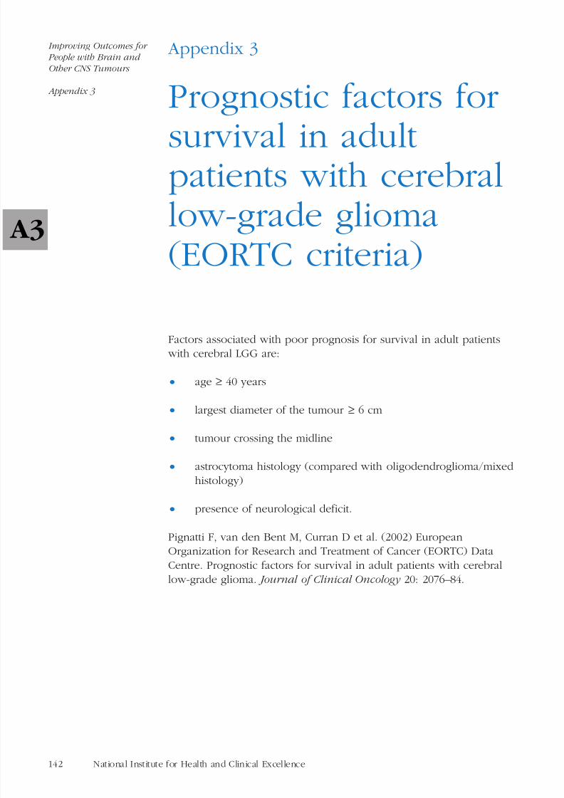
8/14/2019 CSG_brain_manual.pdf
http://slidepdf.com/reader/full/csgbrainmanualpdf 146/185
Appendix 3
Prognostic factors for
survival in adultpatients with cerebrallow-grade glioma(EORTC criteria)
Factors associated with poor prognosis for survival in adult patients with cerebral LGG are:
• age ≥ 40 years
• largest diameter of the tumour ≥ 6 cm
• tumour crossing the midline
• astrocytoma histology (compared with oligodendroglioma/mixed
histology)
• presence of neurological deficit.
Pignatti F, van den Bent M, Curran D et al. (2002) EuropeanOrganization for Research and Treatment of Cancer (EORTC) Data
Centre. Prognostic factors for survival in adult patients with cerebral
low-grade glioma. Journal of Clinical Oncology 20: 2076–84.
142 National Institute for Health and Clinical Excellence
Improving Outcomes for
People with Brain and
Other CNS Tumours
Appendix 3
A3

8/14/2019 CSG_brain_manual.pdf
http://slidepdf.com/reader/full/csgbrainmanualpdf 147/185
Appendix 4
Economic implications
of the guidance
Executive summary
The economic consequences of the recommendations of the‘Guidance on Cancer Services: Improving Outcomes in Brain and
Other CNS Tumours’ in England and Wales are set out in thisdocument. The analysis focuses on those aspects of the key recommendations that are likely to be of greatest consequence in
terms of costs.
The summary of economic implications is outlined in Table A2.
There is uncertainty concerning the estimates presented and there will
be variation at the neuroscience centre and cancer network level.
Sensitivity analyses were conducted where appropriate in the
estimated costs. Further assessments will be needed at cancer networklevel and/or NHS trust level to determine the exact cost implications.
The calculations for employment costs are based on pay levels at2005/06, any future pay awards will also need to be taken into
consideration. Work is currently being carried out in the NHS in
England, in connection with ‘Payment by Results’, to develop a betterunderstanding of costs of treatment and care. This may help these
assessments in the future.
143Guidance on cancer services: brain and other CNS tumours
Improving Outcomes for
People with Brain and
Other CNS Tumours
Appendix 4
A4

8/14/2019 CSG_brain_manual.pdf
http://slidepdf.com/reader/full/csgbrainmanualpdf 148/185
Table A2. Summary of estimated annual economic implications
144 National Institute for Health and Clinical Excellence
Improving Outcomes for
People with Brain and
Other CNS Tumours
Appendix 4
Costs per year (£)
Lower range Upper range
Staffing costs of weekly neuroscience
MDT meetings (per centre) 73,320 188,084
Staffing costs of monthly cancer
network MDT meetings (per network) 7,266 18,336
Site-specific MDTs a
Staffing costs of monthly pituitary
MDT meetings (per MDT) 5,994 15,768
Staffing costs of monthly spinal cord
MDT meetings (per MDT) 5,172 12,924
Staffing costs of monthly skull base
MDT meetings (per MDT) 7,044 18,912
Staffing costs for each MRI scanner b 267,071
Molecular pathology costs (staffing
and test costs for MGMT assay) 1,283,090
Core employment costs ofneuroscience centre 1,308,005 2,180,008
Total employment costs of additional
clinical nurse specialists 1,932,346
Total employment costs of additional
neuropathologists 1,474,208
Employment costs per centre of
additional allied health professionals 48,726 97,453
Total employment costs of additional
palliative care specialists 223,848
Cost of producing information leaflets
(for all patients with brain and other 14,620 (year 1)
central nervous system tumours in 10,400 (subsequent years)
England and Wales)
a It is uncertain how many site-specific multidisciplinary teams (MDTs) will be required.b There is overlap between these figures for employment cost of radiologist and radiographer.
MGMT, O6-methylguanine-DNA methyltransferase.
A4

8/14/2019 CSG_brain_manual.pdf
http://slidepdf.com/reader/full/csgbrainmanualpdf 149/185
Neuroscience and cancer network multidisciplinary teams
The guidance recommends that attendance at multidisciplinary team(MDT) meetings should form part of the timetabled activities for core
MDT members. It is assumed that extra resources will need to be
made available to enable staff to attend MDT meetings. Meeting costsare derived by estimating the time spent attending meetings by
different staff, multiplied by their hourly rate (salary and on-costs), in
addition to preparation time.
It is anticipated that for those cancer networks with no neuroscience
brain and other CNS tumours MDT in place, there will be an annualopportunity cost of between £73,320 for minimum attendance of a
weekly 2-hour neuroscience MDT and up to £188,084 for full
attendance at weekly 3-hour neuroscience MDT meetings.
All cancer networks will have an additional opportunity cost forestablishing a cancer network brain and other CNS tumours MDT.
This is estimated to be between £7266 per year for minimum
attendance at monthly 1-hour MDT meetings and £18,336 for fullattendance at monthly 2-hour MDT meetings.
Site-specific MDTs
For each pituitary MDT in place, there will be annual
opportunity costs of between £5994 for minimum attendance atmonthly 1-hour MDT meetings and £15,768 for full attendance
at monthly 2-hour MDT meetings.
For each spinal cord MDT in place, there will be annual
opportunity costs of between £5172 for minimum attendance at
monthly 1-hour MDT meetings and £12,924 for full attendanceat monthly 2-hour MDT meetings.
For each skull base MDT in place, there will be annual
opportunity costs of between £7044 for minimum attendance atmonthly 1-hour MDT meetings and £18,912 for full attendanceat monthly 2-hour MDT meetings.
Diagnosis – radiology
Government initiatives are under way to improve access to computed
tomography (CT) and magnetic resonance imaging (MRI) facilities.There is a need for imaging facilities to have dedicated time available
for patients with brain and CNS tumours. Existing scanners need to
be adequately staffed to ensure that they are able to operatethroughout the working day. It has been estimated that the staffing
145Guidance on cancer services: brain and other CNS tumours
Improving Outcomes for
People with Brain and
Other CNS Tumours
Appendix 4
A4

8/14/2019 CSG_brain_manual.pdf
http://slidepdf.com/reader/full/csgbrainmanualpdf 150/185
level needed to run an MRI scanner during normal working hours
would be 1.3 full-time equivalent (FTE) consultant neuroradiologists,3.0 FTE neuroradiographers plus additional administrative support.
The annual cost of this level of staffing is £267,071 per MRI scanner.
Diagnosis – pathology
Molecular diagnosis is a fast developing area of clinical practice, andcancer networks will need to plan for expansion over the next 10–20
years. At present, existing services for molecular diagnosis are
adequate for the numbers of patients who require testing. If a specificmolecular diagnostic test such as the MGMT assay becomes available,
then it is anticipated that an additional 30 biomedical scientists would
be required in England and Wales. The total additional annual cost forthe anticipated 2500 MGMT tests in England and Wales, including
consumables, and staffing to perform them is estimated to be£1,283,090. If two more similar molecular tests were to be adopted for
brain and other CNS tumours over a period of 10 years, then the total
costs might double.
Minimum staffing costs at neuroscience centres
The annual employment costs of the core staffing components of a
neuroscience centre treating 100 new patients per year is estimated to
be around £1,744,006 (± 25% range, £1,308,005–2,180,008). There willbe economies of scale for neuroscience centres that have all facilities
on site. As a result of uncertainty in this estimate a sensitivity analysisof ± 25% has been applied. It needs to be emphasised that the costsrepresent opportunity costs, as most of the staff involved in the
treatment centres will already be contracted to the NHS.
Clinical nurse specialists
The needs assessment57 conducted to inform the guidance
development indicated that there was a variation in the numbers of
clinical nurse specialists for neuro-oncology across England and
Wales. Although 3 (of 27) neurosurgical units and 4 (of 45)oncology/radiotherapy units had more than the recommended
minimum staffing for clinical nurse specialists in neuro-oncology, theremainder had less. To ensure that there are a minimum of 1.5
specialist nurses at each unit, a further 39.2 FTE would need to be
recruited at oncology/radiotherapy units and a further 11.5 FTE atneurosurgical units. Therefore, an additional 50.7 FTE clinical nurse
specialists are required in England and Wales at an estimated annual
employment cost of £1,932,346.
146 National Institute for Health and Clinical Excellence
Improving Outcomes for
People with Brain and
Other CNS Tumours
Appendix 4
57 Humphreys C (2005) Health needs assessment: adults with tumours of the brain and
central nervous system in England and Wales . National Public Health Service for Wales,Gwasanaeth Iechyd Cyhoeddus Cenedlaethol Cymru. Available on the CD-ROM thataccompanies this guidance and on the NICE website (www.nice.org.uk/csgbraincns).
A4

8/14/2019 CSG_brain_manual.pdf
http://slidepdf.com/reader/full/csgbrainmanualpdf 151/185
Neuropathologists
A report for the Royal College of Pathologists from the British
Neuropathological Society has recommended that there should be1 FTE neuropathologist per 1,000,000 patients. To achieve this level, a
further 15 FTE posts are required across England and Wales with an
estimated employment cost of around £1,474,208. There are currently seven single-handed neuropathologists in England and Wales, and
these would need to be supported to ensure continual cover of the
adult neuro-oncology service in these centres.
Allied health professionals
It has been estimated by the members of the Guidance Development
Group (GDG) that it is likely that an additional 0.5–1 FTE speech and
language therapist, occupational therapist and physiotherapist may berequired at each neuroscience centre as a result of the guidance.
The employment cost estimate for the three additional allied healthprofessionals (AHPs) is between £48,726 (1.5 FTE) and £97,453
(3 FTE) per centre. This estimate is uncertain, as it may be that some
centres would recruit additional junior staff to enable moreexperienced staff to concentrate on specialist work.
Palliative care specialists
To ensure that there are a minimum of 0.4 FTE palliative care
specialists available at all neuroscience centres additional staff willneed to be recruited. The estimated additional cost for the nine
neurosurgical centres without palliative care support is estimated to
be around £223,848. This estimate is based on a split betweenpalliative care specialist nurses and consultants (60/40). Further
investigation will be required at a local level to establish whether
palliative care consultants, nurse specialists or a mixture of both arerequired. In addition, those centres that stated that palliative care
was available may need additional funding to increase current levelof staffing. As with the other staff discussed in this section, it is
possible that the staffing situation has improved since this survey was conducted.
Patient information
The guidance suggests that patients with brain and other CNS
tumours have specific information needs, particularly when there is
some degree of cognitive impairment, and require information to beprovided in different formats such as spoken, written and audio-
visual. The total cost of producing and distributing generic and
centre-specific information leaflets would be £14,620 in the first yearfor all patients with brain and other CNS tumours in England and
Wales. In subsequent years the costs would be £10,400.
147Guidance on cancer services: brain and other CNS tumours
Improving Outcomes for
People with Brain and
Other CNS Tumours
Appendix 4
A4

8/14/2019 CSG_brain_manual.pdf
http://slidepdf.com/reader/full/csgbrainmanualpdf 152/185
A wide range of high-quality information is available from a variety of
sources on the internet. These mainly include charitable foundation web pages. Set-up costs of having information available are therefore
unlikely to be significant.
148 National Institute for Health and Clinical Excellence
Improving Outcomes for
People with Brain and
Other CNS Tumours
Appendix 4
A4

8/14/2019 CSG_brain_manual.pdf
http://slidepdf.com/reader/full/csgbrainmanualpdf 153/185
Appendix 5
How this guidance
manual was produced
This service guidance is intended to guide health organisations
(strategic health authorities, primary care trusts, local health boards,cancer networks and trusts), their managers and lead clinicians in
improving the effectiveness and efficiency of services for people withbrain and other central nervous system (CNS) tumours. The
information and recommendations in the manual are based on
reviews of the best available evidence on diagnosis, treatment andservice delivery. This evidence was retrieved by information
specialists and assessed by researchers at the National Collaborating
Centre for Cancer (NCC-C) and the recommendations are the productof extensive discussion with the Guidance Development Group
(GDG). A brief overview of the development process to produce the
guidance is provided below.
The first stage in the development of the guidance was the
production of a scope (appendix 1) which defined in detail thepatient population, the healthcare settings and services and key areas
of clinical management that the guidance should cover. This was then
subject to a 4-week consultation with registered stakeholders in line with National Institute for Health and Clinical Excellence (NICE)
methodology. Following this a multidisciplinary GDG was formedcomprising clinicians representing the main stakeholder organisations
and representatives from relevant patient organisations and charities
(appendix 5.2). The GDG was convened by the NCC-C and chairedby Dr Penny Bridger in close association with the Clinical Lead, Dr
Sean Elyan. All GDG members made and updated any declarations of
interest. The Group met on a monthly basis during development of the guidance and NCC-C staff provided methodological support and
leadership for the development.
During the development phase of the guidance the GDG identified
areas where there was a requirement for expert input on particular
specialist topic areas. These topics were addressed by the productionof a position paper by a recognised expert who had been identified
via the relevant registered stakeholder organisation. All relevantexpert positions papers are presented in appendix D of the evidence
review (available on the CD-ROM that accompanies this guidance and
on the NICE website [www.nice.org.uk/csgbraincns]).
149Guidance on cancer services: brain and other CNS tumours
Improving Outcomes for
People with Brain and
Other CNS Tumours
Appendix 5
A5

8/14/2019 CSG_brain_manual.pdf
http://slidepdf.com/reader/full/csgbrainmanualpdf 154/185
The identification and retrieval of evidence to support the
recommendations in the guidance manual is described in detail in theevidence review, which is available on the CD-ROM that
accompanies this guidance and on the NICE website
(www.nice.org.uk/csgbraincns). Briefly, there were three stages tothis process:
• Clinical question development . Members of the GDG wereasked to submit clinical questions to the NCC-C on issues
covered by the project scope.
• Literature searching . All clinical questions were prioritised and
were subject to either a systematic or ‘high-level’ search.
• Critical appraisal . Finally all full papers relevant to each clinical
question were appraised using the methodology described inthe ‘The guidelines manual’ (2006 edition).
It should be noted that most of the published research on cancertopics focuses on clinical evaluations of treatment; little direct
research has been carried out on the organisation and delivery of
services.
To gain insight into the current cancer services provision for patients
with brain and other CNS tumours, a needs assessment wascommissioned by the GDG and NCC-C. This study was carried out by
Dr Ciaran Humphreys at the National Public Health Service Wales.The full results are published separately on the NICE website(www.nice.org.uk/csgbraincns) and are also available on the
CD-ROM that accompanies this guidance.
All the evidence reviews used to inform the manual are summarised
in the document ‘Improving Outcomes in Brain and Other CNSTumours: the Research Evidence’ and include details of all the studies
appraised. This document is published separately on the NICE
website (www.nice.org.uk/csgbraincns) and is also available on the
CD-ROM that accompanies this guidance.
Additional complementary research, designed to quantify thepotential cost of major changes in services, was carried out by the
Centre for the Economics of Health, Institute of Medical and Social
Care Research (IMSCAR) at the University of Bangor. This workinvolves literature searching, interviews with clinicians and managers,
and analyses of costs. The economic analysis document is published
separately on the NICE website (www.nice.org.uk/csgbraincns) and isalso available on the CD-ROM that accompanies this guidance.
150 National Institute for Health and Clinical Excellence
Improving Outcomes for
People with Brain and
Other CNS Tumours
Appendix 5
A5

8/14/2019 CSG_brain_manual.pdf
http://slidepdf.com/reader/full/csgbrainmanualpdf 155/185
The writing of the guidance manual was coordinated by the Chair
and Clinical Lead of the GDG in accordance with all members of theGDG, assisted by staff at the NCC-C.
The production of this guidance was funded by NICE, and has beensubject to the full NICE consultation process.
151Guidance on cancer services: brain and other CNS tumours
Improving Outcomes for
People with Brain and
Other CNS Tumours
Appendix 5
A5

8/14/2019 CSG_brain_manual.pdf
http://slidepdf.com/reader/full/csgbrainmanualpdf 156/185
Appendix 6
People and
organisations involvedin the production of this guidance
6.1 Members of the Guidance Development Group
6.2 Organisations invited to comment on guidancedevelopment
6.3 Researchers carrying out literature reviews and complementary work
6.4 Expert advisers to the Guidance Development
Group
6.5 Members of the Guideline Review Panel
152 National Institute for Health and Clinical Excellence
Improving Outcomes for
People with Brain and
Other CNS Tumours
Appendix 6
A6

8/14/2019 CSG_brain_manual.pdf
http://slidepdf.com/reader/full/csgbrainmanualpdf 157/185
Appendix 6.1
Members of the
GuidanceDevelopment Group(GDG)
GDG Chair
Dr Penny Bridger Deputy Director of Public Health, Surrey
and Sussex SHA (until October 2005)
Consultant in Public Health Medicine,
Information Services, National ServicesScotland (from October 2005)
GDG Lead Clinician
Dr Sean Elyan Consultant Clinical Oncologist, CheltenhamGeneral Hospital
Guidance Development Group Members
Professor Mark Baker Director and Lead Clinician, Yorkshire
Cancer Network
Dr Juliet Britton Consultant Neuroradiologist, St George’s
Hospital, London
Mrs Katherine Carpenter Consultant Clinical Neuropsychologist, The
Radcliffe Infirmary, Oxford
Miss Joanne Carr Physiotherapist, Christie Hospital,Manchester
Professor Garth Professor of Neurosurgery, QueenCruickshank Elizabeth Hospital, Birmingham
153Guidance on cancer services: brain and other CNS tumours
Improving Outcomes for
People with Brain and
Other CNS Tumours
Appendix 6
A6

8/14/2019 CSG_brain_manual.pdf
http://slidepdf.com/reader/full/csgbrainmanualpdf 158/185
Mrs Angela Dickson Patient/Carer Representative, Samantha
Dickson Research Trust
Professor David Ellison Professor in Neuropathology, Newcastle
General Hospital
Dr Robin Grant Consultant Neurologist, Western General
Hospital, Edinburgh
Professor Phil Heywood Professor of Primary Care Development,
University of Leeds
Dr Martine Meyer Consultant in Palliative Medicine, St Helier
Hospital, Carshalton
Professor Roy Rampling Consultant Oncologist, Western Infirmary,
Glasgow
Mr Steve Sparks Assistant Director SpecialistCommissioning, Kent, Surrey and Sussex
Specialist Commissioning Group
Dr Sharon Swain Patient/Carer Representative, The Brain
and Spine Foundation
Miss Emma Townsley Macmillan Clinical Nurse Specialist,
National Hospital for Neurology and
Neurosurgery, London
Dr Esther Waterhouse Consultant in Palliative Medicine, Loros
Hospice, Leicester
154 National Institute for Health and Clinical Excellence
Improving Outcomes for
People with Brain and
Other CNS Tumours
Appendix 6
A6

8/14/2019 CSG_brain_manual.pdf
http://slidepdf.com/reader/full/csgbrainmanualpdf 159/185
Appendix 6.2
Organisations invited
to comment onguidance development
Addenbrooke’s NHS Trust
All Wales Senior Nurses Advisory Group (Mental Health)
Amersham Health
Anglesey Local Health Board
Association for Palliative Medicine of Great Britain and Ireland
Association of British Neurologists
Association of Hospice and Specialist Palliative Care Social Workers
Association of Neuro-oncology Nurses (ANON)
Association of Professional Music Therapists
Association of Surgeons of Great Britain and Ireland
Association of the British Pharmaceuticals Industry (ABPI)
Aventis Pharma
Bard Limited
Barking, Havering & Redbridge NHS Trust
Barts & The London NHS Trust
BASIC (Brain and Spinal Injury Charity)
Bath and North East Somerset PCT
155Guidance on cancer services: brain and other CNS tumours
Improving Outcomes for
People with Brain and
Other CNS Tumours
Appendix 6
A6

8/14/2019 CSG_brain_manual.pdf
http://slidepdf.com/reader/full/csgbrainmanualpdf 160/185
Bayer PLC
Bedfordshire & Hertfordshire NHS Strategic Health Authority
Boehringer Ingelheim Ltd
Boston Scientific Limited
Brain and Spine Foundation
British Association for Counselling and Psychotherapy
British Association of Neuroscience Nurses
British Association of Oral and Maxillofacial Surgeons
British Dietetic Association
British National Formulary (BNF)
British Nuclear Medicine Society
British Oncology Pharmacy Association
British Orthoptic Society
British Paediatric Neurology Association
British Psychological Society
British Psychosocial Oncology Society
British Society of Neuroradiologists
British Society of Paediatric Radiology
British Society of Rehabilitation Medicine
BUPA
Cancer and Leukaemia in Childhood (UK)
CancerBACUP
Cancer Research UK
Cancer Services Collaborative ‘Improvement Partnership’ (CSCIP)
Cancer Services Coordinating Group
156 National Institute for Health and Clinical Excellence
Improving Outcomes for
People with Brain and
Other CNS Tumours
Appendix 6
A6

8/14/2019 CSG_brain_manual.pdf
http://slidepdf.com/reader/full/csgbrainmanualpdf 161/185
Cancer Voices
Chartered Society of Physiotherapy
Children’s and Adolescent Cancer Partnership (CACP)
Clatterbridge Centre for Oncology NHS Trust
Clinovia Ltd
College of Occupational Therapists
Conwy and Denbighshire NHS Trust
Cornwall Partnership Trust
Department of Health
Derbyshire Mental Health Services NHS Trust
Eisai Limited
Elan Pharmaceuticals Ltd
Eli Lilly and Company Ltd
Faculty of Public Health
GE Health Care
Gloucestershire Hospitals NHS Trust
Gorlin Syndrome Group
Great Ormond Street Hospital for Children NHS Trust
Guerbet Laboratories Ltd
Hammersmith Hospitals NHS Trust
Hampshire & Isle of Wight Strategic Health Authority
Headway – The Brain Injury Association
Healthcare Commission
Help Adolescents with Cancer
Help the Hospices
157Guidance on cancer services: brain and other CNS tumours
Improving Outcomes for
People with Brain and
Other CNS Tumours
Appendix 6
A6
8/14/2019 CSG_brain_manual.pdf
http://slidepdf.com/reader/full/csgbrainmanualpdf 162/185
Hertfordshire Partnership NHS Trust
Institute of Biomedical Science
International Brain Tumour Alliance
Joint Committee on Palliative Medicine
Link Pharmaceuticals
Macmillan Cancer Relief
Marie Curie Cancer Care
Medeus Pharma Ltd
Medical Research Council Clinical Trials Unit
Medicines and Healthcare Products Regulatory Agency (MHRA)
Medway NHS Trust
Merck Pharmaceuticals
National Alliance of Childhood Cancer Parent Organisations
National Cancer Alliance
National Cancer Network Clinical Directors Group
National Cancer Research Institute (NCRI) Clinical Studies Group andNational Cancer Research Network (NCRN)
National Patient Safety Agency
National Public Health Service – Wales
Neurological Alliance
NHS Direct
NHS Health and Social Care Information Centre
NHS Modernisation Agency
NHS Quality Improvement Scotland
Novartis Pharmaceuticals UK Ltd
158 National Institute for Health and Clinical Excellence
Improving Outcomes for
People with Brain and
Other CNS Tumours
Appendix 6
A6
8/14/2019 CSG_brain_manual.pdf
http://slidepdf.com/reader/full/csgbrainmanualpdf 163/185
Pfizer Limited
Plymouth Hospitals NHS Trust
Princess Alexandra Hospital NHS Trust
Richmond & Twickenham PCT
Royal College of Anaesthetists
Royal College of General Practitioners
Royal College of General Practitioners Wales
Royal College of Nursing (RCN)
Royal College of Paediatrics and Child Health
Royal College of Pathologists
Royal College of Physicians of London
Royal College of Psychiatrists
Royal College of Radiologists
Royal College of Speech and Language Therapists
Royal College of Surgeons of England
Royal College Patient Liaison Groups
Royal Liverpool Children’s NHS Trust
Royal Pharmaceutical Society of Great Britain
Royal Society of Medicine
Royal West Sussex Trust
Samantha Dickson Research Trust
Schering-Plough
Scottish Intercollegiate Guidelines Network (SIGN)
Sheffield Children’s NHS Trust
Sheffield South West Primary Care Trust
159Guidance on cancer services: brain and other CNS tumours
Improving Outcomes for
People with Brain and
Other CNS Tumours
Appendix 6
A6

8/14/2019 CSG_brain_manual.pdf
http://slidepdf.com/reader/full/csgbrainmanualpdf 164/185
Sheffield Teaching Hospitals NHS Trust
Society and College of Radiographers
Society for Endocrinology
Society of British Neurological Surgeons
Southampton University Hospitals NHS Trust
South West Peninsula Strategic Health Authority
Stroke Association
Teenage Cancer Trust
Thames Valley Strategic Health Authority
UK Children’s Cancer Study Group
University College London Hospital NHS Trust
University Hospital Birmingham NHS Trust
University of Leeds
Vale of Aylesbury PCT
Velindre NHS Trust
Walton Centre for Neurology and Neurosurgery NHS Trust
Welsh Assembly Government (formerly National Assembly for Wales)
160 National Institute for Health and Clinical Excellence
Improving Outcomes for
People with Brain and
Other CNS Tumours
Appendix 6
A6

8/14/2019 CSG_brain_manual.pdf
http://slidepdf.com/reader/full/csgbrainmanualpdf 165/185
Appendix 6.3
Researchers carrying
out literature reviewsand complementary
work
Overall coordinators
Dr Fergus Macbeth National Collaborating Centre for Cancer,
Cardiff
Dr Andrew Champion National Collaborating Centre for Cancer,
Cardiff
Project managers
Dr Nansi Swain National Collaborating Centre for Cancer,Cardiff
Karen Field National Collaborating Centre for Cancer,Cardiff
Senior researcher
Dr Mary Webb National Collaborating Centre for Cancer,Cardiff
Researchers
Dr Nathan Bromham National Collaborating Centre for Cancer,
Cardiff
Andrew Cleves National Collaborating Centre for Cancer,
Cardiff
Dr Karen Francis National Collaborating Centre for Cancer,
Cardiff
Angela Melder National Collaborating Centre for Cancer,
Cardiff
161Guidance on cancer services: brain and other CNS tumours
Improving Outcomes for
People with Brain and
Other CNS Tumours
Appendix 6
A6
8/14/2019 CSG_brain_manual.pdf
http://slidepdf.com/reader/full/csgbrainmanualpdf 166/185
Information specialist
Stephanie Arnold National Collaborating Centre for Cancer,
Cardiff
Health economists
Dr Rhiannon Director, Centre for the Economics of
Tudor Edwards Health, Institute of Medical and Social Care
Research, University of Wales, Bangor
Dr Dyfrig Hughes Locum Director, Centre for the Economics
of Health, Institute of Medical and SocialCare Research, University of Wales, Bangor
Pat Linck Research Officer, Centre for the Economicsof Health, Institute of Medical and Social
Care Research, University of Wales, Bangor
Eugenia Priedane Research Officer, Centre for the Economics
of Health, Institute of Medical and SocialCare Research, University of Wales, Bangor
Needs assessment
Dr Ciaran Humphreys Specialist Registrar, National Public Health
Service for Wales
162 National Institute for Health and Clinical Excellence
Improving Outcomes for
People with Brain and
Other CNS Tumours
Appendix 6
A6

8/14/2019 CSG_brain_manual.pdf
http://slidepdf.com/reader/full/csgbrainmanualpdf 167/185
Appendix 6.4
Expert advisers to
the GuidanceDevelopment Group
Dr Howard Ring Chair of Special Interest Group in
Neuropsychiatry, Royal College of
Psychiatrists
Mr David Sandeman Consultant Neurosurgeon, North Bristol
NHS Trust, Society for Endocrinology
163Guidance on cancer services: brain and other CNS tumours
Improving Outcomes for
People with Brain and
Other CNS Tumours
Appendix 6
A6

8/14/2019 CSG_brain_manual.pdf
http://slidepdf.com/reader/full/csgbrainmanualpdf 168/185
Appendix 6.5
Members of the
Guideline Review Panel
Chair
John Hyslop
Members
Graham Archard
Tony Donovan
Mark Emberton
Patricia Fairbrother
Stephen Karp
164 National Institute for Health and Clinical Excellence
Improving Outcomes for
People with Brain and
Other CNS Tumours
Appendix 6
A6

8/14/2019 CSG_brain_manual.pdf
http://slidepdf.com/reader/full/csgbrainmanualpdf 169/185
Appendix 7
Glossary of terms
Adenoma
Adenomas are benign epithelial tumours of glandular tissue.
Adjuvant chemotherapy
Chemotherapy treatment that is given in addition to the main cancer
treatment.
Adjuvant therapy
Additional treatment that is added to increase the effectiveness of themain treatment.
Aetiology
The cause or origin of disease.
Allied health professional (AHP)
One of the following group of healthcare workers: physiotherapists,
occupational therapists, art therapists, chiropodists/podiatrists,
dietitians, drama therapists, music therapists, orthoptists, paramedics,prosthetists/orthotists, diagnostic radiographers, therapeutic
radiographers, speech and language therapists.
Anaplastic
Cells in ‘anaplastic’ tumours show marked variation in size and shape
(cytological pleomorphism) and signs of rapid proliferation. Anaplastictumours may also contain areas of dead (necrotic) tissue and
formation of new blood vessels (angiogenesis).
Angiography The X-ray visualisation of the internal anatomy of the heart and blood vessels after injection of a radio-opaque (shows up on X-rays)
contrast material.
Asymptomatic
Without obvious signs or symptoms of disease. In early stages, cancer
may develop and grow without producing symptoms.
Audiology
The study of hearing.
165Guidance on cancer services: brain and other CNS tumours
Improving Outcomes for
People with Brain and
Other CNS Tumours
Appendix 7
A7

8/14/2019 CSG_brain_manual.pdf
http://slidepdf.com/reader/full/csgbrainmanualpdf 170/185
Audit
A review and evaluation of healthcare procedures.
Autosomal
Refers to a chromosome that is not involved in determining sex. If adisorder is autosomal it affects both males and females equally.
Base of skull The sloping floor of the cranial cavity.
Benign
Not cancerous; not malignant.
Biopsy
Removal of a sample of tissue or cells from the body to assist in
diagnosis of a disease.
Brachytherapy
Radiotherapy delivered by a temporary or permanent implant of radioactive material into a tissue or organ.
Brain tumour
A tumour found in the intracranial portion of the central nervous
system.
Cancer
Growth of altered body cells that keep on growing and is able to
spread from where it started to another part of the body.
Cancer networks
The organisational model for cancer services to implement the NHSCancer Plan, bringing together health service commissioners and
providers, the voluntary sector and local authorities. There arecurrently 34 cancer networks in England and three in Wales, covering
a population of between 600,000 and 3 million (two thirds serve a
population of between 1 and 2 million people).
Cancer registry
An organisation that systematically collects information about cancerincidence and mortality within its resident population.
Carcinoma
Cancer of the epithelial tissue that covers all the body organs and
lines all the body cavities. Most cancers are carcinomas.
Central nervous system
The portion of the nervous system comprising the brain and spinalcord.
166 National Institute for Health and Clinical Excellence
Improving Outcomes for
People with Brain and
Other CNS Tumours
Appendix 7
A7

8/14/2019 CSG_brain_manual.pdf
http://slidepdf.com/reader/full/csgbrainmanualpdf 171/185
Cerebrospinal fluid
The fluid that flows around the brain and the spinal cord.
Chemosensitivity
The specific sensitivity of an individual to chemotherapy drugs.
Chemotherapy
The use of drugs that kill cancer cells, or prevent or slow theirgrowth.
Clinical nurse specialist
A nurse with specialist knowledge of central nervous system tumours
and skills in communication as defined by the Manual for Cancer
Services.
Clinical oncologist
A doctor who specialises in the treatment of cancer patients,particularly through the use of radiotherapy, but who may also use
chemotherapy.
Clinical oncology
The specialist treatment of cancer patients, particularly through theuse of radiotherapy, but that may also use chemotherapy.
Clinical trial
A research study with patients, usually to evaluate a new treatment or
drug.
Cognitive
Relating to the mental processes of comprehension, judgement,
memory and reasoning.
Cohort studies
Research studies in which groups of patients with a particular
condition or specific characteristic are compared with matched groups
who do not have it.
Community nurse
A nurse who provides care to individuals in the community andoutside of hospital.
Computed tomography (CT)
An X-ray technique that produces cross-sectional images.
Concordance
A new way of defining the process of successful prescribing and
taking medicines, based on a partnership between doctors andpatients (see www.medicines-partnership.org).
167Guidance on cancer services: brain and other CNS tumours
Improving Outcomes for
People with Brain and
Other CNS Tumours
Appendix 7
A7

8/14/2019 CSG_brain_manual.pdf
http://slidepdf.com/reader/full/csgbrainmanualpdf 172/185
Cranial nerves
The 12 pairs of nerves that emerge from the cranial cavity throughopenings in the skull.
Craniotomy
A surgical incision or opening into the skull.
Curative Aiming to cure a disease.
Cytogenetics
The study of chromosomes and chromosomal abnormalities.
Cytological examination
The examination of cells on a microscope, especially for diagnosis,
performed by a specialist in pathology.
Cytopathology
The study of disease changes within individual cells or cell types.
Diagnostic imaging
Visualising body structures to help identify a disease or condition, by using X-ray, ultrasound, radioisotopes or magnetic resonance.
Diagnostic investigations
Tests undertaken to establish the nature of disease.
Diagnostic radiographer
A health professional trained in the technique of obtaining images of
various parts of the body, working closely with a radiologist.
Dietitian
The healthcare professional responsible for the planning andmanaging of the patient’s diet in hospital and providing dietary advice
for a wide range of medical conditions.
Ear, nose and throat (ENT)Diagnosis and treatment of diseases of the ear, nose and throat.
Endocrine
Having to do with glandular tissues that secrete hormones directly
into the bloodstream.
Endocrinologist
A doctor who specialises in treating diseases of the endocrine system.
Endoscopy The visual inspection of any cavity of the body using a special
instrument.
168 National Institute for Health and Clinical Excellence
Improving Outcomes for
People with Brain and
Other CNS Tumours
Appendix 7
A7

8/14/2019 CSG_brain_manual.pdf
http://slidepdf.com/reader/full/csgbrainmanualpdf 173/185
Epidemiology
The study of populations in order to determine the frequency anddistribution of disease and measure risks.
Epilepsy
A group of neurological disorders characterised by recurrent episodes
of either convulsive seizures, sensory disturbances, abnormal
behaviour or loss of consciousness.
Excise/excision
Removal by surgery.
Fractionation
Dividing the total dose of radiation therapy into several smaller, equaldoses delivered over a period of several days.
Germ cell
The reproductive cells of the body: in men, the testicular cell that
divides to produce the immature sperm cells; in women, the ovariancell that divides to form the egg.
Glial
Specialised cells that surround neurones and provide physical support
and insulation.
Glioma
A cancer of the brain that begins in glial cells (cells that surround and
support nerve cells). A ‘high-grade’ glioma is one that grows rapidly and has an aggressive behaviour. High-grade tumours are grades 3
and 4 in the WHO classification. The grade of CNS tumour correlates
with prognosis (1 – best; 4 – worst)
Gorlin syndrome
An inherited condition that can increase an individual’s chance of
developing basal cell carcinoma. Also called basal cell naevus
syndrome.
Gynaecologist
A doctor who specialises in the diagnosis and treatment of disordersaffecting the female reproductive organs.
Haematologist
A doctor who specialises in disorders of the blood and blood-forming
tissues.
Haematopoietic
The process by which blood cells are produced in the bone marrow.
169Guidance on cancer services: brain and other CNS tumours
Improving Outcomes for
People with Brain and
Other CNS Tumours
Appendix 7
A7
8/14/2019 CSG_brain_manual.pdf
http://slidepdf.com/reader/full/csgbrainmanualpdf 174/185
Histological
Relating to the study of cells and tissue at a microscopic level.
Histology
The study of body tissue and cells by examination under amicroscope.
Histopathologist A clinical doctor who specialises in examining abnormal tissue
samples microscopically in order to make a diagnosis and ensure
tumour excision is complete.
Histopathology
The study of microscopic changes in diseased tissues.
Holistic
Looking at the whole system rather than just concentrating onindividual components.
Imaging
The production of a clinical image using radiology, for example an
X-ray or ultrasound.
Immunosuppression
Suppression of the body’s immune system and its ability to fightinfections or disease. Immunosuppression may be deliberately
induced with drugs. It may also result from certain diseases such as
lymphoma or from anticancer drugs.
Intracranial pressure
Pressure that occurs within the cranium (skull).
Intraoperative
Relating to the period during a surgical procedure.
Intrathecal
Into the fluid around the spine.
Ionising radiation
High-energy electromagnetic waves (for example, X-rays) and
particles (for example, electrons) used for radiotherapy treatment.
Irradiation
The use of high-energy radiation from X-rays, gamma rays, neutrons
and other sources to kill cancer cells and shrink tumours.
170 National Institute for Health and Clinical Excellence
Improving Outcomes for
People with Brain and
Other CNS Tumours
Appendix 7
A7

8/14/2019 CSG_brain_manual.pdf
http://slidepdf.com/reader/full/csgbrainmanualpdf 175/185
Key worker
Person who, with the patient’s consent and agreement, takes a key role in coordinating the patient’s care and promoting continuity,
ensuring the patient knows whom to access for information and
advice.
Lesion
An area of abnormal tissue.
Leukaemia
Cancer of the blood-forming system in the bone marrow, usually characterised by the production of abnormal white blood cells, which
may be present in the bone marrow and blood.
Li–Fraumeni syndrome
An inherited family trait carrying an increased risk of cancer during
childhood and early adulthood.
Local health board
Statutory bodies in Wales responsible for commissioning and
providing health services on behalf of the local population.
Low-grade glioma
A CNS tumour that grows slowly, but which may or may not be
successfully treated. Low-grade tumours are grades 1 and 2 in the WHO classification. The grade of CNS tumour correlates with
prognosis (1 – best; 4 –worst).
Lymphoma
Cancer of the lymphatic system. There are two main types of
lymphoma – Hodgkin’s disease and non-Hodgkin’s lymphoma.
Magnetic resonance imaging (MRI)
A non-invasive method of imaging that allows the form and
metabolism of tissues and organs to be visualised (also known as
nuclear magnetic resonance).
Malignant
Cancerous. Malignant tumours can invade and destroy nearby tissueand spread to other parts of the body.
Margin
The edge or border of the tissue removed in cancer surgery.
Maxillofacial
The specialty that combines full surgical training with dental expertise
for the treatment of diseases, injuries, tumours and deformities of theface and jaws.
171Guidance on cancer services: brain and other CNS tumours
Improving Outcomes for
People with Brain and
Other CNS Tumours
Appendix 7
A7

8/14/2019 CSG_brain_manual.pdf
http://slidepdf.com/reader/full/csgbrainmanualpdf 176/185
Medical oncologist
A doctor who treats cancer patients through the use of chemotherapy,and for some tumours immunotherapy.
Medical oncology
The specialist treatment of cancer patients through the use of
chemotherapy, and for some tumours immunotherapy.
Medulloblastoma
A malignant brain tumour that begins in the lower part of the brain
and that can spread to the spine or other parts of the body.
Meninges
The three membranes surrounding the brain and spinal cord.
Meta-analysis
The statistical analysis of the results of a collection of individualresearch studies in order to add the findings together.
Metastases
Cancerous tumours in any part of the body as a result of spread from
the original (primary) origin.
Minimum dataset
A widely agreed-upon and generally accepted set of terms anddefinitions making up a core of data acquired for medical records and
used for developing statistics for different types of analyses and users.
Molecular genetics
The branch of genetics that focuses on the chemical structure and the
functions, replication and mutations of the molecules involved in thetransmission of genetic information, namely DNA and RNA.
Molecular pathology
New techniques for identifying molecular abnormalities in the DNA of
cells.
Morbidity
Either (1) the state of being diseased; or (2) the morbidity rate, whichreflects the number of cases of disease per unit of population in any
specific region, age group, disease or other classification, usually
expressed as cases per 1000, 10,000 or 100,000.
Morphology
The science of the forms and structures of organisms.
172 National Institute for Health and Clinical Excellence
Improving Outcomes for
People with Brain and
Other CNS Tumours
Appendix 7
A7

8/14/2019 CSG_brain_manual.pdf
http://slidepdf.com/reader/full/csgbrainmanualpdf 177/185
Mortality
Either (1) the condition of being subject to death; or (2) the deathrate, which reflects the number of deaths per unit of population in
any specific region, age group, disease or other classification, usually
expressed as deaths per 1000, 10,000 or 100,000.
Nasogastric feeding
Feeding via a tube that is passed through the nose and into thestomach.
Needle biopsy
The removal of tissue or fluid through a needle for examination of
cells under a microscope.
Neoplasm
An abnormal mass of tissue that results from excessive cell division.
Neurofibromatosis
A genetic condition in which people develop multiple, benigntumours of nerve tissue.
Neuroimaging
Production of images of the brain by non-invasive techniques, for
example computed tomography, magnetic resonance imaging or
positron emission tomography.
Neurological
Having to do with the nervous system.
Neurologist
A doctor who diagnoses and treats disorders of the central nervoussystem.
Neuro-oncology
The branch of medical science dealing with tumours of the nervous
system.
Neuropathology
The study of disease processes in the nervous system.
Neuropsychiatry
The branch of psychiatry relating to disorders of the central nervoussystem.
Neuropsychology
A discipline combining neurology and psychology to study the
relationship between the functioning of the brain and cognitiveprocesses or behaviour, using psychological testing and assessment to
assay central nervous system function and diagnose specific
behavioural or cognitive deficits or disorders.
173Guidance on cancer services: brain and other CNS tumours
Improving Outcomes for
People with Brain and
Other CNS Tumours
Appendix 7
A7

8/14/2019 CSG_brain_manual.pdf
http://slidepdf.com/reader/full/csgbrainmanualpdf 178/185
Neuroradiologist
A doctor trained in radiology specialising in creating and interpretingpictures of the nervous system.
Neuroradiology
The branch of radiology that deals with the nervous system.
Neurorehabilitation Rehabilitation that concentrates on improving physical and cognitive
or understanding impairment resulting from damage to the nervous
system.
Neurosurgeon
A doctor who specialises in surgery on the brain, spine and otherparts of the nervous system.
Neurosurgery
Surgery on any part of the nervous system.
Observational study
A non-randomised study that observes the characteristics and
outcomes over time of subjects who do and do not take a particulartherapy.
Occupational therapist
A specialist who is concerned with a person’s ability to participate in
meaningful occupations and the impact of their environment on this;
they will work with a person to design a programme of interventionbased on the individual’s unique lifestyle, environment and
preferences.
Oncologist
A doctor who is trained to treat patients with chemotherapy (medicaloncologist), radiotherapy or both (clinical oncologist).
Oncology
The study of the biology and physical and chemical features of cancers. Also the study of the causes and treatment of cancers.
Ophthalmology
The area of medicine dealing with the eye.
Optic disc
The small blind spot on the surface of the retina.
Optic glioma
A slow-growing tumour on the optic nerve.
174 National Institute for Health and Clinical Excellence
Improving Outcomes for
People with Brain and
Other CNS Tumours
Appendix 7
A7

8/14/2019 CSG_brain_manual.pdf
http://slidepdf.com/reader/full/csgbrainmanualpdf 179/185
Oral
Having to do with the mouth.
Orthopaedics
A branch of surgery that specialises in the study and treatment of theskeletal system, its joints, muscles and associated structures.
Orthotist A skilled professional who fabricates orthotic devices that are
prescribed by a physician.
Otolaryngologist
A doctor who specialises in treating diseases of the ear, nose and
throat.
Palliative
Anything that serves to alleviate symptoms caused by the underlyingcancer but is not expected to cure it.
Palliative care
Active, holistic care of patients with advanced, progressive illness that
may no longer be curable. The aim is to achieve the best quality of life for patients and their families. Many aspects of palliative care are
also applicable in earlier stages of the cancer journey in association
with other treatments.
Papilloedema
Swelling of the optic disc.
Pathologist
A doctor who examines cells and identifies them. The pathologist cantell where a cell comes from in the body and whether it is normal or
a cancer cell. If it is a cancer cell, the pathologist can tell what typeof body cell the cancer developed from. In the hospital, practically all
the diagnostic tests performed with material removed from the body
are evaluated or performed by a pathologist.
Pathology
A branch of medicine concerned with disease, especially its functionaleffects on the body.
Patient pathway
A term used to denote the phases patients who are living with the
consequences of cancer pass through, from the time they first suspect
something might be wrong to beyond the end of treatment. It isdynamic and can be somewhat unpredictable. For some, the pathway
will also include the time when cancer recurs and that leading up totheir death.
175Guidance on cancer services: brain and other CNS tumours
Improving Outcomes for
People with Brain and
Other CNS Tumours
Appendix 7
A7

8/14/2019 CSG_brain_manual.pdf
http://slidepdf.com/reader/full/csgbrainmanualpdf 180/185
Physiotherapist
A specialist trained in using exercise and physical activities tocondition muscles and improve level of activity.
Pineal tumour
A tumour of the pineal body.
Pituitary tumour A tumour of the pituitary gland.
Plastic surgeon
A doctor who specialises in surgery to correct damage to the skin, for
example reducing the amount of scarring or disfigurement that may
happen because of surgery to treat a skin tumour.
Positron emission tomography (PET)
A highly specialised imaging technique using a radioactive tracer toproduce a computerised image of body tissues to find any
abnormalities. PET scans are sometimes used to help diagnose cancerand investigate a tumour’s response to treatment.
Prognosis
A prediction of the likely outcome or course of a disease; the chance
of recovery or recurrence.
Prognostic factor
Patient or disease characteristics, for example age or comorbidity,
which influence the course of the disease under study.
Protocol
An agreed policy that defines appropriate action.
Psychological
Adjective of psychology, which is the scientific study of behaviour and
its related mental processes. Psychology is concerned with such
matters as memory, rational and irrational thought, intelligence,
learning, personality, perceptions and emotions and their relationshipto behaviour.
Psychologist
A specialist who can talk with patients and their families about
emotional, cognitive and personal matters, and can help them makedecisions.
Radiographer
A person who assists the radiologist in imaging (diagnostic radiographer)
or the radiotherapist in treatment (therapeutic radiographer).
176 National Institute for Health and Clinical Excellence
Improving Outcomes for
People with Brain and
Other CNS Tumours
Appendix 7
A7

8/14/2019 CSG_brain_manual.pdf
http://slidepdf.com/reader/full/csgbrainmanualpdf 181/185
Radiologist
A doctor who specialises in creating and interpreting pictures of areasinside the body using X-rays and other specialised imaging
techniques. An interventional radiologist specialises in the use of
imaging techniques to assist treatment, for example catheter insertionfor abscess drainage.
Radiology The use of radiation (such as X-rays) or other imaging technologies
(such as ultrasound and magnetic resonance imaging) to diagnose or
treat disease.
Radiosurgery
A radiation therapy technique that delivers radiation directly to thetumour while sparing the healthy tissue.
Radiotherapy (radiation treatment)
The use of radiation, usually X-rays or gamma rays, to kill cancer cells
and treat tumours.
Randomised controlled trial (RCT)
A type of experiment that is used to compare the effectiveness of different treatments. The crucial feature of this form of trial is that
patients are assigned at random to groups that receive the
interventions being assessed or control treatments. RCTs offer themost reliable (that is least biased) form of evidence of effectiveness.
Reconstructive surgery
Surgery that is done to reshape or rebuild (reconstruct) a part of the
body changed by previous surgery.
Resection
To remove tissue from the body by surgery.
Retrospective study
A study which searches for a relationship between one (usually
current) condition and another that occurred in the past.
Single photon emission computed tomography (SPECT)
A variation of computed tomography (CT).
Specialist palliative care
Services in all sectors that specialise in palliative care and that include
consultants in palliative medicine, clinical nurse specialists in
palliative care and a range of other specialist expertise.
Speech and language therapist A specialist trained in the assessment and management of
communication and swallowing difficulties.
177Guidance on cancer services: brain and other CNS tumours
Improving Outcomes for
People with Brain and
Other CNS Tumours
Appendix 7
A7

8/14/2019 CSG_brain_manual.pdf
http://slidepdf.com/reader/full/csgbrainmanualpdf 182/185
Stereotactic
The precise three-dimensional positioning of a surgical instrument orradiotherapy machine.
Supportive care
Care that helps the patient and their family and carers to cope with
cancer and its treatment throughout the cancer journey, and in the
case of the family and carers, into bereavement. It aims to help thepatient maximise the benefits of treatment and provide the best
possible quality of life.
Systematic review
A systematic review of the literature carried out in order to address a
defined question and using quantitative methods to summarise theresults.
Tertiary
Third level. Relating to medical treatment provided at a specialist
institution.
Therapeutic radiographer
Therapeutic radiographers are responsible for providing radiotherapy treatment.
Toxicity
Refers to the undesirable and harmful side effects of a drug.
Trans-sphenoidal
Performed through the sphenoid bone, part of the base of the skull
below the brain.
Tumour
A mass of excess tissue that results from abnormal cell division.Tumours perform no useful body function.
Unresectable
A tumour or mass that cannot be removed by surgery.
X-ray
A photographic or digital image of the internal organs or bones
produced by the use of ionising radiation.
178 National Institute for Health and Clinical Excellence
Improving Outcomes for
People with Brain and
Other CNS Tumours
Appendix 7
A7

8/14/2019 CSG_brain_manual.pdf
http://slidepdf.com/reader/full/csgbrainmanualpdf 183/185
Appendix 8
Abbreviations
AHP allied health professional
AIDS acquired immune deficiency syndrome
CI confidence interval
CNS central nervous system
CPA clinical pathology accreditation
CSF cerebrospinal fluid
CT computed tomography
CTAAC Clinical Trials Advisory and Awards Committee
DH Department of Health
DNA deoxyribonucleic acid
ENT ear, nose and throat
EORTC European Organisation for Research and
Treatment of Cancer
EQA external quality assurance
EUROCARE European cancer registries study on cancer
patients’ survival and care
FTE full-time equivalent
GCTs germ cell tumours
GDG Guidance Development Group
GP general practitioner
HGG high-grade glioma
HIV human immunodeficiency virus
HR hazard ratio
ICD International Classification of Diseases
IT information technology
LGG low-grade glioma
MDT multidisciplinary team
MGMT O6-methylguanine-DNA methyltransferase
MR magnetic resonance
MRI magnetic resonance imaging
179Guidance on cancer services: brain and other CNS tumours
Improving Outcomes for
People with Brain and
Other CNS Tumours
Appendix 8
A8

8/14/2019 CSG_brain_manual.pdf
http://slidepdf.com/reader/full/csgbrainmanualpdf 184/185
NATCANSAT National Cancer Services Analysis Team
NCC-C National Collaborating Centre for Cancer
NCRI National Cancer Research Institute
NCRN National Cancer Research Network
NHS National Health Service
NICE National Institute for Health and ClinicalExcellence
OR odds ratio
PCNSL primary central nervous system lymphoma
PCT primary care trust
PET positron emission tomography
PNET primitive neuroectodermal tumor
R&D research and developmentRCT randomised controlled trial
SPECT single photon emission computed tomography
UK United Kingdom
UKCCSG United Kingdom Children’s Cancer Study Group
UKCRC United Kingdom Cancer Research Campaign
WBRT whole brain radiotherapy
WHO World Health Organization
180 National Institute for Health and Clinical Excellence
Improving Outcomes for
People with Brain and
Other CNS Tumours
Appendix 8
A8

8/14/2019 CSG_brain_manual.pdf
http://slidepdf.com/reader/full/csgbrainmanualpdf 185/185


