Embed Size (px)
Citation preview
Crystallographic Studies on Ascaris suum NAD-Malic Enzyme Bound to Reduced Cofactor
and Identification of an Effector Site
G.S. Jagannatha Rao†, D. E. Coleman¥, William E. Karsten§, Paul F. Cook§ and Ben G.
Harris†,*
From the †Department of Molecular Biology and Immunology, University of North Texas Health
Science Center, Fort Worth, TX 76107, U.S.A., the ¥Department of Biochemistry, The
University of Texas Southwestern Medical Center, 5323 Harry Hines Blvd, Dallas, TX 75235-
9050, and the §Department of Chemistry and Biochemistry, University of Oklahoma, Norman,
OK 73019
Correspondence author:
E-mail: [email protected], Phone: 817-735-2126 Fax: 817-735-2133
Running Title: Malic Enzyme Structure
This work was supported by grants to B.G.H. from the National Institutes of Health (AI24155;
AI41552), the Robert A.Welch Foundation (BK1309); to P.F.C. from the Oklahoma Center for
Advancement of Science and Technology (HR 990217), the American Chemical Society (PRF
35894-AC4), and the National Science Foundation (MCB 0091207).
Copyright 2003 by The American Society for Biochemistry and Molecular Biology, Inc.
JBC Papers in Press. Published on July 9, 2003 as Manuscript M305145200 by guest on M
arch 30, 2019http://w
ww
.jbc.org/D
ownloaded from
2
Abstract. The crystal structure of the mitochondrial NAD-malic enzyme from Ascaris
suum, in a quaternary complex with NADH, tartronate and magnesium has been determined to
2.0 Å resolution. The structure closely resembles the previously determined structure of the
same enzyme in binary complex with NAD. However, significant difference is observed within
the coenzyme-binding pocket of the active site with the nicotinamide ring of NADH molecule
rotating by 198° over the C1-N1 bond into the active site without causing significant movement
of the other catalytic residues. The implications of this conformational change in the
nicotinamide ring to the catalytic mechanism are discussed. The structure also reveals a binding
pocket for the divalent metal ion in the active site and a binding site for tartronate located in a
highly positively charged environment within the subunit interface that is distinct from the active
site. The tartronate binding site, presumably an allosteric site for the activator fumarate, shows
striking similarities and differences with the activator site of the human NAD-malic enzyme that
has been reported recently. Thus the structure provides additional insights into the catalytic as
well as the allosteric mechanisms of the enzyme.
by guest on March 30, 2019
http://ww
w.jbc.org/
Dow
nloaded from
3
Malic enzyme (ME)1 is an oxidative decarboxylase that catalyzes the conversion of L-
malate to pyruvate and carbon dioxide, using a divalent metal ion (Mg2+ or Mn2+) and NAD+ or
NAD(P)+ as cofactors (1-3). The enzyme is found in prokaryotes and eukaryotes, and
participates in diverse metabolic pathways such as photosynthesis, lipogenesis and energy
metabolism. Mitochondrial and cytosolic isoforms of the enzyme have been identified (1, 4). A
sequence comparison of malic enzymes from different sources shows significant homology
within the family but no homology to other proteins with the exception of the dinucleotide
binding signature motif (5). Because of its functional importance, the enzyme has been isolated
and characterized from several sources (3, 6). The mitochondrial NAD-malic enzyme (NAD-
ME) from the parasitic nematode, Ascaris suum, plays a pivotal role in carbohydrate metabolism
in parasitic worms (7). In the anaerobic metabolism of Ascaris suum, malate, an intermediate in
the worm’s glycolytic pathway, is transported into the mitochondrion. Inside the mitochondria
malate undergoes a dismutation and is converted to pyruvate and NADH via the malic enzyme
reaction and to fumarate via the fumarase reaction. Fumarate is then converted to short,
branched-chain fatty acids via succinate mediated by the NADH produced in the malic enzyme
reaction. The succinate dehydrogenase reaction is also involved in a site 1 oxidative
phosphorylation, the main source of mitochondrial ATP (8). Since malic enzyme generates
reducing equivalents (NADH) for the conversion of fumarate to succinate, it is not surprising
that fumarate regulates its own utilization by activating the malic enzyme reaction (9, 10).
The ascarid malic enzyme has been extensively studied in our laboratories from the
standpoint of its kinetic, regulatory, chemical mechanisms and also physicochemical properties
(3, 6). Recently, the crystal structure of the enzyme complexed with NAD has been determined
to 2.3 Å resolution and compared with the structure of the human mitochondrial enzyme also
by guest on March 30, 2019
http://ww
w.jbc.org/
Dow
nloaded from

4
complexed with NAD (11-13). While the two enzymes have similar tertiary and quaternary
structures and exhibit similarities in domain structure, there are significant differences between
the structures of the two enzymes. The ascarid enzyme has 30 additional residues at its amino
terminus relative to the human enzyme and this extra sequence leads to increased interactions at
the tetramer interface. Although the active site residues of the ascarid enzyme are similar to
those of the human enzyme, residues interacting with NAD differ between the two (11). The two
enzymes also differ in the organization of the tetramer. The Ascarid malic enzyme quaternary
structure is more flattened compared to the human enzyme as a consequence of the difference in
the positioning of the C domain in the two structures relative to the A and B domains and also a
difference in the positioning of the two dimers within each tetramer. The most notable
difference between the two enzymes is that the human enzyme has a second NAD-binding site
(exo site), distinct from the active site, at the tetramer interface which was originally thought to
be an ATP-inhibitory site but later on suggested to be of unknown function (13). The ascarid
enzyme does not have this exo site for NAD (11) and is not inhibited by ATP at physiological
concentrations (Liu and Cook, unpublished observations).
In the previous structure of the Ascarid malic enzyme (11), the active site was located by
the bound NAD and by comparison with the closed and open forms of the human enzyme (12,
14). The NAD cofactor binds with the nicotinamide ring in the anti conformation with the re
face directed towards the solvent. In the bound conformation the nicotinamide ring closely
approaches the pyrophosphate moiety, likely resulting in an ionic interaction between N1 of the
nicotinamide ring and one of the oxygens of the pyrophosphate backbone. In order to determine
the specific effects of reduction of the nicotinamide ring on the coenzyme binding in the active
site and the overall catalytic mechanism, a structure of the Ascaris malic enzyme has been solved
by guest on March 30, 2019
http://ww
w.jbc.org/
Dow
nloaded from
5
with NADH bound at the active site. This is the first report of the structure of a malic enzyme
complexed with the reduced coenzyme, and reveals a huge movement of the nicotinamide ring
closer to the active site residues as a consequence of reduction. Although tartronate was
included in our previous crystallization trials, we were not able to clearly identify bound
tartronate in the electron density map of ME-NAD crystals. However, the higher resolution data
obtained in the present study helped to locate tartronate bound to an allosteric site that is shown
to bind fumarate in the human enzyme.
by guest on March 30, 2019
http://ww
w.jbc.org/
Dow
nloaded from
6
MATERIALS AND METHODS
Crystallization and X-ray Data Collection. Malic enzyme was purified as described
previously (11). Crystals of the ME-NADH complex were obtained by the hanging drop-vapor
diffusion method from a solution containing 100 mM Tris.SO4, pH 7.3, 100 mM sodium acetate,
15% PEG 4000, 5 mM NADH, 10 mM tartronate, 20 mM MgSO4, 10 mM 2-mercaptoethanol
and 0.02% sodium azide. The crystals are typically 0.3 mm x 0.2 mm x 0.2 mm and are
isomorphous with the crystals of ME-NAD complex and belong to the space group P3121 with
an asymmetric unit of a = b = 130.95 Å, c = 149.13 Å, containing two monomers. For cryo-
protection, crystals were first soaked in a stabilizing solution containing 25% (w/v) PEG 4000,
15 mM NAD and 10 mM 2-mercaptoethanol in 100 mM Tris-SO4, pH 7.5 for 2 h. and then
soaked for 24 h. in a cryo-protectant solution containing the above components plus 20%
ethylene glycol (v/v). Crystals were flash frozen on rayon loops by plunging them into liquid
propane, and stored in liquid nitrogen prior to data collection. X-ray diffraction data to 2.0Å
were collected at the Cornell synchrotron source (CHESS) F1 line (λ = 0.947 Å) equipped with a
CCD detector. Data were processed using the DENZO/SCALEPACK programming package
(15).
Model Building and Refinement. The starting model for refinement was a dimer of the
Ascaris-NAD-ME structure solved to 2.3 Å in our laboratory (1LLQ, 11). NAD molecules were
omitted from the model for the first round of refinement. This initial model was refined with the
reflection data for the ME-NADH crystals by simulated annealing with no symmetry constraints
using the CNS (v1.0) programming package (16). Initial 2Fo-Fc and Fo-Fc maps indicated no
major differences between the two structures with the exceptions of the nicotinamide portion of
by guest on March 30, 2019
http://ww
w.jbc.org/
Dow
nloaded from
7
NAD molecule and in the carboxyl-terminal region. The model was manually rebuilt using the
interactive graphics model building program ‘O’ (17). Since differences between the two
crystallographically independent subunits (αa, α b) were observed, as in the structure of the
enzyme-NAD complex, independent models were used for each monomer after the first two
rounds of refinement. A bulk solvent correction was applied throughout all refinements and the
free-R factor method was used to monitor the refinement (16). Sine electron density for the
NADH cofactor and tartronate molecule was clearly observed in the initial model, NADH was
added during the first round of model building. The asymmetric unit for the current model
contains residues 2-6032 (αa) and residues 2-593 (αb) of the two crystallographically independent
subunits, 2 NADH molecules and 2 Mg2+ bound at the active sites, 2 tartronate molecules bound
in the dimer interface, and 410 water molecules. The subunits superimpose with a root mean
square (rms) deviation of 0.34 Å between 593 corresponding α carbons. Analysis of
stereochemistry using the program PROCHECK (18) showed >90% of the residues in the most
favored region of the Ramachandran plot. Final refinement statistics calculated using all data are
given in Table 1. Comparisons between subunits, domains and with the human ME structures
were carried out using ‘O’ (17).
by guest on March 30, 2019
http://ww
w.jbc.org/
Dow
nloaded from
8
RESULTS
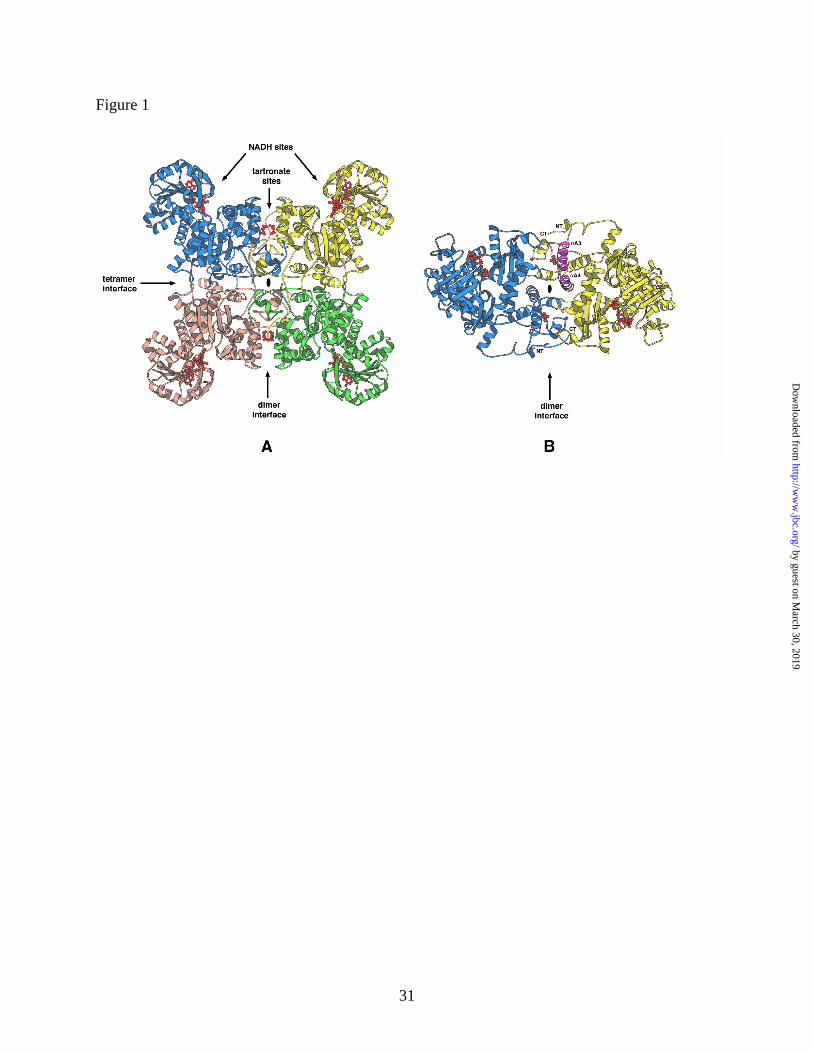
Structure of the ME-NADH complex. The tertiary and quaternary structure of the Ascaris
NAD-malic enzyme in complex with the cofactor NADH is essentially identical to that of the
binary complex with NAD (11). Overall, the structure is a tetramer, which is organized as a
dimer of dimers (Figure 1) (the rms deviation between the alpha carbon backbones of the
corresponding monomers, dimers and tetramers of the two complexes are 0.25, 0.32 and 0.34 Å,
respectively). Two types of subunit interfaces have been defined: a dimer interface between two
subunits composing two monomers, and a tetramer interface formed between two dimers (Figure
1). The monomer is organized into four domains (A, B, C and D) as previously described for
Ascaris (11) and other malic enzymes (14). The active site is a cleft, formed between the C and
B domains, and one NADH cofactor is bound to each of the four active sites in the tetramer. The
B and C domains in the NADH complex superimpose on the corresponding domains in the NAD
complex with rms deviations of 0.18 and 0.30 Å, respectively. The active site, like that of the
NAD-ME complex (11), is in an open conformation, that is, the residues implicated in substrate
binding and catalysis (R181, K199, D295, D272 and N434) are poorly positioned for their
prospective roles.
In the NAD-bound structure electron density for three residues, A601, S602 and M603 at
the C-terminus in the D-domain (11) was either not present or weak. In the present structure, the
three residues could be unambiguously modeled, but do not have a significant impact on the
overall structure.
by guest on March 30, 2019
http://ww
w.jbc.org/
Dow
nloaded from
9
The tetramer in the present structure has four NADH molecules (one per monomer) bound in the
active site and four tartronate molecules (one per monomer) bound within the dimer interface
(Figure 1).
The NADH-Binding Site. The conformation of the NADH cofactor and its interactions with
the enzyme are identical to those of the NAD-ME complex with the exception of a dramatic
difference in the conformation and interactions of the nicotinamide ring (Figure 2A and 2B).
The nicotinamide ring in the NADH complex rotates +198° about the N-glycosylic bond relative
to its orientation in the NAD-ME complex, with the si face exposed to the solvent accessible
region of the active site (Figure 2A). Binding interactions observed in the NAD-ME complex
between the nicotinamide ring amide group and residues G477 and N479 are broken as a
consequence of this rotation, and new hydrogen bonds between the carboxamide side chain and
the enzyme are formed between active site residues D295 and R181 (Table 2 and Figure 2B). A
water molecule, (wat139) occupies the site vacated by the nicotinamide amide group, and forms
hydrogen bonds with G477 and N479.
In addition to NADH, Mg2+ is also bound to the active site (Figure 2B). The metal ion is
coordinated to the carboxylate oxygens of E271, D272, and D295 with distances of about 2.3 Å.
These residues are homologous to the residues that bind Mn2+ in the human enzyme (D255,
D256 and D279) with similar bonding distances (12).
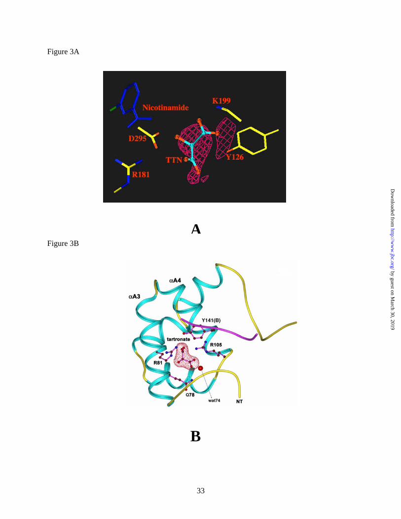
The Tartronate Binding Site. Tartronate, a competitive inhibitor of the Ascarid malic
enzyme with respect to the substrate malate, was included in all Ascaris ME crystallizations in
order to characterize the malate-binding site. Although electron density is observed in the active
site region corresponding to the tartronate binding site as identified in the human NAD-ME (12),
this density in the Ascarid malic enzyme structure is weak and does not conclusively
by guest on March 30, 2019
http://ww
w.jbc.org/
Dow
nloaded from
10
demonstrate that tartronate is bound to the active site (Figure 3A). This may be due to partial
occupancy of the malate-binding site. However, density for tartronate is observed at a different
site, within the dimer interface near a noncrystallographic 2-fold axis (Figure 3B). Here the
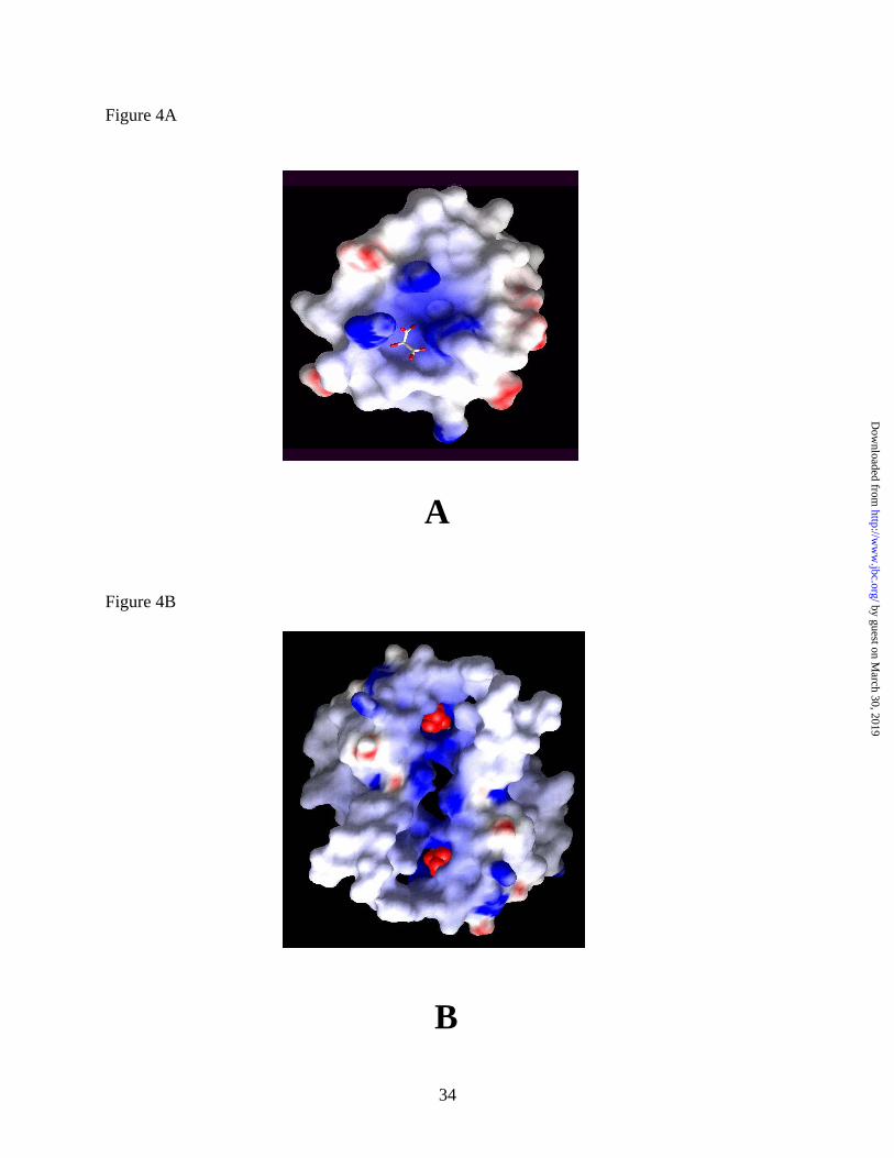
tartronate molecule is bound to a highly positively charged pocket (Figures 4A and 4B) and is
tightly anchored by strong hydrogen bond/ionic interactions with residues from two adjacent
subunits. The majority of the residues contributing to the binding site are in a cleft formed
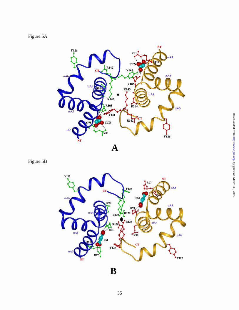
between the αA3 (residues 76 - 87) and αA4 (residues 93 - 104) helices of a subunit (Figure 3B
and 5A). Oxygen atoms O1 and O2 from one tartronate carboxylate group are within hydrogen
bonding distance (Table 3) to the Nε atom of the side chain amide group of Q78 (Q64 in human),
and a salt bridge with the guanidinium group of R105 (R91 in human), respectively (Figure 5A
and 5B). Oxygen atom O4 of the other carboxylate forms a salt bridge with the guanidinium
group of R105, (R91 in human) while its O5 atom forms a salt bridge with the NH2 of the
guanidinium group of R81 (R67 in human). The hydroxyl oxygen atom (O3) of tartronate is
within hydrogen bonding distance to the Nε of R81. The remainder of the tartronate binding site
is formed by residues 140-142 from the adjacent subunit. These residues are part of a coil,
which links the A and B domains of this subunit. The hydroxyl group of Y141 is within
hydrogen bonding distance to a carboxylate oxygen of tartronate. Binding of tartronate to this
site contributes to the subunit interaction across the dimer interface. It is interesting that the
dimer interface in the Ascaris suum malic enzyme contains several charged residues and
provides a continuous, positively charged pocket (Fig. 4A and 4B) for the binding of negatively
charged molecules such as tartronate and malate. The tartronate site also contains a bound
water molecule (wat74, Figure 3B), which is bound to a tartronate carboxylate oxygen and the
by guest on March 30, 2019
http://ww
w.jbc.org/
Dow
nloaded from
11
amide NH of M64. Its location corresponds to a water molecule bound to the fumarate-binding
site in the human NAD-ME.
Identification of bound tartronate in the Ascaris NADH-ME complex also definitively indicated
that tartronate is bound at the same site in the Ascaris NAD-ME complex. In the NAD-ME
complex, although density was observed at the tartronate binding site, the resolution of the data
did not permit identification of this density as tartronate. Comparison of the NADH-ME and
NAD-ME structures revealed that the unidentified density in NAD-ME was also tartronate, and
that the binding interactions are identical to those observed in NADH-ME.
by guest on March 30, 2019
http://ww
w.jbc.org/
Dow
nloaded from

12
DISCUSSION
Overall structure. The present structure, the first of a malic enzyme with reduced
dinucleotide bound to the active site, provides new insights into substrate binding modes and
helps resolve some of the questions associated with the catalytic mechanism of the enzyme. In
addition, the structure reveals the identity of the allosteric site, distinct from the active site and
binds the activator fumarate. The backbone conformation and the quaternary structure observed
in the ME-NADH-complex are very similar to those of the ME-NAD-complex (the rms
deviation between the main chain atoms of the two structures is 0.25 Å, monomer; 0.32 Å,
dimer; and 0.34 Å, tetramer). There are no noticeable differences in the dimer and tetramer
interactions. As stated above, the final three residues at the C-terminus in the D-domain (A601,
S602 and M603) can be visualized, but have no significant impact on the overall structure of the
ME-NADH complex.
In earlier studies on the structure of ME-NAD complex it was speculated that the residues
implicated in metal ion binding (E271, E272 and D295) were not properly positioned for binding
the metal ion. In the present structure the binding site for magnesium is clearly identified (Fig.
2B). While the role of D295 in metal ion binding was further confirmed by earlier site-directed
mutagenesis studies (19), a role of E271 and D272 was not postulated. These two residues are
part of the malic enzyme sequence QFEDFA (positions 269-274), which agrees well with the
consensus metal ion binding sequence XXDDXX where X is an uncharged or hydrophobic
residue (20). All of the residues suggested as metal ion ligands are homologous to residues in
the human enzyme (12). The fact that Mg2+ was not observed bound at the corresponding site in
the NAD-ME complex indicates that the metal ion affinity may be sensitive to the oxidation state
by guest on March 30, 2019
http://ww
w.jbc.org/
Dow
nloaded from
13
of the cofactor or that the low-resolution data for ME-NAD crystals does not reveal the metal
ion-binding site. These suggestions are consistent with the required ordered addition of metal
ion prior to malate. Although the active sites of the NADH•ME and NAD•ME complexes differ
in their cofactor conformations and the presence of Mg2+ bound in NADH•ME complex, the
active site main chain and side chain conformations are very similar to one another.
Conformational Change in the Coenzyme. A comparison of the coenzyme binding pockets
in the present structure and the previously reported ME-NAD structure shows interesting
changes in the conformation of the nicotinamide ring as a consequence of reduction (Figure 2A
and 2B). In the ME•NAD complex, as NAD is bound, the re face of the nicotinamide ring is
directed towards the solvent such that its carboxamide side chain is directed away from the
catalytic pocket into the protein structure. In the present structure, however, the reduced cofactor
has its nicotinamide ring moved into the active site as it has rotated about the N-glycosylic bond
by +198° and its si face is now directed towards the solvent. The carboxamide side chain, which
was originally hydrogen bonded to a backbone and the amide side chain of N479, now interacts
with R181 which in turn interacts with the α-carboxylate of L-malate, and D295, the putative
general base, which in turn interacts with the 2-hydroxyl of malate in the Michaelis complex,
Table 2. The proS proton of the reduced nicotinamide ring is now directed toward the active
site. It is interesting to note that while the nicotinamide ring rotates by almost 200° into the
active site, other catalytic residues are unaffected. As shown in Figure 2A, the density around
the nicotinamide ring in a difference map calculated from ME-NADH complex with and without
NADH is very strong and there are strong H-bonding contacts between carboxamide group of
nicotinamide ring with R181 and D295 suggesting that the nicotinamide ring is indeed locked in
this conformation and that the observed conformational change is not simply due to the
by guest on March 30, 2019
http://ww
w.jbc.org/
Dow
nloaded from
14
conformational flexibility allowed by the Rossman fold as in the case of lactate dehydrogenase
(21, 22). The mobility of the nicotinamide ring reported in this study is also observed in other
enzymes, and has been proposed to play a crucial role in the catalytic mechanism of at least two
other enzymes, aldehyde dehydrogenase (23) and 6-phosphogluconate dehydrogenase (24).
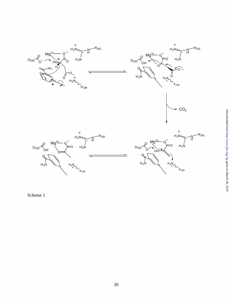
Implications to Catalytic Mechanism. A three step acid-base catalytic mechanism is
proposed for Ascarid malic enzyme based on pH and isotope partitioning studies (25-27). The
assignment of the general acid, general base and the binding groups shown in Scheme 1 is based
on the x-ray structure of the ME-NAD complex (11) and site-directed mutagenesis studies (28).
Generally, L-malate is converted to oxaloacetate facilitated by D295, which acts as a general
base to accept a proton from the 2-hydroxyl of L-malate (28). The hydride at C2 of malate is
transferred to the 4-position of the nicotinamide of NAD with the positively charged pyridinium
nitrogen providing the driving force for reduction. The guanidinium of R181 helps to orient
malate for reaction by forming an ion pair/hydrogen bond with the α-carboxyl of malate, and
K199 acts in a similar role by interacting with the β-carboxyl. The metal ion (Mg2+) aids in the
orientation of malate for catalysis and facilitates proton transfer to D295 by binding to the α-
hydroxyl of malate. In the second step, oxaloacetate is decarboxylated to enolpyruvate with the
metal ion acting as a Lewis acid and protonated D295 acting as a general acid to protonate the
enolate oxygen. Finally, enolpyruvate is tautomerized to pyruvate with D295 acting as a general
base and the ε-amino group of K199 acting as a general acid to protonate C3 of enolpyruvate
(29). Thus the catalytic mechanism requires a protonated form of the general acid (K199) and an
unprotonated form of the general base (D295). The optimum protonation state for D295
(unprotonated) and K199 (protonated) is observed in the V/Kmalate pH-rate profile (26) even
though the catalytic role of K199 is not realized until CO2 is released. The observation of the
by guest on March 30, 2019
http://ww
w.jbc.org/
Dow
nloaded from
15
K199 pK in the V/K profile indicates the importance of the protonated form of K199 in binding
malate. While the existence of the protonated form of K199 is important for optimum binding of
the β-carboxylate of L-malate, it poses a problem in the decarboxylation step, where the
ionic/hydrogen-bonding interaction between K199 and the carboxylate would be anti-catalytic.
Thus during the subsequent hydride transfer step, the interaction between the carboxylate and
K199 must be eliminated.
The crystallographically observed conformational change in the nicotinamide ring may
provide an explanation as to how the hydrogen bond between K199 and the β-carboxyl of malate
may be eliminated. A mechanism incorporating the structural data obtained in the present study
is shown in Scheme 1. Malate is bound such that its α-carboxylate is hydrogen-bonded to the
guanidinium of R181 and coordinated to Mg2+. The β-carboxylate of malate is proposed to
hydrogen bond to K199, and the β-hydroxyl of malate is hydrogen bonded to D295 and
coordinated to Mg2+. The nicotinamide ring of NAD+ is bound with its carboxamide side chain
hydrogen bonded to a backbone NH and carbonyl and to the amide side chain of N479. The
distance between the pyridinium nitrogen of NAD+ and the pyrophosphate moiety of the cofactor
is about 5.7 Å. Hydride transfer from C2 of malate to C4 of the nicotinamide ring occurs, and is
partly driven by the positively charged pyridinium ring and general base catalysis by D295. The
reduction of the nicotinamide ring results in a rotation by 198° about the N-glycosidic bond
placing the carboxamide side chain within hydrogen bonding distance to D295 and R181. The
rotation is thought to result, at least partly, from loss of the ionic interaction between the
pyridinium nitrogen of NAD+ and the pyrophosphate moiety of the cofactor. The movement of
the cofactor is proposed to be reversible but with the equilibrium position favoring that shown in
the structure, Fig. 2. The movement of the nicotinamide ring along with the sp3 to sp2
by guest on March 30, 2019
http://ww
w.jbc.org/
Dow
nloaded from
16
hybridization change at C2 that occurs as oxalacetate is formed is proposed to shift the position
of the bound oxalacetate such that: 1) the β-carboxylate moves away from K199, and 2) the C3-
C4 bond to the β-carboxylate is now orthogonal to the C2-C3 plane of oxalacetate, favoring
decarboxylation. Decarboxylation then occurs facilitated by the Lewis acidity of the Mg2+ and
with D295 acting as a general acid. Finally, tautomerization occurs aided by general base-
general acid catalysis via D295-K199 pair16, line 5 in the revised ms). The products pyruvate
and NADH are then released. Final confirmation of the above hypothesis should come from the
structure of a ternary complex of the enzyme with NADH and an analogue of malate or
oxaloacetate.
Of interest is the change in mechanism observed for the malic enzyme, from 'stepwise' to
'concerted', when the dinucleotide substrate is changed to more oxidizing dinucleotides APAD+,
thioNAD+, or PAAD+ (30). The change in mechanism likely reflects a difference in the binding of
the pyridine ring of the alternative substrates, which results in a difference in the conformation of
the bound malate. A proposed link between the conformation of the bound nicotinamide ring
and malate/oxalacetate was made above. It is not difficult to imagine a difference in the bound
conformation of the pyridine ring given the interaction of the carboxamide side chain of NAD+
with N479. In all cases the side chain of the alternative dinucleotide substrates differ with
respect to the hydrogen-bonding capability, size and hydrophobicity in comparison to the
carboxamide side chain of NAD+.
In order to examine whether the active site of human enzyme could accommodate such a
conformational change in the nicotinamide ring, NADH was modeled into the active site of the
open structure of the human enzyme (14) such that the nicotinamide ring was rotated by 198°
relative to its position in NAD molecule. The new conformation was indeed feasible without any
by guest on March 30, 2019
http://ww
w.jbc.org/
Dow
nloaded from
17
steric hindrance and the contact distances between the nicotinamide ring and the corresponding
enzyme residues were similar to those observed in ascarid enzyme.
The Tartronate Binding Site and Regulatory Mechanism. Tartronate, a dicarboxylic
analogue of malate and fumarate is tightly bound in an allosteric site via hydrogen-bonding
interactions with the side chains of 2 arginines (R105 and R81) and a glutamine (Q78). All of
the residues shown are homologous to those in the fumarate allosteric site depicted in the recent
structure of the human enzyme-ATP complex (Figure 5A and 5B) (13). The human
mitochondrial NAD-ME is allosterically activated by fumarate. Although many of the residues
that bind fumarate in human NAD-ME are homologous or functionally similar to those involved
in tartronate binding in Ascaris NAD-ME, the binding modes of these ligands differ significantly
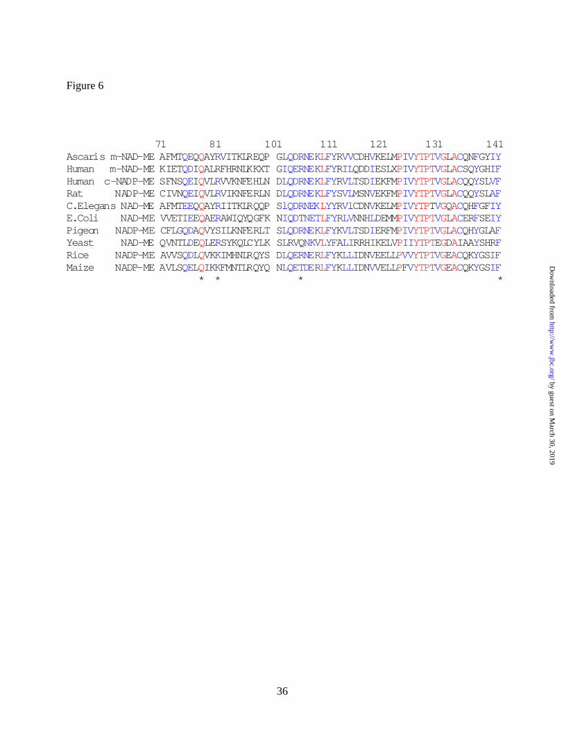
between the two enzymes. A multiple sequence alignment for malic enzymes from selected
species (Figure 6) shows conservation of all of the above residues. It is interesting to note that
fumarate activation has not been reported in all of the species that have the conserved residues
under consideration.
The Ascaris malic enzyme is also allosterically activated by fumarate with an activation
constant of 40 µM (9, 10). Activation is expressed as a decrease in the off-rate for bound malate
(10). Although the activation by fumarate under conditions of saturating NAD+ and Mg2+ is only
2-fold (9), with reactants maintained at estimated physiologic concentrations, the activation is
15-fold (Cook et al, unpublished observations). Thus the allosteric site where tartronate is bound
is most likely an activator site for fumarate. This conclusion is further supported by the
observation that the R105A mutant of the Ascaris malic enzyme, which has a V/Et value
identical to that of the wild type enzyme, is no longer activated by fumarate, consistent with
tartronate being bound to the fumarate activator site (Cook et al, unpublished observations).
by guest on March 30, 2019
http://ww
w.jbc.org/
Dow
nloaded from
18
The distance between the active site and the tartronate allosteric site is ~30 Å. However
there are structural relationships between the sites, which could allow for transmission of the
allosteric signal. Within a subunit, R105 participates in binding tartronate. The backbone
oxygen of the adjacent residue, D104, is hydrogen bonded to the side chain of K143, which is
part of a coil sequence (residues 140-142) that connects the A and B domains of a subunit
(Figure 5A). This coil is also directly involved in forming the second tartronate binding site
within the dimer interface via Y141 (Figure 5A). The amino terminal end of this coil is linked to
the αA6 helix (residues 37-144), which contributes active site residue Y126. Thus, the
tartronate-binding site is linked to the coil connecting the A and B domains of a monomer
(residues 140-143). It is interesting to note that the conformation of this coil differs significantly
from that of the corresponding coil in human ME (residues 126-129) despite almost identical
sequences (Figures 5B and 6). In particular, there is no interaction corresponding to the D104 -
K143 pair, and the positions of the homologous residues R142 (Ascaris) and R91 (human) differ.
In addition, in place of Y141 in Ascaris enzyme, the human enzyme has a phenylalanine (F127).
Overall, the evidence presented in this study identifies an allosteric site and provides new
insights into the mechanism of allosteric activation of the Ascaris suum malic enzyme.
However, the structure of the Ascaris malic enzyme with fumarate bound to this allosteric site
will further confirm the identity of this site and enhance our understanding of the allosteric
mechanism.
by guest on March 30, 2019
http://ww
w.jbc.org/
Dow
nloaded from
19
Acknowledgements
We thank Dr. Hong Zhang for valuable discussions, Dr. Srinivasan Raghunathan for help with
crystallographic analysis, and the staff at the Cornell High Energy Synchrotron Source (CHESS)
for assistance with data collection.
by guest on March 30, 2019
http://ww
w.jbc.org/
Dow
nloaded from
20
REFERENCES
1. Sauer, L. A. (1973) Biochemical & Biophysical Research Communications 50, 524-531
2. Moreadith, R. W., and Lehninger, A. L. (1984) J. Biol Chem. 259, 6222-6227
3. Karsten, W., Cook, P.F. (2000) Protein and Peptide Letters 7, 281-286
4. Sauer, L. A. (1973) FEBS Letters 33, 251-255
5. Kulkarni, G., Cook, P.F. and Harris, B.G. (1993) Arch. Biochem. Biophys. 300, 231-237
6. Rao, J. G. S., Coleman, D.E., Kulkarni, G., Goldsmith, E.J., Cook, P. F., Harris, B.G.
(2000) Protein and Peptide Letters 7, 297-304
7. Saz, H. J. (1981) Ann. Rev. Physiol. 43, 323-341
8. Komuniecki, R., Harris, B. G. (1995) in Biochemistry and Molecular Biology of
Parasites (Marr, J. J., Muller, M., ed), pp. 49-66, Academic Press, London
9. Landsperger, W. J., Harris, B.G. (1976) J. Biol. Chem. 251, 3599-3602
10. Lai, C.-J., Harris, B.G. and Cook, P.F. (1992) Arch. Biochem. Biophys. 299, 214-219
11. Coleman, D. E., Rao, G.S. Jagannatha, Goldsmith, E. J. Cook, Paul F. and Harris, Ben
G. (2002) Biochemistry 41, 6928-6938
12. Yang, Z., Floyd, D.L., Loeber, G., Tong, L. (2000) Nature Struct. Biol. 7, 251-257
13. Yang, Z., Lanks, Charles W. and Tong, Liang. (2002) Structure 10, 951-960
14. Xu, Y., Bhargava, G., Wu, H., Loeber, G., Tong, L. (1999) Structure 7, 877-889
15. Otwinowski, Z., Minor, W. (1997) Methods in Enzymology 276, 307-326
16. Brunger, A. T., Adams, P.D., Clore, G.M., Gros, P., Grosse-Kunstleve, R.W., Jiang, J.-S.,
Kuszewski, J., Nilges, M., Pannu, N.S., Read, R.J., Rice, L.M., Simonson, T., and
Warren, G.L. (1998) Acta Crystallographica D54, 905-921
by guest on March 30, 2019
http://ww
w.jbc.org/
Dow
nloaded from
21
17. Jones, T. A., Zou, J.-Y., Cowan, S. W., and Kjeldgaard, M. (1991) Acta
Crystallographica A47, 110-119
18. Laskowski, R. A., MacArthur, M. W., Moss, D. S., and Thornton, J. M. (1993) Journal of
Applied Crystallography 26, 283-291
19. Karsten, W., Chooback, D. L., Hwang, C., Lynch, C., and Cook, P. F., (1999)
Biochemistry 38, 10527-10532
20. Hlavaty, J., Nowak, T. (1997) Biochemistry 39, 3389-3403
21. Sebastien J. F. Vincent, C. Z., Carol Beth Post, John W. Burgner, and Geoffrey
Bodenhausen. (1994) PNAS, 4383-4388
22. Hall, M. D., and Banaszak, L. J. (1993) J. Mol. Biol 232, 213-222
23. Hammen, P. K., Allali-Hassani, A., Hallenga, K., Hurley, T.D., and Weiner, H. (2002)
Biochemistry 41, 7156-7168.
24. Adams, M. J., Ellis, G. H., Gover, S., Naylor, C. P., and Phillips, C. (1994) Structure 2,
651-668
25. Park, S.-H., Harris, B.G. and Cook, P.F. (1986) Biochemistry 25, 3752-3759
26. Kiick, D. M., Harris, B. G., and Cook, P. F. (1986) Biochemistry 25, 227-236
27. Weiss, P. M., Gavva, S. R., Harris, B. G., Urbauer, J. L., Cleland, W. W., and Cook, P. F.
(1991) Biochemistry 30, 5755-5763
28. Karsten, W. E., Chooback, L., Liu, D., Hwang, C-C., Lynch, C. and Cook, P.F. (1999)
Biochemistry 38, 10527-10532
29. Liu, D., Karsten, W. E., and Cook, P. F. (2000) Biochemistry 39, 11955-11960
30. Karsten, W. E., and Cook, P. F. (1994) Biochemistry 33, 2096-2103
31. Kraulis, P. J. (1991) J. Appl. Crystallogr. 24, 946-950
by guest on March 30, 2019
http://ww
w.jbc.org/
Dow
nloaded from
22
32. Esnouf, R. M. (1999) Acta Crystallographica D255, 938-940
33. Merritt, E. A., and Murphy, M. E. P. (1994) Acta Crystallographica D50, 869-873
34. Nicholls, A., Sharp, K. A., and, Honig, B. (1991) Proteins, 11, 281-296
by guest on March 30, 2019
http://ww
w.jbc.org/
Dow
nloaded from
23
FOOTNOTES
1Abbreviations: NAD, nicotinamide adenine dinucleotide (the plus charge has been omitted for
convenience); NADH, reduced nicotinamide adenine dinucleotide; NADP, nicotinamide adenine
dinucleotide phosphate; NADPH, reduced nicotinamide adenine dinucleotide phosphate; APAD,
acetylpyridine adenine dinucleotide; PAAD, pyridine aldehyde adenine dinucleotide; TTN,
tartronate; FM, fumarate; ME, malic enzyme; c-NADP-ME, cytosolic NADP-dependent ME; m-
NADP-ME, mitochondrial NADP-dependent malic enzyme; NAD-ME, mitochondrial dual
specificity (NAD or NADP)-dependent malic enzyme; DTT, dithiothreitol; EDTA,
ethylenediamine-tetraacetic acid.
by guest on March 30, 2019
http://ww
w.jbc.org/
Dow
nloaded from
24
LEGENDS TO FIGURES
Figure 1: Ribbon diagram of the structure of Ascaris mNAD-ME in complex with NADH, Mg2+
and tartronate. (A) The tetramer viewed down one 2-fold axis (indicated by the black oval) with
the tetramer and dimer interfaces indicated by arrows. The four subunits are colored blue,
yellow, green and tan. The four NADH and four tartronate ligands are shown as red ball-and-
stick models, and their binding sites indicated for one dimer. (B) The mNAD-ME dimer viewed
down the 2-fold axis corresponding to the dimer interface showing the locations of the two
tartronate binding sites more clearly. The NT and CT termini of each monomer are indicated.
For one subunit, the two helices (αA3 and αA4) that contribute most of the residues involved in
tartronate binding are colored purple and labeled. Generated using MOLSCRIPT (31)
Figure 2: NADH bound to the active site of Ascaris mME. (A) Electron density (2Fo-Fc,
contoured at 1σ) for NADH. The NAD molecule bound to the Ascaris NAD-mME complex is
shown for comparision in this and the following diagram. (B) Stereo diagram of the NADH
binding site. A water molecule (Wat139) and Mg2+ bound within the active site are indicated as
spheres. Generated using MOLSCRIPT (31).
Figure 3: Binding of tartronate to Ascaris suum malic enzyme
(A) Fo-Fc electron density map at 2.0 Å resolution for tartronate molecule in the active site
Tartronate (TTN) and active site residues are shown as ball-and stick models. (B) Ribbon
diagram of the tartronate-binding site within the dimer interface of Ascaris ME with tartronate
and key residues indicated as ball-and-stick models, and a water molecule bound to tartronate
by guest on March 30, 2019
http://ww
w.jbc.org/
Dow
nloaded from
25
shown as a sphere. Electron density (2Fo-Fc, contoured at 1σ) for tartronate is also shown.
Generated using MOLSCRIPT (31), BOBSCRIPT (32) and RASTER-3D (33).
Figure 4. (A) Electrostatic potential surface of a monomer of Ascaris mME as viewed in roughly
the same orientation as in Figure 3B. (B) Electrostatic potential surface of two monomers
showing the dimer interface along the vertical axis. Two tartronates are indicated as red CPK
models. Generated using GRASP (34)
Figure 5: Ribbon diagrams for the comparison between the tartronate and fumarate binding sites
of Ascaris and human mME. (A) Tartronate binding site in Ascaris ME; (B) Fumarate binding
site in human mME. The αa and α b subunits in both the enzymes are colored blue and tan
respectively. Tartronate (TTN) in Ascaris ME and fumarate in human \ME (FM) are shown as
red and cyan CPK models. Key residues involved in binding ligands are shown as ball-and-stick
models. The NT and CT termini of each monomer are indicated. Generated using MOLSCRIPT
(31) and RASTER-3D (33)
Figure 6: Sequence alignment of residues in the Ascaris mME tartronate binding site with the
homologous regions in human (and other) MEs. Key residues involved in tartronate binding
(Ascaris) and fumarate binding (human) are indicated.
Scheme 1. Proposed Mechanism for the NAD-Malic Enzyme. The mechanism is described in
the text. R181 is considered to lie above the plane that contains malate, with the nicotinamide
by guest on March 30, 2019
http://ww
w.jbc.org/
Dow
nloaded from
26
ring in the same plane as malate but with its carboxamide pointing back and into the plane of the
paper. Rotation causes the carboxamide side chain to be pointing up out of the paper plane.
by guest on March 30, 2019
http://ww
w.jbc.org/
Dow
nloaded from
27
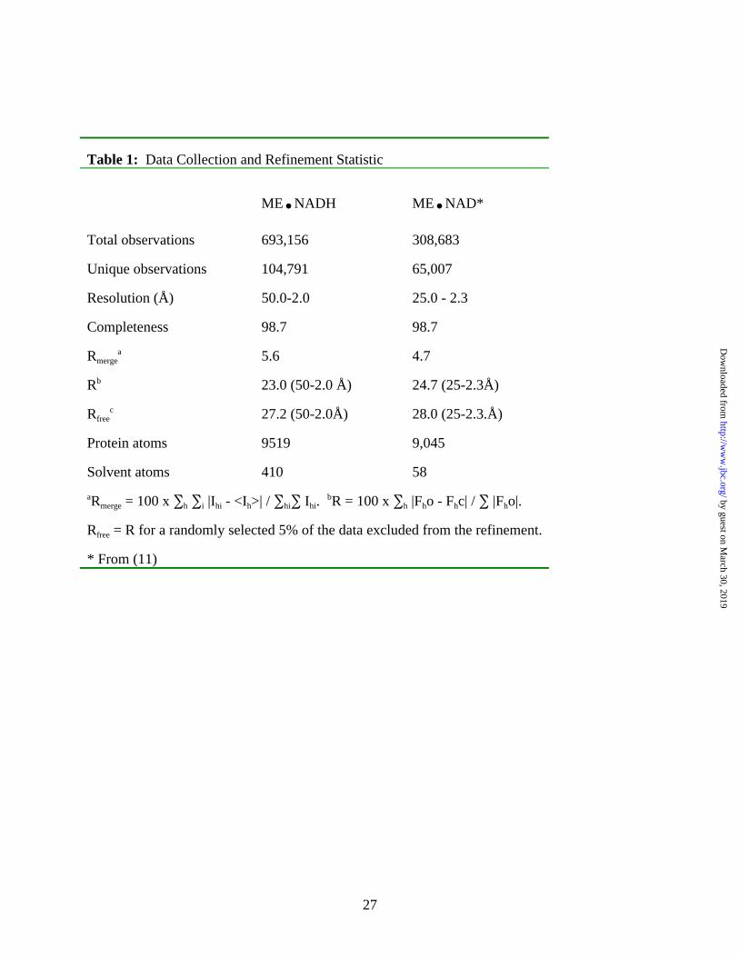
Table 1: Data Collection and Refinement Statistic
ME.NADH ME.NAD*
Total observations 693,156 308,683
Unique observations 104,791 65,007
Resolution (Å) 50.0-2.0 25.0 - 2.3
Completeness 98.7 98.7
Rmergea 5.6 4.7
Rb 23.0 (50-2.0 Å) 24.7 (25-2.3Å)
Rfreec 27.2 (50-2.0Å) 28.0 (25-2.3.Å)
Protein atoms 9519 9,045
Solvent atoms 410 58
aRmerge = 100 x ∑h ∑i |Ihi - <Ih>| / ∑hi∑ Ihi. bR = 100 x ∑h |Fho - Fhc| / ∑ |Fho|.
Rfree = R for a randomly selected 5% of the data excluded from the refinement.
* From (11)
by guest on March 30, 2019
http://ww
w.jbc.org/
Dow
nloaded from
28
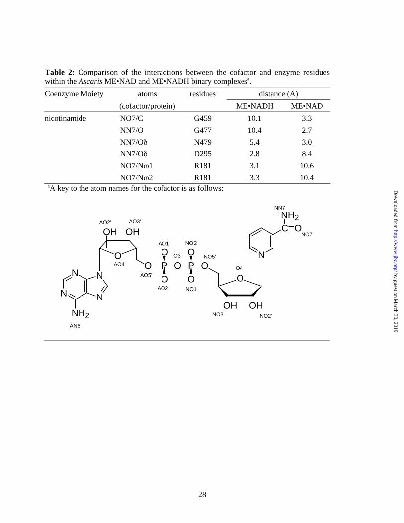
Table 2: Comparison of the interactions between the cofactor and enzyme residueswithin the Ascaris ME•NAD and ME•NADH binary complexesa.
Coenzyme Moiety atoms residues distance (Å)
(cofactor/protein) ME•NADH ME•NAD
nicotinamide NO7/C G459 10.1 3.3
NN7/O G477 10.4 2.7
NN7/Oδ N479 5.4 3.0
NN7/Oδ D295 2.8 8.4
NO7/Nω1 R181 3.1 10.6
NO7/Nω2 R181 3.3 10.4aA key to the atom names for the cofactor is as follows:
O
OHOH
N
C ONH2
OPO
OOP
O
O
N
N N
N
NH2
O
OH OH
O
AN6
NO2
NN7
NO7
O4
NO1
O3 NO5'
AO2' AO3'
AO4'
AO5'
AO1
AO2
NO3' NO2'
by guest on March 30, 2019
http://ww
w.jbc.org/
Dow
nloaded from
29
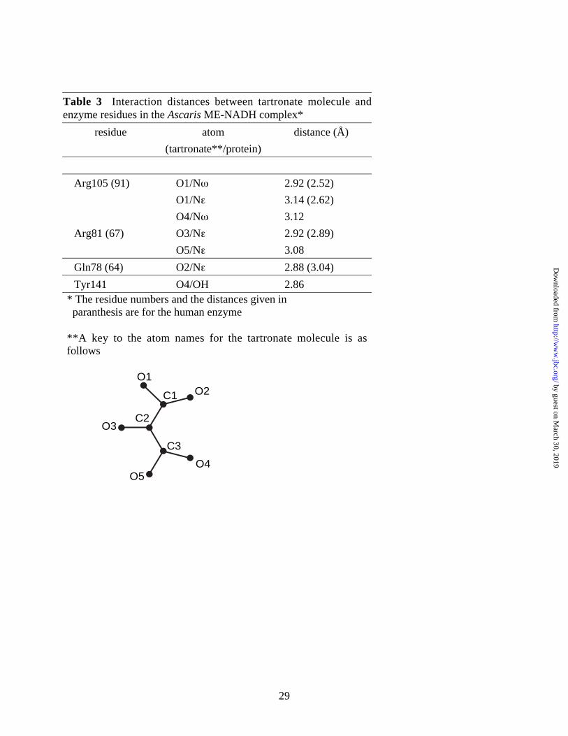
Table 3 Interaction distances between tartronate molecule andenzyme residues in the Ascaris ME-NADH complex*
residue atom distance (Å)
(tartronate**/protein)
Arg105 (91) O1/Nω 2.92 (2.52)
O1/Nε 3.14 (2.62)
O4/Nω 3.12
Arg81 (67) O3/Nε 2.92 (2.89)
O5/Nε 3.08
Gln78 (64) O2/Nε 2.88 (3.04)
Tyr141 O4/OH 2.86* The residue numbers and the distances given in paranthesis are for the human enzyme
**A key to the atom names for the tartronate molecule is asfollows
O1O2C1
C2O3
O5
C3
O4
by guest on March 30, 2019
http://ww
w.jbc.org/
Dow
nloaded from
30
N
NH2O H
HO O
O_
R181NHC
H2N
H2N
+
O
O_
Mg2+
K199
H3N+
+
D295
O
O_
NH2N
O
R181NHC
H2N
H2N
+
Mg2+
K199
H3N+
D295
O
OHO
O
O
O
O_
_
NH2N
O
R181NHC
H2N
H2N
+
Mg2+
K199
H3N+
D295
O
OO
O_
HO_
CO2
NH2N
O
R181NHC
H2N
H2N
+
Mg2+
K199
H3N+
OO
_
O
D295
O
OH
Scheme 1
by guest on March 30, 2019
http://ww
w.jbc.org/
Dow
nloaded from

32
Figure 2A
Figure 2B
B
by guest on March 30, 2019
http://ww
w.jbc.org/
Dow
nloaded from
33
Figure 3A
Figure 3B
B
A
by guest on March 30, 2019
http://ww
w.jbc.org/
Dow
nloaded from
34
Figure 4A
Figure 4B
B
A
by guest on March 30, 2019
http://ww
w.jbc.org/
Dow
nloaded from
35
Figure 5A
AFigure 5B
B
by guest on March 30, 2019
http://ww
w.jbc.org/
Dow
nloaded from
36
Figure 6
71 81 101 111 121 131 141Ascaris m-NAD-ME AFMTQEQQAYRVITKLREQP GLQDRNEKLFYRVVCDHVKELMPIVYTPTVGLACQNFGYIYHuman m-NAD-ME KIETQDIQALRFHRNLKKXT GIQERNEKLFYRILQDDIESLXPIVYTPTVGLACSQYGHIFHuman c-NADP-ME SFNSQEIQVLRVVKNFEHLN DLQDRNEKLFYRVLTSDIEKFMPIVYTPTVGLACQQYSLVFRat NADP-ME CIVNQEIQVLRVIKNFERLN DLQDRNEKLFYSVLMSNVEKFMPIVYTPTVGLACQQYSLAFC.Elegans NAD-ME AFMTEEQQAYRIITKLRQQP SlQDRNEKLYYRVlCDNVKELMPIVYTPTVGQACQHFGFIYE.Coli NAD-ME VVETIEEQAERAWIQYQGFK NIQDTNETLFYRLVNNHLDEMMPIVYTPTVGLACERFSEIYPigeon NADP-ME CFLGQDAQVYSILKNFERLT SLQDRNEKLFYKVLTSDIERFMPIVYTPTVGLACQHYGLAFYeast NAD-ME QVNTLDEQLERSYKQLCYLK SLRVQNKVLYFALIRRHIKELVPIIYTPTEGDAIAAYSHRFRice NADP-ME AVVSQDLQVKKIMHNLRQYS DLQERNERLFYKLLIDNVEELLPVVYTPTVGEACQKYGSIFMaize NADP-ME AVLSQELQIKKFMNTLRQYQ NLQETDERLFYKLLIDNVVELLPFVYTPTVGEACQKYGSIF
* * * *
by guest on March 30, 2019
http://ww
w.jbc.org/
Dow
nloaded from

HarrisJagannatha G.S. Rao, David E. Coleman, William E. Karsten, Paul F. Cook and Ben G.
cofactor and identification of an effector siteo{Crystallographic studies on Ascaris suum NAD-malic enzyme bound to reduced
published online July 9, 2003J. Biol. Chem.
10.1074/jbc.M305145200Access the most updated version of this article at doi:
Alerts:
When a correction for this article is posted•
When this article is cited•
to choose from all of JBC's e-mail alertsClick here
by guest on March 30, 2019
http://ww
w.jbc.org/
Dow
nloaded from


![Ascaris and ascariasis - Semantic Scholar · Ascaris suum is a widespread parasitic nematode that causes infection in pigs with high prevalence rates in host populations [5, 6]. The](https://img.pdfslide.us/doc/110x75/5ca0073588c99350178c8373/ascaris-and-ascariasis-semantic-scholar-ascaris-suum-is-a-widespread-parasitic.jpg)