Embed Size (px)
Citation preview
8/9/2019 Crystallographic and Molecular-Modeling Studies of Lipase B from Candida antarctica Reveal a Stereospecificity Poc…
http://slidepdf.com/reader/full/crystallographic-and-molecular-modeling-studies-of-lipase-b-from-candida-antarctica 1/14
16838
Biochemistry 1995,34,
16838-16851
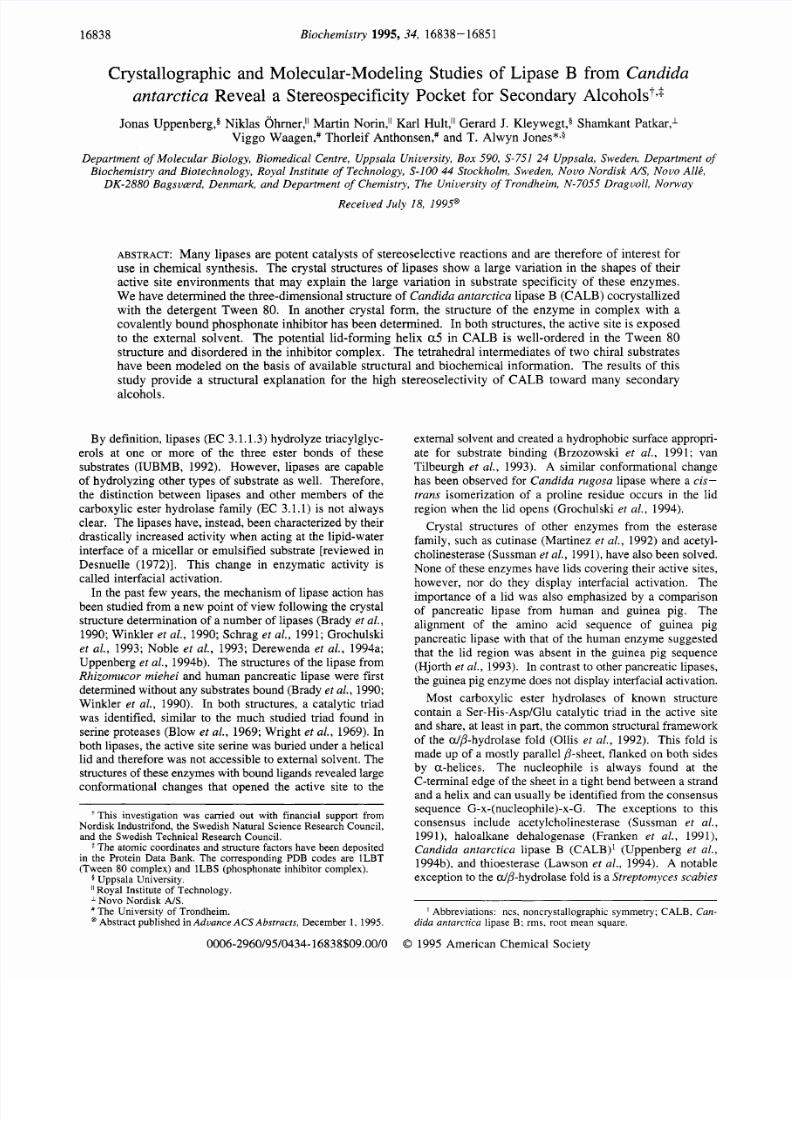
Crystallographic and Molecular-Modeling Studies of Lipase B from Candida
antarctica Reveal a Stereospecificity Pocket for Secondary Alcohols+>
Jona s Uppenberg,a Niklas O hmer," Martin Norin,l' Karl Hult,l' Gerard J. K leyweg t,$ Sham kant Pa tkar ,l
Viggo W aagen,# Thorleif A nthomen,# and T. Alwyn Jones*,$
Department
of
Molecular Biology, Biomedical Centre, Uppsala University, Box 590 S-751 24 Uppsala, Sweden, Department of
Biochemistry and Biotechnology, Royal Institute
of
Technology, S-100 44 Stockholm, Sweden, Novo Nordisk A/S, Novo All t
DK-2880 Bagsulerd, Denmark, and Department
of
Chemistry, The University
of
Trondheim, N-7055 Draguoll, Norway
Received July 18, 1995@
ABSTRACT: Many lipases are potent catalysts of stereoselective reactions and are therefore of interest for
use in chemica l synthesis. The crystal structures of lipases show a large variation in the shape s of their
active site environments that may explain the large variation in substrate specificity of these enzymes.
W e have de termined the three-dimensional structure of Cand ida an tarc t i ca l ipase B (CAL B) cocrystallized
with the detergent Tween 80. In another crystal form, the structure of the enzy me in complex with a
covalently bo und phosp honate inhibitor has been determined. In both structures, the active site is exposed
to the external solvent. The potential lid-forming helix a 5 in CA LB is well-ordered in the Tw een 8 0
structure and disordered in the inhibitor complex. The tetrahedral intermediates of two chiral substrates
have been mo deled on the basis of available structural and biochem ical information. The results of this
study provide a structural explanation for the high stereoselectivity of CALB toward many secondary
alcohols.
By definition, lipases
(EC
3.1.1.3) hydrolyze triacylglyc-
erols at one or more of the three ester bonds of these
substrates (IUB MB , 1992). Howe ver, lipases are capable
of hydrolyzing other types of substrate as well. Therefore,
the distinction between lipases and other members of the
carboxylic ester hydrolase family
(EC
3.1.1) is not always
clear. The lipases have, instead, been characterized by their
drastically increased activity when ac ting at the lipid-water
interface of a m icellar or emulsified sub strate [reviewed in
Desnuelle (1972)l. This change in enzymatic activity is
called interfacial activation.
In the past few years, the me chanism of lipase action has
been studied from a new point of view following the crystal
structure determination of a num ber of lipases (Brady et al.,
1990; Winkler
et al.,
1990; Schrag
et al . ,
1991; Grochulski
et al . , 1993; Noble e t a l . , 1993; Derewenda et al . , 1994a;
Uppenberg et al . , 1994b). The structures of the lipase from
Rhizomucor miehei
and human pancreatic lipase were first
determined without any substrates bound (Brady
et al.,
1990;
Winkler
et al . ,
1990). In both structures, a catalytic triad
was identified, similar to the much studied triad found in
serine proteases (Blow et al., 1969; Wright e t al., 1969). In
both lipases, the active site serine was buried under a helical
lid and therefore was not accessible to external solvent. The
structures of these enz ymes with bound ligan ds revealed large
conformational changes that opened the active site to the
This investigation was carried out with financial support from
Nordisk Industrifond, the Swedish Natural Science Research Council,
and the Swedish Technical Research Council.
The atomic coordinates and structure factors have been deposited
in the Protein Data Bank. The corresponding PDB codes are lLBT
(Tween 80 complex) and
lLBS
(phosphonate inhibitor complex).
5 Uppsala University.
Royal Institute of Technology.
J. Novo Nordisk A I S
The University of Trondheim.
@ Abstract published in Advance ACS
Abstracts,
December 1 1995.
external solvent and created a hydrophobic surface appropri-
ate for substrate binding (Brzozowski e t a l . , 1991; van
Tilbeurgh e t
al.,
1993). A similar conformational change
has been observed for
Candida rugosa
lipase where a cis-
t rans isomerization of a proline residue occurs in the lid
region when the lid opens (Grochulski
et al . ,
1994).
Crystal structures of other enzymes from the esterase
family, such as cutinase (Martinez et al., 1992) and acetyl-
cholinesterase (Sussman
et al.,
1991), have also been solved.
None of these enzymes have lids covering their active sites,
however, nor do they display interfacial activation. The
importance of a lid was also emphasized by a comparison
of pancreatic lipase from human and guinea pig. The
alignment of the amino acid sequence of guinea pig
pancreatic lipase with that of the human e nzym e suggested
that the lid region was absent
in
the guinea pig sequence
(Hjorth et al . , 1993). In contrast to other pancreatic lipases,
the guinea pig enzym e does not display interfacial activation.
Most carboxylic ester hydrolases of known structure
contain a Ser-His-Asp/G lu catalytic triad in the active site
and share , at least in part, the comm on structural framework
of the alp-hydrolase fold (Ollis
et al . ,
1992). This fold is
made up of a mostly parallel ,&sheet, flanked on both sides
by a-helice s. The nucleophile is always found at the
C-terminal edge of the sheet in a tight bend between a strand
and a helix and can usually be identified from the consensus
sequence G-x-(nucleophile)-x-G. The exceptions to this
consensus include acetylcholinesterase (Sussman
et al . ,
199 l), haloalkane dehalogenase (Franken e t a l . , 1991),
Candida antarctica l ipase B (CALB)' (Uppenberg et al . ,
1994b), and thioesterase (L awson
et al . ,
1994). A notable
exception to the d/3-hydrolase fold is a Streptomyces scabies
I Abbreviations: ncs, noncrystallographic symmetry; CALB, Can-
dida antarctica lipase B; rms, root mean square.
0006-2960/95/0434-16838 09.00/0 0
995 American Chemical Society

8/9/2019 Crystallographic and Molecular-Modeling Studies of Lipase B from Candida antarctica Reveal a Stereospecificity Poc…
http://slidepdf.com/reader/full/crystallographic-and-molecular-modeling-studies-of-lipase-b-from-candida-antarctica 2/14
Structure and Function of Lipase B Biochemis try , Vol. 34 No. 51, 1995 16839
FIGURE
: Stereotrace
of
the C a atoms
in CALB.
The structure
consists of a
seven-stranded,
mostly
parallel P-sheet, surrounded
by
helices
on both sides. The side chains of the catalytic residues SerlO5, His224, and Asp187 are shown as well as the side chain of Trpl04.
esterase, where the topology of the P-sheet is different and
the acid in the catalytic triad is replaced by a carbonyl oxyg en
(Wei et al. , 1995).
Lipases have attracted much interest in recent years
because of their potential use in industrial applications, for
instance as additives to washing powder, in the production
of
rare oils and fats through transesterification, an d for use
in other types
of
organic synthesis (Gillis, 1988 ; Bjorkling
et ai . , 1991). They have also proven to be efficient
stereoselective catalysts in the kinetic resolution of a wide
variety
of
chiral compounds (Santaniello et al., 1992). One
of
the most studied features of lipases is their ability to react
selectively with one of two enantiomers of secondary
alcohols.
Empirical studies on different lipases and sub-
strates have allowed the construction of a schematic model
of
the binding sites for the two substituents of the secondary
alcohol (Kazlauskas
et al.,
1991). By comp aring the relative
size
of
the two substituents of the alcohol, one could use
this model to predict with high accuracy which
of
the two
enantiom ers would be converted faster by the lipase. The
crystallographic work on lipases has permitted a more
detailed investigation of their enantioselectivity. This was
first done with the crystal structures of covalent complex es
between C. rugosa lipase and (R ) - and (S)-menthyl ester
transition state analogues (Cygler et al., 1994). These
structures showed that the fast-reacting enantiomer could
bind to the enzyme with an intact hydrogen bond between
the alcohol oxygen and the NE of the active site histidine.
This bond was not form ed with the slow-reacting en antiom er.
The difficulty of the slow-reacting enantiomer in forming
this hydrogen bond has been proposed to account for the
enantioselectivity of this lipase as well as for that of other
lipases and esterases (Cygler er
al.,
1994). The latter
conclusion was based on the observation that lipases share
similar catalytic triads and alcohol binding sites.
CAL B is a lipase with a 33 kDa molecular mass. It has
proven to be a highly stereoselective enzyme fo r the kinetic
resolution
of
secondary alcohols (Frykman et
al.,
1993;
Waagen et
al.,
1993). We have previously reported the
sequence and crystal structure of CALB in two crystal forms
(Uppenberg
et al.
1994b). These structures revealed that
the enzyme has an db-hydrolase-like fold, with an active
site serine triad that is accessible from the solvent through a
narrow channel (Figure 1). Th e open conformatio n of the
enzyme in the absence of any substrate raised the question
of whether CALB undergoes a conformational change upon
activation. A short helix,
a5,
was identified as a potential
lid on the basis
of
its position and an ob served orderldisorder
in different crystal environments. A detergent molecule at
the entrance of the active site channel could be removed by
changing the pH of the crystals, and as a result,
a
changed
from an ordered helix to disordered structure, manifested by
a lack of continuous electron density. A similar disorder
has been observed for the lid in the crystal structure of
Humicola
lanuginosa lipase (Derewenda
et al. ,
1994b).
We now present two new complex structures of CALB.
In the first structure, CALB was incubated with the detergent
Tween
80
prior to crystallization. In the second structure,
the enzyme was inhibited by a racemic mixture of
n-
hexylchlorophosphonate ethyl ester (Bjorkling
et ul. ,
1992).
With information from these crystal structures and those of
other lipase inhibitor complexes and assuming a reaction
mechanism consistent with that of other serine hydrolases,
we have modeled the tetrahedral intermediates
of
two acyl
transfer reactions into the active site in order to exp lain the
high stereoselectivity of CALB toward certain secondary
alcohols. In the first intermediate, the underlying reaction
is an interesterification w ith 1-phenylethanol as acyl accep tor
and an octanoate ester as acyl donor (Frykman
et al.,
1993).
In the second intermediate, the modeled reaction is the
hydrolysis of a glycerol butanoate derivative (Waagen
et al.,
1993).
MATERIALS AND METHODS
For the crystallographic work presented here, enzyme from
the native organism was used exclusively. The purification
of the native enzyme has been described elsewhere (Patkar
et al.,
1993). CA LB crystallizes under a variety of conditions
(Uppenberg et al. , 1994 a), but on ly the crystal form s relevant
to this study will be presented here.
All crystallization
experiments were performed w ith the hanging drop method
(McPherson, 1982), using silane-treated cover slips.
The

8/9/2019 Crystallographic and Molecular-Modeling Studies of Lipase B from Candida antarctica Reveal a Stereospecificity Poc…
http://slidepdf.com/reader/full/crystallographic-and-molecular-modeling-studies-of-lipase-b-from-candida-antarctica 3/14
16840 Biochemistry, Vol.
34
No. 51, 1995 Uppenberg et al.
rigid-body refinement, the R-factor was 29.3% and the free
R-factor was 30.1% for reflections in the 8.0-3.0 8 range.
Positional refinement by energy minimization as well as
B-factor refinement were carried out with strict ncs con-
straints between the two molecules in the asymmetric unit,
including ligand and waters. The ncs operator was obtained
from the model after rigid body refinemen t. Water mol-
ecules, a partial Tween 80 molecule, and two N-acetylglu-
cosamine residues at Am74 were included per enzyme
molecule. A few side chains were manually rebuilt with
0
(Jones et al., 1991) to fit the averaged density (Kleywegt &
Jones, 1994b). B-factor refinement was carried out with
main chain atoms and side chain atoms grouped for each
residue. Well-con nected density for part of the Twee n 80
ligand was observed in the active site. In an attempt to
improve the density, high-temperature simulated annealing
calculations (Briinger et al., 1987) in which harmonic
positional restraints were used for all non-hydrogen atoms
were carried out. Th e rationale behind this approach was
to tether the protein structure to its starting conformation
(Le. the high-resolution structure), except for areas where
the crystallographic data of the complex provided a suf-
ficiently large driving force to effect changes. Th e model
phases improved sufficiently to reveal w ell-connected density
for a Tween 80 molecule extending all the way out to the
solvent interface. A partial model of a Twee n 80 molecule
was built and fitted to the density. The resulting model was
subjected to another high-temperature simulated annealing
calculation, again using harmonic positional restraints for
all atoms except those of the ligand. Th e final model has
an R-factor of 17.1% for reflections between 7.5 and 2.5 A,
and the free R-factor is 19.4%. The rms deviation from the
orthorhombic model is 0.22 8 for Ca's and 0.25 8 for all
non-hydro gen atoms. Refinem ent and model quality statis-
tics are summ arized in Table 2. A plot of the real-space fit
of the model to the final 2F,,b,
- Fcalc
ap and of the main
chain B-factors is shown in Figure 2. The active site Se r
105 adopts an energetically strained conformation typical for
alp-hydrolases, with main chain dihedral angles 4
=
52.0"
and
11 =
19.0'.
Structure Solution of the CALB/Phosphonate Inhibitor
Complex. CA LB was inhibited effectively by a racemic
mixture of the phosphonate inhibitor in equimolar amounts
(Patkar et al., 1993). The protein complex was crystallized
at room temperature. Th e reservoir contained
1.3 M am-
monium sulfate as precipitant, 0.1 M sodium citrate/citric
acid buffer (pH 4.0) and 10% dioxane. The drop contained
5 p L of protein solution
(10
mg/ml), 1 pL of 4% P-octyl
glucoside, and
1
pL from the reservoir. The space group is
C2, and the cell constants are
a
= 229.5
A,
b = 95.6
A,
c
=
86.7 A , and
/3 =
90.0". There are six molecules in the
asymmetric unit, and
V,
is 2.4 A3/Da.
This crystal form exhibits unusual local symmetry, which
manifests itself by systematic absences in the diffraction
pattem and by high peaks in the self Patterson map. There
is one peak at (l/3,0,l/3) and another at
(l/3,0,-l/3),
which
are of unequal height. Th e relative heights of these peaks
also vary between data sets collected from different crystals,
and these data sets do not merge well. While individual data
sets can be merged with intemal R-factors of less than 5%,
combining data sets from different crystals gives merging
R-factors of more than 25%. This suggested that the crystals
suffered from merohedral twinning; in which case, two
Table 1 : Data Collection and Processing Statistics
phosphonate" Tween 80
space group
cell axes
A)
cell , ?-angle deg)
molecules in asymmetric
V ,
( A 3 / ~ a )
resolution A)
highest shell
A)
number of observations
unique reflections
completenessb )
Rmergeb.c ( I
MNb
unit
c 2
a
=
229.5, b
=
95.6,
90.0
6
c
=
86.7
2.4
2.6
2.69-2.60
55 714
26 271
83
( 5 5 )
5.0
(19.1)
13.0 (5.9)
p21
a = 95.1,
b
= 50.2,
c
= 99.5
90.6
2
3.6
2.5
2.59-2.50
45 529
26 677
82 (63)
4.1
(12.3)
14.3
(6.0)
Reflections which were systematically absent due to the special
noncrystallographic symmetry were removed before scaling the data
and have been excluded from the statistics. Values within parentheses
were calculated for reflections in the highest resolution shell. Rmerpe
= xlx, lli, l,)l)/~i~,~lu~,here
I
is the intensity,
is
an index for
all unique reflections, and j is an index
for
all observations of a unique
reflection.
diffraction data for both complexes were collected at 20 "C
on an S DM S Mark I11 multiwire area detector (Hamlin, 1985)
mounted on a R igaku rotating anode operating at 50 kV and
90 mA. The SD MS software was used for collection of the
data, and the stored images were processed with MADNES
(Messerschmidt
&
Pflugrath, 198 7) and profile fitted with
PRO COR (K absch, 1988). Scaling and merging of data were
carried out with the Collaborative Computational Project
Number 4 (CCP4) program package (CC P4, 1994). Statistics
for the data sets are summarized in Table 1.
Structu re Determination
of
the CALBRween 80 Complex.
Prior to crystallization, the protein was incubated for 1 h at
37 "C with Tween 80, poly(oxyethy1ene) sorbitan mo-
nooleate. The Tween 8 0 concentration in the protein solution
was 1-2 mM. Ammonium sulfate (1
O
M) was used as a
precipitant together with 10% dioxane and 0.1 M sodium
citrate/citric acid buffer (pH 4.0). The drop contained 5
pL
of protein solution
(10
mg/mL), 1 p L of P-octyl glucoside
(4% w/v), and 1-2 p L from the reservoir. Crystals grew
within a few days at 20-25 'C (Uppen berg et al., 1994a).
The space group is P21, and the cell constants are a = 95.1
A, b
=
50.2 A, c
=
99.5 A, and
=
90.6'. There are two
molecules in the asymmetric unit, and V , is 3.6 A3/Da
(Matthews, 1968).
The structure was solved by molecular replacement with
X-PLOR (Briinger, 1990) using a partially refined model
from the m onoclinic crystal form described earlier (Uppen-
berg et al., 1994b) as a search model. The rotation function
yielded two strong peaks, 7 0 and
50
above the mean. The
translation functions also gave outstanding peaks, 8 0 and
9 0 above the mean, one for each rotation function solution.
The correct origin relating the two molecules was found by
evaluation of one-dimensional searches along the b axis at
each of the four possible origin choices for this space group.
The structure was refined with X-PLOR (Briinger, 1992a)
using force-field parameters derived from the Cambridge
Structural Data Base (Engh
&
Huber, 1991). The initial
model was based o n the orthorhombic crystal form previously
refined to 1.55 8 (Uppenberg et al., 1994b). The progress
of refinement was monitored with the free R-factor (Briinger,
1992b), with 5% of the data assigned to the test set. After

8/9/2019 Crystallographic and Molecular-Modeling Studies of Lipase B from Candida antarctica Reveal a Stereospecificity Poc…
http://slidepdf.com/reader/full/crystallographic-and-molecular-modeling-studies-of-lipase-b-from-candida-antarctica 4/14
Structure and Function of Lipase B
Biochemistry, Vol.
34 No. 51,
1995 16841
cell at coordinates (x ,y ,z) for molecule 1, ( x + ~ / ~ , Y , z + ~ / ~ )or
molecule 2, and
( x + ~ / ~ , Y , z + ~ / ~ )
or molecule 3. The two
twin compon ents genera te different sets of absent reflections
with the result that some reflections can be attributed to one
lattice only, whereas other reflections have intensity contri-
butions from both lattices (Figure
3).
Prior to refinement
of the structure, the data set was modified so that only the
contribution from the larger twin component remained.
Those reflections for which both twins contributed to the
intensities were modified according to the method described
by Fisher and Swee t (1980). This requires knowled ge of
the relative sizes of the twin components. The se were
calculated from the relative heights of the translation peaks
in the self Patterson function. In the crystal used, the relative
sizes were 0.296 and 0.704 for the small and large twin
component respectively. Since the unit cell P-ang le is
exactly go", there are two possible ways to index the data.
We have arbitrarily chosen the indexing that results in a
( / 3 , 0 , / 3 )
translation in the asymmetric unit for the modeled
twin component.
The structure was solved by molecular replacement
methods using X-PLOR. The rotation function resulted in
two outstanding peaks, both
130
above the mean, which were
related by a 2-fold rotation around an axis parallel to the
crystallographic a axis. The translation functions for the two
orientations gave many peaks, the highest in each being 12 a
above the mean, which were related by the translations
described above. Com bining this information with the
crystallographic symmetry, we constructed an asymmetric
unit with six enzyme molecules (Figure 4). The positions
and orientations of the individual molecules were improved
with rigid-body refinement, followed by positional refine-
ment and grouped B-factor refinement using strict ncs
constraints. Ninety-tw o solvent molecu les per enzym e
molecule were identified in an averaged 2FOb, Fcalc ap.
The N-glycosylation site on Am74 showed density for two
carbohydrate molecules. A few side chains showed different
conformations com pared to the initial model and w ere rebuilt
with 0 In the last step of the refinement, the inhibitor was
added to the model according to the density. The final
R-factor is 20.9% for reflections between 7.5 and 2.6 8
resolution, and the free R-factor is 26.5% using 10% of the
reflections as the test set. Density can be seen for most of
the structure, except around helix a 5 where a number of
side chains have poor or no density. Thi s is reflected in the
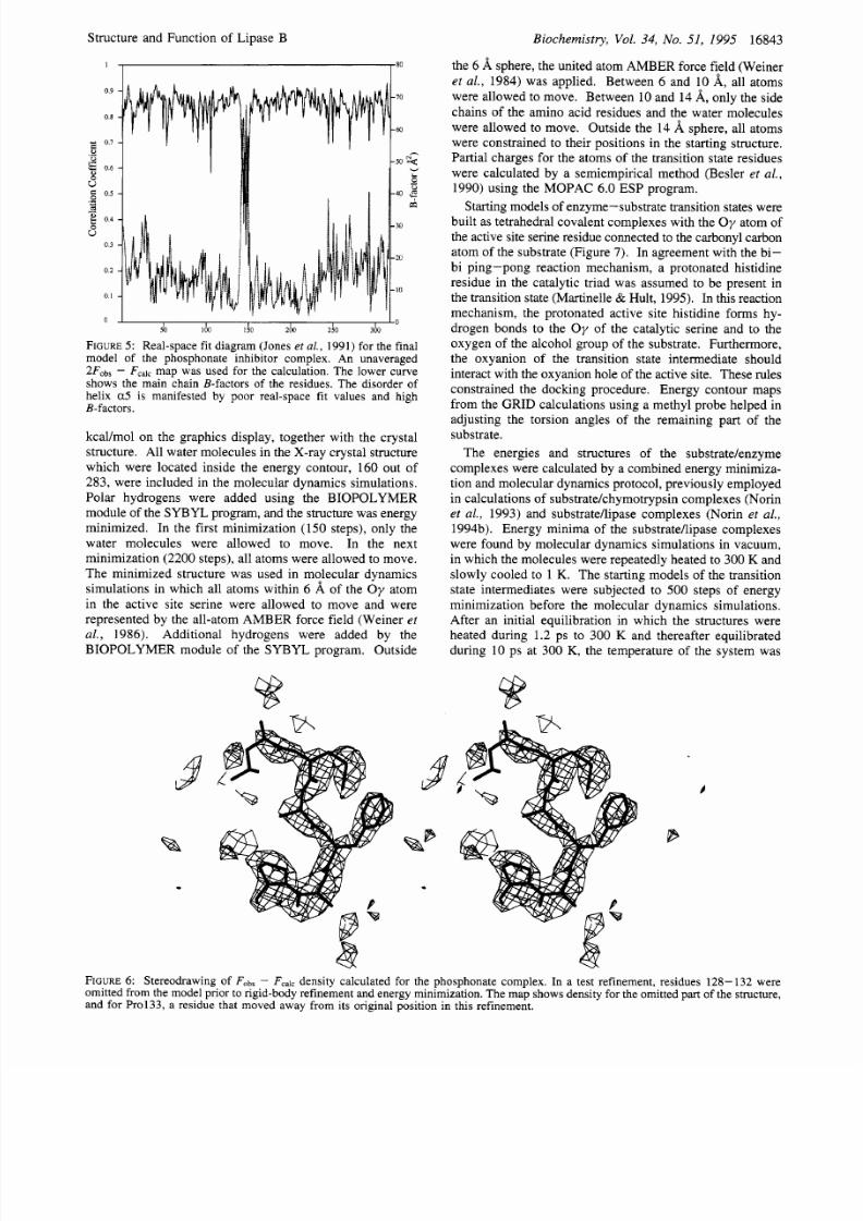
graph of the real-space fit and B-factors (Figure 5). The
final model has an rms deviation of 0.33
8,
rom the initial
orthorhombic model for Ca's and 0.42 A for all non-
hydrogen atoms. Refinement and model quality statistics
are summarized in Table 2. The rms deviations from ideal
bond lengths and angles are higher than we usually obtain
with this refinement protocol and may be due to the special
nature of this crystal form. Th e main chain dihedral angles
for SerlO5 are 4 = 54.5" and 11 = -110.0'. A test
calculation in which residues 128- 132 (corresponding to a
normally we ll-determined part of the structure) were omitted
from the model prior to rigid-body refinement and energy
minimization was also carried out. The resulting model was
used to calculate an
Fobs
calc ap which was averaged
over the six molecules in the asymme tric unit. The omitted
region of the model clearly matches the density as shown in
Figure 6.
Table 2: Refinement and Model Statistics
phosphonate Tween 80
2.69-2.60 2.59-2.50
15 621 25 979
resolution limits A) 7.5-2.6 7.5-2.5
number
of
reflections (F =
0)
completeness ( ) 82" 82
B-factor model group ed grouped
highest shell (
d
NCS model 6-fold 2-fold
constrained constrained
R-facto+
( )
20.7 (29.9) 18.7 (25.2)
R-free'
(%)
26.5 (35.1) 20.9 (30.9)
number of atoms (Z > 1)
protein 2324 2324
active site ligand 11 36
carbohydrate s 28 28
water molecules 92 155
bond lengths (A) 0.020 0.007
bond angles (deg) 2.2 1.3
dihedral angles (deg) 25.0 23.1
improper torsion angles (deg) 2.0 1.3
main chain atoms 17.5 13.7
all protein atoms 18.1 15.4
active site ligand 20.5 54.8
carbohydrate s 46.3 68.8
water molecules 27.6 40.2
rms
deviations from ideal geometryd
average B-factors
(A21
In the calculation of the completeness for the phosphonate data
set, the total number of possible reflections was calculated as the num ber
of reflections
for
the major twin component that were not systematically
absent due to the special local symmetry. bR-factor = x h 1 / F o b s l -
lFCalcl/xhlFabsl,here Fobs and F,,I, are the observed and calculated
structure factors, respectively. R-free has the same definition
as R-factor, but calculated for a subset of reflections not used in
the structure refineme nt. Values calculated with X-PLOR. Param-
eters derived from the Cambridge Database of small molecule struc-
tures (Engh & Huber, 1991) were used for the bond lengths and bond
angles.
I
80
I o
I I I I
I
I
50 100
I50
200
250
300
FIGURE : Real-space fit diagram (Jones et
al.,
1991) of the final
model
of
the Tween 80 complex (solid line). The correlation
coefficient was calculated using an unaveraged 2F0b, Fcalcmap.
The lower curve shows the main chain B-factors of the residues.
independent crystal lattices overlap, and the observed intensi-
ties are the sum s of intensities from the two lattices (Fisher
& Sw eet, 1980 ). In this case, the two twins are related by
a 180" rotation around the x or
z
axis. The absences due to
noncrystallographic symmetry can be explained by position-
ing three molecules with the same orientation in the unit

8/9/2019 Crystallographic and Molecular-Modeling Studies of Lipase B from Candida antarctica Reveal a Stereospecificity Poc…
http://slidepdf.com/reader/full/crystallographic-and-molecular-modeling-studies-of-lipase-b-from-candida-antarctica 5/14
16842
Biochemistry Vol. 34
No.
51 1995
1 = 3 n +
0
2
1
k=2n
Uppenberg et al.
A 1
0 0 0 0 0
0 0 0 0 0
0 e o Q I D o o
Q D o o o e o
o u D o e o o
e o o o Q I D 0
h
d h n n n n n
~ U U V U V I
h = 6 n +
1 2 3 4 5 0
Reflections with intensity contributions from both twin components
Q
Reflections in the lattice of first twin component only
Reflections in the lattice of second twin component only
General condition for reflections in C2: h+k=2
FIGURE
: Reflections in the phosphonate inhibitor data set can be partitioned into different subsets, because of the special noncrystallographic
symmetry and the merohedral twinning. The molecules in the asymmetric unit are related
by
pure translations
in
the
a/c
plane, which
results in a number of systematic absences in the d iffraction pattern. The sets of absences are partly d ifferent for the two twin components
in the crystal, which makes it easy to separate them for parts of the data. Some reflections have to be corrected to remove the contribution
from the unwanted twin.
FIGURE
: CALB/phosphonate inhibitor complex packs with six molecules
in
the asymmetric unit. They are arranged as three closely
packed dimers , in which the hydrophobic surfaces of the molecules face each other. The structural dimer
is
practically identical to that
found in the monoclinic crystal form described earlier (Uppenberg
e t al.,
1994b). The stereofigure shows two asymmetric units related by
the crystallographic centering operation.
Molecular Modeling and Dynamics Simulations.
Models
of the substrates were constructed and energy minimized with
the Biosym Insight I1 software on a Silicon Graphics
workstation . The initial manual modeling was performed
in 0 on an Evans and Sutherland ESV workstation. Mo-
lecular dynamics simulations, energy minimizations, ma-
nipulations of molecules, and graphic evaluations were done
with the Tripos SYBYL version 6.03 molecular-modeling
program on an Evans and Sutherland ESV workstation.
The environ ment of the lipase was modeled as vacuum to
make the computations feasible (Norin
et al .
1994a). The
AMBER force field (Weiner
et al.
1984,1986), using a
distance dependent dielectric constant and a nonbond-
ed cutoff d istance of
10
A ,
was used in all energy minimiza-
tions and molecular dynam ics simulations. Energy mini-
mizations were performed with the Powell minimizer in
SYBYL.
The molecular dynamics simulations were per-
formed using a time step
of
2 fs, with constraints on all bond
lengths using the SHAK E algorithm (Ryckaert
et al.
1977).
The GRID program (Goodford,
1985)
calculates the energy
potentials fo r various functional group s around a protein. Th is
program was used to map possible substrate interactions in
the active site and to assign strongly bonded water molecules
which are not replaced by solvent molecules when reactions
are performed in an organic solvent. A GRID calculation
with a water probe was used to obtain a contour plot at -6.5
8/9/2019 Crystallographic and Molecular-Modeling Studies of Lipase B from Candida antarctica Reveal a Stereospecificity Poc…
http://slidepdf.com/reader/full/crystallographic-and-molecular-modeling-studies-of-lipase-b-from-candida-antarctica 6/14
Structure and Function of Lipase B
Biochemistry , Vol. 34, No. 51, 1995 16843
the 6
8,
sphere, the united atom AMBER force field (Weiner
et al., 1984) was applied. Between 6 and 10 A , all atoms
were allowed to move. Between 10 and 14 8,, only the side
chains of the amino acid residues and the water molecules
were allowed to move. Outside the 14 8, sphere, all atoms
were constrained to their positions in the starting structure.
Partial charges for the atoms of the transition state residues
were calculated by a semiempirical method (Besler et al . ,
1990) using the MOPAC 6.0 ESP program.
Starting models of enzyme-substrate transition states were
built as tetrahedral covalent complexes with the O y atom of
the active site serine residue connected to the carbonyl carbon
atom of the substrate (Figure
7).
In agreement with the bi-
bi ping-pong reaction mechan ism, a protonated histidine
residue in the catalytic triad was assumed to be present in
the transition state (Martinelle & Hult, 1995). In this reaction
mechanism, the protonated active site histidine forms hy-
drogen bonds to the
O y
of the catalytic serine and to the
oxygen of the alcohol group
of
the substrate. Furthermore,
the oxyanion of the transition state intermediate should
interact with the oxyanion hole of the active site. These rules
constrained the docking procedure. Energy contour maps
from the GR ID calculations using a methyl probe helped in
adjusting the torsion angles of the remaining part of the
substrate.
The energies and structures of the substratelenzyme
complexes were calculated by a combined energy minimiza-
tion and molecular dynamics protocol, previously employed
in calculations of substrate khym otrypsin complexes (Norin
et al . , 1993) and substrate/lipase complexes (Norin et al.,
1994b). Energy minima of the substrateAipase complexes
were found by molecular dynamics simulations in vacuum,
in which the molecules were repeatedly heated to 300 K and
slowly cooled to 1 K. The starting models of the transition
state intermediates were subjected to 500 steps of energy
minimization before the molecular dynamics simulations.
After an initial equilibration in which the structures were
heated during 1.2 ps to 300 K and thereafter equilibrated
during 1 0 ps at 300 K, the temperature of the system was
0.9
0.8
0.7
V
5
0.6
U
2
0.5
E
8
e
.-
-
0.4
U
0.3
0.2
0
0 1
I
I o
I
I I
50
100 150 ZW 210 360
FIGURE
:
Real-space fit diagram (Jones
et al., 1991)
for the final
model of the phosphonate inhibitor complex. An unaveraged
2F0b, F,,,, map was used for the calculation. The lower curve
shows the main chain B-factors of the residues. The disorder of
helix a5 is manifested by poor real-space fit values and high
B-factors.
kcaVmol on the graphics display, together with the crystal
structure. All water molecules in the X-ray crystal structure
which w ere located inside the energy contour, 160 out of
283, were included in the molecular dynamics simulations.
Polar hydrogens were added using the BIOPOLYMER
module of the SYB YL program, and the structure was energy
minimized. In the first minimization (150 steps), only the
water molecules were allowed to move. In the next
minimization (2200 steps), all atoms were allowed to move.
The minimized structure was used in molecular dynamics
simulations in which all atoms within 6
8, of the O y atom
in the active site serine were allowed to move and were
represented by the all-atom AMBER force field (Weiner et
al . , 1986). Additional hydrogens were added by the
BIOPOLYM ER module of the SYB YL program. Outside
FIGURE
:
Stereodrawing of Fobs
F
ensity calculated for the phosphonate complex. In a test refinement, residues 128 -132 were
omitted from the mode l prior to rigid-body refinement and energy m inimization. The m ap shows density for the omitted part of the structure,
and for Pro133, a residue that moved away from its original position in this refinement.

8/9/2019 Crystallographic and Molecular-Modeling Studies of Lipase B from Candida antarctica Reveal a Stereospecificity Poc…
http://slidepdf.com/reader/full/crystallographic-and-molecular-modeling-studies-of-lipase-b-from-candida-antarctica 7/14
16844 Biochemistry Vol. 34 No. 51 1995
Uppenberg et al.
FIGURE
7:
Stereodrawing of the solvent accessible surface of the active site pocket and
a
-phenylethyl octanoate modeled
as a
tetrahedral
intermediate. Both the R and S enantiomers are shown, labeled at the methyl group at which they differ. I n the R enantiomer, the methyl
group is pointing into the stereospecificity pocket.
I n
the S enantiomer, it points towards the backbone atoms of GIy39 and Thr40, making
many close contacts. The hydrogen bond between the histidine and the alcohol oxygen is
also
indicated. The surface was calculated with
a 1.4
8 probe radius usin g-VO ID0 0 (Kleywegt & Jones, 1994a).
FIGURE: Stereodrawing of F ,bs Fcalc ensity in the active site pocket of the Tween 80 complex. The Tween 80 molecule was not
included
in
the map calculation. The partial Tween
80
molecule from the final model is
also
shown.
decreased during 1.5 ps to 1 K. Then the temperature was to the surface of the molecule (Figure 8). The Tween 80
raised during 1.2 ps to 300 K, followed by equilibration molecule (Figure 9) is a monoester that can account for the
during 6 ps before the next cooling period. Ten cycles of observed density, although the complete substrate molecule
heating and cooling were run. The total simulation time was cannot be modeled. We have modeled the Tween 80
98 ps. The conformations trapped at 1
K
were subjected to molecule in the active site pocket as an ester prior to
500
steps of energy m inimization.
hydrolysis.
In
order to be hydrolyzed, the alcohol oxygen
of the substrate must be able to receive a proton from
NE
f
RESULTS
the active site histidine. W e have used this biochemical
Crystallography
restriction to assign the two chains of the substrate to the
two tubes of density. The putative lid
a5
adopts the same
Crystal Structure of CALB Cocrystallized with Tween
80.
This structure shows two approximately parallel tubes of
density, originating at the active site serine and leading out
conformation as o bserved in the previously reported CALB
crystal forms and shows no sign of disorder (Figure 2). The
packing in these crystals is different from that of all other

8/9/2019 Crystallographic and Molecular-Modeling Studies of Lipase B from Candida antarctica Reveal a Stereospecificity Poc…
http://slidepdf.com/reader/full/crystallographic-and-molecular-modeling-studies-of-lipase-b-from-candida-antarctica 8/14
Structure
FIGURE
:
and Function of Lipase B
/
Biochemistry Vol. 34 No. 51 1995 16845
inhibitor has side chains extending from the phosphorus
atom, with the acyl part being a hexyl group and the alcohol
part an ethoxy group (Figure
1
I . This compound could be
fitted com pletely into the density (Figure IO). However, the
shorter tube contains som e extra density, in which another
atom can be positioned. At the resolution of this study, it
cannot be ruled out that both enantiomers of the inhibitor
bind compe titively to the serine. This would give rise to a
mixture of conformations for the acyl and alcohol parts of
Tween
80
is
a
nonionic detergent
with
an
approximate
the inhibitor
in
the active site pocket.
In
the hydrolysis of
molecular mass of 1.3 kDa. The sum of x, y. z and w is
-20.
The
oleate part
of
the substrate is shown
in
bold.
crystal forms that have been determined, and the solvent
conte nt of
-63
is unusually high.
It
is also the only crystal
form in which the hydrophobic surface around the active
site pocket is almost fully accessible to the solvent. The
high concen tration of Tween 80
in
the solution may have
contributed to the exposure of this surface.
Crystal Structure of CALB
in
Complex with the Phospho-
nate Inhibitor.
The dimeric packing motif, found in the
earlier monoclinic crystal form (Uppenberg
et al.
1994b),
reappears
in
this structure (Figure 4), although the unit cells
and space groups are different.
In
this crystal form,
a
is
again disordered, with
4-5
residues displayin g weak electron
density.
There is additional electron density in the active
site which we have assigned
to
the inhibitor.
This density
suggests a covalent attachment to the catalytic serine, Ser
105,
as predicted for this compound (Bjorkling et al. 1992). There
are two tubes of density of unequal length which extend from
a large spherical feature in the density connected to the serine
hydroxyl (Figure 10).
Clear density is also present for an
atom pointing toward the oxyanion hole. The phosphon ate
an ester substrate, the alcoho l oxygen receives a proton from
the active site histidine and must therefore be within
hydrogen bonding distance from the Nc of this residue. In
the reaction of the inhibitor with the enzyme, the alcohol
oxygen does not take part in the reaction. The enzyme’s
preference for one of the two enantiomers is therefore
governed predom inantly by the size of the two side chains
and their ability to m ake fav orable interactions in the active
site pocket of the enzyme. The observed density suggests
that the enzyme has been inhibited primarily with the
enantiomer which does not form a hydrogen bond with the
NE of the active site histidine. The other enantiomer can
also be modeled into the active site, albeit with density
missing for parts of its acyl chain. This alternative inter-
pretation of the electro n density supplies information for the
modeling of a tetrahedral interme diate for this enzyme.
Modelling of Chiral Substrates
CALB has been used for kinetic resolution of chiral
secondary alcohols, both
in
hydrolysis of esters and
in
ester
synthesis. In many of these reactions, CALB d isplays a very
high degree of stereoselectivity. To provide a structural
FIGURE: Stereodrawing of averaged Fc bc Fcalc ensity for the phosphonate inhibitor complex. The map was calculated with a model
in which the inhibitor molecule had not yet been included. The inhibitor binds covalently to the active site serine. It is located
in
the active
site pocket,with the alcohol oxygen
in
a
position that would not be favorable for catalysis
in
an
analogous substrate. If the acyl
and
alcohol
parts of the inhibitor traded positions, the inhibitor would represent the alleged tetrahedral intermediate
of
an ester substrate better, with a
hydrogen bond between the active site histidine and the alcohol oxygen. Density can also be seen for the inhibitor oxygen
in
the oxyanion
hole. The map was averaged with programs
in
the
RAVE
package Kleywegt & Jones, 1994b).

8/9/2019 Crystallographic and Molecular-Modeling Studies of Lipase B from Candida antarctica Reveal a Stereospecificity Poc…
http://slidepdf.com/reader/full/crystallographic-and-molecular-modeling-studies-of-lipase-b-from-candida-antarctica 9/14
16846 Biochemistry , Vol. 34 No. 51 1995
Uppenberg et al.
two enantiomers. In the R enantiomer, this methyl group
fits nicely into a small pocket that is delimited by the main
chain of the third strand on one side and by the side chain
of Tr pl 04 on the other. In the S enantiomer, the methyl
group is pointing toward the m ain chain atoms of Gly39 and
Thr40 and the substrate oxyan ion, and it makes many close
van der Waals contacts. These steric clashes make this
interaction highly unfavorable, especially since the two
residues are involved in oxyanion stabilization (Figure 7).
The acyl side
of
the enzymehbstrate complex was fit
manually to the observed density of the CALBhnhibitor
complex.
Energy Minimizat ion and Molecular Dynamics of Sub-
strate 1 To allow this model to relax, we carried out a
number of molecular dynamics simulations. This was done
to identify regions where close contacts could be relaxed by
changes in conformation in either protein or intermediate
and to help formulate test hypotheses. The minimized
structures with the lowest energy are shown in Figure 13
for both enantiomers. The orientation of the alcohol moiety
in the
S
enantiomer differed substantially from that in the
starting structure. The methyl group moved into the small
pocket, and the phenyl g roup pointed out toward the protein
surface. These rearrangements moved the alcohol oxygen
atom toward the oxyanion hole, thereby breaking the
catalytically essential hydrogen bond to His224. This
suggests that, in a transesterification reaction, the S alcohol
cannot easily donate a proton to the catalytic histidine residue
which is essential for nucleophilic attack on the acyl enzym e.
Consequently, in a hydrolysis reaction, the alcohol moiety
of the substrate would be stuck in a position where it cannot
accept a proton from the catalytic histidine and is therefore
not an efficient leaving group. In contrast, the energy
minima of the tetrahedral intermediate
of
the
R
enantiomer
contain a favorable hydrogen bond between the catalytic
histidine and the oxygen atom of the alcohol group which
would enable rapid proton transfer during catalysis.
Substrate
2:
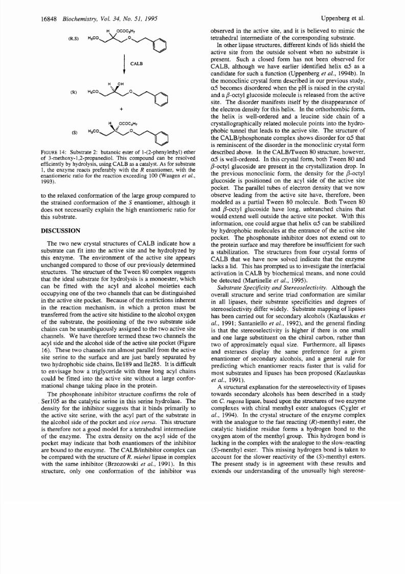
Butanoate of a Glycerol Derivative.
In
another study, CALB has been used for enantiomeric
resolution of a glycerol monoester derivative by hydrolysis
of the
sn-2
ester bond (Waagen e? al . , 1993). This substrate
is similar to phenylethyl oc tano ate in the sense tha t it contains
a chiral carbon on the alcohol group at the same position
relative to the carbonyl carbon (Figure 14). The two free
groups extending from this chiral carbon are a methoxy-
methyl group and a phenyl-co ntaining group. In this
hydrolytic reaction, the R enantiomer of the substrate is the
favored enantiomer, with an enantiomeric ratio exceeding
100.
The R enantiomer was modeled with its methoxymethyl
group pointing into the small stereospecificity pocket,
although this gave rise to a few close contacts with the side
chain of Trp l04 . The long side chain could be positioned
in the channel leading out to the protein surface. The S
enantiomer was more difficult to position, and the long side
chain of the alcohol had to be curved in order to avoid close
contacts with the protein.
Energy Minimizat ion and Molecular Dynamics o Sub-
strate
2. The enzymehbstrate complex was subjected to
the same molecular dynamics simulation and energy mini-
mization protocol as the first com plex. In the resulting
minimized structure, the bonds in the carbon chain of the
large group of the fast-reacting R enantiomer were all in
relaxed trans conformations. The small group was interact-
bl
FIGURE
1: The phosphonate inhibitor used here is a racemic
mixture of n-hexylchlorophosphonate ethyl ester. The chlorine atom
is released when the inhibitor reacts with the enzyme.
I
CALB
FIGURE2: Substrate 1 phenylethyl octanoate. The stereoselective
resolution of 1-phenylethanol s very high in
CALB,
as only the
R
enantiomer of the alcohol reacts with the enzyme. The reaction
was carried out at 39
C,
under atmospheric pressure, in order to
shift the reaction equilibrium
in
favor of the forward reaction. For
a de tailed description of the experimental conditions, see Frykman
et
al. (1993).
explanation for this phenomenon , we have carried out a series
of molecular-modeling experiments.
Substrate
I: 1
Phenylethyl Octanoate.
The first reaction
step to be modeled was the formation of the tetrahedral
intermediate of the esterification of racemic 1 phenylethanol
into 1 phenylethyl octanoate. This reaction (Figure 12)
strongly favors the R-enantiomer
of
the alcohol with an
observed en antiomeric ratio, E greater than 200 (Frykman
et al.,
1993). First, we modeled the atoms associated with
the tetrahedral carbon of the intermed iate. For this, we used
the crystal structure of R. miehei in complex with the
inhibitor n-hexylphosphonate ethyl ester (Brzozowski et al.,
1991). This structure was aligned with CALB to give an
optimal fit with respect to the active site residues and the
residues forming the oxyanion hole. For modeling the
alcohol, we took into account the geometric restrictions
inherent in the tetrahedral intermediate. Thus, we assumed
that the position of the carbo nyl carbon and the oxy anion as
well as the oxygen and the neighboring (chiral) carbon on
the alcohol group must be relatively fixed in order to allow
for the transfer of a proton between the active site histidine
and the alcohol oxygen. Th e corresponding atoms from the
R . muc or inhibitor were therefore assumed to represent a
good starting model. Th e electron density from the CALB/
inhibitor complex also supports such a positioning of these
atoms.
These constraints leave only one degree of rotational
freedom to fit the remaining part of the alcohol in the active
site. This was done manually with guidance from the density
seen in the active site channel in the Twe en 8 0 crystal form
and the van der Waals surface of the surrounding protein
(Figure 7). This led to one unique orientation with the phenyl
group pointing out toward the protein surface in both the
S
and the R form. What remained to be examined was the
orientation of the m ethyl group of the chiral carbon for the

8/9/2019 Crystallographic and Molecular-Modeling Studies of Lipase B from Candida antarctica Reveal a Stereospecificity Poc…
http://slidepdf.com/reader/full/crystallographic-and-molecular-modeling-studies-of-lipase-b-from-candida-antarctica 10/14
Structure and Function of Lipase B
Biochemistry Vol. 34
No. 51
1995
16847
a
b
1187
1187
FIGURE3: Substrate
1.
Modeled structu re of the 1-phenylethy l octanoate tetrahedral interm ediate after minimiza tion of the stru cture with
the lowest energy from the molecular dynamics sim ulation. (a) The
R
enantiom er of the substr ate fits nicely into the active site pocket, with
the hydrogen bond between the histidine and the substrate intact. (b) The S nantiomer adopts a conformation in which the two free groups
of the alcohol have a conformation similar to those in the
R
enantiomer. However, the alcohol oxygen points away from the active site
histidine, making a proton transfer between these two groups impossible.
ing with Trpl04 in the small pocket. Thi s was possible by
a small adjustment of the side chain of Trpl04, which was
pushed back slightly toward the interior
of
the protein (Figure
15a). The large group of the slow-reacting
S
enantiomer
was twisted away from the specificity pocket, mainly
interacting
with
the side chains of Leu278, Ala282, and
Ile285 (Figure 15b). The presence
of
these side chains forced
the carbon chain of this group of the substrate to adopt an
energetically less favorable conformation. The small group
occupied a space between the large alcohol side chain and
the carbon chain o f the acyl group, and the small pocket is
empty.
The acyl group moved toward the bottom of the
active site. The slow-reacting
S
enantiomer retained the
hydrogen bond between the histidine residue of the catalytic
triad and the oxy gen atom of the alcohol group.
It
is possible
that the chiral preference for the R enantiomer is due partly
8/9/2019 Crystallographic and Molecular-Modeling Studies of Lipase B from Candida antarctica Reveal a Stereospecificity Poc…
http://slidepdf.com/reader/full/crystallographic-and-molecular-modeling-studies-of-lipase-b-from-candida-antarctica 11/14
16848 Biochemistry Vol. 34 No. 51 1995
H OCOC3H7
RS) H a c o o T
Uppenberg et al.
observed in the active site, and it is believed to mimic the
tetrahedral intermediate of the corresponding substrate.
In other lipase structures, different kinds
of
lids shield the
active site from the outside solvent when no substrate is
present. Such a closed form has not been observed for
CALB, although we have earlier identified helix
a5
as a
candidate for such a function (Uppenberg et al. 1994b). In
the monoclinic crystal form described in our previous study,
a 5 becomes disordered when the p H is raised in the crystal
and a P-octyl glucoside molecule is released from the active
site. The disorder manifests itself by the disappearance of
the electron density for this helix. In the orthorh omb ic form,
the helix is well-ordered and a leucine
side chain of a
crystallographically related molecule p oints into the hydro-
phobic tunnel that leads to the active site. The structure of
the CALB/phosphonate complex shows disorder for a 5 hat
is reminiscent
of
the disorder in the monoclinic crystal form
described above. In the CA LB Rw een 80 structure, however,
a5
is well-ordered. In this crystal form, both Tween
80
and
P-octyl glucoside are present in the crystallization drop. In
the previous monoclinic form, the density for the P-octyl
glucoside is positioned on the acyl side of the active site
pocket. The parallel tubes of electron density that we now
observe leading from the active site have, therefore, been
modeled as a partial Tween
80
molecule.
Both Tween
80
and P-octyl glucoside have long, unbranched chains that
would extend well outside the active site pocket. With this
information, one could argue that helix a 5 can be stabilized
by hydrophobic molecules at the entrance of the active site
pocket. Th e phosphonate inhibitor does not extend out to
the protein surface and may therefore be insufficient for such
a stabilization.
The structures from four crystal forms of
CALB that we have now solved indicate that the enzyme
lacks a lid. This has prompted us to investigate the interfacial
activation in CALB by biochemical means, and none could
be detected (Martinelle et
al.,
1995).
Substrate Specifc ity and Stereoselectivity.
Although the
overall structure and serine triad conformarion are similar
in all lipases, their substrate specificities and degrees of
stereoselectivity differ widely. Substrate mapping of lipases
has been carried out for secondary alcohols (Kazlauskas
et
al .
1991; Santaniello
et al . ,
1992), and the general finding
is that the stereoselectivity is higher if there is one small
and one large substituent on the chiral carbon, rather than
two of approximately equal size.
Furthermore, all lipases
and esterases display the same preference for a given
enantiomer of secondary alcohols, and a general rule for
predicting which enantiomer reacts faster that is valid for
most substrates and lipases has been proposed (Kazlauskas
et al.
1991).
A structural explanation for the stereoselectivity
of
lipases
towards secondary alcohols has been described in a study
on C. rugosa lipase, based upon the structures of two enzy me
complexes with chiral menthyl ester analogues (Cygler et
al . 1994). In the crystal structure of the enzyme complex
with the analo gue to the fast reacting @)-menthyl ester, the
catalytic histidine residue forms a hydrogen bond to the
oxygen atom of the menthyl group. This hydrogen bond is
lacking in the comp lex with the analogue to the slow-reacting
(S)-menthyl ester. This missing hydrogen bond is taken to
account for the slower reactivity of the (S)-menthyl esters.
The present study is in agreement with these results and
extends our understanding of the unusually high stereose-
CALB
t
H OH
R)
S) H~co3. o 'o
OCOC3H7
FIGURE 4:
Substrate 2: butanoic ester of 1-(2-phenylethyl)ether
of 3-methoxy-l 2-propanediol.
his compound can be resolved
efficiently by hydrolysis, using CALB as a catalyst. As for substrate
1,
the enzyme reacts preferably with
the R
enantiomer,
with
the
enantiomeric ratio for the reaction exceeding
100
(Waagen
et al.,
1993).
to the relaxed conformation of the large group compared to
the strained conformation of the S enantiomer, although it
does not necessarily explain the high enantiomeric ratio for
this substrate.
DISCUSSION
The two new crystal structures of CALB indicate how a
substrate can fit into the active site and be hydrolyzed by
this enzyme. Th e environm ent of the active site appears
unchanged compared to those of our previously determined
structures. The structure of the Tw een 80 complex suggests
that the ideal substrate for hydrolysis is a monoester, which
can be fitted with the acyl and alcohol moieties each
occupy ing one of the two channels that can be distinguished
in the active site pocket. Becaus e
of
the restrictions inherent
in the reaction mechanism, in which a proton must be
transferred from the active site histidine to the alcohol oxygen
of the substrate, the positioning of the two substrate side
chains can be unambiguously assigned to the two active site
channels. We have therefore termed these two channels the
acyl side and the alcohol side of the a ctive site pocket (Fig ure
16).
These tw o channels run alm ost parallel from the active
site serine to the surface and are just barely separated by
tw o hydrophobic side chains, Ile189 and Ile285. It is difficult
to envisage how a triglyceride with three long acyl chains
could be fitted into the active site without a large confor-
mational change taking place in the protein.
The phosphonate inhibitor structure confirms the role
of
SerlO5 as the catalytic serine in this serine hydrolase.
The
density for the inhibitor suggests that it binds primarily to
the active site serine, with the acyl part
of
the substrate in
the alcohol side
of
the pocket and
vice versa.
This structure
is therefore not a good model for a tetrahedral intermediate
of the enzyme. The extra density on the acyl side of the
pocket may indicate that both enantiomers of the inhibitor
are bound
to
the enzyme.
The CA LBhnhibitor complex can
be com pared with the structure of
R.
miehei
lipase in complex
with the same inhibitor (Brzozowski
et al.
1991).
In this
structure, only one conformation
of
the inhibitor was

8/9/2019 Crystallographic and Molecular-Modeling Studies of Lipase B from Candida antarctica Reveal a Stereospecificity Poc…
http://slidepdf.com/reader/full/crystallographic-and-molecular-modeling-studies-of-lipase-b-from-candida-antarctica 12/14
Structure and Function of Lipase B
Biochemistry
Vol.
34 No. 51 1995
16849
a
87
14
b
I87
24
FIGURE5: Substrate 2. The modeled struc ture of the butanoic ester intermediate after minimization of the structure
with
the lowest energy
from the molecular dynamics simulation. (a) The
R
enantiomer of this substrate fits well into the active site pocket after a small movement
of the side chain of Trpl04. The long side chain of the alcohol is
in a
relaxed extended conformation. (b) The
S
nantiomer keeps the
hydrogen bond to the histidine, but the large group extending from the chiral carbon of the alcohol has to make a curve
in
the active site
pocket, forcing it into a strained conformation.
lectivity toward some secondary alcohols that has been
demonstrated for CALB. Although the preference toward
one of the enantiomers of secondary alcohols is similar for
most lipases, the space available for the tw o su bstituents of
the alcohol differs between enzyme species. We have
investigated the alcohol side of the active site pocket with
the enantiomers of two secondary alcohols. This has
revealed a small and well-defined pocket suitable for
accomodation of the small substituent of the fast-reacting
enantiomer. This pocket is buried under a surface helix and
delimited by the side chain of Trpl 04 . The restricted volume
of this pocket allows us to suggest which groups are likely
to fit there. In the first sub strate modeled, the phenylethyl
octanoate complex, a methyl group fits easily into this pocket.
The methoxymethyl group of the second substrate to be
modeled is more troublesom e and causes som e close contacts
with the protein.
However, experimentally,
it
is still ef-
ficiently hydrolyzed with high stereoselectivity, and we,

8/9/2019 Crystallographic and Molecular-Modeling Studies of Lipase B from Candida antarctica Reveal a Stereospecificity Poc…
http://slidepdf.com/reader/full/crystallographic-and-molecular-modeling-studies-of-lipase-b-from-candida-antarctica 13/14
16850 Biochemistry, Vol.
34,
No. 51 1995 Uppenberg et al.
prediction rule works well, which is also exemplified by the
crystal structures of the menthyl ester analogues (Cygler
et
al.,
1994).
Work to examine the specificity pocket of CALB by
further variation of the size of the groups extending from
the chiral carbon is now in progress. Trials to crystallize
chiral inhibitors to gain more insight into the structural
features that govern the stereoselectivity of CALB will also
be undertaken.
Substrates with a chiral center on the acyl side have been
studied less with CAL B as a catalyst. The stereoselectivity
with respect to this side of the substrate is much lower than
for the alcohol side (Quir6s et al., 1993; Sinisterra et al.,
1994). The proposed acyl side of the active site pocket is
more spacious than the alcohol side, and it might be able to
accommodate both enantiomers of such chiral acyl groups.
/
a cy l a l co h o l
s i d e
1 ,
s i d e
S e r l O S
FIGURE
6: Active site pocket can be partitioned into two sides,
an acyl side and an alcohol side, where the corresponding parts of
the substrate will be located during catalysis. This conformational
restriction is imposed on the substrate by the necessary transfer of
a proton between the alcohol oxygen and the NE of the active site
histidine during catalysis.
therefore, assu me that there is som e flexibility of the protein
in this region. The side chain of Trpl 04 is of great interest
since it makes up the bottom of the stereospecificity pocket.
This residue precedes the active site serine, and we believe
it is structurally important since it forms a hydrogen bond
from the nitrogen atom of its indole ring to the backbone
oxygen of the active site histidine. In most other lipases,
however, this residue is a histidine with an imidazole nitrogen
at a position equivalent to that of the indole nitrogen. W e
believe, therefore, that this residue would make an interesting
target for site-directed mutagenesis in many lipases. A single
mutation of this tryptophan to a histidine in our model of
CALB creates a larger pocket for a substrate side chain to
occupy. The molecular dynam ics simulations suggest that
the critical hydrogen bond between the alcohol oxygen of
the substrate and the active site histidine is retained not only
for the
R
enantiomers of both substrates but also for the S
enantiome r of the second subsrate. Th e high enantioselec-
tivity for the second substrate is therefore more difficult to
explain than for the first substrate.
Secondary alcohols have been widely used in reactions
with lipases that display less pronounced stereoselectivity
than CALB. For
R. miehei
lipase, molecular dynamics
studies have been carried out to explain its stereoselectivity
on these substrates (Norin
et
al., 1994). The active site of
this enzyme is more open and allows more flexibility for
substrates to adopt suitable conform ations. In
C. rugosa
lipase, the binding site for the small substituent is more
accessible to the surface than in CAL B. It has also been
reported that this enzym e does not follow the prediction rule
of enantioselectivity for acyclic secondary alcohols (Ka-
zlauskas
et al.,
1991). This means that the large substituent
for the slow-reacting enantiomer of some of these more
flexible substrates can be accommodated in the binding site
for the small substituent. Fo r the cyclic substrates, the
ACKNOWLEDGMENT
We thank Dr. Fredrik Bjorkling, Novo Nordisk
NS
for
providing us with the phosphonate inhibitor.
REFERENCES
Besler, B. H., Merz, K. M. J.,
&
Kollman, P. A. (1990)
J.
Comput.
Chem. 11, 43 1-439.
Bjorkling, F., Godtfredsen,
S. E., &
Kirk,
0
(1991) TIBTech. 9,
360-363.
Bjorkling, F., Godtfredsen, S. E., Kirk,
O.,
Patkar, S. A, , &
Andersen,
0
1992) Microb. Reagents Org. Synth., 1992, 249-
260.
Blow, D. M., Birktoft,
J.
J., & Hartley, B.
S.
(1969) Nature 221,
337-340.
Brady,
L.,
Brzozowski, A. M., Derewenda, Z.
S.,
Dodson,
E.,
Dodson, G., Tolley,
S.,
Turkenburg, J. P., Christiansen, L., Huge-
Jensen, B., Norskov,
L.,
Thim, L., & Menge, U. (1 990) Nature
343, 767-770,
Briinger, A. T. (1990) Acta Crystallogr. A46, 46-57.
Briinger, A. T. (1992a) X-PLOR. Version 3.1. A system fo r X -ray
crystallography and NMR, Yale University Press, New Haven
CT.
Brunger, A. T. (1992b) Nature 355, 472-475.
Brunger, A. T., Krukowski, A. ,
&
Erickson, J. (1990) Acta
Crystallogr. A46, 585-593.
Brzozowski, A. M., Derewenda, U., Derewenda,
Z.
S., Dodson,
G. G., Lawson, D. M., Turkenburg, J. P., Bjorkling, F., Huge-
Jensen, B., Patkar,
S.
A,, & Thim, L. (1991) Nature 351, 491-
494.
Collaborative Computational Project N umber 4. (1 994) Acta
Crystallogr. D50, 760-763.
Cygler, M., Grochulski, P., Kazlaukas, R. J., Schrag, J. D.,
Bouthillier, F. B., Rubin, B., Serreqi, A. N.,
&
Gupta, A. K.
(1994) J . Am. Chem.
SOC.
16, 3180-3186.
Derewenda, U., Swenson, L., Green, R., Wei, Y., Dodson, G. G.,
Yamaguchi, S., Haas, M. J., & Derewenda, Z. S. (1994a) Nut.
Struct.
Biol.
I
36-47.
Derewenda,
U.,
Swenson, L., Wei, Y., Green, R., Kobos, P. M.,
Joerger,
R.,
Haas, M.
J., &
Derewenda, Z. S (1994b)
J .
Lipid
Res. 35, 524-534.
Desnuelle, P. (1972) The Enzymes 7, 575-616.
Engh, R. A.,
&
Huber,
R .
(1991) Acta Crystallogr. A47, 392-
Fisher, R. G., & Sweet, R. M. (1980) Acta Crystallogr. A36, 755-
Franken,
S.
M., Rozeboom, H. J., Kalk, K. H., & Dijkstra, B. (1991)
Frykman, H., Ohmer, N., Norin, T.,
&
Hult,
K.
(1993) Tetrahedron
Gillis, A. (1988)
J .
Am. Oil Chem. SOC.65, 846-850.
Goodford, P. J. (1985)
J .
Med. Chem. 28, 849-857.
400.
760.
EMBO
J.
10 1297-1302.
Lett. 34, 1367- 1370.

8/9/2019 Crystallographic and Molecular-Modeling Studies of Lipase B from Candida antarctica Reveal a Stereospecificity Poc…
http://slidepdf.com/reader/full/crystallographic-and-molecular-modeling-studies-of-lipase-b-from-candida-antarctica 14/14
Structure and Function
of
Lipase
B
Grochulski, P., Li, Y., Schrag, J. D., Bouthillier, F., Smith, P.,
Harrison, D., Rubin, B., & Cygler, M. (1993) J. Biol. Chem.
Grochulski, P., Li, Y., Schrag,
J.
D.,
&
Cygler, M. (1994) Protein
Hamlin, R. (1985) Methods Enzym. 114, 416-452.
Hjorth, A., CarriCrC, F., Cudrey, C., Woldike, H., B oel,
E.,
Lawson,
D. M., Ferrato, F., Cambillau, C., Dodson, G. G., Thim, L., &
Verger, R. (1993) Biochemistry 32, 4702-4707.
IUBMB (1992) Enzyme nomenclature, Academic Press, London.
Jones, T. A., Zou, J.-Y., Cowan, S. W., & Kjeldgaard, M. (1991)
Kabsch, W. (1988) J. Appl. Crystallogr. 21, 916-924.
Kazlauskas, R. J., Weissfloch, A. N. E., Rappaport, A. T., & Cuccia,
L. A. (1991) J. Org. Chem. 56, 2656-2665.
Kleywegt, G. J., & Jones, T. A. (1994a) Acta Crystallogr. D50,
178-185.
Kleywegt, G. J.,
&
Jones,
T.
A. (1994b) in Fromfirst map tofinal
model (Bailey, S. , Hubbard, R., & Waller, D. A., Eds.) pp 59-
66, SER C Daresbury Laboratory, Daresbury, U.K.
Lawson,
D.
M., Derewenda, U., Serre, L., Ferri, S. , Szittner, R.,
Wei, Y., Meighen, E. A.,
&
Derewenda, Z. S. (1994) Biochem-
istry 33, 9382-9388.
Martinelle, M., & Hult, K. (1995) Biochim. Biophys. Acta 1251,
191 97.
Martinelle, M., Holmquist, M., & Hult,
K.
(1995) Biochim. Biophys.
Acta 1258, 272-276.
Martinez, C., DeGeus, P., Lauwereys, M., Matthyssens, G.,
&
Cambillau, C. (1992) Nature 356, 615-618.
Matthews, B. W. (1968)
J .
Mol. Biol.
33,
491-497.
McPherson, A. (1982) in Preparation and analysis
of
protein
Messerschmidt, A ., & Pflugrath, J. W. (1987) J. Appl. Crystallogr.
Noble, M. E. M., C leasby, A., Johnson, L. N., Egmond, M. R.,
&
Norin, M., Hult, K., Mattson, A., & Norin, T. (1993) Biocatalysis
Norin, M., Haeffner,
F.,
Achour, A., Norin, T., & Hult, K. (l994a)
Norin, M., Haeffner, F., Hult, K.,
&
Edholm, 0 1994b) Biophys.
268, 12843-12847.
Sci. 3, 82-91.
Acta Crystallogr.
A4 7
110- 119.
crystals, pp 96-97, J. Wiley
&
Sons, Inc., New York.
30, 306-315.
Frenken, L. G. J. (1993) FEBS Lett. 331, 123-128.
7, 131-147.
Protein Sci. 3, 1493-1503.
J. 67, 548-559.
Biochemistry, Vol.
34
No. 51, 1995 16851
Ollis, D. L., Cheah,
E.,
Cygler, M., Dijkstra, B., Frolow,
F.,
Franken, S. M., Harel, M., Remington,
S.
J., Silman, I., Schrag,
J. D., Sussman, J. L., Verschueren, K. H. G., & Goldman, A.
(1992) Protein Eng. 5, 197-211.
Patkar, S. A., Bjorkling, F., Zundel, M., Schulein, M., Svendsen,
A., Heldt-Hansen, H. P., & Gormsen, E. (1993) Znd. J. Chem.
32B, 76-80.
Quir6s, M., Shnchez, V. M., Brieva, R., Rebolledo, F., & Gotor,
V. (1993) Tetrahedron: Asymmetry 4, 1105- 11 12.
Ryckaert, J.-P., Ciccotti, G.,
&
Berendsen, H. J. C. (1977) J.
Comput. Phys. 23, 327-341.
Santaniello,
E.,
Ferraboschi, P., Grisenti, P.,
&
Manzocchi, A.
(1992) Chem. Rev. 92, 1071-1132.
Schrag, J. D., Li, Y., Wu, S.,
&
Cygler, M. (1991) Nature 351,
761 -764.
Sinisterra, J. V., Llama, E.
F.,
del Campo, C., Cabezas, M. J.,
Moreno, J. M., & Arroyo, M. (1994) J. Chem. SOC.,Perkin Trans.
2, 1333-1336.
Sussman, J. L., Harel, M., Frolow, F., Oefner, C., Goldman, A,,
Toker, L., & Silman, I. (1991) Science 253, 872-879.
Uppenberg, J., Patkar, S. A., Bergfors, T., & Jones, T. A. (1994a)
J.
Mol. Biol. 235, 790-792.
Uppenberg, J., Hansen, M. T., Patkar,
S., &
Jones, T. A. (1994b)
Structure 2, 293-308.
van Tilbeurgh, H., Egloff, M.-P., Ma rtinez, C., Rugani, N., Verger,
R., & Cambillau, C. (1993) Nature 362, 814-820.
Waagen,
V.,
Hollingszter, I., Partali, V., Thorstad,
O.,
& An-
thonsen, T. (1993) Tetrahedron: Asymmetry 4, 2265-2274.
Wei,
Y.,
Schottel, J. L., Derewenda, U., Swenson,
L.,
Patkar,
S.,
&
Derewenda, Z.
S.
(1995) Nut. Struct. Biol. 2, 218-223.
Weiner, S . J., Kollman, P. A., Case, D. A., Singh, U. C., Ghio, C.,
Alagona, G., Profeta, S . J.,
&
Weiner, P. A. (1984) J. Am. Chem.
Weiner, S . J., Kollman, P. A ., Nguyen, D. T.,
&
Case, D. A. (1986)
Winkler,
F.
K., D’Arcy, A., & Hunziker, W. (1990) Nature 343,
Wright, C. S. , Alden, R. A., & Kraut, J. (1969) Nature 221,
SOC 106, 765-784.
J .
Comput. Chem. 7, 230-252.
77 1-774.
235-242.
BI951640V