Embed Size (px)
Citation preview
Published: November 11, 2011
r 2011 American Chemical Society 5443 dx.doi.org/10.1021/nl203216q |Nano Lett. 2011, 11, 5443–5448
LETTER
pubs.acs.org/NanoLett
Crystallization of Fluorescent Quantum Dots within aThree-Dimensional Bio-Organic Template of Actin Filaments and LipidMembranesEtienne Henry,†,‡ Aur�elien Dif,§ Marc Schmutz,|| Loic Legoff,^ Franc-ois Amblard,^ Val�erie Marchi-Artzner,*,§
and Franck Artzner*,†
†Institut de Physique de Rennes, UMR 6251 CNRS, Universit�e Rennes 1, Avenue du G�en�eral Leclerc, 35042 Rennes Cedex, France‡LBPA, UMR CNRS 8113, ENS Cachan, 94235 Cachan, France§Sciences Chimiques de Rennes, UMR 6226 CNRS, Universit�e Rennes 1, Avenue du G�en�eral Leclerc, 35042 Rennes Cedex, France
)Institut Charles Sadron, UPR 22 CNRS, Universit�e de Strasbourg, 23 rue du Loess, 67034 Strasbourg, France^Laboratoire de Physico-chimie, UMR 168 CNRS, Institut Curie, 11 rue Pierre et Marie Curie, 75248 Paris Cedex 04, France
bS Supporting Information
Generating new physical properties using the crystallization ofnanoparticles (NPs) is highly challenging.1�7 The design of
such new materials with unexpected physical properties is thusthe major motivation in the investigation of new strategies forcontrolling the crystallization of nanoparticles. It is now possibleto produce NPs having a variety of specific optical, electronic, ormagnetic properties. The couplings between their individualproperties come from various interactions such as electrostaticor magnetic field, absorption dipole, wave function overlap, andso forth. The emergence of collective properties arising from suchcouplings has been demonstrated in the case of one-dimensional(1D) chains of NPs or two-dimensional (2D) assemblies.5,6,8 Inparticular, the collective optical properties of such ensemble havebeen mainly attributed to the close vicinity between nanoparticles.9
The fabrication of 3D crystalline organizations with well-definedcharacteristic lengths could permit to extend the coupling faraway from the first neighbors so that delocalized eigenstateare expected to appear as observed with 3D crystal of atoms.10
Moreover, as the coupling strength strongly depends on the NPsinterdistance, the spatial organization of NPs, the extension, andthe quality of this order should govern these new properties.11
Colloidal crystallization from a homogeneous suspension ofspherical objects is the most effective process to produce 3D cubicstructures.1�6 NPs surface functionalization has been extensively
developed in order to improve the quality of the crystal and tunethe NPs interdistance. This can be achieved by surfactants, poly-mers, and biomolecules. Recently, double strand DNA interac-tions have successfully demonstrated the unique possibility tomodulate the NPs interdistance.12,13 Another strategy for NPscrystallization is based on their incorporation within three-dimensional crystal templates. Biological molecules1 and molec-ular self-assemblies are promising templates to organize well-defined inorganic nanostructures.11 This approach offers oppor-tunities to generate crystals with new symmetries and a largerrange of distance parameters. An alternative strategy consistsin exploiting three-dimensional (3D) templates to synthesizeNPs in situ.14 This elegant promising method opens access tocrystals with new symmetries that are hardly accessible fromisotropic NPs but is restricted to NPs obtained in mild con-ditions excluding for example the versatile core�shell semicon-ductive NPs such as fluorescent QDs. Our strategy is to incorporatethe NPs during the self-assembling process of a dynamicaltemplate that imposes the symmetry of the emerging NPscrystal (Scheme 1).
Received: September 15, 2011Revised: November 8, 2011
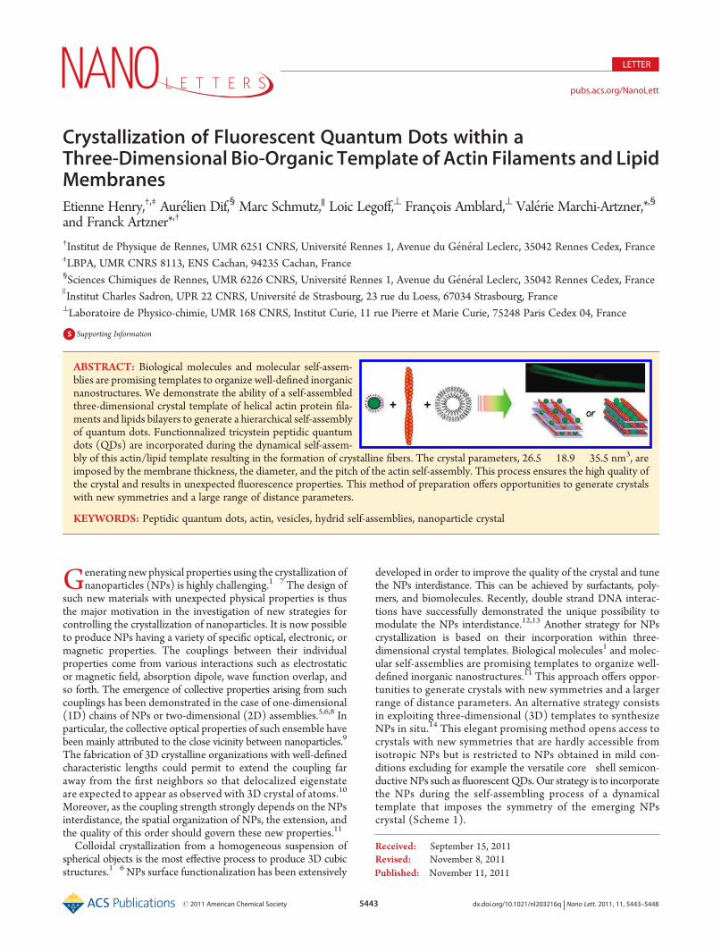
ABSTRACT: Biological molecules and molecular self-assem-blies are promising templates to organize well-defined inorganicnanostructures. We demonstrate the ability of a self-assembledthree-dimensional crystal template of helical actin protein fila-ments and lipids bilayers to generate a hierarchical self-assemblyof quantum dots. Functionnalized tricystein peptidic quantumdots (QDs) are incorporated during the dynamical self-assem-bly of this actin/lipid template resulting in the formation of crystalline fibers. The crystal parameters, 26.5 � 18.9� 35.5 nm3, areimposed by the membrane thickness, the diameter, and the pitch of the actin self-assembly. This process ensures the high quality ofthe crystal and results in unexpected fluorescence properties. This method of preparation offers opportunities to generate crystalswith new symmetries and a large range of distance parameters.
KEYWORDS: Peptidic quantum dots, actin, vesicles, hydrid self-assemblies, nanoparticle crystal
5444 dx.doi.org/10.1021/nl203216q |Nano Lett. 2011, 11, 5443–5448
Nano Letters LETTER
Here, we describe the preparation of a self-assembled 3Dcrystal template of helical actin protein filaments embedded withinlipid bilayers (Figure 1). Functionalized peptidic quantums dots15
(QDs) are incorporated during the self-assembling of the template16
(Scheme 1). This process ensures the high quality of the crystaland results in specific fluorescence properties.
In a first simple approach,15 we used lipid multilayers as atemplate to insert water-soluble functionalized QDs of oppositecharge. Hydrophobic CdSe/ZnS quantum dots were first func-tionalized with a short CCCSSSD heptapeptide 1 bearing threecysteins as an anchor to the ZnS surface sequence and aC-terminal aspartic acid as anionic function according to ourprevious described protocols (Supporting Information 1).15,17
As a result of attractive Coulombic interactions, a lamellar orderis obtained with a spacing forced by the QD diameter (meanhydrodynamical diameter 9(1 nm).15 Meanwhile, the in-planeorder is lost, as indicated by disordered 2D corrugations. In orderto recover the in-plane ordering, we included extraneous objectsproviding additional well-defined length scales and compatiblewith the lamellar structure. In these respects, the most promisingobjects in hand are biomolecules. Because we aim at achieving in-plane order, two length scales are required therefore actinmicrofilament appears to be the most appropriate choice. Thisbiopolymer self-assembles from a globular monomer (43 kDa,∼5 nm diameter) into a left-handed helix with cross-section
having a thick (8 nm) and a thin (6 nm) axes. These two lengthscales are closed to the diameter of the QD used in our firststructure, andmicrofilaments should therefore be compatible withthe lamellar phase reported above. The shape of the cross-sectionresults in an effective modulation of the projected width with a35 nm pitch. In addition, it has been shown that actin microfila-ments can self-assemble on contact with lipid bilayers, leadingeither to paracrystal formation18 or multilayered structures.19
Therefore, we chose the actin microfilaments to enhance theordering of the QD-lipid phase, first investigating the 3D templateself-assembly and then optimizing the QDs incorporation.
The template (Figure 1) was prepared by the addition ofmonomeric actin to DMTAP/DMPC (1:9) small unilamellarvesicles, and polymerization buffer was gradually added after30 min. The morphology observed by polarization microscopyreveals a centimeter long, strongly birefringent fiber (Figure 1a).This structure is obtained after a few days because of the slowmixing of the components in the capillary. Fiber diffraction bySAXS experiments exhibits Bragg peaks that can be interpretedas the sum of a 2D lattice and the vertical diffuse scattering(Figure 1b). The hk plane of the 2D lattice is centered withsystematic extinction for h + k = 2n + 1 with cell dimensionsa = 25 nm and b = 38 nm. Here b is very close to half of the helicalpitch of F-actin and a is approximately twice the thickness of asuperlayer made of one bilayer (4 nm) of lipids and one monolayer
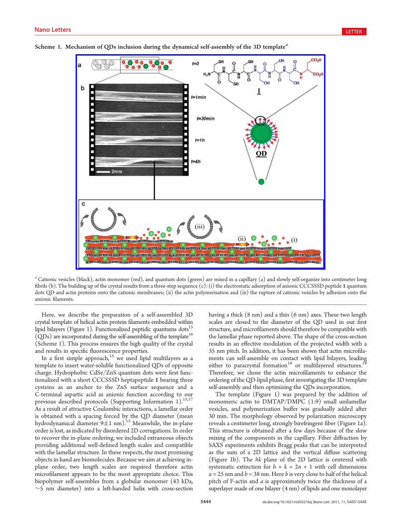
Scheme 1. Mechanism of QDs inclusion during the dynamical self-assembly of the 3D templatea
aCationic vesicles (black), actin monomer (red), and quantum dots (green) are mixed in a capillary (a) and slowly self-organize into centimeter longfibrils (b). The building up of the crystal results from a three-step sequence (c): (i) the electrostatic adsorption of anionic CCCSSSD peptide 1 quantumdots QD and actin proteins onto the cationic membranes; (ii) the actin polymerisation and (iii) the rupture of cationic vesicles by adhesion onto theanionic filaments.
5445 dx.doi.org/10.1021/nl203216q |Nano Lett. 2011, 11, 5443–5448
Nano Letters LETTER
of aligned in phase F-actin microfilaments (8 nm). On the basisof this, a simple and natural structural model is proposed(Figure 1d) where the unit cell perpendicular to the lipid bilayeris composed of two such superlayers, the helical phase of theF-actin microfilaments being π-shifted between adjacent super-layers. The electron density variations along the F-actin micro-filaments are vanishing so that both (1,1) and (0,2) peak intensitiesare very low. The electron density map (Figure 1c) constructedfrom SAXS intensity distributions is in agreement with micro-filament localization and membrane corrugations of our model.High-resolution SAXS profiles (Supporting Information S6)show an additional diffuse scattering peak which is shifted tosmall angles during the lipid chain melting transition from the Lβgel (q = 0.052 Å�1) to the Lα fluid chain state (q = 0.049 Å
�1). Asimilar shift, also observed inDNA/DMTAP/DMPCphases, hasbeen unambiguously attributed to the lateral dilatation of theDNA superlattice.20,21 By analogy, this diffuse scattering is attri-buted to pair interactions between adjacent microfilaments thatcorrespond to a local order along cwithout positional long-rangeorder freeze fracture electron microscopy observations confirmthe above model by showing egg box membrane deformationswith perpendicular directions of undulation and spatial periods of35 and 15 nm (Figure 1f). The former corresponds to an un-dulation of the bilayer imposed by the periodic longitudinalvariation of the apparent thickness of microfilaments (Figure 1e).The latter reflects the local lateral arrangement along c of actinmicrofilament cross sections in the plane perpendicular to actinmicrofilaments (Figure 1e) with the (1,0,1) indexation of thediffuse scattering peak (Figure 1b).
To prepare QD crystals, hydrophilic QDs were added to theactin on contact with lipid biliayers by taking advantage of theslowness of the self-assembling process. The QDs were firstfunctionalized with a negatively charged peptide 115,17 so thatthey can interact with the cationic membranes. The anionic actinproteins (6 nm diameter) and functionalized QDs (8 nm dia-meter) were homogeneously mixed before addition to vesiclessuspension (scheme 1). Since both polyanions (actin and QDs)are attracted to the cationic lipid membranes during the self-assembling of template, one can expect that the insertion of QDsshould not destabilize the 3D organization. Many experimentswere performed varying parameters such as the membrane surfacecharge, the concentration ratio QDs/actin, and the F-buffer con-centration. The best experimental conditions were identified toimprove the reproducibility (10 samples) as followed (Figure 2a):the actin polymerization buffer was not necessary in order to ob-serve actin polymerization as previously observed under cationiclipid monolayers.18 The surface charge has to be increased with alipid mixture DMTAP/DMPC (2:8). The observation of a 3Dorder is very sensitive to a low QDs concentration (1 μmol/L)since an increase to 2.5 μmol/L generates a less ordered phase(Figure 3) composed of QDs chains embedded within lipidbilayers and actin filaments. The formation of actin�QDs�lipidscomplexes required two days and the samples are stable morethan one month at room temperature when they are stored incapped capillaries (Figure 2b).
Regarding their structural analysis, the composite phase con-sists of strongly birefringent macroscopic fibers and exhibits anintense green fluorescence (Figure 2c,d). Powder diffraction obtained
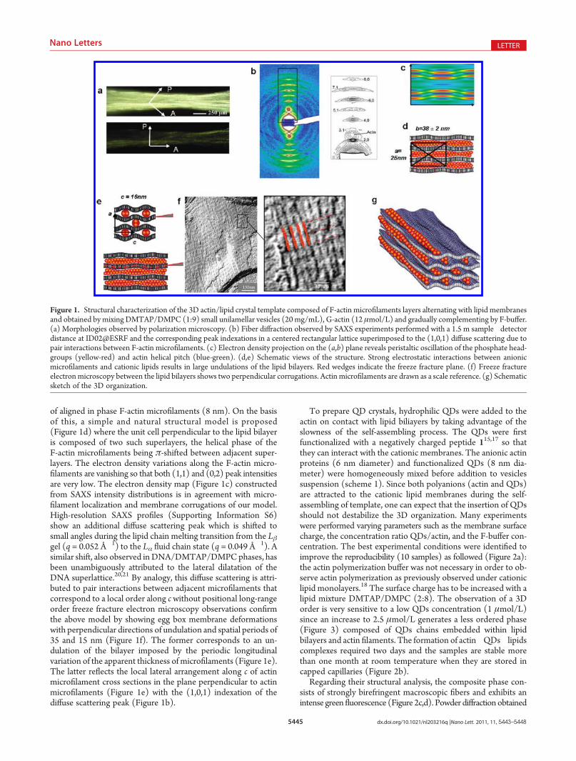
Figure 1. Structural characterization of the 3D actin/lipid crystal template composed of F-actin microfilaments layers alternating with lipid membranesand obtained by mixing DMTAP/DMPC (1:9) small unilamellar vesicles (20 mg/mL), G-actin (12 μmol/L) and gradually complementing by F-buffer.(a) Morphologies observed by polarization microscopy. (b) Fiber diffraction observed by SAXS experiments performed with a 1.5 m sample�detectordistance at ID02@ESRF and the corresponding peak indexations in a centered rectangular lattice superimposed to the (1,0,1) diffuse scattering due topair interactions between F-actin microfilaments. (c) Electron density projection on the (a,b) plane reveals peristaltic oscillation of the phosphate head-groups (yellow-red) and actin helical pitch (blue-green). (d,e) Schematic views of the structure. Strong electrostatic interactions between anionicmicrofilaments and cationic lipids results in large undulations of the lipid bilayers. Red wedges indicate the freeze fracture plane. (f) Freeze fractureelectronmicroscopy between the lipid bilayers shows two perpendicular corrugations. Actin microfilaments are drawn as a scale reference. (g) Schematicsketch of the 3D organization.
5446 dx.doi.org/10.1021/nl203216q |Nano Lett. 2011, 11, 5443–5448
Nano Letters LETTER
by SAXS experiments exhibits Bragg peaks that can be indexed inan orthorhombic lattice (table in Supporting Information S8 andFigure 2e) with the three lattice parameters of a = 35.5 nm, b =26.5 nm, and c = 18.9 nm. Only the (h,k,l) peaks satisfying toh + k + l = 2n (with n an integer) were observed. This is inagreement with a body-centered orthorhombic lattice. These cellparameters are closed from that of the actin/lipid template alonesuggesting that the hybrid QD/actin/lipid organization derivesfrom the template structure. The large changes in the relativepeak intensities confirm that the QD is embedded in the unit cell.Because the QD electron density is much larger than the one oforganic template, the observed X-ray diffraction mainly finds itsorigin from the QD superstructure and gives access to the qualityof the QD crystal. The sharpness of the Bragg peaks demon-strates the long-range positional order of QDs in the crystal(Figure 2e). The hydrophilic QDs can only be located in thechannels delimited by two adjacent lipid membranes and twoneighboring actin filaments. QDs are expected to be located incontact with the thinner part of the channel that is defined by the
actin filament helix (Figure 2f) in order to reduce Coulombrepulsions between anionic QDs and actin. The QDs are notclose to each other in the unit cell because this would produce anadditional diffuse scattering due to pair interactions betweendisordered QDs as observed in the presence of a large excess ofQDs (Figure 3c,d). The QDs localization is confirmed by electrondensity reconstruction (Figure 2g). Complementary freeze frac-ture electron microscopy images (Figure 2h) exhibit only onemode of membrane undulations with a repetition distance ofaround 35( 5 nm, which is induced by the helical modulation ofthe actin filaments along the a direction. The magnitude of themembrane undulation is enhanced by the QDs locations whichare precisely located in the groove of the filaments (Figure 2f).Thus QDs have been incorporated within a template during itsself-assembling process (Scheme 1). The three-dimensionalorder of the template is preserved during the inclusion of theQDs and results in a three-dimensional crystal of QDs (Figure 2i).When the concentration ratio between the QDs and the actin insolution is increased, one can observe a hybrid lamellar phase
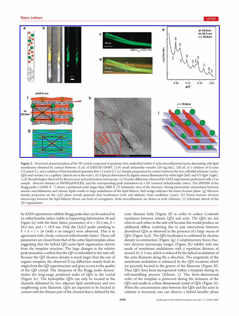
Figure 2. Structural characterization of the 3D crystal composed of quantum-dots embedded within F-actin microfilaments layers alternating with lipidmembranes obtained by contact between 15 μL of DMTAP/DMPC (2:8) small unilamellar vesicles (20 mg/mL), 120 μL of a solution of G-actin(12 μmol/L), and a solution of functionalized quantum dots (1 μmol/L). (a) Sample preparation by contact between the two colloidal solutions (actin/QDs and vesicles) in a capillary (sketch not at the scale). (b) Optical observation by digital camera illuminated by white light (left) and UV light (right).(c,d)Morphologies observed by fluorescence and polarization microscopy. (e) Powder diffraction observed by SAXS experiments performed with a 3msample�detector distance at SWING@SOLEIL and the corresponding peak indexations in a 3D centered orthorhombic lattice. The HWHM of theBragg peaks (<0.004 Å�1) shows a positional order larger than 3000 Å. (f) Schematic view of the structure. Strong electrostatic interactions betweenanionic microfilaments and cationic lipids results in large undulations of the lipid bilayers. Red wedge indicates the freeze fracture plane. (g) Electrondensity projection on the (a,b) plane reveals quantum dots localization (red) and aliphatic chain oscillation (cyan). (h) Freeze fracture electronmicroscopy between the lipid bilayers shows one kind of corrugation. Actin microfilaments are drawn as scale reference. (i) Schematic sketch of the3D organization.
5447 dx.doi.org/10.1021/nl203216q |Nano Lett. 2011, 11, 5443–5448
Nano Letters LETTER
including chains of close-packed QDs forced by the presence ofactin (Figure 3).
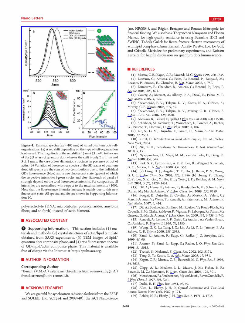
In order to evaluate the relationship between fluorescentproperties and QDs organizations, we investigate fluorescencespectra of different arrays of QDs (Figure 4a). Indeed, fluores-cent properties of dyes are generally related to their aggregationstates.22 In the case of the 3D crystal of QDs, a fluorescence redshift of around 13 nm (53 meV) is observed. This shift is onlyobtained for the 3D crystal organization. The chemical environ-ment composed of actins and lipids cannot explain such a shift.Indeed only a slight red shift of 2 nm (8 eV) was observed forchains of close-packed QDs embedded within membranes andactin filaments (Figure 3). As a complementary control, QDs em-beddedwithin bilayers15 exhibits the same fluorescence spectrumthan a suspension of isolated QDs. In the literature, a fluores-cence red shift observed in linear chains23 or 3D crystals24 ofclose-packed QDs (interdistance below 2 nm) is attributed tofluorescence resonance energy transfer (FRET) between QDs.In the present case, FRET cannot explain the results because theQDs interdistances are larger than 10 nm, which is much higherthan the F€orster radius (5�7 nm).25 Long range interactionsgenerating energy lowering as observed in bacterial photosyn-thetic light harvesting systems26 can also give rise to such a fluo-rescence red shift. Such behavior, called superradiance,27 has beenobserved with QDs with an interdistance as long as 150 nm.8 Totest this hypothesis, fluorescence spectra were recorded indifferent area of the fiber. All spectra exhibit a fluorescenceintensity that is directly related to the fluorescence shift in the 3Dcrystal area (Figure 4b,c). All spectra are the sum of two con-tributions: one due to the isolated QD and the other one due to a
new fluorescent state. The contribution of the second oneincreases linearly with the total intensity of the peak and there-fore can be directly attributed to the emergence of a unique newfluorescent state (Figure 4b,c and Supporting Information S10).This new fluorescence state observed within the 3D array isconsistent with a superradiant emission that is characterized byslight energy loss (<5%) due to the fluorescence wavelength anda large increase (300%) of the fluorescence intensity.28,29 Thelatter result illustrates the possibility to generate new opticalproperties with larger interdistances between QD within anordered assembly.
Bottom-up methods are generally restricted to self-assemblyin solution where the 3D array symmetry is imposed by the shapeand the functionalization of nanoparticles.11 The method re-ported here describes the inclusion of the quantum dots occur-ring during the self-assembling process of the bioorganictemplate. By mixing slowly different bioorganic and inorganicbuilding blocks interacting through electrostatic interaction, wedemonstrate the formation of a very well-defined 3D crystal ofQDs. This strategy gives access to 3D arrays of nanoparticleswhose original low symmetry16 can reveal new physical proper-ties. The fluorescence properties of the 3D crystals of quantumdots demonstrate a direct effect of the nanostructuration of thequantum dots within the crystal. The formation of 3D arrays ofnanoparticles could open new route toward optical materials thatare easily prepared by using self-assembling of vesicles, proteins,and hydrophilic nanoparticles in aqueous environment. Thisstrategy could be extended to any kind of hydrophilic nanopar-ticles with various morphologies. Furthermore, the range ofcharacteristic lengths can be extended by using another biological
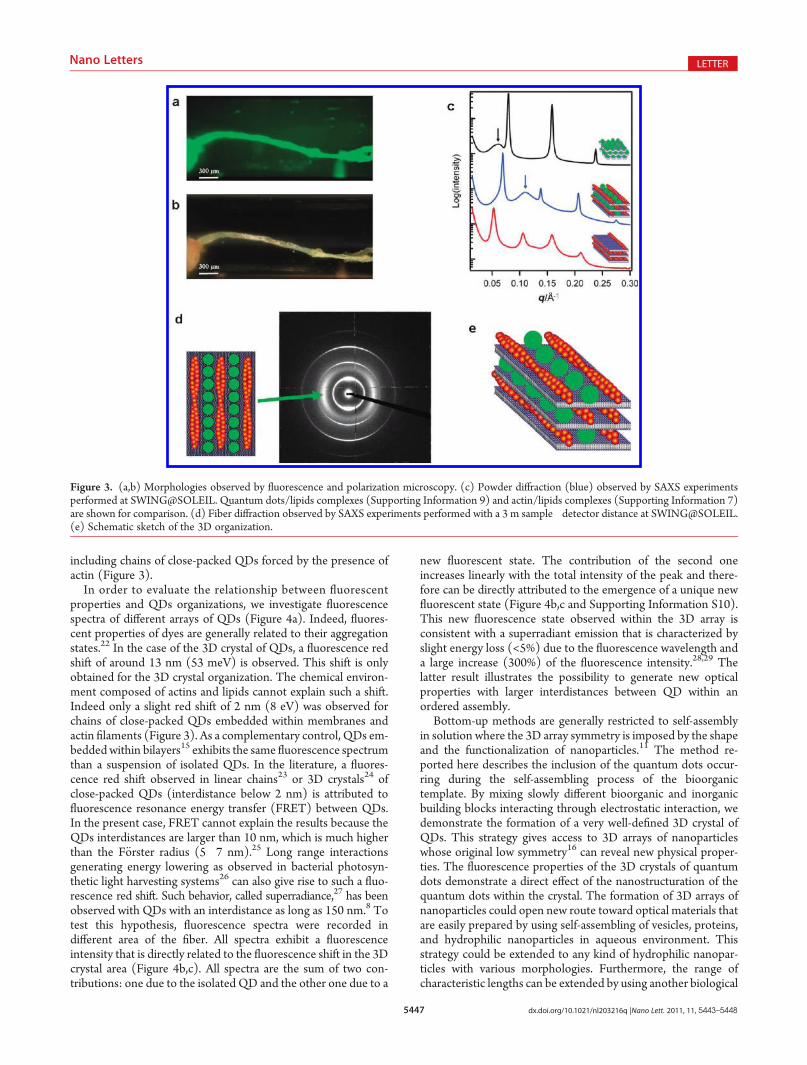
Figure 3. (a,b) Morphologies observed by fluorescence and polarization microscopy. (c) Powder diffraction (blue) observed by SAXS experimentsperformed at SWING@SOLEIL. Quantum dots/lipids complexes (Supporting Information 9) and actin/lipids complexes (Supporting Information 7)are shown for comparison. (d) Fiber diffraction observed by SAXS experiments performed with a 3 m sample�detector distance at SWING@SOLEIL.(e) Schematic sketch of the 3D organization.
5448 dx.doi.org/10.1021/nl203216q |Nano Lett. 2011, 11, 5443–5448
Nano Letters LETTER
polyelectrolyte (DNA, microtubules, polysaccharides, amyloidsfibers, and so forth) instead of actin filament.
’ASSOCIATED CONTENT
bS Supporting Information. This section includes (1) ma-terials and methods, (2) crystal structures of actin/lipid templateobtained from SAXS experiments, (3) TEM images of lipid/quantum dots composite phase, and (4) raw fluorescence spectraof QD/lipid/actin composite phase. This material is availablefree of charge via the Internet at http://pubs.acs.org.
’AUTHOR INFORMATION
Corresponding Author*E-mail: (V.M.-A.) [email protected]; (F.A.)[email protected].
’ACKNOWLEDGMENT
We are grateful for synchrotron radiation facilities from the ESRFand SOLEIL (no. SC2384 and 2008740), the ACI Nanoscience
(no. NR0004), and R�egion Bretagne and Rennes M�etropole forfinancial funding. We also thank Theyencheri Narayanan and FlorianMeneau for high quality assistance in using Beamline ID02 andSWING, Tadeck Gulick for freeze fracture electron microscopy ofactin lipid complexes, Anne Renault, Aur�elie Parette, Loic Le Goff,and Cristelle Meriadec for preliminary experiments, and RobsonFerreira for helpful discussion on quantum dots luminescence.
’REFERENCES
(1) Murray, C. B.; Kagan, C. R.; Bawendi, M. G. Science 1995, 270, 1335.(2) Desvaux, C.; Amiens, C.; Fejes, P.; Renaud, P.; Respaud, M.;
Lecante, P.; Snoeck, E.; Chaudret, B. Nat. Mater. 2005, 4, 750.(3) Dumestre, F.; Chaudret, B.; Amiens, C.; Renaud, P.; Fejes, P.
Science 2004, 303, 821.(4) Courty, A.; Mermet, A.; Albouy, P. A.; Duval, E.; Pileni, M. P.
Nat. Mater. 2005, 4, 395.(5) Shevchenko, E. V.; Talapin, D. V.; Kotov, N. A.; O’Brien, S.;
Murray, C. B. Nature 2006, 439, 55.(6) Shevchenko, E. V.; Talapin, D. V.; Murray, C. B.; O’Brien, S.
J. Am. Chem. Soc. 2006, 128, 3620.(7) Abecassis, B.;Testard, F.; Spalla,O.Phys. Rev. Lett.2008,100, 115504.(8) Scheibner, M.; Schmidt, T.; Worschech, L.; Forchel, A.; Bacher,
G.; Passow, T.; Hommel, D. Nat. Phys. 2007, 3, 106.(9) Lin, S.; Li, M.; Dujardin, E.; Girard, C.; Mann, S. Adv. Mater.
2005, 17, 2553.(10) Kittel, C. Introduction to Solid State Physics, 8th ed.; Wiley:
New York, 2004.(11) Nie, Z. H.; Petukhova, A.; Kumacheva, E. Nat. Nanotechnol.
2010, 5, 15.(12) Nykypanchuk, D.; Maye, M. M.; van der Lelie, D.; Gang, O.
Nature 2008, 451, 549.(13) Park, S. Y.; Lytton-Jean, A. K. R.; Lee, B.; Weigand, S.; Schatz,
G. C.; Mirkin, C. A. Nature 2008, 451, 553.(14) (a) Liang, H. J.; Angelini, T. E.; Ho, J.; Braun, P. V.; Wong,
G. C. L. J. Am. Chem. Soc. 2003, 125, 11786. (b) Huang, Y.; Chiang,C.-Y.; Lee, S. K.; Gao, Y.; Hu, E. L.; Yoreo, J. D.; Belcher, A. M. NanoLett. 2005, 5 (7), 1429–1434.
(15) Dif, A.; Henry, E.; Artzner, F.; Baudy-Floc’h, M.; Schmutz, M.;Dahan, M.; Marchi-Artzner, V. J. Am. Chem. Soc. 2008, 130, 8289.
(16) Pouget, E.; Dujardin, E.; Cavalier, A.; Moreac, A.; Valery, C.;Marchi-Artzner, V.; Weiss, T.; Renault, A.; Paternostre, M.; Artzner, F.Nat. Mater. 2007, 6, 434.
(17) Dif, A.; Boulmedais, F.; Pinot, M.; Roullier, V.; Baudy-Floc’h, M.;Coquelle, F.M.;Clarke, S.;Neveu, P.; Vignaux, F.; Leborgne, R.;Dahan,M.;Gueroui, G.;Marchi-Artzner, V. J. Am. Chem. Soc. 2009, 131, 14738–14746.
(18) Renault, A.; Lenne, P. F.; Zakri, C.; Aradian, A.; Venien-Bryan,C.; Amblard, F. Biophys. J. 1999, 76, 1580.
(19) Wong, G. C. L.; Tang, J. X.; Lin, A.; Li, Y. L.; Janmey, P. A.;Safinya, C. R. Science 2000, 288, 2035.
(20) Zantl, R.; Artzner, F.; Rapp, G.; Radler, J. O. Europhys. Lett.1999, 45, 90.
(21) Artzner, F.; Zantl, R.; Rapp, G.; Radler, J. O. Phys. Rev. Lett.1998, 81, 5015.
(22) Tretiak, S.; Mukamel, S. Chem. Rev. 2002, 102, 3171.(23) Tang, Z. Y.; Kotov, N. A. Adv. Mater. 2005, 17, 951.(24) Kagan, C. R.; Murray, C. B.; Bawendi, M. G. Phys. Rev. B 1996,
54, 8633.(25) Clapp, A. R.; Medintz, I. L.; Mauro, J. M.; Fisher, B. R.;
Bawendi, M. G.; Mattoussi, H. J. Am. Chem. Soc. 2004, 126, 301.(26) Monshouwer, R.;Abrahamsson,M.; vanMourik, F.; vanGrondelle, R.
J. Phys. Chem. B 1997, 101, 7241.(27) Dicke, R. H. Phys. Rev. 1954, 93, 99.(28) Allen, L.; Elerby, J. H. In Optical Resonance and Two-Level
Atoms; Dover: New York, 1987; p 174.(29) Rehler, N. E.; Eberly, J. H. Phys. Rev. A 1971, 3, 1735.
Figure 4. Emission spectra (ex = 405 nm) of varied quantum dots self-organizations. (a) A red shift depending on the type of self-organizationis observed. Themagnitude of the red shift is 13 nm (53meV) in the caseof the 3D arrays of quantum dots whereas the shift is only 2( 1 nm and3 ( 1 nm in the case of low dimension structures in presence or not ofactin. (b) Variation of fluorescence spectra of the 3D arrays of quantumdots. All spectra are the sum of two contributions due to the individualQDs fluorescence (blue) and a new fluorescent state (green) of whichthe respective intensities (green circles and blue diamonds of panel c)strongly depend on the total fluorescence intensity. For comparison, allintensities are normalized with respect to the maximal intensity (100).Note that the fluorescence intensity increase is mainly due to this newfluorescent state. All spectra and fits are shown in Supporting Informa-tion 10.