Embed Size (px)
Citation preview

Crystal Structures of the DNA-binding Domain Tetramer ofthe p53 Tumor Suppressor Family Member p73 Bound toDifferent Full-site Response Elements□S
Received for publication, August 13, 2012, and in revised form, December 3, 2012 Published, JBC Papers in Press, December 14, 2012, DOI 10.1074/jbc.M112.408039
Abdul S. Ethayathulla, H. Thien Nguyen, and Hector Viadiu1
From the Laboratory of Structural Biochemistry, Department of Chemistry and Biochemistry, University of California, San Diego,La Jolla, California 92093
Background: Members of the p53 protein family bind to full-site response elements (REs) to trigger specific cellularpathways.Results:We solved two crystal structures of the p73 DNA-binding domain in complex with full-site REs.Conclusion: Lys-138 in loop L1 distinguishes between consensus REs.Significance: Conformational changes in Lys-138 might explain specificity between cell arrest and apoptosis target genes.
How cells choose between developmental pathways remains afundamental biological question. In the case of the p53 proteinfamily, its three transcription factors (p73, p63, and p53) eachtrigger a gene expression pattern that leads to specific cellularpathways. At the same time, these transcription factors recog-nize the same response element (RE) consensus sequences, andtheir transactivation of target genes overlaps. We aimed tounderstand target gene selectivity at the molecular level bydetermining the crystal structures of the p73 DNA-bindingdomain (DBD) in complex with full-site REs that vary insequence. We report two structures of the p73 DBD bound as atetramer to 20-bp full-site REs based on two distinct quarter-sites: GAACA and GAACC. Our study confirms that the DNA-binding residues are conserved within the p53 family, whereasthe dimerization and tetramerization interfaces diverge. More-over, a conserved lysine residue in loop L1 of theDBD senses thepresence of guanines in positions 2 and 3 of the quarter-site RE,whereas a conserved arginine in loop 3 adapts to changes inposition 5. Sequence variations in the RE elicit a p73 conforma-tional response that might explain target gene specificity.
Differential gene expression is a fundamental biologicalmechanism that underlies cell differentiation and pathologicalmechanisms such as cancer. The p53 family of transcriptionfactors, formed by p73, p63, and p53, can activate �100 genes(1, 2). Each member of the family triggers a unique pattern ofgene expression. For example, p73was initially recognized to beinvolved in brain development and immune systemmaturation(3, 4), p63 in limb and epithelial growth (5, 6), and p53 in cellarrest (7, 8) and apoptosis (9, 10). After the initial discovery ofp73 and p63, transcription activation by the p53 family mem-
bers has been recognized as a complex networkwhere any com-bination of the three transcription factors could trigger theexpression of some target genes (11, 12). For instance, p73and p63 have been found to also have a role in tumor sup-pression by activating DNA repair and apoptosis pathways(13, 14). However, the factors that induce the p53 familymembers to trigger unique or overlapping transcription pat-terns are not understood.The p53 family members share a basic gene structure: an
N-terminal transactivation domain, a central DNA-bindingdomain (DBD),2 and aC-terminal tetramerization domain (15).The longer C terminus in both p73 and p63 includes a SAM(sterile alphamotif) domain, which is absent in the shorter pos-itively charged p53 C terminus. The DBD is a 200-amino acidimmunoglobulin-like domain with two �-sheets packed as a�-sandwich (16). Several structures of the members of the p53transcription factor family in complex with DNA have beensolved (17–27). The DBD binds as a tetramer to response ele-ments (REs), with each monomer recognizing a 5-bp quarter-site of the 20-bp full-site RE. A full-site RE has two half-site REs,where each half-site follows the 5�-PuPuPuC(A/T)(A/T)GPy-PyPy-3� consensus sequence, and in some cases, both half-sitescan be separated by 1–3 nucleotides (1). The DBDs of the p53family members have an amino acid sequence identity of�50%(28). Once the DBD binds to DNA, the N terminus promotesthe recruitment of other factors that eventually lead to the tran-scription of downstream target genes.This work builds upon our previous structural research on
RE spacers that highlights, using oligonucleotides with half-siteREs, how the p73 DBD recognizes REs with spacers of 0, 1, 2,and 4 nucleotides between two half-sites (27). In this study, weanalyze the oligomerization state and DNA affinity of the p73DBD upon binding to full-site REs and present two crystalstructures of the p73DBD tetramer bound to continuous 20-bpfull-site REs. We compare all of the solved structures of p73DBD tetramers in complex with different DNA sequences and
□S This article contains supplemental Figs. S1–S5, Tables S1–S5, and addi-tional references.
The atomic coordinates and structure factors (codes 4G82 and 4G83) have beendeposited in the Protein Data Bank (http://wwpdb.org/).
1 To whom correspondence should be addressed: Lab. of Structural Biochem-istry, Dept. of Chemistry and Biochemistry, University of California, SanDiego, 9500 Gilman Dr., MC 0378, La Jolla, CA 92093. E-mail: [email protected].
2 The abbreviations used are: DBD, DNA-binding domain; RE, response ele-ment; Bistris propane, 1,3-bis[tris(hydroxymethyl)methylamino]propane;r.m.s.d., root mean square deviation(s).
THE JOURNAL OF BIOLOGICAL CHEMISTRY VOL. 288, NO. 7, pp. 4744 –4754, February 15, 2013© 2013 by The American Society for Biochemistry and Molecular Biology, Inc. Published in the U.S.A.
4744 JOURNAL OF BIOLOGICAL CHEMISTRY VOLUME 288 • NUMBER 7 • FEBRUARY 15, 2013
by guest on January 30, 2018http://w
ww
.jbc.org/D
ownloaded from

correlate changes in the RE sequence with changes in the con-formation of the protein that could explain p73 target geneselectivity.
EXPERIMENTAL PROCEDURES
Protein Expression, Purification, and Crystallization—Thehuman p73 DBD (residues 115–312) was purified as describedpreviously (27). Commercially purchased 20-bp oligonucleo-tides were used in crystallization (5�-GAACATGTTCGAA-CATGTTC-3� for the GAACA crystals and 5�-GAACCG-GTTCGAACCGGTTC-3� for the GAACC crystals). Prior tocrystallization, the p73 DBD (20 mg/ml) was mixed with DNA(6mg/ml) at a 1:1molar ratio and incubated for 30min at roomtemperature. Both protein-DNA complexes were crystallizedusing the hanging drop vapor diffusion method at room tem-perature. The GAACA crystals grew in 100 mM trihydratedsodium acetate, 100 mM Tris base (pH 9.0), and 18–20% PEG3350. TheGAACC crystals grew in 100mM trihydrated sodiumacetate, 100 mM Bistris propane (pH 7.5), and 12–15% PEG3350.DataCollection and StructureDetermination—Crystalswere
frozen in liquid nitrogen using 30% PEG 3350 as a cryopro-tectant. Initial crystals that allowed optimization were dif-fracted at beamline BL7-1 of the Stanford Synchrotron Radia-tion Lightsource, allowing optimization. The final data setswere collected at beamline 8.2.2 of the Advanced Light Sourceat the Lawrence BerkeleyNational Laboratory. Crystal data andintensity statistics are given in Table 1. Diffraction data wereindexed, integrated, and scaled using HKL2000 (29). In bothcomplexes, the asymmetric unit consists of two p73 DBDmol-ecules (198 amino acids in each molecule) and half of the pal-indromic 20-bp oligonucleotide used in crystallization (10-bpdsDNA). The calculated VM values are 2.77 Å3/Da for theGAACA crystal and 2.14 Å3/Da for the GAACC crystal, whichcorrespond to solvent contents of 55.6% and 42.3%, respec-tively. The structures were solved by molecular replacementusing Phaser (30). First, we solved the GAACA structure usinga p73 DBD dimer with chains A and B from Protein Data Bank3VD0 Model 1 bound to a modeled 10-bp dsDNA moleculewith the sequence 5�-GAACATGTTC-3� as a search model(27). A unique molecular replacement solution was obtained.The initial solution was refined using rigid body, simulatedannealing, and positional refinement in CNS 1.3 (31). Initialrefinement yielded a clear electron density for the DNA mole-cule, and the 10-bp half-site was built 1 bp at a time. Consider-ing the adjacent symmetry-related molecule, a continuous20-bp DNA is present in the model. Extra steps of positionaland individual B-factor refinement were calculated with CNS1.3 (31). Cycles of manual and then real-space refinement werecarried out for the protein, DNA, and waters with the programCoot 0.6.1 (32). The model was verified using composite omitmaps as implemented in CNS 1.3 (31). For the GAACC struc-ture, the GAACA dimer with DNA was used as a search modelto find a molecular replacement solution. As the crystal dif-fracted only to 4.0 Å, the structure was refined imposing tightnon-crystallographic symmetry constraints between mono-mers A and B to reduce the number of refined parameters.Initial refinement was carried out using CNS 1.3 (31), followed
by TLS refinement with tight non-crystallographic symmetryconstraints between the two protein chains using REFMAC 5.0(33) as implemented in CCP4 (34). The final Rfactor/Rfree statis-tics are 25.0/27.4 for the GAACA structure and 24.4/30.7 forthe GAACC structure. All residues were either in the mostfavored or additionally allowed regions of the Ramachandranplot as evaluated by the programPROCHECK (35). Refinementand stereochemical quality statistics of the final model areshown in Table 1. The analysis of DNA structural parameterswas carried out using the program 3DNA (36). Chimera wasused to calculate structural alignments and root mean squaredeviations (r.m.s.d.) and to generate figures (37).Analytical Ultracentrifugation—Sedimentation velocity ex-
periments were performed and analyzed as described previ-ously (27). A fluorescein-labeled dsDNA (5�-6-FAM-GAA-CATGTTCGAACATGTTC-3�, where 6-FAM is fluoresceinamidite) was used. The data collected were analyzed usingSEDFIT software to calculate sedimentation coefficient distri-butions (38).Fluorescence Anisotropy—For the binding studies, 15 sam-
ples were prepared in a total volume of 500 �l containing purep73 DBD at 1 nM to 8 �M in buffer containing 100 mMNaCl, 10mM sodium citrate (pH 6.1), 5 mM DTT, and 5 �M ZnCl2 inaddition to a constant 10 nM final concentration of 5�-fluores-cein-labeled 20-bp oligonucleotide (5�-FAM-GAACATGTTC-GAACATGTTC-3� or 5�-FAM-GAACCGGTTCGAACCG-GTTC-3�). Measurements and analysis were carried out asdescribed previously (27).
RESULTS
Crystal Structures of the p73 DBD Tetramer Bound to 20-bpFull-site REs—We solved the structures of the p73 DBD boundas a tetramer to two REs (Fig. 1A). The structure of a p73 DBDtetramer in complex with a 20-bp full-site RE with a5�-GAACA-3� quarter-site sequence, hereafter termed theGAACA structure (Protein Data Bank code 4G82), crystallizesin space group P61, and it was solved to a resolution of 3.1 Å(Table 1 and supplemental Fig. 1A). The p73 DBD tetramerstructure bound to a 20-bp full-site RE with a 5�-GAACC-3�quarter-site sequence, hereafter termed the GAACC structure(Protein Data Bank code 4G83), crystallizes in space groupP212121, and it was solved to a resolution of 4.0 Å (Table 1 andsupplemental Fig. S1B). The final maps show electron densityfor all of the p73 DBD residues and DNA base pairs (supple-mental Figs. S2 and S3,A and B). Although both crystals belongto different space groups, they contain a protein dimer and ahalf-site RE in the asymmetric unit. The adjacent molecule inthe crystal, related by translation, completes the p73 DBDtetramer bound to the continuous 20-bp DNA molecule usedduring crystallization (supplemental Figs. S1 and S3, A and B).Structure of the p73 DBD Monomer—The p73 DBD shows
the characteristic immunoglobulin-like �-sandwich also pres-ent in the DBDs of the other twomembers of the p53 transcrip-tion factor family (p63 and p53). The DBD has a �-sandwichfold with two highly twisted �-sheets, one formed by fourstrands (S1, S3, S8, and S5) and the other by five strands (S6, S7,S4, S9, and S10) (Fig. 1B). At one end of the �-sandwich, theshort loops are compacted, whereas at the other end, two large
Crystal Structures of the p73 DBD Bound to Full-site REs
FEBRUARY 15, 2013 • VOLUME 288 • NUMBER 7 JOURNAL OF BIOLOGICAL CHEMISTRY 4745
by guest on January 30, 2018http://w
ww
.jbc.org/D
ownloaded from
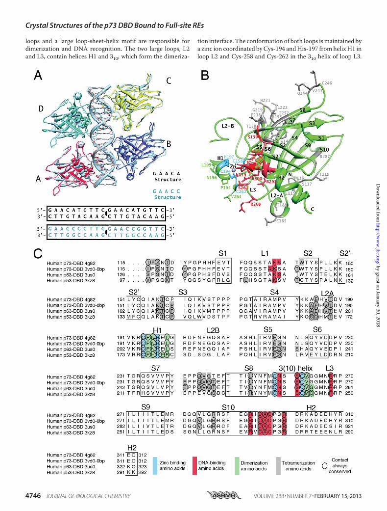
loops and a large loop-sheet-helix motif are responsible fordimerization and DNA recognition. The two large loops, L2and L3, contain helices H1 and 310, which form the dimeriza-
tion interface. The conformation of both loops ismaintained bya zinc ion coordinated byCys-194 andHis-197 fromhelixH1 inloop L2 and Cys-258 and Cys-262 in the 310 helix of loop L3.
Crystal Structures of the p73 DBD Bound to Full-site REs
4746 JOURNAL OF BIOLOGICAL CHEMISTRY VOLUME 288 • NUMBER 7 • FEBRUARY 15, 2013
by guest on January 30, 2018http://w
ww
.jbc.org/D
ownloaded from
The larger motif at that end of the �-sandwich is a loop-sheet-helix motif that positions helix H2 to recognize DNA, togetherwith residues in loop L3. The p73 DBD shares 85% and 58%sequence identity and all of the secondary structure elementswith the p63 and p53 DBDs, respectively (Fig. 1C).Due to crystallographic symmetry, there are four unique p73
DBD monomers in the GAACA and GAACC crystals, two ineach crystal form. All of the unique monomer structures arestill very similar among them, with an r.m.s.d. of 1 or less. Forexample, only three regions differ significantly between mono-mers A and B of the GAACA structure. The N terminus, theloop between strands S3 and S4, and the loop between strandsS7 and S8 all have significantly higher r.m.s.d. values (1.79, 2.84,and 2.12, respectively) than the rest of the structure. Due to thelower resolution of the GAACC crystal, non-crystallographicsymmetry constraints were applied to restrain the monomerstructures to each other and decrease the number of parame-
ters to be refined. In summary, the monomer structure is con-served, and its flexibility is limited to individual side chains orfew secondary structure elements.Structure of the p73 DBD Dimer—The ensuing structural
description of the p73 DBD dimers and tetramers is simplifiedbecause, due to the symmetry present in both crystal forms,monomers A and B are identical tomonomers D andC, respec-tively (Fig. 2A). As a consequence, the dimerization interfacebetween monomers A and B is identical to the one betweenmonomers D and C. Thus, the p73 DBD tetramer bound to a20-bp full-site RE in both crystals is formed by two identicaldimers bound to two identical half-sites.The dimerization interface of the p73DBD tetramer is found
�13 Å above the main DNA axis, allowing the dimer toembrace the DNA. The interface is formed by three residueslocated in helix H1 (Pro-195, Asn-196, and Leu-199) and oneresidue located in the 310 helix found in loop L3 (Val-263) (Fig.
FIGURE 1. Structures of the p73 DBD tetramer in complex with a full-site RE. A, GAACA and GAACC tetramer structures. Monomers A and B in complex withhalf of the 20-bp DNAs used for crystallization form the asymmetric unit. Monomers C and D and the other half of the 20-bp DNA are related by translation andare identical to the content of the asymmetric unit. B, p73 DBD monomer. The secondary structure elements are labeled following the established nomencla-ture for the p53 DBD (17). There are 10 �-strands (S1–S10), two helices (H1 and H2), and three long loops (L1, L2A/L2B, and L3). Residues involved in dimerization(green), tetramerization (gray), zinc binding (blue), and DNA binding (red) are labeled. C, sequence alignment of the DBDs of the three members of the p53protein family. Amino acids forming the secondary structure elements are boxed. Colored boxes indicate the residues involved in dimerization (green), tetramer-ization (gray), zinc binding (blue), and DNA binding (red) for four structures that have a similar tetrameric arrangement (4G82 described in this study and 3VD0Model 1 for the p73 DBD-DNA complex (27), 3US0 for the p63-DNA complex (26), and 3KZ8 for the p53-DNA complex (21)). Residues where the same contactis conserved for all of the monomers of the tetramer are circled.
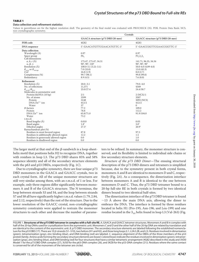
TABLE 1Data collection and refinement statisticsValues in parentheses are for the highest resolution shell. The geometry of the final model was evaluated with PROCHECK (35). PDB, Protein Data Bank; NCS,non-crystallographic symmetry.
CrystalsGAACA structure (p73 DBD/20-mer) GAACC structure (p73 DBD/20-mer)
PDB code 4G82 4G83DNA sequence 5�-GAACATGTTCGAACATGTTC-3� 5�-GAACCGGTTCGAACCGGTTC-3�
Data collectionWavelength (Å) 0.97 0.97Space group P61 P212121Cell dimensionsa, b, c (Å) 175.47, 175.47, 34.31 141.71, 96.35, 34.34�, �, � 90°, 90°, 120° 90°, 90°, 90°
Resolution (Å) 100.0-3.1 (3.21-3.1) 50.0-4.0 (4.09-4.0)Rsym or Rmerge 7.0 (33.0) 13.0 (45.3)I/�I 16.8 (1.9) 8.8 (2.7)Completeness (%) 99.7 (98.1) 99.8 (99.0)Redundancy 8.9 (4.5) 7.6 (8.0)
RefinementResolution (Å) 20.0–3.1 79.0–4.0No. of reflections 11,371 4349Rwork/Rfree 25.0/27.4 24.4/30.7Molecules in asymmetric unitProtein/dsDNA (10 bp) 2/1 2 (NCS)/1No. of atoms 3599 3507Protein 3148 3093 (NCS)DNA/Zn2� ion 412/2 412/2Water 37 0
B-factors 77.2 109.0Protein 78.8 112.2DNA/Zn2� ion 65.8/71.0 91.9/104.0Water 73.2
r.m.s.d.Bond lengths (Å) 0.010 0.03Bond angles 1.8° 3.0°Dihedral angles 27.3° 20.9°
Ramachandran plot (%)Residues in most favored region 87.8 97.5Residues in additionally allowed region 12.2 1.5Residues in generously allowed region 0.0 1.0Residues in disallowed region 0.0 0.0
Crystal Structures of the p73 DBD Bound to Full-site REs
FEBRUARY 15, 2013 • VOLUME 288 • NUMBER 7 JOURNAL OF BIOLOGICAL CHEMISTRY 4747
by guest on January 30, 2018http://w
ww
.jbc.org/D
ownloaded from

2B). The dimerization interface has a surface area of 225Å2, andit is formed by two helices, helix H1 from each monomer. It isstabilized by hydrophobic interactions and two hydrogenbonds. The OD1 of Asn-196 in monomer B interacts with theamide nitrogen of Asn-196 in monomer A, and the ND2 ofAsn-196 in monomer B interacts with the carbonyl oxygen ofVal-263 inmonomerA.There are also numerous van derWaalsinteractions between the carbon atoms of the four residues thatstabilize the dimer interface, most notably between the sidechains of both Leu-199 residues and the intertwining betweenthe side chains of Pro-195 and Asn-196 from both monomers.The zinc-binding residues in helix H1 and loop L3 are adjacentto the residues involved in dimerization. Thus, the bound zinc
atom is essential tomaintain the structure that allows dimeriza-tion of p73 DBD.Structure of the p73 DBD Tetramer—As observed in the pre-
viously solved structures of the p73DBD in complexwithDNA,the p73 DBD binds as a tetramer to its REs. In this work, wereport two structures of the p73 DBD tetramer bound to twodistinct 20-bp full-site REs that are continuousDNAmolecules.Previous p73DBD structures had been solvedwith half-site REsthat were only assembled as a full-site RE in the crystal by thestacking of two adjacentDNAmolecules (27). TheDNA-boundp73 DBD tetramer is stabilized by dimer-dimer contactsformed between two interfaces. The AD tetramerization inter-face is stabilized by contacts between monomers A and D, and
FIGURE 2. Dimerization and tetramerization interfaces of the p73 DBD tetramer bound to a full-site RE. A, surface area model of the protein tetramershowing the two dimerization interfaces (AB and DC) and the two tetramerization interfaces (AD and CB). B, the DC (green/yellow) and AB (red/blue) dimeriza-tion interfaces are identical due to crystal symmetry. The same two secondary structure elements, helix H2 and loop L3, are involved in the DC and ABdimerization interfaces. Three of the four residues that coordinate the zinc ion are displayed (Cys-194, His-197, and Cys-262), together with the four residuesinvolved in the dimerization (Pro-195, Asn-196, Leu-199, and Val-263). C, atomic details of the AD tetramerization interface. Residues in the N terminus and loopL2A of monomer A (red) contact residues in the S7–S8 loop and strands S3, S5, and S8 of monomer D (green). D, atomic details of the CB tetramerizationinterface. Residues in the N terminus and loop L2A of monomer C (yellow) contact residues in the S7–S8 loop and strands S2, S3, S5, and S8 of monomer B (blue).
Crystal Structures of the p73 DBD Bound to Full-site REs
4748 JOURNAL OF BIOLOGICAL CHEMISTRY VOLUME 288 • NUMBER 7 • FEBRUARY 15, 2013
by guest on January 30, 2018http://w
ww
.jbc.org/D
ownloaded from
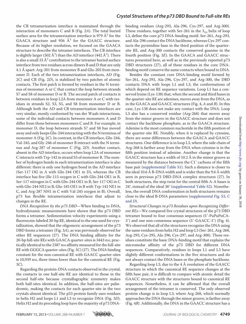
the CB tetramerization interface is maintained through theinteraction of monomers C and B (Fig. 2A). The total buriedsurface area for the tetramerization interface is 979 Å2 for theGAACA structure and 956 Å2 for the GAACC structure.Because of its higher resolution, we focused on the GAACAstructure to describe the tetramer interfaces. The CB interfaceis slightly larger (502 Å2) than the AD interface (442 Å2). Thereis also a small 33 Å2 contribution to the tetramer buried surfaceinterface from two residues across dimers B andD that are only4.1 Å apart: Arg-201 frommonomer B and Glu-205 frommon-omer D. Each of the two tetramerization interfaces, AD (Fig.2C) and CB (Fig. 2D), is stabilized by two patches of atomiccontacts. The first patch is formed by residues in the N termi-nus of monomer A or C that contact the loop between strandsS7 and S8 of monomer D or B. The second patch of contacts isbetween residues in loop L2A from monomer A or C with res-idues in strands S2, S3, S5, and S8 from monomer D or B.Although both the AD and CB tetramerization interfaces arevery similar, mostly conformed by van der Waals interactions,some of the individual contacts between monomers A and Ddiffer from those between monomers C and B. For example, inmonomer D, the loop between strands S7 and S8 has movedaway andonly keepsGln-244 interactingwith theN terminus ofmonomer A (Fig. 2C). In contrast, in the CB interface, Gln-244,Val-245, and Gly-246 of monomer B interact with the N termi-nus and Arg-287 of monomer C (Fig. 2D). Another contact,absent in the AD interface, occurs when loop L2A of monomerC interacts with Trp-142 in strand S3 ofmonomer B. The num-ber of hydrogen bonds in each tetramerization interface is alsodifferent: there is only one hydrogen bond in the AD interface(Ser-117 OG in A with Gln-244 OE1 in D), whereas the CBinterface has five (Ile-115 oxygen in C with Gln-244 OE1 in B,Ser-117 nitrogen in C with Gln-244 OE1 in B, Ser-117 OG in Cwith Gln-244 NE2 in B, Glu-185 OE1 in B with Trp-142 NE1 inC, and Arg-287 NH1 in C with Val-245 oxygen in B). Overall,p73 has flexible tetramerization interfaces that adjust tochanges in the RE.DNA Recognition by the p73 DBD—When binding to DNA,
hydrodynamic measurements have shown that the p73 DBDforms a tetramer. Sedimentation velocity experiments using afluorescein-labeled 20-bp RE, identical to the one used for crys-tallization, showed that the oligomeric arrangement of the p73DBD forms a tetramer (Fig. 3A), as was previously observed forother RE sequences (27). The DNA binding affinity for the20-bp full-site REs with GAACAquarter-sites is 3443 nM, prac-tically identical to the 2387 nM affinitymeasured for the full-siteREwithGGGCAquarter-sites (Fig. 3C) (27). TheDNAbindingconstant for the non-canonical RE with GAACC quarter-sitesis 10,939 nM, three times lower than for the canonical RE (Fig.3C).Regarding the protein-DNA contacts observed in the crystal,
the contacts in one half-site RE are identical to those in thesecond half-site because crystallographic symmetry makesboth half-sites identical. In addition, the half-sites are palin-dromic, making the contacts for each quarter-site in the twocrystals almost identical. The p53 protein family uses residuesin helix H2 and loops L1 and L3 to recognize DNA (Fig. 3D).Helix H2 and its preceding loop have themajority of p73 DNA-
binding residues (Arg-293, Ala-296, Cys-297, and Arg-300).These residues, together with Ser-261 in the 310 helix of loopL3, define the core p73 DNA-binding motif. Ser-261, Arg-293,andAla-296 contact theDNAbackbone, whereas Cys-297 con-tacts the pyrimidine base in the third position of the quarter-site RE, and Arg-300 contacts the conserved guanine in thefourth position (Fig. 3E). In the GAACA and GAACC struc-tures presented here, as well as in the previously reported p73DBD structures (27), all of these residues in the core DNA-bindingmotif conserve the same contacts when bound toDNA.Besides the constant core DNA-binding motif formed by
Ser-261, Arg-293, Ala-296, Cys-297, and Arg-300, the DBDcontacts DNA with loops L1 and L3, the conformations ofwhich depend on RE sequence variations. Loop L1 has a con-served lysine (Lys-138) that, when the second and third bases inthe quarter-site RE are adenines, moves away from theDNA, asin the GAACA and GAACC structures (Fig. 4, A and B). In thiscase, Lys-138 does not make any contact with the DNA. LoopL3 also has a conserved residue (Arg-268) that moves awayfrom the minor groove in the GAACC structure and does notcontact the backbone phosphate, as in the GAACA structure.Adenine is the most common nucleotide in the fifth position ofthe quarter-site RE. Notably, when it is replaced by cytosine,there are some differences between the GAACA and GAACCstructures. One difference is in loop L3, where the side chain ofArg-268 is farther away from the DNA when cytosine is in thefifth position instead of adenine. Another change is that theGAACC structure has a width of 10.2 Å in the minor groove asmeasured by the distance between the C1� carbons of the fifthbase pair (supplemental Table S1). Such a distance is closer tothe ideal 10.6 Å B-DNAwidth and is wider than the 9.6 Åwidthseen in previous p73 DBD-DNA complex structures (27). Inboth structures, the step twist for the central fifth A-A bases is24°, instead of the ideal 36° (supplemental Table S2). Nonethe-less, the overall DNA conformation in both structures remainsclose to the ideal B-DNA parameters (supplemental Fig. S3, Cand D).Structural Changes in p73 Residues upon Recognizing Differ-
ent REs—We compared five crystal structures of the p73 DBDtetramer bound to four consensus sequences (5�-PuPuPuCA-3�) and one non-consensus sequence (5�-GAACC-3�) (Fig. 4).We observed that all of the structures recognize the DNAusingthe same residues fromhelixH2 and loop L3 (Ser-261, Arg-268,Arg-293, Cys-295, Ala-296, Cys-297, and Arg-300). These res-idues constitute the basic DNA-binding motif that explains themicromolar affinity of the p73 DBD for different DNAsequences. Comparatively, residues in loops L1 and L3 haveslightly different conformations in the five structures and donot always contact the DNA bases or the phosphate backbone.Regarding loop L3, due to the 4 Å resolution of the GAACC
structure in which the canonical RE sequence changes at thefifth base pair, it is difficult to compare with atomic detail theGAACC structure with the structures bound to canonical REsequences. Nonetheless, it can be affirmed that the overallarrangement of the tetramer is conserved. The only observedprotein change is in loop L3, where Arg-268, which normallyapproaches the DNA through theminor groove, is farther away(Fig. 4B). Additionally, the DNA in the GAACC structure has a
Crystal Structures of the p73 DBD Bound to Full-site REs
FEBRUARY 15, 2013 • VOLUME 288 • NUMBER 7 JOURNAL OF BIOLOGICAL CHEMISTRY 4749
by guest on January 30, 2018http://w
ww
.jbc.org/D
ownloaded from

FIGURE 3. Binding of the p73 DBD to a full-site RE. A, sedimentation coefficient distribution of the p73 DBD bound to a 20-bp DNA containing the samefull-site RE used in crystallization experiments obtained in a sedimentation velocity experiment. B, binding affinity constant of the p73 DBD measured byfluorescence anisotropy using fluorescein-labeled DNA for the full-site RE used in crystallization of the GAACA structure. C, binding affinity constant of the p73DBD measured by fluorescence anisotropy using fluorescein-labeled DNA for the full-site RE used in crystallization of the GAACC structure. D, stereo view of theresidues of monomer A from the GAACA crystal structure that have been described as contacting DNA (27). E, schematic diagram of the atomic interactionsbetween the p73 DBD and a quarter-site RE of the GAACA structure.
Crystal Structures of the p73 DBD Bound to Full-site REs
4750 JOURNAL OF BIOLOGICAL CHEMISTRY VOLUME 288 • NUMBER 7 • FEBRUARY 15, 2013
by guest on January 30, 2018http://w
ww
.jbc.org/D
ownloaded from
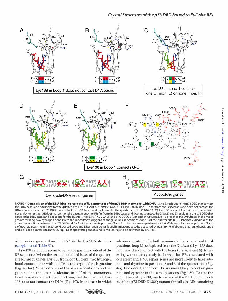
wider minor groove than the DNA in the GAACA structure(supplemental Table S1).Lys-138 in loop L1 seems to sense the guanine content of the
RE sequence. When the second and third bases of the quarter-site RE are guanines, Lys-138 from loop L1 forms two hydrogenbond contacts, one with the O6 keto oxygen of each guanine(Fig. 4,D–F). When only one of the bases in positions 2 and 3 isguanine and the other is adenine, in half of the monomers,Lys-138 makes contacts with the bases, and the other half, Lys-138 does not contact the DNA (Fig. 4C). In the case in which
adenines substitute for both guanines in the second and thirdpositions, loop L1 is displaced from theDNA, and Lys-138 doesnot make direct contact with the bases (Fig. 4, A and B). Inter-estingly, microarray analysis showed that REs associated withcell arrest and DNA repair genes are more likely to have ade-nine and thymine in positions 2 and 3 of the quarter-site (Fig.4G). In contrast, apoptotic REs are more likely to contain gua-nine and cytosine in the same positions (Fig. 4H). To test theimportance of Lys-138, we characterized theDNAbinding abil-ity of the p73 DBD K138Q mutant for full-site REs containing
FIGURE 4. Comparison of the DNA-binding residues of five structures of the p73 DBD in complex with DNA. A and B, residues in the p73 DBD that contactthe DNA bases and backbone for the quarter-site REs (5�-GAACA-3� and 5�-GAACC-3�). Lys-138 in loop L1 is far from the DNA bases and does not contact theDNA. C, residues in the p73 DBD that contact the DNA bases and backbone for the quarter-site RE (5�-GGACA-3�). Lys-138 in loop L1 acquires two conforma-tions. Monomer (mon.) E does not contact the bases; monomer F is far from the DNA bases and does not contact the DNA. D and E, residues in the p73 DBD thatcontact the DNA bases and backbone for the quarter-site REs (5�-AGGCA-3� and 5�-GGGCC-3�). In both structures, Lys-138 reaches the DNA bases in the majorgroove forming two hydrogen bonds with the O2 carbonyl oxygens of the guanines in positions 2 and 3 of the quarter-site RE. F, schematic diagram of theatomic interactions between the p73 DBD and DNA with guanines in positions 2 and 3 of the consensus quarter-site RE. G, WebLogo diagram of positions 2 and3 of each quarter-site in the 20-bp REs of cell cycle and DNA repair genes found in microarrays to be activated by p73 (39). H, WebLogo diagram of positions 2and 3 of each quarter-site in the 20-bp REs of apoptotic genes found in microarrays to be activated by p73 (39).
Crystal Structures of the p73 DBD Bound to Full-site REs
FEBRUARY 15, 2013 • VOLUME 288 • NUMBER 7 JOURNAL OF BIOLOGICAL CHEMISTRY 4751
by guest on January 30, 2018http://w
ww
.jbc.org/D
ownloaded from

theGGGCA andGAACAquarter-site sequences.We observeda drastic drop of at least 100-fold in DNA binding affinity forboth sequences (supplemental Fig. S4). In sum, the p73 DBDhas a stable core of DNA-binding residues (Ser-261, Arg-293,Ala-296, Cys-297, and Arg-300), plus Lys-138 in loop L1 andArg-268 in loop L3, that adjust their interactions with DNAdepending on the RE sequence.
DISCUSSION
The p53 family of transcription factors triggers the expres-sion of �100 genes that lead cells to diverse pathways (3).Recent experiments revealed that target gene transcription forthe three members often overlaps, and the three factors can actas tumor suppressor genes, triggering cell arrest and apoptosis(13, 14). Microarray analysis of gene expression in mouseembryonic fibroblasts identified 620 genes regulated by amem-ber of the p53 transcription family (39). Of this total, 86 geneswere regulated at the same time by p73, p63, and p53; 131 geneswere uniquely controlled by p73; 58 genes were regulated byp73 and p63; and 41 genes were activated by both p73 and p53.How the p53 family members distinguish between sequencesthat match the RE consensus sequence but trigger differentcellular pathways remains unexplained.In this work, we studied how the transcription factor p73
recognizes a full-site RE and how it distinguishes between dif-ferent RE consensus sequences. We have described the oligo-merization state, DNA binding affinities, and crystal structuresof the p73DBDbound to two different full-site REs. The data inthis study, in conjunction with previous results, show p73 DBDflexible dimerization and tetramerization interfaces and loopsL1 and L3 adjusting their conformation to nucleotide changesin theRE sequence. Such structural adjustmentsmight underliedifferences in the transactivation level of p73 target genes.Flexibility in p73 DBD Quaternary Structure—The solved
p73 DBD-DNA complex structures show some variation intheir dimerization and tetramerization interfaces (Fig. 2).Among all of the previously reported p73 DBD tetramer struc-tures bound to different spacers, the comparisons between theGAACA and GAACC structures and the 0-bp AGGCA struc-ture (ProteinData Bank 3VD0Model 1) yield the lowest r.m.s.d.(1.25) (supplemental Fig. S5A and Table S3) (27). The totaldimerization surface for the GAACA, GAACC, and 0-bpAGGCA structures is between 210 and 225 Å2. Four residues(Pro-195, Asn-196, Leu-199, and Val-263) form the dimeriza-tion interface for the two identical dimerization interfaces ofthe tetramer bound to the 5�-GAACA-3� sequence (Fig. 2B).Comparatively, in the 0-bp AGGCA structure, one dimeriza-tion interface is formed by the same four residues as in theGAACA structure, whereas the second dimer loses the Pro-195and Val-263 contacts, resulting in an 8° rotation of helix H1.When Lys-138 in loop L1 and Arg-268 in loop L3 contact theDNA, the angle between the H1 helices is broader, and Pro-195and Val-263 frommonomers B and D do not participate in thedimerization interface. Therefore, we suggest that DNA recog-nition is paired with the angle kept by the dimerization helices.By comparing the structures of the p73 DBD bound to an REwith different sequences, we can begin to understand howDNArecognition affects dimerization.
Once DNA recognition influences dimerization, the quater-nary movements of the dimer also lead to changes in thetetramer. The tetramerization interfaces in the p73 DBD aremore variable and extended than the dimerization interfaces.The GAACA structure presents two tetramerization interfacesthat are distinct from each other and, at the same time, distinctfrom the interfaces that are observed in the 0-bpAGGCAstruc-ture (Fig. 2). There are seven residues that are conserved in thetetramerization interfaces of both structures: Ile-115, Ser-117,Glu-185, and Thr-188 from monomers A and C and Thr-158,Gln-244, and Leu-253 from monomers B and D. Nonetheless,minor changes in the dimerization interface lead to largermovements of some of the secondary structures involved intetramerization that consequently rearrange some of the inter-face contacts. The tetramerization buried surface area is largerfor the 0-bp AGGCA structure (a total of 1186 Å2, with 561 Å2
for AD, 608 Å2 for CB, and 17 Å2 for BD) than for the GAACAstructure (a total of 977 Å2, with 442 Å2 for AD, 502 Å2 for CB,and 33 Å2 for BD) (supplemental Table S3) (27). The main dif-ferences are that, in theGAACAstructure, residues in loopL2A(Ala-184 and Val-187) and strand S10 (Val-284) of monomersA and C lose contacts with residues in loop L1 (Thr-141) andthe loop between strands S7 and S8 (Thr-247) of monomers Band D.Comparison with Structures of the p53 Transcription Factor
Family—Several structures of members of the p53 transcrip-tion factor family in complex with DNA have been solved (sup-plemental Table S3) (17–27). A common denominator of all ofthe tetramer structures is the arrangement of the tetramer as adimer of dimers. A 2-fold rotational symmetry axis runningperpendicular to the center of the DNA axis relates one dimerwith the other, resulting in a C2 molecular symmetry. Thestructural alignment of all of the solved structures shows thattetrameric arrangements vary broadly (supplemental Table S3).In comparing theGAACA structurewith the structures of its
close homolog p63 bound to DNA, we found that the p63 DBDtetramer with a 0-bp spacer (Protein Data Bank code 3US0) hasthe most similar oligomerization to the GAACA structure. Inthe 3US0 structure, the asymmetric unit contains a tetramerseparated by a 2-bp spacer, but considering a symmetry-relateddimer, the p63 DBD forms a 0-bp spacer tetramer that we usedfor our comparison (supplemental Fig. S5B) (26). The overallr.m.s.d. between the p73DBD tetramer and the 0-bp spacer p63DBD tetramer is 2.31 (4G82 versus 3US0 for the 426 atomswithin a 5.0 Å distance cutoff), where three of the four mono-mers superimpose with an r.m.s.d. of 2 or more (supplementalFig. S5B and Table S3). The 0-bp spacer p63 DBD structureresembles evenmore the 0-bpp73DBDstructure (3VD0Model1), with an overall r.m.s.d. of 0.93 (for the 724 atomswithin a 5.0Å distance cutoff) (27). All of the residues in the oligomeriza-tion interfaces are conserved between p73 and p63, except fortwo residues (Ile-115 and Asn-221) in p73 that become Ser-126and Ser-232 in the tetramerization interface of p63 (Fig. 1C). Inconclusion, the dimerization and tetramerization interfaces ofthe DBDs of p73 and p63 are almost identical.Finally, we compared the GAACA tetramer with the p53
DBD tetramer bound to a full-site RE (Protein Data Bank code3KZ8). This is the p53 DBD tetramer structure with the lowest
Crystal Structures of the p73 DBD Bound to Full-site REs
4752 JOURNAL OF BIOLOGICAL CHEMISTRY VOLUME 288 • NUMBER 7 • FEBRUARY 15, 2013
by guest on January 30, 2018http://w
ww
.jbc.org/D
ownloaded from
r.m.s.d. for the two p73 DBD tetramers presented here (supple-mental Table S3) (21). Although the p53 DBD tetramer closelysuperimposes with the GAACA and GAACC structures (sup-plemental Fig. S5C), p53 has very different dimerization andtetramerization interfaces compared with p73 and p63. Theonly p73 dimerization residues that are conserved in p53 arePro-177 and Gly-244. His-178, Arg-181, and Met-243 in p53replace Asn-196, Leu-199, and Val-263 in p73. Glu-180 in p53makes a salt bridge contact with Arg-181 of the opposite mon-omer, but the arginine is absent in p73 and p63. As a result, p53has a significantly larger dimerization interface (453 Å2/dimer)compared with p73 (210–225 Å2). Regarding the tetrameriza-tion interface, the buried surface in p53 is smaller than that inp73 (745 versus 979Å2). In brief, p53 has conserved the fold andDNA recognition of its homologs p73 and p63, but thedimerization and tetramerization interfaces have diverged.In summary, the p73, p63, and p53 tetramers discussed here
retain a similar quaternary organization (supplemental Fig.S5D) and an identical core DNA recognition motif formed byresidues in helix H2 and loop L3 (Ser-261, Arg-268, Arg-293,Cys-295, Ala-296, Cys-297, and Arg-300 in p73 or their con-served equivalent residues in p63 and p53) (Fig. 1C). On theother hand, structural differences could be linked to the abilityto form a correct scaffold to bind other transcription factorsand influence transactivation activity. For example, in p73, con-formational movements of Lys-138 in loop L1 (Lys-149 in p63or Lys-120 in p53) or Arg-268 in loop L3 (Arg-279 in p63 orArg-248 in p53) might induce the tetramer to acquire the cor-rect scaffold structure to recruit other factors.Lys-138Conformation Switches betweenCell Arrest andApo-
ptotic REs—The most interesting aspect of our work is that theLys-138 conformation adapts differently to the sequence inpositions 2 and 3 of each quarter-site RE (Fig. 4). Moreover, theguanine content in such positions is low for cell arrest andDNArepair genes and high for apoptosis genes, as identified inmicroarrays assays (Fig. 4, G–H, and supplemental Tables S4and S5). One can speculate on the role of Lys-138 in determin-ing the target gene specificity of p73. In the case of p53, acety-lation of Lys-120 by acetyltransferases of the MYST family hasbeen shown to be important to distinguish between the apopto-tic cell response and cell arrest (40, 41). Moreover, biochemi-cally, it has been demonstrated that Lys-120 is important forDNA binding and that its acetylation increases the DNA spec-ificity of p53 (42–44). In the case of p73, Lys-138 (where weobserved different conformations depending on the REsequence) is the conserved equivalent residue of Lys-120 in p53.Considering the importance of Lys-120 acetylation in p53 andthe structural differences seen for Lys-138 in p73 DBD-DNAcomplexes, the existing data suggest that the potential Lys-138acetylation could influence how p73 distinguishes between REsthat trigger different cellular responses. A potential modelwould require loop L1 to be detached from the DNA to activatetranscription, initially favoring transcription of genes with ade-nines in positions 2 and 3. In the case of apoptotic genes withguanines in positions 2 and 3, which trap loop L1 conformationby binding to Lys-138, lysine acetylation would be required torelease loop L1.
Despite extensive research on DNA recognition and targetgene specificity of the p53 protein family, fundamental ques-tions about target gene selection remain to be answered. Targetgene selectivity is a complex process involving many proteinsand steps of regulation. The interaction of transcription factorswith REs is only the first step leading to the transcription of agene. Here, we have provided structural evidence of conforma-tional changes that occur when the p73 DBD distinguishesbetween closely related REs. Clearly, much work remains to becompleted to decipher the structural basis of target geneselectivity.
Acknowledgments—Preliminary crystals were diffracted at beamlineBL7-1 of the Stanford Synchrotron Radiation Lightsource (supportedby the United States Department of Energy and National Institutes ofHealth Grant P41RR001209), and diffraction data were collected atbeamline 8.2.2 of the Advanced Light Source at the Lawrence BerkeleyNational Laboratory (supported by Grant DE-AC02-05CH11231from the Director, Basic Energy Sciences, Office of Science, UnitedStates Department of Energy).
REFERENCES1. Riley, T., Sontag, E., Chen, P., and Levine, A. (2008) Transcriptional con-
trol of human p53-regulated genes. Nat. Rev. Mol. Cell Biol. 9, 402–4122. Menendez, D., Inga, A., and Resnick,M.A. (2009) The expanding universe
of p53 targets. Nat. Rev. Cancer 9, 724–7373. Yang, A., Walker, N., Bronson, R., Kaghad, M., Oosterwegel, M., Bonnin,
J., Vagner, C., Bonnet,H., Dikkes, P., Sharpe, A.,McKeon, F., andCaput, D.(2000) p73-deficient mice have neurological, pheromonal and inflamma-tory defects but lack spontaneous tumours. Nature 404, 99–103
4. Kommagani, R., Whitlatch, A., Leonard, M. K., and Kadakia, M. P. (2010)p73 is essential for vitamin D-mediated osteoblastic differentiation. CellDeath Differ. 17, 398–407
5. Mills, A. A., Zheng, B., Wang, X.-J., Vogel, H., Roop, D. R., and Bradley, A.(1999) p63 is a p53 homologue required for limb and epidermal morpho-genesis. Nature 398, 708–713
6. Yang, A., Schweitzer, R., Sun, D., Kaghad, M., Walker, N., Bronson, R. T.,Tabin, C., Sharpe, A., Caput, D., Crum, C., and McKeon, F. (1999) p63 isessential for regenerative proliferation in limb, craniofacial and epithelialdevelopment. Nature 398, 714–718
7. Scheffner, M.,Werness, B. A., Huibregtse, J. M., Levine, A. J., and Howley,P.M. (1990) The E6 oncoprotein encoded by human papillomavirus types16 and 18 promotes the degradation of p53. Cell 63, 1129–1136
8. Michalovitz, D., Halevy, O., andOren,M. (1990) Conditional inhibition oftransformation and of cell proliferation by a temperature-sensitivemutantof p53. Cell 62, 671–680
9. Yonish-Rouach, E., Resnitzky, D., Lotem, J., Sachs, L., Kimchi, A., andOren, M. (1991) Wild-type p53 induces apoptosis of myeloid leukaemiccells that is inhibited by interleukin-6. Nature 352, 345–347
10. Shaw, P., Bovey, R., Tardy, S., Sahli, R., Sordat, B., and Costa, J. (1992)Induction of apoptosis by wild-type p53 in a human colon tumor-derivedcell line. Proc. Natl. Acad. Sci. U.S.A. 89, 4495–4499
11. Levrero, M., De Laurenzi, V., Costanzo, A., Gong, J., Wang, J. Y., andMelino, G. (2000) The p53/p63/p73 family of transcription factors: over-lapping and distinct functions. J. Cell Sci. 113, 1661–1670
12. Yang, A., Kaghad, M., and Caput, D. (2002) On the shoulders of giants:p63, p73 and the rise of p53. Trends Genet. 18, 90–95
13. Flores, E. R., Tsai, K. Y., Crowley, D., Sengupta, S., Yang, A., McKeon, F.,and Jacks, T. (2002) p63 and p73 are required for p53-dependent apoptosisin response to DNA damage. Nature 416, 560–564
14. Flores, E. R., Sengupta, S.,Miller, J. B., Newman, J. J., Bronson, R., Crowley,D., Yang, A., McKeon, F., and Jacks, T. (2005) Tumor predisposition inmice mutant for p63 and p73: evidence for broader tumor suppressorfunctions for the p53 family. Cancer Cell 7, 363–373
Crystal Structures of the p73 DBD Bound to Full-site REs
FEBRUARY 15, 2013 • VOLUME 288 • NUMBER 7 JOURNAL OF BIOLOGICAL CHEMISTRY 4753
by guest on January 30, 2018http://w
ww
.jbc.org/D
ownloaded from
15. Arrowsmith, C. H. (1999) Structure and function in the p53 family. CellDeath Differ. 6, 1169–1173
16. Joerger, A. C., and Fersht, A. R. (2008) Structural biology of the tumorsuppressor p53. Annu. Rev. Biochem. 77, 557–582
17. Cho, Y., Gorina, S., Jeffrey, P. D., and Pavletich, N. P. (1994) Crystal struc-ture of a p53 tumor suppressor-DNA complex: understanding tumori-genic mutations. Science 265, 346–355
18. Kitayner, M., Rozenberg, H., Kessler, N., Rabinovich, D., Shaulov, L., Ha-ran, T. E., and Shakked, Z. (2006) Structural basis of DNA recognition byp53 tetramers.Mol. Cell 22, 741–753
19. Ho, W. C., Fitzgerald, M. X., and Marmorstein, R. (2006) Structure of thep53 core domain dimer bound to DNA. J. Biol. Chem. 281, 20494–20502
20. Malecka, K. A., Ho,W.C., andMarmorstein, R. (2009) Crystal structure ofa p53 core tetramer bound to DNA. Oncogene 28, 325–333
21. Kitayner, M., Rozenberg, H., Rohs, R., Suad, O., Rabinovich, D., Honig, B.,and Shakked, Z. (2010) Diversity in DNA recognition by p53 revealed bycrystal structures with Hoogsteen base pairs. Nat. Struct. Mol. Biol. 17,423–429
22. Chen, Y., Dey, R., and Chen, L. (2010) Crystal structure of the p53 coredomain bound to a full consensus site as a self-assembled tetramer. Struc-ture 18, 246–256
23. Petty, T. J., Emamzadah, S., Costantino, L., Petkova, I., Stavridi, E. S.,Saven, J. G., Vauthey, E., and Halazonetis, T. D. (2011) An induced fitmechanism regulates p53 DNA binding kinetics to confer sequence spec-ificity. EMBO J. 30, 2167–2176
24. Emamzadah, S., Tropia, L., andHalazonetis, T. D. (2011) Crystal structureof a multidomain human p53 tetramer bound to the natural CDKN1A(p21) p53-response element.Mol. Cancer Res. 9, 1493–1499
25. Chen, C., Gorlatova, N., Kelman, Z., andHerzberg, O. (2011) Structures ofp63 DNA binding domain in complexes with half-site and with spacer-containing full response elements. Proc. Natl. Acad. Sci. U.S.A. 108,6456–6461
26. Chen, C., Gorlatova, N., and Herzberg, O. (2012) Pliable DNA conforma-tion of response elements bound to transcription factor p63. J. Biol. Chem.287, 7477–7486
27. Ethayathulla, A. S., Tse, P.-W., Monti, P., Nguyen, S., Inga, A., Fronza, G.,and Viadiu, H. (2012) Structure of p73 DNA-binding domain tetramermodulates p73 transactivation. Proc. Natl. Acad. Sci. U.S.A. 109,6066–6071
28. Belyi, V. A., Ak, P., Markert, E., Wang, H., Hu, W., Puzio-Kuter, A., andLevine, A. J. (2010) The origins and evolution of the p53 family of genes.Cold Spring Harb. Perspect. Biol. 2, a001198
29. Otwinowski, Z., andMinor,W. (1997) Processing of X-ray diffraction datacollected in oscillation mode.Method Enzymol. 276, 307–326
30. McCoy, A. J., Grosse-Kunstleve, R. W., Adams, P. D., Winn, M. D., Sto-
roni, L. C., and Read, R. J. (2007) Phaser crystallographic software. J. Appl.Crystallogr. 40, 658–674
31. Brunger, A. T. (2007) Version 1.2 of the crystallography andNMR system.Nat. Protoc. 2, 2728–2733
32. Emsley, P., and Cowtan, K. (2004) Coot: model-building tools for molec-ular graphics. Acta Crystallogr. D 60, 2126–2132
33. Winn, M. D., Murshudov, G. N., and Papiz, M. Z. (2003)MacromolecularTLS refinement in REFMAC at moderate resolutions.Methods Enzymol.374, 300–321
34. Winn,M. D., Ballard, C. C., Cowtan, K. D., Dodson, E. J., Emsley, P., Evans,P. R., Keegan, R. M., Krissinel, E. B., Leslie, A. G., McCoy, A., McNicholas,S. J., Murshudov, G. N., Pannu, N. S., Potterton, E. A., Powell, H. R., Read,R. J., Vagin, A., and Wilson, K. S. (2011) Overview of the CCP4 suite andcurrent developments. Acta Crystallogr. D 67, 235–242
35. Laskowski, R. A., MacArthur, M. W., Moss, D. S., and Thornton, J. M.(1993) PROCHECK: a program to check the stereochemical quality ofprotein structures. J. Appl. Crystallogr. 26, 283–291
36. Lu, X.-J., and Olson, W. K. (2008) 3DNA: a versatile, integrated softwaresystem for the analysis, rebuilding and visualization of three-dimensionalnucleic-acid structures. Nat. Protoc. 3, 1213–1227
37. Pettersen, E. F., Goddard, T. D., Huang, C. C., Couch, G. S., Greenblatt,D.M.,Meng, E. C., and Ferrin, T. E. (2004) UCSFChimera–a visualizationsystem for exploratory research and analysis. J. Comput. Chem. 25,1605–1612
38. Schuck, P. (2000) Size-distribution analysis of macromolecules by sedi-mentation velocity ultracentrifugation and Lamm equation modeling.Biophys. J. 78, 1606–1619
39. Lin, Y. L., Sengupta, S., Gurdziel, K., Bell, G.W., Jacks, T., and Flores, E. R.(2009) p63 and p73 transcriptionally regulate genes involved in DNA re-pair. PLoS Genet. 5, e1000680
40. Sykes, S. M., Mellert, H. S., Holbert, M. A., Li, K., Marmorstein, R., Lane,W. S., and McMahon, S. B. (2006) Acetylation of the p53 DNA-bindingdomain regulates apoptosis induction.Mol. Cell 24, 841–851
41. Tang, Y., Luo, J., Zhang, W., and Gu, W. (2006) Tip60-dependent acety-lation of p53modulates the decision between cell-cycle arrest and apopto-sis.Mol. Cell 24, 827–839
42. Luo, J., Li, M., Tang, Y., Laszkowska, M., Roeder, R. G., and Gu,W. (2004)Acetylation of p53 augments its site-specific DNA binding both in vitroand in vivo. Proc. Natl. Acad. Sci. U.S.A. 101, 2259–2264
43. Zupnick, A., and Prives, C. (2006) Mutational analysis of the p53 coredomain L1 loop. J. Biol. Chem. 281, 20464–20473
44. Arbely, E., Natan, E., Brandt, T., Allen, M. D., Veprintsev, D. B., Robinson,C. V., Chin, J. W., Joerger, A. C., and Fersht, A. R. (2011) Acetylation oflysine 120 of p53 endows DNA-binding specificity at effective physiolog-ical salt concentration. Proc. Natl. Acad. Sci. U.S.A. 108, 8251–8256
Crystal Structures of the p73 DBD Bound to Full-site REs
4754 JOURNAL OF BIOLOGICAL CHEMISTRY VOLUME 288 • NUMBER 7 • FEBRUARY 15, 2013
by guest on January 30, 2018http://w
ww
.jbc.org/D
ownloaded from

Abdul S. Ethayathulla, H. Thien Nguyen and Hector ViadiuSuppressor Family Member p73 Bound to Different Full-site Response Elements
Crystal Structures of the DNA-binding Domain Tetramer of the p53 Tumor
doi: 10.1074/jbc.M112.408039 originally published online December 14, 20122013, 288:4744-4754.J. Biol. Chem.
10.1074/jbc.M112.408039Access the most updated version of this article at doi:
Alerts:
When a correction for this article is posted•
When this article is cited•
to choose from all of JBC's e-mail alertsClick here
Supplemental material:
http://www.jbc.org/content/suppl/2012/12/14/M112.408039.DC1
http://www.jbc.org/content/288/7/4744.full.html#ref-list-1
This article cites 44 references, 12 of which can be accessed free at
by guest on January 30, 2018http://w
ww
.jbc.org/D
ownloaded from