Embed Size (px)
Citation preview
Life Science: Structural Biology
18
Crystal structures of innate immune RNA receptor human TLR8
The Toll- l ike receptors (TLRs) are a family of pattern-recognition receptors that recognize pathogen-associated molecular patterns and activate innate immune system [1]. The TLR consists of the extracellular leucine-rich repeat (LRR) domain, transmembrane domain, and intracellular TIR domain. In general, unliganded forms of TLRs are believed to exist as monomers that transform into the activated dimer upon ligand binding. TLR7 and TLR8 are expressed in the endosome and recognize single-stranded (ss) RNA from viruses [2]. Moreover, TLR7 and TLR8 are known to be activated by synthetic imidazoquinoline compounds, some of which are applied in medical treatments [3], but unavailability of the structural information of TLR8 hampers the further development of drugs targeting TLR8. We determined the crystal structures of unliganded and liganded forms of TLR8 to clarify the ligand recognition and activation mechanisms of TLR8 [4].
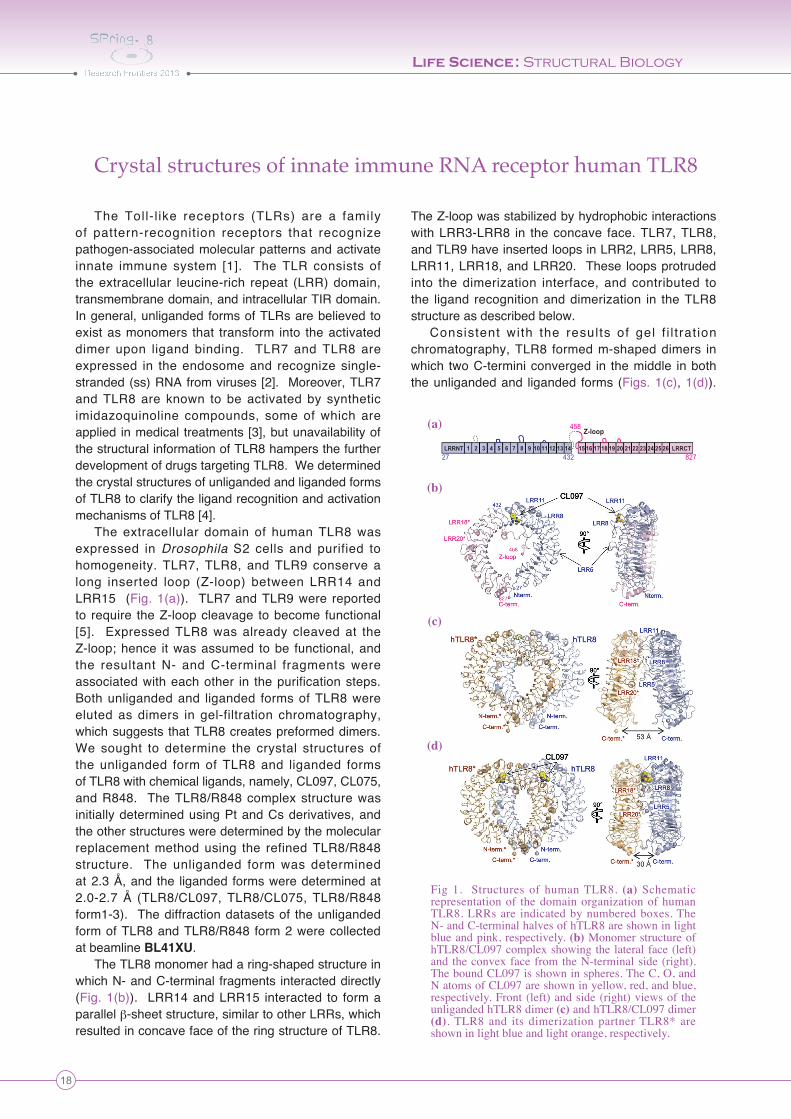
The extracellular domain of human TLR8 was expressed in Drosophila S2 cells and purified to homogeneity. TLR7, TLR8, and TLR9 conserve a long inserted loop (Z-loop) between LRR14 and LRR15 (Fig. 1(a)). TLR7 and TLR9 were reported to require the Z-loop cleavage to become functional [5]. Expressed TLR8 was already cleaved at the Z-loop; hence it was assumed to be functional, and the resultant N- and C-terminal fragments were associated with each other in the purification steps. Both unliganded and liganded forms of TLR8 were eluted as dimers in gel-filtration chromatography, which suggests that TLR8 creates preformed dimers. We sought to determine the crystal structures of the unliganded form of TLR8 and liganded forms of TLR8 with chemical ligands, namely, CL097, CL075, and R848. The TLR8/R848 complex structure was initially determined using Pt and Cs derivatives, and the other structures were determined by the molecular replacement method using the refined TLR8/R848 structure. The unliganded form was determined at 2.3 Å, and the liganded forms were determined at 2.0-2.7 Å (TLR8/CL097, TLR8/CL075, TLR8/R848 form1-3). The diffraction datasets of the unliganded form of TLR8 and TLR8/R848 form 2 were collected at beamline BL41XU.
The TLR8 monomer had a ring-shaped structure in which N- and C-terminal fragments interacted directly (Fig. 1(b)). LRR14 and LRR15 interacted to form a parallel β-sheet structure, similar to other LRRs, which resulted in concave face of the ring structure of TLR8.
The Z-loop was stabilized by hydrophobic interactions with LRR3-LRR8 in the concave face. TLR7, TLR8, and TLR9 have inserted loops in LRR2, LRR5, LRR8, LRR11, LRR18, and LRR20. These loops protruded into the dimerization interface, and contributed to the ligand recognition and dimerization in the TLR8 structure as described below.
Consistent with the results of gel f i l t rat ion chromatography, TLR8 formed m-shaped dimers in which two C-termini converged in the middle in both the unliganded and liganded forms (Figs. 1(c), 1(d)).
Fig 1. Structures of human TLR8. (a) Schematic representation of the domain organization of human TLR8. LRRs are indicated by numbered boxes. The N- and C-terminal halves of hTLR8 are shown in light blue and pink, respectively. (b) Monomer structure of hTLR8/CL097 complex showing the lateral face (left) and the convex face from the N-terminal side (right). The bound CL097 is shown in spheres. The C, O, and N atoms of CL097 are shown in yellow, red, and blue, respectively. Front (left) and side (right) views of the unliganded hTLR8 dimer (c) and hTLR8/CL097 dimer (d). TLR8 and its dimerization partner TLR8* are shown in light blue and light orange, respectively.
(a)
(b)
(c)
(d)
LRRNT LRRCT
Z-loop
27 432
458
8271 2 3 4 5 6 7 8 9 10 11 12 13 14 15 16 17 18 19 20 21 22 23 24 25 26
Life Science: Structural Biology
19
Throughout this report, we indicate the second TLR8 and its residues in the dimeric TLR8 with asterisks. The C-termini of the two TLR8 protomers were separated by 53 Å in the unliganded form, whereas the C-termini were brought into close proximity (~30 Å) in the liganded forms.
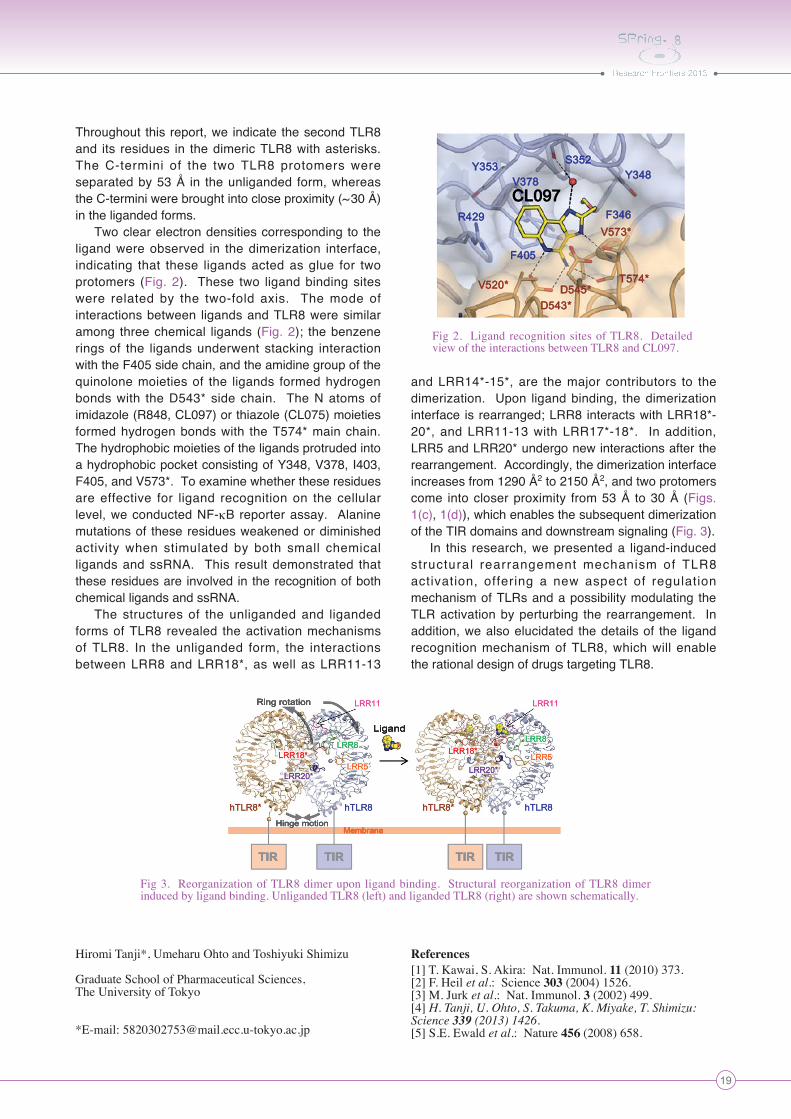
Two clear electron densities corresponding to the ligand were observed in the dimerization interface, indicating that these ligands acted as glue for two protomers (Fig. 2). These two ligand binding sites were related by the two-fold axis. The mode of interactions between ligands and TLR8 were similar among three chemical ligands (Fig. 2); the benzene rings of the ligands underwent stacking interaction with the F405 side chain, and the amidine group of the quinolone moieties of the ligands formed hydrogen bonds with the D543* side chain. The N atoms of imidazole (R848, CL097) or thiazole (CL075) moieties formed hydrogen bonds with the T574* main chain. The hydrophobic moieties of the ligands protruded into a hydrophobic pocket consisting of Y348, V378, I403, F405, and V573*. To examine whether these residues are effective for ligand recognition on the cellular level, we conducted NF-κB reporter assay. Alanine mutations of these residues weakened or diminished activity when stimulated by both small chemical ligands and ssRNA. This result demonstrated that these residues are involved in the recognition of both chemical ligands and ssRNA.
The structures of the unliganded and liganded forms of TLR8 revealed the activation mechanisms of TLR8. In the unliganded form, the interactions between LRR8 and LRR18*, as well as LRR11-13
and LRR14*-15*, are the major contributors to the dimerization. Upon ligand binding, the dimerization interface is rearranged; LRR8 interacts with LRR18*-20*, and LRR11-13 with LRR17*-18*. In addition, LRR5 and LRR20* undergo new interactions after the rearrangement. Accordingly, the dimerization interface increases from 1290 Å2 to 2150 Å2, and two protomers come into closer proximity from 53 Å to 30 Å (Figs. 1(c), 1(d)), which enables the subsequent dimerization of the TIR domains and downstream signaling (Fig. 3).
In this research, we presented a ligand-induced structural rearrangement mechanism of TLR8 activation, offering a new aspect of regulation mechanism of TLRs and a possibility modulating the TLR activation by perturbing the rearrangement. In addition, we also elucidated the details of the ligand recognition mechanism of TLR8, which will enable the rational design of drugs targeting TLR8.
Hiromi Tanji*, Umeharu Ohto and Toshiyuki Shimizu
Graduate School of Pharmaceutical Sciences, The University of Tokyo
*E-mail: [email protected]
References[1] T. Kawai, S. Akira: Nat. Immunol. 11 (2010) 373.[2] F. Heil et al.: Science 303 (2004) 1526.[3] M. Jurk et al.: Nat. Immunol. 3 (2002) 499.[4] H. Tanji, U. Ohto, S. Takuma, K. Miyake, T. Shimizu: Science 339 (2013) 1426.[5] S.E. Ewald et al.: Nature 456 (2008) 658.
Fig 2. Ligand recognition sites of TLR8. Detailed view of the interactions between TLR8 and CL097.
Ring rotation
Hinge motion
Ligand
TIR TIR TIR TIR
Membrane
Ring rotation
Hinge motion
Ligand
TIR TIR TIR TIR
Membrane
Ring rotation
Hinge motion
Ligand
TIR TIR TIR TIR
Membrane
Ring rotation
Hinge motion
Ligand
TIR TIR TIR TIR
Membrane
Fig 3. Reorganization of TLR8 dimer upon ligand binding. Structural reorganization of TLR8 dimer induced by ligand binding. Unliganded TLR8 (left) and liganded TLR8 (right) are shown schematically.