Embed Size (px)
Citation preview
Crystal Structure of theRibosome at 5.5 Å Resolution
Marat M. Yusupov,1*† Gulnara Zh. Yusupova,1* Albion Baucom,1
Kate Lieberman,1 Thomas N. Earnest,2 J. H. D. Cate,3†Harry F. Noller1†
We describe the crystal structure of the complete Thermus thermophilus 70Sribosome containing bound messenger RNA and transfer RNAs (tRNAs) at 5.5angstrom resolution. All of the 16S, 23S, and 5S ribosomal RNA (rRNA) chains,the A-, P-, and E-site tRNAs, and most of the ribosomal proteins can be fittedto the electron density map. The core of the interface between the 30S smallsubunit and the 50S large subunit, where the tRNA substrates are bound, isdominated by RNA, with proteins located mainly at the periphery, consistentwith ribosomal function being based on rRNA. In each of the three tRNA bindingsites, the ribosome contacts all of the major elements of tRNA, providing anexplanation for the conservation of tRNA structure. The tRNAs are closelyjuxtaposed with the intersubunit bridges, in a way that suggests coupling of the20 to 50 angstrom movements associated with tRNA translocation with in-tersubunit movement.
Ribosomes are large ribonucleoprotein com-plexes that are responsible for protein synthe-sis in all cells. Unlike other cellular poly-merases, their mechanism of action appearsto be based fundamentally on RNA; i.e., theyare ribozymes (1–3). Understanding thestructural basis for the functional capabilitiesof rRNA is essential to explain why theseancient organelles use RNA, instead of pro-tein, for the complex and biologically crucialtask of translation. Bacterial ribosomes,which have been the most extensively inves-tigated, are composed of small (30S) sub-units, containing 16S rRNA and about 20proteins, and large (50S) subunits, which con-tain 23S rRNA, 5S rRNA, and over 30 pro-teins (4). The complete 70S ribosome isformed by association of the 30S and 50Ssubunits through a network of intermolecularbridges (5). Its intersubunit space is occupiedby the transfer RNAs (tRNAs), whose anti-codons base pair with messenger RNA(mRNA) codons in the 30S subunit, whereastheir 39-CCA ends, which carry the growingpolypeptide chain and the incoming aminoacid, reach into the 50S subunit, the locationof the peptidyl transferase center, where pep-tide bond formation is catalyzed.
The structure of the ribosome began to
emerge from the efforts of electron micros-copists, using methods such as immunoelec-tron microscopy (IEM) to identify the loca-tions of the different macromolecular compo-nents at low resolution [reviewed in (6)].Classical electron microscopy (EM) has giv-en way to cryo-EM reconstruction methods,which have provided increasingly higher res-olution views of the ribosome and its subunitsand functional complexes (5, 7, 8). Morerecently, the possibility of obtaining atomic-resolution structures of the ribosome by x-raycrystallography has come from the ability toprepare well-diffracting crystals of ribosomesand subunits (9) and to overcome the daunt-ing phase problem (10–12). During the pastyear, this has led to high-resolution structuresof Haloarcula marismortui 50S subunits (13)and of Thermus thermophilus 30S subunits(14, 15), providing detailed views of thesevast and intricate ribonucleoprotein complex-es. In addition, the structures of peptidyltransferase substrate analogs bound to the50S subunit (3) and of a rRNA stem loopbound to the P site of the 30S subunit (16)begin to suggest how the ribosome interactswith tRNA at atomic resolution.
Previously, we described the cocrystalli-zation of complete T. thermophilus 70S ribo-somes in functional complexes with mRNAand A-, P-, and E-site tRNAs and the solutionof their crystal structures at resolutions of upto 7.8 Å (11). Here, we describe the three-dimensional structure of the 70S ribosomecontaining mRNA and tRNAs bound to the Pand E sites at 5.5 Å resolution and to the Asite at 7 Å resolution. Features of the 50Ssubunit that were disordered in the high-resolution Haloarcula structure are found tobe ordered in the 70S Thermus structure,providing a nearly complete view of the 50S
subunit. The three tRNAs are closely juxta-posed with certain intersubunit bridges, assuggested by chemical probing and cross-linking studies (17–20) and in previous cryo-EM and x-ray structures (11, 21). Severallines of evidence indicate that these bridgesare mobile, suggesting that tRNA transloca-tion is in some way coupled with intersubunitmovement.
Overall structure of the 70S ribosome.T. thermophilus 70S ribosomes containing asynthetic mRNA analog and tRNAs bound tothe P and E sites were crystallized as de-scribed earlier, and their diffraction was im-proved to 5 Å resolution (Table 1). Experi-mental phases to 7.5 Å were obtained fromMAD experiments (11) and extended to aneffective resolution of 5.5 Å by use of densitymodification algorithms involving solventflipping (22) (Table 1). The quality of thephases was confirmed by the electron densityof the bound tRNAs, which provided internalstandards of known structure (Fig. 1). At 5.5Å, the RNA backbones can be traced withhigh confidence, and proteins of knownstructure can be fitted readily to the electrondensity (23). Using 70S complexes crystal-lized with and without tRNA bound to the Asite, we obtained a 7 Å Fourier differencemap that provided the position of the A-sitetRNA.
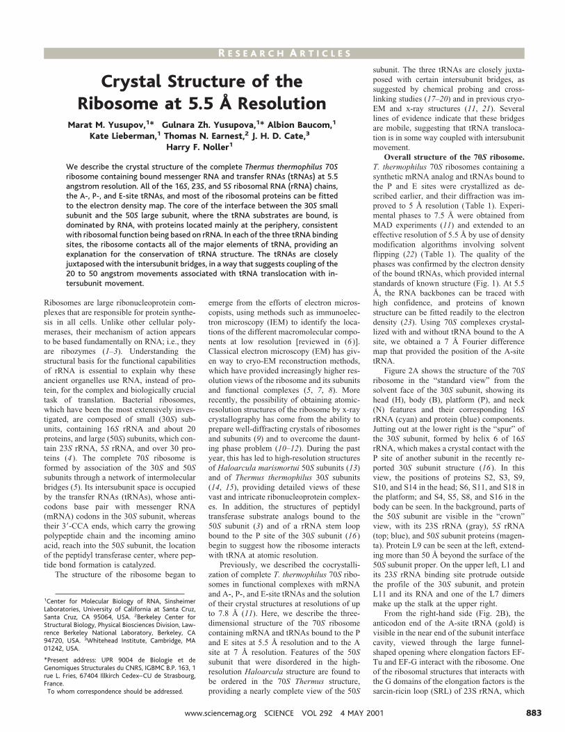
Figure 2A shows the structure of the 70Sribosome in the “standard view” from thesolvent face of the 30S subunit, showing itshead (H), body (B), platform (P), and neck(N) features and their corresponding 16SrRNA (cyan) and protein (blue) components.Jutting out at the lower right is the “spur” ofthe 30S subunit, formed by helix 6 of 16SrRNA, which makes a crystal contact with theP site of another subunit in the recently re-ported 30S subunit structure (16). In thisview, the positions of proteins S2, S3, S9,S10, and S14 in the head; S6, S11, and S18 inthe platform; and S4, S5, S8, and S16 in thebody can be seen. In the background, parts ofthe 50S subunit are visible in the “crown”view, with its 23S rRNA (gray), 5S rRNA(top; blue), and 50S subunit proteins (magen-ta). Protein L9 can be seen at the left, extend-ing more than 50 Å beyond the surface of the50S subunit proper. On the upper left, L1 andits 23S rRNA binding site protrude outsidethe profile of the 30S subunit, and proteinL11 and its RNA and one of the L7 dimersmake up the stalk at the upper right.
From the right-hand side (Fig. 2B), theanticodon end of the A-site tRNA (gold) isvisible in the near end of the subunit interfacecavity, viewed through the large funnel-shaped opening where elongation factors EF-Tu and EF-G interact with the ribosome. Oneof the ribosomal structures that interacts withthe G domains of the elongation factors is thesarcin-ricin loop (SRL) of 23S rRNA, which
1Center for Molecular Biology of RNA, SinsheimerLaboratories, University of California at Santa Cruz,Santa Cruz, CA 95064, USA. 2Berkeley Center forStructural Biology, Physical Biosciences Division, Law-rence Berkeley National Laboratory, Berkeley, CA94720, USA. 3Whitehead Institute, Cambridge, MA01242, USA.
*Present address: UPR 9004 de Biologie et deGenomiques Structurales du CNRS, IGBMC B.P. 163, 1rue L. Fries, 67404 Illkirch Cedex–CU de Strasbourg,France.†To whom correspondence should be addressed.
R E S E A R C H A R T I C L E S
www.sciencemag.org SCIENCE VOL 292 4 MAY 2001 883

is visible between the A-tRNA and proteinL14. Also evident in the right-hand view areproteins S9, S12, S13, S19, S20, L3, L5, L6,L7, L11, L13, L14, L19, L22, L25, and L30,as well as the positions of proteins L21 andL32 (whose structures are not known), andthe positions of electron density labeled LU,LV, and LX that we ascribe to as yet uniden-tified large subunit ribosomal proteins (whichmay include the three unassigned known pro-teins L31, L35, and L36). 5S rRNA (5S) isvisible at the top of the 50S subunit, alongwith two of its binding proteins, L5 and L25.
The view from the back of the 50S subunit(Fig. 2C) reveals the locations of additional50S subunit proteins L4, L15, L16, L21, L24,L27, L28, L29, L32, L33, and L34, the third5S rRNA-binding protein L18, and unidenti-fied proteins LW and LY. The opening of thepolypeptide exit channel (EC) is at the bot-tom of the back side of the 50S subunit,surrounded by proteins L22, L24, and L29 inaddition to elements of domains I and III of23S rRNA.
In the left-hand view (Fig. 2D), the closeapproach of the two subunits at the interfaceis much more evident. The platform of the30S subunit, around proteins S11, S6, andS15, contacts the 50S subunit near protein L2,mainly through RNA-RNA interactions andRNA-protein interactions involving proteinsS15 and L2. The E-site tRNA (red) can beseen at the near side of the interface cavity,partly shielded from view by L1 and its RNAbinding site, which appear to block the pathfor its exit from the ribosome. In the top view(Fig. 2E), the orientations of all three tRNAs(A, gold; P, orange; and E, red) in the inter-face cavity can be seen more clearly. In thisview, protein S13 in the head of the 30Ssubunit can be seen to contact helix 38 of 23SrRNA (the A-site finger; ASF) to form bridgeB1a. Also evident is the close approach be-tween proteins L5 and S13, whose electrondensities merge to form the single protein-
protein intersubunit contact (bridge B1b),which lies directly above and parallel to theanticodon arm of the P-site tRNA (seebelow).
Viewed from the interface (Fig. 2, F andG), fewer proteins are visible on the 30S and50S subunits, and they are located mainlyaround the periphery, leaving large exposedsurfaces of ribosomal RNA. The three tRNAsare aligned on the 30S subunit with theiranticodon ends bound in the RNA-richgroove between the head, body, and platform(Fig. 2F). The rest of all three tRNAs, includ-ing their D stems, elbows, and acceptor arms,interact with the 50S subunit. The acceptorarms of the A and P tRNAs point downwardinto the peptidyl transferase cavity, wheretheir acceptor ends come within 5 Å of eachother, whereas the E-tRNA acceptor arm isdirected into a separate cleft next to the L1ridge, placing its acceptor end nearly 50 Åfrom that of the P-tRNA. The tRNA bindingsite neighborhoods are dominated by rRNA,as are the interface contact surfaces.
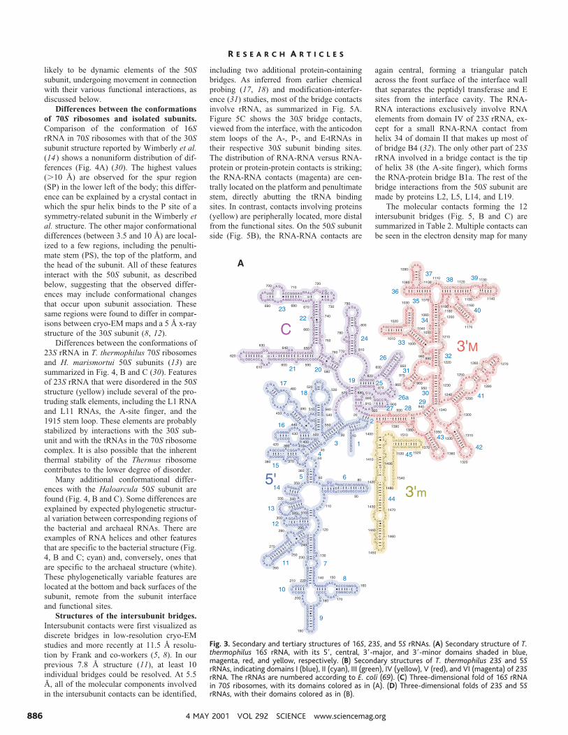
The secondary structure of 16S rRNA(Fig. 3A) (24, 25) falls into four recognizabledomains, called the 59, central, 39-major and39-minor domains. As observed in the struc-tures of the isolated ribosomal subunits (14,15), the secondary structure domains of 16SrRNA (Fig. 3A) do indeed correspond tothree-dimensional domains that are nearlystructurally autonomous (Fig. 3C). This orga-nization immediately suggests that the do-mains are designed to move relative to one
another during protein synthesis. In particu-lar, the very minimal interaction between thehead and the rest of the subunit is consistentwith movement of the head during transloca-tion (26-28). The four domains converge nearthe geometric center of the subunit, next tothe sites of its functional interactions withmRNA and tRNA, further suggesting cou-pling of interdomain movement with biolog-ical function.
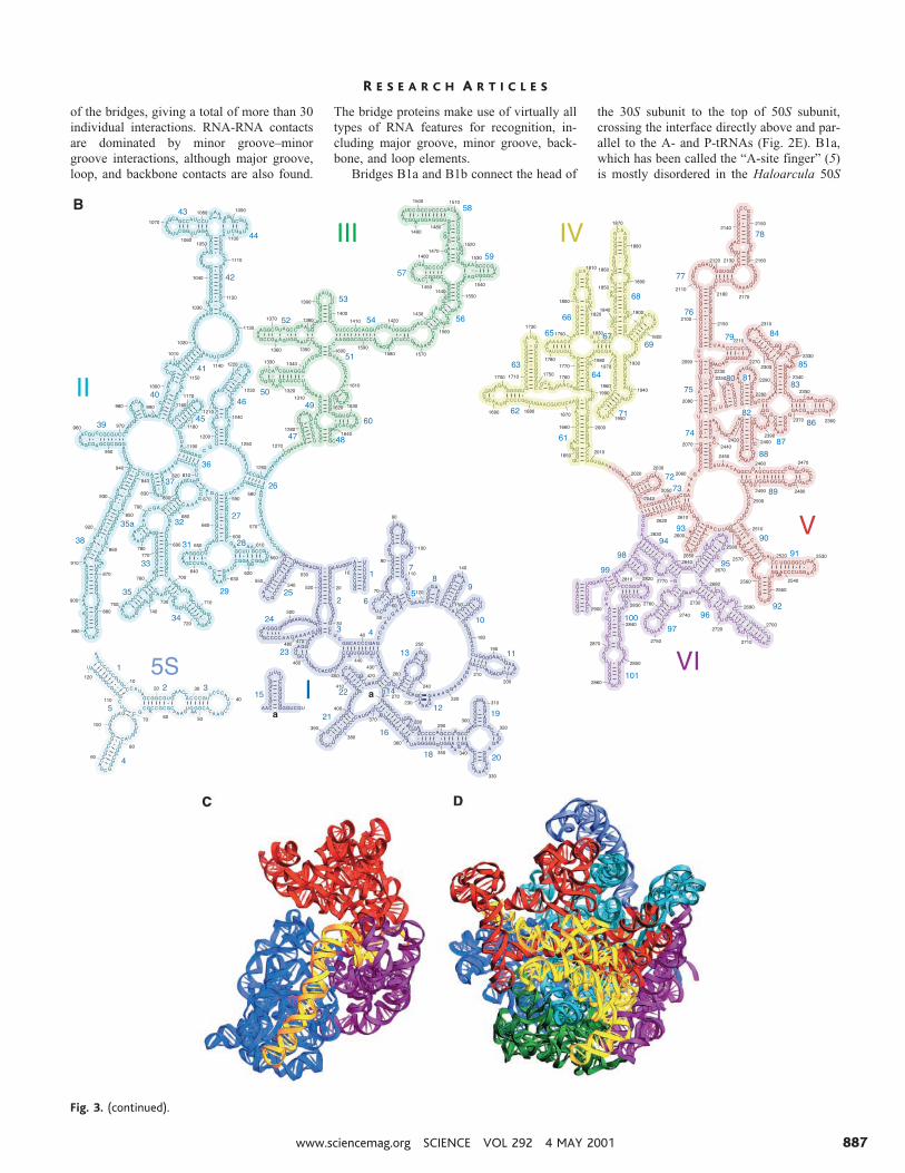
The more than 130 individual helices pre-dicted from comparative sequence analysis of23S rRNA (29) are found in its x-ray crystalstructure, except for a predicted Thermus-specific helix inserted around position 650 of23S rRNA, relative to the Escherichia colisecondary structure for which no electrondensity is found (25). The 23S rRNA and 5SrRNA together form seven secondary struc-tural domains (Fig. 3B). In contrast to thedesign of the 30S subunit, the domains of 23SrRNA are extensively intertwined with eachother, as first noted for the H. marismortui50S subunit (13), creating the single large,hemispherical domain that forms the body ofthe 50S subunit (Fig. 3D). From the bodyproject a number of molecular stalks, madeup of RNA elements from domains II, IV, V,and VI, some of which are extended coaxialhelical arms and others of which are mush-roomlike globular RNA domains tethered tothe body of the subunit by helical stems.Some of the stalks form bridges with the 30Ssubunit, whereas others interact with thetRNAs and elongation factors; the stalks are
Fig. 1. Electron density of tRNAMetf bound to
the P site of the 70S ribosome, at 5.5 Åresolution.
Table 1. Crystallographic statistics and scaling. Crystals of Thermus thermophilus 70S ribosome func-tional complexes were grown as described (11). All complexes contained 70S ribosomes, a 36-mer mRNAfragment derived from phage T4 gene 32 mRNA, and a tRNA of unknown identity bound to the E site.The other ligands were as follows: (ASL), a synthetic 19-nucleotide analog of the tRNAPhe anticodonstem-loop bound to the 30S subunit P site; (P site), tRNAMetf bound to the P site; (No mRNA), as for Psite, except lacking the mRNA fragment; and (A site), as for P site, but also including tRNAPhe bound tothe A site. Crystals grew in space group I422 with cell dimensions of a 5 b 5 507.2 Å and c 5 803.7 Å.Structure factor amplitudes were measured at the Advanced Light Source (ALS), essentially as described(11). Structure factor phases determined experimentally from a crystal containing an anticodon stemloop tRNA analog in the P site (“ASL”) (11) were used as a starting point for structure factor phasing ofdiffraction data measured from crystals containing tRNAMet
f in the P site (“P site”). Phase extension to5.0 Å was carried out by density modification and solvent flipping in CNS (22). The quality of the phaseswas assessed by monitoring the appearance of intact tRNA in the P site (Fig. 1).
Crystal ASL P site No mRNA A site
High-resolution limit (Å) 7.5 5 6.5 6.5Rsym* 8.9 9.4 8.9 7.2
Mean I/s(I) 3.1 (at 7.8 Å) 3.3 (at 5.5 Å) 4.4 (at 7.0 Å) 3 (at 7.0 Å)Number of reflections
Unique 124,437# 209,044 95,127 95,671Observational redundancy 4.4 2.8 3.6 2.3Completeness (%) 97.7 95.3 96.6 93.9
Riso† (%) 23.6x2, cross-crystal‡ 36.9Mean figure of merit for
starting phase set(at 7.5 Å)§
0.505
*Rsym 5 S?I 2 ^I&?/SI. †Riso 5 S?FPH 2 ^FP&?/SFPH, where FPH and FP are the structure factor amplitudes from theASL-containing ribosome crystal and the P-site tRNA-containing ribosome crystal, respectively. ‡x2 analysis, from20 to 7.5 Å, was taken from Scalepack (70). §The mean figure of merit, or mean cosine of the phase error, wascalculated from experimental phases measured from the ASL-containing crystal (11). #Data set taken from thepreviously reported MAD phasing experiment (11).
R E S E A R C H A R T I C L E S
4 MAY 2001 VOL 292 SCIENCE www.sciencemag.org884
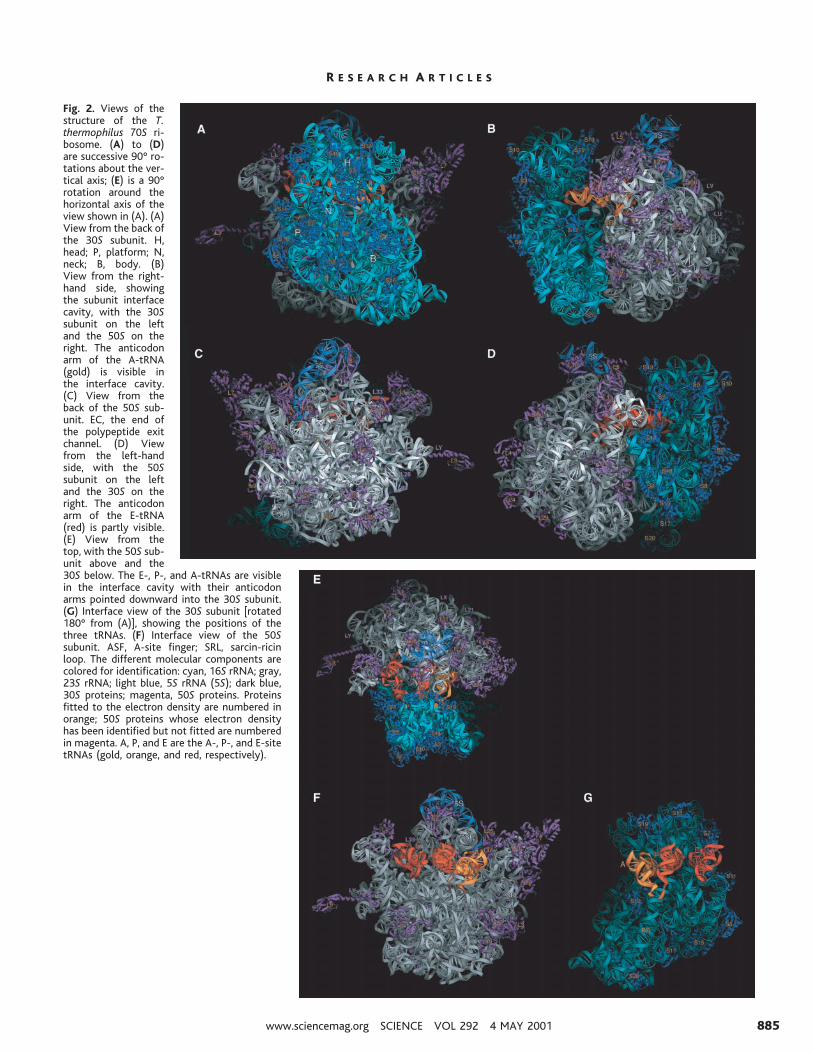
Fig. 2. Views of thestructure of the T.thermophilus 70S ri-bosome. (A) to (D)are successive 90° ro-tations about the ver-tical axis; (E) is a 90°rotation around thehorizontal axis of theview shown in (A). (A)View from the back ofthe 30S subunit. H,head; P, platform; N,neck; B, body. (B)View from the right-hand side, showingthe subunit interfacecavity, with the 30Ssubunit on the leftand the 50S on theright. The anticodonarm of the A-tRNA(gold) is visible inthe interface cavity.(C) View from theback of the 50S sub-unit. EC, the end ofthe polypeptide exitchannel. (D) Viewfrom the left-handside, with the 50Ssubunit on the leftand the 30S on theright. The anticodonarm of the E-tRNA(red) is partly visible.(E) View from thetop, with the 50S sub-unit above and the30S below. The E-, P-, and A-tRNAs are visiblein the interface cavity with their anticodonarms pointed downward into the 30S subunit.(G) Interface view of the 30S subunit [rotated180° from (A)], showing the positions of thethree tRNAs. (F) Interface view of the 50Ssubunit. ASF, A-site finger; SRL, sarcin-ricinloop. The different molecular components arecolored for identification: cyan, 16S rRNA; gray,23S rRNA; light blue, 5S rRNA (5S); dark blue,30S proteins; magenta, 50S proteins. Proteinsfitted to the electron density are numbered inorange; 50S proteins whose electron densityhas been identified but not fitted are numberedin magenta. A, P, and E are the A-, P-, and E-sitetRNAs (gold, orange, and red, respectively).
R E S E A R C H A R T I C L E S
www.sciencemag.org SCIENCE VOL 292 4 MAY 2001 885
likely to be dynamic elements of the 50Ssubunit, undergoing movement in connectionwith their various functional interactions, asdiscussed below.
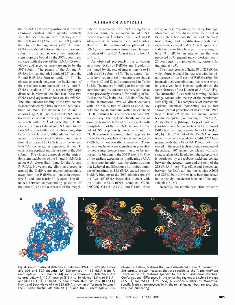
Differences between the conformationsof 70S ribosomes and isolated subunits.Comparison of the conformation of 16SrRNA in 70S ribosomes with that of the 30Ssubunit structure reported by Wimberly et al.(14) shows a nonuniform distribution of dif-ferences (Fig. 4A) (30). The highest values(.10 Å) are observed for the spur region(SP) in the lower left of the body; this differ-ence can be explained by a crystal contact inwhich the spur helix binds to the P site of asymmetry-related subunit in the Wimberly etal. structure. The other major conformationaldifferences (between 3.5 and 10 Å) are local-ized to a few regions, including the penulti-mate stem (PS), the top of the platform, andthe head of the subunit. All of these featuresinteract with the 50S subunit, as describedbelow, suggesting that the observed differ-ences may include conformational changesthat occur upon subunit association. Thesesame regions were found to differ in compar-isons between cryo-EM maps and a 5 Å x-raystructure of the 30S subunit (8, 12).
Differences between the conformations of23S rRNA in T. thermophilus 70S ribosomesand H. marismortui 50S subunits (13) aresummarized in Fig. 4, B and C (30). Featuresof 23S rRNA that were disordered in the 50Sstructure (yellow) include several of the pro-truding stalk elements, including the L1 RNAand L11 RNAs, the A-site finger, and the1915 stem loop. These elements are probablystabilized by interactions with the 30S sub-unit and with the tRNAs in the 70S ribosomecomplex. It is also possible that the inherentthermal stability of the Thermus ribosomecontributes to the lower degree of disorder.
Many additional conformational differ-ences with the Haloarcula 50S subunit arefound (Fig. 4, B and C). Some differences areexplained by expected phylogenetic structur-al variation between corresponding regions ofthe bacterial and archaeal RNAs. There areexamples of RNA helices and other featuresthat are specific to the bacterial structure (Fig.4, B and C; cyan) and, conversely, ones thatare specific to the archaeal structure (white).These phylogenetically variable features arelocated at the bottom and back surfaces of thesubunit, remote from the subunit interfaceand functional sites.
Structures of the intersubunit bridges.Intersubunit contacts were first visualized asdiscrete bridges in low-resolution cryo-EMstudies and more recently at 11.5 Å resolu-tion by Frank and co-workers (5, 8). In ourprevious 7.8 Å structure (11), at least 10individual bridges could be resolved. At 5.5Å, all of the molecular components involvedin the intersubunit contacts can be identified,
including two additional protein-containingbridges. As inferred from earlier chemicalprobing (17, 18) and modification-interfer-ence (31) studies, most of the bridge contactsinvolve rRNA, as summarized in Fig. 5A.Figure 5C shows the 30S bridge contacts,viewed from the interface, with the anticodonstem loops of the A-, P-, and E-tRNAs intheir respective 30S subunit binding sites.The distribution of RNA-RNA versus RNA-protein or protein-protein contacts is striking;the RNA-RNA contacts (magenta) are cen-trally located on the platform and penultimatestem, directly abutting the tRNA bindingsites. In contrast, contacts involving proteins(yellow) are peripherally located, more distalfrom the functional sites. On the 50S subunitside (Fig. 5B), the RNA-RNA contacts are
again central, forming a triangular patchacross the front surface of the interface wallthat separates the peptidyl transferase and Esites from the interface cavity. The RNA-RNA interactions exclusively involve RNAelements from domain IV of 23S rRNA, ex-cept for a small RNA-RNA contact fromhelix 34 of domain II that makes up most ofof bridge B4 (32). The only other part of 23SrRNA involved in a bridge contact is the tipof helix 38 (the A-site finger), which formsthe RNA-protein bridge B1a. The rest of thebridge interactions from the 50S subunit aremade by proteins L2, L5, L14, and L19.
The molecular contacts forming the 12intersubunit bridges (Fig. 5, B and C) aresummarized in Table 2. Multiple contacts canbe seen in the electron density map for many
Fig. 3. Secondary and tertiary structures of 16S, 23S, and 5S rRNAs. (A) Secondary structure of T.thermophilus 16S rRNA, with its 59, central, 39-major, and 39-minor domains shaded in blue,magenta, red, and yellow, respectively. (B) Secondary structures of T. thermophilus 23S and 5SrRNAs, indicating domains I (blue), II (cyan), III (green), IV (yellow), V (red), and VI (magenta) of 23SrRNA. The rRNAs are numbered according to E. coli (69). (C) Three-dimensional fold of 16S rRNAin 70S ribosomes, with its domains colored as in (A). (D) Three-dimensional folds of 23S and 5SrRNAs, with their domains colored as in (B).
R E S E A R C H A R T I C L E S
4 MAY 2001 VOL 292 SCIENCE www.sciencemag.org886
of the bridges, giving a total of more than 30individual interactions. RNA-RNA contactsare dominated by minor groove–minorgroove interactions, although major groove,loop, and backbone contacts are also found.
The bridge proteins make use of virtually alltypes of RNA features for recognition, in-cluding major groove, minor groove, back-bone, and loop elements.
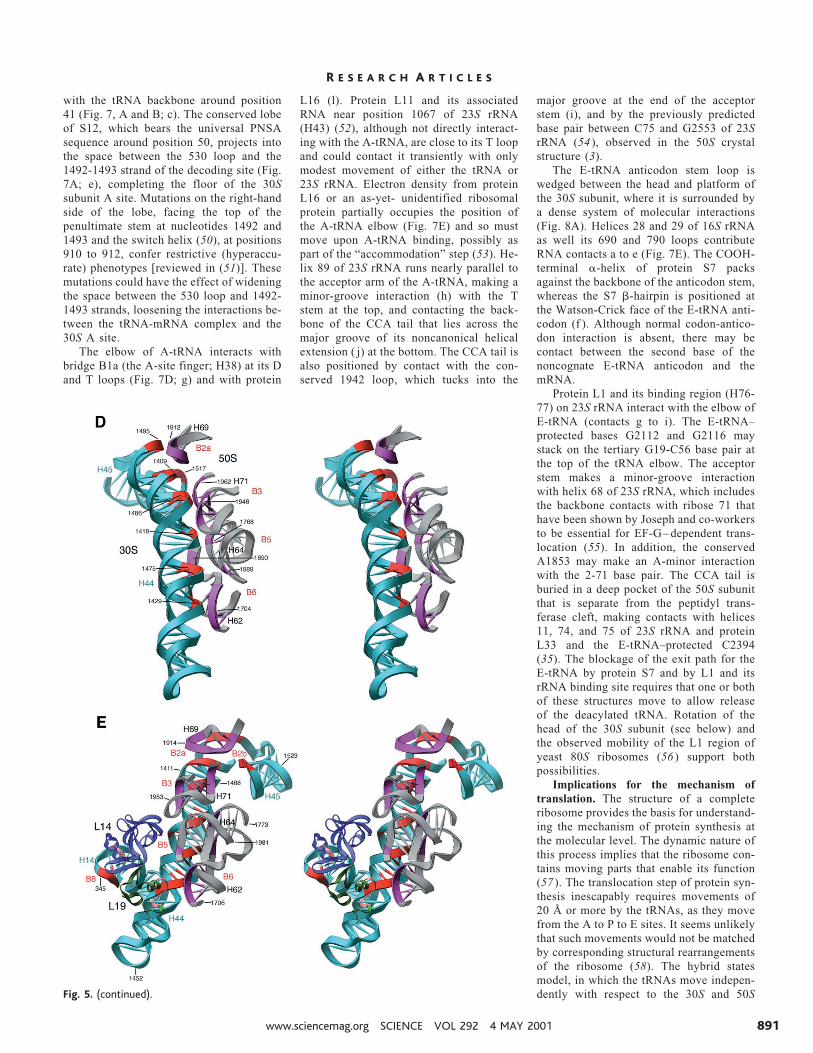
Bridges B1a and B1b connect the head of
the 30S subunit to the top of 50S subunit,crossing the interface directly above and par-allel to the A- and P-tRNAs (Fig. 2E). B1a,which has been called the “A-site finger” (5)is mostly disordered in the Haloarcula 50S
Fig. 3. (continued).
R E S E A R C H A R T I C L E S
www.sciencemag.org SCIENCE VOL 292 4 MAY 2001 887

subunit structure (13). It consists of a longhelical RNA arm (helix 38 of 23S rRNA)reaching from the right side of the centralprotuberance of the 50S subunit to the middleof the head of the 30S subunit, where itsapical 890 loop contacts the conserved basicsequence around position 92 of protein S13.Bridge B1b is the sole protein-protein contactbetween the subunits. Helix 84 of 23S rRNAreaches partway toward the head of the 30Ssubunit above the P-tRNA; the remainingdistance is bridged by protein L5, which con-tacts the NH2-terminal tail of S13 from a20–amino acid loop formed by residues 134to 153 of L5 (Hm positions 109 to 127),which are also disordered in the H. marismor-tui 50S structure.
Bridges B2a, B3, B5, and B6 (Fig. 5, Band C) all involve interactions between the50S subunit and the penultimate stem (helix44) of 16S rRNA, the dominant structuralcomponent of the 30S subunit interface. Fig-ure 5D shows the arrangement of the RNAelements forming these four bridges. At thetop, bridge B2a is made by the 1914 loop ofhelix 69 of 23S rRNA, another feature that isdisordered in the Haloarcula 50S subunitstructure. It contacts the decoding site of 16SrRNA around position 1408, as predictedfrom cross-linking experiments (19) in thefirst of a series of three consecutive minorgroove–minor groove interactions. In thenext one (B3), helix 71 of 23S rRNA contactsthe penultimate stem at its two consecutivenoncanonical A-G pairs around position1418. Just below B3, a major groove contact(B5) is made by the minor groove of helix 64of 23S rRNA, followed by the third minor-minor interaction (B6) formed by contactwith helix 62. A further contact with thepenultimate stem at bridge B6 is made byprotein L19 (Fig. 5E). L14, which interactswith L19 by forming an intermolecularb-sheet, contacts the major groove side of the345 loop of helix 14 of 16S rRNA to formbridge B8 (Fig. 5E).
Helices 68 and 71 of 23S rRNA form along, largely noncanonical coaxial arm thatlies horizontally along the top of the interfacewall of the 50S subunit, containing the 50Scomponents of bridges B2b and B7a, in ad-dition to the aforementioned B3 (Fig. 5C).Figure 5F shows the complex set of interac-tions that form B2b and B7a, viewed from thetop of the platform. The electron density forbridge B7a suggests that A702, which isstrongly protected from diethyl pyrocarbon-ate modification in 70S ribosomes (18),makes an “A-minor” contact (32a) with theminor groove of helix 68 of 23S rRNA. Thetwo remaining protein-RNA bridges areshown in Fig. 5G. Protein L2 makes twodistinct contacts with 16S rRNA (B7b), athelices 23 and 24; L2 is also very close toprotein S6 (not shown in figure) and may
make transient contacts with it during trans-lation. The proposed role of L2 in peptidyltransferase activity (33) might be related toits participation in bridge B7b, which couldserve as a relay between tRNA interactions inthe small subunit and the catalytic center inthe large subunit. Bridge B4 is primarily aninteraction between protein S15 and the 715loop of helix 34 of 23S rRNA, as shownpreviously (32); the 715 loop also makes amodest RNA-RNA contact with helix 20 of16S rRNA (Fig. 5G).
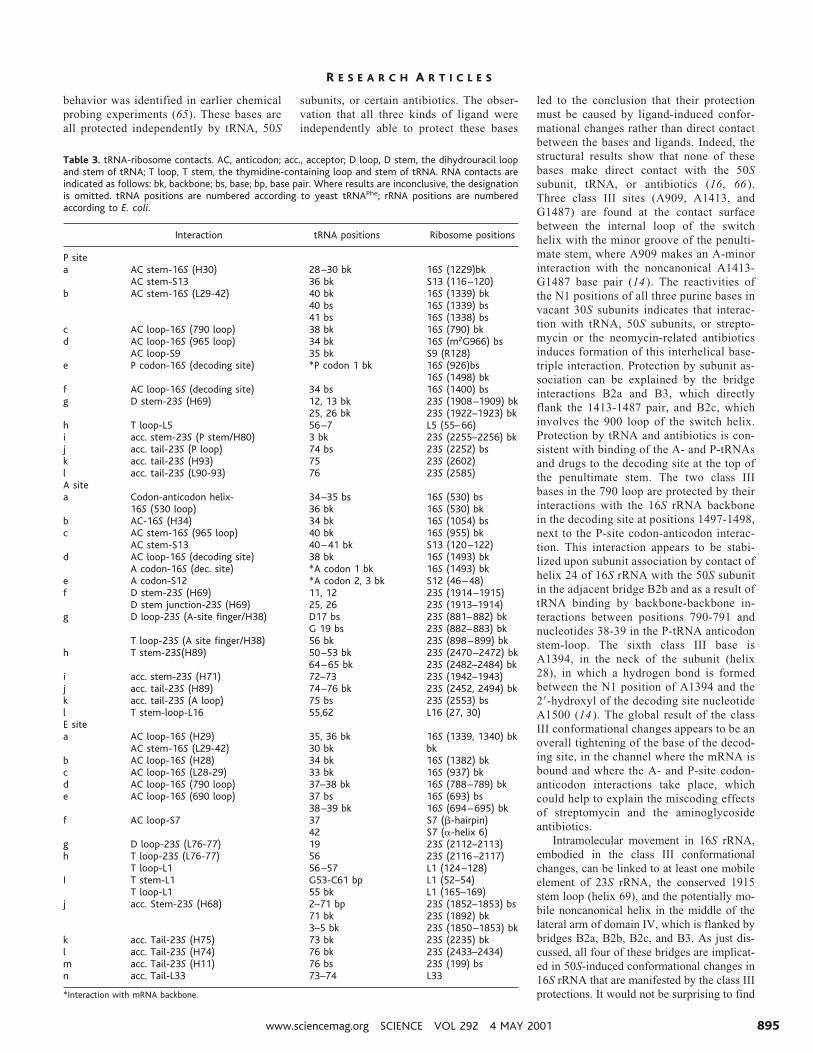
tRNA-ribosome interactions. Most im-portant for understanding the translationalmechanism is how the ribosome interactswith its substrates, the tRNAs. In addition totheir well-known interactions with mRNA,through base pairing between the codons andanticodons, tRNAs also interact with the ri-bosome itself. These interactions not onlyhelp to stabilize the binding of tRNA to theribosome but are involved directly in func-tional processes such as discrimination mech-anisms that increase the accuracy of amino-acyl-tRNA selection, maintaining the correcttranslational reading frame, translocationalmovement of tRNAs within the ribosome,and catalysis of peptide bond formation.Knowledge of the molecular contacts be-tween tRNA and the ribosome thus provides
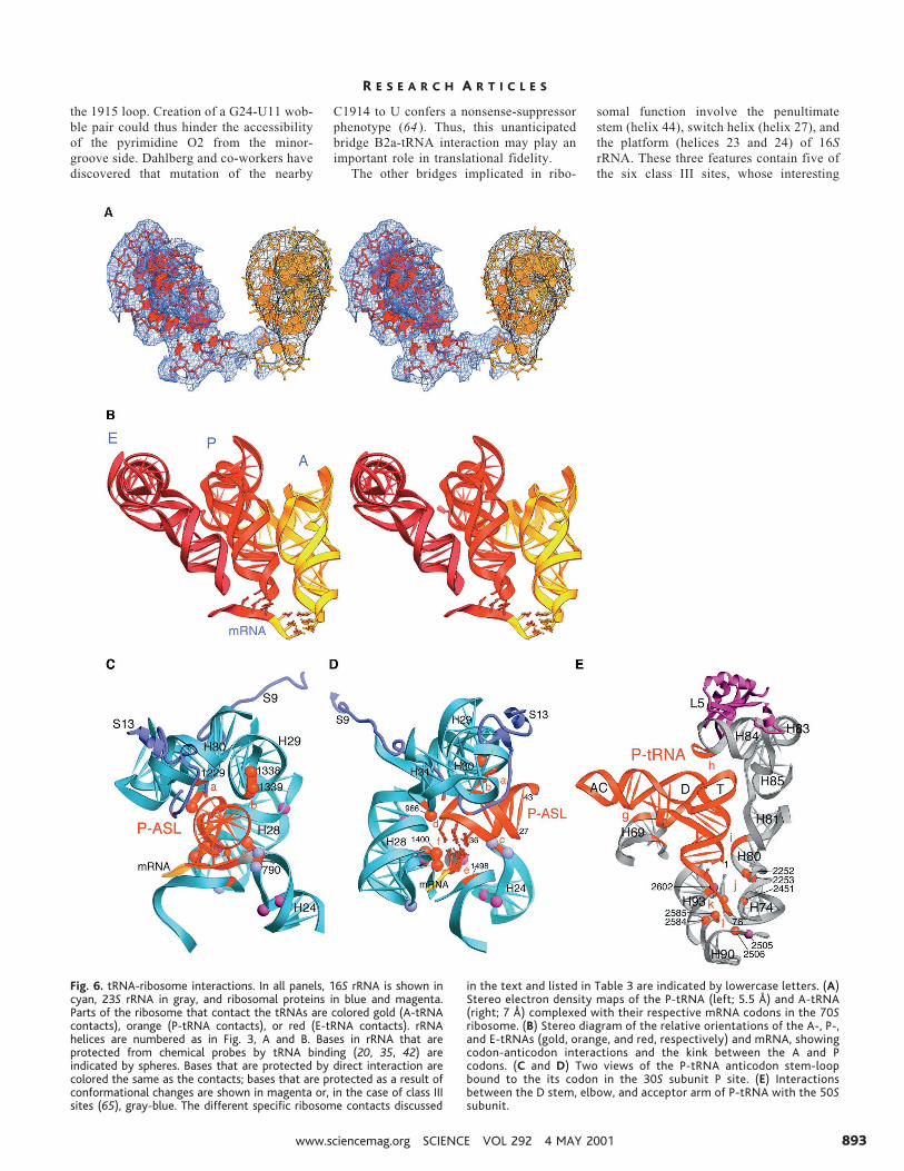
a structural framework for elucidation ofmechanisms for these processes. As predictedby many earlier studies [reviewed in (2)], thetRNAs are mainly surrounded by elements ofrRNA in the ribosome, most of which wereidentified in footprinting, cross-linking, anddirected hydroxyl radical probing studies (20,34, 35). Not surprisingly, we find that allthree tRNA binding sites (A, P, and E) of theribosome contact all three tRNAs at univer-sally conserved parts of their structures; thisallows the ribosome to bind different tRNAspecies in precisely the same way.
The A-tRNA density could be fitted to thestructure of tRNAPhe (36) without modifica-tion, whereas the P-tRNA is kinked slightlyaround the junction of the D and anticodonstems, angling the anticodon loop toward theA site and narrowing the major groove of theanticodon stem. The E-tRNA is substantiallydistorted relative to known tRNA crystalstructures. The angle of its elbow is moreopen, and the anticodon stem pivots near itsjunction with the D stem, pinching the majorgroove of the D stem. Its anticodon loopmakes an unusually sharp U-turn.
Figure 6A shows the electron density ofthe A- and P-tRNAs bound to their respectivecodons, and Fig. 6B shows the overall rela-tive geometry of the A-, P-, and E-tRNAs and
Table 2. Intersubunit bridges. Bridges are numbered B1a, B1b, etc., as shown in Fig. 5, B and C. rRNAcontacts are to 16S rRNA for the 30S subunit and to 23S rRNA for the 50S subunit, listed by the numberof the proximal helix (H44, etc.), numbered as shown in Fig. 3, A and B. rRNA nucleotide numbers areaccording to E. coli numbering. Molecular contacts are scored in parentheses as follows: M, major groove;m, minor groove; L, loop; B, backbone; Lm refers to the minor groove side of the loop, LB to the loopbackbone, etc.
Bridge Type
30S subunit 50S subunit
16S rRNA helixor S protein
RNA or proteinpositions
23S rRNA helixor L protein
RNA or proteinpositions
B1a Prot-RNA S13 92–94 H38(L) 886–888B1b Prot-Prot S13 NH2-term L5 134–153B2a RNA-RNA H44(m) 1408–1410,
1494–1495H69(Lm) 1913–1914, 1918
B2b RNA-RNA H24(m,LM) 784–785,794 H67(m),H69(M)
1836–1837, 1922
RNA-RNA H45(LM,Lm) 1516–1519 H71(M),H69(B)
1919–1920, 1932
B2c RNA-RNA H24(Bm) 770–771 H67(B) 1832–1833RNA-RNA H27(Bm) 900–1 H67(B) 1832–1833
B3 RNA-RNA H44(m) 1484–1486 H71(m) 1947–1948, 1960–1961B4 RNA-RNA H20(m) 763–764 H34(Lm) 717–718
Prot-RNA S15 40–44,COOH-term
H34(LB,LM) 713, 717
B5 RNA-RNA H44(m) 1418–1419 H64(m) 1768–1769RNA-Prot H44(B) 1420–1422 L14 44–49RNA-RNA H44(B) 1474–1476 H62(Bm) 1689–1690RNA-RNA H44(B) 1474–1476 H64(m) 1989
B6 RNA-RNA H44(m) 1429–1430,1474–1476
H62(m) 1689–1690, 1702–1705
RNA-prot H44(B) 1431 L19 (Hm24e:R44)B7a RNA-RNA H23(L,m) 698,702 H68(m) 1848–1849, 1896B7b RNA-Prot H23(M,m) 712–713 L2 162–164, 172–174,
177–178RNA-Prot H24(M,m) 773–776 L2 177–178, 198–202
B8 RNA-Prot H14(LM) 345–347 L14 116–119
R E S E A R C H A R T I C L E S
4 MAY 2001 VOL 292 SCIENCE www.sciencemag.org888
the mRNA as they are positioned in the 70Sribosome crystals. Their specific contactswith the ribosome indicate that they are intheir “classical” (A/A, P/P, and E/E), ratherthan hybrid binding states (37). All threetRNAs are shared between the two ribosomalsubunits in a similar way; their anticodonstem loops are bound by the 30S subunit, andcontacts with the rest of the tRNA—D stem,elbow, and acceptor arm—are made by the50S subunit. The planes of the A- and P-tRNAs form an included angle of 26°, and theP- and E-tRNAs form an angle of 46°. Theclosest approach between the backbones ofthe anticodon stem loops of the A- and P-tRNAs is about 10 Å, a surprisingly largedistance, in view of the fact that these twotRNAs read adjacent codons on the mRNA.The simultaneous reading of the two codonsis accomodated by a kink in the mRNA back-bone of about 45° between the A and Pcodons (Fig. 6B). The A- and P-tRNA back-bones are closest at the acceptor stems, whichapproach within 5 Å of each other. At theelbow, the bases D16 of A-tRNA and U47 ofP-tRNA are actually within H-bonding dis-tance of each other, although we are notaware of prior evidence that such an interac-tion takes place. The CCA tails of the A- andP-tRNAs converge, as expected, at their 39ends in the peptidyl transferase site of the 50Ssubunit. The closest approach of the antico-don stem backbones of the P- and E-tRNAs isabout 6 Å, closer than found for the A- andP-tRNAs. However, the elbow and acceptorarm of the E-tRNA are rotated substantiallyaway from the P-tRNA, so that their respec-tive 39 ends are nearly 50 Å apart. The dis-tances between corresponding positions ofthe three tRNAs are a measure of the magni-
tude of the movement of tRNA during trans-location. Thus, the anticodon end of tRNAmoves about 28 Å between the 30S A and Psites, and 20 Å between the P and E sites.Because of the rotation of the plane of thetRNA, the elbow moves through much largerdistances of 40 and 55 Å, as it transits from Ato P to E.
As observed previously, the anticodonstem loop (ASL) of P-tRNA and P codon ispositioned by six sets of interactions (a to f )with the 30S subunit (11). The structural fea-tures involved in these interactions are shownin Fig. 6, C and D, and summarized in Table3 (38). The mode of binding of the anticodonstem loop and its contacts are very similar tothose previously observed for binding of he-lix 6 to the 30S P site (16). All six of the 30SP-site interactions involve direct contactswith 16S rRNA, two of which (a and d) arebolstered by interactions with the extendedCOOH-terminal tails of proteins S13 and S9,respectively. The phylogenetically somewhatvariable lysine-rich tail of S13 interacts withphosphate 36 of the P-tRNA. In contrast, thetail of S9 is precisely conserved, and itsCOOH-terminal arginine, which appears tointeract with phosphate 35 in the anticodon ofP-tRNA, is universally conserved. Thesesame phosphates were identified in phospho-rothioate-interference experiments to be im-portant for binding to the 30S P site (39). Oneof the earliest experiments implicating rRNAin ribosome function was the demonstrationthat kethoxal modification of a limited num-ber of guanines in 16S rRNA caused loss ofP-tRNA binding to the 30S subunit (40). Ofthe five 16S rRNA bases that interact withthe P-site mRNA-tRNA complex, G926,2mG966, G1338, A1339, and C1400, three
are guanines, explaining the early findings.Moreover, all five bases were identified asP-site interactions on the basis of chemicalfootprinting and modification-interferenceexperiments (20, 41, 42). C1400 appears tostabilize the wobble base pair by stacking onbase 34 of tRNA, an arrangement that waspredicted by Ofengand and co-workers nearly20 years ago, from photochemical cross-link-ing studies (43).
The minor groove of helix 69 of 23S rRNA,which forms bridge B2a, interacts with the mi-nor groove of the D stem of P-tRNA (Fig. 6E;interaction g), extending into the A site whereits conserved loop interacts with almost thesame features of the D stem of A-tRNA (Fig.7D; interaction f ), as well as forming the B2abridge contact with the top of the penultimatestem (Fig. 5D). This complex set of interactionsexplains chemical footprinting results thatshowed partial protection of bases in the 1915loop of helix 69 by the 30S subunit, whichbecame complete upon binding of tRNA (35).At its elbow, a b-hairpin loop of protein L5(positions 54 to 66) interacts with the T loop ofP-tRNA at the minor-groove face of C56 (Fig.6E; h). The CCA tail of the P-tRNA is posi-tioned to allow the predicted C74-G2252 basepairing with the 23S rRNA P loop (44), ob-served in the recent high-resolution structure ofthe archaeal 50S subunit complexed with sub-strate analogs (3). In addition, the acceptor endis positioned by a backbone-backbone contactbetween the acceptor stem and the stem of the23S rRNA P loop (Fig. 6E; i) and interactionsbetween the CCA tail and nucleotides A2602and U2585, both of which have been implicatedin the peptidyl transferase function of the largesubunit (35, 45).
Recently, the atomic-resolution structure
Fig. 4. Conformational differences between rRNAs in 70S ribosomesand 30S and 50S subunits. (A) Differences in 16S rRNA from T.thermophilus 30S subunits (14) and 70S ribosomes. Differences arecolored yellow (.10 Å), orange (5.5 Å to 10 Å), red (3.5 Å to 5.5 Å),and blue (,3.5 Å). H, head; PS, penultimate stem; SP, spur. (B and C)Front and back views of the 23S rRNA, showing differences betweenthe H. marismortui 50S subunit (13) and the T. thermophilus 70S
ribosome. Yellow, features that were disordered in the H. marismortui50S structure; cyan, features that are specific to the T. thermophilusstructure; white, features specific to the H. marismortui structure.Conformational differences in the remaining regions are colored orange(.5.5 Å) and red (3.5 Å to 5.5 Å). Nucleotide numbers of Haloarcula-specific features are preceded by H; the remaining numbers are accordingto E. coli numbering.
R E S E A R C H A R T I C L E S
www.sciencemag.org SCIENCE VOL 292 4 MAY 2001 889

of the Haloarcula 50S subunit has beensolved in complex with the compoundCCdAp-Puromycin (3), which is believed tobe a transition-state analog of the peptidyltransferase reaction (46). This structure hasled to a proposal for a mechanism for catal-ysis of peptide bond formation by the ribo-some (3). At 5.5 Å resolution, most of theconformation of the rRNA backbone in thevicinity of the 39-CCA end of P-site tRNAshows few discernible differences betweenthe two structures. The few apparent differ-ences are localized to the P loop and aroundpositions 2451, 2506, 2585, and 2602, whichmay move in a concerted way. In the 70Sribosome complex, the position of the 39-CCA end of the P-tRNA, relative to nearbyfeatures of 23S rRNA, appears to differ fromthat of the corresponding part of the transi-tion-state analog, possibly because of the ab-sence of an acyl group.
Surrounding the A-tRNA anticodon
loop in the 30S subunit are G530, A1492,and A1493, the three universally conservedbases originally identified as A-site–specif-ic features by chemical footprinting studies(20, 42) and shown to affect A-site bindingby mutational and biochemical studies (47,48). All three bases are positioned close tothe site of codon-anticodon interaction inthe 30S A site (Fig. 7, A and B; a and d).The tRNA-protected N1 positions of basesA1492 and 1493 point away from thecodon-anticodon base pairs and are separat-ed from them by the 16S rRNA backbone,when the 30S subunit A site is vacant (14 ),consistent with the electron density of the70S ribosome in the absence of A-tRNA. Inthe presence of the aminoglycoside antibi-otic paromomycin, the conformations ofnucleotides 1492 and 1493 have been foundto rearrange (16 ), raising the possibilitythat they may also rearrange in response tobinding tRNA to the 30S A site. In the 7 Å
Fourier difference map of the A-site tRNAbound to the 70S ribosome (Fig. 7C), apatch of negative electron density is seen atthe position of bases 1492-1493, providingsupport for the possibility that they rear-range to interact with the first and secondbase pairs in the minor groove of the A-sitecodon-anticodon helix, as suggested byCarter et al. (16 ). The N1 position of G530is also protected upon A-tRNA binding (20,42) and mutations of this base confer adominant lethal phenotype and defectiveA-tRNA binding (47 ). G530 is also posi-tioned in the minor groove of the codon-anticodon helix, near the second and thirdbase pairs. The bulged base C1054, muta-tions in which have been shown to suppressUGA nonsense mutations (49), projects to-ward the apex of the A-tRNA anticodonloop (Fig. 7B; b).
Lysine 120 of protein S13 and phos-phate 955 are both close enough to interact
Fig. 5. Intersubunitbridges. (A) Secondarystructures of 16S and23S rRNAs, showingfeatures involved inintersubunit contacts(red). (B and C) Inter-face views of the 50Sand 30S subunits, withthe bridges numbered(5, 11). RNA-RNA con-tacts are shown inmagenta; protein-RNAand protein-proteincontacts are shown inyellow. The bridgecontact B1b with pro-tein L5 is not explicitlymodeled, but its ap-proximate position isindicated in the figure.A, P, and E indicatethe three tRNAs (23SrRNA) or tRNA antico-don stem loops (16SrRNA). (D to G) De-tailed stereo views ofthe bridge interactions,viewed as in (D) Fig.1B, (E) Fig. 1C, (F)Fig. 1D, rotated 90°around the horizontalaxis, and (G) Fig. 1D.
R E S E A R C H A R T I C L E S
4 MAY 2001 VOL 292 SCIENCE www.sciencemag.org890
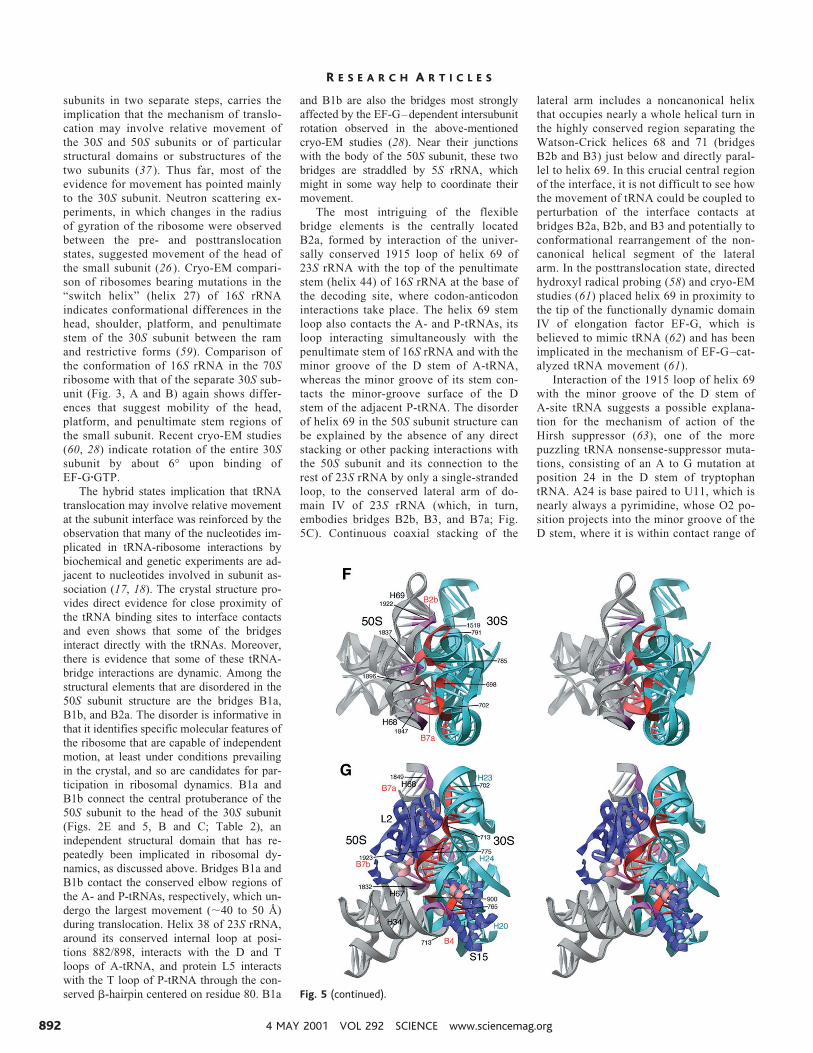
with the tRNA backbone around position41 (Fig. 7, A and B; c). The conserved lobeof S12, which bears the universal PNSAsequence around position 50, projects intothe space between the 530 loop and the1492-1493 strand of the decoding site (Fig.7A; e), completing the floor of the 30Ssubunit A site. Mutations on the right-handside of the lobe, facing the top of thepenultimate stem at nucleotides 1492 and1493 and the switch helix (50), at positions910 to 912, confer restrictive (hyperaccu-rate) phenotypes [reviewed in (51)]. Thesemutations could have the effect of wideningthe space between the 530 loop and 1492-1493 strands, loosening the interactions be-tween the tRNA-mRNA complex and the30S A site.
The elbow of A-tRNA interacts withbridge B1a (the A-site finger; H38) at its Dand T loops (Fig. 7D; g) and with protein
L16 (l). Protein L11 and its associatedRNA near position 1067 of 23S rRNA(H43) (52), although not directly interact-ing with the A-tRNA, are close to its T loopand could contact it transiently with onlymodest movement of either the tRNA or23S rRNA. Electron density from proteinL16 or an as-yet- unidentified ribosomalprotein partially occupies the position ofthe A-tRNA elbow (Fig. 7E) and so mustmove upon A-tRNA binding, possibly aspart of the “accommodation” step (53). He-lix 89 of 23S rRNA runs nearly parallel tothe acceptor arm of the A-tRNA, making aminor-groove interaction (h) with the Tstem at the top, and contacting the back-bone of the CCA tail that lies across themajor groove of its noncanonical helicalextension ( j) at the bottom. The CCA tail isalso positioned by contact with the con-served 1942 loop, which tucks into the
major groove at the end of the acceptorstem (i), and by the previously predictedbase pair between C75 and G2553 of 23SrRNA (54 ), observed in the 50S crystalstructure (3).
The E-tRNA anticodon stem loop iswedged between the head and platform ofthe 30S subunit, where it is surrounded bya dense system of molecular interactions(Fig. 8A). Helices 28 and 29 of 16S rRNAas well its 690 and 790 loops contributeRNA contacts a to e (Fig. 7E). The COOH-terminal a-helix of protein S7 packsagainst the backbone of the anticodon stem,whereas the S7 b-hairpin is positioned atthe Watson-Crick face of the E-tRNA anti-codon (f ). Although normal codon-antico-don interaction is absent, there may becontact between the second base of thenoncognate E-tRNA anticodon and themRNA.
Protein L1 and its binding region (H76-77) on 23S rRNA interact with the elbow ofE-tRNA (contacts g to i). The E-tRNA–protected bases G2112 and G2116 maystack on the tertiary G19-C56 base pair atthe top of the tRNA elbow. The acceptorstem makes a minor-groove interactionwith helix 68 of 23S rRNA, which includesthe backbone contacts with ribose 71 thathave been shown by Joseph and co-workersto be essential for EF-G– dependent trans-location (55). In addition, the conservedA1853 may make an A-minor interactionwith the 2-71 base pair. The CCA tail isburied in a deep pocket of the 50S subunitthat is separate from the peptidyl trans-ferase cleft, making contacts with helices11, 74, and 75 of 23S rRNA and proteinL33 and the E-tRNA–protected C2394(35). The blockage of the exit path for theE-tRNA by protein S7 and by L1 and itsrRNA binding site requires that one or bothof these structures move to allow releaseof the deacylated tRNA. Rotation of thehead of the 30S subunit (see below) andthe observed mobility of the L1 region ofyeast 80S ribosomes (56 ) support bothpossibilities.
Implications for the mechanism oftranslation. The structure of a completeribosome provides the basis for understand-ing the mechanism of protein synthesis atthe molecular level. The dynamic nature ofthis process implies that the ribosome con-tains moving parts that enable its function(57 ). The translocation step of protein syn-thesis inescapably requires movements of20 Å or more by the tRNAs, as they movefrom the A to P to E sites. It seems unlikelythat such movements would not be matchedby corresponding structural rearrangementsof the ribosome (58). The hybrid statesmodel, in which the tRNAs move indepen-dently with respect to the 30S and 50SFig. 5. (continued).
R E S E A R C H A R T I C L E S
www.sciencemag.org SCIENCE VOL 292 4 MAY 2001 891
subunits in two separate steps, carries theimplication that the mechanism of translo-cation may involve relative movement ofthe 30S and 50S subunits or of particularstructural domains or substructures of thetwo subunits (37 ). Thus far, most of theevidence for movement has pointed mainlyto the 30S subunit. Neutron scattering ex-periments, in which changes in the radiusof gyration of the ribosome were observedbetween the pre- and posttranslocationstates, suggested movement of the head ofthe small subunit (26 ). Cryo-EM compari-son of ribosomes bearing mutations in the“switch helix” (helix 27) of 16S rRNAindicates conformational differences in thehead, shoulder, platform, and penultimatestem of the 30S subunit between the ramand restrictive forms (59). Comparison ofthe conformation of 16S rRNA in the 70Sribosome with that of the separate 30S sub-unit (Fig. 3, A and B) again shows differ-ences that suggest mobility of the head,platform, and penultimate stem regions ofthe small subunit. Recent cryo-EM studies(60, 28) indicate rotation of the entire 30Ssubunit by about 6° upon binding ofEF-GzGTP.
The hybrid states implication that tRNAtranslocation may involve relative movementat the subunit interface was reinforced by theobservation that many of the nucleotides im-plicated in tRNA-ribosome interactions bybiochemical and genetic experiments are ad-jacent to nucleotides involved in subunit as-sociation (17, 18). The crystal structure pro-vides direct evidence for close proximity ofthe tRNA binding sites to interface contactsand even shows that some of the bridgesinteract directly with the tRNAs. Moreover,there is evidence that some of these tRNA-bridge interactions are dynamic. Among thestructural elements that are disordered in the50S subunit structure are the bridges B1a,B1b, and B2a. The disorder is informative inthat it identifies specific molecular features ofthe ribosome that are capable of independentmotion, at least under conditions prevailingin the crystal, and so are candidates for par-ticipation in ribosomal dynamics. B1a andB1b connect the central protuberance of the50S subunit to the head of the 30S subunit(Figs. 2E and 5, B and C; Table 2), anindependent structural domain that has re-peatedly been implicated in ribosomal dy-namics, as discussed above. Bridges B1a andB1b contact the conserved elbow regions ofthe A- and P-tRNAs, respectively, which un-dergo the largest movement (;40 to 50 Å)during translocation. Helix 38 of 23S rRNA,around its conserved internal loop at posi-tions 882/898, interacts with the D and Tloops of A-tRNA, and protein L5 interactswith the T loop of P-tRNA through the con-served b-hairpin centered on residue 80. B1a
and B1b are also the bridges most stronglyaffected by the EF-G–dependent intersubunitrotation observed in the above-mentionedcryo-EM studies (28). Near their junctionswith the body of the 50S subunit, these twobridges are straddled by 5S rRNA, whichmight in some way help to coordinate theirmovement.
The most intriguing of the flexiblebridge elements is the centrally locatedB2a, formed by interaction of the univer-sally conserved 1915 loop of helix 69 of23S rRNA with the top of the penultimatestem (helix 44) of 16S rRNA at the base ofthe decoding site, where codon-anticodoninteractions take place. The helix 69 stemloop also contacts the A- and P-tRNAs, itsloop interacting simultaneously with thepenultimate stem of 16S rRNA and with theminor groove of the D stem of A-tRNA,whereas the minor groove of its stem con-tacts the minor-groove surface of the Dstem of the adjacent P-tRNA. The disorderof helix 69 in the 50S subunit structure canbe explained by the absence of any directstacking or other packing interactions withthe 50S subunit and its connection to therest of 23S rRNA by only a single-strandedloop, to the conserved lateral arm of do-main IV of 23S rRNA (which, in turn,embodies bridges B2b, B3, and B7a; Fig.5C). Continuous coaxial stacking of the
lateral arm includes a noncanonical helixthat occupies nearly a whole helical turn inthe highly conserved region separating theWatson-Crick helices 68 and 71 (bridgesB2b and B3) just below and directly paral-lel to helix 69. In this crucial central regionof the interface, it is not difficult to see howthe movement of tRNA could be coupled toperturbation of the interface contacts atbridges B2a, B2b, and B3 and potentially toconformational rearrangement of the non-canonical helical segment of the lateralarm. In the posttranslocation state, directedhydroxyl radical probing (58) and cryo-EMstudies (61) placed helix 69 in proximity tothe tip of the functionally dynamic domainIV of elongation factor EF-G, which isbelieved to mimic tRNA (62) and has beenimplicated in the mechanism of EF-G–cat-alyzed tRNA movement (61).
Interaction of the 1915 loop of helix 69with the minor groove of the D stem ofA-site tRNA suggests a possible explana-tion for the mechanism of action of theHirsh suppressor (63), one of the morepuzzling tRNA nonsense-suppressor muta-tions, consisting of an A to G mutation atposition 24 in the D stem of tryptophantRNA. A24 is base paired to U11, which isnearly always a pyrimidine, whose O2 po-sition projects into the minor groove of theD stem, where it is within contact range of
Fig. 5 (continued).
R E S E A R C H A R T I C L E S
4 MAY 2001 VOL 292 SCIENCE www.sciencemag.org892
the 1915 loop. Creation of a G24-U11 wob-ble pair could thus hinder the accessibilityof the pyrimidine O2 from the minor-groove side. Dahlberg and co-workers havediscovered that mutation of the nearby
C1914 to U confers a nonsense-suppressorphenotype (64 ). Thus, this unanticipatedbridge B2a-tRNA interaction may play animportant role in translational fidelity.
The other bridges implicated in ribo-
somal function involve the penultimatestem (helix 44), switch helix (helix 27), andthe platform (helices 23 and 24) of 16SrRNA. These three features contain five ofthe six class III sites, whose interesting
Fig. 6. tRNA-ribosome interactions. In all panels, 16S rRNA is shown incyan, 23S rRNA in gray, and ribosomal proteins in blue and magenta.Parts of the ribosome that contact the tRNAs are colored gold (A-tRNAcontacts), orange (P-tRNA contacts), or red (E-tRNA contacts). rRNAhelices are numbered as in Fig. 3, A and B. Bases in rRNA that areprotected from chemical probes by tRNA binding (20, 35, 42) areindicated by spheres. Bases that are protected by direct interaction arecolored the same as the contacts; bases that are protected as a result ofconformational changes are shown in magenta or, in the case of class IIIsites (65), gray-blue. The different specific ribosome contacts discussed
in the text and listed in Table 3 are indicated by lowercase letters. (A)Stereo electron density maps of the P-tRNA (left; 5.5 Å) and A-tRNA(right; 7 Å) complexed with their respective mRNA codons in the 70Sribosome. (B) Stereo diagram of the relative orientations of the A-, P-,and E-tRNAs (gold, orange, and red, respectively) and mRNA, showingcodon-anticodon interactions and the kink between the A and Pcodons. (C and D) Two views of the P-tRNA anticodon stem-loopbound to the its codon in the 30S subunit P site. (E) Interactionsbetween the D stem, elbow, and acceptor arm of P-tRNA with the 50Ssubunit.
R E S E A R C H A R T I C L E S
www.sciencemag.org SCIENCE VOL 292 4 MAY 2001 893
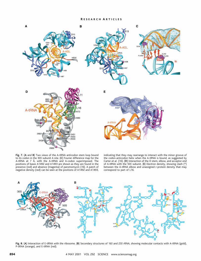
Fig. 7. (A and B) Two views of the A-tRNA anticodon stem loop boundto its codon in the 30S subunit A site. (C) Fourier difference map for theA-tRNA at 7 Å, with the A-tRNA and A-codon superimposed. Thepositions of bases A1492 and A1493 are shown as they are found in thepresence (red) and absence (magenta) of paromomycin (16). A patch ofnegative density (red) can be seen at the positions of A1492 and A1493,
indicating that they may rearrange to interact with the minor groove ofthe codon-anticodon helix when the A-tRNA is bound, as suggested byCarter et al. (16). (D) Interaction of the D stem, elbow, and acceptor endof A-tRNA with the 50S subunit. (E) Electron density, showing clash (*)between the A-tRNA elbow and unassigned r-protein density that maycorrespond to part of L16.
Fig. 8. (A) Interaction of E-tRNA with the ribosome. (B) Secondary structures of 16S and 23S rRNA, showing molecular contacts with A-tRNA (gold),P-tRNA (orange), and E-tRNA (red).
R E S E A R C H A R T I C L E S
4 MAY 2001 VOL 292 SCIENCE www.sciencemag.org894
behavior was identified in earlier chemicalprobing experiments (65). These bases areall protected independently by tRNA, 50S
subunits, or certain antibiotics. The obser-vation that all three kinds of ligand wereindependently able to protect these bases
led to the conclusion that their protectionmust be caused by ligand-induced confor-mational changes rather than direct contactbetween the bases and ligands. Indeed, thestructural results show that none of thesebases make direct contact with the 50Ssubunit, tRNA, or antibiotics (16, 66 ).Three class III sites (A909, A1413, andG1487) are found at the contact surfacebetween the internal loop of the switchhelix with the minor groove of the penulti-mate stem, where A909 makes an A-minorinteraction with the noncanonical A1413-G1487 base pair (14 ). The reactivities ofthe N1 positions of all three purine bases invacant 30S subunits indicates that interac-tion with tRNA, 50S subunits, or strepto-mycin or the neomycin-related antibioticsinduces formation of this interhelical base-triple interaction. Protection by subunit as-sociation can be explained by the bridgeinteractions B2a and B3, which directlyflank the 1413-1487 pair, and B2c, whichinvolves the 900 loop of the switch helix.Protection by tRNA and antibiotics is con-sistent with binding of the A- and P-tRNAsand drugs to the decoding site at the top ofthe penultimate stem. The two class IIIbases in the 790 loop are protected by theirinteractions with the 16S rRNA backbonein the decoding site at positions 1497-1498,next to the P-site codon-anticodon interac-tion. This interaction appears to be stabi-lized upon subunit association by contact ofhelix 24 of 16S rRNA with the 50S subunitin the adjacent bridge B2b and as a result oftRNA binding by backbone-backbone in-teractions between positions 790-791 andnucleotides 38-39 in the P-tRNA anticodonstem-loop. The sixth class III base isA1394, in the neck of the subunit (helix28), in which a hydrogen bond is formedbetween the N1 position of A1394 and the29-hydroxyl of the decoding site nucleotideA1500 (14 ). The global result of the classIII conformational changes appears to be anoverall tightening of the base of the decod-ing site, in the channel where the mRNA isbound and where the A- and P-site codon-anticodon interactions take place, whichcould help to explain the miscoding effectsof streptomycin and the aminoglycosideantibiotics.
Intramolecular movement in 16S rRNA,embodied in the class III conformationalchanges, can be linked to at least one mobileelement of 23S rRNA, the conserved 1915stem loop (helix 69), and the potentially mo-bile noncanonical helix in the middle of thelateral arm of domain IV, which is flanked bybridges B2a, B2b, B2c, and B3. As just dis-cussed, all four of these bridges are implicat-ed in 50S-induced conformational changes in16S rRNA that are manifested by the class IIIprotections. It would not be surprising to find
Table 3. tRNA-ribosome contacts. AC, anticodon; acc., acceptor; D loop, D stem, the dihydrouracil loopand stem of tRNA; T loop, T stem, the thymidine-containing loop and stem of tRNA. RNA contacts areindicated as follows: bk, backbone; bs, base; bp, base pair. Where results are inconclusive, the designationis omitted. tRNA positions are numbered according to yeast tRNAPhe; rRNA positions are numberedaccording to E. coli.
Interaction tRNA positions Ribosome positions
P sitea AC stem-16S (H30) 28–30 bk 16S (1229)bk
AC stem-S13 36 bk S13 (116–120)b AC stem-16S (L29-42) 40 bk 16S (1339) bk
40 bs 16S (1339) bs41 bs 16S (1338) bs
c AC loop-16S (790 loop) 38 bk 16S (790) bkd AC loop-16S (965 loop) 34 bk 16S (m2G966) bs
AC loop-S9 35 bk S9 (R128)e P codon-16S (decoding site) *P codon 1 bk 16S (926)bs
16S (1498) bkf AC loop-16S (decoding site) 34 bs 16S (1400) bsg D stem-23S (H69) 12, 13 bk 23S (1908–1909) bk
25, 26 bk 23S (1922–1923) bkh T loop-L5 56–7 L5 (55–66)i acc. stem-23S (P stem/H80) 3 bk 23S (2255–2256) bkj acc. tail-23S (P loop) 74 bs 23S (2252) bsk acc. tail-23S (H93) 75 23S (2602)l acc. tail-23S (L90-93) 76 23S (2585)A sitea Codon-anticodon helix- 34–35 bs 16S (530) bs
16S (530 loop) 36 bk 16S (530) bkb AC-16S (H34) 34 bk 16S (1054) bsc AC stem-16S (965 loop) 40 bk 16S (955) bk
AC stem-S13 40–41 bk S13 (120–122)d AC loop-16S (decoding site) 38 bk 16S (1493) bk
A codon-16S (dec. site) *A codon 1 bk 16S (1493) bke A codon-S12 *A codon 2, 3 bk S12 (46–48)f D stem-23S (H69) 11, 12 23S (1914–1915)
D stem junction-23S (H69) 25, 26 23S (1913–1914)g D loop-23S (A-site finger/H38) D17 bs 23S (881–882) bk
G 19 bs 23S (882–883) bkT loop-23S (A site finger/H38) 56 bk 23S (898–899) bk
h T stem-23S(H89) 50–53 bk 23S (2470–2472) bk64–65 bk 23S (2482–2484) bk
i acc. stem-23S (H71) 72–73 23S (1942–1943)j acc. tail-23S (H89) 74–76 bk 23S (2452, 2494) bkk acc. tail-23S (A loop) 75 bs 23S (2553) bsl T stem-loop-L16 55,62 L16 (27, 30)E sitea AC loop-16S (H29) 35, 36 bk 16S (1339, 1340) bk
AC stem-16S (L29-42) 30 bk bkb AC loop-16S (H28) 34 bk 16S (1382) bkc AC loop-16S (L28-29) 33 bk 16S (937) bkd AC loop-16S (790 loop) 37–38 bk 16S (788–789) bke AC loop-16S (690 loop) 37 bs 16S (693) bs
38–39 bk 16S (694–695) bkf AC loop-S7 37 S7 (b-hairpin)
42 S7 (a-helix 6)g D loop-23S (L76-77) 19 23S (2112–2113)h T loop-23S (L76-77) 56 23S (2116–2117)
T loop-L1 56–57 L1 (124–128)I T stem-L1 G53-C61 bp L1 (52–54)
T loop-L1 55 bk L1 (165–169)j acc. Stem-23S (H68) 2–71 bp 23S (1852–1853) bs
71 bk 23S (1892) bk3–5 bk 23S (1850–1853) bk
k acc. Tail-23S (H75) 73 bk 23S (2235) bkl acc. Tail-23S (H74) 76 bk 23S (2433–2434)m acc. Tail-23S (H11) 76 bs 23S (199) bsn acc. Tail-L33 73–74 L33
*Interaction with mRNA backbone.
R E S E A R C H A R T I C L E S
www.sciencemag.org SCIENCE VOL 292 4 MAY 2001 895
that these same conformational changes,which are also induced in 16S rRNA bytRNA and mRNA interactions in the decod-ing site of the 30S subunit, could reciprocallyaffect the conformation of this interface re-gion of 23S rRNA, through the same set ofbridge interactions. This could have interest-ing implications for the mechanism of trans-lation, because the lateral arm of domain IVpacks directly against the 2600 stem loop(helix 93) and the A loop (helix 92) of 23SrRNA, both of which are directly involved ininteractions in the peptidyl transferase center(3, 35, 54, 67). Furthermore, the 2563-2564loop at the base of helix 92 interacts directlywith the base of helix 95, the sarcin-ricinloop, which is directly implicated in the ac-tivities of elongation factors EF-Tu and EF-G. Finally, the far left-hand end of the lateralarm of domain IV, near bridge B7a, makesinteractions with the acceptor end of the E-tRNA that have been shown to be crucial forEF-G–dependent translocation (55). Knowl-edge of the structure of the complete ribo-some complexed with mRNA and tRNA nowprovides the possibility to test these and otherspecific molecular models for the mechanismof translation.
References and Notes1. H. F. Noller, V. Hoffarth, L. Zimniak, Science 256,
1416 (1992).2. R. Green, H. F. Noller, Annu. Rev. Biochem. 66, 679
(1997).3. P. Nissen, J. Hansen, N. Ban, P. B. Moore, T. A. Steitz,
Science 289, 920 (2000).4. A. T. Matheson, P. P. Dennis, J. E. Davies, W. E. Hill,
Biochem. Cell Biol. 73 (1995); R. A. Garrett et al.,Eds., The Ribosome ( American Society for Microbi-ology, Washington, DC, 2000); W. E. Hill et al., Eds.,The Ribosome: Structure, Function and Evolution(American Society for Microbiology, Washington,DC, 1990).
5. J. Frank et al., Biochem. Cell Biol. 73, 757 (1995).6. M. Stoffler-Meilecke, G. Stoffler, in The Ribosome:
Structure, Function and Evolution, W. E. Hill et al.,Eds. (American Society for Microbiology, Washing-ton, DC, 1990), pp. 123–133; M. O. Oakes, A.Scheinman, T. Atha, G. Shankweiler, J. A. Lake, inThe Ribosome: Structure, Function, and Evolution,W. E. Hill et al., Eds. (American Society for Micro-biology, Washington, DC, 1990), pp. 180 –193.
7. R. K. Agrawal et al., Science 271, 1000 (1996); H.Stark et al., Cell 88, 19 (1997); H. Stark et al., Nature389, 403 (1997); R. Matadeen et al., Structure FoldDesign 7, 1575 (1999).
8. I. S. Gabashvili et al., Cell 100, 537 (2000).9. M. M. Yusupov, M. B. Garber, V. D. Vasiliev, A. S.
Spirin, Biochimie 73, 887 (1991); K. von Bohlen et al.,J. Mol. Biol. 222, 11 (1991); S. Trakhanov et al., J.Mol. Biol. 209, 327 (1989); I. Makowski et al., J. Mol.Biol. 193, 819 (1987).
10. N. Ban et al., Cell 93, 1105 (1998); A. Tocilj et al.,Proc. Natl. Acad. Sci. U.S.A. 96, 14252 (1999).
11. J. H. Cate, M. M. Yusupov, G. Z. Yusupova, T. N.Earnest, H. F. Noller, Science 285, 2095 (1999).
12. W. M. Clemons et al., Nature 400, 833 (1999).13. N. Ban, P. Nissen, J. Hansen, P. B. Moore, T. A. Steitz,
Science 289, 905 (2000).14. B. T. Wimberly et al., Nature 407, 327 (2000).15. F. Schluenzen et al., Cell 102, 615 (2000).16. A. P. Carter et al., Nature 407, 340 (2000).17. C. Merryman, D. Moazed, G. Daubresse, H. F. Noller, J.
Mol. Biol. 285, 107 (1999).18. C. Merryman, D. Moazed, J. McWhirter, H. F. Noller, J.
Mol. Biol. 285, 97 (1999).
19. P. Mitchell, M. Osswald, R. Brimacombe, Biochemistry31, 3004 (1992).
20. D. Moazed, H. F. Noller, Cell 47, 985 (1986).21. A. Malhotra et al., J. Mol. Biol. 280, 103 (1998).22. A. T. Brunger et al., Acta Crystallogr. D Biol Crystal-
logr. 54, 905 (1998).23. After fitting the backbone of 16S rRNA in our
electron density map, high-resolution structures of30S subunits appeared (14, 15). Our original modeldiffered from the structure of Wimberly et al. (14)by 5.7 Å rmsd and by 7.0 Å from the structure ofSchluenzen et al. (15). After correction of errors inregister, our structure was within 2.7 Å and 5.2 Årmsd, respectively, of the same two structures.Availability of the Haloarcula 50S subunit structure(13) facilitated fitting the 50S subunit portion ofour electron density in regions that are conservedbetween the bacterial and archaeal structures; ini-tial rigid-body docking of large fragments of thearchaeal structure was followed by detailed fittingof smaller fragments and individual phosphates toour map. Small subunit proteins were docked ini-tially as rigid bodies with the coordinates for theindividual proteins from the T. thermophilus 30Ssubunit structure (14). Structures for most of theT. thermophilus large subunit proteins are notknown; therefore, the structures of proteins fromthe most closely related organisms were modeled,after deleting any extra residues. Details of thefitting of the ribosomal proteins will be presentedelsewhere.
24. C. R. Woese et al., Nucleic Acids Res. 8, 2275 (1980);R. R. Gutell, Nucleic Acids Res. 22, 3502 (1994).
25. See www.rna.icmb.utexas.edu/26. I. Serdyuk et al., Biochimie 74, 299 (1992).27. M. Mougel et al., J. Mol. Biol. 198, 91 (1987).28. J. Frank, R. K. Agrawal, Nature 406, 318 (2000).29. H. F. Noller et al., Nucleic Acids Res. 9, 6167 (1981);
R. R. Gutell, M. W. Gray, M. N. Schnare, Nucleic AcidsRes. 21, 3055 (1993).
30. Differences between the positions of phosphorus at-oms were calculated after carrying out least-squaressuperimpositions of the respective 16S (14) and 23SrRNAs (13) as follows. First, a distance matrix wascalculated independently for each RNA coordinateset. Then a set of the 214 atoms whose intramolec-ular distance values varied the least between the twocomparison molecules was used to superimpose theentire molecules by a least-squares fit, with theprogram MIDAS [T. E. Ferrin, C. C. Huang, L. E. Jarvis,R. Langridge, J. Mol. Graphics 6, 13 (1988)].
31. W. Herr, N. M. Chapman, H. F. Noller, J. Mol. Biol.130, 433 (1979).
32. G. M. Culver, J. H. Cate, G. Z. Yusupova, M. M.Yusupov, H. F. Noller, Science 285, 2133 (1999).
32a.P. Nissen, J. A. Ippolito, N. Ban, P. B. Moore, T. A.Steitz, Proc. Natl. Acad. Sci. U.S.A. 10 April 2001(epub ahead of print).
33. B. S. Cooperman, T. Wooten, D. P. Romero, R. R. Traut,Biochem. Cell. Biol. 73, 1087 (1995); M. Uhlein, W.Weglohner, H. Urlaub, B. Wittmann-Liebold, Bio-chem. J. 331, 423 (1998).
34. T. Doring, P. Mitchell, M. Osswald, D. Bochkariov, R.Brimacombe, EMBO J. 13, 2677 (1994); S. Joseph,H. F. Noller, EMBO J. 15, 910 (1996); S. Joseph, B.Weiser, H. F. Noller, Science 278, 1093 (1997).
35. D. Moazed, H. F. Noller, Cell 57, 585 (1989).36. L. Jovine, S. Djordjevic, D. Rhodes, J. Mol. Biol. 301,
401 (2000).37. D. Moazed, H. F. Noller, Nature 342, 142 (1989).38. At the present resolution, actual atomic interac-
tions are not resolved. However, known RNA ste-reochemistry, combined with the docked high-res-olution structures, strongly constrains, for exam-ple, whether interactions with RNA involve thesugar-phosphate backbone or the bases and insome cases allows us to predict which chemicalgroups are most likely to be involved.
39. W. Schnitzer, U. von Ahsen, Proc. Natl. Acad. Sci.U.S.A. 94, 12823 (1997).
40. H. F. Noller, J. B. Chaires, Proc. Natl. Acad. Sci. U.S.A.69, 3113 (1972).
41. U. v. Ahsen, H. F. Noller, Science 267, 234 (1995).42. D. Moazed, H. F. Noller, J. Mol. Biol. 211, 135 (1990).43. J. Ofengand, J. Ciesiolka, R. Denman, K. Nurse, in
Structure, Function, and Genetics of Ribosomes, B.Hardesty, G. Kramer, Eds. (Springer-Verlag, NewYork, 1986), pp. 473–494.
44. R. R. Samaha, R. Green, H. F. Noller, Nature 377, 309(1995).
45. A. Barta, G. Steiner, J. Brosius, H. F. Noller, E. Kuechler,Proc. Natl. Acad. Sci. U.S.A. 81, 3607 (1984).
46. M. Welch, J. Chastang, M. Yarus, Biochemistry 34, 385(1995).
47. T. Powers, H. F. Noller, Proc. Natl. Acad. Sci. U.S.A.87, 1042 (1990).
48. S. Yoshizawa, D. Fourmy, J. D. Puglisi, Science 285,1722 (1999).
49. E. J. Murgola, K. A. Hijazi, H. U. Goringer, A. E.Dahlberg, Proc. Natl. Acad. Sci. U.S.A. 85, 4162(1988).
50. J. S. Lodmell, A. E. Dahlberg, Science 277, 1262 (1997).51. C. G. Kurland et al., in The Ribosome: Structure,
Function, and Evolution, W. E. Hill et al., Eds. (Amer-ican Society for Microbiology, Washington, DC,1990), pp. 513–526.
52. G. L. Conn, D. E. Draper, E. E. Lattman, A. G. Gittis,Science 284, 1171 (1999). B. T. Wimberly, R. Guy-mon, J. P. McCutcheon, S. W. White, V. Ramakrishnan,Cell 97, 491 (1999).
53. T. Pape, W. Wintermeyer, M. Rodnina, EMBO J. 18,3800 (1999).
54. D. F. Kim, R. Green, Mol. Cell 4, 859 (1999).55. J. S. Feinberg, S. Joseph, personal communication.56. M. G. Gomez-Lorenzo et al., EMBO J. 19, 2710 (2000).57. A. S. Spirin, Cold Spring Harbor Symp. Quant. Biol. 34,
197 (1969).58. K. Wilson, H. F. Noller, Cell 92, 337 (1998).59. I. S. Gabashvili et al., EMBO J. 18, 6501 (1999).60. R. K. Agrawal, A. B. Heagle, P. Penczek, R. A. Grassucci,
J. Frank, Nature Struct. Biol. 6, 643 (1999).61. R. K. Agrawal et al., J. Cell Biol. 150, 447 (2000).62. P. Nissen et al., Science 270, 1464 (1995).63. D. Hirsh, J. Mol. Biol. 58, 439 (1971).64. M. O’Connor, A. E. Dahlberg, J. Mol. Biol. 254, 838
(1995).65. D. Moazed, H. F. Noller, Nature 327, 389 (1987).66. D. Fourmy, M. I. Recht, S. C. Blanchard, J. D. Puglisi,
Science 274, 1367 (1996).67. D. Moazed, H. F. Noller, Proc. Natl. Acad. Sci. U.S.A.
88, 3725 (1991); R. Green, C. Switzer, H. F. Noller,Science 280, 286 (1998).
68. B. Weiser, H. F. Noller, unpublished data.69. J. Brosius, T. J. Dull, H. F. Noller, Proc. Natl. Acad. Sci.
U.S.A. 77, 201 (1980).70. Z. Otwinowski, in Data Collection and Processing, L.
Sawyer, N. Isaacs, D. Bailey, Eds. (SERC DaresburyLaboratory, Warrington, UK, 1993), pp. 52–62.
71. M. Carson, Methods Enzymol. 277B, 493 (1997).72. T. A. Jones, J. Y. Zou, S. W. Cowan, M. Kjeldgaard, Aota
Crystallogr. A 47, 110 (1991).73. We thank L. Lancaster for fitting all of the small subunit
ribosomal proteins, A. Szoke, H. Szoke, S. Ray, B. Scott,J. Murray, and C. Wilson for stimulating discussions; R.Gutell and F. Michel for generously providing unpub-lished sequence analysis results and alignments and fordiscussions; G. McDermott, K. Henderson, J. Nix, andL.-W. Hung for advice and help with data collection; S.Joseph for communicating unpublished results; and B.Weiser for generating secondary structure figures.Three-dimensional model renderings were generatedwith RIBBONS (71), the electron density maps withRIBBONS and with O (72), and secondary structurediagrams with XRNA (68). This work was supported bygrants from the NIH and the Agouron Institute (toH.F.N), from the W. M. Keck Foundation (to the Centerfor Molecular Biology of RNA), and from the WhiteheadInstitute for Biomedical Research and from the SearleScholars Program (J.H.D.C). The Macromolecular Crys-tallography Facility at the Advanced Light Source, Law-rence Berkeley National Laboratory is supported by theDepartment of Energy and by grants from the NIH andthe Agouron Institute (T.N.E.). Coordinates for the mo-lecular structures have been deposited with the RCSB(PDB accession number 1GIX and 1GIY ).
21 February 2001; accepted 20 March 2001Published online 29 March 2001;10.1126/science 1060089Include this information when citing this paper.
R E S E A R C H A R T I C L E S
4 MAY 2001 VOL 292 SCIENCE www.sciencemag.org896

![Ribosome Stoichiometry: From Form to Function · Ribosome abundance: A major model, also termed the ribosome concentration hypothesis [3], that explains how ribosomes could exert](https://img.pdfslide.us/doc/110x75/60de31e56d30fc4fb30719b8/ribosome-stoichiometry-from-form-to-function-ribosome-abundance-a-major-model.jpg)