Embed Size (px)
Citation preview
Crystal Structure of the Catalytica Subunit of E. coli Replicative DNAPolymerase IIIMeindert H. Lamers,1 Roxana E. Georgescu,3 Sang-Gyu Lee,3 Mike O’Donnell,3 and John Kuriyan1,2,*1Howard Hughes Medical Institute, Department of Molecular and Cell Biology and Department of Chemistry, University of
California, Berkeley, CA 94720, USA2Physical Biosciences Division, Lawrence Berkeley National Laboratory, Berkeley, CA 94720, USA3Laboratory of DNA Replication, Howard Hughes Medical Institute, The Rockefeller University, New York, NY 10021, USA
*Contact: [email protected]
DOI 10.1016/j.cell.2006.07.028
SUMMARY
Bacterial replicative DNA polymerases such asPolymerase III (Pol III) share no sequence simi-larity with other polymerases. The crystal struc-ture, determined at 2.3 A resolution, of a largefragment of Pol III (residues 1–917), revealsa unique chain fold with localized similarity inthe catalytic domain to DNA polymerase b andrelated nucleotidyltransferases. The structureof Pol III is strikingly different from those ofmembers of the canonical DNA polymerasefamilies, which include eukaryotic replicativepolymerases, suggesting that the DNA replica-tion machinery in bacteria arose independently.A structural element near the active site in Pol IIIthat is not present in nucleotidyltransferasesbut which resembles an element at the activesites of some canonical DNA polymerases sug-gests that, at a more distant level, all DNA poly-merases may share a common ancestor. Thestructure also suggests a model for interactionof Pol III with the sliding clamp and DNA.
INTRODUCTION
Replicative DNA polymerases are multiprotein holoen-
zyme complexes that carry out highly processive DNA
replication during cell division, with tight coordination of
leading and lagging strand synthesis (Johnson and
O’Donnell, 2005; Waga and Stillman, 1998). The replica-
tive polymerase of E. coli, DNA Polymerase III, is a ten
subunit complex, with the 130 kDa a subunit (referred to
as Pol III in this paper) being the catalytic DNA polymerase
subunit. Although much is now known about the struc-
tures of DNA polymerases in general (Brautigam and
Steitz, 1998; Rothwell and Waksman, 2005), it is remark-
able that the catalytic subunit of Pol III is of completely
unknown structure.
One of the most striking aspects of the Pol III holoen-
zyme is its ability to move with the advancing replication
fork at speeds approaching 1000 bp/s, with the catalytic
subunit making only 1 error in �105 steps prior to proof-
reading (Bloom et al., 1997). The speed of E. coli DNA
Pol III may be contrasted with that of the eukaryotic repli-
cative polymerases, which operate at replication forks that
move �20 times slower (Raghuraman et al., 2001). In
E. coli and other Gram-negative bacteria a proofreading
30-50 exonuclease forms a separate subunit named 3,
which is bound tightly to Pol III. Gram-positive bacteria
have two DNA polymerases that are related to E. coli Pol
III. One of these, Pol C, is the replicase and has the 30-50
exonuclease activity as part of the same polypeptide
chain as the DNA polymerase (Huang et al., 1997). The
other polymerase is more closely related to E. coli Pol III
in its domain organization, but its role in chromosomal rep-
lication is unclear (Bruck et al., 2003).
The sequences of Pol III and Pol C share no detectable
similarity to any DNA polymerase of known structure. Par-
ticularly surprising is the lack of sequence similarity to the
replicative polymerases in eukaryotes, such as human
DNA Pol d and 3 and the archaeal replicative DNA poly-
merases (Braithwaite and Ito, 1993). These polymerases
are homologs of bacterial DNA Pol II, which does not func-
tion in chromosomal replication (Bonner et al., 1990). Al-
though the structures of Pol d and Pol 3 remain unknown,
crystal structures have been determined for the homolo-
gous DNA polymerase of bacteriophage RB69 (Franklin
et al., 2001; Wang et al., 1997), as well as for archaeal
DNA polymerases (Hopfner et al., 1999; Rodriguez et al.,
2000; Zhao et al., 1999).
All replicative DNA polymerases acquire high processiv-
ity by the coupling of the catalytic subunits to sliding DNA
clamps that encircle DNA (Kong et al., 1992; Krishna et al.,
1994). Sliding clamps are loaded onto DNA by the ATP-
dependent clamp loader complexes, the subunits of
which are related in sequence and in structure in bacteria,
archaea, and eukaryotes (Bowman et al., 2005). The con-
servation of the clamp loader subunits suggests that
the mechanisms for processive replication in all three
Cell 126, 881–892, September 8, 2006 ª2006 Elsevier Inc. 881
domains of life have diverged from that of a common an-
cestral replication complex.
This view is countered by the fact that, in addition to the
apparent unrelatedness of the replicative polymerases,
the bacterial primase enzymes that are critical for Okazaki
fragment synthesis are unrelated to those in eukaryotes
and archaea (Leipe et al., 1999). These points of distinc-
tion have led to the suggestion that DNA replication may
have evolved twice independently after divergence from
the last common ancestor of the bacteria on the one
hand and the eukaryotes and archaea on the other (For-
terre, 2006; Leipe et al., 1999).
DNA polymerases resemble a right hand in overall
shape, with Palm, Fingers, and Thumb domains (Brauti-
gam and Steitz, 1998; Rothwell and Waksman, 2005).
The three domains form a deep cleft, with the active site
located within the Palm domain at the bottom of the cleft.
The Fingers domain binds the incoming nucleotide, while
the Thumb domain guides the nascent DNA duplex as it
leaves the active site (Doublie et al., 1998; Eom et al.,
1996; Franklin et al., 2001; Kiefer et al., 1998; Li et al.,
1998; Pelletier et al., 1994).
DNA polymerases can be divided into two main groups,
the A/B/Y families and the X family, based on the topology
of the secondary structural elements and the location of
catalytic residues in the Palm domain. The A family poly-
merases include E. coli DNA Pol I, T7 DNA polymerase,
and Taq polymerase (Brautigam and Steitz, 1998; Roth-
well and Waksman, 2005). The eukaryotic replicative
DNA polymerases Pol d and Pol 3 belong to the B family
of polymerases. The Y family members, such as bacterial
DinB and UmuC, are involved in movement of the replica-
tion machinery past lesions in the template (Goodman,
2002). The catalytic centers of HIV reverse transcriptase
and bacteriophage T7 RNA polymerase are similar to
that of A/B/Y polymerases (Brautigam and Steitz, 1998;
Rothwell and Waksman, 2005). We refer to the A/B/Y fam-
ily members as canonical DNA polymerases.
The X family DNA polymerases, of which Pol b is the
prototype, are structurally different from the A/B/Y poly-
merases in the organization of the active site in the Palm
domain. These polymerases belong to the superfamily of
nucleotidyltransferases (Aravind and Koonin, 1999; Holm
and Sander, 1995), many of which are involved in RNA ed-
iting. Pol b functions in processes related to DNA repair
(Beard and Wilson, 2006). The original description of the
structure of Pol b (Pelletier et al., 1994) introduced a do-
main notation in which the Fingers and Thumb designa-
tions are flipped with respect to the notation used in this
paper. Our designation of the Fingers and Thumb domains
of Pol b corresponds with the domain notation used for ca-
nonical DNA polymerases after superposition of the DNA
bound at the active site.
Pol III and Pol C constitute the C family of polymerases
that are unique in terms of sequence. In order to help re-
solve the uncertainty concerning the evolutionary origins
of the bacterial replicative DNA polymerases and to define
the structural features that facilitate very high speed DNA
882 Cell 126, 881–892, September 8, 2006 ª2006 Elsevier Inc.
synthesis, we have determined the structure of the cata-
lytic a subunit of E. coli DNA Polymerase III. Although
much of the chain fold is unrelated to that of other poly-
merases, the structure contains clearly recognizable Fin-
gers, Thumb, and Palm domains. The Palm domain of
Pol III is similar to that of X family polymerases, and it is
likely that Pol III and Pol b are related evolutionary. Intrigu-
ingly, additional structural elements in the Palm domain of
Pol III but not Pol b point to a much more distant relation-
ship between Pol III and the A family DNA polymerases,
such as Pol I. The Pol III structure allows us to model
how the catalytic subunit interacts with DNA, the sliding
clamp, and the 30-50 editing exonuclease.
RESULTS AND DISCUSSION
Overall Structure of Pol III
The crystal structure of a large fragment of Pol III (residues
1–917) was determined at 2.3 A (Experimental Proce-
dures). Based on analogy to a cupped right hand, the
structure of Pol III consists of Fingers, Palm, and Thumb
domains that together constitute the catalytic core of the
enzyme (Figure 1). The detailed structural topology of
Pol III is quite different from that of any known polymerase,
except for specific localized features within the Palm do-
main. Pol III has a much larger Fingers domain than is typ-
ically seen, as well as a PHP (Polymerases and Histidinol
Phosphatase) domain.
The heart of the protein is formed by the Palm domain
(residues 271–432 and 511–560). The Fingers domain
(residues 561–911) is located on the left side of the Palm
domain in the reference view shown in Figures 1A and
1B. Four subdomains can be assigned within the ex-
tended Fingers domain of Pol III, and these are named
the index (residues 641–756), middle (561–640 and 757–
778), ring (779–838), and little Finger (839–911) subdo-
mains.
The Thumb domain (residues 433–510) is located at the
right side of the Palm domain and arches above it. To-
gether, the Palm, Thumb, and Fingers domains form
a deep cleft whose dimensions can readily accommodate
duplex DNA such that it interacts with the active site lo-
cated at the bottom of the cleft. The barrel-shaped
PHP domain (residues 2–270) is located in the arch of
the wrist, adjacent to the Thumb domain. The C-terminal
segment of full-length Pol III, including the oligonucleotide
binding (OB) fold and the two contact sites to the
sliding clamp (Dohrmann and McHenry, 2005; Lopez
de Saro et al., 2003), is not present in the structure
but would connect to the extreme end of the little finger
subdomain.
Similarity between the Active Sites of Pol III and DNA
Polymerase b
The Palm domain is the most extensive contiguous region
of high-sequence conservation on the surface of Pol III
(Figure 5C and Figure S3). Located within a 5-stranded
b sheet of the Palm domain are three strictly conserved
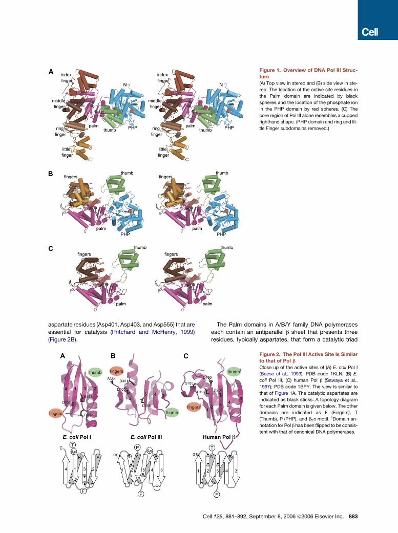
Figure 1. Overview of DNA Pol III Struc-
ture
(A) Top view in stereo and (B) side view in ste-
reo. The location of the active site residues in
the Palm domain are indicated by black
spheres and the location of the phosphate ion
in the PHP domain by red spheres. (C) The
core region of Pol III alone resembles a cupped
righthand shape. (PHP domain and ring and lit-
tle Finger subdomains removed.)
aspartate residues (Asp401, Asp403, and Asp555) that are
essential for catalysis (Pritchard and McHenry, 1999)
(Figure 2B).
The Palm domains in A/B/Y family DNA polymerases
each contain an antiparallel b sheet that presents three
residues, typically aspartates, that form a catalytic triad
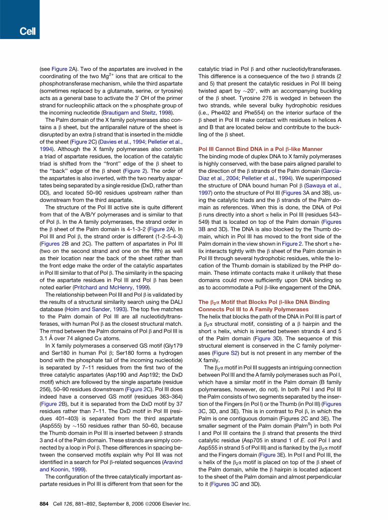
Figure 2. The Pol III Active Site Is Similar
to that of Pol b
Close up of the active sites of (A) E. coli Pol I
(Beese et al., 1993); PDB code 1KLN, (B) E.
coli Pol III, (C) human Pol b (Sawaya et al.,
1997); PDB code 1BPY. The view is similar to
that of Figure 1A. The catalytic aspartates are
indicated as black sticks. A topology diagram
for each Palm domain is given below. The other
domains are indicated as F (Fingers), T
(Thumb), P (PHP), and b2a motif. xDomain an-
notation for Pol b has been flipped to be consis-
tent with that of canonical DNA polymerases.
Cell 126, 881–892, September 8, 2006 ª2006 Elsevier Inc. 883
(see Figure 2A). Two of the aspartates are involved in the
coordinating of the two Mg2+ ions that are critical to the
phosphotransferase mechanism, while the third aspartate
(sometimes replaced by a glutamate, serine, or tyrosine)
acts as a general base to activate the 30 OH of the primer
strand for nucleophilic attack on the a phosphate group of
the incoming nucleotide (Brautigam and Steitz, 1998).
The Palm domain of the X family polymerases also con-
tains a b sheet, but the antiparallel nature of the sheet is
disrupted by an extra b strand that is inserted in the middle
of the sheet (Figure 2C) (Davies et al., 1994; Pelletier et al.,
1994). Although the X family polymerases also contain
a triad of aspartate residues, the location of the catalytic
triad is shifted from the ‘‘front’’ edge of the b sheet to
the ‘‘back’’ edge of the b sheet (Figure 2). The order of
the aspartates is also inverted, with the two nearby aspar-
tates being separated by a single residue (DxD, rather than
DD), and located 50–90 residues upstream rather than
downstream from the third aspartate.
The structure of the Pol III active site is quite different
from that of the A/B/Y polymerases and is similar to that
of Pol b. In the A family polymerases, the strand order in
the b sheet of the Palm domain is 4-1-3-2 (Figure 2A). In
Pol III and Pol b, the strand order is different (1-2-5-4-3)
(Figures 2B and 2C). The pattern of aspartates in Pol III
(two on the second strand and one on the fifth) as well
as their location near the back of the sheet rather than
the front edge make the order of the catalytic aspartates
in Pol III similar to that of Pol b. The similarity in the spacing
of the aspartate residues in Pol III and Pol b has been
noted earlier (Pritchard and McHenry, 1999).
The relationship between Pol III and Pol b is validated by
the results of a structural similarity search using the DALI
database (Holm and Sander, 1993). The top five matches
to the Palm domain of Pol III are all nucleotidyltrans-
ferases, with human Pol b as the closest structural match.
The rmsd between the Palm domains of Pol b and Pol III is
3.1 A over 74 aligned Ca atoms.
In X family polymerases a conserved GS motif (Gly179
and Ser180 in human Pol b; Ser180 forms a hydrogen
bond with the phosphate tail of the incoming nucleotide)
is separated by 7–11 residues from the first two of the
three catalytic aspartates (Asp190 and Asp192; the DxD
motif) which are followed by the single aspartate (residue
256), 50–90 residues downstream (Figure 2C). Pol III does
indeed have a conserved GS motif (residues 363–364)
(Figure 2B), but it is separated from the DxD motif by 37
residues rather than 7–11. The DxD motif in Pol III (resi-
dues 401–403) is separated from the third aspartate
(Asp555) by �150 residues rather than 50–60, because
the Thumb domain in Pol III is inserted between b strands
3 and 4 of the Palm domain. These strands are simply con-
nected by a loop in Pol b. These differences in spacing be-
tween the conserved motifs explain why Pol III was not
identified in a search for Pol b-related sequences (Aravind
and Koonin, 1999).
The configuration of the three catalytically important as-
partate residues in Pol III is different from that seen for the
884 Cell 126, 881–892, September 8, 2006 ª2006 Elsevier In
catalytic triad in Pol b and other nucleotidyltransferases.
This difference is a consequence of the two b strands (2
and 5) that present the catalytic residues in Pol III being
twisted apart by �20�, with an accompanying buckling
of the b sheet. Tyrosine 276 is wedged in between the
two strands, while several bulky hydrophobic residues
(i.e., Phe402 and Phe554) on the interior surface of the
b sheet in Pol III make contact with residues in helices A
and B that are located below and contribute to the buck-
ling of the b sheet.
Pol III Cannot Bind DNA in a Pol b-like Manner
The binding mode of duplex DNA to X family polymerases
is highly conserved, with the base pairs aligned parallel to
the direction of the b strands of the Palm domain (Garcia-
Diaz et al., 2004; Pelletier et al., 1994). We superimposed
the structure of DNA bound human Pol b (Sawaya et al.,
1997) onto the structure of Pol III (Figures 3A and 3B), us-
ing the catalytic triads and the b strands of the Palm do-
main as references. When this is done, the DNA of Pol
b runs directly into a short a helix in Pol III (residues 543–
549) that is located on top of the Palm domain (Figures
3B and 3D). The DNA is also blocked by the Thumb do-
main, which in Pol III has moved to the front side of the
Palm domain in the view shown in Figure 2. The short a he-
lix interacts tightly with the b sheet of the Palm domain in
Pol III through several hydrophobic residues, while the lo-
cation of the Thumb domain is stabilized by the PHP do-
main. These intimate contacts make it unlikely that these
domains could move sufficiently upon DNA binding so
as to accommodate a Pol b-like engagement of the DNA.
The b2a Motif that Blocks Pol b-like DNA Binding
Connects Pol III to A Family Polymerases
The helix that blocks the path of the DNA in Pol III is part of
a b2a structural motif, consisting of a b hairpin and the
short a helix, which is inserted between strands 4 and 5
of the Palm domain (Figure 3D). The sequence of this
structural element is conserved in the C family polymer-
ases (Figure S2) but is not present in any member of the
X family.
The b2a motif in Pol III suggests an intriguing connection
between Pol III and the A family polymerases such as Pol I,
which have a similar motif in the Palm domain (B family
polymerases, however, do not). In both Pol I and Pol III
the Palm consists of two segments separated by the inser-
tion of the Fingers (in Pol I) or the Thumb (in Pol III) (Figures
3C, 3D, and 3E). This is in contrast to Pol b, in which the
Palm is one contiguous domain (Figures 2C and 3E). The
smaller segment of the Palm domain (PalmS) in both Pol
I and Pol III contains the b strand that presents the third
catalytic residue (Asp705 in strand 1 of E. coli Pol I and
Asp555 in strand 5 of Pol III) and is flanked by the b2a motif
and the Fingers domain (Figure 3E). In Pol I and Pol III, the
a helix of the b2a motif is placed on top of the b sheet of
the Palm domain, while the b hairpin is located adjacent
to the sheet of the Palm domain and almost perpendicular
to it (Figures 3C and 3D).
c.
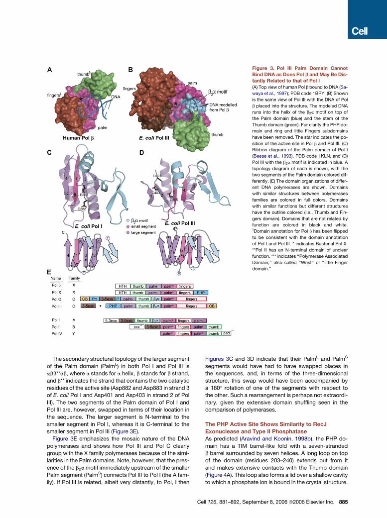
Figure 3. Pol III Palm Domain Cannot
Bind DNA as Does Pol b and May Be Dis-
tantly Related to that of Pol I
(A) Top view of human Pol b bound to DNA (Sa-
waya et al., 1997); PDB code 1BPY. (B) Shown
is the same view of Pol III with the DNA of Pol
b placed into the structure. The modeled DNA
runs into the helix of the b2a motif on top of
the Palm domain (blue) and the stem of the
Thumb domain (green). For clarity the PHP do-
main and ring and little Fingers subdomains
have been removed. The star indicates the po-
sition of the active site in Pol b and Pol III. (C)
Ribbon diagram of the Palm domain of Pol I
(Beese et al., 1993), PDB code 1KLN, and (D)
Pol III with the b2a motif is indicated in blue. A
topology diagram of each is shown, with the
two segments of the Palm domain colored dif-
ferently. (E) The domain organizations of differ-
ent DNA polymerases are shown. Domains
with similar structures between polymerases
families are colored in full colors. Domains
with similar functions but different structures
have the outline colored (i.e., Thumb and Fin-
gers domain). Domains that are not related by
function are colored in black and white.xDomain annotation for Pol b has been flipped
to be consistent with the domain annotation
of Pol I and Pol III. * indicates Bacterial Pol X.
**Pol II has an N-terminal domain of unclear
function. *** indicates ‘‘Polymerase Associated
Domain,’’ also called ‘‘Wrist’’ or ‘‘little Finger
domain.’’
The secondary structural topology of the larger segment
of the Palm domain (PalmL) in both Pol I and Pol III is
abb**ab, where a stands for a helix, b stands for b strand,
and b** indicates the strand that contains the two catalytic
residues of the active site (Asp882 and Asp883 in strand 3
of E. coli Pol I and Asp401 and Asp403 in strand 2 of Pol
III). The two segments of the Palm domain of Pol I and
Pol III are, however, swapped in terms of their location in
the sequence. The larger segment is N-terminal to the
smaller segment in Pol I, whereas it is C-terminal to the
smaller segment in Pol III (Figure 3E).
Figure 3E emphasizes the mosaic nature of the DNA
polymerases and shows how Pol III and Pol C clearly
group with the X family polymerases because of the simi-
larities in the Palm domains. Note, however, that the pres-
ence of the b2a motif immediately upstream of the smaller
Palm segment (PalmS) connects Pol III to Pol I (the A fam-
ily). If Pol III is related, albeit very distantly, to Pol, I then
Figures 3C and 3D indicate that their PalmL and PalmS
segments would have had to have swapped places in
the sequences, and, in terms of the three-dimensional
structure, this swap would have been accompanied by
a 180� rotation of one of the segments with respect to
the other. Such a rearrangement is perhaps not extraordi-
nary, given the extensive domain shuffling seen in the
comparison of polymerases.
The PHP Active Site Shows Similarity to RecJ
Exonuclease and Type II Phosphatase
As predicted (Aravind and Koonin, 1998b), the PHP do-
main has a TIM barrel-like fold with a seven-stranded
b barrel surrounded by seven helices. A long loop on top
of the domain (residues 203–240) extends out from it
and makes extensive contacts with the Thumb domain
(Figure 4A). This loop also forms a lid over a shallow cavity
to which a phosphate ion is bound in the crystal structure.
Cell 126, 881–892, September 8, 2006 ª2006 Elsevier Inc. 885
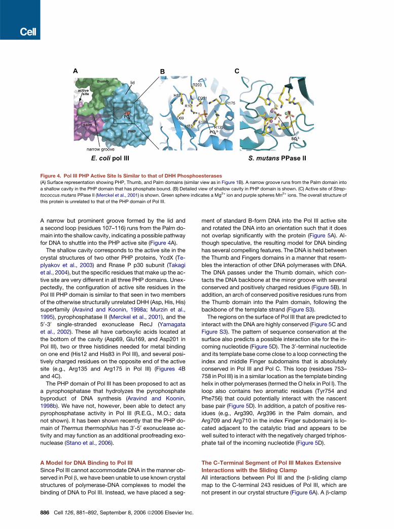
Figure 4. Pol III PHP Active Site Is Similar to that of DHH Phosphoesterases
(A) Surface representation showing PHP, Thumb, and Palm domains (similar view as in Figure 1B). A narrow groove runs from the Palm domain into
a shallow cavity in the PHP domain that has phosphate bound. (B) Detailed view of shallow cavity in PHP domain is shown. (C) Active site of Strep-
tococcus mutans PPase II (Merckel et al., 2001) is shown. Green sphere indicates a Mg2+ ion and purple spheres Mn2+ ions. The overall structure of
this protein is unrelated to that of the PHP domain of Pol III.
A narrow but prominent groove formed by the lid and
a second loop (residues 107–116) runs from the Palm do-
main into the shallow cavity, indicating a possible pathway
for DNA to shuttle into the PHP active site (Figure 4A).
The shallow cavity corresponds to the active site in the
crystal structures of two other PHP proteins, YcdX (Te-
plyakov et al., 2003) and Rnase P p30 subunit (Takagi
et al., 2004), but the specific residues that make up the ac-
tive site are very different in all three PHP domains. Unex-
pectedly, the configuration of active site residues in the
Pol III PHP domain is similar to that seen in two members
of the otherwise structurally unrelated DHH (Asp, His, His)
superfamily (Aravind and Koonin, 1998a; Murzin et al.,
1995), pyrophosphatase II (Merckel et al., 2001), and the
50-30 single-stranded exonuclease RecJ (Yamagata
et al., 2002). These all have carboxylic acids located at
the bottom of the cavity (Asp69, Glu169, and Asp201 in
Pol III), two or three histidines needed for metal binding
on one end (His12 and His83 in Pol III), and several posi-
tively charged residues on the opposite end of the active
site (e.g., Arg135 and Arg175 in Pol III) (Figures 4B
and 4C).
The PHP domain of Pol III has been proposed to act as
a pyrophosphatase that hydrolyzes the pyrophosphate
byproduct of DNA synthesis (Aravind and Koonin,
1998b). We have not, however, been able to detect any
pyrophosphatase activity in Pol III (R.E.G., M.O.; data
not shown). It has been shown recently that the PHP do-
main of Thermus thermophilus has 30-50 exonuclease ac-
tivity and may function as an additional proofreading exo-
nuclease (Stano et al., 2006).
A Model for DNA Binding to Pol III
Since Pol III cannot accommodate DNA in the manner ob-
served in Pol b, we have been unable to use known crystal
structures of polymerase-DNA complexes to model the
binding of DNA to Pol III. Instead, we have placed a seg-
886 Cell 126, 881–892, September 8, 2006 ª2006 Elsevier In
ment of standard B-form DNA into the Pol III active site
and rotated the DNA into an orientation such that it does
not overlap significantly with the protein (Figure 5A). Al-
though speculative, the resulting model for DNA binding
has several compelling features. The DNA is held between
the Thumb and Fingers domains in a manner that resem-
bles the interaction of other DNA polymerases with DNA.
The DNA passes under the Thumb domain, which con-
tacts the DNA backbone at the minor groove with several
conserved and positively charged residues (Figure 5B). In
addition, an arch of conserved positive residues runs from
the Thumb domain into the Palm domain, following the
backbone of the template strand (Figure S3).
The regions on the surface of Pol III that are predicted to
interact with the DNA are highly conserved (Figure 5C and
Figure S3). The pattern of sequence conservation at the
surface also predicts a possible interaction site for the in-
coming nucleotide (Figure 5D). The 30-terminal nucleotide
and its template base come close to a loop connecting the
index and middle Finger subdomains that is absolutely
conserved in Pol III and Pol C. This loop (residues 753–
758 in Pol III) is in a similar location as the template binding
helix in other polymerases (termed the O helix in Pol I). The
loop also contains two aromatic residues (Tyr754 and
Phe756) that could potentially interact with the nascent
base pair (Figure 5D). In addition, a patch of positive res-
idues (e.g., Arg390, Arg396 in the Palm domain, and
Arg709 and Arg710 in the index Finger subdomain) is lo-
cated adjacent to the catalytic triad and appears to be
well suited to interact with the negatively charged triphos-
phate tail of the incoming nucleotide (Figure 5D).
The C-Terminal Segment of Pol III Makes Extensive
Interactions with the Sliding Clamp
All interactions between Pol III and the b-sliding clamp
map to the C-terminal 243 residues of Pol III, which are
not present in our crystal structure (Figure 6A). A b-clamp
c.
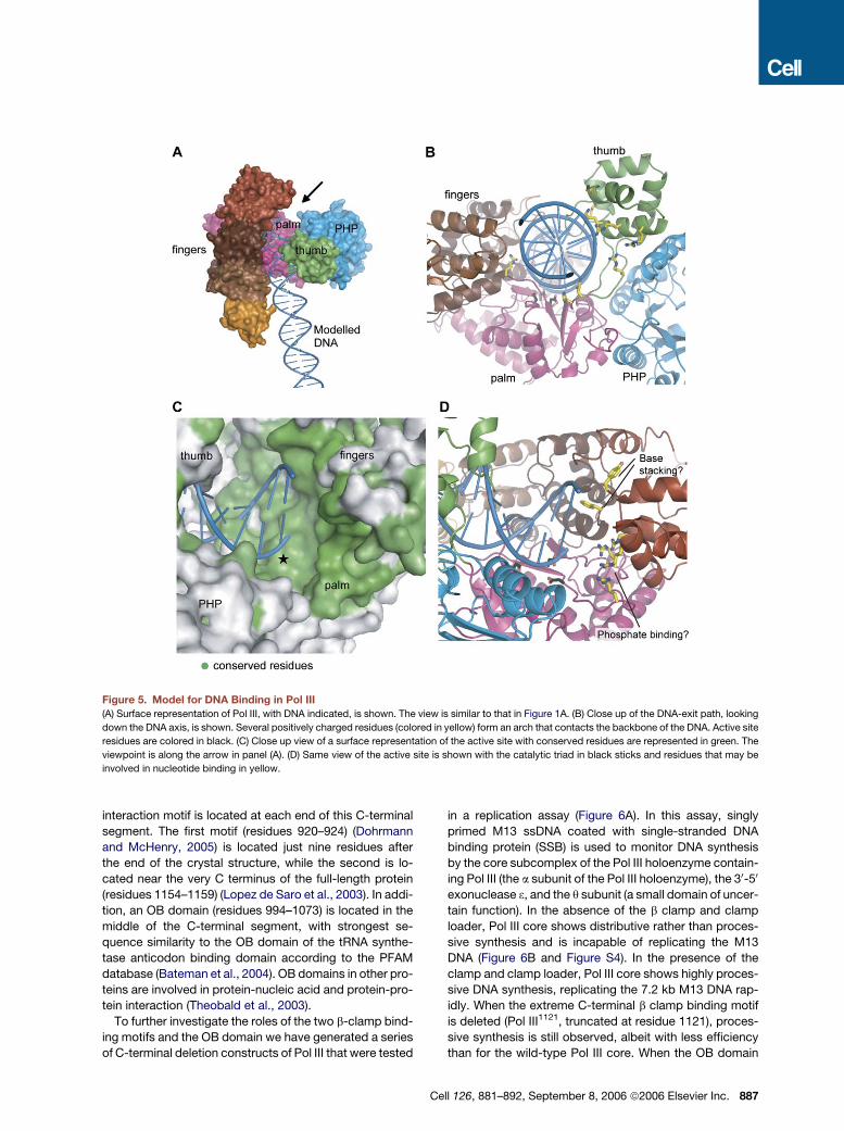
Figure 5. Model for DNA Binding in Pol III
(A) Surface representation of Pol III, with DNA indicated, is shown. The view is similar to that in Figure 1A. (B) Close up of the DNA-exit path, looking
down the DNA axis, is shown. Several positively charged residues (colored in yellow) form an arch that contacts the backbone of the DNA. Active site
residues are colored in black. (C) Close up view of a surface representation of the active site with conserved residues are represented in green. The
viewpoint is along the arrow in panel (A). (D) Same view of the active site is shown with the catalytic triad in black sticks and residues that may be
involved in nucleotide binding in yellow.
interaction motif is located at each end of this C-terminal
segment. The first motif (residues 920–924) (Dohrmann
and McHenry, 2005) is located just nine residues after
the end of the crystal structure, while the second is lo-
cated near the very C terminus of the full-length protein
(residues 1154–1159) (Lopez de Saro et al., 2003). In addi-
tion, an OB domain (residues 994–1073) is located in the
middle of the C-terminal segment, with strongest se-
quence similarity to the OB domain of the tRNA synthe-
tase anticodon binding domain according to the PFAM
database (Bateman et al., 2004). OB domains in other pro-
teins are involved in protein-nucleic acid and protein-pro-
tein interaction (Theobald et al., 2003).
To further investigate the roles of the two b-clamp bind-
ing motifs and the OB domain we have generated a series
of C-terminal deletion constructs of Pol III that were tested
C
in a replication assay (Figure 6A). In this assay, singly
primed M13 ssDNA coated with single-stranded DNA
binding protein (SSB) is used to monitor DNA synthesis
by the core subcomplex of the Pol III holoenzyme contain-
ing Pol III (the a subunit of the Pol III holoenzyme), the 30-50
exonuclease 3, and the q subunit (a small domain of uncer-
tain function). In the absence of the b clamp and clamp
loader, Pol III core shows distributive rather than proces-
sive synthesis and is incapable of replicating the M13
DNA (Figure 6B and Figure S4). In the presence of the
clamp and clamp loader, Pol III core shows highly proces-
sive DNA synthesis, replicating the 7.2 kb M13 DNA rap-
idly. When the extreme C-terminal b clamp binding motif
is deleted (Pol III1121, truncated at residue 1121), proces-
sive synthesis is still observed, albeit with less efficiency
than for the wild-type Pol III core. When the OB domain
ell 126, 881–892, September 8, 2006 ª2006 Elsevier Inc. 887
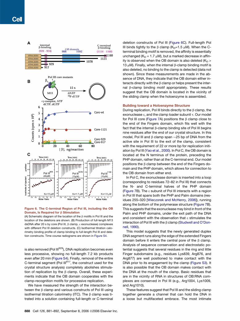
is also removed (Pol III929), DNA replication becomes even
less processive, showing no full-length 7.2 kb products
even after 20 min (Figure S4). Finally, removal of the entire
C-terminal segment (Pol III917, the construct used for the
crystal structure analysis) completely abolishes stimula-
tion of replication by the b clamp. Overall, these experi-
ments indicate that the OB domain cooperates with the
clamp-recognition motifs for processive replication.
We have measured the strength of the interaction be-
tween the b clamp and various constructs of Pol III using
isothermal titration calorimetry (ITC). The b clamp was ti-
trated into a solution containing full-length or C-terminal
Figure 6. The C-terminal Region of Pol III, including the OB
Domain, Is Required for b Stimulation
(A) Schematic diagram of the location of the b motifs in Pol III and the
location of the deletions are shown. (B) Production of full-length M13
dsDNA after 20 s by core (Pol III, b clamp, 3 exonuclease) complexes
with different Pol III deletion constructs. (C) Isothermal titration calo-
rimetry binding profile of clamp binding to full-length Pol III and dele-
tion constructs. Time course radiograms are shown in Figure S4.
888 Cell 126, 881–892, September 8, 2006 ª2006 Elsevier Inc.
deletion constructs of Pol III (Figure 6C). Full-length Pol
III binds tightly to the b clamp (KD=1.5 mM). When the C-
terminal binding motif is removed, the affinity is essentially
unchanged (KD = 1.7 mM), but a marked decrease in affin-
ity is observed when the OB domain is also deleted (KD >
13 mM). Finally, when the internal b-clamp binding motif is
also deleted, no binding to the clamp is detected (data not
shown). Since these measurements are made in the ab-
sence of DNA, they indicate that the OB domain either in-
teracts directly with the b clamp or helps present the inter-
nal b-clamp binding motif appropriately. These results
suggest that the OB domain is located in the vicinity of
the sliding clamp when the holoenzyme is assembled.
Building toward a Holoenzyme Structure
During replication, Pol III binds directly to the b clamp, the
exonuclease 3, and the clamp loader subunit t. Our model
for Pol III core (Figure 7A) positions the b clamp close to
the end of the Fingers domain, which fits well with the
fact that the internal b-clamp binding site of Pol III begins
nine residues after the end of our crystal structure. In this
model, Pol III and b clamp span �25 bp of DNA from the
active site in Pol III to the exit of the clamp, consistent
with the requirement of 22 or more bp for replication initi-
ation by Pol III (Yao et al., 2000). In Pol C, the OB domain is
located at the N terminus of the protein, preceding the
PHP domain, rather than at the C-terminal end. Our model
positions the b clamp between the end of the Fingers do-
main and the PHP domain, which allows for connection to
the OB domain from either end.
In Pol C, the exonuclease domain is inserted into a loop
(corresponding to residues 72–82 in Pol III) that connects
the N- and C-terminal halves of the PHP domain
(Figure 7B). The 3 subunit of Pol III interacts with a region
in Pol III that spans both the PHP and Palm domains (res-
idues 255–320 [Wieczorek and McHenry, 2006]), running
along the bottom of the polymerase structure (Figure 7B).
This suggests that the exonuclease may bind in front of the
Palm and PHP domains, under the exit path of the DNA
and consistent with the observation that 3 stimulates the
interaction of Pol III with the b clamp (Studwell and O’Don-
nell, 1990).
This model suggests that the newly generated duplex
DNA segment runs along the edge of the extended Fingers
domain before it enters the central pore of the b clamp.
Analysis of sequence conservation and electrostatic po-
tential suggests that several residues in the ring and little
Finger subdomains (e.g., residues Lys839, Arg876, and
Arg877) are well positioned to make contact with the
DNA prior to its engagement by the clamp (Figure S3). It
is also possible that the OB domain makes contact with
the DNA at the mouth of the clamp. Basic residues that
are in the vicinity of RNA in structures of OB:RNA com-
plexes are conserved in Pol III (e.g., Arg1004, Lys1009,
and Arg1010).
These features suggest that Pol III and the sliding clamp
together generate a channel that can hold the DNA in
a loose but multifaceted embrace. The most intimate
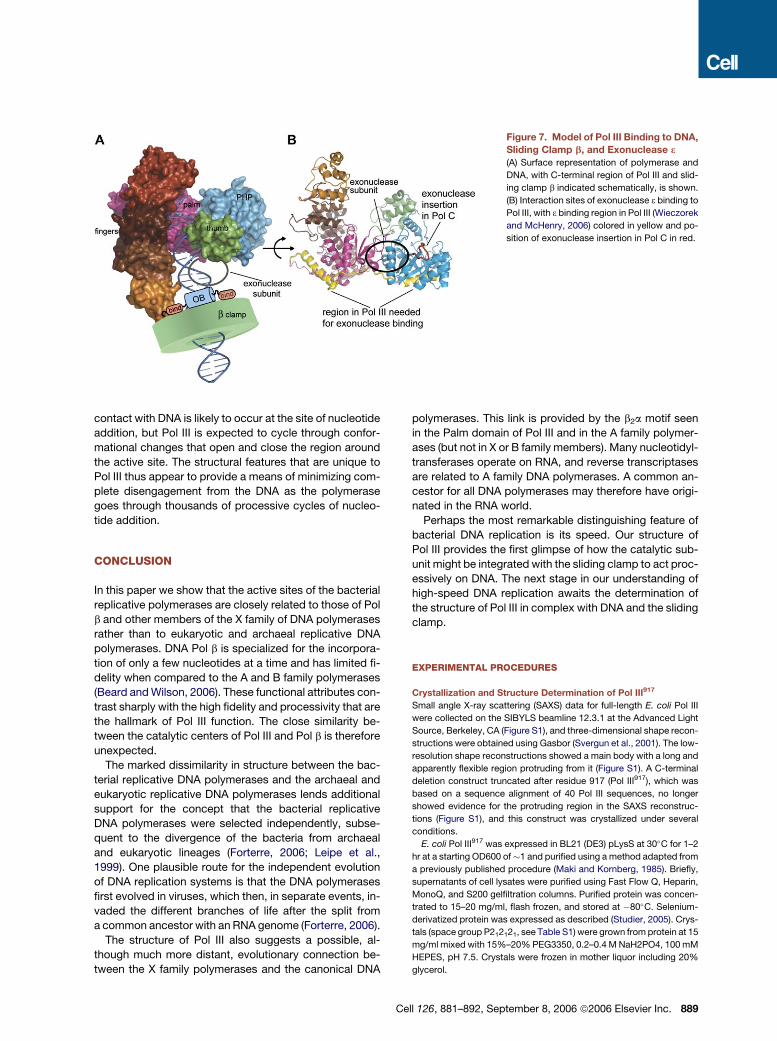
Figure 7. Model of Pol III Binding to DNA,
Sliding Clamp b, and Exonuclease 3
(A) Surface representation of polymerase and
DNA, with C-terminal region of Pol III and slid-
ing clamp b indicated schematically, is shown.
(B) Interaction sites of exonuclease 3 binding to
Pol III, with 3 binding region in Pol III (Wieczorek
and McHenry, 2006) colored in yellow and po-
sition of exonuclease insertion in Pol C in red.
contact with DNA is likely to occur at the site of nucleotide
addition, but Pol III is expected to cycle through confor-
mational changes that open and close the region around
the active site. The structural features that are unique to
Pol III thus appear to provide a means of minimizing com-
plete disengagement from the DNA as the polymerase
goes through thousands of processive cycles of nucleo-
tide addition.
CONCLUSION
In this paper we show that the active sites of the bacterial
replicative polymerases are closely related to those of Pol
b and other members of the X family of DNA polymerases
rather than to eukaryotic and archaeal replicative DNA
polymerases. DNA Pol b is specialized for the incorpora-
tion of only a few nucleotides at a time and has limited fi-
delity when compared to the A and B family polymerases
(Beard and Wilson, 2006). These functional attributes con-
trast sharply with the high fidelity and processivity that are
the hallmark of Pol III function. The close similarity be-
tween the catalytic centers of Pol III and Pol b is therefore
unexpected.
The marked dissimilarity in structure between the bac-
terial replicative DNA polymerases and the archaeal and
eukaryotic replicative DNA polymerases lends additional
support for the concept that the bacterial replicative
DNA polymerases were selected independently, subse-
quent to the divergence of the bacteria from archaeal
and eukaryotic lineages (Forterre, 2006; Leipe et al.,
1999). One plausible route for the independent evolution
of DNA replication systems is that the DNA polymerases
first evolved in viruses, which then, in separate events, in-
vaded the different branches of life after the split from
a common ancestor with an RNA genome (Forterre, 2006).
The structure of Pol III also suggests a possible, al-
though much more distant, evolutionary connection be-
tween the X family polymerases and the canonical DNA
polymerases. This link is provided by the b2a motif seen
in the Palm domain of Pol III and in the A family polymer-
ases (but not in X or B family members). Many nucleotidyl-
transferases operate on RNA, and reverse transcriptases
are related to A family DNA polymerases. A common an-
cestor for all DNA polymerases may therefore have origi-
nated in the RNA world.
Perhaps the most remarkable distinguishing feature of
bacterial DNA replication is its speed. Our structure of
Pol III provides the first glimpse of how the catalytic sub-
unit might be integrated with the sliding clamp to act proc-
essively on DNA. The next stage in our understanding of
high-speed DNA replication awaits the determination of
the structure of Pol III in complex with DNA and the sliding
clamp.
EXPERIMENTAL PROCEDURES
Crystallization and Structure Determination of Pol III917
Small angle X-ray scattering (SAXS) data for full-length E. coli Pol III
were collected on the SIBYLS beamline 12.3.1 at the Advanced Light
Source, Berkeley, CA (Figure S1), and three-dimensional shape recon-
structions were obtained using Gasbor (Svergun et al., 2001). The low-
resolution shape reconstructions showed a main body with a long and
apparently flexible region protruding from it (Figure S1). A C-terminal
deletion construct truncated after residue 917 (Pol III917), which was
based on a sequence alignment of 40 Pol III sequences, no longer
showed evidence for the protruding region in the SAXS reconstruc-
tions (Figure S1), and this construct was crystallized under several
conditions.
E. coli Pol III917 was expressed in BL21 (DE3) pLysS at 30�C for 1–2
hr at a starting OD600 of�1 and purified using a method adapted from
a previously published procedure (Maki and Kornberg, 1985). Briefly,
supernatants of cell lysates were purified using Fast Flow Q, Heparin,
MonoQ, and S200 gelfiltration columns. Purified protein was concen-
trated to 15–20 mg/ml, flash frozen, and stored at �80�C. Selenium-
derivatized protein was expressed as described (Studier, 2005). Crys-
tals (space group P212121, see Table S1) were grown from protein at 15
mg/ml mixed with 15%–20% PEG3350, 0.2–0.4 M NaH2PO4, 100 mM
HEPES, pH 7.5. Crystals were frozen in mother liquor including 20%
glycerol.
Cell 126, 881–892, September 8, 2006 ª2006 Elsevier Inc. 889
A 3 wavelength MAD (multiple-wavelength anomalous diffraction)
data set to 3.1 A was collected at beamline 8.2.1 at the Advanced Light
Source, Berkeley, CA. All data were processed with HKL2000 (Otwi-
nowski and Minor, 1997). The locations of selenium sites were found
with SHELXD (Schneider and Sheldrick, 2002) using the peak wave-
length data set only. The sites were refined in SHARP (Bricogne
et al., 2003) using all three wavelengths of the MAD data set. A
higher-resolution data set to 2.6 A was collected at beamline 8.3.1
and phase extension to 2.6 A was performed using RESOLVE (Terwil-
liger, 2000), producing a map in which the larger part of the molecule
could be traced. A large flexible segment of the structure (residues
�600–917) could only be built using electron density maps produced
with BUSTER-TNT (Blanc et al., 2004), with model improvement by
simulated annealing using CNS (crystallography and NMR system)
(Brunger et al., 1998). A third data set to 2.3 A was collected after con-
trolled dehydration using the Free Mounting System (Rigaku) at the
SIBYLS beamline 12.3.1 at the Advanced Light Source. This data set
showed significant shrinkage in unit cell dimensions (from a = 83.7 A,
b = 100.7 A, c = 143.7 A to a = 83.7 A, b = 94.0 A, c = 131.2 A) and
the resulting electron density map was much improved, particularly
for the flexible segment. Except for a �5� rotation of the flexible seg-
ment no significant structural changes were observed. The model
was built using O (Jones et al., 1991) and Coot (Emsley and Cowtan,
2004) and refined using Refmac5 with the TLS option (Winn et al.,
2001). The final model (Rwork = 19.7%, Rfree = 26.5%) spans residues
2–911. Complete statistics for the model are in Table S1. Figures
were prepared using Pymol (www.pymol.org).
Primed M13mp18 ssDNA Extension Assays
Replication assays were performed in 25 ml reactions containing 30
fmol of M13mp18 ssDNA primed with a 30-mer ssDNA, 0.8 mg SSB,
0.5 mM ATP, 60 mM dCTP, 60 mM dGTP, 740 fmol of b clamp, 20
fmol of clamp loader, and 0.5 pmol Pol III or mutant Pol III cores. Pol
III core was reconstituted by incubating 0.5 pmol (Pol III wild-type
and its truncations at residues 917, 929, 1078, and 1121, to which 3
and q subunits were added in 1:1.5 ratio) and preincubated 5 min at
37�C. Replication reaction was started upon addition of dATP and
[a-32P] dTTP to final concentrations of 60 and 20 mM, respectively.
For time course reactions (Figure S4) the replication times are indi-
cated in the Figure S4, and reactions were quenched upon addition
of an equal volume of 1% SDS/40 mM EDTA. Products were analyzed
in a 0.8% agarose gel in 1 3 TBE. For Pol III replication reactions, var-
ious amounts of b clamp were used (0–740 pmoles), and reactions
were quenched after 15 s by spotting reactions on DEAE-cellulose cir-
cles (Whatman) followed by washing and counting in a liquid scintilla-
tion counter.
Isothermal Titration Calorimetry Experiments
Binding studies were performed by isothermal titration calorimetry
(ITC) using a VP-ITC microcalorimeter (MicroCal, Northampton, MA).
b clamp and Pol III (Pol III929, Pol III1078, Pol III1160) were dialyzed exten-
sively against 20 mM Hepes buffer pH 7.5, 0.5 mM EDTA, and 50 mM
NaCl. The reaction cell (1.4 ml) was filled with degassed solutions and
equilibrated at 25�C. Stirring speed was 400 rpm, the thermal power
was recorded every second, and instrumental feedback (reference
power) was 10%. In a typical experiment, b clamp (100–220 mM in
the syringe) was titrated over 28 injections of 10 ml into the cell contain-
ing 10–40 mM solution of purified Pol III1160 or its truncations. An initial
injection of 2 ml was made before each titration to ensure that the titrant
(b clamp) concentration was at its loading value. The heats of dilution
were obtained at 25�C from separate titration experiments of b clamp
into buffer. The corrected quantity of heat released due to b clamp
binding to Pol III and Pol III deletion constructs was measured by inte-
grating the area of each titration peak. Calculation of binding enthalpy,
entropy, and stoichiometry from protein titration data was accom-
plished using ORIGIN software (Microcal).
890 Cell 126, 881–892, September 8, 2006 ª2006 Elsevier Inc.
Supplemental Data
Supplemental Data include four figures and two tables and can be
found with this article online at http://www.cell.com/cgi/content/full/
126/5/881/DC1/.
ACKNOWLEDGMENTS
We thank the staff at beamlines 8.2.1, 8.2.2, 8.3.1, and 12.3.1 of the
Advanced Light Source, Berkeley, CA, for their help and technical sup-
port. We are thankful to Clemens Vonrhein for help with BUSTER-TNT,
Axel Brunger for help with CNS, and Anastassis Perrakis for helpful
suggestions. We thank members of the Kuriyan laboratory, particularly
Joel Guenther, Steven Kazmirsky, and Xuewu Zhang for help and ad-
vice. This work was supported in part by grants from the NIH to J.K.
(GM45547) and M.O. (GM38839). M.H.L. was supported by a Jane
Coffin Childs Memorial Fund Fellowship.
Received: May 16, 2006
Revised: June 29, 2006
Accepted: July 29, 2006
Published: September 7, 2006
REFERENCES
Aravind, L., and Koonin, E.V. (1998a). A novel family of predicted phos-
phoesterases includes Drosophila prune protein and bacterial RecJ
exonuclease. Trends Biochem. Sci. 23, 17–19.
Aravind, L., and Koonin, E.V. (1998b). Phosphoesterase domains as-
sociated with DNA polymerases of diverse origins. Nucleic Acids
Res. 26, 3746–3752.
Aravind, L., and Koonin, E.V. (1999). DNA polymerase beta-like nucle-
otidyltransferase superfamily: identification of three new families, clas-
sification and evolutionary history. Nucleic Acids Res. 27, 1609–1618.
Bateman, A., Coin, L., Durbin, R., Finn, R.D., Hollich, V., Griffiths-
Jones, S., Khanna, A., Marshall, M., Moxon, S., Sonnhammer, E.L.,
et al. (2004). The Pfam protein families database. Nucleic Acids Res.
32, D138–D141.
Beard, W.A., and Wilson, S.H. (2006). Structure and mechanism of
DNA polymerase Beta. Chem. Rev. 106, 361–382.
Beese, L.S., Derbyshire, V., and Steitz, T.A. (1993). Structure of DNA
polymerase I Klenow fragment bound to duplex DNA. Science 260,
352–355.
Blanc, E., Roversi, P., Vonrhein, C., Flensburg, C., Lea, S.M., and Bri-
cogne, G. (2004). Refinement of severely incomplete structures with
maximum likelihood in BUSTER-TNT. Acta Crystallogr. D Biol. Crystal-
logr. 60, 2210–2221.
Bloom, L.B., Chen, X., Fygenson, D.K., Turner, J., O’Donnell, M., and
Goodman, M.F. (1997). Fidelity of Escherichia coli DNA polymerase III
holoenzyme. The effects of beta, gamma complex processivity pro-
teins and epsilon proofreading exonuclease on nucleotide misincorpo-
ration efficiencies. J. Biol. Chem. 272, 27919–27930.
Bonner, C.A., Hays, S., McEntee, K., and Goodman, M.F. (1990). DNA
polymerase II is encoded by the DNA damage-inducible dinA gene of
Escherichia coli. Proc. Natl. Acad. Sci. USA 87, 7663–7667.
Bowman, G.D., Goedken, E.R., Kazmirski, S.L., O’Donnell, M., and
Kuriyan, J. (2005). DNA polymerase clamp loaders and DNA recogni-
tion. FEBS Lett. 579, 863–867.
Braithwaite, D.K., and Ito, J. (1993). Compilation, alignment, and phy-
logenetic relationships of DNA polymerases. Nucleic Acids Res. 21,
787–802.
Brautigam, C.A., and Steitz, T.A. (1998). Structural and functional in-
sights provided by crystal structures of DNA polymerases and their
substrate complexes. Curr. Opin. Struct. Biol. 8, 54–63.
Bricogne, G., Vonrhein, C., Flensburg, C., Schiltz, M., and Paciorek, W.
(2003). Generation, representation and flow of phase information in
structure determination: recent developments in and around SHARP
2.0. Acta Crystallogr. D Biol. Crystallogr. 59, 2023–2030.
Bruck, I., Goodman, M.F., and O’Donnell, M. (2003). The essential C
family DnaE polymerase is error-prone and efficient at lesion bypass.
J. Biol. Chem. 278, 44361–44368.
Brunger, A.T., Adams, P.D., Clore, G.M., DeLano, W.L., Gros, P.,
Grosse-Kunstleve, R.W., Jiang, J.S., Kuszewski, J., Nilges, M., Pannu,
N.S., et al. (1998). Crystallography & NMR system: A new software
suite for macromolecular structure determination. Acta Crystallogr.
D Biol. Crystallogr. 54, 905–921.
Davies, J.F., II, Almassy, R.J., Hostomska, Z., Ferre, R.A., and Hos-
tomsky, Z. (1994). 2.3 A crystal structure of the catalytic domain of
DNA polymerase beta. Cell 76, 1123–1133.
Dohrmann, P.R., and McHenry, C.S. (2005). A bipartite polymerase-
processivity factor interaction: only the internal beta binding site of
the alpha subunit is required for processive replication by the DNA
polymerase III holoenzyme. J. Mol. Biol. 350, 228–239.
Doublie, S., Tabor, S., Long, A.M., Richardson, C.C., and Ellenberger,
T. (1998). Crystal structure of a bacteriophage T7 DNA replication
complex at 2.2 A resolution. Nature 391, 251–258.
Emsley, P., and Cowtan, K. (2004). Coot: model-building tools for mo-
lecular graphics. Acta Crystallogr. D Biol. Crystallogr. 60, 2126–2132.
Eom, S.H., Wang, J., and Steitz, T.A. (1996). Structure of Taq polymer-
ase with DNA at the polymerase active site. Nature 382, 278–281.
Forterre, P. (2006). Three RNA cells for ribosomal lineages and three
DNA viruses to replicate their genomes: a hypothesis for the origin of
cellular domain. Proc. Natl. Acad. Sci. USA 103, 3669–3674.
Franklin, M.C., Wang, J., and Steitz, T.A. (2001). Structure of the rep-
licating complex of a pol alpha family DNA polymerase. Cell 105,
657–667.
Garcia-Diaz, M., Bebenek, K., Krahn, J.M., Blanco, L., Kunkel, T.A.,
and Pedersen, L.C. (2004). A structural solution for the DNA polymer-
ase lambda-dependent repair of DNA gaps with minimal homology.
Mol. Cell 13, 561–572.
Goodman, M.F. (2002). Error-prone repair DNA polymerases in pro-
karyotes and eukaryotes. Annu. Rev. Biochem. 71, 17–50.
Holm, L., and Sander, C. (1993). Protein structure comparison by align-
ment of distance matrices. J. Mol. Biol. 233, 123–138.
Holm, L., and Sander, C. (1995). DNA polymerase beta belongs to an
ancient nucleotidyltransferase superfamily. Trends Biochem. Sci. 20,
345–347.
Hopfner, K.P., Eichinger, A., Engh, R.A., Laue, F., Ankenbauer, W.,
Huber, R., and Angerer, B. (1999). Crystal structure of a thermostable
type B DNA polymerase from Thermococcus gorgonarius. Proc. Natl.
Acad. Sci. USA 96, 3600–3605.
Huang, Y., Braithwaite, D.K., and Ito, J. (1997). Evolution of dnaQ, the
gene encoding the editing 30 to 50 exonuclease subunit of DNA poly-
merase III holoenzyme in Gram-negative bacteria. FEBS Lett. 400,
94–98.
Johnson, A., and O’Donnell, M. (2005). Cellular DNA replicases: com-
ponents and dynamics at the replication fork. Annu. Rev. Biochem. 74,
283–315.
Jones, T.A., Zou, J.-Y., Cowan, S.W., and Kjeldgaard, M. (1991). Im-
proved methods for the building of protein models in electron density
maps and the location of errors in these models. Acta Crystallogr. A47,
110–119.
Kiefer, J.R., Mao, C., Braman, J.C., and Beese, L.S. (1998). Visualizing
DNA replication in a catalytically active Bacillus DNA polymerase crys-
tal. Nature 391, 304–307.
Kong, X.P., Onrust, R., O’Donnell, M., and Kuriyan, J. (1992). Three-di-
mensional structure of the beta subunit of E. coli DNA polymerase III
holoenzyme: a sliding DNA clamp. Cell 69, 425–437.
Krishna, T.S., Kong, X.P., Gary, S., Burgers, P.M., and Kuriyan, J.
(1994). Crystal structure of the eukaryotic DNA polymerase processiv-
ity factor PCNA. Cell 79, 1233–1243.
Leipe, D.D., Aravind, L., and Koonin, E.V. (1999). Did DNA replication
evolve twice independently? Nucleic Acids Res. 27, 3389–3401.
Li, Y., Korolev, S., and Waksman, G. (1998). Crystal structures of open
and closed forms of binary and ternary complexes of the large frag-
ment of Thermus aquaticus DNA polymerase I: structural basis for nu-
cleotide incorporation. EMBO J. 17, 7514–7525.
Lopez de Saro, F.J., Georgescu, R.E., and O’Donnell, M. (2003). A
peptide switch regulates DNA polymerase processivity. Proc. Natl.
Acad. Sci. USA 100, 14689–14694.
Maki, H., and Kornberg, A. (1985). The polymerase subunit of DNA
polymerase III of Escherichia coli. II. Purification of the alpha subunit,
devoid of nuclease activities. J. Biol. Chem. 260, 12987–12992.
Merckel, M.C., Fabrichniy, I.P., Salminen, A., Kalkkinen, N., Baykov,
A.A., Lahti, R., and Goldman, A. (2001). Crystal structure of Strepto-
coccus mutans pyrophosphatase: a new fold for an old mechanism.
Structure 9, 289–297.
Murzin, A.G., Brenner, S.E., Hubbard, T., and Chothia, C. (1995).
SCOP: a structural classification of proteins database for the investiga-
tion of sequences and structures. J. Mol. Biol. 247, 536–540.
Otwinowski, Z., and Minor, W. (1997). Processing of X-ray Diffraction
Data Collected in Oscillation Mode. Methods Enzymol. 276, 307–326.
Pelletier, H., Sawaya, M.R., Kumar, A., Wilson, S.H., and Kraut, J.
(1994). Structures of ternary complexes of rat DNA polymerase beta,
a DNA template-primer, and ddCTP. Science 264, 1891–1903.
Pritchard, A.E., and McHenry, C.S. (1999). Identification of the acidic
residues in the active site of DNA polymerase III. J. Mol. Biol. 285,
1067–1080.
Raghuraman, M.K., Winzeler, E.A., Collingwood, D., Hunt, S., Wo-
dicka, L., Conway, A., Lockhart, D.J., Davis, R.W., Brewer, B.J., and
Fangman, W.L. (2001). Replication dynamics of the yeast genome.
Science 294, 115–121.
Rodriguez, A.C., Park, H.W., Mao, C., and Beese, L.S. (2000). Crystal
structure of a pol alpha family DNA polymerase from the hyperthermo-
philic archaeon Thermococcus sp. 9 degrees N-7. J. Mol. Biol. 299,
447–462.
Rothwell, P.J., and Waksman, G. (2005). Structure and mechanism of
DNA polymerases. Adv. Protein Chem. 71, 401–440.
Sawaya, M.R., Prasad, R., Wilson, S.H., Kraut, J., and Pelletier, H.
(1997). Crystal structures of human DNA polymerase beta complexed
with gapped and nicked DNA: evidence for an induced fit mechanism.
Biochemistry 36, 11205–11215.
Schneider, T.R., and Sheldrick, G.M. (2002). Substructure solution with
SHELXD. Acta Crystallogr. D Biol. Crystallogr. 58, 1772–1779.
Stano, N.M., Chen, J., and McHenry, C.S. (2006). A coproofreading
Zn(2+)-dependent exonuclease within a bacterial replicase. Nat.
Struct. Mol. Biol. 13, 458–459.
Studier, F.W. (2005). Protein production by auto-induction in high den-
sity shaking cultures. Protein Expr. Purif. 41, 207–234.
Studwell, P.S., and O’Donnell, M. (1990). Processive replication is con-
tingent on the exonuclease subunit of DNA polymerase III holoenzyme.
J. Biol. Chem. 265, 1171–1178.
Svergun, D.I., Petoukhov, M.V., and Koch, M.H. (2001). Determination
of domain structure of proteins from X-ray solution scattering. Bio-
phys. J. 80, 2946–2953.
Takagi, H., Watanabe, M., Kakuta, Y., Kamachi, R., Numata, T., Ta-
naka, I., and Kimura, M. (2004). Crystal structure of the ribonuclease
Cell 126, 881–892, September 8, 2006 ª2006 Elsevier Inc. 891
P protein Ph1877p from hyperthermophilic archaeon Pyrococcus ho-
rikoshii OT3. Biochem. Biophys. Res. Commun. 319, 787–794.
Teplyakov, A., Obmolova, G., Khil, P.P., Howard, A.J., Camerini-Otero,
R.D., and Gilliland, G.L. (2003). Crystal structure of the Escherichia coli
YcdX protein reveals a trinuclear zinc active site. Proteins 51, 315–318.
Terwilliger, T.C. (2000). Maximum-likelihood density modification.
Acta Crystallogr. D Biol. Crystallogr. 56, 965–972.
Theobald, D.L., Mitton-Fry, R.M., and Wuttke, D.S. (2003). Nucleic acid
recognition by OB-fold proteins. Annu. Rev. Biophys. Biomol. Struct.
32, 115–133.
Waga, S., and Stillman, B. (1998). The DNA replication fork in eukary-
otic cells. Annu. Rev. Biochem. 67, 721–751.
Wang, J., Sattar, A.K., Wang, C.C., Karam, J.D., Konigsberg, W.H.,
and Steitz, T.A. (1997). Crystal structure of a pol alpha family replica-
tion DNA polymerase from bacteriophage RB69. Cell 89, 1087–1099.
Wieczorek, A., and McHenry, C.S. (2006). The NH2-terminal php do-
main of the alpha subunit of the Escherichia coli replicase binds the ep-
silon proofreading subunit. J. Biol. Chem. 281, 12561–12567.
892 Cell 126, 881–892, September 8, 2006 ª2006 Elsevier Inc.
Winn, M.D., Isupov, M.N., and Murshudov, G.N. (2001). Use of TLS pa-
rameters to model anisotropic displacements in macromolecular re-
finement. Acta Crystallogr. D Biol. Crystallogr. 57, 122–133.
Yamagata, A., Kakuta, Y., Masui, R., and Fukuyama, K. (2002). The
crystal structure of exonuclease RecJ bound to Mn2+ ion suggests
how its characteristic motifs are involved in exonuclease activity.
Proc. Natl. Acad. Sci. USA 99, 5908–5912.
Yao, N., Leu, F.P., Anjelkovic, J., Turner, J., and O’Donnell, M. (2000).
DNA structure requirements for the Escherichia coli gamma complex
clamp loader and DNA polymerase III holoenzyme. J. Biol. Chem.
275, 11440–11450.
Zhao, Y., Jeruzalmi, D., Moarefi, I., Leighton, L., Lasken, R., and Kur-
iyan, J. (1999). Crystal structure of an archaebacterial DNA polymer-
ase. Structure 7, 1189–1199.
Accession Numbers
The structures described in our paper have been deposited in the Pro-
tein Data Bank under ID codes 2HQA and 2HNH.