Embed Size (px)
Citation preview
Crystal structure of a translation terminationcomplex formed with release factor RF2Andrei Korosteleva,b,1, Haruichi Asaharaa,b,1,2, Laura Lancastera,b,1, Martin Laurberga,b, Alexander Hirschia,b,Jianyu Zhua,b, Sergei Trakhanova,b, William G. Scotta,c, and Harry F. Nollera,b,3
aCenter for Molecular Biology of RNA and Departments of bMolecular, Cell and Developmental Biology and cChemistry and Biochemistry,University of California, Santa Cruz, CA 95064
Contributed by Harry F. Noller, October 30, 2008 (sent for review October 22, 2008)
We report the crystal structure of a translation termination com-plex formed by the Thermus thermophilus 70S ribosome boundwith release factor RF2, in response to a UAA stop codon, solvedat 3 Å resolution. The backbone of helix �5 and the side chain ofserine of the conserved SPF motif of RF2 recognize U1 and A2 of thestop codon, respectively. A3 is unstacked from the first 2 bases,contacting Thr-216 and Val-203 of RF2 and stacking on G530 of 16SrRNA. The structure of the RF2 complex supports our previousproposal that conformational changes in the ribosome in responseto recognition of the stop codon stabilize rearrangement of theswitch loop of the release factor, resulting in docking of theuniversally conserved GGQ motif in the PTC of the 50S subunit. Asseen for the RF1 complex, the main-chain amide nitrogen ofglutamine in the GGQ motif is positioned to contribute directly tocatalysis of peptidyl-tRNA hydrolysis, consistent with mutationalstudies, which show that most side-chain substitutions of theconserved glutamine have little effect. We show that when theH-bonding capability of the main-chain N-H of the conservedglutamine is eliminated by substitution with proline, peptidyl-tRNA esterase activity is abolished, consistent with its proposedrole in catalysis.
70S ribosome structure � stop codon recognition � polypeptide release
In bacteria, termination of protein synthesis depends on thetype I release factors, RF1 and RF2, which are required for
recognition of the stop codons and for hydrolysis of the peptidyl-tRNA ester bond. Our understanding of the mechanism oftermination faces three main questions: (i) How are stop codonsrecognized? Unlike the sense codons, there are no correspond-ing cognate tRNAs to recognize nonsense codons. Are theyrecognized directly by the release factors, or indirectly, forexample through ribosomal RNA? (ii) What is the mechanism ofpeptidyl-tRNA hydrolysis? Is the esterase reaction catalyzeddirectly by the release factors, or by the ribosome? And (iii) Howis peptidyl-tRNA hydrolysis coupled to stop codon recognition?
Termination at the UAG stop codon depends on RF1, UGAon RF2, and UAA on either of the two factors (1–3). Thus,although the two release factors have similar overall structures(4–6) and both recognize codons of the general type URR, RF1is able to discriminate between A and G at the second positionwhereas RF2 discriminates between A and G at the thirdposition. Determinants for codon specificity were localized todomain 2 of the release factors, in particular to the conservedPxT and SPF motifs of RF1 and RF2, respectively, based ongenetic studies in which swapping these motifs was found toswitch codon specificity (7, 8). A ‘‘tripeptide anticodon’’ mech-anism for stop-codon recognition was proposed, in which thePxT and SPF motifs recognize the corresponding stop codons (7,8). In a recent 3.2 Å crystal structure of a termination complexcontaining RF1, Thr-186 of the PxT motif was indeed found tobe a critical recognition element of RF1, interacting directly withthe UA dinucleotide in the first and second positions of the UAAstop codon (9). The third-position A was seen to be unstackedfrom the rest of the codon, sandwiched between Ile-192 of RF1
and G530 of 16S rRNA, and recognized separately by interac-tions with Gln-181 and Thr-194. Stop codon recognition by RF1also involves a network of interactions with other structuralelements of RF1, including critical main-chain atoms and con-served features of 16S rRNA (9).
Many studies have implicated the conserved GGQ motif indomain 3, present in the release factors of all three primarydomains of life, in the hydrolysis reaction. Although the sidechain of the conserved glutamine has been proposed to play arole in catalysis (10, 11), elimination of its side-chain amidegroup by mutation of this residue to alanine, for example,confers only a small decrease in catalytic activity (12–14). Thiswas rationalized by the structure of the RF1 termination com-plex, which showed that the side chain of the glutamine isdirected away from the scissile bond, whereas its main-chainamide is positioned to participate in catalysis through productand/or transition-state stabilization (9). This unexpected resultalso explains why substitutions of the neighboring glycine causesevere defects in peptide release (14–16): introduction of a sidechain would block access of the main-chain amide of theglutamine to the reaction center.
The structure of the RF1 complex also suggested a mechanismfor how codon recognition is coupled to peptidyl-tRNA hydro-lysis. Upon recognition of the UAA stop codon, G530 and A1492flip out, but A1493, which would clash with domain 2 of RF1 iff lipped as in sense codon recognition (17), remains stackedwithin helix 44; A1913 of 23S rRNA then stacks on A1493 of 16SrRNA. The interface between the rearranged decoding site andthe reading head of the factor thus forms a binding site for analtered conformation of the ‘‘switch’’ loop, which links domains3 and 4 of RF2 (Fig. 1), forming a rigid connector that placesdomain 3 and its GGQ motif in contact with the peptidyl-tRNAester linkage in the peptidyl transferase center of the 50Ssubunit. This scenario is consistent with the observations thatdeletion of helix 69 of 23S rRNA, whose apical loop containsA1913, results in a specific defect in RF1-dependent peptidyl-tRNA hydrolysis (18), and that paromomycin, which inducesflipping out of both A1492 and A1493 (17), and which occupiesthe site vacated by the flipped A1493, inhibits termination, butnot sense codon recognition (19).
Here, we report the crystal structure of a translation termi-nation complex containing release factor RF2 bound in response
Author contributions: A.K., H.A., L.L., W.G.S., and H.F.N. designed research; A.K., H.A., L.L.,M.L., A.H., J.Z., and S.T. performed research; and A.K., M.L., and H.F.N. wrote the paper.
The authors declare no conflict of interest.
Data deposition: The atomic coordinates and structure factors have been deposited withthe Protein Data Bank, www.pdb.org (PDB ID codes 3F1E, 3F1F, 3F1G, and 3F1H).
1A.K., H.A., and L.L. contributed equally to this work.
2Present address: New England Biolabs, Ipswich, MA 01938.
3To whom correspondence should be addressed. E-mail: [email protected].
This article contains supporting information online at www.pnas.org/cgi/content/full/0810953105/DCSupplemental.
© 2008 by The National Academy of Sciences of the USA
19684–19689 � PNAS � December 16, 2008 � vol. 105 � no. 50 www.pnas.org�cgi�doi�10.1073�pnas.0810953105
to a UAA codon, solved at 3Å resolution. The different codonrecognition specificities of RF1 and RF2 can be rationalized bystructural differences in the decoding center, where Ser-206 ofthe SPF motif of RF2 interacts directly with the second base andThr-216 recognizes A3 of the stop codon. Despite considerablesequence divergence in the sequences of the switch loops of RF1and RF2, the switch loop of RF2 also undergoes a conforma-tional change that is likely involved in coupling codon recogni-tion to the positioning of domain 3 (4). The GGQ motif of RF2is positioned essentially identically to that seen for the RF1complex, again implicating the main-chain amide nitrogen of theconserved glutamine in catalysis of peptidyl-tRNA hydrolysis.Finally, when the H-bonding capability of the main-chain N-H ofthe Gln is eliminated by substitution with proline, activity isabolished, consistent with its proposed role in catalysis.
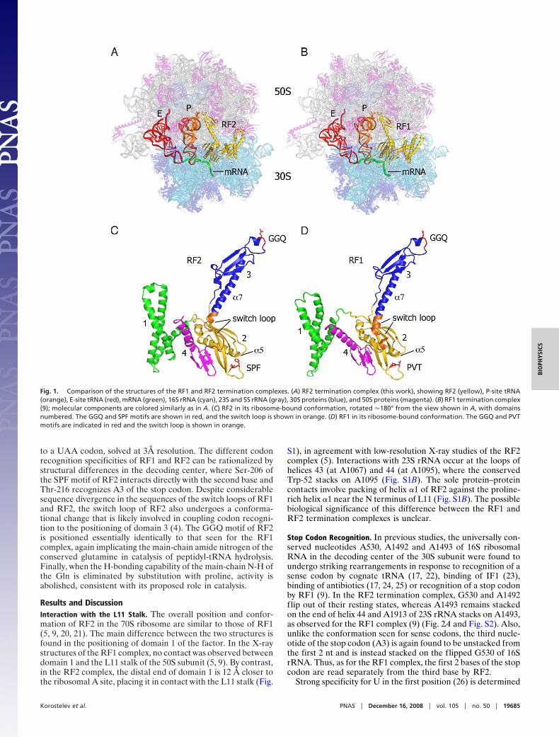
Results and DiscussionInteraction with the L11 Stalk. The overall position and confor-mation of RF2 in the 70S ribosome are similar to those of RF1(5, 9, 20, 21). The main difference between the two structures isfound in the positioning of domain 1 of the factor. In the X-raystructures of the RF1 complex, no contact was observed betweendomain 1 and the L11 stalk of the 50S subunit (5, 9). By contrast,in the RF2 complex, the distal end of domain 1 is 12 Å closer tothe ribosomal A site, placing it in contact with the L11 stalk (Fig.
S1), in agreement with low-resolution X-ray studies of the RF2complex (5). Interactions with 23S rRNA occur at the loops ofhelices 43 (at A1067) and 44 (at A1095), where the conservedTrp-52 stacks on A1095 (Fig. S1B). The sole protein–proteincontacts involve packing of helix �1 of RF2 against the proline-rich helix �1 near the N terminus of L11 (Fig. S1B). The possiblebiological significance of this difference between the RF1 andRF2 termination complexes is unclear.
Stop Codon Recognition. In previous studies, the universally con-served nucleotides A530, A1492 and A1493 of 16S ribosomalRNA in the decoding center of the 30S subunit were found toundergo striking rearrangements in response to recognition of asense codon by cognate tRNA (17, 22), binding of IF1 (23),binding of antibiotics (17, 24, 25) or recognition of a stop codonby RF1 (9). In the RF2 termination complex, G530 and A1492flip out of their resting states, whereas A1493 remains stackedon the end of helix 44 and A1913 of 23S rRNA stacks on A1493,as observed for the RF1 complex (9) (Fig. 2A and Fig. S2). Also,unlike the conformation seen for sense codons, the third nucle-otide of the stop codon (A3) is again found to be unstacked fromthe first 2 nt and is instead stacked on the flipped G530 of 16SrRNA. Thus, as for the RF1 complex, the first 2 bases of the stopcodon are read separately from the third base by RF2.
Strong specificity for U in the first position (26) is determined
Fig. 1. Comparison of the structures of the RF1 and RF2 termination complexes. (A) RF2 termination complex (this work), showing RF2 (yellow), P-site tRNA(orange), E-site tRNA (red), mRNA (green), 16S rRNA (cyan), 23S and 5S rRNA (gray), 30S proteins (blue), and 50S proteins (magenta). (B) RF1 termination complex(9); molecular components are colored similarly as in A. (C) RF2 in its ribosome-bound conformation, rotated �180° from the view shown in A, with domainsnumbered. The GGQ and SPF motifs are shown in red, and the switch loop is shown in orange. (D) RF1 in its ribosome-bound conformation. The GGQ and PVTmotifs are indicated in red and the switch loop is shown in orange.
Korostelev et al. PNAS � December 16, 2008 � vol. 105 � no. 50 � 19685
BIO
PHYS
ICS
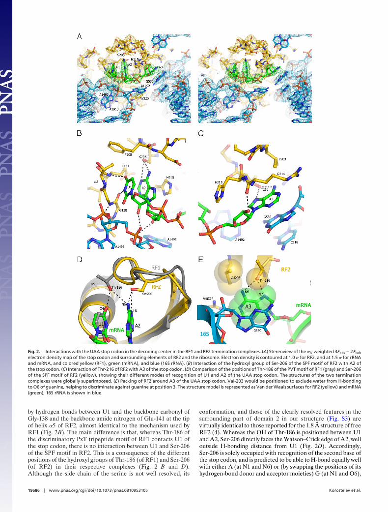
by hydrogen bonds between U1 and the backbone carbonyl ofGly-138 and the backbone amide nitrogen of Glu-141 at the tipof helix �5 of RF2, almost identical to the mechanism used byRF1 (Fig. 2B). The main difference is that, whereas Thr-186 ofthe discriminatory PxT tripeptide motif of RF1 contacts U1 ofthe stop codon, there is no interaction between U1 and Ser-206of the SPF motif in RF2. This is a consequence of the differentpositions of the hydroxyl groups of Thr-186 (of RF1) and Ser-206(of RF2) in their respective complexes (Fig. 2 B and D).Although the side chain of the serine is not well resolved, its
conformation, and those of the clearly resolved features in thesurrounding part of domain 2 in our structure (Fig. S3) arevirtually identical to those reported for the 1.8 Å structure of freeRF2 (4). Whereas the OH of Thr-186 is positioned between U1and A2, Ser-206 directly faces the Watson–Crick edge of A2, welloutside H-bonding distance from U1 (Fig. 2D). Accordingly,Ser-206 is solely occupied with recognition of the second base ofthe stop codon, and is predicted to be able to H-bond equally wellwith either A (at N1 and N6) or (by swapping the positions of itshydrogen-bond donor and acceptor moieties) G (at N1 and O6),
Fig. 2. Interactions with the UAA stop codon in the decoding center in the RF1 and RF2 termination complexes. (A) Stereoview of the �A-weighted 3Fobs � 2Fcalc
electron density map of the stop codon and surrounding elements of RF2 and the ribosome. Electron density is contoured at 1.0 � for RF2, and at 1.5 � for rRNAand mRNA, and colored yellow (RF1), green (mRNA), and blue (16S rRNA). (B) Interaction of the hydroxyl group of Ser-206 of the SPF motif of RF2 with A2 ofthe stop codon. (C) Interaction of Thr-216 of RF2 with A3 of the stop codon. (D) Comparison of the positions of Thr-186 of the PVT motif of RF1 (gray) and Ser-206of the SPF motif of RF2 (yellow), showing their different modes of recognition of U1 and A2 of the UAA stop codon. The structures of the two terminationcomplexes were globally superimposed. (E) Packing of RF2 around A3 of the UAA stop codon. Val-203 would be positioned to exclude water from H-bondingto O6 of guanine, helping to discriminate against guanine at position 3. The structure model is represented as Van der Waals surfaces for RF2 (yellow) and mRNA(green); 16S rRNA is shown in blue.
19686 � www.pnas.org�cgi�doi�10.1073�pnas.0810953105 Korostelev et al.
in keeping with the codon specificity of RF2 for UAA or UGA(Fig. S2 G and H). Conservation of the remaining residues of theSPF (RF2) or PxT (RF1) motifs could be explained by require-ments for excluding water from the decoding pocket of the firstand second bases, in addition to maintaining the correct fold ofthe loop.
In the RF1 complex, recognition of the third base (A3) occursseparately from the first 2 bases, and is shared between Gln-181and Thr-194 (9) (Fig. S2F). Gln-181 is positioned to accept anH-bond from N6 of adenine with its amide carbonyl oxygen andThr-194 can H-bond to the N6 and N7 positions; for recognitionof G in the third position by RF1, the amide group of Gln-181could donate an H-bond to O6. In the RF2 complex, Thr-216occupies the same position as Thr-194 of RF1, enabling recog-nition of adenine; however, no residue equivalent to Gln-181 isfound. Moreover, the position of the hydrophobic side chain ofVal-203 would prevent H-bonding of the O6 of guanine to water(Fig. 2E). Discrimination against guanine in the third positioncould be the result of a large free-energy penalty due todesolvation of guanine, whose dipole moment is significantlylarger than that of adenine (27, 28). The next 2 nt downstreamfrom the A-site codon are also well ordered, enabling theirdetailed conformations to be seen here for the first time (Fig.S4). The nucleotide following the stop codon (A4), which hasbeen implicated in translation termination efficiency (29), in-tercalates between U1196 and C1397 of 16S rRNA. Its Hoogs-teen edge faces Ser-211, located on the opposite side of thespecificity loop from the SPF motif (Fig. S4B), consistent withthe observed cross-linking of RF2 to the base immediatelydownstream of the stop codon (30).
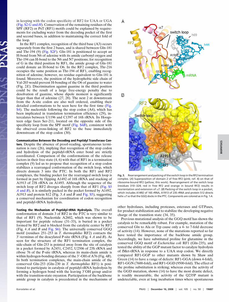
Communication Between the Decoding and Peptidyl Transferase Cen-ters. Despite the absence of proof-reading, spontaneous termi-nation is rare (26), implying that recognition of the stop codonand hydrolysis of the peptidyl-tRNA ester bond are strictlycoordinated. Comparison of the conformations of the releasefactors in their free state (4, 6) with that of RF1 in a terminationcomplex (9) led us to propose that recognition of a stop codonstabilizes a rearranged conformation of the switch loop, whichdirects domain 3 into the PTC. In both the RF1 and RF2complexes, the binding pocket for the rearranged switch loop isformed in part by flipping A1492 of 16S rRNA and stacking ofA1913 of 23S rRNA on A1493. Although the sequence of theswitch loop of RF2 diverges sharply from that of RF1 (Fig. S5A and B), it is similarly packed in the pocket formed by A1492,A1913 and protein S12 (Fig. 3 A and B and Fig. S5), suggestinga conserved mechanism for coordination of codon recognitionand peptidyl-tRNA hydrolysis.
Testing the Mechanism of Peptidyl-tRNA Hydrolysis. The overallconformation of domain 3 of RF2 in the PTC is very similar tothat of RF1 (9). Nucleotide A2602, which was shown to beimportant for peptide release (31–33), is buried in a pocketformed by RF2 and is blocked from the catalytic center by RF2(Fig. 4 A and B and Fig. S6). The universally conserved GGQmotif (residues 251–253 in T. thermophilus RF2) contacts the3�-terminus of the deacylated P-site tRNA (Fig. 4 A and B). Asseen for the structure of the RF1 termination complex, theside-chain of Gln-253 is pointed away from the site of catalysisin a pocket formed by A2451, C2452, U2506 of 23S rRNA andribose 76 of the P-site tRNA, whereas its main-chain amide iswithin hydrogen-bonding distance of the 3�-OH of A76 (Fig. 4B).In both termination complexes, the main-chain amide of theconserved Gln-253 (Gln-230 in T. thermophilus RF1) is posi-tioned to participate in catalysis of peptidyl-tRNA hydrolysis byforming a hydrogen bond with the leaving 3�OH group and/orwith the transition-state oxyanion. Participation of the backboneamide group in catalysis is precedented in the mechanisms of
other hydrolases, including proteases, esterases and GTPases,for product stabilization and to stabilize the developing negativecharge of the transition state (34, 35).
Previous mutational analysis of the GGQ motif has shown thecatalysis to be remarkably robust. For example, mutation of theconserved Gln to Ala or Trp cause only a 4- to 7-fold decreaseof activity (14). However, none of the mutations reported so farhave tested the importance of the backbone amide group.Accordingly, we have substituted proline for glutamine in theconserved GGQ motif of Escherichia coli RF1 (Gln-235), andtested the ability of the GGP mutant factor to catalyze hydrolysisof fMet-tRNA in response to a UAA stop codon. We directlycompared RF1-GGP to other mutants shown by Shaw andGreen (14) to have a range of defects: RF1-GGA (down 4-fold),RF1-GGN (7600-fold), and RF1-GGD (9500-fold). The effect ofthe proline substitution is striking; whereas even the activity ofthe GGD mutation, shown (14) to have the most drastic defect,is readily measurable, the activity of the Q235P mutant isundetectable, even at long incubation times where spontaneous
Fig. 3. Rearrangement and packing of the switch loop in the RF2 terminationcomplex. (A) Superposition of domain 2 of free RF2 (pink; ref. 4) on that ofribosome-bound RF2 (yellow; this work). Rearrangement of the switch loop(residues 310–324; red in free RF2 and orange in bound RF2) results inreorientation and extension of �7. (B) Packing of the switch loop in a pocket,which includes A1492 of 16S rRNA, A1913 of 23S rRNA and protein S12 directshelix �7 so that the GGQ docks in the PTC. Components are colored as in Fig. 1A.
Korostelev et al. PNAS � December 16, 2008 � vol. 105 � no. 50 � 19687
BIO
PHYS
ICS
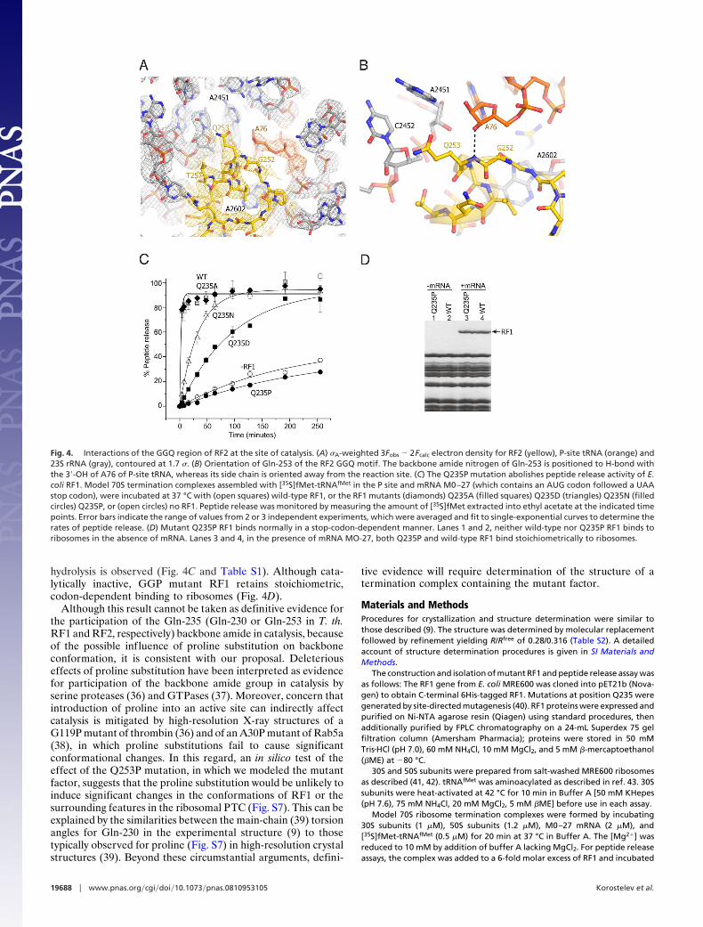
hydrolysis is observed (Fig. 4C and Table S1). Although cata-lytically inactive, GGP mutant RF1 retains stoichiometric,codon-dependent binding to ribosomes (Fig. 4D).
Although this result cannot be taken as definitive evidence forthe participation of the Gln-235 (Gln-230 or Gln-253 in T. th.RF1 and RF2, respectively) backbone amide in catalysis, becauseof the possible influence of proline substitution on backboneconformation, it is consistent with our proposal. Deleteriouseffects of proline substitution have been interpreted as evidencefor participation of the backbone amide group in catalysis byserine proteases (36) and GTPases (37). Moreover, concern thatintroduction of proline into an active site can indirectly affectcatalysis is mitigated by high-resolution X-ray structures of aG119P mutant of thrombin (36) and of an A30P mutant of Rab5a(38), in which proline substitutions fail to cause significantconformational changes. In this regard, an in silico test of theeffect of the Q253P mutation, in which we modeled the mutantfactor, suggests that the proline substitution would be unlikely toinduce significant changes in the conformations of RF1 or thesurrounding features in the ribosomal PTC (Fig. S7). This can beexplained by the similarities between the main-chain (39) torsionangles for Gln-230 in the experimental structure (9) to thosetypically observed for proline (Fig. S7) in high-resolution crystalstructures (39). Beyond these circumstantial arguments, defini-
tive evidence will require determination of the structure of atermination complex containing the mutant factor.
Materials and MethodsProcedures for crystallization and structure determination were similar tothose described (9). The structure was determined by molecular replacementfollowed by refinement yielding R/Rfree of 0.28/0.316 (Table S2). A detailedaccount of structure determination procedures is given in SI Materials andMethods.
The construction and isolation of mutant RF1 and peptide release assay wasas follows: The RF1 gene from E. coli MRE600 was cloned into pET21b (Nova-gen) to obtain C-terminal 6His-tagged RF1. Mutations at position Q235 weregenerated by site-directed mutagenesis (40). RF1 proteins were expressed andpurified on Ni-NTA agarose resin (Qiagen) using standard procedures, thenadditionally purified by FPLC chromatography on a 24-mL Superdex 75 gelfiltration column (Amersham Pharmacia); proteins were stored in 50 mMTris�HCl (pH 7.0), 60 mM NH4Cl, 10 mM MgCl2, and 5 mM �-mercaptoethanol(�ME) at �80 °C.
30S and 50S subunits were prepared from salt-washed MRE600 ribosomesas described (41, 42). tRNAfMet was aminoacylated as described in ref. 43. 30Ssubunits were heat-activated at 42 °C for 10 min in Buffer A [50 mM KHepes(pH 7.6), 75 mM NH4Cl, 20 mM MgCl2, 5 mM �ME] before use in each assay.
Model 70S ribosome termination complexes were formed by incubating30S subunits (1 �M), 50S subunits (1.2 �M), M0–27 mRNA (2 �M), and[35S]fMet-tRNAfMet (0.5 �M) for 20 min at 37 °C in Buffer A. The [Mg2�] wasreduced to 10 mM by addition of buffer A lacking MgCl2. For peptide releaseassays, the complex was added to a 6-fold molar excess of RF1 and incubated
Fig. 4. Interactions of the GGQ region of RF2 at the site of catalysis. (A) �A-weighted 3Fobs � 2Fcalc electron density for RF2 (yellow), P-site tRNA (orange) and23S rRNA (gray), contoured at 1.7 �. (B) Orientation of Gln-253 of the RF2 GGQ motif. The backbone amide nitrogen of Gln-253 is positioned to H-bond withthe 3�-OH of A76 of P-site tRNA, whereas its side chain is oriented away from the reaction site. (C) The Q235P mutation abolishes peptide release activity of E.coli RF1. Model 70S termination complexes assembled with [35S]fMet-tRNAfMet in the P site and mRNA M0–27 (which contains an AUG codon followed a UAAstop codon), were incubated at 37 °C with (open squares) wild-type RF1, or the RF1 mutants (diamonds) Q235A (filled squares) Q235D (triangles) Q235N (filledcircles) Q235P, or (open circles) no RF1. Peptide release was monitored by measuring the amount of [35S]fMet extracted into ethyl acetate at the indicated timepoints. Error bars indicate the range of values from 2 or 3 independent experiments, which were averaged and fit to single-exponential curves to determine therates of peptide release. (D) Mutant Q235P RF1 binds normally in a stop-codon-dependent manner. Lanes 1 and 2, neither wild-type nor Q235P RF1 binds toribosomes in the absence of mRNA. Lanes 3 and 4, in the presence of mRNA MO-27, both Q235P and wild-type RF1 bind stoichiometrically to ribosomes.
19688 � www.pnas.org�cgi�doi�10.1073�pnas.0810953105 Korostelev et al.
in Buffer A (10 mM MgCl2) at 37 °C. Aliquots were removed from the reactionat each time point and quenched in 5 vol of 0.1 M HCl; hydrolyzed [35S]fMetwas extracted with 1 mL of ethyl acetate, 0.7 mL of which was added toscintillation mixture and counted.
For RF1 binding assays, the 70S complex was formed as above, but with 2�M [35S]fMet-tRNAfMet, and with or without M0–27 mRNA. As above, eachcomplex was added to a 6-fold excess of RF1, then incubated at 37 °C for 5 minin Buffer A (10 mM MgCl2). Reactions were passed through a SephacrylS200-HR resin (Sigma) 1-mL spin column, then precipitated with 4 vol of
acetone (�20 °C for 1 h), dissolved in loading buffer, and electrophoresed ona 12% acrylamide SDS gel.
ACKNOWLEDGMENTS. We thank John Paul Donohue for assistance with dataprocessing and computational support; Eric Maklan for helpful discussionsregarding kinetic measurements; and the beamline staffs at SSRL, ALS, andAPS for their expert support during screening and data collection. Thesestudies were supported by grants from the National Institutes of Health andthe National Science Foundation (to H.F.N.) and a fellowship from the DanishResearch Council (to M.L.).
1. Capecchi MR, Klein HA (1969) Characterization of three proteins involved in polypep-tide chain termination. Cold Spring Harbor Symp Quant Biol 34:469–477.
2. Scolnick E, Tompkins R, Caskey T, Nirenberg M (1968) Release factors differing inspecificity for terminator codons. Proc Natl Acad Sci USA 61:768–774.
3. Scolnick EM, Caskey CT (1969) Peptide chain termination. V. The role of release factorsin mRNA terminator codon recognition. Proc Natl Acad Sci USA 64:1235–1241.
4. Vestergaard B, et al. (2001) Bacterial polypeptide release factor RF2 is structurallydistinct from eukaryotic eRF1. Mol Cell 8:1375–1382.
5. Petry S, et al. (2005) Crystal structures of the ribosome in complex with release factorsRF1 and RF2 bound to a cognate stop codon. Cell 123:1255–1266.
6. Shin DH, et al. (2004) Structural analyses of peptide release factor 1 from Thermotogamaritima reveal domain flexibility required for its interaction with the ribosome. J MolBiol 341:227–239.
7. Ito K, Uno M, Nakamura Y (2000) A tripeptide ‘‘anticodon’’ deciphers stop codons inmessenger RNA. Nature 403:680–684.
8. Nakamura Y, Ito K (2002) A tripeptide discriminator for stop codon recognition. FEBSLett 514:30–33.
9. Laurberg M, et al. (2008) Structural basis for translation termination on the 70Sribosome. Nature 454:852–857.
10. Song H, et al. (2000) The crystal structure of human eukaryotic release factor eRF1–mechanism of stop codon recognition and peptidyl-tRNA hydrolysis. Cell 100:311–321.
11. Trobro S, Aqvist J (2007) A model for how ribosomal release factors induce peptidyl-tRNA cleavage in termination of protein synthesis. Mol Cell 27:758–766.
12. Seit Nebi A, Frolova L, Ivanova N, Poltaraus A, Kiselev L (2000) Mutation of a glutamineresidue in the universal tripeptide GGQ in human eRF1 termination factor does notcause complete loss of its activity. Mol Biol (Moscow) 34:899–900.
13. Seit-Nebi A, Frolova L, Justesen J, Kisselev L (2001) Class-1 translation terminationfactors: Invariant GGQ minidomain is essential for release activity and ribosomebinding but not for stop codon recognition. Nucleic Acids Res 29:3982–3987.
14. Shaw JJ, Green R (2007) Two distinct components of release factor function uncoveredby nucleophile partitioning analysis. Mol Cell 28:458–467.
15. Frolova LY, et al. (1999) Mutations in the highly conserved GGQ motif of class 1polypeptide release factors abolish ability of human eRF1 to trigger peptidyl-tRNAhydrolysis. Rna 5:1014–1020.
16. Zavialov AV, Mora L, Buckingham RH, Ehrenberg M (2002) Release of peptide pro-moted by the GGQ motif of class 1 release factors regulates the GTPase activity of RF3.Mol Cell 10:789–798.
17. Ogle JM, et al. (2001) Recognition of cognate transfer RNA by the 30S ribosomalsubunit. Science 292:897–902.
18. Ali IK, Lancaster L, Feinberg J, Joseph S, Noller HF (2006) Deletion of a conserved,central ribosomal intersubunit RNA bridge. Mol Cell 23:865–874.
19. Youngman EM, He SL, Nikstad LJ, Green R (2007) Stop codon recognition by releasefactors induces structural rearrangement of the ribosomal decoding center that isproductive for peptide release. Mol Cell 28:533–543.
20. Rawat UB, et al. (2003) A cryo-electron microscopic study of ribosome-bound termi-nation factor RF2. Nature 421:87–90.
21. Rawat U, et al. (2006) Interactions of the release factor RF1 with the ribosome asrevealed by cryo-EM. J Mol Biol 357:1144–1153.
22. Selmer M, et al. (2006) Structure of the 70S ribosome complexed with mRNA and tRNA.Science 313:1935–1942.
23. Carter AP, et al. (2001) Crystal structure of an initiation factor bound to the 30Sribosomal subunit. Science 291:498–501.
24. Borovinskaya MA, et al. (2007) Structural basis for aminoglycoside inhibition ofbacterial ribosome recycling. Nat Struct Mol Biol 14:727–732.
25. Francois B, et al. (2005) Crystal structures of complexes between aminoglycosides anddecoding A site oligonucleotides: Role of the number of rings and positive charges inthe specific binding leading to miscoding. Nucleic Acids Res 33:5677–5690.
26. Freistroffer DV, Kwiatkowski M, Buckingham RH, Ehrenberg M (2000) The accuracy ofcodon recognition by polypeptide release factors. Proc Natl Acad Sci USA 97:2046–2051.
27. Basu G, Sivanesan D, Kawabata T, Go N (2004) Electrostatic potential of nucleotide-freeprotein is sufficient for discrimination between adenine and guanine-specific bindingsites. J Mol Biol 342:1053–1066.
28. Sponer J, Leszczynski J, Hobza P (1996) Structures and energies of hydrogen-bondedDNA base pairs. A nonempirical study with inclusion of electron correlation. J PhysChem 100:1965–1974.
29. Tate WP, et al. (1995) Translational termination efficiency in both bacteria andmammals is regulated by the base following the stop codon. Biochem Cell Biol73:1095–1103.
30. Poole ES, Major LL, Mannering SA, Tate WP (1998) Translational termination inEscherichia coli: Three bases following the stop codon cross-link to release factor 2 andaffect the decoding efficiency of UGA-containing signals. Nucleic Acids Res 26:954–960.
31. Amort M, et al. (2007) An intact ribose moiety at A2602 of 23S rRNA is key to triggerpeptidyl-tRNA hydrolysis during translation termination. Nucleic Acids Res 35:5130–5140.
32. Polacek N, et al. (2003) The critical role of the universally conserved A2602 of 23Sribosomal RNA in the release of the nascent peptide during translation termination.Mol Cell 11:103–112.
33. Youngman EM, Brunelle JL, Kochaniak AB, Green R (2004) The active site of theribosome is composed of two layers of conserved nucleotides with distinct roles inpeptide bond formation and peptide release. Cell 117:589–599.
34. Wilmouth RC, et al. (2001) X-ray snapshots of serine protease catalysis reveal atetrahedral intermediate. Nat Struct Biol 8:689–694.
35. Jaeger KE, Dijkstra BW, Reetz MT (1999) Bacterial biocatalysts: Molecular biology,three-dimensional structures, and biotechnological applications of lipases. Annu RevMicrobiol 53:315–351.
36. Bobofchak KM, Pineda AO, Mathews FS, Di Cera E (2005) Energetic and structuralconsequences of perturbing Gly-193 in the oxyanion hole of serine proteases. J BiolChem 280:25644–25650.
37. Liang Z, Mather T, Li G (2000) GTPase mechanism and function: New insights fromsystematic mutational analysis of the phosphate-binding loop residue Ala30 of Rab5.Biochem J 346 (Pt 2):501–508.
38. Zhu G, et al. (2003) High resolution crystal structures of human Rab5a and five mutantswith substitutions in the catalytically important phosphate-binding loop. J Biol Chem278:2452–2460.
39. Ramachandran GN, Ramakrishnan C, Sasisekharan V (1963) Stereochemistry ofpolypeptide chain configurations. J Mol Biol 7:95–99.
40. Kunkel TA (1985) Rapid and efficient site-specific mutagenesis without phenotypicselection. Proc Natl Acad Sci USA 82:488–492.
41. Moazed D, Noller HF (1989) Interaction of tRNA with 23S rRNA in the ribosomal A, P,and E sites. Cell 57:585–597.
42. Moazed D, Noller HF (1986) Transfer RNA shields specific nucleotides in 16S ribosomalRNA from attack by chemical probes.Cell 47:985–994.
43. Lancaster L, Noller HF (2005) Involvement of 16S rRNA nucleotides G1338 and A1339in discrimination of initiator tRNA.Mol Cell 20:623–632.
Korostelev et al. PNAS � December 16, 2008 � vol. 105 � no. 50 � 19689
BIO
PHYS
ICS
Supporting InformationKorostelev et al. 10.1073/pnas.0810953105SI Materials and MethodsCrystallization. Ribosomes were purified from Thermus ther-mophilus as described (1) with the following modifications. Aftercolumn chromatography on Toyo-Pearl Butyl 650S, the buffer ofthe eluted ribosome fraction was replaced with buffer E (25 mMTris-OAc, pH 7.0, 50 mM KOAc, 10 mM NH4OAc, 10 mMMg(OAc)2), using Centricon Plus YM-100 (Amicon). An equalvolume of microcrystallization solution (100 mM Tris-OAc, pH7.0, 200 mM KSCN, 7% PEG20k, 15% PEG200, 2.8% deoxy-BigChap) was then added at room temperature, and the mixturewas stored at 4 °C. After a few days, microcrystals were harvestedand dissolved in buffer E. The ribosome concentration wasadjusted to 20 mg/ml, and stored in aliquots at -80 °C until usedfor complex formation.
The mRNA M0–27 [GGC AAG GAG GUA AAA AUGUAA AAA AAA] was chemically synthesized (IDT) (1). Esch-erichia coli tRNAfMet was purchased from Sigma-Aldrich. Thegene for T. thermophilus RF2 lacking T52 from the start codonwhere a frameshift occurs was cloned into pET24b. RF2 waspurified using the same procedure reported for RF1 (1). The70S:mRNA:tRNAfMet:RF2 termination complex was formed asdescribed for the RF1 complex (1), using the molar ratios1:2:2.4:4, respectively. Crystallization and cryoprotection proce-dures were as described (1).
x-ray data collection and structure determination. Crystals werescreened at beamlines 7.1, 9.1, 9.2 and 11.1 at the StanfordSynchrotron Radiation Laboratory, and at beamline 4.2.2 at the
Advanced Light Source, Lawrence Berkeley National Labora-tory. x-ray diffraction data were recorded at beamline 23 ID-Dat the Advanced Photon Source at Argonne National Laboratoryusing an x-ray wavelength of 0.9537 Å and an oscillation angle of0.2°. Data from four datasets obtained from different positionsof the same crystal were integrated and merged using the XDSpackage (2), scaled in SCALA (3) and truncated in TRUNCATE(4). 1% of reflections were marked as test-set (Rfree set) reflec-tions to monitor the progress of refinement.
Structure determination started with rigid-body refinement ofthe previously determined structure of the RF1 terminationcomplex, which was obtained from the same crystal form (1). Atthis stage of refinement, release factor was not included in thestructure. Secondary structure elements and some side chains ofRF2 were visible in the starting Fourier difference maps (Fig.S3). The 1.8 Å structure of free E. coli RF2 (5) was modified tofit the difference map obtained after rigid-body refinement. Thesequence was modified to that of T. thermophilus RF2 employingT-COFFEE (6) and MODELLER (7). The 2.5 Å structure offree T. thermophilus RF2 (8) was used as an aid in modelingseveral f lexible parts of the release factor. After simulatedannealing and B-factor refinement in CNS (9), the structure ofthe RF2 termination complex was subjected to TLS refinementin PHENIX (10), yielding good stereochemistry and crystallo-graphic statistics (Table S2). NCS restraints were used through-out the refinement as described (1). PYMOL (11), O (12) andlocal real-space refinement (13) were used for model building.Figs. were rendered using PYMOL (11).
1. Laurberg M, et al. (2008) Structural basis for translation termination on the 70Sribosome. Nature 454:852–857.
2. Kabsch W (1993) Automatic processing of rotation diffraction data from crystals ofinitially unknown symmetry and cell constants. J Appl Crystallogr 26:795–800.
3. Evans P (2006) Scaling and assessment of data quality. Acta Crystallogr D 62:72–82.4. CCP4 (1994) The CCP4 suite: Programs for protein crystallography. Acta Crystallogr D
Biol Crystallogr 50:760–763.5. Vestergaard B, et al. (2001) Bacterial polypeptide release factor RF2 is structurally
distinct from eukaryotic eRF1. Mol Cell 8:1375–1382.6. Notredame C, Higgins DG, Heringa J (2000) T-Coffee: A novel method for fast and
accurate multiple sequence alignment. J Mol Biol 302:205–217.7. Eswar N, et al. (2007) Comparative protein structure modeling using MODELLER. Curr
Protocols Protein Sci Chapter 2, Unit 29.8. Zoldak G, et al. (2007) Release factors 2 from Escherichia coli and Thermus thermophi-
lus: structural, spectroscopic and microcalorimetric studies. Nucleic Acids Res 35:1343–1353.
9. Brunger AT, et al. (1998) Crystallography & NMR system: A new software suite formacromolecular structure determination. Acta Crystallogr D 54:905–921.
10. Adams PD, et al. (2002) PHENIX: Building new software for automated crystallographicstructure determination. Acta Crystallogr D 58:1948–1954.
11. DeLano WL (2002) The PyMOL Molecular Graphics System (Delano Scientific, Palo Alto,CA).
12. Jones TA., Zou JY, Cowan SW, Kjeldgaard M (1991) Improved methods for buildingprotein models in electron density maps and the location of errors in these models.Acta Crystallogr A 47 (Pt 2):110–119.
13. Korostelev A, Bertram R, Chapman MS (2002) Simulated-annealing real-space refine-ment as a tool in model building. Acta Crystallogr D 58:761–767.
Korostelev et al. www.pnas.org/cgi/content/short/0810953105 1 of 10
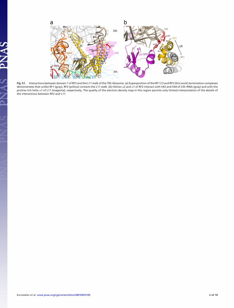
Fig. S1. Interactions between domain 1 of RF2 and the L11 stalk of the 70S ribosome. (a) Superposition of the RF1 (1) and RF2 (this work) termination complexesdemonstrates that unlike RF1 (gray), RF2 (yellow) contacts the L11 stalk. (b) Helices �2 and �1 of RF2 interact with h43 and h44 of 23S rRNA (gray) and with theproline-rich helix �1 of L11 (magenta), respectively. The quality of the electron density map in this region permits only limited interpretation of the details ofthe interactions between RF2 and L11.
Korostelev et al. www.pnas.org/cgi/content/short/0810953105 2 of 10
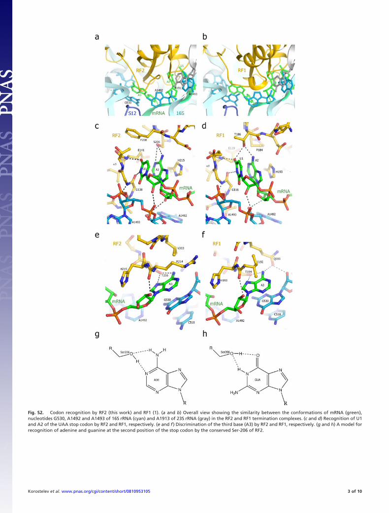
Fig. S2. Codon recognition by RF2 (this work) and RF1 (1). (a and b) Overall view showing the similarity between the conformations of mRNA (green),nucleotides G530, A1492 and A1493 of 16S rRNA (cyan) and A1913 of 23S rRNA (gray) in the RF2 and RF1 termination complexes. (c and d) Recognition of U1and A2 of the UAA stop codon by RF2 and RF1, respectively. (e and f ) Discrimination of the third base (A3) by RF2 and RF1, respectively. (g and h) A model forrecognition of adenine and guanine at the second position of the stop codon by the conserved Ser-206 of RF2.
Korostelev et al. www.pnas.org/cgi/content/short/0810953105 3 of 10
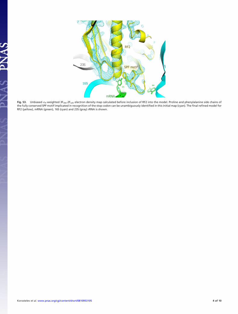
Fig. S3. Unbiased �A-weighted 3Fobs-2Fcalc electron density map calculated before inclusion of RF2 into the model. Proline and phenylalanine side chains ofthe fully conserved SPF motif implicated in recognition of the stop codon can be unambiguously identified in this initial map (cyan). The final refined model forRF2 (yellow), mRNA (green), 16S (cyan) and 23S (gray) rRNA is shown.
Korostelev et al. www.pnas.org/cgi/content/short/0810953105 4 of 10
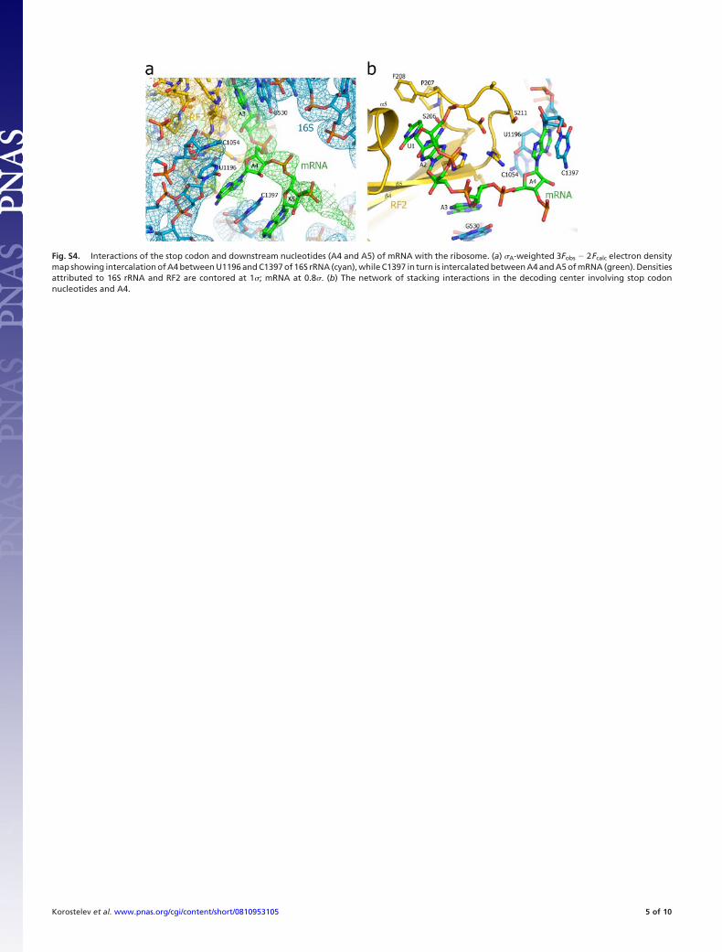
Fig. S4. Interactions of the stop codon and downstream nucleotides (A4 and A5) of mRNA with the ribosome. (a) �A-weighted 3Fobs � 2Fcalc electron densitymap showing intercalation of A4 between U1196 and C1397 of 16S rRNA (cyan), while C1397 in turn is intercalated between A4 and A5 of mRNA (green). Densitiesattributed to 16S rRNA and RF2 are contored at 1�; mRNA at 0.8�. (b) The network of stacking interactions in the decoding center involving stop codonnucleotides and A4.
Korostelev et al. www.pnas.org/cgi/content/short/0810953105 5 of 10
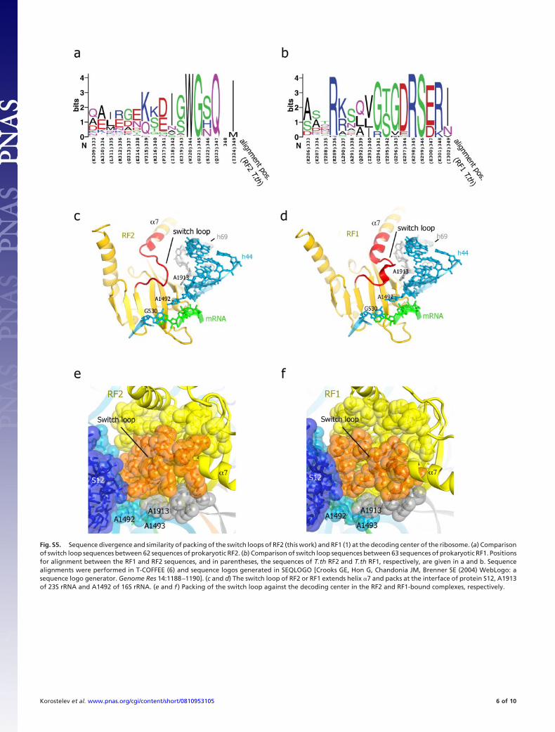
Fig. S5. Sequence divergence and similarity of packing of the switch loops of RF2 (this work) and RF1 (1) at the decoding center of the ribosome. (a) Comparisonof switch loop sequences between 62 sequences of prokaryotic RF2. (b) Comparison of switch loop sequences between 63 sequences of prokaryotic RF1. Positionsfor alignment between the RF1 and RF2 sequences, and in parentheses, the sequences of T.th RF2 and T.th RF1, respectively, are given in a and b. Sequencealignments were performed in T-COFFEE (6) and sequence logos generated in SEQLOGO [Crooks GE, Hon G, Chandonia JM, Brenner SE (2004) WebLogo: asequence logo generator. Genome Res 14:1188–1190]. (c and d) The switch loop of RF2 or RF1 extends helix �7 and packs at the interface of protein S12, A1913of 23S rRNA and A1492 of 16S rRNA. (e and f ) Packing of the switch loop against the decoding center in the RF2 and RF1-bound complexes, respectively.
Korostelev et al. www.pnas.org/cgi/content/short/0810953105 6 of 10
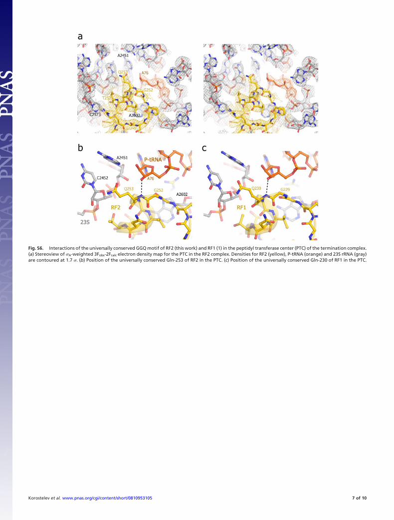
Fig. S6. Interactions of the universally conserved GGQ motif of RF2 (this work) and RF1 (1) in the peptidyl transferase center (PTC) of the termination complex.(a) Stereoview of �A-weighted 3Fobs-2Fcalc electron density map for the PTC in the RF2 complex. Densities for RF2 (yellow), P-tRNA (orange) and 23S rRNA (gray)are contoured at 1.7 �. (b) Position of the universally conserved Gln-253 of RF2 in the PTC. (c) Position of the universally conserved Gln-230 of RF1 in the PTC.
Korostelev et al. www.pnas.org/cgi/content/short/0810953105 7 of 10

Fig. S7. Conformation of the computationally modeled GGP loop of the Q253P mutant (a) is similar to that of the GGQ loop in the crystal structure of (b) theRF2 complex (this work), consistent with its stoichiometric binding to the 70S ribosome (Fig. 4C). (c) Backbone torsion angles (box) for the modeled proline arein a favorable region (green) of the Ramachandran plot and are similar to (d) those of glutamine. Ramachandran plots were generated using PROCHECK (4).The in silico mutation Q253P was performed in the RF2 termination complex using the mutagenesis option in PyMOL (11) followed by minimization of repulsivevan der Waals and geometry energy terms in CNS (9) of all RF2 and 70S ribosome residues within 5Å of each other.
Korostelev et al. www.pnas.org/cgi/content/short/0810953105 8 of 10
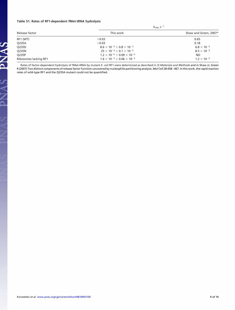
Table S1. Rates of RF1-dependent fMet-tRNA hydrolysis
Release factor
kobs, s�1
This work Shaw and Green, 2007*
RF1 (WT) �0.03 0.65Q235A �0.03 0.18Q235D 8.6 � 10�5 � 0.8 � 10�5 6.8 � 10�5
Q235N 25 � 10�5 � 0.1 � 10�5 8.5 � 10�5
Q235P 1.2 � 10�5 � 0.09 � 10�5 NDRibosomes lacking RF1 1.6 � 10�5 � 0.06 � 10�5 1.2 � 10�5
Rates of factor-dependent hydrolysis of fMet-tRNA by mutant E. coli RF1 were determined as described in SI Materials and Methods and in Shaw JJ, GreenR (2007) Two distinct components of release factor function uncovered by nucleophile partitioning analysis. Mol Cell 28:458–467. In this work, the rapid reactionrates of wild-type RF1 and the Q235A mutant could not be quantified.
Korostelev et al. www.pnas.org/cgi/content/short/0810953105 9 of 10
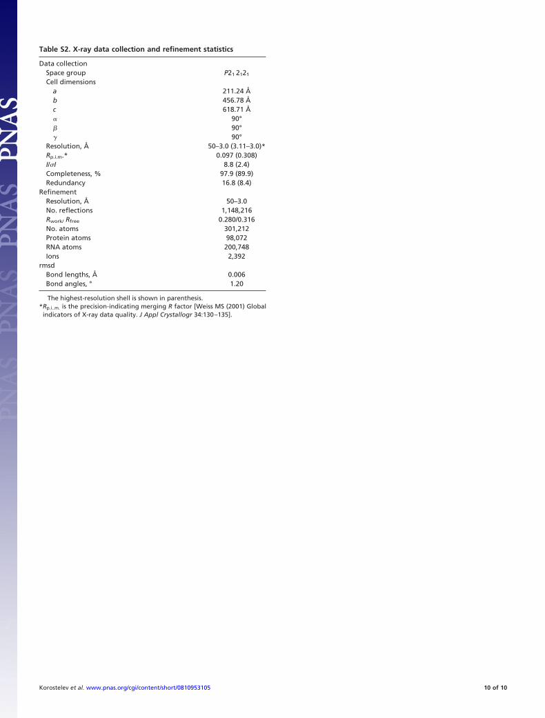
Table S2. X-ray data collection and refinement statistics
Data collectionSpace group P21 2121
Cell dimensionsa 211.24 Åb 456.78 Åc 618.71 Å� 90°� 90°� 90°
Resolution, Å 50–3.0 (3.11–3.0)*Rp.i.m.* 0.097 (0.308)I/�I 8.8 (2.4)Completeness, % 97.9 (89.9)Redundancy 16.8 (8.4)
RefinementResolution, Å 50–3.0No. reflections 1,148,216Rwork/ Rfree 0.280/0.316No. atoms 301,212Protein atoms 98,072RNA atoms 200,748Ions 2,392
rmsdBond lengths, Å 0.006Bond angles, ° 1.20
The highest-resolution shell is shown in parenthesis.*Rp.i..m. is the precision-indicating merging R factor [Weiss MS (2001) Globalindicators of X-ray data quality. J Appl Crystallogr 34:130–135].
Korostelev et al. www.pnas.org/cgi/content/short/0810953105 10 of 10