Embed Size (px)
Citation preview

Crystal Structure and Site-Directed Mutagenesis Analyses ofHaloalkane Dehalogenase LinB from Sphingobium sp. Strain MI1205
Masahiko Okai,a Jun Ohtsuka,a Lica Fabiana Imai,a Tomoko Mase,a Ryota Moriuchi,b Masataka Tsuda,b Koji Nagata,a Yuji Nagata,b
Masaru Tanokuraa
Department of Applied Biological Chemistry, Graduate School of Agricultural and Life Sciences, University of Tokyo, Tokyo, Japana; Department of Environmental LifeSciences, Graduate School of Life Sciences, Tohoku University, Sendai, Japanb
The enzymes LinBUT and LinBMI (LinB from Sphingobium japonicum UT26 and Sphingobium sp. MI1205, respectively) catalyzethe hydrolytic dechlorination of �-hexachlorocyclohexane (�-HCH) and yield different products, 2,3,4,5,6-pentachlorocyclo-hexanol (PCHL) and 2,3,5,6-tetrachlorocyclohexane-1,4-diol (TCDL), respectively, despite their 98% identity in amino acid se-quence. To reveal the structural basis of their different enzymatic properties, we performed site-directed mutagenesis and X-raycrystallographic studies of LinBMI and its seven point mutants. The mutation analysis revealed that the seven amino acid resi-dues uniquely found in LinBMI were categorized into three groups based on the efficiency of the first-step (from �-HCH toPCHL) and second-step (from PCHL to TCDL) conversions. Crystal structure analyses of wild-type LinBMI and its seven pointmutants indicated how each mutated residue contributed to the first- and second-step conversions by LinBMI. The dynamicssimulation analyses of wild-type LinBMI and LinBUT revealed that the entrance of the substrate access tunnel of LinBUT was moreflexible than that of LinBMI, which could lead to the different efficiencies of dehalogenation activity between thesedehalogenases.
Hexachlorocyclohexane (HCH) is a six-chlorine-substitutedcyclohexane. One of its isomers, the � isomer, has insecticidal
properties and has been widely used as an insecticide around theworld (1). Although the use of �-HCH has been prohibited inmost countries due to its toxicity and long persistence, the large-scale production, widespread use, and dumping of the other non-insecticidal isomers (�-, �-, and �-HCHs) in past decades stillcontinue to create problems with HCH contamination in soil andgroundwater (2). �-HCH in particular is a persistent and prob-lematic isomer of HCH.
Several �-HCH-degrading bacteria whose �-HCH-degradingenzymes can be utilized for bioremediation have been identified(3–5). LinBMI and LinBUT are haloalkane dehalogenases isolatedfrom Sphingobium sp. MI1205 and Sphingobium japonicum UT26,respectively, that can cleave the carbon-halogen bond in �-HCH.Haloalkane dehalogenases belong to the �/�-hydrolase family,and their catalytic mechanism consists of the following steps: (i)substrate binding, (ii) cleavage of the carbon-halogen bond in thesubstrate and formation of an intermediate covalently bound tothe nucleophile, (iii) hydrolysis of the alkyl-enzyme intermediate,and (iv) release of halide ion and alcohol (6). LinBMI and LinBUT
share 98% sequence identity, with only 7 different amino acidresidues (at positions 81, 112, 134, 135, 138, 247, and 253) out of296 residues, but these enzymes exhibit different enzymatic prop-erties (Fig. 1). LinBMI catalyzes the two-step dehalogenation andconverts �-HCH to 2,3,4,5,6-pentachlorocyclohexanol (PCHL)and further to 2,3,5,6-tetrachlorocyclohexane-1,4-diol (TCDL)(7) in the manner of LinB2 from Sphingomonas sp. BHC-A (8) andLinB from Sphingobium indicum B90A (9), whereas LinBUT cata-lyzes only the first-step dehalogenation of �-HCH to PCHL (10)and cannot degrade PCHL further. Moreover, LinBMI can catalyzethe first-step conversion eight times as efficiently as LinBUT (7).
In a previous site-directed mutagenesis study, the V134I,H247A, and V134I H247A mutants of LinBMI, in which one or twoLinBMI-specific residues were mutated to a LinBUT-type resi-
due(s), showed reduced activities in both the first- and second-step dehalogenations, with the exception that there was no reduc-tion in the first-step dehalogenation activity of the H247A mutant(7). However, the activities of these mutants were still higher thanthat of LinBUT in both the first- and second-step dehalogenations,which suggested that one or more of the other five residues (T81,V112, T135, L138, and I253) uniquely found in LinBMI were alsoimportant for the high dehalogenation activity of LinBMI. To date,the crystal structure of LinBUT has been described (11–14),whereas the crystal structure of LinBMI has not. To investigate howthe seven residues that are different between LinBMI and LinBUT
contribute to their different enzymatic properties, we performedsite-directed mutagenesis and X-ray crystallographic studies ofLinBMI and its seven point mutants, where each LinBMI-specificresidue is mutated to the LinBUT-type residue (T81A, V112A,V134I, T135A, L138I, H247A, and I253M). Activity measure-ments were made for all the mutants except for those carrying theV134I and H247A mutations, whose measurements were reportedpreviously (7).
MATERIALS AND METHODSExpression, purification, and crystallization. The expression plasmids ofwild-type LinBMI and the seven mutants (carrying T81A, V112A, V134I,T135A, L138I, H247A, and I253M) were constructed using the vectorpAQNM, where the target proteins were expressed under the control ofthe tac promoter and lacIq (7). Wild-type LinBMI and the seven mutantswere expressed and purified by the following procedures. Escherichia coli
Received 27 October 2012 Accepted 26 March 2013
Published ahead of print 5 April 2013
Address correspondence to Masaru Tanokura, [email protected].
Copyright © 2013, American Society for Microbiology. All Rights Reserved.
doi:10.1128/JB.02020-12
2642 jb.asm.org Journal of Bacteriology p. 2642–2651 June 2013 Volume 195 Number 11
on Septem
ber 24, 2018 by guesthttp://jb.asm
.org/D
ownloaded from

strain BL21(DE3) cells (Novagen) were cultured in Luria-Bertani (LB)medium containing 50 �g ml�1 ampicillin until an optical density at 600nm (OD600) of 0.6 at 37°C. Protein expression was induced by addingisopropyl �-D-thiogalactopyranoside (IPTG) to a final concentration of 1mM, and the culture was continued at 25°C for 12 h. The cells wereharvested by centrifugation at 4,500 � g at 4°C for 10 min. The harvestedcells were suspended in Sol A (50 mM Tris-HCl [pH 7.5], 400 mM NaCl,and 5 mM imidazole) and disrupted by sonication. After centrifugation at40,000 � g for 30 min at 4°C, the supernatant was loaded onto a 3-ml NiSepharose 6 Fast Flow column (GE Healthcare) at room temperature.After a wash step with Sol B (50 mM Tris-HCl [pH 7.5], 400 mM NaCl,and 50 mM imidazole), the protein was eluted with Sol C (50 mM Tris-HCl [pH 7.5], 400 mM NaCl, and 200 mM imidazole). The purified pro-tein was dialyzed against 20 mM Tris-HCl (pH 8.0) and then concentratedto 25 mg ml�1 using a Vivaspin 20 concentrator (Sartorius) at 4°C.
Initial crystallization trials of LinBMI were performed by the sitting-drop vapor diffusion method in 96-well Intelli-Plate plates (Art RobbinsInstruments) using Crystal Screen HT, Index HT (Hampton Research),and Wizard I and II (Emerald Biosystems) sparse-matrix screening kits.Each drop was prepared by mixing equal volumes (0.7 �l) of the proteinsolution and a reservoir solution and equilibrated against 70 �l of thereservoir solution at 4°C or 20°C. Further crystallization trials were car-ried out based on the crystallization conditions of the untagged (100 mMTris-HCl [pH 8.8 to 9.0], 200 mM CaCl2, and 17 to 19% [wt/vol] poly-ethylene glycol [PEG] 6000) and His-tagged (100 mM Tris-HCl [pH 8.5],200 mM MgCl2. and 20% [wt/vol] PEG 4000) LinBUT by the sitting-dropvapor diffusion method in 24-well plates (Hampton Research) (14, 15).The crystallization drops were prepared by mixing 1.0 �l protein solutionand 1.0 �l reservoir solution and were equilibrated against 0.3 ml reser-voir solution.
Data collection and processing. The crystals of wild-type LinBMI andthe seven mutants were transferred to the reservoir solution containing25% (vol/vol) glycerol as the cryoprotectant. The X-ray diffraction datawere collected at a wavelength of 1.0000 Å in a cryogenic nitrogen gasstream at beamlines BL-5A and AR-NW12A of the Photon Factory(Ibaraki, Japan). The data sets were obtained by collecting 360 frames,with an oscillation step of 0.5°. The diffraction data were indexed, inte-grated, and scaled using the HKL-2000 software package (16).
Structure modeling and refinement. The crystal structure of wild-type LinBMI was determined by the molecular replacement method usingthe software program MOLREP (17) and the crystal structure of LinBUT
(PDB code 1CV2) (11) as the initial model. Refinements were performed
using the Coot (18) and Refmac5 (19) programs. Water molecules wereadded using ARP/wARP software (20). Then, the crystal structures of theseven mutants were solved by molecular replacement using the wild-typestructure of LinBMI as the initial model. The stereochemical quality ofeach final model was assessed using the Ramachandran plots obtained bythe RAMPAGE software program (21).
Molecular dynamics simulation. The atomic coordinates of the crys-tal structures of wild-type LinBMI (PDB code 4H77), solved in this study,and LinBUT (PDB code 1CV2) (11) were used as the initial models. Thefollowing dynamics simulations were performed using the software pro-gram MOE2011.10 with the default parameter settings unless otherwisestated. The missing hydrogen atoms of wild-type LinBMI and LinBUT weregenerated and energy minimized using the MMFF94x (Merck molecularforce field 94x) force field with distance-dependent dielectric electrostat-ics. Then, a few potassium ions for neutralization and explicit water mol-ecules were added within a sphere of 10 Å from the protein surfaces. Theresulting protein and solvent molecules in the spherical droplet were en-ergy minimized using the MMFF94x force field with R-field electrostatics.Tether weight was applied to all nonhydrogen atoms during the energyminimization steps. The molecular-dynamics simulations were per-formed using the NVT ensemble and the Nosé-Poincaré-Anderson(NPA) algorithm at 303 K with a time step of 1 fs and without any bondconstraint. As for the first 100-ps dynamics, the tether weight was appliedto all nonhydrogen atoms and gradually reduced. After the first 100-psdynamics, the dynamics simulations were performed for 14 ns withoutany positional restraint. The atomic coordinates were recorded every 1 psafter the first 100-ps dynamics and used for trajectory analysis.
Ligand-docking simulation. The ligand-docking simulations wereperformed using the ASEDock software program, a docking programbased on a shape similarity assessment between a concave portion on aprotein and a ligand, in the Molecular Operating Environment (MOE)software package (Chemical Computing Group, Montreal, Canada). Thethree-dimensional structures of �-HCH and PCHL were modeled usingthe Molecule Builder in MOE. The initial models were energy minimized,employing the MMFF94x force field. The active site of the LinB structurewas detected using the Alpha Site Finder in MOE. For each ligand, 250conformations were generated using the default LowModeMD search pa-rameters. The scoring function used by ASEDock was based on the pro-tein-ligand interaction energies. The interaction energy (Udock) of a givenconformation was calculated as the sum of Uele (electric energy), Uvdw
(van der Waals energy), and Ustrain (difference of the minimal energiesbetween the docked ligand and the ligand which was located nearest thedocked ligand).
Enzymatic assays. For enzymatic assays, E. coli BL21 Star(DE3) cells(Invitrogen) expressing LinB and its mutants were disrupted by bacterio-lysis using a CelLytic B reagent (Sigma), and His-tagged enzymes werepurified by using BD Talon metal affinity resins (BD Biosciences). Thepurified enzymes were incubated with 17 �M �-HCH in 50 mM potas-sium phosphate buffer (pH 7.5) containing 10% (vol/vol) glycerol at30°C. The enzyme concentration in the reaction mixture was 150 �g/ml.The mixture (100 �l) was extracted with an equal volume of ethyl acetateand then analyzed using a Shimadzu GC-17A gas chromatograph with an63Ni electron capture detector (ECD) and Rtx-1 capillary column (30 mby 0.25 �m by 0.25 �m; Restek). The column temperature was increasedfrom 160°C to 200°C at a rate of 4°C/min for the separation of the peak ofPCHL from that of TCDL and then from 200°C to 260°C at a rate of20°C/min. The gas flow rate was 30 ml/min. As the internal standard, 10�M 2,4,5-trichlorophenol was used. Kinetic data were fitted to the irre-versible two-step reaction structure of HCH conversion to TCDL viaPCHL (Scheme 1) by using the GEPASI 3.2 software program (22). Thespecificity constants and their standard errors for both reaction steps (k1
and k2) were obtained from the calculation. Evolutionary programming(23) was used to optimize the kinetic constants during the fitting of thekinetic data to Scheme 1. Values given are the means of triplicates. Due to
FIG 1 Different enzymatic properties between LinBMI and LinBUT. (A)�-HCH degradation reactions catalyzed by LinBMI and LinBUT. LinBMI con-verts �-HCH to PCHL and further to TCDL, while LinBUT catalyzes only thefirst-step conversion of �-HCH to PCHL. The activity of LinBMI is approxi-mately eight times as high as that of LinBUT in the first-step dehalogenation of�-HCH to PCHL (7). (B) The seven amino acid residues that are differentbetween LinBMI and LinBUT.
Structure of LinB from Sphingobium sp. Strain MI1205
June 2013 Volume 195 Number 11 jb.asm.org 2643
on Septem
ber 24, 2018 by guesthttp://jb.asm
.org/D
ownloaded from

the low solubility (17 �M) of �-HCH in water, the kcat and Km values ofthese mutants could not be calculated.
Scheme 1Protein structure accession numbers. The atomic coordinates and
structure factors (PDB codes 4H77, 4H7D, 4H7E, 4H7F, 4H7H, 4H7I,4H7J and 4H7K) have been deposited in the Protein Data Bank.
RESULTS AND DISCUSSIONSite-directed mutagenesis. The wild-type LinBMI enzyme used inreference 7 and this study gave comparable data with the sameresearch group, as shown in Fig. 2A and G. We examined thedehalogenation activities of the point mutants of LinBMI, in whicheach of the five residues (T81, V112, T135, L138, and I253) wasmutated to the corresponding residue in LinBUT (Table 1 and Fig.2B to F). The V112A (Fig. 2C), T135A (Fig. 2D), and L138I (Fig.2E) mutants showed reduced activities in both the first- and sec-ond-step dehalogenations. The I253M (Fig. 2F) mutant retainedfull activity in the first-step dehalogenation but showed reducedactivity in the second-step dehalogenation as in the case of theH247A (Fig. 2I) mutant (7). On the other hand, the T81A (Fig. 2B)
mutant showed reduced activity in the first-step dehalogenationbut retained full activity in the second-step dehalogenation. Ourmutational data combined with the previous data reported by Itoet al. (7) revealed that one (T81), two (H247 and I253), and four
FIG 2 Degradation of �-HCH (black circles) and appearance of its metabolites, PCHL (black triangle) and TCDL (white triangle), in reaction mixturescontaining LinBMI wild type (A), LinBMI T81A (B), LinBMI V112A (C), LinBMI T135A (D), LinBMI L138I (E), LinBMI I253M (F), LinBMI wild type (G), LinBMI
V134I (H), LinBMI H247A (I), LinBMI V134I/H247A (J), or LinBUT wild type (K). The same data (G to K) used in reference 7 are also shown in this study. Theactivity data (A to F) of this study were obtained by the same research group as for reference 7 under the same reaction conditions except for the concentrationof purified enzyme (100 and 150 �g/ml in the work described in reference 7 and this study, respectively).
TABLE 1 Specificity constants of wild-type LinBMI and its mutants
Enzyme
Specificity constant, kcat/Km (mM�1 s�1)
HCH ¡ PCHL PCHL ¡ TCDL
LinBMI wild type 0.19 � 0.008 1.0 � 0.3LinBMI T81A 0.070 � 0.003 0.95 � 0.5LinBMI V112A 0.10 � 0.009 0.23 � 0.04LinBMI T135A 0.080 � 0.005 0.42 � 0.1LinBMI L138I 0.13 � 0.01 0.22 � 0.04LinBMI I253 M 0.21 � 0.03 0.14 � 0.02LinBMI wild typea 0.205 � 0.005 0.716 � 0.052LinBMI V134Ia 0.124 � 0.005 0.080 � 0.003LinBMI H247Aa 0.210 � 0.015 0.240 � 0.021LinBMI V134I H247Aa 0.104 � 0.003 0.027 � 0.001LinBUT
b 0.0271 � 0.0002 0.0036 � 0.0006a The same data used in reference 7 are shown.b LinBUT is identical to LinBMI T81A V112A V134I T135A L138I H247A I253M.
Okai et al.
2644 jb.asm.org Journal of Bacteriology
on Septem
ber 24, 2018 by guesthttp://jb.asm
.org/D
ownloaded from

TA
BLE
2D
atacollection
and
refin
emen
tstatistics
forw
ild-typeLin
BM
I and
the
sevenm
utan
ts
Statistic
Valu
efor
LinB
MI w
ithm
utation
(PD
Bcode)
Non
e(4H
77)T
81A(4H
7D)
V112A
(4H7E
)V
134I(4H
7F)T
135A(4H
7H)
L138I(4H
7I)H
247A(4H
7J)I253M
(4H7K
)
Diffraction
datacollection
Beam
line
Ph
otonFactory
AR
-NW
12AP
hoton
FactoryB
L-5AP
hoton
FactoryB
L-5AP
hoton
FactoryB
L-5AP
hoton
FactoryA
R-N
W12A
Ph
otonFactory
BL-5A
Ph
otonFactory
BL-5A
Ph
otonFactory
AR
-NW
12AD
etectorA
DSC
Qu
antu
m210
AD
SCQ
uan
tum
210A
DSC
Qu
antu
m210
AD
SCQ
uan
tum
315rA
DSC
Qu
antu
m210
AD
SCQ
uan
tum
315rA
DSC
Qu
antu
m210
AD
SCQ
uan
tum
210W
avelength
(Å)
1.00001.0000
1.00001.0000
1.00001.0000
1.00001.0000
Spacegrou
pP
21 2
1 2P
21 2
1 2P
21 2
1 2P
21 2
1 2P
21 2
1 2P
21 2
1 2P
21 2
1 2P
21 2
1 2U
nit-cellparam
eters(Å
)a
50.4
a
50.4a
50.4
a
50.5a
50.4
a
50.4a
50.4
a
50.5b
72.1
b
72.1b
72.2
b
72.3b
71.7
b
72.3b
72.2
b
72.2c
73.5
c
73.5c
73.9
c
73.6c
73.1
c
73.9c
73.2
c
73.6R
esolution
(Å)
a20–1.60
(1.66–1.60)20–1.95
(1.98–1.95)20–1.80
(1.86–1.80)20–1.80
(1.86–1.80)20–2.10
(2.14–2.10)20–1.80
(1.86–1.80)20–1.80
(1.83–1.80)20–1.75
(1.78–1.75)N
o.ofmeasu
remen
ts258,109
125,465174,986
165,87692,137
176,684167,404
190,853N
o.ofun
ique
reflection
s36,060
19,95425,428
25,64916,096
25,64024,924
27,832C
ompleten
ess(%
)a
99.9(99.9)
99.2(91.7)
100.0(100.0)
99.8(98.7)
99.9(99.6)
99.8(98.2)
97.3(97.7)
99.8(98.2)
Rsy
ma
,b0.068
(0.314)0.087
(0.279)0.101
(0.339)0.084
(0.283)0.110
(0.397)0.075
(0.270)0.071
(0.194)0.093
(0.367)
I�/
�(I)�
a32.0
(5.3)37.7
(9.3)31.1
(6.1)39.0
(7.9)23.7
(4.6)30.5
(5.6)41.7
(9.4)37.3
(5.3)
Refi
nem
ent
Resolu
tionran
ge(Å
)20–1.60
20–1.9520–1.80
20–1.8020–2.10
20–1.8020–1.80
20–1.75R
wo
rkc(%
)15.9
17.717.4
17.718.6
17.217.5
17.2R
freed
(%)
19.121.0
19.820.0
25.019.3
19.620.5
RM
SDB
onds
(Å)
0.0100.008
0.0070.007
0.0090.007
0.0060.007
An
gles(°)
1.3711.310
1.1601.253
1.3711.230
1.2201.298
Ram
achan
dranplot
Favoredregion
(%)
96.696.9
96.996.9
96.296.6
96.996.2
Allow
edregion
(%)
3.43.1
3.13.1
3.83.4
3.13.8
Ou
tlierregion
(%)
0.30
00
00
00
aV
alues
inparen
theses
arefor
the
high
est-resolution
shell.
bR
sym
hkl
i |Ii (hkl)�
I(hkl)�
|/ h
kl
i Ii (hkl),wh
ere
I(hkl)�is
the
averagein
tensity
ofsymm
etryrelation
reflection
s.cR
wo
rk
h
kl ||F
ob
s |�|F
cal ||/
hkl |F
ob
s |.d
Rfree
was
calculated
byu
sing
the
5%ofrefl
ections
excluded
inth
erefi
nem
ent.
Structure of LinB from Sphingobium sp. Strain MI1205
June 2013 Volume 195 Number 11 jb.asm.org 2645
on Septem
ber 24, 2018 by guesthttp://jb.asm
.org/D
ownloaded from
(V112, V134, T135, and L138) of the seven different residues be-tween LinBMI and LinBUT contributed to their different efficien-cies in the first step, the second step, and both steps of dehaloge-nation, respectively.
Crystallization and data collection. We obtained LinBMI crys-tals by combining the reported crystallization conditions for un-tagged and His-tagged LinBUT (14, 15). The best crystals, withtypical dimensions of 0.2 by 0.4 by 0.01 mm, were obtained bymixing 1.0 �l of the protein solution (25 mg ml�1) and 1.0 �l ofthe reservoir solution (100 mM Tris-HCl (pH 8.0), 20% (wt/vol)PEG 4000, and 200 mM CaCl2) at 5°C. Similarly, the crystals of theseven mutants of LinBMI were obtained by mixing 1.0 �l of theprotein solution (25 mg ml�1) and 1.0 �l of the reservoir solution(100 mM Tris-HCl (pH 7.8 to 8.1), 17 to 20% (wt/vol) PEG 4000,and 200 mM CaCl2) at 5°C.
The crystal of wild-type LinBMI belonged to the space groupP21212 with the following unit cell dimensions: a 50.4 Å, b 72.1 Å, and c 73.5 Å. It contained one LinBMI molecule perasymmetric unit. The Matthews coefficient (24) and the solventcontent were 1.96 Å3 Da�1 and 37%, respectively. The crystals ofthe seven mutants had the same space group, P21212, with unit celldimensions similar to those of the crystal of wild-type LinBMI. Thediffraction data statistics for these crystals are given in Table 2.
Overall structures of the wild type and seven mutants ofLinBMI. We have solved the crystal structures of wild-type LinBMI
at a 1.60-Å resolution and of the seven mutants at 1.75- to 2.10-Åresolutions by molecular replacement. The LinBMI molecule ex-isted as a monomer in the crystal and consisted of two domains,the core domain and the cap domain (Fig. 3A). The core domain(residues 2 to 132 and 214 to 295) had a typical �/�-hydrolasefold, as seen in other haloalkane dehalogenases (25–29). Unlikethe core domain, the cap domain varied in the number and ori-entations of helices among haloalkane dehalogenases, and the capdomain (residues 133 to 213) of LinBMI was composed of four 310
and six �-helices. The crystal structures of the wild type and theseven mutants of LinBMI were very similar to one another, withroot mean square deviations (RMSDs) for C� atoms (residues 2 to295) of 0.095 to 0.31 Å.
In LinBMI, D108, H272, and E132 formed the catalytic triad asin LinBUT (Fig. 3A). D108, located on the �5 strand, acted as thenucleophile. The O�2 atom of D108 formed a hydrogen bond withthe Nε atom of H272, which was located on the loop between the�8 strand and the �8 helix. The N� atom of H272 formed a hydro-gen bond with the Oε1 atom of E132, which was located on the �6strand.
The reservoir solution used contained 200 mM CaCl2, and theelectron density of one calcium ion was clearly observed betweentwo adjacent LinBMI molecules aligned in the crystal. The calciumion was coordinated with the O�1 and O�2 atoms in the side chainof D166 in a LinBMI molecule, the main chain O atoms of P175and I178 of an adjacent LinBMI molecule, and three water mole-cules. Thus, the calcium ion plays an important role for the growthof this crystal by mediating the above intermolecular interaction.
The electron density of one chloride ion was observed in theactive site, and the chloride ion formed hydrogen bonds with twohalide-stabilizing residues, N38 and W109. These hydrogen bondswould reflect the manner of recognition of a chloride ion releasedfrom the substrate.
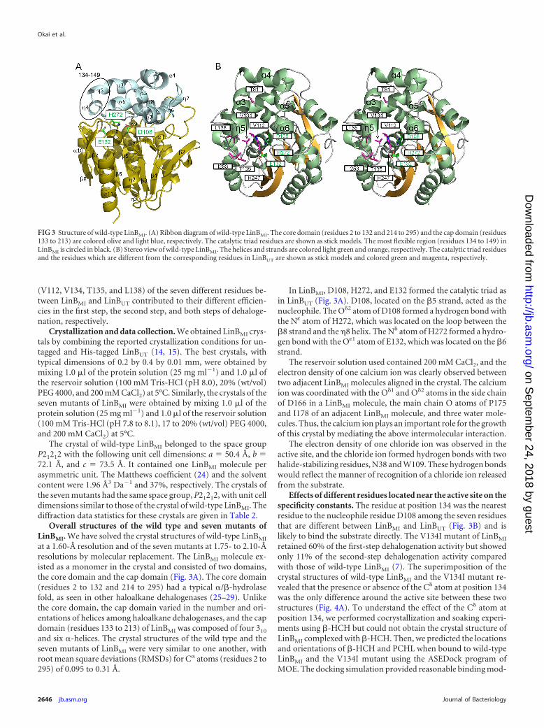
Effects of different residues located near the active site on thespecificity constants. The residue at position 134 was the nearestresidue to the nucleophile residue D108 among the seven residuesthat are different between LinBMI and LinBUT (Fig. 3B) and islikely to bind the substrate directly. The V134I mutant of LinBMI
retained 60% of the first-step dehalogenation activity but showedonly 11% of the second-step dehalogenation activity comparedwith those of wild-type LinBMI (7). The superimposition of thecrystal structures of wild-type LinBMI and the V134I mutant re-vealed that the presence or absence of the C� atom at position 134was the only difference around the active site between these twostructures (Fig. 4A). To understand the effect of the C� atom atposition 134, we performed cocrystallization and soaking experi-ments using �-HCH but could not obtain the crystal structure ofLinBMI complexed with �-HCH. Then, we predicted the locationsand orientations of �-HCH and PCHL when bound to wild-typeLinBMI and the V134I mutant using the ASEDock program ofMOE. The docking simulation provided reasonable binding mod-
FIG 3 Structure of wild-type LinBMI. (A) Ribbon diagram of wild-type LinBMI. The core domain (residues 2 to 132 and 214 to 295) and the cap domain (residues133 to 213) are colored olive and light blue, respectively. The catalytic triad residues are shown as stick models. The most flexible region (residues 134 to 149) inLinBMI is circled in black. (B) Stereo view of wild-type LinBMI. The helices and strands are colored light green and orange, respectively. The catalytic triad residuesand the residues which are different from the corresponding residues in LinBUT are shown as stick models and colored green and magenta, respectively.
Okai et al.
2646 jb.asm.org Journal of Bacteriology
on Septem
ber 24, 2018 by guesthttp://jb.asm
.org/D
ownloaded from
els of �-HCH for both wild-type LinBMI and the V134I mutant.The �-HCH molecules docked in wild-type LinBMI and the V134Imutant were located at the same position with almost the sameorientations (data not shown). On the other hand, the dockingsimulation with PCHL gave different results for wild-type LinBMI
and the V134I mutant. In the top three solutions, the interac-tion energies of the PCHL molecule with wild-type LinBMI
were �1.6, 1.8, and 3.4 kcal/mol, and those with the V134Imutant were �14.7, �1.1, and 2.3 kcal/mol. In wild-typeLinBMI, the manner of binding of PCHL in the top solutioncould explain the occurrence of the second-step conversionfrom PCHL to TCDL, with the distance between the O�2 atomof D108 and the C-4 atom of PCHL being 3.1 Å (Fig. 4B).However, in the case of the V134I mutant, the positions andorientations of the bound PCHL models in the top two solu-tions (Fig. 4C, cyan and magenta) were different from those inthe top solution for wild-type LinBMI. The C-4 atoms in the twoPCHL models were 4.7 Å away from the O�2 atom of D108, andthus the second-step conversion from PCHL to TCDL was un-likely to occur. The binding manner of the third solution (Fig.4C, yellow) for the V134I mutant was almost the same as that inthe top solution for wild-type LinBMI. These docking simula-tion results suggested that the V134I mutant of LinBMI was notlikely to bind PCHL properly for the second-step conversion tooccur because of the presence of the C� atom at position 134.
The residue at position 112 was located at the bottom of thesubstrate binding pocket (Fig. 3B). The V112A mutant of LinBMI
retained 53% of the first-step dehalogenation activity but showedonly 23% of the second-step dehalogenation activity of wild-typeLinBMI (7). In the V112A mutant, the main chain of V134 wasshifted by 0.3 Å toward the catalytic residue (D108) comparedwith the corresponding region in wild-type LinBMI, and the sidechain of W109, one of the two halide-stabilizing residues, wasrotated 6° relative to that in wild-type LinBMI around the C�-C�1
bond (Fig. 4D). Such structural differences at the two residuesshould be due to the absence of the C�2 atom rather than the C�1
atom in the V112A mutant of LinBMI. These structural changeswithin the active-site pocket should cause a reduction in first- andsecond-step dehalogenation activities in the V112A mutant ofLinBMI.
Effects of different residues lining the substrate access tunnelon specificity constants. The active site of LinBMI was burieddeeply inside the enzyme. Three entrances to the substrate accesstunnels were found in LinBMI using the software program CAVER(Fig. 5A). Two tunnel entrances (Fig. 5A, purple and cyan) wereformed by the �5, �3, �5, and �6 helices, and the other tunnelentrance (Fig. 5A, pink) was formed by the two helices (�4 and�10) and a loop between the �7 strand and the �10 helix. A tunnelentrance (Fig. 5B, purple) found in LinBUT, which was formed bythe �6 helix and two loops between the �4 and �5 helices andbetween the �7 strand and the �10 helix, was not observed inLinBMI because the side chain of His247 covered the entrance. Itoet al. reported that H247 in LinBMI was important for the second-step conversion of PCHL to TCDL (7). In the H247A mutantstructure, the �5 helix was shifted toward the �6 helix because theH247A mutation created an extra space, which resulted in confor-mational changes of the side chains of F143 and P144 (Fig. 5C).Thus, the side chain of H247 would contribute to the tunnel for-mation suitable for substrate (PCHL) entry and product (TCDL)release.
L138 and I253 of LinBMI were involved in the formation of oneaccess tunnel (Fig. 5A, pink), while T135 was located approxi-mately 6 Å away from the tunnel. The orientations of the sidechain at position 253 were divided into two groups among thewild type and mutants of LinBMI. In wild-type LinBMI and the
FIG 4 Different amino acid residues located around the active site betweenLinBMI and LinBUT. (A) Superimposition of the active sites of the wild type(light green) and the V134I mutant (slate) of LinBMI. The catalytic triad resi-dues (D108, E132, and H272) and V134/I134 are labeled. (B and C) Dockingsimulations of the wild type (B) or the V134I mutant (C) with PCHL. Thechlorine, oxygen, and hydrogen atoms of the PCHL molecules are coloredgreen, red, and white, respectively. In wild-type LinBMI, the PCHL model withthe lowest binding energy is shown, and its carbon atoms are colored yellow. Inthe V134I mutant, the carbon atoms are colored cyan, magenta, and yellowin the PCHL models with the lowest binding, the second-lowest binding andthe highest interation energies, respectively. (D) Superimposition of the activesites between the wild type (light green) and the V112A mutant (orange).
Structure of LinB from Sphingobium sp. Strain MI1205
June 2013 Volume 195 Number 11 jb.asm.org 2647
on Septem
ber 24, 2018 by guesthttp://jb.asm
.org/D
ownloaded from

T81A, V112A, V134I, and H247A mutants, the C�-C�1-C�1 chainof I253 faced toward the side chain of T135 (Fig. 5D). In contrast,in the T135A and L138I mutants, the C�-C�1-C�1 chains of I253faced toward the side chain of L138 (Fig. 5E and F). Thus, theT135A and L138I mutations caused the conformational changesof the side chain of I253, which resulted in the changes of the sizeand position of a tunnel entrance (Fig. 5D to F). In the I253Mmutant (Fig. 5G), the side chain of M253 faced toward the sidechain of L138, and the side chain of L138 was rotated approxi-mately 90° relative to that in wild-type LinBMI along the C�-C�
bond. In contrast, in LinBUT, the side chain of M253 faced towardthe side chain of A135 (Fig. 6) (11). Thus, the orientation of theside chain of M253 could be influenced by the residues at posi-tion(s) 135 and/or 138. Since the residue at position 253 was lo-cated at an entrance of the access tunnel, the irregular orientationof the side chain at position 253 affected the shape of the entranceof the access tunnel in the T135A, L138I, and I253M mutants (Fig.5E to G). The irregular forms of the tunnel entrances in thesemutants should lead to the reductions in the dehalogenase activi-ties, especially the second-step dehalogenation activity.
FIG 5 Different amino acid residues lining the access tunnel between LinBMI and LinBUT. (A and B) The three access tunnels (pink, purple, and cyan) to theactive site of wild-type LinBMI (A) or LinBUT (B). The catalytic triad residues (green) and amino acid residues (magenta) that are different between wild-typeLinBMI and LinBUT are shown as stick models. The red circles represent the entrances of the access tunnels. (C) Superimposition between the wild type (lightgreen) and the H247A mutant (cyan) of LinBMI. The tunnel (purple) observed in wild-type LinBMI is shown. The catalytic triad residues and the residues atpositions 135, 138, and 253 are shown as sticks in the wild-type (D), T135A mutant (E), L138I mutant (F), or I253M mutant (G) structure of LinBMI. The redcircles show the entrances of the access tunnels of the wild type and three mutants.
FIG 6 Structural comparison between wild-type LinBMI and LinBUT. Superimposition between wild-type LinBMI (light green) and LinBUT (cyan and dark gray)is shown. The most noteworthy difference between wild-type LinBMI and LinBUT is colored cyan in LinBUT. The catalytic triad residues, one (W109) of twohalide-stabilizing residues, and six of the residues that are different between wild-type LinBMI and LinBMI are shown as stick models and labeled.
Okai et al.
2648 jb.asm.org Journal of Bacteriology
on Septem
ber 24, 2018 by guesthttp://jb.asm
.org/D
ownloaded from

In wild-type LinBMI, T81 was positioned outside the active site,and the side chain of T81 formed hydrogen bonds with one watermolecule and the amide nitrogen of E84. The main-chain struc-ture of the T81A mutant was very similar to that of wild-typeLinBMI, with an RMSD of 0.12 Å, and no conformational changewas observed either at the active site or in the access tunnel be-tween wild-type LinBMI and the T81A mutant.
Structural comparison between LinBMI and LinBUT. LinBMI
and LinBUT share 98% sequence identity. Their overall structureswere very similar to each other, with an RMSD of 0.27 Å for 292 C�
atoms. The most remarkable structural difference between LinBMI
and LinBUT was observed at the N-terminal region of the capdomain (residues 134 to 149) (Fig. 3A and 6), which plays animportant role in determining the shape and size of the active siteand the substrate access tunnels. A structural difference similar tothat between LinBMI and LinBUT was observed between wild-typeLinBMI and the H247A mutant (Fig. 5C). The main chain of I134in LinBUT was shifted by 0.8 Å toward the catalytic residue (D108)compared with that in LinBMI, and the side chain of W109 inLinBUT was rotated approximately 5° relative to that in LinBMI
(Fig. 6). These structural differences would be due to the size of theamino acid residue at position 112 (Val in LinBMI versus Ala inLinBUT), considering the structural difference between wild-typeLinBMI and the V112A mutant.
We performed molecular dynamics simulations to reveal themolecular mechanisms of the different efficiencies in dehalogena-tion between LinBMI and LinBUT. In both the cases of LinBMI andLinBUT, the C� RMSDs against the initial coordinates increasedsharply in the first nanosecond of the simulations, and the RMSDswere in the range of 1.4 to 1.8 Å in the last two nanoseconds (12 to14 ns) (Fig. 7A), indicating that no global conformational changeoccurred. Figures 7B and C show the superpositions of the crystalstructures and the structures after the simulation of LinBMI andLinBUT, respectively. In the core domain of LinBUT, the crystal andthe simulated structures were almost identical. In contrast, in thecore domain of LinBMI, a conformational change was observed ina loop (residues 76 to 81) between the �4 strand and the �1 helix,which would be due to T81, the only residue in this region uniqueto LinBMI. The conformational change in the loop could lead to amovement of the interacting �7 helix in the cap domain towardthe �4 helix and a concomitant change in the shape of the sub-strate binding pocket, which might cause the different efficienciesin first-step dehalogenation activities between two enzymes. Asfor the cap domain, similar conformational changes were ob-served in both LinBMI and LinBUT in the following regions: �4-(loop)-�5, �4, �5, and �7. The conformational change at the en-trance of the substrate access tunnel from the �4 to �5 helices inLinBUT was larger than that in LinBMI, allowing the substrates to
FIG 7 Molecular dynamics (MD) simulations of wild-type LinBMI and LinBUT. (A) Time course of C� RMSDs from the initial structures of wild-type LinBMI
(green) and LinBUT (black) during MD simulations. (B) Superposition of the crystal structure (gray and green) and the structure after the simulation (gray andyellow) of wild-type LinBMI. (C) Superposition of the crystal structure (gray and black) and the structure after the simulation (gray and yellow) of wild-typeLinBUT. Green, black, and yellow in panels B and C indicate the most different regions observed between the crystal structures and the structures after thesimulations (gray and yellow). (D) C� RMSFs for LinBMI (green) and LinBUT (black) residues over the last 2-ns simulations.
Structure of LinB from Sphingobium sp. Strain MI1205
June 2013 Volume 195 Number 11 jb.asm.org 2649
on Septem
ber 24, 2018 by guesthttp://jb.asm
.org/D
ownloaded from

enter the tunnel easily (Fig. 7B and C). The different residues atpositions 247 and 253 should cause the different conformationalchanges in this region. Root mean square fluctuation (RMSF) wasused as an index of structural flexibility. The RMSF analysis (Fig.7D) clearly shows that the entrance of the substrate access tunnelfrom the �4 to �5 helices (residues 142 to 146) of LinBUT is muchmore flexible than that of LinBMI. This high flexibility in LinBUT
would lead to the large conformational change at the entrance ofthe substrate access tunnel, as shown in Fig. 7B. In DhaA, a mem-ber of the same �/�-hydrolase family as LinB, the molecular dy-namics simulation analysis revealed that the narrower substrateaccess tunnel in a variant than in the wild-type enzyme shieldedthe active site from the solvent and showed higher activity thanthat of the wild-type enzyme (30). Similarly, the low flexibility ofthe tunnel entrance in LinBMI would contribute to the increase inits dehalogenation activity by inhibiting the influx of water mole-cules into the active site, particularly for second-step dehalogena-tion activity, where the water molecules can compete with thehydroxyl group of PCHL.
Concluding remarks. We have analyzed the dehalogenationactivities of five of the seven amino acid residues that differ be-tween LinBMI and LinBUT. This and previous mutagenesis analy-ses revealed that most of the seven residues had effects on second-step dehalogenation and none of the seven residues were criticalfor degradation activity. We have determined the crystal struc-tures of the wild type and the seven mutants of LinBMI. The struc-tural comparisons among wild-type LinBMI, LinBUT, and theseven mutants of LinBMI indicated that each mutant except theT81A mutant caused a small conformational change in the accesstunnels or the active site that resulted in a reduction in the first-and second-step dehalogenation activities of LinBUT comparedwith those of LinBMI. The dynamics simulations of wild-typeLinBMI and LinBUT suggested that the flexibility of the entrance ofthe substrate access tunnel led to the difference in dehalogenationactivity, peculiarly the second-step activity.
ACKNOWLEDGMENTS
We thank the beamline staff at the Photon Factory for their kind help withdata collection. We thank Zbynek Prokop for assistance in calculating thespecificity constants of enzymatic activities. Synchrotron radiation exper-iments were done at the Photon Factory (Ibaraki, Japan) (proposal no.2009G122).
This work was supported in part by the Targeted Proteins ResearchProgram and Grants-in-Aid of the Ministry of Education, Culture, Sports,Science, and Technology of Japan.
REFERENCES1. Willett KL, Ulrich EM, Hites RA. 1998. Differential toxicity and envi-
ronmental fates of hexachlorocyclohexane isomers. Environ. Sci. Technol.32:2197–2207.
2. Walker K, Vallero DA, Lewis RG. 1999. Factors influencing the distri-bution of lindane and other hexachlorocyclohexanes in the environment.Environ. Sci. Technol. 33:4373– 4378.
3. Johri AK, Dua M, Tuteja D, Saxena R, Saxena DM, Lal R. 1998.Degradation of �, �, � and �-hexachlorocyclohexanes by Sphingomonaspaucimobilis. Biotechnol. Lett. 20:885– 887.
4. Gupta A, Kaushik CP, Kaushik A. 2000. Degradation of hexachlorocy-clohexane (HCH; �, �, � and �) by Bacillus circulans and Bacillus brevisisolated from soil contaminated with HCH. Soil Biol. Biochem. 32:1803–1805.
5. Gupta A, Kaushik CP, Kaushik A. 2001. Degradation of hexachlorocy-clohexane isomers by two strains of Alcaligenes faecalis isolated from acontaminated site. Bull. Environ. Contam. Toxicol. 66:794 – 800.
6. Prokop Z, Monincová M, Chaloupková R, Klvana M, Nagata Y, JanssenDB, Damborský J. 2003. Catalytic mechanism of the haloalkane dehalo-genase LinB from Sphingomonas paucimobilis UT26. J. Biol. Chem. 278:45094 – 45100.
7. Ito M, Prokop Z, Klvana M, Otsubo Y, Tsuda M, Damborský J, NagataY. 2007. Degradation of �-hexachlorocyclohexane by haloalkane dehalo-genase LinB from �-hexachlorocyclohexane-utilizing bacterium Sphingo-bium sp. MI1205. Arch. Microbiol. 188:313–325.
8. Wu J, Hong Q, Han P, He J, Li S. 2007. A gene linB2 responsible for theconversion of �-HCH and 2,3,4,5,6-pentachlorocyclohexanol in Sphingomo-nas sp. BHC-A. Appl. Microbiol. Biotechnol. 73:1097–1105.
9. Sharma P, Raina V, Kumari R, Malhotra S, Dogra C, Kumari H,Kohler HP, Buser HR, Holliger C, Lal R. 2006. Haloalkane dehalo-genase LinB is responsible for �- and �-hexachlorocyclohexane trans-formation in Sphingobium indicum B90A. Appl. Environ. Microbiol.72:5720 –5727.
10. Nagata Y, Prokop Z, Sato Y, Jerabek P, Kumar A, Ohtsubo Y, TsudaM, Damborský J. 2005. Degradation of �-hexachlorocyclohexane byhaloalkane dehalogenase LinB from Sphingomonas paucimobilis UT26.Appl. Environ. Microbiol. 71:2183–2185.
11. Marek J, Vévodová J, Smatanová IK, Nagata Y, Svensson LA, NewmanJ, Takagi M, Damborský J. 2000. Crystal structure of the haloalkanedehalogenase from Sphingomonas paucimobilis UT26. Biochemistry 39:14082–14086.
12. Oakley AJ, Prokop Z, Bohác M, Kmunicek J, Jedlicka T, Monincová M,Kuta-Smatanová I, Nagata Y, Damborský J, Wilce MC. 2002. Exploringthe structure and activity of haloalkane dehalogenase from Sphingomonaspaucimobilis UT26: evidence for product- and water-mediated inhibition.Biochemistry 41:4847– 4855.
13. Streltsov VA, Prokop Z, Damborský J, Nagata Y, Oakley A, Wilce MC.2003. Haloalkane dehalogenase LinB from Sphingomonas paucimobilisUT26: X-ray crystallographic studies of dehalogenation of brominatedsubstrates. Biochemistry 42:10104 –10112.
14. Oakley AJ, Klvana M, Otyepka M, Nagata Y, Wilce MC, Damborský J.2004. Crystal structure of haloalkane dehalogenase LinB from Sphingomo-nas paucimobilis UT26 at 0.95 Å resolution: dynamics of catalytic residues.Biochemistry 43:870 – 878.
15. Smatanová I, Nagata Y, Svensson LA, Takagi M, Marek J. 1999. Crys-tallization and preliminary X-ray diffraction analysis of haloalkane deha-logenase LinB from Sphingomonas paucimobilis UT26. Acta Crystallogr. D55:1231–1233.
16. Otwinowski Z, Minor W. 1997. Processing of X-ray diffraction datacollected in oscillation mode. Methods Enzymol. 276:307–326.
17. Vagin A, Teplyakov A. 1997. MOLREP: an automated program for mo-lecular replacement. J. Appl. Crystallogr. 30:1022–1025.
18. Emsley P, Cowtan K. 2004. Coot: model-building tools for moleculargraphics. Acta Crystallogr. D 60:2126 –2132.
19. Murshudov GN, Vagin AA, Dodson EJ. 1997. Refinement of macromo-lecular structures by the maximum-likelihood method. Acta Crystallogr.D 53:240 –255.
20. Perrakis A, Morris R, Lamzin VS. 1999. Automated protein modelbuilding combined with iterative structure refinement. Nat. Struct. Biol.6:458 – 463.
21. Lovell SC, Davis IW, Arendall WB, III, de Bakker PI, Word JM, PrisantMG, Richardson JS, Richardson DC. 2003. Structure validation by C�geometry: �,� and C� deviation. Proteins 50:437– 450.
22. Mendes P. 1997. Biochemistry by numbers: simulation of biochemicalpathways with Gepasi 3. Trends Biochem. Sci. 22:361–363.
23. Baeck T, Fogel DB, Michalewicz Z. 1997. Handbook of evolutionarycomputation. IOP Publishing/Oxford University Press, Oxford, UnitedKingdom.
24. Matthews BW. 1968. Solvent content of protein crystals. J. Mol. Biol.33:491– 497.
25. Verschueren KH, Franken SM, Rozeboom HJ, Kalk KH, Dijkstra BW.1993. Refined X-ray structures of haloalkane dehalogenase at pH 6.2 andpH 8.2 and implications for the reaction mechanism. J. Mol. Biol. 232:856 – 872.
26. Newman J, Peat TS, Richard R, Kan L, Swanson PE, Affholter JA,Holmes IH, Schindler JF, Unkefer CJ, Terwilliger TC. 1999. Haloalkanedehalogenases: structure of a Rhodococcus enzyme. Biochemistry 38:16105–16114.
27. Pavlová M, Klvana M, Jesenská A, Prokop Z, Konecná H, Sato T, TsudaM, Nagata Y, Damborský J. 2007. The identification of catalytic pentad in
Okai et al.
2650 jb.asm.org Journal of Bacteriology
on Septem
ber 24, 2018 by guesthttp://jb.asm
.org/D
ownloaded from
the haloalkane dehalogenase DhmA from Mycobacterium avium N85: re-action mechanism and molecular evolution. J. Struct. Biol. 157:384 –392.
28. Mazumdar PA, Hulecki JC, Cherney MM, Garen CR, James MN. 2008.X-ray crystal structure of Mycobacterium tuberculosis haloalkane dehalo-genase Rv2579. Biochim. Biophys. Acta 1784:351–362.
29. Hesseler M, Bogdanovic X, Hidalgo A, Berenguer J, Palm GJ, HinrichsW, Bornscheuer UT. 2011. Cloning, functional expression, biochemical
characterization, and structural analysis of a haloalkane dehalogenasefrom Plesiocystis pacifica SIR-1. Appl. Microbiol. Biotechnol. 91:1049 –1060.
30. Pavlová M, Klvana M, Prokop Z, Chaloupková R, Banás P, Otyepka M,Wade RC, Tsuda M, Nagata Y, Damborský J. 2009. Redesigning deha-logenase access tunnels as a strategy for degrading an anthropogenic sub-strate. Nat. Chem. Biol. 5:727–733.
Structure of LinB from Sphingobium sp. Strain MI1205
June 2013 Volume 195 Number 11 jb.asm.org 2651
on Septem
ber 24, 2018 by guesthttp://jb.asm
.org/D
ownloaded from