Embed Size (px)
Citation preview

8/6/2019 Crystal is at Ion
http://slidepdf.com/reader/full/crystal-is-at-ion 1/12
Methods 34 (2004) 254–265
www.elsevier.com/locate/ymeth
1046-2023/$ - see front matter 2004 Elsevier Inc. All rights reserved.
doi:10.1016/j.ymeth.2004.03.019
Introduction to protein crystallization
Alexander McPherson¤
Department of Molecular Biology and Biochemistry, University of Califonia, Irvine 560 Steinhaus Hall, Irvine, CA 92697-3900, USA
Accepted 24 March 2004
Abstract
Biological macromolecules can be crystallized by a variety of techniques, and using a wide range of reagents which produce super-saturated mother liquors. These may, in turn, be applied under diV erent physical conditions such as temperature. The fundamental
approaches to devising successful crystallization conditions and the factors that inXuence them are summarized here. For the novice,
it is hoped that this brief review might serve as a useful introduction and a stepping-stone to a successful X-ray strucutre determina-
tion. In addition, it may provide a framework in which to place the articles that follow.
2004 Elsevier Inc. All rights reserved.
1. Some history
Protein crystallization developed in the latter half of
the 19th century for three reasons, (a) it provided a
means for the puriWcation of speciWc proteins from anotherwise impure mixture at a time when few other
means existed, (b) it served as a demonstration that a
protein had been puriWed (which even now is taken as a
pretty good measure), and (c) it was an interesting labo-
ratory curiosity. Initially, the crystallization of hemoglo-
bin from a variety of sources was really nothing more
than that [36], though a thoughtful attempt to relate
hemoglobin crystal form to evolution was made around
1900 [48]. The Wrst two reasons, however, were dominant
in the last quarter of the 19th century and biochemists
such as Osborne used it extensively to isolate and char-
acterize proteins, particularly those from seeds. Themeaning of this early research for us today is to show
Wrst of all that speciWc proteins can often be isolated
from quite impure preparations, but more important,
these pioneers deduced many of the approaches for the
growth of protein crystals that are still in use.
Between 1900 and 1940, the emphasis was on
enzymes, and again, crystallization in the hands of Sum-
mer, Northrup, Kunitz, Herriott, and their colleagues
[50–52,43] proved an important tool in establishing the
properties and nature of catalytic macromolecules. In
the late 1930s, however, a new application for protein
crystallization appeared as a result of the studies, using
X-ray diV raction analysis, of Bernal and Crowfoot [2],Perutz [45], and others. Today, though crystallization is
still an admired and respected procedure by enzymolo-
gists and protein chemists, X-ray crystallography, and
the structure determination of macromolecules and their
complexes stand as the principal objective of those
involved in crystallization.
A fundamental change in protein crystallization, its
investigation and its application, occurred in the 1980s.
This was due to the development of recombinant DNA
technology which permitted researchers, for the Wrst
time, to prepare ample amounts of otherwise rare and
elusive proteins. Currently, the majority of proteinsaddressed by X-ray diV raction are derived from recom-
binant sources. Among the other consequences is that
today we are generally working with more homogeneous
protein samples which exhibit greater reproducibility
than ever before. Needless to say, this has both acceler-
ated the progress of X-ray crystallography, and greatly
expanded both its applicability and its appeal to bio-
chemists and molecular biologists [37,38].
Ultimately, structural biologists would like to describe
all living systems, and the materials they produce, in
molecular and even atomic terms. This Wrst requires a
precise knowledge of the building block molecules, the
¤Fax: 1-949-824-5992.
E-mail address: [email protected].
8/6/2019 Crystal is at Ion
http://slidepdf.com/reader/full/crystal-is-at-ion 2/12
A. McPherson / Methods 34 (2004) 254–265 255
proteins, nucleic acids, lipids, and polysaccharides. It
further requires information of a somewhat diV erent
nature, rules or guidelines to specify how the building
block molecules are joined, organized, and assembled
into higher order structures. Those greater structures
include macromolecular complexes, assemblies, organ-
elles, cell walls, membranes, cytoskeletons, etc. Fromthese assemblies we can Wnally reach a molecular and
atomic level description of the living cell, and from that
understand, in terms of classical chemistry and physics,
the architecture and mechanics of living matter.
The dynamics within the cell, the mechanisms respon-
sible for the dynamics, and the cell's interactions with
exterior inXuences are equally important. To understand
those, however, we Wrst need to delineate how the build-
ing block molecules respond to chemical and physical
forces, how the responses are regulated, and how the
responses are transmitted through the hierarchy of
assemblies and higher structures. This in turn means
visualizing the building block molecules, not in a single
state, but in all of those available as a consequence of
their molecular interactions.
The salient elements of this more detailed and com-
prehensive understanding of life's design and processes
are the structures of the building block molecules, and
the principles of how they assemble and interact. To the
precision required, these properties can only be
addressed by X-ray crystallography. The atomic struc-
tures of the building block molecules, the proteins and
other macromolecules, must be elucidated. This includes
not only those easily solubilized and crystallized, but
also those that resist current techniques.Progress in molecular biology and its application to
human medicine, agriculture, and industrial processes
have for the past two decades been crucially dependent
on a detailed knowledge of macromolecular structure at
the atomic level. This has included proteins, nucleic
acids, viruses, and other large macromolecular com-
plexes and assemblies. Redundancies in structural ele-
ments emerging from the now constant Xow of newly
determined molecular structures suggest that the num-
ber of naturally occurring structural motifs and sub-
structures (domains) may be Wnite. Ultimately then, all
macromolecular structures may be classiWed and cata-
logued according to polypeptide folds. Once all, or most
of the folds which are utilized by nature, are known, then
this will provide predictive insight, based primarily on
amino acid sequence, into the structures and functions of
unknown proteins. The sequences of most proteins, it is
important to note, are currently being elucidated by a
broad array of sequencing eV orts, such as the human
genome project, carried out both by government and the
private sector. Extension of these genome projects to the
three-dimensional structural level appears the next logi-
cal step, and this eV ort, under the broad rubric of struc-
tural genomics, is now in the initial stages.
In addition to the dramatic impact that knowledge of
three-dimensional structures of proteins has had on fun-
damental research in biochemistry and biology, macro-
molecular structure, is of formidable value in
biotechnology as well. Here, it provides the essential
knowledge required to apply the technique of rational
drug design in the creation and discovery of new drugsand pharmaceutical products [37,38]. It serves as the
basis of powerful approaches now being applied in
emerging biotechnology enterprises, as well as major
pharmaceutical companies, to identify lead compounds
to treat a host of human ailments, veterinary problems,
and crop diseases in agriculture. The underlying hypoth-
esis is that if the structure of the active site of a salient
enzyme in a metabolic or regulatory pathway is known,
then chemical compounds, such as drugs, can be ratio-
nally designed to inhibit or otherwise aV ect the behavior
of that enzyme.
A second approach, of equal importance to biotech-
nology that also requires knowledge of three-dimen-
sional macromolecular structure is the genetic
engineering of proteins. Although recombinant DNA
techniques provide the essential synthetic role that per-
mits modiWcation of proteins, structure determination
by X-ray crystallography provides the analytical func-
tion. It serves as a structural guide for rational and pur-
poseful changes, in place of random and chance amino
acid substitutions. Direct visualization of the structural
alterations that are introduced by mutation oV ers new
directions for chemical and physical enhancements.
Presently, and in the foreseeable future, the only tech-
nique that can yield atomic level structural images of biological macromolecules is X-ray diV raction analysis
as applied to single crystals. While other methods may
produce important structural and dynamic data, for the
purposes described above, only X-ray crystallography is
adequate. As its name suggests, application of X-ray
crystallography is absolutely dependent on crystals of
the macromolecule, and not simply crystals, but crystals
of suYcient size and quality to permit accurate data col-
lection. The quality of the Wnal structural image is
directly determined by the perfection, size, and physical
properties of the crystalline specimen, hence the crystal
becomes the keystone element of the entire process, and
the ultimate determinant of its success [35].
When crystallizing proteins for X-ray diV raction
analysis, one is usually dealing with homogeneous, often
exceptionally pure, macromolecules, and the objective
may be to grow only a few large, perfect crystals. It is
important to emphasize that while the number of crys-
tals needed may be few, often the amount of protein
available may be severely limited. This in turn places
grave constraints on the approaches and strategies that
may be used to obtain those crystals. While new method-
ologies such as synchrotron radiation [21] and cryocrys-
tallography [15] have driven the necessary size of

8/6/2019 Crystal is at Ion
http://slidepdf.com/reader/full/crystal-is-at-ion 3/12
256 A. McPherson / Methods 34 (2004) 254–265
specimen crystals consistently downward, they have not
alleviated the need for crystal perfection.
It is also well to remember that X-ray analysis is a sin-
gular event conWned to the research laboratory and the
Wnal product is basic scientiWc knowledge. The crystals
themselves have no medicinal or pharmaceutical value,
but simply serve as intermediaries in the crystallographyprocess. The crystals provide the X-ray diV raction pat-
terns that in turn serve as the raw data which allow the
direct visualization of the macromolecules or their com-
plexes composing the crystals.
2. General approach
Macromolecular crystallization, which includes the
crystallization of proteins, nucleic acids, and larger mac-
romolecular assemblies such as viruses and ribosomes, is
based on a rather diverse set of principles, experiences,
and ideas. There is no comprehensive theory, or even a
very good base of fundamental data, to guide our eV orts,
though that is being accumulated at this time. As a con-
sequence, macromolecular crystal growth is largely
empirical in nature, and demands patience, perseverance,
and intuition.
Complicating the entire process, in addition to our lim-
ited understanding of the phenomena involved, is the
astonishing complexity and the range of macromolecules
before us. Even in the case of rather small proteins, such as
cytochrome c or myoglobin for example, there are roughly
a thousand atoms with hundreds of bonds and thousands
of degrees of freedom. For viruses of weights measured inthe millions of daltons, the possibilities for conformation,
interaction, and mobility are almost unimaginable.
Only now are we beginning to develop rational
approaches to macromolecular crystallization based on an
understanding of the fundamental properties of the sys-
tems. We are only now using, in a serious and systematic
manner, the classical methods of physical-chemistry to
determine the characteristics of those mechanisms respon-
sible for the self-organization of large biological molecules
into crystal lattices. As an alternative to the precise and
reasoned strategies that we commonly apply to scientiWc
problems, we rely, for the time being at least, on what isfundamentally a trial and error approach. Macromolecu-
lar crystallization is generally a matter of searching, as sys-
tematically as possible, the ranges of the individual
parameters that inXuence crystal formation, Wnding a set,
or multiple sets of factors that yield some kind of crystals,
and then optimizing the individual variables to obtain the
best possible crystals. This is usually achieved by carrying
out an extensive series, or establishing a vast matrix of
crystallization trials, evaluating the results, and using what
information is obtained to improve conditions in succes-
sive rounds of trials. Because the number of variables is
so large, and the ranges so broad, experience and insight
into designing and evaluating the individual and collec-
tive trials becomes an important consideration.
3. The nature of protein crystals
Macromolecular crystals like those seen in Fig. 1 arecomposed of approximately 50% solvent on average,
though this may vary from 25 to 90% depending on the
particular macromolecule. Protein or nucleic acid occu-
pies the remaining volume so that the entire crystal is in
many ways an ordered gel permeated by extensive inter-
stitial spaces through which solvent and other small mol-
ecules freely diV use.
In proportion to molecular mass, the number of bonds
(salt bridges, hydrogen bonds, and hydrophobic interac-
tions) that a conventional molecule forms with its neigh-
bors in a crystal far exceeds the very few exhibited by
crystalline macromolecules. Since these contacts provide
the lattice interactions essential for crystal maintenance,
this largely explains the diV erences in properties between
crystals of salts or small molecules and macromolecules.
Living systems are based almost exclusively on aque-
ous chemistry within narrow ranges of temperature and
pH. Macromolecules have, thus, evolved an appropriate
compatibility. Serious deviations or perturbations are
rarely tolerated. As a consequence, all protein and
nucleic acid crystals must be grown from aqueous solu-
tions, ones to which they are tolerant, and these solu-
tions are called mother liquors. Macromolecular crystals
have not yet been grown except from such medium.
Although comparable in their morphologies andappearance, there are important practical diV erences
between crystals of low-molecular-mass compounds and
crystals of proteins and nucleic acids. Crystals of con-
ventional molecules are characterized by Wrm lattice
forces, are relatively highly ordered, generally physically
hard and brittle, easy to manipulate, usually can be
exposed to air, have strong optical properties, and
diV ract X-rays intensely. Macromolecular crystals are by
comparison usually more limited in size, are very soft
and crush easily, disintegrate if allowed to dehydrate,
exhibit weak optical properties, and diV ract X-rays
poorly. Macromolecular crystals are temperature sensi-tive and undergo extensive damage after prolonged
exposure to radiation. Frequently, several crystals must
be analyzed for a structure determination to be success-
ful although the advent of cryocrystallography [46],
CCD area detectors of very high photon counting
eYciency [19], high intensity synchrotron X-ray sources
[21,46], and new phasing methods (Chapters 12–16) [49]
have greatly lessened this constraint.
The extent of the diV raction pattern from a crystal is
directly correlated with its degree of internal order. The
more vast the pattern, or the higher the resolution to
which it extends, the more structurally uniform are the
8/6/2019 Crystal is at Ion
http://slidepdf.com/reader/full/crystal-is-at-ion 4/12
A. McPherson / Methods 34 (2004) 254–265 257
molecules in the crystal and the more precise is their peri-odic arrangement. The level of detail to which atomic
positions can be determined by crystal structure analysis
corresponds closely with that degree of crystalline order.
While conventional crystals often diV ract to their theoreti-
cal limit of resolution, protein crystals, by comparison,
produce diV raction patterns of more limited extent.
The liquid channels and solvent Wlled cavities that per-
meate macromolecular crystals are primarily responsible
for the limited resolution of the diV raction patterns.
Because of the relatively large spaces between adjacent
molecules and the consequent weak lattice forces, all mole-
cules in the crystal may not occupy exactly equivalent ori-
entations and positions but may vary slightly within or
between unit cells. Furthermore, because of their structural
complexity and their potential for conformational dynam-
ics, protein molecules in a particular crystal may exhibit
slight variations in the course of their polypeptide chains
or the dispositions of side groups from one to another.
Although the presence of extensive solvent regions is a
major contributor to the generally modest diV raction
quality of protein crystals, it is also responsible for their
value to biochemists. Because of the high solvent content,
the individual macromolecules in protein crystals are sur-
rounded by layers of water that maintain their structure
virtually unchanged from that found in solution. As aconsequence, ligand binding, enzymatic, spectroscopic
characteristics, and most other biochemical features are
essentially the same as for the fully solvated molecule.
Conventional chemical compounds, which may be ions,
ligands, substrates, coenzymes, inhibitors, drugs, or other
eV ector molecules, may be freely diV used into and out of
the crystals. Crystalline enzymes, though immobilized,
are completely accessible for experimentation simply
through alteration of the surrounding mother liquor.
Polymorphism is a common phenomenon with both
protein, nucleic acid, and virus crystals. Presumably this
is a consequence of their conformational dynamic range
and the sensitivity of the lattice contacts involved. Thus,
diV erent habits and diV erent unit cells may arise from
what, by most standards, would be called identical con-
ditions. In fact, multiple crystal forms are sometimes
seen coexisting in the same sample of mother liquor.
There are further diV erences which complicate the
crystallization of macromolecules as compared with
conventional, small molecules [10,12,13,34,39,41]. First,
macromolecules may assume multiple distinctive states
that include amorphous precipitates, oils, or gels as well as
crystals, and most of these are kinetically favored. Second,
macromolecular crystals nucleate, or initiate development
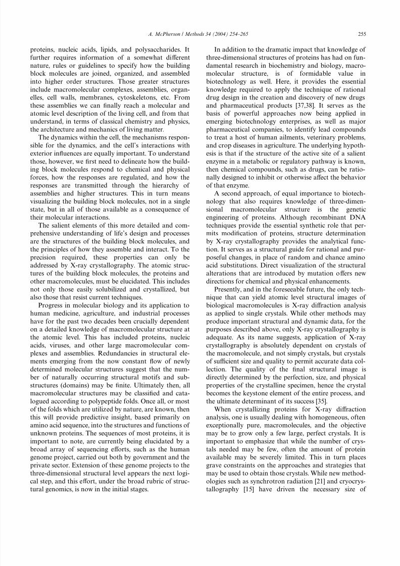
Fig. 1. An array of protein and virus crystals grown for X-ray diV raction analysis under a variety of conditions. In (A) the -subunit of leutinizing
hormone, (B) satellite tobacco mosaic virus, (C) a CH2 domain deleted human antibody, (D) the sweet protein thaumatin (E) the T D 1 particle of
brome mosaic virus, and (F) the seed storage protein canavalin.
8/6/2019 Crystal is at Ion
http://slidepdf.com/reader/full/crystal-is-at-ion 5/12
258 A. McPherson / Methods 34 (2004) 254–265
only at very high levels of supersaturation, often two to
three orders of magnitude greater than required to sustain
growth. Finally, the kinetics of macromolecular crystal
nucleation and growth are generally two to three orders
of magnitude slower than for conventional molecules
[27,30,32]. This latter diV erence arises from the consider-
ably larger size, lowered diV usivity, and weaker associa-tion tendencies compared with small molecules or ions, as
well as a lower probability of incorporation of an incom-
ing macromolecule into a growth step [4].
4. Screening and optimization
There are really two phases in the pursuit of protein
crystals for an X-ray diV raction investigation, and these
are (a) the identiWcation of chemical, biochemical, and
physical conditions that yield some crystalline material,
though it may be entirely inadequate, and (b) the system-
atic alteration of those initial conditions by incremental
amounts to obtain optimal samples for diV raction analy-
sis. The Wrst of these is fraught with the greater risk, as
some proteins simply refuse to form crystals, and any
clues as to why are elusive or absent. The latter, however,
often proves the more demanding, time consuming, and
frustrating.
There are basically two approaches to screening for
crystallization conditions. The Wrst is a systematic varia-
tion of what are believed to be the most important vari-
ables, precipitant type and concentration, pH,
temperature, etc. The second is what we might term a
shotgun approach, but a shotgun aimed with intelli-gence, experience, and accumulated wisdom. While far
more thorough in scope and more congenial to the scien-
tiWc mind, the Wrst method usually does require a signiW-
cantly greater amount of protein. In those cases where
the quantity of material is limiting, it may simply be
impractical. The second technique provides much more
opportunity for useful conditions to escape discovery,
but in general requires less precious material.
The second approach also has, presently at least, one
other major advantage, and that is convenience. There is
currently on the commercial market, from numerous
companies, a wide variety of crystallization screeningkits. The availability and ease of use of these relatively
modestly priced kits, which may be used in conjunction
with a variety of crystallization methods (hanging and
sitting drop vapor diV usion, dialysis, etc. see below),
make them the Wrst tool of choice in attacking a new
crystallization problem. With these kits, nothing more is
required than combining a series of potential crystalliza-
tion solutions with one's protein of interest using a
micropipette, sealing the samples, and waiting for suc-
cess to smile. Often it does, but sometimes not, and this is
when the crystal grower must begin using his own intelli-
gence to diagnose the problem and devise a remedy.
Once some crystals, even if only microcrystals are
observed and shown to be of protein origin (and one
ardently hopes for this event) then optimization begins.
Every component in the solution yielding crystals must be
noted and considered (buV er, salt, ions, etc.), along with
pH, temperature, and whatever other factors (see below)
might have an impact on the quality of the results. Eachof these parameters or factors is then carefully incre-
mented in additional trial matricies encompassing a range
spanning the conditions which gave the “hit.” Because the
problem is non linear, and one variable may be coupled to
another, this process is often more complex and diYcult
than one might expect [1,9,34,39]. It is here that the
amount of protein and the limits of the investigator's
patience may prove a formidable constraint.
5. Supersaturation, nucleation, and growth
Crystallization of a molecule, or of any chemical spe-
cies including proteins proceeds in two rather distinct but
inseparable steps, nucleation and growth. Nucleation is
the most diYcult problem to address theoretically and
experimentally because it represents a Wrst order phase
transition by which molecules pass from a wholly disor-
dered state to an ordered one. Presumably this occurs
through the formation of partially ordered or paracrystal-
line intermediates, in this case protein aggregates having
short-range order, and ultimately yields small, completely
ordered assemblies which we refer to as critical nuclei.
Critical nuclei must be considered in terms of the
molecular dimensions, the supersaturation, and the sur-face free energy of molecular addition. Currently the
critical nuclear size has only been described for a few
systems, and for several cases these were only investi-
gated in terms of two-dimensional nuclei developing on
the surfaces of already existent crystals [30,32]. Recently,
a theory has emerged which attempts to explain the
nucleation phenomenon in terms of statistical Xuctua-
tions in solution properties [20,47,53]. This idea holds
that a distinctive “liquid protein phase” forms in concen-
trated protein solutions, and that this “phase” ultimately
gives rise to critical nuclei with comprehensive order.
This idea is now under study by a variety of experimen-tal techniques in numerous laboratories.
Growth of macromolecular crystals is a better-char-
acterized process than nucleation, and its mechanisms
are reasonably well understood. Protein crystals grow
principally by the classical mechanisms of dislocation
growth, and growth by two-dimensional nucleation,
along with two other less common mechanisms known
as normal growth and three-dimensional nucleation
[31,42]. A common feature of nucleation and growth is
that both are critically dependent on what is termed the
supersaturation of the mother liquor giving rise to the
crystals. Supersaturation is the variable that drives both

8/6/2019 Crystal is at Ion
http://slidepdf.com/reader/full/crystal-is-at-ion 6/12
A. McPherson / Methods 34 (2004) 254–265 259
processes and determines their occurrence, extent, and
the kinetics that govern them.
Crystallization of a macromolecule absolutely requires
the creation of a supersaturated state. This is a non-equi-
librium condition in which some quantity of the macro-
molecule in excess of the solubility limit, under speciWc
chemical and physical conditions, is nonetheless presentin solution. Equilibrium is re-established by formation
and development of a solid state, such as crystals, as the
saturation limit is attained. To produce the supersatu-
rated solution, the properties of an undersaturated solu-
tion must be modiWed to reduce the ability of the medium
to solubilize the macromolecule (i.e., reduce its chemical
activity), or some property of the macromolecules must
be altered to reduce their solubility and/or to increase the
attraction of one macromolecule for another. In all cases,
the relationships between solvent and solute, or between
the macromolecules in solution are perturbed so as to
promote formation of the solid state.
If no crystals or other solid is present as conditions are
changed, then solute will not immediately partition into
two phases, and the solution will remain in the supersatu-
rated state. The solid state does not develop spontane-
ously as the saturation limit is exceeded because energy,
analogous to the activation energy of a chemical reaction,
is required to create the second phase, the stable nucleus
of a crystal or a precipitate. Thus, an or energy (or proba-
bility) barrier allows conditions to proceed further from
equilibrium and further into the zone of supersaturation.
Once a stable nucleus appears in a supersaturated solu-
tion, however, it will proceed to grow until the system
regains equilibrium. So long as non-equilibrium forcesprevail and some degree of supersaturation exists to drive
events, a crystal will grow or precipitate continue to form.
6. Creating a state of supersaturation
In practice, one begins (with the exception of the
batch method, see below) with a solution, a potential
mother liquor, which contains some concentration of the
protein below its solubility limit, or alternatively at its
solubility maximum. The objective is then to alter mat-
ters so that the solubility of the protein in the sample issigniWcantly reduced, thereby rendering the solution
supersaturated. This may be done through several
approaches, (a) altering the protein itself (e.g., by change
of pH which alters the ionization state of surface amino
acid residues), (b) by altering the chemical activity of the
water (e.g., by addition of salt), (c) by altering the degree
of attraction of one protein molecule for another (e.g.,
change of pH, addition of bridging ions), or (d) altering
the nature of the interactions between the protein mole-
cules and the solvent (e.g., addition of polymers or ions).
Table 1 is a compilation of the methods upon which
one might develop strategies for crystallizing a protein
for the Wrst time. Indeed there may be others, the limit is
only a function of the imagination and cunning of the
investigator. The details of these various approaches
have been described in detail numerous times elsewhere
[1,9,34,39,40] and need receive no more attention here. It
is probably suYcient to say that if a protein has any pro-
pensity to crystallize readily, it can probably be accom-
plished by variation of precipitant type, precipitant
concentration, pH, to a lesser extent temperature, but
with all due consideration to the biochemical properties
and eccentricities of the protein under investigation.
Finally, we are all advised that with real estate there are
three important factors, and they are location, location,
and location. With protein crystallization there are simi-
larly three, and they are purity, purity, and homogeneity.
7. Methodology
The growth of protein crystals must be carried out in
some physical apparatus that allows the investigator to
alter the solubility of the protein or the properties of the
mother liquor using one of the strategies in Table 1. Cur-
rently, these use almost exclusively microtechniques.
Thus, crystallization “trials” with a particular matrix of
conditions may be carried out with volumes of only a
few microliters or less. Increasingly these employ plastic,
multichambered trays for hanging and sitting drops,
plexiglass buttons for dialysis, or microdrops under oil.
Other approaches are found in Table 2.
Again, all of these devices and their methodologies have
been described in detail elsewhere (and also elaborated
upon in other chapters of this volume). It is unnecessary
to comment on each of them again. In addition, detailed
instructions are frequently provided by the manufactur-
ers of the crystallization kits, supplies, and plasticware
along with much helpful material. SuYce it to say that
currently the hanging drop and sitting drop procedures
for vapor diV usion, and the batch method using micro-
drops under oil are most in favor, and are recommended
for most investigations. In those cases where mother
liquor components cannot be transported through the
Ta le 1
Methods for creating supersaturation
1 Direct mixing to immediately create a supersaturated
condition (Batch Method)
2 Alter temperature
3 Alter salt concentration (salting in or out)
4 Alter pH
5 Add a ligand that changes the solubility of the macromolecule
6 Alteration of the dielectric of the medium
7 Direct removal of water (evaporation)
8 Addition of a polymer that produces volume exclusion
9 Addition of a cross bridging agent
10 Concentration of the macromolecule
11 Removal of a solubilizing agent

8/6/2019 Crystal is at Ion
http://slidepdf.com/reader/full/crystal-is-at-ion 7/12
260 A. McPherson / Methods 34 (2004) 254–265
vapor phase (e.g., metal ions, detergents) then microdial-
ysis may be the only recourse. An important point, how-
ever, is that the best method for screening conditions
and obtaining an initial set of crystallization parametersmay not be the optimal means. Thus, one may start with
one technique but ultimately Wnd that another gives
larger crystals of higher quality.
As illustrated particularly in the article by Sommers,
et al. in this volume, screening for crystallization condi-
tions, and even optimization in some cases, has been con-
signed in high throughput laboratories to robotic devices.
This is particularly true in those of large pharmaceutical
companies where many proteins may be under simulta-
neous investigation. Robotic systems have the advanta-
ges of exceptional sample record maintenance, most of
them can deploy sub microliter amounts of motherliquor, and they can be used to screen vast matrices of
conditions that might otherwise be impossible in a prac-
tical sense for a lone investigator. Robotic systems are, in
addition, now being used to examine and evaluate the
results of crystallization trials using optical subsystems
and image processing techniques [8,24,29]. Evaluation of
trial arrays of conditions, however, continues to be
problematic because of the continuing diYculty in devis-
ing meaningful criteria in the absence of actual crystals.
That is, the sole presence of various kinds of precipitates
or other phases in an individual crystallization trial gives
only very murky indications of how near the conditions
were to a successful mother liquor.
8. Precipitants
If one were to examine the reagents utilized in any of
the commercial crystallization screens which are based
on shotgun approaches, or examined the crystallization
databases which have been compiled (see below), then it
would become immediately apparent that a very wide
range of precipitating (crystallizing) agents are used.
Indeed, many agents have been employed usefully, and
some, such as ammonium sulfate or polyethylene glycol,
for a great number of successes. It is often necessary,
however, to explore many, and it is diYcult to know inadvance which might oV er the greatest likelihood of
obtaining crystals.
Individual precipitants and their properties have also
been reviewed [39] and will not be extensively discussed
here. To simplify, however, it is possible to group the pre-
cipitants into categories based on their mechanisms for
promoting crystallization, and this is done in Table 3. Pre-
cipitants of macromolecules fall into four broad categories
(1) salts, (2) organic solvents, (3) long chain polymers, and
(4) low molecular weight polymers and non-volatile
organic compounds. The Wrst two classes are typiWed by
ammonium sulfate and ethyl alcohol, respectively, andhigher polymers such as polyethylene glycol 4000 are char-
acteristic of the third. In the fourth category, we might
place compounds such as methylpentanediol and polyeth-
ylene glycols of molecular weight less than about 1000.
The solubility of macromolecules in concentrated salt
solutions is complicated, but it can be viewed naively as
a competition between salt ions, principally the anions,
Ta le 2
Methods for promoting a solubility minimum
1 Bulk crystallization
2 Batch method in vials
3 Evaporation
4 Bulk dialysis
5 Concentration dialysis
6 Microdialysis
7 Liquid bridge
8 Free interface diV usion
9 Vapor diV usion on plates (sitting drop)
10 Vapor diV usion in hanging drops
11 Sequential extraction
12 pH-induced crystallization
13 Temperature-induced crystallization
14 Crystallization by eV ector addition
Table 3
Precipitants used in macromolecular crystallization
Salts Volatile organic solvents Polymers Non-Volatile organic solvents
Ammonium phosphate sulfate Ethanol Poly(ethylene glycol) 1000, 3350, 6000,
8000, 20,000
2-Methyl-2, 4-pentanediol
Lithium sulfate Propanol and isopropanol JeV amine T, JeV amine M
Sodium or ammonium citrate 1,3-Propanediol Poly(ethylene glycol) monomethyl ester 2,5-Hexandediol
Sodium or potassium phosphate 2-Methyl-2, 4-pentanediol Poly(ethylene glycol) monostearate Ethylene glycol 400
Sodium or potassium or
ammonium chloride
Dioxane Polyamine
Sodium or ammonium acetate Acetone
Magnesium or calcium sulfate Butanol
Cetyltriethyl ammonium salts Acetonitrile
Calcium chloride Dimethyl sulfoxide
Ammonium or sodium nitrate
Sodium or magnesium formate Methanol
Sodium or potassium tartrate 1,3-Butyrolactone
Cadmium sulfate Ethylene glycol 400

8/6/2019 Crystal is at Ion
http://slidepdf.com/reader/full/crystal-is-at-ion 8/12
A. McPherson / Methods 34 (2004) 254–265 261
and the macromolecules for the binding of water mole-
cules which are essential for the maintenance of solubil-
ity [5,6,22,23]. At suYciently high salt concentrations the
macromolecules become so uncomfortably deprived of
solvent that they seek association with one another in
order to satisfy their electrostatic requirements. In this
environment, ordered crystals as well as disorderedamorphous precipitate may form. Some salt ions, chieXy
cations, are also necessary to insure macromolecular sol-
ubility. At very low ionic strengths, cation availability is
insuYcient to maintain macromolecule solubility, and
under those conditions too, crystals may form. The
behavior of typical proteins over the entire range of salt
concentrations, including both the “salting in” and “salt-
ing out” regions, is illustrated by Fig. 2.
As described above, salts exert their eV ect principally
by dehydrating proteins through competition for water
molecules. A measure of their eYciency in this is the
ionic strength whose value is the product of the molarity
of each ion in solution with the square of their valences.
Thus, multivalent ions, particularly anions, are the most
eYcient precipitants. Sulfates, phosphates, and citrates
have, for example, traditionally been employed.
One might anticipate little variation among diV erent
salts so long as the valences of their ions were the same.
Thus, there should be little expected variation between
two diV erent salts such as (NH4)2PO4 and (NH4)2SO4 if
only ionic strength were involved. This, however, is often
observed not to be the case. In addition to salting out,
which is a general dehydration eV ect, or reduction of the
chemical activity of water, there are also speciWc protein–
ion interactions that may have other consequences. This isperhaps not unexpected given the unique polyvalent char-
acter of individual proteins, their structural complexity,
and the intimate dependence of their physical properties
on their surroundings. It is inadequate, therefore, when
attempting to crystallize a protein to examine only one or
two salts and ignore the broader range. Alternative salts
can sometimes produce crystals of varied quality, mor-
phology, and in some cases diV raction properties.
It is usually not possible to predict the degree of satu-
ration or molarity of a precipitating agent required forthe crystallization of a particular protein or nucleic acid
without some prior knowledge of its behavior. In general,
however, it is a concentration just a few percent less than
that which yields an amorphous precipitate [52], and this
can be determined for a macromolecule under a given set
of conditions using only minute amounts of material [34].
To determine the approximate insolubility points with a
particular precipitant, a 10l droplet of a 5–15 mg/ml
protein solution can be placed in the well of a depression
slide and observed under a low-power light microscope
as increasing amounts of saturated salt solution or
organic solvent (in 1- or 2-l increments) are added. If
the well is sealed between additions with a coverslip, the
increases can be made over a period of many hours.
Along with ionic strength, pH is one of the most
important variables inXuencing the solubility of pro-
teins. As such, it provides another powerful approach to
creating supersaturated solutions, and hence eV ecting
crystallization. Its manipulation at various ionic
strengths and in the presence of diverse precipitants is a
fundamental idea in formulating screening matrices and
discovering successful crystallization conditions. An
example of the eV ect of pH on two diV erent proteins is
illustrated in Fig. 3.
Organic solvents reduce the dielectric of the medium,hence the screening of the electric Welds that mediate
macromolecular interactions in solution. As the concen-
tration of organic solvent is increased, attraction
between macromolecules increases, solvent becomes less
Fig. 2. The solubility of a typical protein, enolase, is shown here as a
function of ionic strength produced by two diV erent, widely used salts.
The regions of the end points of the curves where solubility decreases
are called, at low ionic strength, the “salting in” region, and at high
ionic strength, the “salting out” region. Both provide opportunities for
the creation of supersaturated macromolecular solutions and crystal
growth.
Fig. 3. Solubility of two typical proteins, hen egg albumin and hemo-
globin, as a function of pH. All parameters are otherwise constant.
Both proteins show dramatic decreases in their solubilities at charac-
teristic pH values, a feature that can be used to advantage in creating
supersaturated solution of the proteins.
8/6/2019 Crystal is at Ion
http://slidepdf.com/reader/full/crystal-is-at-ion 9/12
262 A. McPherson / Methods 34 (2004) 254–265
eV ective (the activity coeYcient of water is reduced), and
the solid state is favored [7,11]. Organic solvents should
be used at a low temperature, at or below 0 °C, and they
should be added very slowly with good mixing [39]. Since
they are usually volatile, vapor diV usion techniques are
equally applicable for either bulk or micro amounts.
Ionic strength should, in general, be maintained low andwhatever means are otherwise available should be pur-
sued to protect against denaturation.
Some polymers, polyethylene glycols are most popular
[33,44], produce volume exclusion eV ects that also induce
separation of macromolecules from solution [26,33]. The
polymeric precipitants, unlike proteins, have no consis-
tent conformation, writhe and twist randomly in solu-
tion, and occupy far more space than they otherwise
deserve. This results in less solvent available space for the
other macromolecules which then segregate, aggregate,
and ultimately form a solid state, often crystals.
Many protein structures have now been solved using
crystals grown from polyethylene glycol. These conWrm
that the protein molecules are in as native a condition in
this medium as in any other. This is reasonable because
the larger molecular weight polyethylene glycols proba-
bly do not even enter the crystals and therefore do not
directly contact the interior molecules. In addition, it
appears that crystals of many proteins when grown from
polyethylene glycol are essentially isomorphous with,
and exhibit the same unit cell symmetry and dimensions
as those grown by other means.
PEG sizes from M rD400 to 20,000 have successfully
provided protein crystals, but the most useful are those
in the range 2000–8000. A number of cases haveappeared, however, in which a protein could not easily
be crystallized using this range but yielded in the pres-
ence of PEG 400 or 20,000. The molecular weight sizes
are generally not completely interchangeable for a given
protein even within the mid range. Some produce the
best-formed and largest crystals only at, say, M rD 3350
and less perfect examples at other weights. This is a
parameter which is best optimized by empirical means
along with concentration and temperature. The very low
molecular weight PEGs such as 200 and 400 are rather
similar in character to MPD and hexanediol. There does
not appear to be any correlation between the molecular
weight of a protein and that of the PEG best used for its
crystallization. The higher molecular weight PEGs do,
however, have a proportionally greater capacity to force
proteins from solution.A distinct advantage of polyethylene glycol over
other precipitating agents is that most proteins crystal-
lize within a fairly narrow range of PEG concentrations;
this being from about 4 to 18% (although there are
numerous examples where either higher or lower con-
centrations were necessary). In addition, the exact PEG
concentration at which crystals form is rather insensi-
tive. If one is within a few percent of the optimal value,
some success is likely to be achieved. With most crystalli-
zations from high ionic strength solutions or from
organic solvents, one must be within 1 or 2% of an opti-
mum lying anywhere between 15% and 85% saturation.
The great advantage of PEG is that when conducting a
series of initial trials to determine what conditions will
give crystals, one can use a fairly coarse selection of con-
centrations and over a rather narrow total range.
Since PEG solutions are not volatile, PEG must be
used like salt or MPD and equilibrated with the protein
by dialysis, slow mixing, or vapor equilibration. When
the reservoir concentration is in the range of 5–12%, the
protein solution to be equilibrated should be at an initial
concentration of about half, conveniently obtained by
mixing equal volumes of the reservoir and protein solu-
tion. When the Wnal PEG concentration to be attained is
much higher than 12%, it is probably advisable to initi-ate the mother liquor at no more than 4–5% below the
Wnal value.
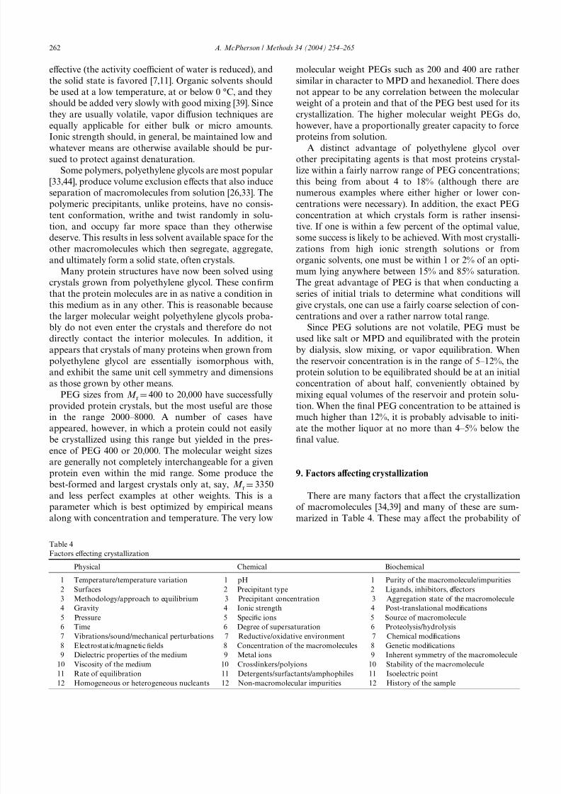
9. Factors aV ecting crystallization
There are many factors that aV ect the crystallization
of macromolecules [34,39] and many of these are sum-
marized in Table 4. These may aV ect the probability of
Table 4
Factors eV
ecting crystallizationPhysical Chemical Biochemical
1 Temperature/temperature variation 1 pH 1 Purity of the macromolecule/impurities
2 Surfaces 2 Precipitant type 2 Ligands, inhibitors, eV ectors
3 Methodology/approach to equilibrium 3 Precipitant concentration 3 Aggregation state of the macromolecule
4 Gravity 4 Ionic strength 4 Post-translational modiWcations
5 Pressure 5 SpeciWc ions 5 Source of macromolecule
6 Time 6 Degree of supersaturation 6 Proteolysis/hydrolysis
7 Vibrations/sound/mechanical perturbations 7 Reductive/oxidative environment 7 Chemical modiWcations
8 Electrostatic/magnetic Welds 8 Concentration of the macromolecules 8 Genetic modiWcations
9 Dielectric properties of the medium 9 Metal ions 9 Inherent symmetry of the macromolecule
10 Viscosity of the medium 10 Crosslinkers/polyions 10 Stability of the macromolecule
11 Rate of equilibration 11 Detergents/surfactants/amphophiles 11 Isoelectric point
12 Homogeneous or heterogeneous nucleants 12 Non-macromolecular impurities 12 History of the sample

8/6/2019 Crystal is at Ion
http://slidepdf.com/reader/full/crystal-is-at-ion 10/12
A. McPherson / Methods 34 (2004) 254–265 263
its occurring at all, the nucleation probability and rate,
crystal growth rate, and the ultimate sizes and quality of
the products. As noted above, pH and salt, or the con-
centrations of other precipitants are of great importance.
The concentration of the macromolecule, which may
vary from as low as 2mg/ml to as much as 100mg/ml, is
an additional, signiWcant variable.Other parameters may be less important but often
play crucial roles. The presence or absence of ligands or
inhibitors, the variety of salt or buV er, the equilibration
technique used, the temperature, or the presence of
detergents, these are all pertinent considerations. Param-
eters of somewhat lesser signiWcance are things like grav-
ity, electric and magnetic Welds, or viscosity. It can, in
general, not be predicted which of these many variables
may be of importance for a particular macromolecule,
and the inXuence of any one must be deWned by a series
of empirical trials.
The most intriguing problem, or opportunity depend-
ing on one's perspective, is what additional components
or compounds should comprise the mother liquor in
addition to solvent, protein, and precipitating agent. The
most probable eV ectors are those which maintain the pro-
tein in a single, homogeneous, and invariant state. Reduc-
ing agents such as glutathione or -mercaptoethanol are
useful to preserve sulfhydryl groups and prevent oxida-
tion. EDTA and EGTA are eV ective if one wishes to pro-
tect the protein from heavy or transition metal ions.
Inclusion of these components may be particularly desir-
able when crystallization requires a long period of time to
reach completion. When crystallization is carried out at
room temperature in polyethylene glycol or low ionicstrength solutions, then attention must be given to pre-
venting the growth of microbes. These generally secrete
proteolytic enzymes that may have serious eV ects on the
integrity of the protein under study. Inclusion of sodium
azide or thymol or chlorobutanol at low levels may be
necessary to suppress invasive bacteria and fungi.
Substrates, coenzymes, and inhibitors often serve to
maintain an enzyme in a more compact and stable form.
Thus, a greater degree of structural homogeneity may be
imposed on a population of macromolecules, and a
reduced level of statistical variation achieved by com-
plexing the protein with a natural ligand before attempt-
ing its crystallization. In some cases, an apoprotein and
its ligand complexes may be signiWcantly diV erent in
their physical behavior and can, in terms of crystalliza-
tion, be treated as almost entirely separate problems.
Complexes may provide additional opportunities for
growing crystals if the native apoprotein is refractile. It is
worthwhile, therefore, when searching for crystallization
conditions, to explore complexes of the macromolecule
with substrates, coenzymes, analogues, and inhibitors at
an early stage. Such complexes are, in addition, inher-
ently more interesting in a biochemical sense than the
apoprotein.
Various metal ions have occasionally been observed
to promote the crystallization of proteins and nucleic
acids. In some instances, these ions were essential for
activity. It was, therefore, reasonable to expect that they
might aid in maintaining certain structural features of
the molecule. In other cases, however, metal ions,
particularly divalent metal ions of the transition series,were found that encouraged crystal growth but played
no known role in the macromolecule's activity. Likely,
they serve as bridging agents between molecules in the
crystal lattice.
9.1. Membrane proteins
Proteins that are naturally membrane associated or
otherwise unusually hydrophobic or lipophilic in nature
invariable present unusual problems. Such proteins are,
in general, only sparingly soluble in normal aqueous
media, some virtually insoluble, and this in turn makes
the application of conventional protein crystallization
techniques problematic. Such cases are diYcult but not
intractable. To address these diYculties the use of deter-
gents, particularly non-ionic detergents, has been devel-
oped. No attempt will be made here to describe the
various techniques or the combinations of detergents
and accessory molecules that have been used, as that
involves a number of complexities and considerations
that are inappropriate here.
The essential diYculty with the necessity of including
a solubilization agent, such as a detergent, is that it adds
an additional dimension to the matrix of conditions that
must otherwise be evaluated. For example, if one is con-tent in using a standard 48-well screen of conditions, at
least initially, then the additional search for a useful
detergent means that the 48 sample screen must then be
multiplied by the number of detergent candidates. The
problem is that there are a lot of potentially useful deter-
gents. Hampton Research, a major source of screening
reagents, oV ers three diV erent detergent kits of 24 sam-
ples each. Were one to simply apply the basic 48-well
screen with each detergent, then that would require a
total of 3456 individual trials. While this may actually be
possible with highly automated systems, and where a
substantial amount of material is available, it is imprac-tical for most laboratories.
The basic crystal screens, whether they are systematic
screens or shotgun screens, cannot be abandoned, how-
ever. Thus, it becomes essential to reduce, at least in ini-
tial screens, the number of detergents to be considered.
If, for example, a set of 6 highly promising detergents
could be identiWed, then less than 300 trials would be
called for initially, an undertaking well within the capa-
bilities of most laboratories. No one, however, has yet
reduced the set to a favored few, everyone has their own
opinion as to which detergents should constitute it, and cer-
tainly no consensus set has yet emerged from databases
8/6/2019 Crystal is at Ion
http://slidepdf.com/reader/full/crystal-is-at-ion 11/12
264 A. McPherson / Methods 34 (2004) 254–265
or from analyses of experiments and the successful struc-
ture determinations that have been carried out. Hope-
fully, such a reduction in the detergent variable will be
among the Wrst important products of the structural
genomics enterprise. This will be true, however, only if
membrane and lipophilic proteins are addressed with the
same enthusiasm and intensity as are the soluble macro-molecules.
To make matters in this area even worse, it appears
that some, perhaps many detergents function best when
accompanied by small amphiphilic molecules such as
LDAO. This would of course add yet another dimension
to the screening problem and seem to convert it into a
hopeless exercise. Again, we can only hope that experi-
ence and the careful recording of data will provide us
with a reduced set of most promising amphiphiles.
While not as valuable as naming actual candidate
detergents, the author can point to a number of useful
reviews and discussions that illustrate the properties and
virtues of various detergents for membrane crystalliza-
tion, and also call attention to the chapter by Nollert in
this volume. Michel (1990) [55] is a good review of work
up until that time, more recently, there are Wne dis-
courses by Loll [28], CaV rey [3], Garavito and Ferguson-
Miller [14], Hunte et al. [25], and Wiener [54].
9.2. The protein as a variable
At the risk of belaboring a point, a factor of particu-
lar importance is the purity of the macromolecule [16]
and this deserves special emphasis. Some proteins, it is
true, may crystallize even from very heterogeneous mix-tures, and indeed, crystallization has long been used as a
powerful puriWcation tool. In general, however, the like-
lihood of success in crystal growth is greatly advanced
by increased homogeneity of the sample. Investment in
further puriWcation is always warranted and usually
proWtable. When every eV ort to crystallize a macromole-
cule fails, the best recourse is to further purify.
Upon entering the Weld of macromolecular crystallog-
raphy one is struck by the extraordinary range of mole-
cules and their properties that one must contend with,
and the extensive variety of techniques and conditions
that must be tested in order to grow crystals suitable forX-ray diV raction analysis. It would indeed be useful if
some comprehensive database existed that at least con-
tained the experiences accumulated over the years.
Indeed, such a knowledge base, combined with a system
to search and sift for all kinds of relevant information
regarding protein crystal growth, has been compiled and
is readily available. This is the crystallization database
devised by Gilliland [17,18] and distributed through the
National Institute of Standards and Technology
([email protected]). This database provides a valu-
able tool for the novice as well as the experienced crystal-
lographer. It includes virtually all of those conditions used
to grow crystals of individual proteins, and it provides
innumerable ideas regarding procedures and techniques.
Recombinant DNA technology provided an enor-
mous impetus to crystal growth research and X-ray crys-
tallography 25 years ago, and it may be on the verge of
providing another at this very time. Arguably, but
hardly so, the most important parameter in protein crys-tallization is the protein itself. Until recently we have
had little or no direct control over most of the important
features of that parameter. ModiWcation at the genetic
level, however, now provides us that opportunity, and its
possibilities are only now beginning to be realized.
Through truncations, mutations, chimeric conjugates,
and many other protein engineering contrivances, the
probability of crystallization may be signiWcantly
enhanced. If we can learn how to go about this in a ratio-
nal and systematic manner then advances may occur in
the succeeding years that match the progress of the past.
Even, so, the mother liquor must still be made, and the
optimal conditions identiWed in order to achieve success.
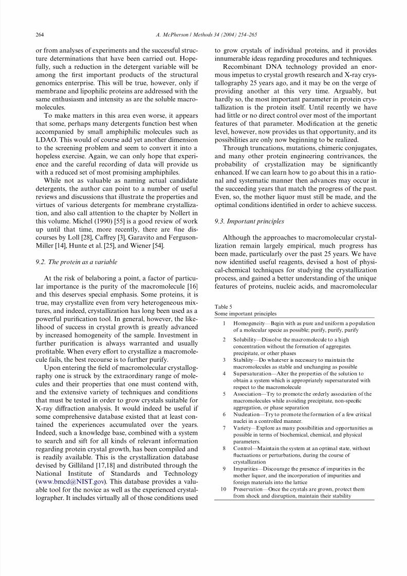
9.3. Important principles
Although the approaches to macromolecular crystal-
lization remain largely empirical, much progress has
been made, particularly over the past 25 years. We have
now identiWed useful reagents, devised a host of physi-
cal-chemical techniques for studying the crystallization
process, and gained a better understanding of the unique
features of proteins, nucleic acids, and macromolecular
Table 5
Some important principles
1 Homogeneity—Begin with as pure and uniform a population
of a molecular specie as possible; purify, purify, purify
2 Solubility—Dissolve the macromolecule to a high
concentration without the formation of aggregates,
precipitate, or other phases
3 Stability—Do whatever is necessary to maintain the
macromolecules as stable and unchanging as possible
4 Supersaturation—Alter the properties of the solution to
obtain a system which is appropriately supersaturated with
respect to the macromolecule
5 Association—Try to promote the orderly association of the
macromolecules while avoiding precipitate, non-speciWc
aggregation, or phase separation
6 Nucleation—Try to promote the formation of a few critical
nuclei in a controlled manner.
7 Variety—Explore as many possibilities and opportunities as
possible in terms of biochemical, chemical, and physical
parameters.
8 Control—Maintain the system at an optimal state, without
Xuctuations or perturbations, during the course of
crystallization
9 Impurities—Discourage the presence of impurities in the
mother liquor, and the incorporation of impurities and
foreign materials into the lattice
10 Preservation—Once the crystals are grown, protect them
from shock and disruption, maintain their stability

8/6/2019 Crystal is at Ion
http://slidepdf.com/reader/full/crystal-is-at-ion 12/12
A. McPherson / Methods 34 (2004) 254–265 265
assemblies that aV ect their capacity to crystallize. Some
principles now stand out regarding the crystallization
problem, and these are summarized in Table 5. It
remains to the individual investigator to Wnd practical
means to institute these ideas and determine for a spe-
ciWc problem which are of critical importance, and which
will have greatest inXuence on the likelihood of success.
References
[1] T.M. Bergfors, Protein Crystallization: Techniques, Strategies and
Tips, International University Line, La Jolla, CA, 1999.
[2] J.D. Bernal, D. Crowfoot, Nature 133 (1934) 794.
[3] M. CaV rey, J. Struc. Biol. 142 (2003) 108–132.
[4] A.A. Chernov, J. Struc. Biol. 142 (2003) 3–21.
[5] E.J. Cohn, J.T. Edsall (Eds.), Proteins, Amino Acids and Peptides as
Ions and Dipolar Ions, Van Nostrand-Reinhold, Princeton, NJ, 1943.
[6] E.J. Cohn, J.D. Ferry (Eds.), The Interactions of Proteins with
Ions and Dipolar Ions. Proteins, Amino Acids and Peptides, Van
Nostrand-Rheinhold, New Jersey, 1943.[7] E.J. Cohn, W.L. Hughes, et al., J. Am. Chem. Soc. 69 (1974) 1753–
1761.
[8] L.J. DeLucas, T.L. Bray, et al., J. Struc. Biol. 142 (2003) 188–206.
[9] A. Ducruix, R. Giége, Crystallization of Nucleic Acids and Pro-
teins, A Practical Approach, IRL Press, Oxford, 1992.
[10] S.D. Durbin, G. Feher, Annu. Rev. Phys. Chem. 47 (1996) 171–204.
[11] S. Englard, S. Seifter, Precipitation Techniques, Methods Enzy-
mol. 182 (1990) 301–306.
[12] G. Feher, J. Cryst. Growth 76 (1986) 545–546.
[13] R.S. Feigelson, J. Cryst. Growth 90 (1988) 1–13.
[14] R.M. Garavito, S. Ferguson-Miller, J. Biol. Chem. 276 (2001)
32403–32406.
[15] E.F. Garmen, T.R. Schneider, J. Appl. Cryst. 30 (1997) 211–237.
[16] R. Giege, B. Lorber, et al., Acta Cryst. D 50 (1994) 339–350.
[17] G.L. Gilliland, J. Cryst. Growth 90 (1998) 51–59.[18] G.L. Gilliland, M. Tung, et al., Acta Cryst. D 50 (1994) 408–413.
[19] S.M. Gruner, E.F. Eikenberry, et al., Comparison of X-ray Detec-
tors, in: M.G. Rossmann, E. Arnold (Eds.), International Tables
for Crystallography, F, Kluwer Academic Publishers, Dordrecht/
Boston/London, 2001, pp. 143–153.
[20] C. Haas, J. Drenth, J. Cryst. Growth 196 (1999) 388–394.
[21] J.R. Helliwell, Macromolecular Crystallography with Synchrotron
Radiation, Cambridge University Press, Cambridge, UK, 1992.
[22] R.M. Herriott, Chem. Rev. 30 (1942) 413.
[23] T. Hofmeister, Z. Physiol. Chem. 14 (1890) 165.
[24] D. HosWeld, J. Palan, et al., J. Struc. Biol. 142 (2003) 207–217.
[25] C. Hunte, V. Jagow, et al., Membrane Protein PuriWcation and Crys-
tallization: A Practical Guide, Academic Press, San Diego, 2003.
[26] K.G. Ingham, Methods Enzymol. 182 (1990) 301–306.
[27] Y.G. Kuznetsov, A.J. Malkin, et al., J. Struc. Biol. 114 (3) (1995)
184–196.
[28] P.J. Loll, J. Struct. Biol. 142 (1) (2003) 144–153.
[29] J.R. Luft, R.J. Collins, et al., J. Struc. Biol. 142 (2003) 170–179.
[30] A.J. Malkin, Y.G. Kuznetsov, et al., J. Phys. Chem 100 (1996)
11736–11743.
[31] A.J. Malkin, Y.G. Kuznetsov, et al., Nat. Struc. Biol 2 (11) (1995)
956–959.
[32] A.J. Malkin, Y.G. Kuznetsov, et al., Surface Sci 393 (1997) 95–107.
[33] A. McPherson, J. Biol. Chem. 251 (1976) 3600–6303.
[34] A. McPherson, The Preparation and Analysis of Protein Crystals,
Wiley, New York, 1982.
[35] A. McPherson, Sci. Am. 260 (1989) 62.
[36] A. McPherson, J. Cryst. Growth 110 (1991) 1–10.
[37] A. McPherson, J Cryst. Growth 122 (1992) 161–167.
[38] A. McPherson, The Role of X-ray crystallography in structure
based rational drug design, in: Chemical and Structural
Approaches to Rational Drug Design, CRC Press, Boca Raton,
FL, 1994.
[39] A. McPherson, Crystallization of Biological Macromolecules,
Cold Spring Harbor Laboratory Press, Cold Spring Harbor, NY,
1999.
[40] A. McPherson, R. Cudney, et al., The crystallization of proteins,nucleic acids, and viruses for X-ray diV raction analysis, in: Bio-
polymers, vol. 18 (2003) 427–468.
[41] A. McPherson, A.J. Malkin, et al., Structure 3 (8) (1995) 759–768.
[42] A. McPherson, A.J. Malkin, et al., Ann. Rev. Biophys. Biomol.
Struc. 29 (2000) 361–410.
[43] J.H. Northrop, M. Kunitz, et al., Crystalline Enzymes, Columbia
University Press, New York, 1948.
[44] S. Patel, R. Cudney, et al., Biochemistry and Biophysics Research
Communications 207 (1995) 819–828.
[45] M.F. Perutz, Sci. Am. 211 (1964) 64.
[46] J.W. PXugrath, Curr. Opin. Struct. Biol. 2 (1992) 811–815.
[47] R. Piazza, J. Cryst. Growth 196 (1999) 415–423.
[48] E.T. Reichert, A.P. Brown, The DiV erentiation and SpeciWcity of
Corresponding Proteins and Other Vital Substances in Relation
To Biological ClassiW
cation and Organic Evolution: The Crystal-lography of Hemoglobins, Carnegie Institute, Washington, DC,
1909.
[49] M.G. Rossmann, E. Arnold, Crystallography of Biological Mac-
romolecules, Kluwer Academic Publishers, Dordrecht/Boston/
London, 2001.
[50] J.B. Sumner, J. Biol. Chem. 69 (1926) 435.
[51] J.B. Sumner, A.L. Dounce, J. Biol. Chem. 1231 (1937) 417.
[52] J.B. Sumner, G.F. Somers, The Enzymes, Academic Press, New
York, 1943.
[53] R. Ten Wolde, D. Fraenkel, Science 277 (1997) 1975–1978.
[54] M.C. Weiner, Curr. Opin. Struct. Biol. 6 (2001) 412–419.
[55] H. Michel (Ed.), General and practical aspects of membrane pro-
tein crystallization. Crystallization of membrane proteins. CRC
Press, Boca Raton, FL, 1990.