Embed Size (px)
Citation preview
Peer ReviewedConsultant on Call Infectious Disease
December 2012 • clinician’s brief 39
MORE
�
Cryptococcosis
Rebecca Norton, DVMGulf Coast Veterinary Specialists, Houston, Texas
Derek P. Burney, DVM, PhD, DACVIMVeterinary Specialists of North Texas, Dallas, Texas
Profile
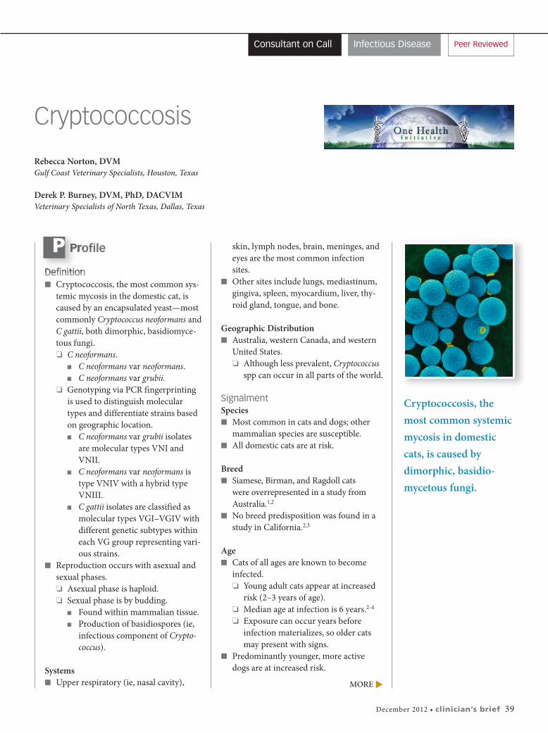
Definition� Cryptococcosis, the most common sys-
temic mycosis in the domestic cat, iscaused by an encapsulated yeast—mostcommonly Cryptococcus neoformans andC gattii, both dimorphic, basidiomyce-tous fungi.� C neoformans.
� C neoformans var neoformans.� C neoformans var grubii.
� Genotyping via PCR fingerprintingis used to distinguish moleculartypes and differentiate strains basedon geographic location.� C neoformans var grubii isolates
are molecular types VNI andVNII.
� C neoformans var neoformans istype VNIV with a hybrid typeVNIII.
� C gattii isolates are classified asmolecular types VGI–VGIV withdifferent genetic subtypes withineach VG group representing vari-ous strains.
� Reproduction occurs with asexual andsexual phases.� Asexual phase is haploid.� Sexual phase is by budding.
� Found within mammalian tissue.� Production of basidiospores (ie,
infectious component of Crypto-coccus).
Systems� Upper respiratory (ie, nasal cavity),
skin, lymph nodes, brain, meninges, andeyes are the most common infectionsites.
� Other sites include lungs, mediastinum,gingiva, spleen, myocardium, liver, thy-roid gland, tongue, and bone.
Geographic Distribution� Australia, western Canada, and western
United States.� Although less prevalent, Cryptococcus
spp can occur in all parts of the world.
SignalmentSpecies� Most common in cats and dogs; other
mammalian species are susceptible.� All domestic cats are at risk.
Breed � Siamese, Birman, and Ragdoll cats
were overrepresented in a study fromAustralia.1,2
� No breed predisposition was found in astudy in California.2,3
Age� Cats of all ages are known to become
infected.� Young adult cats appear at increased
risk (2–3 years of age).� Median age at infection is 6 years.2-4� Exposure can occur years before
infection materializes, so older catsmay present with signs.
� Predominantly younger, more activedogs are at increased risk.
P
Cryptococcosis, themost common systemicmycosis in domesticcats, is caused bydimorphic, basidio-mycetous fungi.
Consultant on Call
Sex� No known sex predilection.
Causes� Inhalation of spores from avian guano
or affected soils.� C neoformans is also found in decaying
plant matter harboring in some treehollows.
� C gattii has been isolated from air, sea-water, freshwater, and tree bark.
Risk Factors� Cats with retroviruses (ie, FeLV, FIV)
are not predisposed to infection withCryptococcus spp, but difficultyresponding to therapy and relapse ofcryptococcosis may be common.2,5� These patients may be predisposed
to neoplasms (eg, lymphoma, ade-nocarcinoma, mast cell tumor).
� Opportunistic infections (eg, from Toxo-plasma gondii) have been reported.
Pathophysiology� Encapsulated spores (basidiospores)
are typically inhaled, initiating infec-tion in the nasal cavities of cats (primarily) and dogs.
� Disease can spread through the cribri-form plate, causing meningitis.� May involve the optic nerve and eye.� May extend into lungs, skin, bones,
brain, and other body sites viahematogenous routes.� May be detected in the lungs via
histopathology without produc-ing clinical signs.
� Direct contact with openwounds may cause skin lesions.
� Multiple granulomatous skinlesions are more likely associ-ated with hematogenous spread.
History� Outdoor cats more susceptible.� Indoor cats exposed from open win-
dows/doors or indoor plants/soil.� Transfer from clothing can occur.
� Signs:� Sneezing, head shaking, stertor.� Inappetence from blocked sinus
passages and invasion into the CNS.� Blindness.
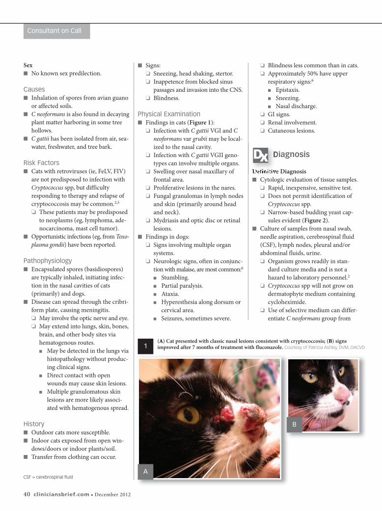
Physical Examination� Findings in cats (Figure 1):
� Infection with C gattii VGI and Cneoformans var grubii may be local-ized to the nasal cavity.
� Infection with C gattii VGII geno-types can involve multiple organs.
� Swelling over nasal maxillary offrontal area.
� Proliferative lesions in the nares.� Fungal granulomas in lymph nodes
and skin (primarily around headand neck).
� Mydriasis and optic disc or retinallesions.
� Findings in dogs:� Signs involving multiple organ
systems.� Neurologic signs, often in conjunc-
tion with malaise, are most common:6� Stumbling.� Partial paralysis.� Ataxia.� Hyperesthesia along dorsum or
cervical area.� Seizures, sometimes severe.
� Blindness less common than in cats.� Approximately 50% have upper
respiratory signs:6� Epistaxis.� Sneezing.� Nasal discharge.
� GI signs.� Renal involvement.� Cutaneous lesions.
Diagnosis
Definitive Diagnosis � Cytologic evaluation of tissue samples.
� Rapid, inexpensive, sensitive test.� Does not permit identification of
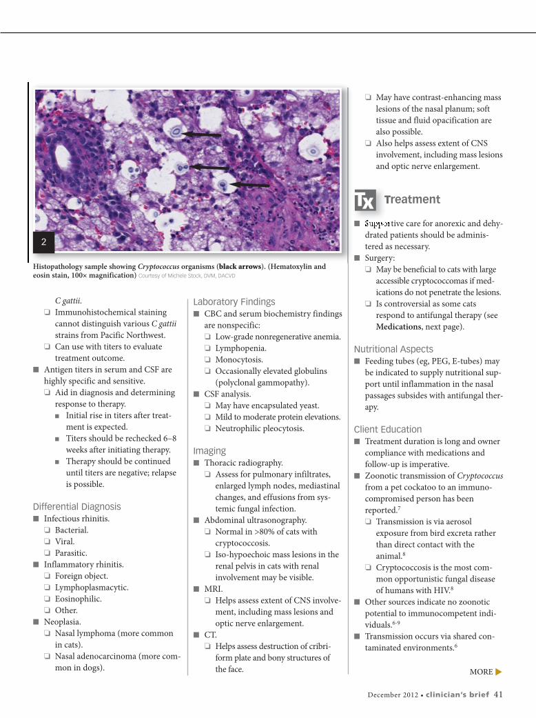
Cryptococcus spp.� Narrow-based budding yeast cap-
sules evident (Figure 2).� Culture of samples from nasal swab,
needle aspiration, cerebrospinal fluid(CSF), lymph nodes, pleural and/orabdominal fluids, urine.� Organism grows readily in stan-
dard culture media and is not ahazard to laboratory personnel.2
� Cryptococcus spp will not grow ondermatophyte medium containingcycloheximide.
� Use of selective medium can differ-entiate C neoformans group from
40 cliniciansbrief.com • December 2012
(A) Cat presented with classic nasal lesions consistent with cryptococcosis; (B) signsimproved after 7 months of treatment with fluconazole. Courtesy of Patricia Ashley, DVM, DACVD
CSF = cerebrospinal fluid
1
A
B
C gattii.� Immunohistochemical staining
cannot distinguish various C gattiistrains from Pacific Northwest.
� Can use with titers to evaluatetreatment outcome.
� Antigen titers in serum and CSF arehighly specific and sensitive.� Aid in diagnosis and determining
response to therapy.� Initial rise in titers after treat-
ment is expected.� Titers should be rechecked 6–8
weeks after initiating therapy.� Therapy should be continued
until titers are negative; relapseis possible.
Differential Diagnosis� Infectious rhinitis.
� Bacterial.� Viral.� Parasitic.
� Inflammatory rhinitis.� Foreign object.� Lymphoplasmacytic.� Eosinophilic.� Other.
� Neoplasia.� Nasal lymphoma (more common
in cats).� Nasal adenocarcinoma (more com-
mon in dogs).
Laboratory Findings� CBC and serum biochemistry findings
are nonspecific:� Low-grade nonregenerative anemia.� Lymphopenia.� Monocytosis.� Occasionally elevated globulins
(polyclonal gammopathy).� CSF analysis.
� May have encapsulated yeast.� Mild to moderate protein elevations.� Neutrophilic pleocytosis.
Imaging� Thoracic radiography.
� Assess for pulmonary infiltrates,enlarged lymph nodes, mediastinalchanges, and effusions from sys-temic fungal infection.
� Abdominal ultrasonography.� Normal in >80% of cats with
cryptococcosis.� Iso-hypoechoic mass lesions in the
renal pelvis in cats with renalinvolvement may be visible.
� MRI.� Helps assess extent of CNS involve-
ment, including mass lesions andoptic nerve enlargement.
� CT.� Helps assess destruction of cribri-
form plate and bony structures ofthe face.
� May have contrast-enhancing masslesions of the nasal planum; softtissue and fluid opacification arealso possible.
� Also helps assess extent of CNSinvolvement, including mass lesionsand optic nerve enlargement.
Treatment
� Supportive care for anorexic and dehy-drated patients should be adminis-tered as necessary.
� Surgery:� May be beneficial to cats with large
accessible cryptococcomas if med-ications do not penetrate the lesions.
� Is controversial as some catsrespond to antifungal therapy (seeMedications, next page).
Nutritional Aspects� Feeding tubes (eg, PEG, E-tubes) may
be indicated to supply nutritional sup-port until inflammation in the nasalpassages subsides with antifungal ther-apy.
Client Education� Treatment duration is long and owner
compliance with medications and follow-up is imperative.
� Zoonotic transmission of Cryptococcusfrom a pet cockatoo to an immuno-compromised person has beenreported.7� Transmission is via aerosol
exposure from bird excreta ratherthan direct contact with theanimal.8
� Cryptococcosis is the most com-mon opportunistic fungal diseaseof humans with HIV.8
� Other sources indicate no zoonoticpotential to immunocompetent indi-viduals.6-9
� Transmission occurs via shared con-taminated environments.6
December 2012 • clinician’s brief 41
Histopathology sample showing Cryptococcus organisms (black arrows). (Hematoxylin andeosin stain, 100¥ magnification) Courtesy of Michele Stock, DVM, DACVD
2
MORE
�
Consultant on Call
Medications
Azoles� Fungistatic compounds that alter cell
membrane permeability and allow cellcontents to leak into the periphery.
� Fluconazole is treatment of choice formaximum penetration into the CNS,eye, and urinary tract with minimalside effects.� Standard dose for dogs and cats is 5
mg/kg PO q12h until antigen testingof blood or CSF (if CNS disease ispresent) is negative (mean duration,8 months).8
� Some cryptococcal isolates showresistance.
� Itraconazole has been used success-fully to treat cryptococcal meningitis.� Cats may experience anorexia,
vomiting, and hepatocellular dam-age (all dose-dependent).
� Oral suspension has greaterbioavailability and should be dosedlower than tablet formulation (3 mg/kg PO q24h rather than 10 mg/kg PO q24h).
� Compounded formulations havebeen associated with inadequateblood levels.
� For cats with localized disease andresistance to fluconazole, ketoconazolemay be preferred by cost-consciousowners.� Standard dose of ketoconazole for
dogs and cats is 5–10 mg/kg POq12–24h until antigen testing ofblood or CSF (if CNS disease ispresent) is negative (~6–18months).8
� Other azoles (eg, voriconazole,posaconazole) are effective but expensive.� Cats may experience neurologic
effects from voriconazole.
Amphotericin B � Fungicidal compound that also dis-
rupts fungal cell membranes.
� Must be given parenterally.� SC protocol may minimize hospital-
ization and permit outpatient care.� 0.5–0.75 mg/kg diluted in large
volumes of 0.45% NaCl and 2.5%dextrose 2–3 times/week.
� Median cumulative dose of 16 mg/kg.
� Newer liposomal and lipid complexpreparations are less nephrotoxic thanprevious formulations.
Flucytosine� Pyrimidine analog that interferes with
fungal nucleic acid synthesis.� Standard dose (cats only) is 30 mg/kg
PO q6h, 50 mg/kg PO q8h, or 75mg/kg PO q12h, not to exceed 250 mgq6–8h.8
� Should not be used as a monotherapybecause of risk for resistance.
� May cause bone marrow suppression,GI disturbance, and worsening of pre-existing renal insufficiency.
� Should not be used in conjunctionwith amphotericin B.
� Use only in cats; dogs tend to developsevere cutaneous drug eruptions.
Follow-up
Complications� Hepatocellular damage associated
with chronic ketoconazole and itra-conazole administration is possibleand warrants monthly monitoring ofliver enzymes.
� Renal toxicity associated with ampho-tericin B is possible and warrantsmonthly monitoring of renal valuesand consideration of using liposome-encapsulated formulations of ampho-tericin B to reduce risk for renaltoxicity.
Future Follow-up� Serum antigen titers should be moni-
tored monthly for seroconversion during treatment.
� Patients that seroconvert to a nega-tive status should be retested 1month after therapy.
� Successful treatment occurs whenthe titer reaches zero.
� Treatment may be indicated formonths to years.
� For cats in carrier state, periodic anti-gen testing is warranted.� Antigen titer should decrease by
1 dilution each month during therapy.� Failure to achieve this suggests
the need for more aggressivetherapy (ie, additional medica-tions or change in protocol).8
In General
Relative Cost� Treatment is costly, as it is required for
months to years: $$$$$� Fluconazole and ketoconazole are less
expensive than itraconazole.
Prognosis� Animals that survive the first 2 weeks
of therapy have a reasonable butguarded prognosis.� Rapid rate of improvement in the
first month can lead to decreasedowner compliance, precluding ahigher rate of relapse.
� Relapse is possible.� CNS involvement is the only signifi-
cant predictor of mortality. � cb
See Aids & Resources, back page, for references & suggested reading.
42 cliniciansbrief.com • December 2012
*
Cost Key$ = up to $100 $$ = $101–$250 $$$ = $251–$500$$$$ = $501–$1000$$$$$ = more than $1000
CSF = cerebrospinal fluid