Embed Size (px)
Citation preview
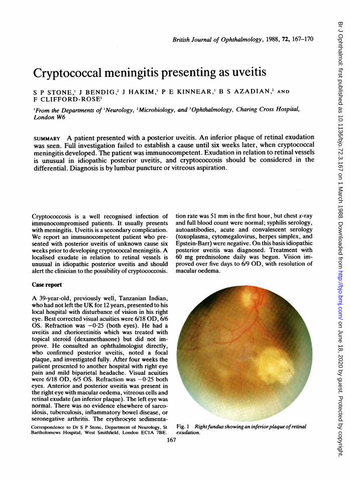
British Journal of Ophthalmology, 1988, 72, 167-170
Cryptococcal meningitis presenting as uveitisS P STONE,' J BENDIG,2 J HAKIM,3 P E KINNEAR,3 B S AZADIAN,2 ANDF CLIFFORD-ROSE'
'From the Departments of 'Neurology, 2Microbiology, and 3Ophthalmology, Charing Cross Hospital,London W6
SUMMARY A patient presented with a posterior uveitis. An inferior plaque of retinal exudationwas seen. Full investigation failed to establish a cause until six weeks later, when cryptococcalmeningitis developed. The patient was immunocompetent. Exudation in relation to retinal vesselsis unusual in idiopathic posterior uveitis, and cryptococcosis should be considered in thedifferential. Diagnosis is by lumbar puncture or vitreous aspiration.
Cryptococcosis is a well recognised infection ofimmunocompromised patients. It usually presentswith meningitis. Uveitis is a secondary complication.We report an immunocompetent patient who pre-sented with posterior uveitis of unknown cause sixweeks prior to developing cryptococcal meningitis. Alocalised exudate in relation to retinal vessels isunusual in idiopathic posterior uveitis and shouldalert the clinician to the possibility of cryptococcosis.
Case report
A 39-year-old, previously well, Tanzanian Indian,who had not left the UK for 12 years, presented to hislocal hospital with disturbance of vision in his righteye. Best corrected visual acuities were 6/18 OD, 6/6OS. Refraction was -0-25 (both eyes). He had auveitis and chorioretinitis which was treated withtopical steroid (dexamethasone) but did not im-prove. He consulted an ophthalmologist directly,who confirmed posterior uveitis, noted a focalplaque, and investigated fully. After four weeks thepatient presented to another hospital with right eyepain and mild biparietal headache. Visual acuitieswere 6/18 OD, 6/5 OS. Refraction was -0-25 botheyes. Anterior and posterior uveitis was present inthe right eye with macular oedema, vitreous cells andretinal exudate (an inferior plaque). The left eye wasnormal. There was no evidence elsewhere of sarco-idosis, tuberculosis, inflammatory bowel disease, orseronegative arthritis. The erythrocyte sedimenta-Correspondence to Dr S P Stone, Department of Neurology, StBartholomews Hospital, West Smithfield, London EClA 7BE.
tion rate was 51 mm in the first hour, but chest x-rayand full blood count were normal; syphilis serology,autoantibodies, acute and convalescent serology(toxoplasma, cytomegalovirus, herpes simplex, andEpstein-Barr) were negative. On this basis idiopathicposterior uveitis was diagnosed. Treatment with60 mg prednisolone daily was begun. Vision im-proved over five days to 6/9 OD, with resolution ofmacular oedema.
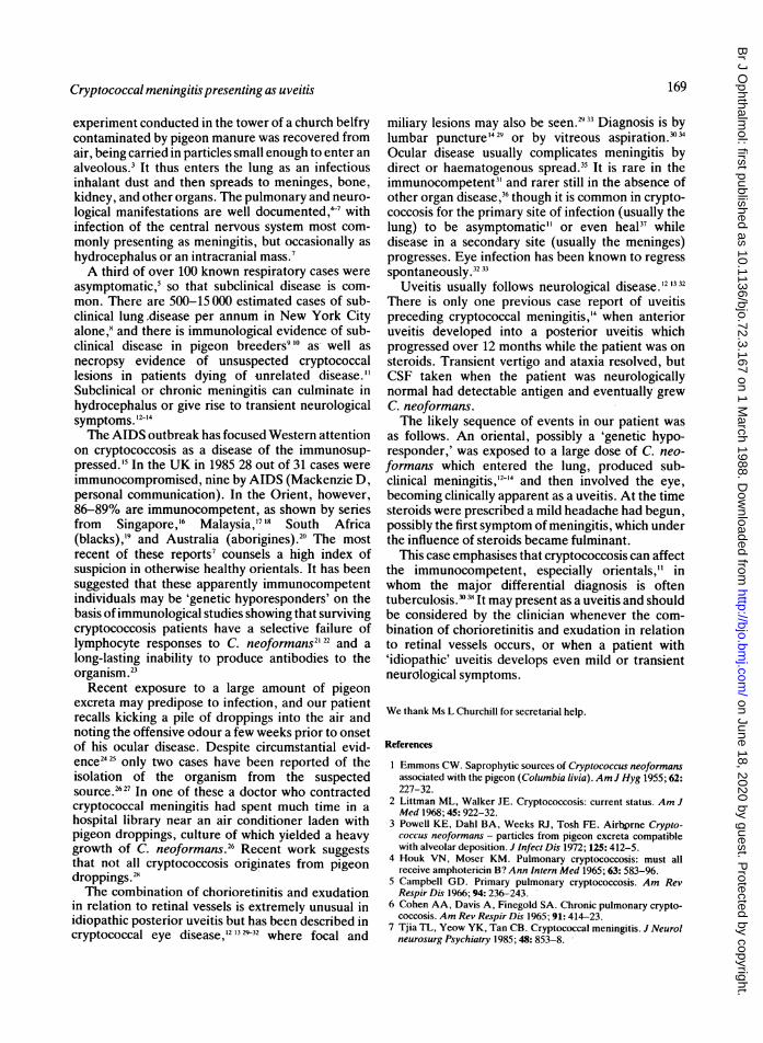
Fig. 1 Rightfundusshowingan inferiorplaque ofretinalexudation.
167
on June 18, 2020 by guest. Protected by copyright.
http://bjo.bmj.com
/B
r J Ophthalm
ol: first published as 10.1136/bjo.72.3.167 on 1 March 1988. D
ownloaded from
S P Stone, J Bendig, JHakim, P E Kinnear, B SAzadian, andF Clifford-Rose
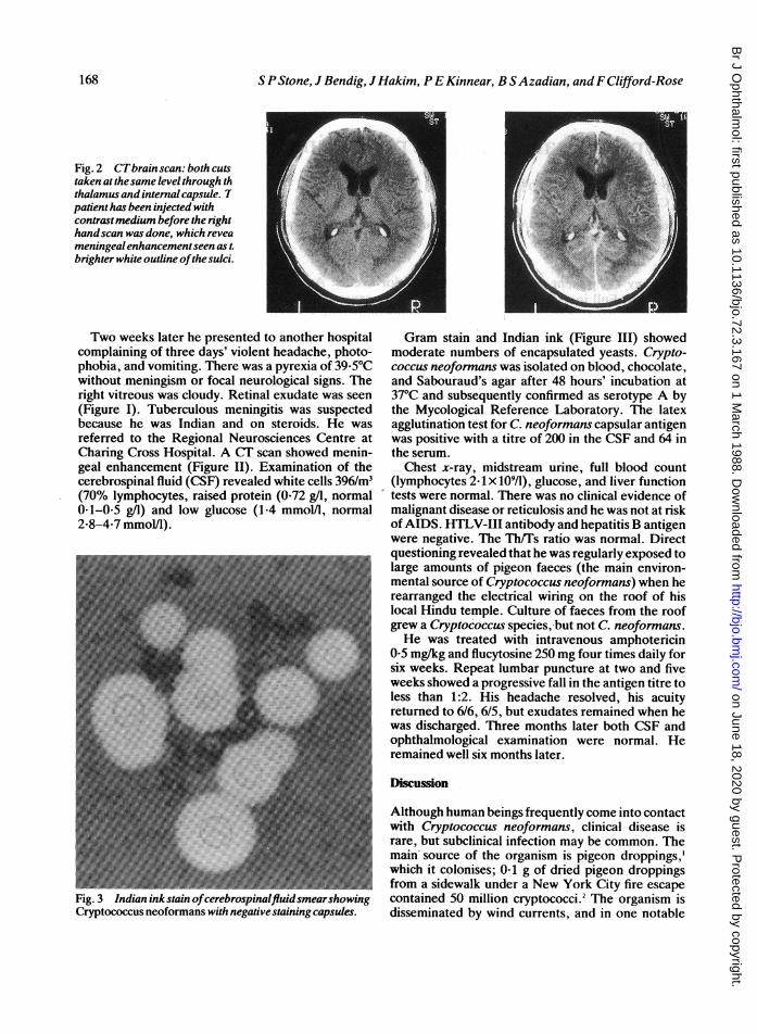
Fig. 2 CTbrain scan: both cutstaken at thesame level through ththalamus and internal capsule. Tpatient has been injected withcontrastmedium before the righthandscan was done, which reveameningeal enhancementseen as t.brighter white outline ofthe sulci.
Two weeks later he presented to another hospitalcomplaining of three days' violent headache, photo-phobia, and vomiting. There was a pyrexia of 39-50Cwithout meningism or focal neurological signs. Theright vitreous was cloudy. Retinal exudate was seen(Figure I). Tuberculous meningitis was suspectedbecause he was Indian and on steroids. He wasreferred to the Regional Neurosciences Centre atCharing Cross Hospital. A CT scan showed menin-geal enhancement (Figure II). Examination of thecerebrospinal fluid (CSF) revealed white cells 396/m3(70% lymphocytes, raised protein (0-72 g/l, normal0-1-0-5 g/l) and low glucose (1.4 mmol/l, normal2-8-4-7 mmol/l).
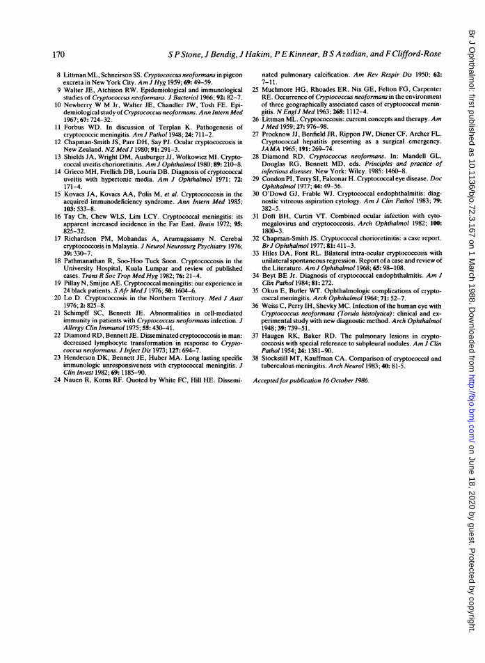
Fig. .3 Indian inkstain ofcerebrospinalftuidsmearshowingCryptococcus neoformans with negative staining capsules.
Gram stain and Indian ink (Figure III) showedmoderate numbers of encapsulated yeasts. Crypto-coccus neoformans was isolated on blood, chocolate,and Sabouraud's agar after 48 hours' incubation at370C and subsequently confirmed as serotype A bythe Mycological Reference Laboratory. The latexagglutination test for C. neoformans capsular antigenwas positive with a titre of 200 in the CSF and 64 inthe serum.
Chest x-ray, midstream urine, full blood count(lymphocytes 2-1 x 10⁹/1), glucose, and liver functiontests were normal. There was no clinical evidence ofmalignant disease or reticulosis and he was not at riskofAIDS. HTLV-III antibody and hepatitis B antigenwere negative. The Th/Ts ratio was normal. Directquestioning revealed that he was regularly exposed tolarge amounts of pigeon faeces (the main environ-mental source of Cryptococcus neoformans) when herearranged the electrical wiring on the roof of hislocal Hindu temple. Culture of faeces from the roofgrew a Cryptococcus species, but not C. neoformans.He was treated with intravenous amphotericin
0-5 mg/kg and flucytosine 250 mg four times daily forsix weeks. Repeat lumbar puncture at two and fiveweeks showed a progressive fall in the antigen titre toless than 1:2. His headache resolved, his acuityreturned to 6/6, 6/5, but exudates remained when hewas discharged. Three months later both CSF andophthalmological examination were normal. Heremained well six months later.
Discussion
Although human beings frequently come into contactwith Cryptococcus neoformans, clinical disease israre, but subclinical infection may be common. Themain source of the organism is pigeon droppings,'which it colonises; 0-1 g of dried pigeon droppingsfrom a sidewalk under a New York City fire escapecontained 50 million cryptococci.2 The organism isdisseminated by wind currents, and in one notable
168
on June 18, 2020 by guest. Protected by copyright.
http://bjo.bmj.com
/B
r J Ophthalm
ol: first published as 10.1136/bjo.72.3.167 on 1 March 1988. D
ownloaded from
Cryptococcal meningitis presenting as uveitis
experiment conducted in the tower of a church belfrycontaminated by pigeon manure was recovered fromair, being carried in particles small enough to enter analveolous.3 It thus enters the lung as an infectiousinhalant dust and then spreads to meninges, bone,kidney, and other organs. The pulmonary and neuro-logical manifestations are well documented,"4 withinfection of the central nervous system most com-monly presenting as meningitis, but occasionally ashydrocephalus or an intracranial mass.7A third of over 100 known respiratory cases were
asymptomatic,5 so that subclinical disease is com-mon. There are 500-15 000 estimated cases of sub-clinical lung disease per annum in New York Cityalone," and there is immunological evidence of sub-clinical disease in pigeon breeders9 "' as well asnecropsy evidence of unsuspected cryptococcallesions in patients dying of unrelated disease."Subclinical or chronic meningitis can culminate inhydrocephalus or give rise to transient neurologicalsymptoms. 12-14The AIDS outbreak has focused Western attention
on cryptococcosis as a disease of the immunosup-pressed.'5 In the UK in 1985 28 out of 31 cases wereimmunocompromised, nine by AIDS (Mackenzie D,personal communication). In the Orient, however,86-89% are immunocompetent, as shown by seriesfrom Singapore,'6 Malaysia,'7"" South Africa(blacks),'9 and Australia (aborigines).20 The mostrecent of these reports7 counsels a high index ofsuspicion in otherwise healthy orientals. It has beensuggested that these apparently immunocompetentindividuals may be 'genetic hyporesponders' on thebasis of immunological studies showing that survivingcryptococcosis patients have a selective failure oflymphocyte responses to C. neoformans2'22 and along-lasting inability to produce antibodies to theorganism.23
Recent exposure to a large amount of pigeonexcreta may predipose to infection, and our patientrecalls kicking a pile of droppings into the air andnoting the offensive odour a few weeks prior to onsetof his ocular disease. Despite circumstantial evid-ence2425 only two cases have been reported of theisolation of the organism from the suspectedsource.2627 In one of these a doctor who contractedcryptococcal meningitis had spent much time in ahospital library near an air conditioner laden withpigeon droppings, culture of which yielded a heavygrowth of C. neoformans.2" Recent work suggeststhat not all cryptococcosis originates from pigeondroppings.2"The combination of chorioretinitis and exudation
in relation to retinal vessels is extremely unusual inidiopathic posterior uveitis but has been described incryptococcal eye disease,12132932 where focal and
miliary lesions may also be seen.2933 Diagnosis is bylumbar puncture'429 or by vitreous aspiration.?"Ocular disease usually complicates meningitis bydirect or haematogenous spread.35 It is rare in theimmunocompetent3' and rarer still in the absence ofother organ disease,3" though it is common in crypto-coccosis for the primary site of infection (usually thelung) to be asymptomatic" or even heal37 whiledisease in a secondary site (usually the meninges)progresses. Eye infection has been known to regressspontaneously.32 33
Uveitis usually follows neurological disease.121332There is only one previous case report of uveitispreceding cryptococcal meningitis,'4 when anterioruveitis developed into a posterior uveitis whichprogressed over 12 months while the patient was onsteroids. Transient vertigo and ataxia resolved, butCSF taken when the patient was neurologicallynormal had detectable antigen and eventually grewC. neoformans.The likely sequence of events in our patient was
as follows. An oriental, possibly a 'genetic hypo-responder,' was exposed to a large dose of C. neo-formans which entered the lung, produced sub-clinical meningitis,'2 14 and then involved the eye,becoming clinically apparent as a uveitis. At the timesteroids were prescribed a mild headache had begun,possibly the first symptom of meningitis, which underthe influence of steroids became fulminant.This case emphasises that cryptococcosis can affect
the immunocompetent, especially orientals," inwhom the major differential diagnosis is oftentuberculosis.?3"3 It may present as a uveitis and shouldbe considered by the clinician whenever the com-bination of chorioretinitis and exudation in relationto retinal vessels occurs, or when a patient with'idiopathic' uveitis develops even mild or transientneurological symptoms.
We thank Ms L Churchill for secretarial help.
References
1 Emmons CW. Saprophytic sources of Cryptococcus neoformansassociated with the pigeon (Columbia livia). AmJ Hyg 1955; 62:227-32.
2 Littman ML, Walker JE. Cryptococcosis: current status. Am JMed 1968; 45: 922-32.
3 Powell KE, Dahl BA, Weeks RJ, Tosh FE. Airborne Crypto-coccus neoformans - particles from pigeon excreta compatiblewith alveolar deposition. J Infect Dis 1972; 125: 412-5.
4 Houk VN, Moser KM. Pulmonary cryptococcosis: must allreceive amphotericin B? Ann Intern Med 1965; 63: 583-96.
5 Campbell GD. Primary pulmonary cryptococcosis. Am RevRespir Dis 1966; 94: 236-243.
6 Cohen AA, Davis A, Finegold SA. Chronic pulmonary crypto-coccosis. Am Rev Respir Dis 1965; 91: 414-23.
7 Tjia TL, Yeow YK, Tan CB. Cryptococcal meningitis. J Neurolneurosurg Psychiatry 1985; 48: 853-8.
169
on June 18, 2020 by guest. Protected by copyright.
http://bjo.bmj.com
/B
r J Ophthalm
ol: first published as 10.1136/bjo.72.3.167 on 1 March 1988. D
ownloaded from
S P Stone, J Bendig, J Hakim, P E Kinnear, B S Azadian, and F Clifford-Rose
8 Littman ML, Schneirson SS. Cryptococcus neoformans in pigeonexcreta in New York City. Am J Hyg 1959; 69: 49-59.
9 Walter JE, Atchison RW. Epidemiological and immunologicalstudies of Cryptococcus neoformans. J Bacteriol 1966; 92: 82-7.
10 Newberry W M Jr, Walter JE, Chandler JW, Tosh FE. Epi-demiological study of Cryptococcus neoformans. Ann Intern Med1967; 67: 724-32.
11 Forbus WD. In discussion of Terplan K. Pathogenesis ofcryptococcic meningitis. Am J Pathol 1948; 24: 711-2.
12 Chapman-Smith JS, Parr DH, Say PJ. Ocular cryptococcosis inNew Zealand. NZ MedJ 1980; 91: 291-3.
13 Shields JA, Wright DM, Ausburger JJ, Wolkowicz MI. Crypto-coccal uveitis chorioretinitis. Am J Ophthalmol 1980; 89: 210-8.
14 Grieco MH, Frellich DB, Louria DB. Diagnosis of cryptococcaluveitis with hypertonic media. Am J Ophthalmol 1971; 72:171-4.
15 Kovacs JA, Kovacs AA, Polis M, et al. Cryptococcosis in theacquired immunodeficiency syndrome. Ann Intern Med 1985;103: 533-8.
16 Tay Ch, Chew WLS, Lim LCY. Cryptococcal meningitis: itsapparent increased incidence in the Far East. Brain 1972; 95:825-32.
17 Richardson PM, Mohandas A, Arumugasamy N. Cerebalcryptococcosis in Malaysia. J Neurol Neurosurg Psychiatry 1976;39: 330-7.
18 Pathmanathan R, Soo-Hoo Tuck Soon. Cryptococcosis in theUniversity Hospital, Kuala Lumpar and review of publishedcases. Trans R Soc Trop Med Hyg 1982; 76: 21-4.
19 Pillay N, Smijee AE. Cryptococcal meningitis: our experience in24 black patients. SAfr Med J 1976; 50: 1604-6.
20 Lo D. Cryptococcosis in the Northern Territory. Med J Aust1976; 2:825-8.
21 Schimpff SC, Bennett JE. Abnormalities in cell-mediatedimmunity in patients with Cryptococcus neoformans infection. JAllergy Clin immunol 1975; 55: 430-41.
22 Diamond RD, Bennett JE. Disseminated cryptococcosis in man:decreased lymphocyte transformation in response to Crypto-coccus neoformans. J Infect Dis 1973; 127: 694-7.
23 Henderson DK, Bennett JE, Huber MA. Long lasting specificimmunologic unresponsiveness with cryptococcal meningitis. J
Clin Invest 1982; 69: 1185-90.24 Nauen R, Korns RF. Quoted by White FC, Hill HE. Dissemi-
nated pulmonary calcification. Am Rev Respir Dis 1950; 62:7-11.
25 Muchmore HG, Rhoades ER, Nix GE, Felton FG, CarpenterRE. Occurrence of Cryptococcus neoformans in the environmentof three geographically associated cases of cryptococcal menin-gitis. N Engl J Med 1963; 268: 1112-4.
26 Littman ML. Cryptococcosis: current concepts and therapy. AmJ Med 1959; 27: 976-98.
27 Procknow JJ, Benfield JR, Rippon JW, Diener CF, Archer FL.Cryptococcal hepatitis presenting as a surgical emergency.JAMA 1965; 191: 269-74.
28 Diamond RD. Cryptococcus neoformans. In: Mandell GL,Douglas RG, Bennett MD, eds. Principles and practice ofinfectious diseases. New York: Wiley. 1985: 1460-8.
29 Condon PI, Terry SI, Falconar H. Cryptococcal eye disease. DocOphthalmol 1977; 44: 49-56.
30 O'Dowd GJ, Frable WJ. Cryptococcal endophthalmitis: diag-nostic vitreous aspiration cytology. Am J Clin Pathol 1983; 79:382-5.
31 Doft BH, Curtin VT. Combined ocular infection with cyto-megalovirus and cryptococcosis. Arch Ophthalmol 1982; 100:18OO-3.
32 Chapman-Smith JS. Cryptococcal chorioretinitis: a case report.BrJ Ophthalmol 1977; 81: 411-3.
33 Hiles DA, Font RL. Bilateral intra-ocular cryptococcosis withunilateral spontaneous regression. Report of a case and review ofthe Literature. Am J Ophthalmol 1968; 65: 98-108.
34 Beyt BE Jr. Diagnosis of cryptococcal endophthalmitis. Am JClin Pathol 1984; 81: 272.
35 Okun E, Butler WT. Ophthalmologic complications of crypto-coccal meningitis. Arch Ophthalmol 1964; 71: 52-7.
36 Weiss C, Perry IH, Shevky MC. Infection of the human eye withCryptococcus neoformans (Torula histolytica): clinical and ex-
perimental study with new diagnostic method. Arch Ophthalmol1948; 39: 739-51.
37 Haugen RK, Baker RD. The pulmonary lesions in crypto-coccosis with special reference to subpleural nodules. Am J ClinPathol 1954; 24: 1381-90.
38 Stockstill MT, Kauffman CA. Comparison of cryptococcal andtuberculous meningitis. Arch Neurol 1983; 40: 81-5.
Acceptedforpublication 16 October 1986.
170
on June 18, 2020 by guest. Protected by copyright.
http://bjo.bmj.com
/B
r J Ophthalm
ol: first published as 10.1136/bjo.72.3.167 on 1 March 1988. D
ownloaded from