Embed Size (px)
Citation preview

SC I ENCE ADVANCES | R E S EARCH ART I C L E
PROTE INS
1Department of Structural Biology, University of Pittsburgh School of Medicine,Pittsburgh, PA 15260, USA. 2Pittsburgh Center for HIV Protein Interactions, Univer-sity of Pittsburgh School of Medicine, Pittsburgh, PA 15260, USA. 3MRC Laboratoryof Molecular Biology, Francis Crick Avenue, Cambridge CB2 0QH, UK. 4Departmentof Physics and Beckman Institute, University of Illinois at Urbana-Champaign, Urbana,IL 61801, USA. 5Department of Cancer Immunology and Virology, Dana-FarberCancer Institute, Boston, MA 02215, USA. 6Department of Medicine, Harvard MedicalSchool, Boston, MA 02115, USA. 7Division of Structural Biology, Henry WellcomeBuilding for Genomic Medicine, University of Oxford, Headington, Oxford OX37BN, UK. 8Electron Bio-Imaging Centre, Diamond Light Source, Harwell Science andInnovation Campus, Didcot OX11 0DE, UK.*Present address: Department of Chemistry and Biochemistry, University of Delaware,Newark, DE 19716, USA.†Deceased.‡Corresponding author. Email: [email protected]
Alvarez et al., Sci. Adv. 2017;3 : e1701264 15 September 2017
Copyright © 2017 The Authors, some rights reserved; exclusive licensee American Association for the Advancement of Science. No claim to original U.S. Government Works. Distributed under a Creative Commons Attribution License 4.0 (CC BY).
CryoEM structure of MxB reveals a noveloligomerization interface critical for HIV restrictionFrances J. D. Alvarez,1,2 Shaoda He,3 Juan R. Perilla,4* Sooin Jang,2,5,6 Klaus Schulten,4†
Alan N. Engelman,2,5,6 Sjors H. W. Scheres,3 Peijun Zhang1,2,7,8‡
Human dynamin–like, interferon-induced myxovirus resistance 2 (Mx2 or MxB) is a potent HIV-1 inhibitor. Antiviralactivity requires both the amino-terminal region of MxB and protein oligomerization, each of which has eludedstructural determination due to difficulties in protein preparation. We report that maltose binding protein–fused,full-length wild-type MxB purifies as oligomers and further self-assembles into helical arrays in physiological salt.Guanosine triphosphate (GTP), but not guanosine diphosphate, binding results in array disassembly, whereas sub-sequent GTP hydrolysis allows its reformation. Using cryo-electron microscopy (cryoEM), we determined the MxBassembly structure at 4.6 Å resolution, representing the first near-atomic resolution structure in the mammaliandynamin superfamily. The structure revealed previously described and novel MxB assembly interfaces. Mutationalanalyses demonstrated a critical role for one of the novel interfaces in HIV-1 restriction.
Dow
n
on June 29, 2020http://advances.sciencemag.org/
loaded from
INTRODUCTIONMyxovirus resistance (Mx) proteins are important restriction factors inthe interferon response against viruses (1). There are two isoforms inmammals, grouped into Mx1 (MxA)–like and Mx2 (MxB)–like pro-teins, based on evolutionary analysis (2). Although human MxA hasbeen well documented to restrict a broad range of viruses, including in-fluenza, vesicular stomatitis, and Thogoto (3), the antiviral activity ofhuman MxB has only been recently discovered, decades after its initialidentification (4) and characterization (5). Studies have shown stronginhibition of HIV-1 replication by ectopic (6–8) or endogenous expres-sion (8) ofMxB.MxB primarily targets the viral core after cell entry andafter reverse transcription (9). In certain cell types, this interaction isdependent on the HIV-1 capsid protein (CA) host factor cyclophilinA (8, 10). MxB also blocks nuclear import of preintegration complexesand proviral integration (6, 11). Several primateMxBproteins have sincebeen reported to show species-dependent variation in inhibiting the in-fection of lentiviruses (12). Naturally occurring HIV-1 CA mutations(13) and transmitted/founder virus strains (10) have also been foundto escape inhibition by MxB, suggesting an active selective pressure onHIV-1 evolution.
Mx proteins belong to the dynamin superfamily of large guanosinetriphosphatases (GTPases) (fig. S1), which share a core structure com-prising a GTPase, a bundle signaling element (BSE), and a stalk domain(1). The structural homology among these proteins is exemplified in thecrystal structures of human MxA (14) and human MxB lacking itsN-terminal region (NTR) (D1–83) (15), where individual domains are
practically superimposable (root mean square deviation of 0.8 to 1.1 Å)(15). Dynamin family proteins share common properties of self-assembling into ordered helical arrays and exhibiting guanosine tri-phosphate (GTP)–dependent assembly or disassembly (16). Althoughthese properties, along with the L4 loop that confers antiviral specificity(17), were found to be important forMxA function, the L4 loop (11, 18)and GTPase activity (6, 7, 11) appear to be dispensable for MxB anti–HIV-1 activity. Instead, the antiviral activity ofMxB requires its capsid-binding NTR (6) and the ability to oligomerize (18, 19). However, nostructural information is available for either the protein oligomers or theNTR, owing to difficulties with protein preparation of wild-type MxB.Current knowledge of protein oligomerization and assembly of the dy-namin family is largely based on the cryo-electron microscopy(cryoEM) structure of dynamin–DPRD (deleted proline-rich domain)tubes at 12 Å resolution (20). Therefore, we sought to obtain essentialstructural information of MxB oligomers and to dissect the specific in-terfaces responsible for the protein’s anti–HIV-1 activity.
RESULTS AND DISCUSSIONPurification of full-length wild-type MxBTo obtain full-length wild-type MxB, we expressed and purified anN-terminal maltose binding protein (MBP) fusion protein from mam-malian cells. MBP-MxB is purified as oligomers when eluted from aSephacryl S-500 HR gel filtration column following amylose affinitychromatography (Fig. 1A) such that a gradient of different oligomericspecies was observed (Fig. 1, B to E). Single-particle analysis and two-dimensional (2D) classification of the negatively stained EM imagesfrom the main peak fraction (Fig. 1C) revealed that these MxB oligo-mers have various extents of packing (Fig. 1F), indicating that the sam-ple was too heterogeneous for further structural analysis. Like otherdynamin family members (16), MxB spontaneously assembled intohighly ordered long helical tubes at 150 mM NaCl (Fig. 2A), even atlowprotein concentrations (0.05mg/ml; 0.4 mM). Immunogold labelinglocalized the MBP fusion tag to the outer surface of the tube (Fig. 2B),suggesting that the MxB NTR is oriented toward its outer circumfer-ence. The helical assembly was not induced by the MBP tag becauseremoval of the tag did not affect tube formation and instead inducedtube bundling/aggregation (fig. S2).
1 of 9
SC I ENCE ADVANCES | R E S EARCH ART I C L E
on June 29, 2020http://advances.sciencem
ag.org/D
ownloaded from
Effect of GTP binding on MxB helical assemblyMxB was previously shown to have GTPase activity in immunoprecip-itates (5). We determined that purified MxB hydrolyzes GTP with theGTPase activity (kobs) comparable to the basal GTPase hydrolysis ratesof other dynamin-like proteins (fig. S3) (16). The addition of GTP(Fig. 2G and fig. S4B) or nonhydrolyzable GTP analogs, such as gua-nosine 5′-O-(3′-thiotriphosphate) (GTP-g-S) or guanosine-5′-[(b,g)-methylene] triphosphate (GMP-PCP) (Fig. 2, E and F), to theMxB tubes completely disrupted them, whereas the addition of guano-sine diphosphate (GDP) (Fig. 2, C and D) or GTP without MgCl2 (fig.S4A) had no effect. Overnight treatment with GTP (Fig. 2H), but notwith GMP-PCP orGTP-g-S (Fig. 2F), resulted inMxB tube reassembly.These results suggest that GTP binding, but not hydrolysis, is sufficientto exert conformational changes that disrupt the MxB helical assembly,wherein upon hydrolysis, MxB reverts back to the assembly-competentconformation. It should be noted that although GTP-g-S or GMP-PCP binding disassembles the MxB tubes or depolymerizes the MxArings (21), binding of these GTP analogs, on the contrary, promoteshelical assembly of other dynamin family members such as human dy-namin 1 (22), yeast Dnm1 (23), and human Drp1 (24).
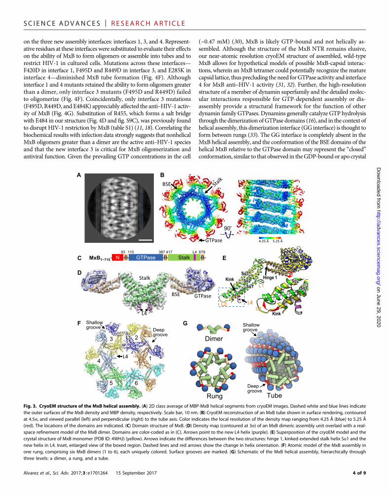
CryoEM structure of the MxB assemblyThe assembledMxB tubes were highly ordered with an inner and outerdiameter of 55 and 275 Å, respectively (Fig. 3A and fig. S5). There wasan undifferentiated density out to 360 Å, as shown in one of the 2Dclasses (Fig. 3A, between white and blue dashed lines), presumablycorresponding to the MxB NTR and MBP tag. The Fourier transformsof theMxB tubes indicated that they belong to a one-start helical familyof (−6, 1) (fig. S5), a right-handed helix (fig. S6) with a rise of 8.237 Åand a twist of 58.4°. Using cryoEMand real-space helical reconstruction
Alvarez et al., Sci. Adv. 2017;3 : e1701264 15 September 2017
(25), we determined the 3D density map of theMxB helical assembly at4.6 Å resolution (Fig. 3B and fig. S7). The local resolution of the densitymap varies (Fig. 3B); a-helical turns (fig. S8, D to G) and some bulkyside-chain densities (fig. S8, D and G) are resolved at the inner core(stalk and BSE), whereas the GTPase domain appears to be more flex-ible. The NTR, together with the MBP tag, is not resolved, probably be-cause of their flexibility. Initial rigid-body docking of individualdomains from the crystal structure of the NTR-truncated MxB dimer[Protein Data Bank (PDB) ID: 4WHJ] (15) resulted in a reasonableoverall fit, although it revealed substantial deviations, particularly atthe first stalk helix Sa1 and unaccounted extra helical density (fig.S8A, arrows). This helical region is part of the L4 loop, which conveysantiviral specificity for MxA-like proteins (17). It was previouslythought to be completely unstructured and was not observed in eitherthe MxA (26) or MxB (15) crystal structure.
We modeled the L4 helix de novo using Rosetta into the MxBdimer structure and carried out molecular dynamics flexible fitting(MDFF), followed by real-space refinement to obtain an all-atomcryoEM structure model of the MxB assembly (Fig. 3, D and F, fig. S8,and movie S1). The resulting model displays a good match to the exper-imental density (Fig. 3D and fig. S8, C toG). In the cryoEMstructure, theentireMxB assembly ismade up ofMxBdimer units. Six dimers go handin hand, interlockingwith each other through the stalk andBSE domainsto form one rung, where the sixth dimer comes around to interact withthe first dimer, forming the one-start right-handed helix (Fig. 3, F andG,and movie S2). The tube surface displays a shallow groove, where theGTPase domains cluster, and a deep groove, where the NTRs are pre-sumably located (Fig. 3, F and G). The MxB dimer in the assembly issubstantially different from the crystal dimer (PDB ID: 4WHJ), display-ing (i) a highly kinked and extended stalk Sa1 helix C terminus (Sa1c),
670
kDa
158
kDa
Voi
d
44 k
Da
Retention volume (ml)
AU
at 2
80 n
mB
C
D
E
B D E
C FA
Fig. 1. Full-length wild-type MxB purified as oligomers. (A) Purification of MBP-MxB from Expi293F cells by amylose affinity chromatography, followed by gelfiltration through a Sephacryl S-500 HR column. The fractions indicated by arrowheads were visualized by negative stain EM. Inset, Coomassie-stained SDS–polyacrylamide gel electrophoresis (PAGE) gel of untransfected cells (1), transfected cells (2), and elution from amylose resin (3). Molecular weight (MW) markersare shown in kilodaltons (kDa). AU, absorbance units. (B to E) Representative negative stain micrographs of the respective fractions indicated in (A). (F) 2D classaverages of the negatively stained MxB sample from fraction “C.” Scale bars, 200 nm.
2 of 9
SC I ENCE ADVANCES | R E S EARCH ART I C L E
http://advances.sciencemag.o
Dow
nloaded from
which effectively displaces the base of Sa1c by 27°, (ii) a shift in the do-main orientations about hinge 1 between the BSE and the stalk by 22°,and (iii) a well-ordered L4 loop helix (Fig. 3E). As a result, these newfeatures led to the formation of completely new assembly interfaces,which are distinct from those previously inferred from crystal contacts(table S1) (discussed in detail below). There is no swapped dimer present,as previously thought for the dynamin tube (20).
Novel MxB assembly interfacesThe cryoEM structure revealed three levels of assembly interfaces thatare likely shared by members of the dynamin superfamily of GTPases:dimer interface [interface 2; following theMxA convention (26)], oligo-mer interfaces (interfaces 1 and 3), and helical (higher-order) assemblyinterface (interface 4) (Figs. 3G and 4, A and B). The dimer interface(interface 2) is essentially the same, as seen in the MxB crystal structure(fig. S9A). However, the other three interfaces are novel in the cryoEMstructure, as described in detail below.
Interface 1 is formed by the symmetric interaction of the tip of thestalk domain (Sa1n) of one dimer and the BSE domain (Ba3) fromanother, mediated by a salt bridge (D417-K693) and hydrophobiccontacts (F420, M419, and I696) (Fig. 4C and fig. S9B). This interfaceis markedly different from the putative interface 1 (fig. S10A and tableS1) gleaned from the crystallographic symmetries of MxA and MxBstructures, which shows stacking of the stalk domains (Sa1n and Sa4)of the dimers to form a linear array (14, 15).
Interface 3 is completely novel and composed of the new extendedbase of kinked Sa1c and its connecting loops at the either end of Sa1c,L1 and L2 (Fig. 4D and fig. S9C). Conserved hydrophobic L2 residues(F495 and V496) from adjacent dimers form a symmetric interaction(Fig. 4D, blue and orange, and fig. S9C). Additional salt bridges, R449-E491 between L1 and L2 from neighboring dimers as well as R455-E484between L1 and the base of Sa1c, further stabilize the interface (Fig. 4Dand fig. S9C). Because of the kinking of Sa1c and consequential posi-
Alvarez et al., Sci. Adv. 2017;3 : e1701264 15 September 2017
tioning of its base and L2 (Fig. 3E and movie S3), these interactions areabsent from the previous crystal structures (fig. S10B). However, a kinkin Sa1c was observed in a crystal structure of tetrameric human dynam-in 3 (fig. S11C, black arrow) where interface contacts in L2 similar toMxB were also observed (fig. S11E) (27), further emphasizing the crit-ical role of this new interface in MxB oligomerization (movie S4).
Interface 4 is formed between the GTPase domain of dimer 1 andthe stalk–hinge 1 region of dimer 6, as it completes a full rung (Fig. 4, Band E). Our structure lacks evidence for GTPase domain dimerization,as proposed previously for dynamin helical assembly (20), supportingstalk-driven oligomerization. The GTP hydrolysis–deficient MxB mu-tant, T151A,was shown to retain the ability to inhibitHIV-1 (6, 11) with-out affecting GTP binding (28). The T151A alteration does not affectthe MxB helical assembly (fig. S12A); however, unlike wild-typeMxB,GTP orGTP-g-S binding has amarginal effect onMxB T151A assem-blies (fig. S12). The residues involved in interface 4 include P284, E285,and K250 from the GTPase domain of dimer 1 and R674, W677, andQ680 from the stalk–hinge 1 region of dimer 6, mediating charge inter-actions and hydrophobic contacts (Fig. 4E and fig. S9D). This is the onlyassembly interface involving the GTPase domain and is the interfaceresponsible for helical assembly by stabilizing rung stacking (Figs. 3Gand 4B). Thus, it explains our observations that the MxB tubes are dis-rupted into oligomers upon GTP binding (Fig. 2, E and G). Low-saltconditions, which promote helical assembly of MxB (Fig. 2A) andother dynamin familymembers (29), likely facilitate hydrophobic in-teractions at interface 4.
Functional importance of MxB interface 3 in assembly andantiviral activityWith detailed knowledge of all interfaces responsible for assembly,we tested which of these interfaces is relevant to the MxB function.Because the dimer interface was previously well characterized forits important role in MxB antiviral function (15, 18, 19), we focused
on June 29, 2020rg/
A B C D
+GTP-γ-S (O/N)+GTP-γ-S (2 h) +GTP (2 h) +GTP (O/N)E F HG
+GDP (2 h) +GDP (O/N)
Fig. 2. GTP binding induces depolymerization of the helical MxB assembly. (A) MBP-MxB self-assembled into helical structures at 150 mM NaCl. (B) Gold labelingof MBP shows that MBP is located on the surface of assembled MBP-MxB tubes. End-on view (top) and side view (bottom) are shown. (C to H) Representative micro-graphs of MBP-MxB in the presence of 1 mM GDP (C and D), GTP-g-S (E and F), and GTP (G and H), incubated at room temperature for 2 hours (C, E, and G) or overnight(O/N) (D, F, and H). Scale bars, 50 nm (A and B) and 100 nm (H). Panels (C) to (H) are of the same scale.
3 of 9
SC I ENCE ADVANCES | R E S EARCH ART I C L E
on the three new assembly interfaces: interfaces 1, 3, and 4. Represent-ative residues at these interfaces were substituted to evaluate their effectson the ability of MxB to form oligomers or assemble into tubes and torestrict HIV-1 in cultured cells. Mutations across these interfaces—F420D in interface 1, F495D and R449D in interface 3, and E285K ininterface 4—diminished MxB tube formation (Fig. 4F). Althoughinterface 1 and 4 mutants retained the ability to form oligomers greaterthan a dimer, only interface 3 mutants (F495D and R449D) failedto oligomerize (Fig. 4F). Coincidentally, only interface 3 mutations(F495D, R449D, and E484K) appreciably affected the anti–HIV-1 activ-ity of MxB (Fig. 4G). Substitution of R455, which forms a salt bridgewith E484 in our structure (Fig. 4D and fig. S9C), was previously foundto disrupt HIV-1 restriction byMxB (table S1) (11, 18). Correlating thebiochemical results with infection data strongly suggests that nonhelicalMxB oligomers greater than a dimer are the active anti–HIV-1 speciesand that the new interface 3 is critical for MxB oligomerization andantiviral function. Given the prevailing GTP concentrations in the cell
Alvarez et al., Sci. Adv. 2017;3 : e1701264 15 September 2017
(~0.47 mM) (30), MxB is likely GTP-bound and not helically as-sembled. Although the structure of the MxB NTR remains elusive,our near-atomic resolution cryoEM structure of assembled, wild-typeMxB allows for hypothetical models of possible MxB-capsid interac-tions, wherein anMxB tetramer could potentially recognize the maturecapsid lattice, thus precluding the need forGTPase activity and interface4 for MxB anti–HIV-1 activity (31, 32). Further, the high-resolutionstructure of a member of dynamin superfamily and the detailed molec-ular interactions responsible for GTP-dependent assembly or dis-assembly provide a structural framework for the function of otherdynamin family GTPases. Dynamins generally catalyze GTP hydrolysisthrough the dimerization ofGTPase domains (16), and in the context ofhelical assembly, this dimerization interface (GG interface) is thought toform between rungs (33). The GG interface is completely absent in theMxB helical assembly, and the conformation of the BSE domains of thehelical MxB relative to the GTPase domain may represent the “closed”conformation, similar to that observed in theGDP-bound or apo crystal
on June 29, 2020http://advances.sciencem
ag.org/D
ownloaded from
A
C
B
GF
90
1
23
4
5 6
Deepgroove
Shallow groove
Rung
Dimer
Tube
D
N GTPase StalkB B BMxB1−715
93 110 387 417 679L4
Shallow groove
Deepgroove
E
Fig. 3. CryoEM structure of the MxB helical assembly. (A) 2D class average of MBP-MxB helical segments from cryoEM images. Dashed white and blue lines indicatethe outer surfaces of the MxB density and MBP density, respectively. Scale bar, 10 nm. (B) CryoEM reconstruction of an MxB tube shown in surface rendering, contouredat 4.5s, and viewed parallel (left) and perpendicular (right) to the tube axis. Color indicates the local resolution of the density map ranging from 4.25 Å (blue) to 5.25 Å(red). The locations of the domains are indicated. (C) Domain structure of MxB. (D) Density map (contoured at 3s) of an MxB dimeric assembly unit overlaid with a real-space refinement model of the MxB dimer. Domains are color-coded as in (C). Arrows point to the new L4 helix (purple). (E) Superposition of the cryoEM model and thecrystal structure of MxB monomer (PDB ID: 4WHJ) (yellow). Arrows indicate the differences between the two structures: hinge 1, kinked extended stalk helix Sa1 and thenew helix in L4. Inset, enlarged view of the boxed region. Dashed lines and red arrows show the change in helix orientation. (F) Atomic model of the MxB assembly inone rung, comprising six MxB dimers (1 to 6), each uniquely colored. Surface grooves are marked. (G) Schematic of the MxB helical assembly, hierarchically throughthree levels: a dimer, a rung, and a tube.
4 of 9

SC I ENCE ADVANCES | R E S EARCH ART I C L E
on June 29, 2020http://advances.sciencem
ag.org/D
ownloaded from
structures of dynamin proteins (fig. S13) (16). We speculate that thetrans-dimerization of the GTPase domains upon GTP binding of MxBis sensed by the BSE domains, causing them to transition to the openconformation, which effectively disrupts the helical assemblies.
MATERIALS AND METHODSPlasmid constructionCloning vectors containing the gene for the full-lengthwild-type humanMxB (UniProt ID: P20592-1) and the MBP tag were gifts from J. Ahn(PittsburghCenter forHIVProtein Interactions, University of PittsburghSchool of Medicine). The expression vector pcDNA3.1(+) was obtainedfrom Life Technologies (Invitrogen). TheMxB gene and the MBP tagwere amplified by polymerase chain reaction (PCR) and then sub-cloned, using the NEBuilder HiFi Assembly kit (New England Biolabs
Alvarez et al., Sci. Adv. 2017;3 : e1701264 15 September 2017
Inc.), into pcDNA3.1(+) that had been linearized by the restrictionenzymes Eco RV and Xba I. The resulting insert, designated asMBP-MxB-H6, has a leading Kozak sequence, an N-terminal MBPtag, followed by a human rhinovirus 3C protease cut site, the full-length wild-type MxB, and a C-terminal hexahistidine tag (H6). Theplasmid used for the infectivity assay was generated by transferringthe PCR construct for MBP-MxB-H6 or MxB-hemagglutinin frompcDNA3.1(+) to pIRES2–enhanced green fluorescent protein (eGFP)(34).The sequences of the inserts were confirmed by DNA sequencing(Genewiz Inc.).
Sequence alignmentAmino acid sequences ofMx anddynaminproteinswere obtained fromUniProt using the indicated IDs and were then aligned usingMUSCLE(35). The alignment was visualized using ESPript3 (36).
Fig. 4. MxB intermolecular assembly interfaces and their role in the MxB assembly and HIV-1 inhibition. (A) Intermolecular interfaces in an MxB oligomer: thecanonical MxB dimer interface, interface 2 (black circle), and the lateral interfaces that link adjacent MxB dimers, interfaces 1 (blue circle) and 3 (red circle). The samecolor scheme is used as in Fig. 3F. (B) Interface 4 (magenta circles), the vertical interface between adjacent rungs. MxB dimer 7 starts the next rung in the helix, coloredthe same as dimer 1. (C to E) Expanded views of the intermolecular interfaces: interface 1 (C), interface 3 (D), and interface 4 (E). Specific residues at the interfaces arelabeled, along with the secondary structures. Underlined amino acids were subjected to mutational analysis. (F) Effects of interface mutations on the MxB assembly:E285K (interface 4), F420D (interface 1), F495D, and R449D (interface 3). Negatively stained images of purified interface mutant proteins under helical assembly conditions areshown. Inset, wild-type MxB. Scale bar, 50 nm. (G) Effects of interface mutations on MxB anti–HIV-1 activity (top) (mean ± SD for minimally n = 3 independent experiments) andWestern blot analysis of MxB protein expression (bottom). MBP-tagged wild-type (WT) MxB, which does not inhibit HIV-1, was used as a control.
5 of 9

SC I ENCE ADVANCES | R E S EARCH ART I C L E
on June 29, 2020http://advances.sciencem
ag.org/D
ownloaded from
Expression and purification of MxBRecombinant MxB was transiently expressed in mammalian cellsusing the Expi293 Expression kit from Life Technologies (Invitrogen).Suspension-adapted Expi293F cells were grown in Expi293 ExpressionMedium to a density of 3.5 × 10⁶ to 4 × 10⁶ cells/ml and a viability of>95% 24 hours before transfection. Plasmid DNA and ExpiFectaminereagent (Invitrogen) were diluted in Opti-MEM I Reduced Serum(Invitrogen) into separate tubes, incubated for 5 min at room tem-perature, and then mixed together for 25 min. Cells were transfectedwith the DNA-ExpiFectamine complex at a DNA/transfection reagent/cell culture volume ratio of 30 mg/1.5 ml/30 ml and to a final celldensity of 2.9 × 10⁶ cells/ml. Cells were then incubated at 37°C and125-rpm agitation with 8% CO2 in air. After 18 hours of incubation,150 ml of transfection enhancer 1 and 1.5 ml of transfection enhancer2 for every 30 ml of cells were added into the suspension. Twenty-fourhours after the addition of the enhancers, cells were harvested by cen-trifugation at low speed (100g). Cells were washed once with coldphosphate-buffered saline, and the cell pellet was flash-frozen andstored at −80°C for later use.
The thawed cell pellet was resuspended in buffer A [50 mMHepes-KOH (pH 8), 250 mM NaCl, and 5% glycerol] supplemented with de-tergents (1%Tween 20 and0.3%NP-40), deoxyribonuclease I (50mg/ml;Sigma-Aldrich) in thepresenceof 5mMMgCl2, 10mMb-mercaptoethanol,and a cocktail of protease inhibitors (Roche). After 1 hour of rotation at4°C, the lysate was homogenized by 15 strokes in an ice-cold, tight-fitting Dounce homogenizer. The homogenate was then centrifugedat 21,000g at 4°C for 30 min. After centrifugation, the supernatant wascollected and mixed with 1 ml of amylose agarose resin (New EnglandBiolabs Inc.) (per 50ml of cell suspension) pre-equilibratedwith bufferA.The mixture was incubated with rotation at 4°C for 1 hour and thentransferred to a column to flow through. The resin was washed with50× resin volume of buffer A. To elute the recombinant protein, the resinwas incubated, in batch, with buffer A containing 50 mMmaltose for15min at 4°C, and then, the flow throughwas collected as elution. Thepurified protein was detected byWestern blot using antibodies againstthe MBP tag (Abcam) and MxB (N-17) (Santa Cruz Biotechnology)andHisProbe–horseradish peroxidase (HRP) (ThermoFisher Scientific)for the hexahistidine tag.
GTPase assayThe GTPase function of MxB was assessed using a continuous NADH(reduced form of nicotinamide adenine dinucleotide)–coupled assay(37). The reaction mixture was prepared to achieve the following finalconcentrations in the assay solution: 50mMHepes-KOH(pH7), 150mMNaCl, 10 mMMgCl2, 2 mM dithiothreitol (DTT), 4 mM phosphenol-pyruvate, 0.35mMNADH,25Uofpyruvate kinase/lactatedehydrogenase,and1mMGTP.MxBwas added just beforemeasurement. Thedecrease intheNADHabsorbance at 340 nmwasmonitored in a 96-well plate using aSynergyH1HybridReader (BioTek) at 37°C.The rate ofNADHoxidationwasmeasured and used to calculate the kobs ofMxB.NADHoxidationwasalso monitored in the absence of protein using buffer only as control. Theexperimental valueswerenormalized to correct for background.Results arerepresentative of three independent measurements.
Mass spectrometry analysisMxB was digested overnight at 37°C with sequencing grade trypsin inthe presence of 1M urea. Digested protein (100 pmol) was captured ona Phenomenex Aeris C18 column (2.1 × 50 mm) (Phenomenex) andeluted with a 0 to 65% gradient of acetonitrile at 400 ml/min. Lock mass–
Alvarez et al., Sci. Adv. 2017;3 : e1701264 15 September 2017
corrected mass spectra were acquired in MSE (all-ion) mode using aWatersQ-ToF Premier, and peptides were identified using ProteinLynxGlobal Server.
Electron microscopySample preparationTo prepare samples for initial screening by negative stain, the elutionfrom the amylose resin was immediately filtered through a HiPrepSephacryl S-500 HR (GE Healthcare) in buffer A with an additional2 mMDTT. To induce the formation of the long tubes, the elution fromthe amylose resin was diluted in assembly buffer [20 mM Hepes-KOH(pH 7), 150 mMNaCl, 1 mMMgCl2, 2 mMEGTA, and 2mMDTT] at0.5 to 1 mg/ml, and then incubated for the indicated period of time.Negative stain EMAliquots (3 ml) from the gel filtration samples or the helical assemblywere adsorbed to a glow-discharged, 400-mesh, carbon-coated coppergrid and stained with fresh uranyl formate (2%). Images were recordedon a TF20 electron microscope (FEI) equipped with a field-emissiongun at the indicated magnification on a 4k × 4k Gatan UltraScancharge-coupled device camera (Gatan).Immunogold labelingImmunogold labeling, modified from the study by Mears et al. (38),was performed to determine the orientation of the NTR of MxB inits helical form. Samples containing the MxB tubes were preparedand applied to a grid, as described above. The grid was successivelyfloated on the following solutions: (i) twice with blocking buffer [bovineserum albumin (BSA;10 mg/ml) in oligomerization buffer] for 5 min,(ii) with blocking buffer containing primary antibody against MBP tag(Abcam) (1:250 dilution) for 1 hour, (iii) twice with blocking buffer for5 min, and (iv) with blocking buffer containing a 5-mm gold-labeledsecondary antibody (Ted Pella) (1:250 dilution) for 1 hour. All incuba-tions were carried out at 4°C, and the grid was washed once withblocking buffer and twice with oligomerization buffer before stainingwith uranyl formate.CryoEMThree microliters of the MxB tubes (0.5 mg/ml) was applied on thecarbon side of glow-discharged holey R2/1 Quantifoil grids (QuantifoilMicro Tools GmbH), manually blotted from the backside, and thenplunge-frozen in liquid ethane using a homemade manual plunger.Images were collected under low-dose conditions (~40 e−/Å2 total)using a Polara 300-kV microscope with a field-emission gun and anFEI Falcon II detector. Movies (~1000, each with seven frames) weremanually collected using SerialEM (39) at a nominal magnificationof ×98,000 (1.147 Å/pixel), with under-focus values ranging from 1.5to 3.5 mm.
Image processing and helical reconstructionMovie frames were aligned using UCSF (University of California,SanFrancisco)MotionCorr v2.1 (40), and the resultingmotion-correctedsumswere used for contrast transfer function estimationusingGctf v0.50(41). Micrographs were then sorted on the basis of image quality, and~630 micrographs were used for subsequent helical reconstructionusing RELION 2.0 beta (25), which is a software package in develop-ment that integrates a helical processing workflow. Helical segmentswere boxed using EMAN 2.0 helixboxer.py (42), and diffraction pat-terns from individual tubes or 2D class averages generated using Spring(43) were used to estimate the helical parameters. A small data set with3486 segments (500-pixel box size) was used in the helical processingworkflow in RELION 2.0 beta to refine the helical parameters, which
6 of 9

SC I ENCE ADVANCES | R E S EARCH ART I C L E
on June 29, 2020http://advances.sciencem
ag.org/D
ownloaded from
converged to a rotation angle of 58.4° and a rise of 8.25 Å. Using thepreviously generated 2D classes as templates, helical filaments wereautomatically picked from the full data set, resulting in 51,553 segments(450-pixel box size, 90.41% overlap between neighboring boxes, and in-terbox distance six times the helical rise). The segments were then ana-lyzed by 2D classification, and 64 of 100 classes showing clear structuraldetails were then selected for further processing (44,955 segments cor-respond to ~270,000 subunits). The first round of refinement, usinga featureless cylinder (236 pixels in diameter and low-pass–filtered to30Å), resulted in amapwith a resolution of 6.9 Å. For the next roundof refinement, the previous reconstruction was used as an initial ref-erence and converged to a map with 6.6 Å overall resolution, whichwas improved to 5.0Å after postprocessing (sharpening and applicationof soft-edged mask). A final round of refinement was performed usingpolished particles, which gave amapwith 5.3Å resolution. Postprocessingby soft-edge masking and B-factor sharpening resulted in a map with4.6 Å resolution. The finalmapwas then low-pass–filtered, according tothe local resolution estimated using RELION 2.0. Further 3D classifica-tion did not reveal distinguishable reconstructions, suggesting that therewas no mixing of helical symmetry in the data set. Attempts to resolvethe outer portion of the tube by 3D classification using cylindricalmasks(150 to 360 Å in diameter) did not show any improvement to thefeatures of the outer portion of the map.
Infectivity assayHuman embryonic kidney (HEK) 293T cells were cultured inDulbecco’smodified Eagle’s medium supplemented with 10% fetal bovine serum,penicillin (100 IU/ml), and streptomycin (0.1 mg/ml). For virus pro-duction, cells were plated 1 day before transfection. Single-roundHIV-1 harboring the gene for firefly luciferase (HIV-Luc) was gener-ated by cotransfecting HEK293T with pNLX.Luc.(R−).DAvrII and pCG-VSV-G(44).Thevirus yieldwas assessedbyHIV-1CAp24enzyme-linkedimmunosorbent assay (ABL Inc.).
Before infection, HEK293T cells were transiently transfected withparental pIRES2-eGFP vector or derivatives expressingMxB. At 24 hoursafter transfection, GFP-positive cells selected by fluorescence-activatedcell sorting were infected in duplicate with HIV-Luc (0.1 pg of p24 percell) in the presence of polybrene (4 mg/ml). Forty-eight hours after infec-tion, cells were lysed, and luciferase activity was determined as described(34, 44). Luciferase values were normalized to the level of total proteins incell lysates, as determined using a bicinchoninic acid (BCA) assay(Pierce).
Western blottingCells were lysed in 50 mM tris-HCl (pH 8), 150 mM NaCl, 1% NP-40,0.5% sodium deoxycholate, and 0.1% SDS, and total protein concentra-tion was determined using the BCA assay. Samples (10 mg of total pro-tein) were separated by SDS-PAGE, transferred to polyvinylidenedifluoride membranes, and reacted with goat polyclonal antibody toMxB (N-17), followed by HRP-conjugated secondary bovine anti-goatantibody (Santa Cruz Biotechnology). As an internal control, HRP-conjugated antibody to b-actin (Sigma-Aldrich) was used. Membraneswere developed using the ECL Prime reagent (Amersham Biosciences)and imaged with a ChemiDoc MP imager (Bio-Rad).
De novo structure modeling of the L4 helix(residues 579 to 598)Structure modeling of the L4 helix, which was apparent in the cryoEMdensity map but was missing in the crystal structure (PDB ID: 4WHJ),
Alvarez et al., Sci. Adv. 2017;3 : e1701264 15 September 2017
wasmodeled usingRosetta (45). Fragments containing sequence 3-mersand 9-mers were generated using the Rosetta server, using the MxBwild-type sequence (46). One thousand candidate folds were generatedusing theTopologyBroker protocol in RosettaScripts (47). The structuralvariability of the predicted folds was modest because all the predictedstructures corresponded to an extended a helix. Using the Talaris2014potential (48), the lowest energy fold was selected for further refine-ment, as explained in the following sections.
Molecular dynamics flexible fittingThe x-ray–derived structure of the MxB dimer (PDB ID: 4WHJ) wasused as the initial model. Missing loops were modeled using Modellerv9.17 (49). In addition, residues 487 to 490, which contain four alaninemutations in the structure (PDB: 4WHJ), were reverted back to the wild-type sequence. For the initialmodeling of full-lengthMxB, the L4 helixwas omitted, and residues 577 and 626 were treated as C-terminaland N-terminal, respectively, to cap the ends. Secondary structural as-signments used as structural restraints (ss-restraints) during theMDFFprotocol (50) were derived using a secondary structure predictionprogram, DSSP (51). To preserve the cis/trans conformations presentin the initial model (PDB ID: 4WHJ), cis-peptide restraints were alsoused. Themodelwas then subjected toMDFF,with the backbone atomscoupled to the experimental density, with a coupling constant rampingfrom 0.05 to 0.15 over 5 ns, resulting in a cross-correlation between themap and the structure of 0.85. Because of the ss-restraints, the fitting ofthe stalk helix Sa1c into the experimental density was poor; therefore,the ss-restraints for residues 472 to 474 were manually removed. Sub-sequently, the model was subjected to MDFF, with a coupling constantramping from 0.05 to 0.15 over 5 ns, resulting in a cross-correlation of0.86. All molecular dynamics simulations were performed usingNAMD 2.10 (52), with an integration time step of 2 fs, bonded interac-tions computed every time step, and electrostatics updated every 4 fs.Particle mesh Ewald was used for long-range electrostatics with a gridsize of 1 Å. The CHARMM36 force field (53) and the TIP3P watermodel (54) were used in all simulations.
Iterative refinement of the MxB dimer by Rosetta and MDFFThe MDFF-derived MxB dimer model was further refined usingRosetta by following a similar procedure as the one developed by Lindertand McCammon (55). In particular, the MxB model was refined usingthe CartesianSampler available in RosettaScripts. For this purpose, thede novo model of L4 helix was docked into the experimental densityusing UCSF Chimera (56). First, a hybrid model of dimeric MxBcontaining the docked L4 helix and the MDFF-derived MxB dimerwas obtained usingModeller (49). The hybridmodel was then subjectedto further refinement using theCartesianSampler in RosettaScripts (47).The model, which consists of the MDFF-derived MxB dimer and theRosetta-derivedmodel for residues 579 to 598, was further refined usingMDFF, with a density coupling ramping from 0.05 to 0.15 over 5 ns.
Modeling of the MxB helical assemblyUsing UCSF Chimera, starting from the Rosetta-MDFFMxB dimer, anentire MxB helical assembly was constructed by using a helicalsymmetry with a rise of 8.327Å and an angle of 58.4°. The helicalmodelwas refined using MDFF with symmetry restraints (57). In addition,fitting of L1 and L2 into the density was further improved by usingthe interactive MDFF protocol with a coupling between the backboneatoms and a density of 0.1 (50). The interactive MDFF protocol man-ually guides the backbone of the loops into the experimental density.
7 of 9

SC I ENCE ADVANCES | R E S EARCH ART I C L E
The final cross-correlation between the helical model and the densitymap was 0.92.
Molecular dynamics simulations and analysisAn equilibrium molecular dynamics simulation of the entire MxBtube was performed starting from the refined helical MDFF-Rosettamodel. The molecular dynamics simulations were performed usingNAMD 2.10 (52), with an integration time step of 2 fs, bonded inter-actions computed every time step, and electrostatics updated every 4 fs.Particle mesh Ewald was used for long-range electrostatics with a gridsize of 1 Å. The CHARMM36 force field (53) and the TIP3P (54)water model were used in all simulations. Analysis of salt bridges,hydrogen bonds, and hydrophobic contacts was performed in VMD(visual molecular dynamics) (58) and averaged over all interfaces ofthe tube. A probability score was assigned to each contact, based on theoccupancy of the contact in the assembled structure during the moleculardynamics simulation.
on Juhttp://advances.sciencem
ag.org/D
ownloaded from
SUPPLEMENTARY MATERIALSSupplementary material for this article is available at http://advances.sciencemag.org/cgi/content/full/3/9/e1701264/DC1table S1. Effects of MxB mutations at intermolecular interfaces 1, 3, and 4.fig. S1. Sequence alignment of Mx and dynamin proteins.fig. S2. MxB assembles into helical tubes with and without the MBP tag.fig. S3. GTPase activity of MBP-MxB at different protein and salt concentrations.fig. S4. MBP-MxB tubes in the presence of GTP without or with MgCl2.fig. S5. CryoEM of the MBP-MxB assembly.fig. S6. Handedness of the MxB helical assembly map.fig. S7. Gold-standard Fourier shell correlation curve of the MBP-MxB density map.fig. S8. MDFF and real-space refinement of the MxB helical assembly model.fig. S9. Enlarged views of the intermolecular interfaces in the MxB assembly.fig. S10. Comparison of interfaces between cryoEM and crystal structures.fig. S11. Comparison of L1 and L2 contacts of MxB and dynamin 3.fig. S12. Interface 4 mutations relative to GTP-binding site.fig. S13. Comparison of MxB and MxA GTPase-BSE domains.movie S1. Molecular dynamics flexible fitting of MxB into the cryoEM map.movie S2. MxB assembles into a helical array.movie S3. Sa1c helix kinks from linear to tubular array.movie S4. Formation of Interface 3 during helical assembly.
ne 29, 2020
REFERENCES AND NOTES1. O. Haller, P. Staeheli, M. Schwemmle, G. Kochs, Mx GTPases: Dynamin-like antiviral
machines of innate immunity. Trends Microbiol. 23, 154–163 (2015).2. P. S. Mitchell, J. M. Young, M. Emerman, H. S. Malik, Evolutionary analyses suggest a
function of MxB immunity proteins beyond lentivirus restriction. PLOS Pathog. 11,e1005304 (2015).
3. J. Verhelst, P. Hulpiau, X. Saelens, Mx proteins: Antiviral gatekeepers that restrain theuninvited. Microbiol. Mol. Biol. Rev. 77, 551–566 (2013).
4. M. Aebi, J. Fäh, N. Hurt, C. E. Samuel, D. Thomis, L. Bazzigher, J. Pavlovic, O. Haller,P. Staeheli, cDNA structures and regulation of two interferon-induced human Mxproteins. Mol. Cell. Biol. 9, 5062–5072 (1989).
5. K. Melén, P. Keskinen, T. Ronni, T. Sareneva, K. Lounatmaa, I. Julkunen, Human MxBprotein, an interferon-a-inducible GTPase, contains a nuclear targeting signal and islocalized in the heterochromatin region beneath the nuclear envelope. J. Biol. Chem. 271,23478–23486 (1996).
6. M. Kane, S. S. Yadav, J. Bitzegeio, S. B. Kutluay, T. Zang, S. J. Wilson, J. W. Schoggins,C. M. Rice, M. Yamashita, T. Hatziioannou, P. D. Bieniasz, MX2 is an interferon-inducedinhibitor of HIV-1 infection. Nature 502, 563–566 (2013).
7. C. Goujon, O. Moncorgé, H. Bauby, T. Doyle, C. C. Ward, T. Schaller, S. Hué, W. S. Barclay,R. Schulz, M. H. Malim, Human MX2 is an interferon-induced post-entry inhibitor ofHIV-1 infection. Nature 502, 559–562 (2013).
8. Z. Liu, Q. Pan, S. Ding, J. Qian, F. Xu, J. Zhou, S. Cen, F. Guo, C. Liang, The interferon-inducibleMxB protein inhibits HIV-1 infection. Cell Host Microbe 14, 398–410 (2013).
9. T. Fricke, T. E. White, B. Schulte, D. A. de Souza Aranha Vieira, A. Dharan, E. M. Campbell,A. Brandariz-Nuñez, F. Diaz-Griffero, MxB binds to the HIV-1 core and prevents theuncoating process of HIV-1. Retrovirology 11, 68 (2014).
Alvarez et al., Sci. Adv. 2017;3 : e1701264 15 September 2017
10. Z. Liu, Q. Pan, Z. Liang, W. Qiao, S. Cen, C. Liang, The highly polymorphic cyclophilinA-binding loop in HIV-1 capsid modulates viral resistance to MxB. Retrovirology 12,1 (2015).
11. K. A. Matreyek, W. Wang, E. Serrao, P. K. Singh, H. L. Levin, A. Engelman, Host and viraldeterminants for MxB restriction of HIV-1 infection. Retrovirology 11, 90 (2014).
12. I. Busnadiego, M. Kane, S. J. Rihn, H. F. Preugschas, J. Hughes, D. Blanco-Melo,V. P. Strouvelle, T. M. Zang, B. J. Willett, C. Boutell, P. D. Bieniasz, S. J. Wilson, Host and viraldeterminants of Mx2 antiretroviral activity. J. Virol. 88, 7738–7752 (2014).
13. W. Wei, H. Guo, M. Ma, R. Markham, X.-F. Yu, Accumulation of MxB/Mx2-resistantHIV-1 capsid variants during expansion of the HIV-1 epidemic in human populations.EBioMedicine 8, 230–236 (2016).
14. S. Gao, A. von der Malsburg, A. Dick, K. Faelber, G. F. Schröder, O. Haller, G. Kochs,O. Daumke, Structure of myxovirus resistance protein a reveals intra- and intermoleculardomain interactions required for the antiviral function. Immunity 35, 514–525 (2011).
15. J. L. Fribourgh, H. C. Nguyen, K. A. Matreyek, F. J. D. Alvarez, B. J. Summers, T. G. Dewdney,C. Aiken, P. Zhang, A. Engelman, Y. Xiong, Structural insight into HIV-1 restriction byMxB. Cell Host Microbe 16, 627–638 (2014).
16. O. Daumke, G. J. K. Praefcke, Invited review: Mechanisms of GTP hydrolysis andconformational transitions in the dynamin superfamily. Biopolymers 105, 580–593 (2016).
17. P. S. Mitchell, C. Patzina, M. Emerman, O. Haller, H. S. Malik, G. Kochs, Evolution-guidedidentification of antiviral specificity determinants in the broadly acting interferon-inducedinnate immunity factor MxA. Cell Host Microbe 12, 598–604 (2012).
18. M. D. J. Dicks, C. Goujon, D. Pollpeter, G. Betancor, L. Apolonia, J. R. C. Bergeron,M. H. Malim, Oligomerization requirements for MX2-mediated suppression ofHIV-1 infection. J. Virol. 90, 22–32 (2015).
19. C. Buffone, B. Schulte, S. Opp, F. Diaz-Griffero, Contribution of MxB oligomerization toHIV-1 capsid binding and restriction. J. Virol. 89, 3285–3294 (2015).
20. J. S. Chappie, J. A. Mears, S. Fang, M. Leonard, S. L. Schmid, R. A. Milligan, J. E. Hinshaw,F. Dyda, A pseudoatomic model of the dynamin polymer identifies a hydrolysis-dependent powerstroke. Cell 147, 209–222 (2011).
21. P. E. Nigg, J. Pavlovic, Oligomerization and GTP-binding requirements of MxA for viraltarget recognition and antiviral activity against influenza A virus. J. Biol. Chem. 290,29893–29906 (2015).
22. P. Zhang, J. E. Hinshaw, Three-dimensional reconstruction of dynamin in the constrictedstate. Nat. Cell Biol. 3, 922–926 (2001).
23. E. Ingerman, E. M. Perkins, M. Marino, J. A. Mears, J. M. McCaffery, J. E. Hinshaw, J. Nunnari,Dnm1 forms spirals that are structurally tailored to fit mitochondria. J. Cell Biol. 170,1021–1027 (2005).
24. Y. Yoon, K. R. Pitts, M. A. McNiven, Mammalian dynamin-like protein DLP1 tubulatesmembranes. Mol. Biol. Cell 12, 2894–2905 (2001).
25. S. He, S. H. W. Scheres, Helical reconstruction in RELION. J. Struct. Biol. 198, 163–176(2017).
26. S. Gao, A. von der Malsburg, S. Paeschke, J. Behlke, O. Haller, G. Kochs, O. Daumke,Structural basis of oligomerization in the stalk region of dynamin-like MxA. Nature 465,502–506 (2010).
27. T. F. Reubold, K. Faelber, N. Plattner, Y. Posor, K. Ketel, U. Curth, J. Schlegel, R. Anand,D. J. Manstein, F. Noé, V. Haucke, O. Daumke, S. Eschenburg, Crystal structure of thedynamin tetramer. Nature 525, 404–408 (2015).
28. M. C. King, G. Raposo, M. A. Lemmon, Inhibition of nuclear import and cell-cycleprogression by mutated forms of the dynamin-like GTPase MxB. Proc. Natl. Acad.Sci. U.S.A. 101, 8957–8962 (2004).
29. G. J. K. Praefcke, H. T. McMahon, The dynamin superfamily: Universal membranetubulation and fission molecules? Nat. Rev. Mol. Cell Biol. 5, 133–147 (2004).
30. T. W. Traut, Physiological concentrations of purines and pyrimidines. Mol. Cell. Biochem.140, 1–22 (1994).
31. J. Kong, M. Ma, S. He, X. Qin, Mx oligomer: A novel capsid pattern sensor? Future Microbiol.11, 1047–1055 (2016).
32. G. Zhao, J. R. Perilla, E. L. Yufenyuy, X. Meng, B. Chen, J. Ning, J. Ahn, A. M. Gronenborn,K. Schulten, C. Aiken, P. Zhang, Mature HIV-1 capsid structure by cryo-electronmicroscopy and all-atom molecular dynamics. Nature 497, 643–646 (2013).
33. A. C. Sundborger, S. Fang, J. A. Heymann, P. Ray, J. S. Chappie, J. E. Hinshaw, A dynaminmutant defines a superconstricted prefission state. Cell Rep. 8, 734–742 (2014).
34. M.-C. Shun, N. K. Raghavendra, N. Vandegraaff, J. E. Daigle, S. Hughes, P. Kellam,P. Cherepanov, A. Engelman, LEDGF/p75 functions downstream from preintegrationcomplex formation to effect gene-specific HIV-1 integration. Genes Dev. 21, 1767–1778(2007).
35. R. C. Edgar, MUSCLE: A multiple sequence alignment method with reduced time andspace complexity. BMC Bioinformatics 5, 113 (2004).
36. X. Robert, P. Gouet, Deciphering key features in protein structures with the newENDscript server. Nucleic Acids Res. 42, W320–W324 (2014).
37. E. Ingerman, J. Nunnari, A continuous, regenerative coupled GTPase assay for dynamin-related proteins. Methods Enzymol. 404, 611–619 (2005).
8 of 9

SC I ENCE ADVANCES | R E S EARCH ART I C L E
on June 29http://advances.sciencem
ag.org/D
ownloaded from
38. J. A. Mears, P. Ray, J. E. Hinshaw, A corkscrew model for dynamin constriction. Structure15, 1190–1202 (2007).
39. D. N. Mastronarde, Automated electron microscope tomography using robust predictionof specimen movements. J. Struct. Biol. 152, 36–51 (2005).
40. X. Li, P. Mooney, S. Zheng, C. R. Booth, M. B. Braunfeld, S. Gubbens, D. A. Agard, Y. Cheng,Electron counting and beam-induced motion correction enable near-atomic-resolutionsingle-particle cryo-EM. Nat. Methods 10, 584–590 (2013).
41. K. Zhang, Gctf: Real-time CTF determination and correction. J. Struct. Biol. 193, 1–12(2016).
42. G. Tang, L. Peng, P. R. Baldwin, D. S. Mann, W. Jiang, I. Rees, S. J. Ludtke, EMAN2: Anextensible image processing suite for electron microscopy. J. Struct. Biol. 157, 38–46(2007).
43. A. Desfosses, R. Ciuffa, I. Gutsche, C. Sachse, SPRING—An image processing packagefor single-particle based helical reconstruction from electron cryomicrographs.J. Struct. Biol. 185, 15–26 (2014).
44. Y. Koh, X. Wu, A. L. Ferris, K. A. Matreyek, S. J. Smith, K. Lee, V. N. KewalRamani,S. H. Hughes, A. Engelman, Differential effects of human immunodeficiency virus type1 capsid and cellular factors nucleoporin 153 and LEDGF/p75 on the efficiency andspecificity of viral DNA integration. J. Virol. 87, 648–658 (2013).
45. A. Leaver-Fay, M. Tyka, S. M. Lewis, O. F. Lange, J. Thompson, R. Jacak, K. Kaufman,P. D. Renfrew, C. A. Smith, W. Sheffler, I. W. Davis, S. Cooper, A. Treuille, D. J. Mandell,F. Richter, Y.-E. A. Ban, S. J. Fleishman, J. E. Corn, D. E. Kim, S. Lyskov, M. Berrondo,S. Mentzer, Z. Popović, J. J. Havranek, J. Karanicolas, R. Das, J. Meiler, T. Kortemme,J. J. Gray, B. Kuhlman, D. Baker, P. Bradley, ROSETTA3: An object-oriented software suitefor the simulation and design of macromolecules. Methods Enzymol. 487, 545–574 (2011).
46. D. Chivian, D. E. Kim, L. Malmström, P. Bradley, T. Robertson, P. Murphy, C. E. M. Strauss,R. Bonneau, C. A. Rohl, D. Baker, Automated prediction of CASP-5 structures using theRobetta server. Proteins 53, 524–533 (2003).
47. S. J. Fleishman, A. Leaver-Fay, J. E. Corn, E.-M. Strauch, S. D. Khare, N. Koga, J. Ashworth,P. Murphy, F. Richter, G. Lemmon, J. Meiler, D. Baker, RosettaScripts: A scripting languageinterface to the Rosetta macromolecular modeling suite. PLOS ONE 6, e20161 (2011).
48. M. J. O’Meara, A. Leaver-Fay, M. D. Tyka, A. Stein, K. Houlihan, F. DiMaio, P. Bradley,T. Kortemme, D. Baker, J. Snoeyink, B. Kuhlman, Combined covalent-electrostatic modelof hydrogen bonding improves structure prediction with Rosetta. J. Chem. TheoryComput. 11, 609–622 (2015).
49. B. Webb, A. Sali, Comparative protein structure modeling using MODELLER, in CurrentProtocols in Bioinformatics (John Wiley & Sons Inc., 2002).
50. L. G. Trabuco, E. Schreiner, J. Gumbart, J. Hsin, E. Villa, K. Schulten, Applications of themolecular dynamics flexible fitting method. J. Struct. Biol. 173, 420–427 (2011).
51. W. Kabsch, C. Sander, Dictionary of protein secondary structure: Pattern recognition ofhydrogen-bonded and geometrical features. Biopolymers 22, 2577–2637 (1983).
52. J. C. Phillips, R. Braun, W. Wang, J. Gumbart, E. Tajkhorshid, E. Villa, C. Chipot, R. D. Skeel,L. Kalé, K. Schulten, Scalable molecular dynamics with NAMD. J. Comput. Chem. 26,1781–1802 (2005).
53. R. B. Best, X. Zhu, J. Shim, P. E. M. Lopes, J. Mittal, M. Feig, A. D. Mackerell Jr., Optimizationof the additive CHARMM all-atom protein force field targeting improved sampling of
Alvarez et al., Sci. Adv. 2017;3 : e1701264 15 September 2017
the backbone ϕ, y and side-chain c1 and c2 dihedral angles. J. Chem. Theory Comput. 8,3257–3273 (2012).
54. W. L. Jorgensen, J. Chandrasekhar, J. D. Madura, R. W. Impey, M. L. Klein, Comparison ofsimple potential functions for simulating liquid water. J. Chem. Phys. 79, 926–935 (1983).
55. S. Lindert, J. A. McCammon, Improved cryoEM-guided iterative molecular dynamics–Rosetta protein structure refinement protocol for high precision protein structureprediction. J. Chem. Theory Comput. 11, 1337–1346 (2015).
56. E. F. Pettersen, T. D. Goddard, C. C. Huang, G. S. Couch, D. M. Greenblatt, E. C. Meng,T. E. Ferrin, UCSF Chimera—A visualization system for exploratory research and analysis.J. Comput. Chem. 25, 1605–1612 (2004).
57. K.-Y. Chan, J. Gumbart, R. McGreevy, J. M. Watermeyer, B. T. Sewell, K. Schulten,Symmetry-restrained flexible fitting for symmetric EM maps. Structure 19, 1211–1218(2011).
58. W. Humphrey, A. Dalke, K. Schulten, VMD: Visual molecular dynamics. J. Mol. Graph. 14,33–38 (1996).
Acknowledgments: We thank P. E. Prevelige Jr. for the mass spectrometry analysis,T. Brosenitsch for reading the manuscript, and X. Fu, G. Zhao, C. Liu, M. DeLucia, M. Ford,and T. Krzysiak for the technical assistance. Funding: This work was supported by the NIH(GM082251, GM085043, GM104601, GM067887, OD019995, and AI039394), the WellcomeTrust Investigator Award (206422/Z/17/Z), and the UK Medical Research Council(MC_UP_A025_1013). Molecular dynamics simulations were performed on the Blue WatersComputer, financed by the NSF (OCI 07-25070 and ACI-1238993). Author contributions: F.J.D.A. and P.Z. designed the research. F.J.D.A. expressed and purified the MxB, carried out thebiochemical analysis and EM imaging, prepared the samples for cryoEM, and collectedthe cryoEM data. F.J.D.A., S.H., S.H.W.S., and P.Z. carried out the cryoEM image analysis and3D reconstruction. J.R.P. and K.S. performed the molecular dynamics simulations andreal-space refinement and, with F.J.D.A., built the atomic model of the MxB assembly. F.J.D.A.,J.R.P., and P.Z. analyzed the model and designed the mutations. F.J.D.A. carried out themutagenesis. S.J. and A.N.E. performed the viral infectivity assays. F.J.D.A. and P.Z. wrote themanuscript, with support from all the authors. Competing interests: The authors declare thatthey have no competing interests. Data and materials availability: All data needed toevaluate the conclusions in the paper are present in the paper and/or the SupplementaryMaterials. Additional data related to this paper may be requested from the authors. ThecryoEM density map of wild-type MxB has been deposited in the Electron Microscopy DataBank under the accession code EMD-8577. The resulting atomic model has been deposited inthe PDB under the accession code PDB-5UOT.
Submitted 20 April 2017Accepted 17 August 2017Published 15 September 201710.1126/sciadv.1701264
Citation: F. J. D. Alvarez, S. He, J. R. Perilla, S. Jang, K. Schulten, A. N. Engelman, S. H. W. Scheres,P. Zhang, CryoEM structure of MxB reveals a novel oligomerization interface critical for HIVrestriction. Sci. Adv. 3, e1701264 (2017).
, 2
9 of 9
020

CryoEM structure of MxB reveals a novel oligomerization interface critical for HIV restriction
Peijun ZhangFrances J. D. Alvarez, Shaoda He, Juan R. Perilla, Sooin Jang, Klaus Schulten, Alan N. Engelman, Sjors H. W. Scheres and
DOI: 10.1126/sciadv.1701264 (9), e1701264.3Sci Adv
ARTICLE TOOLS http://advances.sciencemag.org/content/3/9/e1701264
MATERIALSSUPPLEMENTARY http://advances.sciencemag.org/content/suppl/2017/09/11/3.9.e1701264.DC1
REFERENCES
http://advances.sciencemag.org/content/3/9/e1701264#BIBLThis article cites 57 articles, 11 of which you can access for free
PERMISSIONS http://www.sciencemag.org/help/reprints-and-permissions
Terms of ServiceUse of this article is subject to the
is a registered trademark of AAAS.Science AdvancesYork Avenue NW, Washington, DC 20005. The title (ISSN 2375-2548) is published by the American Association for the Advancement of Science, 1200 NewScience Advances
BY).Science. No claim to original U.S. Government Works. Distributed under a Creative Commons Attribution License 4.0 (CC Copyright © 2017, The Authors, some rights reserved; exclusive licensee American Association for the Advancement of
on June 29, 2020http://advances.sciencem
ag.org/D
ownloaded from