Embed Size (px)
Citation preview
Crucial role of dynamic linker histone bindingand divalent ions for DNA accessibility andgene regulation revealed by mesoscale modelingof oligonucleosomesRosana Collepardo-Guevara1 and Tamar Schlick1,2,*
1Department of Chemistry, New York University, 100 Washington Square East, New York, NY 10003and 2Courant Institute of Mathematical Sciences, New York University, 251 Mercer Street, New York,NY 10012, USA
Received March 7, 2012; Revised May 15, 2012; Accepted May 28, 2012
ABSTRACT
Monte Carlo simulations of a mesoscale model ofoligonucleosomes are analyzed to examine therole of dynamic-linker histone (LH) binding/unbind-ing in high monovalent salt with divalent ions, and tofurther interpret noted chromatin fiber softening bydynamic LH in monovalent salt conditions. We findthat divalent ions produce a fiber stiffening effectthat competes with, but does not overshadow,the dramatic softening triggered by dynamic-LHbehavior. Indeed, we find that in typical in vivoconditions, dynamic-LH binding/unbinding reducesfiber stiffening dramatically (by a factor of almost 5,as measured by the elasticity modulus) comparedwith rigidly fixed LH, and also the force needed toinitiate chromatin unfolding, making it consistentwith those of molecular motors. Our data alsoshow that, during unfolding, divalent ions togetherwith LHs induce linker-DNA bending and DNA–DNArepulsion screening, which guarantee formation ofheteromorphic superbeads-on-a-string structuresthat combine regions of loose and compact fiberindependently of the characteristics of the LH–corebond. These structures might be important for generegulation as they expose regions of the DNA se-lectively. Dynamic control of LH binding/unbinding,either globally or locally, in the presence of divalentions, might constitute a mechanism for regulation ofgene expression.
INTRODUCTION
Understanding how chromatin fibers fold and unfold aswell as details of their structure and dynamics on a range
of spatial and temporal scales is important for interpretingfundamental template-directed processes such as DNAreplication, transcription, and repair. Indeed, the tightlypacked complex array of DNA with histone proteinsundergoes continuous chemical modification anddynamic association of proteins, such as linker histones(LHs), which control the accessibility of the geneticmaterial. Together with internal variations, such as thenucleosome repeat length (NRL) associated with thebasic repeating unit of DNA wrapped around the nucleo-some core (�147 bp) plus the variable linker-DNA length,and external factors such as the ionic environment, thesechanges determine the shape of the chromatin fiber at dif-ferent stages of the cell cycle.Although it is clear that LHs are essential for under-
standing chromatin compaction (1–3), many questions re-garding the structure and behavior of LH, and its role ingene regulation remain open [for a thorough review, see(4)]. We have been intrigued by suggestions that thedynamic binding/unbinding of LHs in vivo (5,6) andin vitro (7) may function to alter chromatin organizationby generating complex interaction networks that impartglobal changes from local rearrangements (5,8–11). Suchnetworks are plausible because LHs, sandwiched betweenentering and exiting linker DNA, lead to LH/linker DNAassociation called DNA stems that rigidify chromatin;conversely, LH dissociation can disrupt these networksand trigger unfolding rearrangements. Growing evidencepoints to a key role for LH dynamic binding during regu-lation of chromatin structure and gene expression (12);that is in vivo LH dynamic binding behavior might allowremodeling factors to bind to temporarily availablenucleosomal sites and induce chromatin structuralchanges to either activate or repress genes (13). In fact,lower H1 mobility, resulting for instance from LHdephosphorylation, is suggested to maintain chromatinin a compact structure and shut down gene expression,
*To whom correspondence should be addressed. Tel: +1 212 998 3596; Fax: +1 212 995 4152; Email: [email protected]
Published online 11 July 2012 Nucleic Acids Research, 2012, Vol. 40, No. 18 8803–8817doi:10.1093/nar/gks600
� The Author(s) 2012. Published by Oxford University Press.This is an Open Access article distributed under the terms of the Creative Commons Attribution Non-Commercial License (http://creativecommons.org/licenses/by-nc/3.0), which permits unrestricted non-commercial use, distribution, and reproduction in any medium, provided the original work is properly cited.
Dow
nloaded from https://academ
ic.oup.com/nar/article-abstract/40/18/8803/2411172 by guest on 12 April 2019

while enhanced H1 mobility is linked to undifferentiatedcells that require flexible chromatin to enable transcription(14); this is consistent with the presence of LHs with lowermobility causing inhibition of stem cell differentiation (15)and higher LH mobility observed in pluripotent stem cells(14). Furthermore, experiments (14,16) and our previousmodeling of chromatin in monovalent salt (17) have linkedincreased mobility of LH (e.g. induced by phosphoryl-ation of LH or acetylation of core histones) to facile chro-matin fiber opening. Our work (17) also suggested thatfast and slow LH dynamic binding populations, foundsimultaneously in vivo (10,18), cooperate to promote chro-matin unfolding with selective DNA exposure at lowforces. However, it remains unclear how such localexchange processes occur and how they affect chromatinorganization and accessibility at physiological ionic con-ditions (i.e. with divalent ions).Divalent ions enhance DNA compaction as they bring
linker-DNA segments closer to one another by screeningtheir electrostatic repulsion (19). Modeling and experi-ments have revealed that chromatin fibers with bothLHs and magnesium ions adopt a compact heteromorphicarchitecture that combines straight and bent DNA linkers(19). Although it is clear that divalent ions favor structuralvariations in compact chromatin, it is unknown what thecombined effects of divalent ions and LH dynamicbinding/unbinding are during chromatin’s fiber opening.Fiber heteromorphicity in divalent conditions has led us tospeculate that together divalent ions and dynamic-LHbinding/unbinding might serve a fundamental functionfor chromatin unfolding: increasing fiber fluidity byaccommodating more easily structural perturbations andmultiple fiber forms.Recent chromatin applications, by atomic force micros-
copy (20), optical tweezers (21–25), and magnetic tweezers(26), measured the fiber’s stretching response using force–extension curves and provided information on chroma-tin’s unfolding behavior. Based on optical tweezers experi-ments of medium-NRL chromatin, the Bustamante groupoffered evidence for an irregular zigzag structure stabilizedby nucleosome–nucleosome interactions <7 pN (21,27).The van Noort group studied chromatin with two differ-ent NRLs (short=167 bp and medium=197 bp) subjectto stretching at physiological salt with magnesium ionsand suggested that the response of the short-NRL arrayis consistent with a zigzag organization, while that of themedium-NRL arrays supports a solenoid topology (26).The latter study also showed that the presence of LHsdoes not change the stiffness of medium-NRL fiberswhen magnesium ions are present.Here, we develop and apply a computational approach
to simulate dynamic-LH binding/unbinding underdivalent salt conditions (see ‘Materials and Methods’section) and study the stretching response of single chro-matin fibers as a function of LH concentration andmobility. Our simulation approach is grounded on amesoscale model derived from experimental data of thechromatin fiber and atomistic properties of its compo-nents. This model combines different coarse-grainedstrategies for the nucleosome core, histone tails, DNA,and LHs (Figure 1). Such a modeling strategy allows us
to capture the essential physics of the chromatin fiber (likeits electrostatics, DNA and nucleosome mechanics, struc-tural irregularity, and histone-tail flexibility), and at thesame time to reduce the system dimensionality markedlyand sample the chromatin conformational space exhaust-ively. This approach has been developed, validated againstexperiments, and refined over the past few years, asdetailed extensively in (28–34).
Our results suggest a dramatic softening effect of thechromatin fiber when LHs associate/dissociate dynamic-ally compared with fixed (or static) LH species; thisproduces a stretching resistance compatible with unfold-ing at natural forces of molecular motors. Moreover, thedetailed fiber configurations corresponding to each stageof the unfolding process reveal that divalent ions favortransitional states that combine regions of fully stretchedfiber with prominent compact clusters or superbeads.These unusual ‘superbeads-on-a-string’ forms, observedexperimentally at different cell types (35) or in thepresence of low concentrations of magnesium ions (36),suggest mechanisms by which intrinsic and externalfactors, such as LH binding and divalent ions, lead tohigher order fiber forms. Although related unfoldingarrays emerged in our studies without divalent ionsfocusing on two binding/unbinding scenarios for LHs,the clumps were sparse (17). This work indicates thatdivalent ions are necessary to enhance nucleosome-clusterstability. Thus, LH mobility and divalent ions may intro-duce a combined regulatory mechanism for gene expres-sion through a localized transient exposure ofchromosomal DNA.
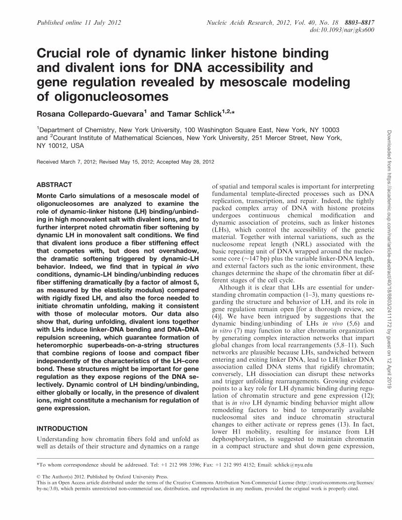
Figure 1. Mesoscale model of the basic chromatin building blockincluding: nucleosome core surface with wrapped DNA modeled asan irregularly shaped rigid body with uniformly distributed charges;linker DNA treated using the discrete worm-like chain model; andhistone tails and linker histones coarse-grained as bead models. Thesolvent around the oligonucleosome is treated implicitly as a con-tinuum. The screening of electrostatic interactions due to monovalentions in solution (0.15M NaCl) is treated using a Debye–Huckel poten-tial. A low concentration of magnesium ions is considered by a first-order approximation as developed in (32), by reducing the DNAbending persistence length and allowing the DNA beads to nearlytouch one another.
8804 Nucleic Acids Research, 2012, Vol. 40, No. 18
Dow
nloaded from https://academ
ic.oup.com/nar/article-abstract/40/18/8803/2411172 by guest on 12 April 2019
MATERIALS AND METHODS
Because of the intrinsic complexity of the chromatinfiber in solution (i.e. the many atoms that constitute it,the multiple time and length scales involved in itsdynamics, and the interplay of factors that intervene inits structural reorganization), simulating chromatin’sfolding/unfolding represents a grand challenge for bimol-ecular modelers. On the one hand, the accuracy of atom-istic models is desirable to capture the details of thechemical interactions, but on the other hand an all-atomstudy of oligonucleosomes is prohibitive due to themassive system dimensionality (e.g. a 50-nucleosomearray without solvent already contains >1 millionatoms). Coarse-graining represents one of the only alter-natives to model chromatin, and indeed several otherlarge-scale bimolecular systems [for an excellent review,see (37)], because it dramatically reduces the system sizeby averaging out many effects (e.g. protein/DNA sequenceeffects, hydrogen bonding, atomistic fluctuations, andsolvation), while simultaneously maintains the essentialphysical and chemical information required to analyzeits structural organization. This dimensionality reductionis a major advantage as it allows extensive sampling of thecoarse-grained phase space.
Coarse-grained modeling has been useful to comple-ment experimental analyses and help dissect the factorsthat control the structure and dynamics of the chromatinfiber (38). Among the existing chromatin coarse-grainedmodels, those that treat the DNA and nucleosome by geo-metric descriptions have provided insight on the physicalproperties of the chromatin polymer (27,39–44), whilemore realistic approaches based on all-atom studies and/or experimental data have dissected the influence of LHs,nucleosome interactions, and ionic environment on chro-matin structure (17,19,32–34,45,46).
Indeed, in silico stretching experiments have beeninstrumental for analyzing chromatin’s unfoldingbehavior and its implications for DNA accessibility(17,27,40,44,47). In particular, they have shown that chro-matin’s unfolding response is consistent with an irregularzigzag architecture (27), that the persistence length ofmedium-NRL fibers is strongly affected by the excludedvolume of the nucleosomes (47), that chromatin is muchmore resistant to stretching than to bending (40), that fiberstiffness decreases with the NRL and increases in thepresence of rigid DNA stems (17,40), and that simulatedconformations aid the analysis of chromatin force exten-sion experiments (17,44). Analytic approaches have alsoprovided information on the mechanical properties of thechromatin fiber (48–50). Here, we refine our mesoscalemodel of chromatin, which uses different coarse-grainedinterpretations for the different oligonucleosome compo-nents, to analyze the effect of LH dynamic binding duringforce-induced fiber unfolding.
Mesoscale model
Our model has been recently described in detail in (32,34),and below we summarize the main features of the
strategies used to treat each oligonocleosome element(Figure 1).
. Nucleosome cores: the nucleosome protein core,excluding histone tails, with wrapped DNA ismodeled as a rigid irregular body with 300 Debye–Huckel charges uniformly distributed on the nucleo-some molecular surface. The charges are optimizedto reproduce the full atom electric field around thenucleosome core by the discrete surface charge opti-mization (DiSCO) algorithm (28), which solves thecomplete nonlinear Poisson–Boltzmann equation.
. Histone tails: we model the 10 histone tails protrudingout of each core (the N-termini of each H2A, H2B,H3, and H4, plus the C-termini of each H2A) asflexible chains of beads (each bead comprises fiveamino acids) with the first bead rigidly attached tothe parent core. The stretching and bending flexibilityconstants of each tail inter-bead segment are modeledby harmonic potentials with parameters developed tomimic their atomistic flexibilities (32). The charges ofthe tail beads are also modeled to reproduce the atom-istic properties of the amino acids it represents.
. DNA linkers: the DNA that connects consecutive nu-cleosomes is treated as a chain of spherical beads thathave a salt-dependent charge parameterized using theStigter procedure (51). The mechanical properties ofthe linker-DNA chains are also considered anddescribed with the worm-like chain model (52,53).The equilibrium DNA inter-bead segment is 3 nm(�9 bp), thus to model an NRL of 209 bp, we usesix DNA beads (seven segments) per linker. Theexiting and entering DNA linkers attached to the nu-cleosome define an angle of 108�, which correspondsto the 147 DNAbp tightly wound �1.7 times aroundthe core.
. Linker histones: the LH proteins are modeled based onrat H1d LH. The family of eukaryotic LH proteins H1are formed by a globular domain (�80 amino acids)and two highly positively charged (lysine-rich) terminaltails (a short N-tail of �14 amino acids and a longC-tail of �100 amino acids) (54). We neglect the shortrelatively uncharged N-terminal domain and model theC-terminal domain by two charged beads, and theglobular domain by a single bead. The LH beads areplaced on the dyad axis of each nucleosome and areseparated by a inter-bead distance of 2.6 nm, as sug-gested by Bharath et al. (55). Each LH bead isassigned a Debye–Huckel charge, also optimized withDiSCO. Here, we have refined our LH model byintroducing moderate bending and stretching flexibilityto the LH beads through semi-stiff harmonic poten-tials (with stretching and bending constants empiricallyset one order of magnitude greater than thosedescribing histone-tail motion), and by allowing theLH beads to interact electrostatically with all chroma-tin components (see Supplementary Material).Through the ‘LH reorganization’ MC move,described below, we produce oligonucleosome ensem-bles with LH beads in optimized positions near thedyad axis. Dynamic-LH binding/unbinding is
Nucleic Acids Research, 2012, Vol. 40, No. 18 8805
Dow
nloaded from https://academ
ic.oup.com/nar/article-abstract/40/18/8803/2411172 by guest on 12 April 2019

accounted for by the new ‘LH on and off’ movepresented below.
. Solvent and ionic environment: the water around theoligonucleosome is treated implicitly as a continuum.The screening of electrostatic interactions due to thepresence of monovalent ions in solution (0.15M NaCl)is treated using a Debye–Huckel potential (electro-static screening length of 1.27 nm�1) (32) and, asdescribed above, with the charges on each componentparameterized considering salt-dependent screening. Alow concentration of magnesium ions is considered asdeveloped in (32), by reducing the DNA bending per-sistence length (from 50 to 30 nm) and the repulsionamong DNA linkers (DNA–DNA screening length of2.5 nm�1). Note that only the screening length of theDNA linkers is adjusted because this is a phenomeno-logical approach based on the argument that, withincompact chromatin, divalent ion screening allowslinker DNAs to almost touch each other. The changein screening length for all interactions due to a lowconcentration of magnesium ions in solution is small(i.e. from 1.27 to 1.31 nm�1) and does not modify theresults (32).
To prevent overlap among chromatin components, eachnucleosome charge, linker-DNA bead, histone-tail bead,and LH bead are assigned an excluded volume. Specificexpressions for the oligonuclesome energy are given in theSupplementary Material. Furthermore details and allvalues of parameters can be found in (17,32,34); inaddition, Supplementary Table S1 provides a list ofmodel parameters and the appropriate reference wheretheir values can be found.
Model limitationsIn the past years, innovative chromatin coarse-grainedmodels, considering very different approximations, havesprouted [for a recent review, see (56)]. Our model cansimulate moderate oligonucleosome sizes (i.e. 12–48 nu-cleosomes per fiber) and considers, among otherfeatures, the charged and contoured nucleosome surface,the flexibility of histone tails, and the presence of LHproteins. At the same time, compromises are made asfollows. First, although our histone-tail treatment con-siders them as unstructured protein regions, some experi-ments (57–59) and modeling (60) have suggested theintermittent presence of local secondary structure motifsamong the nucleosome histone tails. Our tail models havebeen parameterized to reproduce the average atomisticbehavior of the tails. Second, we model the histoneprotein core plus DNA around it as a rigid entity, andthus omit the effects of nucleosome unwrapping andsliding. For chromatin under tension, nucleosomewrapping/unwrapping might become relevant at lowforces [�4.5 pN for no-LH chromatin in monovalent salt(44)]. Nonetheless, our coarse-grained model is suitablefor capturing structural rearrangements at the level ofwhole chromatin fibers, which take place at longer timescales. Third, as discussed previously (32), our mesoscalemodel does not account for charge–charge correlationeffects, specific protein–protein interactions, and
desolvation effects. Although charge correlation effectsbecome important in systems with highly chargedsurfaces and multivalent counterions, an accuratemodeling of these effects requires explicit treatment ofions and solvent, and is not feasible for the largeoligonucleosome systems studied here. Specific protein–protein interactions are needed to properly describeinternucleosome interactions, but they are expected to berelatively weak when compared with the strong electro-static interactions among chromatin components.Similarly, the desolvation effects are expected to be negli-gible in comparison with the electrostatic interactions.A comprehensive treatment of these effects is not yetfeasible for large oligonucleosomes as it requires adetailed description of the nucleosome and explicit treat-ment of the solvent.
Monte Carlo algorithm
We sample oligonucleosome conformations at constanttemperature with five different Monte Carlo (MC)moves (global pivot, local translation, local rotation, tailregrowth, and LH reorganization) and one optional moveto account for dynamic-LH binding/unbinding (LH onand off, described in ‘LH dynamic binding modeling’below).
. Pivot, translation, and rotation chain moves: the globalpivot move is implemented by randomly choosing onelinker-DNA bead or nucleosome core and a randomaxis passing through the chosen component. Theshorter part of the oligonucleosome about this axis isrotated by an angle chosen from a uniform distribu-tion within [0, 20�]. The local translation and rotationmoves also select randomly a oligonucleosome chaincomponent (linker-DNA bead or core) and an axispassing through it. In the translation move, the com-ponent is moved along the axis by a distance sampledfrom a uniform distribution within [0, 0.6 nm]. In therotation move, the component is rotated about theaxis by an angle sampled from a uniform distributionin the range [0, 36�]. All three MC moves are acceptedor rejected based on the Metropolis criterion.
. Tail regrowth move: the tail regrowth move is imple-mented to sample histone-tail conformations basedon the configurational bias MC method (61,62). Themove randomly selects a histone-tail chain andregrows it bead-by-bead using the Rosenbluthscheme (63). To prevent histone-tail beads frompenetrating the nucleosome core, the volume enclosedwithin the nucleosome surface is discretized, and anytrial configurations that place the beads within thisvolume are rejected automatically.
. LH reorganization move: the LH reorganization moveis implemented by randomly selecting one LH beadand an axis passing through it, and then translatingthe bead along that axis by a distance sampled from auniform distribution within [0, 0.3 nm]. As done in thetail regrowth move, any trial configurations that placethe LH beads within the nucleosome discretizedvolume are rejected automatically. The rest of the
8806 Nucleic Acids Research, 2012, Vol. 40, No. 18
Dow
nloaded from https://academ
ic.oup.com/nar/article-abstract/40/18/8803/2411172 by guest on 12 April 2019

trial configurations are selected based on theMetropolis criterion.
The pivot, translation, rotation, tail regrowth, and LHreorganization moves are attempted with probabilities of0.2, 0.1, 0.1, 0.4, and 0.2, respectively. In simulations thatconsider LH dynamic binding, the probabilities for thepivot, translation, rotation, tail regrowth, LH reorganiza-tion, and LH on and off moves are 0.2, 0.1, 0.1, 0.4, 0.1,and 0.1, respectively.
LH dynamic binding modeling
Modeling LH’s behavior is intricate. In vivo, H1 bindsdynamically to the cores, exchanging continuouslyamong nucleosomal binding sites (5,6). To approximatethe dynamic-LH binding behavior (5,6), we have de-veloped an MC procedure that allows the LH proteinsto continuously bind and unbind to different coresduring the simulations. This ‘LH on and off move’proceeds as follows:
(1) One LH–core binding site is selected at random.(2) If the LH is bound to a core, a trial configuration is
formed by either leaving the LH bound or with aprobability Pd2 (0, 1), dissociating it and diffusingit to infinity so that its contribution to the totalenergy is zero. If the LH is unbound, the trial con-figuration is formed by re-associating it to its corewith a probability Pa2 (0, 1).
(3) The trial configuration is then accepted or rejectedbased on the Metropolis criterion.
The values of Pd and Pa describe the dissociation-and-diffusion and association probabilities of LH, respect-ively. They also measure the LH–core binding affinity anddetermine the average number of cores that have a LHbound to them at any given MC step. Throughout thesimulations, each core can be bound to one or zero LHmolecules, and there are always enough LH proteins tosaturate the nucleosome array. Low LH concentrationsthus reflect low LH–core binding affinities.
The two parameters, Pd and Pa are determined by theLH–core binding affinity but are difficult to obtain experi-mentally due to the complexity of the chromatin fiber. Asrevealed by fluorescent recovery after photobleaching(FRAP) experiments, the complex and dynamic LH–chro-matin interaction is controlled cooperatively by two DNAbinding sites on LH’s globular domain and the C-terminaltail (10). The role of the N-tail of LH has not been clearlyidentified, and the exact mechanism by which LH’sglobular domain and C-tail act during binding is stillunder debate. In addition, the binding properties of LHalso depend on many factors, such as the specific subtypeof LH [H1.1 and H1.2 have low affinity, H1.0 and H1.3moderate affinity, and H1.4 and H1.5 high affinity (64)],the presence of post-translational modifications of LHsdomains, replacement of core histones by variants, andthe interaction of LH with other chromatin structuralproteins (12). Thus, in this work, we consider a range ofvalues that span possible binding scenarios (Table 1).
Our MC sampling of oligonucleosome conformationsallows us to extract qualitative structural and mechanisticinformation of chromatin’s unfolding process. Clearly,our MC procedure is statistical and by ‘dynamicbinding’ we mean a non-zero probability of binding and/or unbinding. The different chromatin conformations inthe resulting equilibrium ensemble have similar numbersof total LH–core bonds; however, the specific locations ofthe bound sites change among the different conform-ations. The resulting equilibrium ensemble thus mimicsan array of chromatin fibers in which the LH proteinsbind/unbind in a stop-and-go mode (5).
Implementation of fiber stretching
We mimic the extension experiments by fixing the geomet-ric center position of the first nucleosome core to its initialposition and applying a constant force to the last nucleo-some in the oligonucleosome chain, as done previously byAumann et al. (40). In practice, we add an stretchingenergy term (Epull) to the total oligonucleosome energy(see Supplementary Material); this term is proportionalto the product of the stretching force (Fpull) and thedistance in the z-direction between the geometric centersof the first (z1) and last (zNc
) nucleosomes, i.e.Epull ¼ �FpulljzNc
� z1j.
Simulation details
Our simulations are performed at 293K and high mono-valent salt concentration (0.15M of NaCl). Simulationsfor fibers with LH (dynamic and fixed) also consider alow concentration of magnesium ions. Every simulationset includes 12 trajectories that cover the mean DNAtwist angle and two DNA twist deviations (±12�) fromthe mean twist to mimic natural variations, as done pre-viously (34). Each simulation trajectory was run for up to50 million MC steps. Convergence of our simulations isreached well before 45 million MC steps as shown else-where (34). For statistical analysis, the last 5 million stepswere used and only conformations spaced by 100 000 stepswere considered. For our initial configurations, we userepresentative zigzag equilibrium conformations at zeropulling force obtained previously in the presence of fixedLHs and magnesium ions (34).
Table 1. LH dynamic binding model parameters and resulting pro-
portion of LH–core bonds and fiber resting length
Case Pa Pd %LH–corebonds±SD
Restinglength (nm)
Fixed LH 1 0 100 49Effective diffusion (very highaffinity): Pa�Pd
1 0.25 80.45±8.09 53
High affinity: Pa > Pd 1 0.5 67.16±9.54 60Moderate affinity: Pa= Pd 1 1 50.29±10.19 71Low affinity: Pa < Pd 0.5 1 33.40±9.64 73No LH 0 1 0 74
All fibers with LHs were modeled in high monovalent salt and lowconcentration of magnesium ions. Fibers without LH were modeledin high monovalent salt only.
Nucleic Acids Research, 2012, Vol. 40, No. 18 8807
Dow
nloaded from https://academ
ic.oup.com/nar/article-abstract/40/18/8803/2411172 by guest on 12 April 2019

RESULTS AND DISCUSSION
Dramatic effect of dynamic-LH binding on chromatinfiber’s elasticity
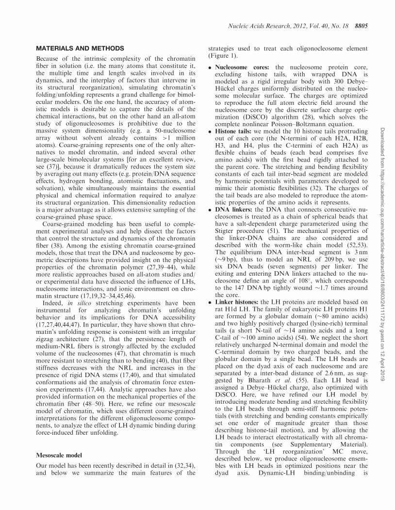
Using our mesoscale model, we analyze the unfoldingbehavior of 24-core 209-bp repeat oligonucleosomes withone LH permanently attached to each core and with LHsthat are allowed to bind/unbind to their cores with differ-ent affinities (as described in Table 1). These simulationsare performed in physiological conditions (high monova-lent salt: 0.15M NaCl, low concentration of magnesiumions, and room temperature) and are started fromconverged zigzag configurations. As reference, weperform additional simulations of 24-core 209-bp repeatarrays without LH with monovalent ions only; simula-tions analyzing the effect of the NRL, different ratios offixed LH–core bonds, and two LH/core binding affinitiesin monovalent ions only are reported elsewhere (17),where longer NRLs were shown to soften the fiber andan interesting mechanism to facilitate controlled fiber un-folding by the simultaneous presence of fast and slow LHbinding pools was described. The 209-bp repeat fiber issuitable to dissect the role of LH in chromatin unfolding;experiments (65) and modeling (17,34) have shown thatLH’s compacting and structural effect is strongest formedium-NRL arrays [medium NRLs range roughlyfrom 190 to 210 bp (34)]. In addition, the 209-bp repeatlength is common in nature and close to the value ofchicken erythrocyte chromatin used widely in manystudies [(19,21,36)]. The stretching experiment is imple-mented by fixing the position of the first nucleosomecore and applying a stretching force of up to 40 pN tothe last core (Figure 2a). Such forces mimic cellular con-straints exerted by molecular machines [e.g. RNA andDNA polymerases (66–70)].
Exploration of various LH dynamic binding scenariosIn Figure 2b, we compare the stretching behavior of chro-matin fibers without LH nor magnesium ions, with LHsand magnesium ions, and with LHs that bind/unbind dy-namically with different affinities and magnesium ions;Supplementary Figure S1 presents additional data forfibers without LH and with magnesium ions. In Table 1,we present the values for the association anddissociation-and-difussion probabilities we use to modelthe different binding affinities. Each binding regimeproduces chromatin fibers with different average LH–core bond concentrations. In the table we provide theresulting LH–core bond concentrations at zero pullingforce; for each combination of probabilities, similar con-centrations are obtained at higher forces. The cases ofPd=0 and Pa=0 reproduce fixed-LH (100% LHbound) and no-LH (0% LH bound) behavior, respectively(red and blue curves in Figure 2b). Other probability com-binations produce ensembles with intermediate concentra-tions (four other colored curves in Figure 2b). Forexample, the probability combination with Pa�Pd (i.e.Pa=1 and Pd=0.25) mimics dynamic-LH chromatinfibers where LHs exhibit effective diffusion behavior(10). This means that, following dissociation, rebindingoccurs much faster than diffusion to infinity.
Consequently, such parameters yield equilibriumensemble values of oligonucleosomes in which themajority of LH proteins (�80%) are bound to cores. Onthe other hand, probability combinations with Pa<Pd
describe a low LH-core binding affinity and yield chroma-tin fibers with a reduced number of LH–core bonds.Table 1 also indicates that the individual conformationsin the ensemble have a LH-core bond concentration thatdeviates �10% from the mean, reflecting a fluctuation inthe number of LH-core bonds consistent with dynamicregimes.
The force–extension curves in Figure 2b demonstratemarked differences between fibers with fixed LHs (redcurve), dynamic LHs (green, turquoise, purple andmagenta curves), and no LH (blue curve). Note that alldynamic-LH curves lie between the two extreme cases(fixed-LH, red and no-LH, blue). Fibers without LH(blue curve) require only 2 pN to produce a 2-fold exten-sion from their resting length (equilibrium fiber length atzero pulling force; values given in Table 1). In comparison,
0
5
10
15
20
25
30
35
40
0 100 200 300 400 500 600
For
ce (
pN)
Extension (nm)
+LH (fixed) +Mg2+; 100% LH
+LH (dyn) +Mg2+; ~80% LH
+LH (dyn) +Mg2+; ~67%LH
+LH (dyn) +Mg2+; ~50% LH
+LH (dyn) +Mg2+; ~33% LH
−LH −Mg2+; 0% LH
0 2 4 6 8 10 120
0.5
1
1.5
k − nucl. num.
I(k)
(a)
(b)
Figure 2. Effect of LH dynamic binding in the force–extensionresponse of 24-core 209-bp repeat oligonucleosomes. (a) Representationof the force–extension experiment (left) and internucleosome interactionpattern of our converged zigzag starting conformation characterized bydominant k±2 and moderate k±5 interactions (right). (b) Force–extension curves for fibers pulled below 40 pN at room temperature.Fibers with LHs (fixed or dynamic) are simulated in high monovalentsalt (0.150M NaCl) and low concentration of magnesium ions. Fiberswithout LH are simulated in high monovalent salt only. The curvelabels show the average LH–core bond concentration in each bindingregime; such concentrations are obtained with the binding probabilitiesgiven in Table 1. The force–extension curve is a plot of the magnitudeof the applied force versus the ‘end-to-end’ extension or distance (in thedirection of the stretching force) between the geometric centers of thefirst and last nucleosome cores. Error bars in the force–extension curvedenote the standard deviation from the mean of the ensemble average.Dynamic LH binding dramatically reduces LHs stiffening effect andmore so as the LH–core binding affinity decreases.
8808 Nucleic Acids Research, 2012, Vol. 40, No. 18
Dow
nloaded from https://academ
ic.oup.com/nar/article-abstract/40/18/8803/2411172 by guest on 12 April 2019

the force needed to double the resting length of fixed-LHfibers (red curve) is well above experimental values (21,26),reaching 25 pN; these fibers are extremely stiff.
By considering dynamic-LH binding/unbinding, weobserve a dramatic reduction of fiber stiffness. Theforces needed to double the resting length of fibers withdynamic LH–core bonds approach the no-LH value asthe binding affinity decreases; for example, the forces forhigh, moderate, and low affinities are 4.5, 4, and 3 pN,respectively. Even for dynamic-LH fibers with thehighest binding affinity considered (effective diffusioncase), we observe a remarkable softening with respect tofixed-LH fibers: the force needed to double their restinglength is only 6 pN (compared with 25 pN for fixed-LHspecies).
The high stiffness of fixed-LH fibers results from thepresence of magnesium ions and the formation of rigidDNA stems—209-bp chromatin with fixed-LH in mono-valent salt only doubles its resting length at a much lowervalue, 10 pN (17). Divalent ions increase chromatin stiff-ness significantly by (i) bending the DNA linkers andscreening the DNA–DNA repulsion, which allows the nu-cleosomes to come closer together, and (ii) permitting thenucleosome cores to rearrange in a wider range of con-formations that yield optimum contacts within anextended array; both effects favor strong internucleosomeinteractions and make unfolding more challenging. RigidDNA stems further screen DNA repulsion and lock thechromatin fiber in a compact form. This observationsuggests that DNA stems and divalent ions worktogether to stabilize chromatin compaction, increasingthe forces needed to unfold the fiber.
Dynamic LH with high binding affinityIn the remainder of this article, we elaborate upon thebehavior of fibers with LHs that exhibit effective diffusion(highest binding affinity) by comparing it with that offibers without LH and with fixed LHs. Hereafter, werefer to the fibers with LH molecules that display effectivediffusion as ‘dynamic-LH fibers’. Results from this regimeare the most relevant biologically because the majority ofLH molecules are bound to cores, as in vivo (10).
Softening caused by this dynamic-LH binding behaviorcan also be noted by comparing the slopes of the force–extension curves. In Figure 2b, separate regimes (forceranges), in which the force–extension curves of fixed-LH(red curve), dynamic-LH (green curve), and no-LH (bluecurve) fibers present linear extension behavior individu-ally, can be recognized. For each fiber, the force regimescan be distinguished by their different slopes; the slope
quantifies the fiber’s propensity to extend during suchregime. The forces at which these regimes occur and thecorresponding slopes of the force–extension curves areshown in Table 2.Fibers with fixed and dynamic LHs display two separate
force regimes. The slopes in both regimes are muchsmaller for dynamic-LH fibers than for the rigidfixed-LH arrays. In the first force regime, both curveshave their largest slopes, which suggests that the fibersstraighten and then open partially, maintaining theiroriginal zero-force structure. During the second forceregime, the slopes drastically decrease; a small forceincrease now produces a notable fiber extension, signalingfiber unfolding. The slope for dynamic-LH fibers in thissecond force regime is �0.03 pN/nm, in excellent agree-ment with the 0.02–0.028 pN/nm values estimated formedium-NRL chromatin with magnesium ions (26).In comparison, the force–extension curve of no-LH
fibers shows three different force regimes. Instead of aninitial straightening regime, the fiber unfolds (minimumslope) and the force–extension curve resembles aplateau; the slope in this regime is six times smaller thanthat for fixed-LH fibers (Table 2). A similar plateau at lowforces was previously reported by Cui and Bustamante(21) for medium-NRL chromatin at low salt, where aloose structure is expected. At intermediate forces, the in-creased slope suggests reorganization into a stiffer struc-ture. At high forces, nonlinear behavior due to DNAover-stretching occurs. This fiber is further analyzedin (17).To further quantify chromatin’s stiffness, we estimate
the stretching elasticity modulus s, defined as the forceneeded to produce a 2-fold extension (71). Fixed-LHfibers show a stiff elasticity modulus of s& 25 pN, closeto values calculated by Aumann et al. (40) for fibers withrigid DNA stems. In comparison, the softer dynamic-LHfibers have an elastic modulus of s& 6 pN, in agreementwith the s=5–8 pN experimental values (21,72). This re-markable stiffness reduction (by a factor of almost 5)caused by LHs dynamic binding behavior demonstratesthat LH mobility plays an important role in facilitatingforce-induced chromatin unfolding.Note that our force–extension curves mimic the
behavior of integral chromatin fibers, i.e. fibers in whichall nucleosomes remain intact; the effects of nucleosomeunwrapping and sliding are not included in our model.Experiments indicate that the presence of histonevariants and post-translational modifications throughoutthe dyad DNA entry/exit region can induce nucleosome
Table 2. Force-extension slope during two force regimes
Fiber Straightening regime Unfolding regime
Force (pN) Slope±SD (pN/nm) Force (pN) Slope±SD (pN/nm)
Dynamic LH (effective diffusion) 0–6 0.1211±0.0624 >6 0.0319±0.0089Fixed LH 0–25 0.4478±0.0966 >25 0.0611±0.0160No LH – – <4 0.01077±0.0005
Fibers with LHs were modeled in high monovalent salt and low concentration of magnesium ions. Fibers without LH were modeled in highmonovalent salt only.
Nucleic Acids Research, 2012, Vol. 40, No. 18 8809
Dow
nloaded from https://academ
ic.oup.com/nar/article-abstract/40/18/8803/2411172 by guest on 12 April 2019

unwrapping even at zero force (73). For instance, H3lysine 56 is located in the dyad region and its acetylationencourages transient nucleosome unwrapping (74). Someremodeling complexes also function by favoring nucleo-some unwrapping. For example, the SWI/SNF complex isthought to strip off a section of DNA around the dyadposition (75). It has been speculated that nucleosomeunwrapping creates a DNA bulge on the nucleosomesurface close to the dyad that propagates sliding thecores (76), and as such might increase the degree ofpositional delocalization (or ‘fuzziness’) of nucleosomesacross the genome (77). In addition, modeling ofmedium-NRL chromatin without LH and withoutdivalent ions has suggested that in native nucleosomesunwrapping becomes significant above �4.5 pN (44).We expect individual nucleosome unwrapping to
modify the electrostatic properties of the partiallyunwrapped cores. Multiple transient nucleosome unwra-ppings and enhanced fuzziness might favor fiber compac-tion and fiber stiffening. Note, however, that for theconditions analyzed here (divalent ions and LHs), the ef-fects of nucleosome unwrapping and sliding are expectedto be reduced because experiments show that unwrappingin divalent ion conditions occurs only at high forces[>15–25 pN, approximately (22,23)] and that LH-induced stems restrict nucleosome movement. Consis-tently, other experiments have also found that, in thepresence of magnesium ions, chromatin unfolds by ini-tially forming an open beads-on-a-string structure withmost internucleosome contacts broken and no unwrappednucleosomes (26).
Unfolding mechanism of chromatin with dynamic LHs
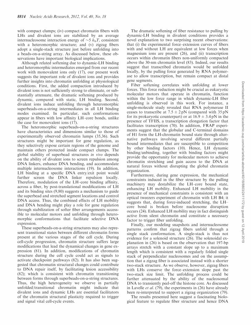
To describe how the internal organization of nucleosomesin the chromatin fiber changes with the pulling force, wecomplement the force–extension curves with an analysis ofrepresentative simulation snapshots and internucleosomeinteraction patterns (32,34) (Figure 3). These patternsmeasure the relative intensity of histone-tail mediatedinteractions between nucleosomes separated by k neigh-bors (i± k). For example, dominant i±2 contacts areindicative of a compact two-start conformation (thezigzag), while dominant i±1 and i±6 interactionssuggest a 6-nucleosomes-per-turn solenoid model (34).The combined information in Figure 3 reveals different
fiber opening mechanisms. For dynamic-LH fibers (leftpanels), we have the following steps: (i) within 0–6 pN,the fiber straightens while maintaining a zigzag organiza-tion with DNA stems (dominant i±2 and moderate i±5contacts) (ii) within 7–12 pN, individual linker-DNAstems rupture, causing significant fiber extension andyielding partially unfolded heteromorphic structures weterm ‘superbeads-on-a-string’; these structures combinestretched fiber regions with ‘superbeads’, or compactzigzag clusters, in which the nucleosomes interactstrongly with their zigzag neighbors (i±2) and moderatelywith other cores (i±1 and i±3); (iii) between 13 and16 pN, the DNA stems continue to break reducing thepresence of superbeads and their intense i±2 contacts;(iv) at 16 pN, most DNA stems have ruptured and
the fiber forms a distinctive ‘single-stack’ conformationwith dominant i±1 interactions; the nucleosomes in thisstructure are vertically aligned, but instead of stackingparallel on top of one another, they are irregularlyoriented to maximize the strength of i±1 interactions;and (v) above 16 pN, the single-stack fiber stretches toform an open beads-on-a-string conformation with negli-gible internucleosome interactions.
Unfolding of fixed-LH fibers (middle panels) proceedsthough the same five steps described above fordynamic-LH fibers. However, the forces needed toinitiate each unfolding step are much higher when LHsare fixed due to destabilization of the rigid DNA stemsand an enhanced configurational heterogeneity. Indeed,dynamic-LH DNA stems start rupturing above 6 pNcompared with fixed-LH stems at only 25 pN. Theseforces coincide with those at which the force–extensionslopes change, confirming that these slope changes signalstructural transitions.
Without LH (panels at right), the three force regimescorrespond to conformations of irregular opening,single-stack, and unfolded chain, respectively. In thesingle-stack organization (4–10 pN), the parallel alignmentof nucleosomes facilitates optimum internucleosome inter-actions and explains the higher resistance to stretching(increased slope) in this force range. Although for fiberswithout LH this transition into a single-stack conform-ation coincides with an increased force–extension slope,for fibers with dynamic and fixed LH this transitiondoes not alter the shape of the force–extension curveand is revealed only by our simulation analysis. This dif-ferent correlation between the slope change and actualconformational transition shows that the interactionsbetween immediate nucleosomes, which stabilize thesingle-stack conformation, stiffen the fiber without LHat low forces (4–10 pN) but can be ruptured easily athigher forces (16 and 36 pN for dynamic and fixed LH,respectively). Notably, these single-stack conformationsemerge for simulations started from zigzag fibers and arenot evidence of an initial solenoid fiber organization (26).This underscores the utility of modeling to extract struc-tural information from stretching experiments.
Formation of superbeads-on-a-string intermediates isobserved for all fibers in divalent conditions, regardlessof LH binding rate (see representative snapshots inSupplementary Figure S2). In contrast, in fibers withoutLH and without divalent ions, formation of such struc-tures is negligible. Stabilization of these structures requiresthe presence of LHs and divalent ions to decrease DNA–DNA repulsion, increase DNA bending, and bring thenucleosomes in closer contact inside compact clumps.This is consistent with the observation that chromatinfibers with LHs and divalent ions are heteromorphic andexhibit zigzag features of straight linker DNA combinedwith moderate DNA bending (19).
The stability of superbeads-on-a-string structures in thepresence of LHs and divalent ions is supported by severalexperimental observations. First, stable structurescombining unfolded fiber regions and small aggregatesof nucleosomes have been visualized by atomic force mi-croscopy of trypsin-digested chromatin with LHs and
8810 Nucleic Acids Research, 2012, Vol. 40, No. 18
Dow
nloaded from https://academ
ic.oup.com/nar/article-abstract/40/18/8803/2411172 by guest on 12 April 2019

magnesium ions (36). This is relevant to our study becausetrypsin digestion removes the N-terminal histone tails andtriggers an equivalent effect to that of applying a pullingforce: inducing fiber unfolding (78). Second, electron mi-croscopy of digested chromatin fibers with NRLs of 200and 212 bp, both with LHs and in high monovalent salt,has already revealed the formation of regular clumps ofnucleosomes, or superbeads, measuring 32 and 35 nm, re-spectively (35). Simple estimation in our chromatin snap-shots indicates similar dimensions (see green lines inFigure 3). Animations of the three unfolding mechanismsare available on http://www.biomath.nyu.edu/index/gallery.html.
Internucleosome interaction energy during fiber unfolding
The folded state of the chromatin fiber is stabilized byelectrostatic interactions between the positively chargedand flexible histone tails, which extend from the nucleo-some surface, and the charged surfaces of neighboringcores. During force-induced stretching, this stabilizinginternucleosome interaction energy must be overcomebefore the fiber unfolds. Figure 4 plots this energy as afunction of the pulling force. We define this energy as theaverage nucleosome/nucleosome electrostatic interactionenergy per core, considering all nucleosome charges inthe surface and histone tails.
The maximum strength of the stabilizing inter-nucleosome interaction energy occurs at zero pullingforce, when the fiber is most compact. Fibers withoutLH have a loose zero-force structure stabilized by alow internucleosome energy of �1 kbT (i.e. mean energyper core± standard deviation: �1.03±0.11 kbT). Fiberswith fixed and dynamic LHs have compact structures inthe absence of pulling force maintained by internucleo-some energies of �5 kbT (i.e. Fixed LH=�5.04±0.21kbT and dynamic LH=�4.72±0.26 kbT). Equilibriumdata in Supplementary Figure S3 confirm these compac-tion trends. Internucleosome energies of 1 kbT and 5 kbTfor loose and compact chromatin, respectively, are reason-able, considering that a value of 3 kbT has beendetermined for medium-NRL fibers with LHs in theabsence of magnesium ions, where fibers are expected tobe moderately compact (21,29). Our internucleosomeenergy for compact chromatin is weak when comparedwith the �14 kbT estimate for 197-bp fibers with LHsand magnesium ions (26). However, as recently challengedby Lavelle et al. (79), such a high internucleosome energywas derived assuming a solenoid conformation and amodel with five variable parameters. Although a high 14kbT internucleosome interaction energy is necessary to sta-bilize a solenoid arrangement with highly bent linkers andstrong DNA–DNA repulsion, a weaker 3 kbT-value canmaintain a zigzag fiber with straight linkers (29). Our
(a), (b)
(c)
Figure 3. Effect of dynamic LH binding on chromatin’s unfolding mechanism, as characterized by (a) force–extension curves, (b) patterns ofinternucleosome interactions, and (c) simulation snapshots (space filling models in which alternating nucleosomes are colored white and navy,DNA as red and LH turquoise). The three vertical panels are for 24-core 209-bp oligonucleosomes at 0.15 M monovalent salt with: dynamic LHand Mg2+ (with Pa=1 and Pd=0.25), fixed LH and Mg2+, and no LH and no Mg2+ (green, red, and blue curves in Figure 2, respectively). Thesesnapshots are presented for different force regimes: low (yellow background), moderate (blue background), high (pink background) and very high(purple background). Some longer structures are only shown partially at bottom, as denoted by three dots. Fibers with dynamic LHs unfold viasuperbeads-on-a-string (SBS) structures at much lower forces than fibers with fixed LHs.
Nucleic Acids Research, 2012, Vol. 40, No. 18 8811
Dow
nloaded from https://academ
ic.oup.com/nar/article-abstract/40/18/8803/2411172 by guest on 12 April 2019

intermediate internucleosome energy of 5 kbT agrees wellwith the compact hetermorphic chromatin structureobserved in divalent conditions, which combines straightand bent DNA linkers (19).
Comparison with previous modeling and experimentalworks
Our integrated chromatin coarse-grained model has beenextensively validated against different available experi-mental data (32,34). In past work, we have shown thatour model reproduces the following experimental results:salt-dependent sedimentation coefficients and packingratios of 12-unit oligonucleosomes of chicken erythrocytechromatin over a broad range of monovalent salt concen-trations, with/without magnesium ions, and with/withoutLHs (32); diffusion and salt-dependent behavior of mono-nucleosomes, dinucleosomes, and trinucleosomes (30);salt-dependent extension of histone tails (30); the irregularzigzag and solenoid topologies of chromatin fibers (29,34),and their enhanced compaction upon LH binding (32,34);linker crossing orientations (32); internucleosome inter-action patterns consistent with cross-linking and EM
experiments (19,34); and force–extension behavior inmonovalent salt conditions (17).
A comparison of chromatin force–extension curvesfrom different studies is complex because the results arehighly sensitive to the experimental conditions; import-antly, the many studies available analyze chromatinfibers of different characteristics (e.g. NRL, number ofnucleosomes, and LH presence/absence) and undervarying experimental conditions (e.g. ionic environmentand applied forces). Thus, no single chromatin force–extension experimental or modeling benchmark exists.Nonetheless, here we plot our results together withselected available data to interpret general trends.
In Figure 5, we compare our force–extension curveswith experimental and modeling studies of medium-NRL chromatin fibers (190–210 bp). Given that theend-to-end equilibrium extension is correlated withthe number of cores in the chromatin fiber, to facilitatethe comparison in Figure 5, we plot the force versus theextension-per-core. Considering the expected effects ofthe different conditions used in each study, the figureconfirms that our simulated curves are consistent withprevious works.
Fixed-LH curveOur stiffer fixed-LH curve lies closely below to that ofno-LH fibers modeled by Aumann et al. (40). Eventhough Aumann’s fibers were simulated in the absenceof LH (a softening factor), their relatively high stiffnesscan be explained by their much shorter NRL (190 bpversus 209 bp in our work). Decreased NRLs have beenshown to significantly stiffen the chromatin fiber(17,26,40), because they hinder reorganization intoextended arrays and favor intense internucleosome inter-actions (17).
No-LH curveAt low forces (<4 pN, see figure inset), the force–extensioncurve of our no-LH fibers lies closely above that of no-LHfibers simulated by Kepper et al. (44). This low force rangeis most relevant for analyzing unfolding behavior of ourno-LH fibers, given that all but some i±1 internucleosomeinteractions are broken above such force. Comparedwith our arrays, the Kepper fibers have a slightly shorterNRL (199 bp versus 209 bp in this work) but weresimulated at lower monovalent salt concentration(0.10M NaCl versus 0.15 M in this work); these differ-ences produce competing stiffening and softening effectsin the stretching response, and explain the similitudebetween curves. This agreement supports both modelingapproaches, which consider different approximations butnonetheless yield similar chromatin behavior. At lowforces, our no-LH curve is also consistent with the experi-mental curve for 210-bp fiber of Cui and Bustamante (21).Although Cui and Bustamante’s curve is for chromatinwith LHs, this curve is only moderately stiffer than ourno-LH result because the former was handled at lowermonovalent salt concentration (0.04M NaCl versus 0.15M in this work), which produces fiber loosening (34)similar to that observed in the absence of LH. At lowforces, our no-LH curve also runs under the no-LH
Figure 4. Top: internucleosome interaction energy per nucleosomeversus force for 24-core 209-bp oligonucleosomes at 0.15 M monova-lent salt: fixed LH and Mg2+ (red) and dynamic LH and Mg2+ (green),versus no LH and no Mg2+ (blue). Error bars denote the standarddeviation from the mean of the ensemble average. For visualizationpurposes, the low-force–low-energy region is expanded on the right.Bottom: space-filling models based on MC simulations of fibers withdynamic LHs and Mg2+ show DNA in red and alternating nucleosomesin white and blue. The zero-force energy is similar for fibers with fixedand dynamic LHs. The different force regions in which the curve withdynamic LHs has a constant slope correspond to: (1) low force wherecompact zigzag fibers form; (2) moderate force where superbeads-on-a-string arrangements occur; (3) high force where a one-start struc-ture is stable and (4) very high force where a fully extended array formswith no internucleosome interactions (zero energy).
8812 Nucleic Acids Research, 2012, Vol. 40, No. 18
Dow
nloaded from https://academ
ic.oup.com/nar/article-abstract/40/18/8803/2411172 by guest on 12 April 2019

curve of Kruithof et al. (26); this is also reasonablebecause the experimental fibers have a slightly shorterNRL (197 bp) and, unlike our no-LH fibers, weremanipulated in the presence of magnesium ions, whichinduce compact folding and fiber stiffening. Consideringthe effects of nucleosome unwrapping might be importantto model behavior of these no-LH fibers without divalentions at higher forces (>4 pN).
Dynamic-LH curveOur dynamic-LH fiber has an intermediate stiffnessbetween the extremes of fixed-LH and no-LH curves. Asexpected, it is softer than Aumann’s 190-bp fiber (40)—consistently with softening induced by longer NRLs(17,26,40)—and stiffer than the low-salt fiber of Cui andBustamante (21) and the no-LH fibers of Kepper et al.(44) and Kruithof et al. (26). Moreover, our resultsoffer an explanation to Kruithof’s observation that LHmolecules do not affect the stiffness of the chromatinfiber: as shown in Figure 2, fibers with highly mobileLH molecules (low affinity) can exhibit the same stiffness
as fibers without LH. High LH mobility is consistent withthe experiment because the strain induced in the fiber bythe stretching process can destabilize the LH–core bonds.
CONCLUSIONS
Our suggestion for a critical role for dynamic linkerhistone binding/unbinding during force-induced chroma-tin fiber unfolding at divalent ion conditions is basedon analyses of the force–extension curves, internucleo-some interaction patterns, simulation snapshots, andinternucleosome energy. These data also indicate that(i) fixed-LH DNA stems require forces >25 pN to break;(ii) dynamic-LH binding/unbinding behavior destabilizesthe rigid DNA stems, reducing dramatically chromatin’sstiffness (stretching modulus) and the forces needed toinitiate unfolding to 6 pN; (iii) in the presence ofdivalent ions and LHs, chromatin unfolding proceedsthrough irregular superbeads-on-a-string intermediatestructures that combine fully extended DNA regions
Figure 5. Comparison of our results with previous experimental (Exp.) and modeling (Mod.) force–extension curves. Bottom: force-extension curvesof the various works. Top: low-force region expanded for visualization purposes. Results from this work are shown in solid lines (red = fixed LH,green = dynamic LH with Pa=1 and Pd=0�25, blue = no LH), results from other works are shown in dashed lines. The references andexperimental/modeling conditions for each work are summarized in the legend. Data shown for other works has been taken directly from thedifferent paper figures. For comparison purposes, instead of the extension, the extension-per-core (extension divided by the total number of cores) isused.
Nucleic Acids Research, 2012, Vol. 40, No. 18 8813
Dow
nloaded from https://academ
ic.oup.com/nar/article-abstract/40/18/8803/2411172 by guest on 12 April 2019
with compact clumps; (iv) compact chromatin fibers withLHs and divalent ions are stabilized by an averageinternucleosome interaction energy of �5 kbT, consistentwith a heteromorphic structure; and (v) zigzag fibersadopt a single-stack structure just before unfolding intoa beads-on-a-string array. As discussed below, these ob-servations have important biological implications.Although related softening due to dynamic-LH binding
and heteromorphic intermediates emerged from our recentwork with monovalent ions only (17), our present worksuggests the important role of divalent ions and providesfurther insights into chromatin unfolding at physiologicalconditions. First, the added compaction introduced bydivalent ions is not sufficiently strong to eliminate, or sub-stantially attenuate, the dramatic softening produced bydynamic, compared with static, LH binding. Second,divalent ions induce unfolding through heteromorphicsuperbeads-on-a-string intermediates in all LH bindingmodes examined. In particular, such conformationsappear in fibers with low affinity LH–core bonds, unlikethe case for monovalent ions (17).The heteromorphic superbeads-on-a-string structures
have characteristics and dimensions similar to those ofexperimentally observed chromatin lumps (35,36). Suchstructures might be important for gene regulation, asthey selectively expose certain regions of the genome andmaintain others protected inside compact clumps. Theglobal stability of superbead structures in vivo dependson the ability of divalent ions to screen repulsion amongDNA linkers, enhance DNA bending, and accommodatemultiple internucleosome interactions (19). In addition,LH binding at a specific DNA entry/exit point wouldfurther screen the DNA linker repulsion locally.Therefore, modulation of the LH–core binding affinity,across a fiber, by post-translational modifications of LHand its binding sites (9,80) suggests a mechanism to guidethe superbead and stretched segment locations and controlDNA access. Thus, the combined effects of LH mobilityand DNA bending might play a role for gene regulationthrough stabilization of softer fiber arrangements access-ible to molecular motors and unfolding through hetero-morphic conformations that facilitate selective DNAexpression.These superbeads-on-a-string structures may also repre-
sent transitional states between different chromatin formspresent at the various stages of the cell cycle. Duringcell-cycle progression, chromatin structure suffers largemodifications that lead the dynamical changes in gene ex-pression (81). In addition, modifications of chromatinstructure during the cell cycle could act as signals toactivate checkpoint pathways (82). It has also been sug-gested that chromatin structural changes could contributeto DNA repair itself, by facilitating lesion accessibility(82); which is consistent with chromatin transitioningbetween forms through superbeads-on-a-string structures.Thus, the high heterogeneity we observe in partiallyunfolded/transitional chromatin might indicate thatdivalent ions and dynamic LHs are essential facilitatorsof the chromatin structural plasticity required to triggerand signal vital cell-cycle events.
The dramatic softening of fiber resistance to pulling bydynamic-LH binding in divalent conditions provides anovel explanation to two surprising recent observations:that (i) the experimental force–extension curves of fiberswith and without LH are equivalent at low forces whenmagnesium ions are present (26), and (ii) transcriptionoccurs within chromatin fibers non-uniformly compactedabove the 30-nm chromatin level (83). Indeed, our resultssuggest that transcribed chromatin would be unfoldedlocally, by the pulling force generated by RNA polymer-ase to allow transcription, but remain compact at distalgene segments.
Fiber softening correlates with unfolding at lowerforces. This force reduction might be crucial as eukaryoticmolecular motors that operate in chromatin, functionwithin the low force range in which dynamic-LH fiberunfolding is observed in this work. For instance, asingle-molecule study revealed that RNA polymerase IIceases to transcribe at 7.5±2pN (compared with 35 pNfor its prokaryotic counterpart) or at 16.9±3.4 pN in thepresence of TFIIS, a transcription elongation factor thatfacilitates transcription (70). In addition, FRAP experi-ments suggest that the globular and C-terminal domainsof H1 form the LH-chromatin bound state through alter-native pathways involving several different partiallybound intermediates that are susceptible to competitionby other binding factors (10). Hence, LH dynamicbinding/unbinding, together with binding factors mightprovide the opportunity for molecular motors to achievechromatin stretching and gain access to the DNA atnatural forces without disrupting the global chromatinorganization.
Furthermore, during gene expression, the mechanicalconstraints induced in the fiber structure by the pullingmachinery may destabilize the LH–core bound state,enhancing LH mobility. Enhanced LH mobility in thepresence of mechanical constraints is consistent with anoptical tweezers experiment of chromatin with LH B4; itsuggests that, during force-induced stretching, the LH–core bond is broken before nucleosome disruptionoccurs (24). Enhanced LH mobility may in fact distinguishactive from silent chromatin and constitute a necessaryfactor to trigger fiber unfolding.
Finally, our modeling snapshots and internucleosomepatterns confirm that zigzag fibers unfold through asingle stack conformation. A single-stack is thus notevidence for a solenoid structure (26). The solenoidal ex-planation in (26) is based on the observation that 197-bparrays stretch with a constant slope up to a maximumlength which is consistent with a regularly folded singlestack of perpendicular nucleosomes and on the assump-tion that a zigzag fiber is associated instead with a shortertwo-stack structure. As we observe, however, zigzag fiberswith LHs conserve the force–extension slope past thetwo-stack size limit. The unfolding process could befurther attenuated by the ability of the nucleosomalDNA to transiently peel-off the histone core. As discussedin Lavelle et al. (79), the experiments in (26) have alreadybeen re-interpreted to support a zigzag organization (79).
The results presented here suggest a fascinating biolo-gical feature to regulate fiber structure and hence DNA
8814 Nucleic Acids Research, 2012, Vol. 40, No. 18
Dow
nloaded from https://academ
ic.oup.com/nar/article-abstract/40/18/8803/2411172 by guest on 12 April 2019

accessibility, cell-cycle progression, and cell-cycle check-points, by divalent ions effects and a dynamic LHbinding/unbinding mechanism. The possible crucial rolethat dynamic LH binding has in facilitating transientchromatin unfolding also underscores the importance ofconsidering the highly dynamic nature of chromatin todecipher the external and internal factors that drive andalter gene expression. Recent data suggest that the pullingforce created during transcription may organize the globalarchitecture of an extended chromosomal domain in situ(84). Other architectural factors may include HMGproteins that accelerate LH mobility (9) or chromatin-condensing proteins such as HP1 (85) and MENT (86)that may polymerize on the nucleosomes to inhibit tran-sient unfolding of the chromatin fiber. Together with othervariables that affect chromatin compaction and configur-ational transitions, such proteins suggest how small localdifferences can have profound global effects.
SUPPLEMENTARY DATA
Supplementary Data are available at NAR Online:Supplementary Table 1, Supplementary Figures 1–3 andSupplementary Methods.
ACKNOWLEDGEMENTS
The authors thank Dr Sergei Grigoryev for his invaluablecomments and insights concerning this work. Computingsupport from the NYU HPC USQ and Cardiac clusters isalso acknowledged.
FUNDING
National Science Foundation [MCB-0316771 to T.S.];National Institutes of Health (NIH) [R01 GM55164 toT.S.]; American Chemical Society [PRF39225-AC4];Petroleum Research Fund (to T.S.); Philip Morris USA(to T.S.); Philip Morris International (to T.S.);Schlumberger Faculty for the Future Program (to R.C.-G.).Funding for open accees charge: National ScienceFoundation [MCB-0316771 to T.S.]; NIH [R01GM55164 to T.S.].
Conflict of interest statement. None declared.
REFERENCES
1. Finch,J.T. and Klug,A. (1976) Solenoidal model forsuperstructure in chromatin. Proc. Natl Acad. Sci. USA, 73,1897–1901.
2. Worcel,A., Strogatz,S. and Riley,D. (1981) Structure of chromatinand the linking number of DNA. Proc. Natl Acad. Sci. USA, 78,1461–1465.
3. Woodcock,C.L., Frado,L.L. and Rattner,J.B. (1984) Thehigher-order structure of chromatin: evidence for a helical ribbonarrangement. J. Cell Biol., 99, 42–52.
4. McBryant,S.J., Lu,X. and Hansen,J.C. (2010) Multifunctionalityof the linker histones: an emerging role for protein-proteininteractions. Cell Res., 20, 519–528.
5. Misteli,T., Gunjan,A., Hock,R., Bustin,M. and Brown,D.T. (2000)Dynamic binding of histone H1 to chromatin in living cells.Nature, 408, 877–881.
6. Lever,M.A., Th’ng,J.P., Sun,X. and Hendzel,M.J. (2000) Rapidexchange of histone H1.1 on chromatin in living human cells.Nature, 408, 873–876.
7. Caron,F. and Thomas,J.O. (1981) Exchange of histone H1between segments of chromatin. J. Mol. Biol., 146, 513–537.
8. Brown,D.T. (2003) Histone H1 and the dynamic regulation ofchromatin function. Biochem. Cell Biol., 81, 221–227.
9. Catez,F., Ueda,T. and Bustin,M. (2006) Determinants of histoneH1 mobility and chromatin binding in living cells. Nat. Struct.Mol. Biol., 13, 305–310.
10. Stasevich,T.J., Mueller,F., Brown,D.T. and McNally,J.G. (2010)Dissecting the binding mechanism of the linker histone inlive cells: an integrated FRAP analysis. EMBO J., 29,1225–1234.
11. Caterino,T.L., Fang,H. and Hayes,J.J. (2011) Nucleosome linkerDNA contacts and induces specific folding of the intrinsicallydisordered H1 carboxyl terminal domain. Mol. Cell. Biol., 31,2341–2348.
12. Happel,N. and Doenecke,D. (2008) Histone H1 and its isoforms:Contribution to chromatin structure and function. Gene, 431,1–12.
13. Zlatanova,J., Caiafa,P. and van Holde,K. (2000) Linker histonebinding and displacement: versatile mechanism for transcriptionalregulation. FASEB J., 14, 1697–1704.
14. Yellajoshyula,D. and Brown,D.T. (2006) Global modulation ofchromatin dynamics mediated by dephosphorylation of linkerhistone H1 is necessary for erythroid differentiation. Proc. NatlAcad. Sci. USA, 103, 18568–18573.
15. Meshorer,E., Yellajoshula,D., George,E., Scambler,P.J.,Brown,D.T. and Misteli,T. (2006) Hyperdynamic plasticity ofchromatin proteins in pluripotent embryonic stem cells. Dev. Cell,10, 105–116.
16. Dou,Y., Bowen,J., Liu,Y. and Gorovsky,M.A. (2002)Phosphorylation and an ATP-dependent process increase thedynamic exchange of H1 in chromatin. J. Cell Biol., 158,1161–1170.
17. Collepardo-Guevara,R. and Schlick,T. (2011) The effect of linkerhistone’s nucleosome binding affinity on chromatin unfoldingmechanisms. Biophys. J., 101, 1670–1680.
18. Carrero,G., Crawford,E., Hendzel,M.J. and de Vries,G. (2004)Characterizing fluorescence recovery curves for nuclearproteins undergoing binding events. Bull. Math. Biol., 66,1515–1545.
19. Grigoryev,S.A., Arya,G., Correll,S., Woodcock,C.L. andSchlick,T. (2009) Evidence for heteromorphic chromatin fibersfrom analysis of nucleosome interactions. Proc. Natl Acad. Sci.USA, 106, 13317–13322.
20. Leuba,S.H., Yang,G., Robert,C., Samori,B., van Holde,K.,Zlatanova,J. and Bustamante,C. (1994) Three-dimensionalstructure of extended chromatin fibers as revealed bytapping-mode scanning force microscopy. Proc. Natl Acad. Sci.USA, 91, 11621–11625.
21. Cui,Y. and Bustamante,C. (2000) Pulling a single chromatin fiberreveals the forces that maintain its higher-order structure. Proc.Natl Acad. Sci. USA, 97, 127–132.
22. Bennink,M.L., Leuba,S.H., Leno,G.H., Zlatanova,J., deGrooth,B.G. and Greve,J. (2001) Unfolding individualnucleosomes by stretching single chromatin fibers with opticaltweezers. Nat. Struct. Biol0, 8, 606–610.
23. Brower-Toland,B.D., Smith,C.L., Yeh,R.C., Lis,J.T.,Peterson,C.L. and Wang,M.D. (2002) Mechanical disruption ofindividual nucleosomes reveals a reversible multistage release ofDNA. Proc. Natl Acad. Sci. USA, 99, 1960–1965.
24. Pope,L.H., Bennink,M.L., van Leijenhorst-Groener,K.A.,Nikova,D., Greve,J. and Marko,J.F. (2005) Single chromatin fiberstretching reveals physically distinct populations of disassemblyevents. Biophys. J., 88, 3572–3583.
25. Roopa,T. and Shivashankar,G.V. (2006) Direct measurement oflocal chromatin fluidity using optical trap modulation forcespectroscopy. Biophys. J., 91, 4632–4637.
26. Kruithof,M., Chien,F.-T., Routh,A., Logie,C., Rhodes,D. and vanNoort,J. (2009) Single-molecule force spectroscopy reveals ahighly compliant helical folding for the 30-nm chromatin fiber.Nat. Struct. Mol. Biol., 16, 534–540.
Nucleic Acids Research, 2012, Vol. 40, No. 18 8815
Dow
nloaded from https://academ
ic.oup.com/nar/article-abstract/40/18/8803/2411172 by guest on 12 April 2019

27. Katritch,V., Bustamante,C. and Olson,W.K. (2000) Pullingchromatin fibers: computer simulations of direct physicalmicromanipulations. J. Mol. Biol., 295, 29–40.
28. Zhang,Q., Beard,D.A. and Schlick,T. (2003) Constructingirregular surfaces to enclose macromolecular complexes formesoscale modeling using the Discrete Surface ChargeOptimization (DiSCO) algorithm. J. Compt. Chem., 24,2063–2074.
29. Arya,G. and Schlick,T. (2006) Role of histone tails in chromatinfolding revealed by a mesoscopic oligonucleosome model. Proc.Natl Acad. Sci. USA, 103, 16236–16241.
30. Arya,G., Zhang,Q. and Schlick,T. (2006) Flexible histone tails ina new mesoscopic oligonucleosome model. Biophys. J., 91,133–150.
31. Arya,G. and Schlick,T. (2007) Efficient global biopolymersampling with end-transfer configurational bias Monte Carlo.J. Chem. Phys., 126, 044107.
32. Arya,G. and Schlick,T. (2009) A tale of tails: How histone tailsmediate chromatin compaction in different salt and linker histoneenvironments. J. Phys. Chem. A, 113, 4045–4059.
33. Schlick,T. and Perisic,O. (2009) Mesoscale simulations of twonucleosome-repeat length oligonucleosomes. Phys. Chem. Chem.Phys., 11, 10729–10737.
34. Perisic,O., Collepardo-Guevara,R. and Schlick,T. (2010) Modelingstudies of chromatin fiber structure as a function of DNA linkerlength. J. Mol. Biol., 403, 777–802.
35. Zentgraf,H. and Franke,W.W. (1984) Differences ofsupranucleosomal organization in different kinds of chromatin:cell type-specific globular subunits containing different numbers ofnucleosomes. J. Cell. Biol., 99, 272–286.
36. Cano,S., Caravaca,J.M., Martın,M. and Daban,J.-R. (2006)Highly compact folding of chromatin induced by cellular cationconcentrations. Evidence from atomic force microscopy studies inaqueous solution. Eur. Biophys. J., 35, 495–501.
37. Hyeon,C. and Thirumalai,D. (2011) Capturing the essence offolding and functions of biomolecules using coarse-grainedmodels. Nat. Commun., 2, 1–11.
38. Schlick,T., Hayes,J.J. and Grigoryev,S. (2012) Towardconvergence of experimental studies and theoretical modeling ofthe chromatin fiber. J. Biol. Chem., 287, 5183–5191.
39. Woodcock,C.L., Grigoryev,S.A., Horowitz,R.A. andWhitaker,N.A. (1993) Chromatin folding model that incorporateslinker variability generates fibers resembling the native structures.Proc. Natl Acad. Sci. USA, 90, 9021–9025.
40. Aumann,F., Lankas,F., Caudron,M. and Langowski,J. (2006)Monte Carlo simulation of chromatin stretching. Phys. Rev. E,73, 041927.
41. Wu,C., Bassett,A. and Travers,A. (2007) A variable topology forthe 30-nm chromatin fibre. EMBO Rep., 8, 1129–1134.
42. Kepper,N., Foethke,D., Stehr,R., Wedemann,G. and Rippe,K.(2008) Nucleosome geometry and internucleosomal interactionscontrol the chromatin fiber conformation. Biophys. J., 95,3692–3705.
43. Aumann,F., Suhnel,J., Langowski,J. and Diekmann,S. (2010)Rigid assembly and Monte Carlo models of stable and unstablechromatin structures: the effect of nucleosomal spacing. Theor.Chem. Acc., 125, 217–231.
44. Kepper,N., Ettig,R., Stehr,R., Marnach,S., Wedemann,G. andRippe,K. (2011) Force spectroscopy of chromatin fibers:extracting energetics and structural information from MonteCarlo simulations. Biopolymers, 95, 435–447.
45. Mozziconacci,J., Wong,H. and Victor,J.M. (2007) All-atom modelof the chromatin fiber containing linker histones: a versatilestructure tuned by the repeat length. Febs. J., 274, 78.
46. Voltz,K., Trylska,J., Tozzini,V., Kurkal-Siebert,V., Langowski,J.and Smith,J. (2008) Coarse-grained force field for thenucleosome from self-consistent multiscaling. J. Comp. Chem., 29,1429–1439.
47. Mergell,B., Everaers,R. and Schiessel,H. (2004) Nucleosomeinteractions in chromatin: fiber stiffening and hairpin formation.Phys. Rev. E., 70, 011915.
48. Schiessel,H., Gelbart,W.M. and Bruinsma,R. (2001) DNA folding:Structural and mechanical properties of the two-angle model forchromatin. Biophys. J., 80, 1940–1956.
49. Ben-Haım,E., Lesne,A. and Victor,J.-M. (2001) Chromatin: atunable spring at work inside chromosomes. Phys. Rev. E., 64,051921.
50. Stehr,R., Kepper,N., Rippe,K. and Wedemann,G. (2008) Theeffect of internucleosomal interaction on folding of the chromatinfiber. Biophys. J., 95, 3677–3691.
51. Stigter,D. (1977) Interactions of highly charged colloidal cylinderswith applications to double-stranded. Biopolymers, 16, 1435–1448.
52. Allison,S., Austin,R. and Hogan,M. (1989) Bending and twistingdynamics of short linear DNAs. Analysis of the triplet anisotropydecay of a 209 base pair fragment by Brownian simulation. J.Chem. Phys., 90, 3843–3854.
53. Heath,P.J., Gebe,J.A., Allison,S.A. and Schurr,J.M. (1996)Comparison of analytical theory with Brownian dynamicssimulations for small linear and circular DNAs. Macromolecules,29, 3583–3596.
54. Allan,J., Staynov,D. and Gould,H. (1980) Reversible dissociationof linker histone from chromatin with preservation ofinternucleosomal repeat. Proc. Natl Acad. Sci. USA, 77, 885–889.
55. Bharath,M.M., Chandra,N.R. and Rao,M.R. (2002) Prediction ofan HMG-box fold in the C-terminal domain of histone H1:insights into its role in DNA condensation. Proteins, 49, 71–81.
56. Korolev,N., Yanping,F., Lyubartsev,A.P. and Nordenskiold,L.(2012) Modelling chromatin structure and dynamics: Status andprospects. Curr. Opio. Struct. Biol., 22, 1–9.
57. Baneres,J., Martin,A. and Parello,J. (1997) The N tails ofhistones H3 and H4 adopt a highly structured conformation inthe nucleosome. J. Mol. Biol., 273, 503–508.
58. Wang,X., Moore,S.C., Laszckzak,M. and Ausi,J. (2000) Proteinsynthesis post-translation modification and degradation. J. Biol.Chem., 275, 35013–35020.
59. Kato,H., Gruschus,J., Ghirlando,R., Tjandra,N. and Bai,Y.(2009) Characterization of the N-terminal tail domain of histoneH3 in condensed nucleosome arrays by hydrogen exchange andNMR. J. Am. Chem. Soc., 131, 15104–15105.
60. Potoyan,D.A. and Papoian,G.A. (2011) Energy landscape analysesof disordered histone tails reveal special organization of theirconformational dynamics. J. Am. Chem. Soc., 133, 7405–7415.
61. Frenkel,D., Mooij,G.C.A. and Smit,B. (1992) Novel scheme tostudy structural and thermal-properties of continuouslydeformable molecules. J. Phy. Condens. Matte., 4, 3053–3076.
62. de Pablo,J.J., Laso,M. and Suter,U.W. (1992) Simulation ofpolyethylene above and below the melting point. J. Chem. Phys.,96, 2395–2403.
63. Rosenbluth,M.N. and Rosenbluth,A.W. (1955) Monte Carlocalculation of the average extension of molecular chains.J. Chem. Phys., 23, 356–359.
64. Th’ng,R., Sung,M.Y. and Hendzel,M.J. (2005) H1 family histonesin the nucleus. Control of binding and localization by theC-terminal domain. J. Biol. Chem., 280, 27809–27814.
65. Routh,A., Sandin,S. and Rhodes,D. (2008) Nucleosome repeatlength and linker histone stoichiometry determine chromatin fiberstructure. Proc. Natl Acad. Sci. USA, 105, 8872–8877.
66. Yin,H., Wang,M.D., Svoboda,K., Landick,R., Block,S.M. andGelles,J. (1995) Transcription against an applied force. Science,270, 1653–1657.
67. Wang,M.D., Schnitzer,M.J., Yin,H., Landick,R., Gelles,J. andBlock,S.M. (1998) Force and velocity measured for singlemolecules of RNA polymerase. Science, 282, 902–907.
68. Wuite,G.J., Smith,S.B., Young,M., Kelle,D. and Bustamante,C.(2000) Single-molecule studies of the effect of template tension onT7 DNA polymerase activity. Nature, 404, 103–106.
69. Maier,B., Bensimon,D. and Croquette,V. (2000) Replication by asingle DNA polymerase of a stretched single-stranded DNA.Proc. Natl Acad. Sci. USA, 97, 12002–12007.
70. Galburt,E.A., Grill,S.W., Wiedmann,A., Lubkowska,L., Choy,J.,Nogales,E., Kashlev,M. and Bustamante,C. (2007) Backtrackingdetermines the force sensitivity of RNAP II in a factor-dependentmanner. Nature, 446, 820–823.
71. Smith,S.B., Cui,Y. and Bustamante,C. (1996) OverstretchingB-DNA: the elastic response of individual double-stranded andsingle-stranded. Science, 271, 795–799.
72. Bancaud,A., Conde e Silva,N., Barbi,M., Wagner,G.,Allemand,J.F., Mozziconacci,J., Lavelle,C., Croquette,V.,
8816 Nucleic Acids Research, 2012, Vol. 40, No. 18
Dow
nloaded from https://academ
ic.oup.com/nar/article-abstract/40/18/8803/2411172 by guest on 12 April 2019
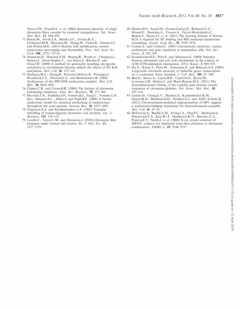
Victor,J.M., Prunell,A. et al. (2006) Structural plasticity of singlechromatin fibers revealed by torsional manipulation. Nat. Struct.Mol. Biol., 13, 444–450.
73. Simon,M., North,J.A., Shimko,J.C., Forties,R.A.,Ferdinand,M.B., Manohar,M., Zhang,M., Fishel,R., Ottesen,J.J.and Poirier,M.G. (2011) Histone fold modifications controlnucleosome unwrapping and disassembly. Proc. Natl Acad. Sci.USA, 108, 12711–12716.
74. Neumann,H., Hancock,S.M., Buning,R., Routh,A., Chapman,L.,Somers,J., Owen-Hughes,T., van Noort,J., Rhodes,D. andChin,J.W. (2009) A method for genetically installing site-specificacetylation in recombinant histones defines the effects of H3 K56acetylation. Mol. Cell, 36, 153–163.
75. Dechassa,M.L., Zhang,B., Horowitz-Scherer,R., Persinger,J.,Woodcock,C.L., Peterson,C.L. and Bartholomew,B. (2008)Architecture of the SWI/SNF-nucleosome complex. Mol. Cell.Biol., 28, 6010–6021.
76. Clapier,C.R. and Cairns,B.R. (2009) The biology of chromatinremodeling complexes. Annu. Rev. Biochem., 78, 273–304.
77. Mavrich,T.N., Ioshikhes,I.P., Venters,B.J., Jiang,C., Tomsho,L.P.,Qi,J., Schuster,S.C., Albert,I. and Pugh,B.F. (2008) A barriernucleosome model for statistical positioning of nucleosomesthroughout the yeast genome. Genome Res., 18, 1073–1083.
78. Grigoryev,S.A. and Krasheninnikov,I.A. (1982) Transientunfolding of trypsin-digested chromatin core particles. Eur. J.Biochem., 129, 119–125.
79. Lavelle,C., Victor,J.-M. and Zlatanova,J. (2010) Chromatin fiberdynamics under tension and torsion. Int. J. Mol. Sci., 11,1557–1579.
80. Shukla,M.S., Syed,S.H., Goutte-Gattat,D., Richard,J.L.C.,Montel,F., Hamiche,A., Travers,A., Faivre-Moskalenko,C.,Bednar,J., Hayes,J.J. et al. (2011) The docking domain of histoneH2A is required for H1 binding and RSC-mediated nucleosomeremodeling. Nucleic Acids Res., 39, 2559–2570.
81. Cremer,T. and Cremer,C. (2001) Chromosome territories, nucleararchitecture and gene regulation in mammalian cells. Nat. Rev.Genet., 2, 292–301.
82. Koundrioukoff,S., Polo,S. and Almouzni,G. (2004) Interplaybetween chromatin and cell cycle checkpoints in the context ofATR/ATM-dependent checkpoints. DNA Repair, 3, 969–978.
83. Hu,Y., Kireev,I., Plutz,M., Ashourian,N. and Belmont,A.S. (2009)Large-scale chromatin structure of inducible genes: transcriptionon a condensed, linear template. J. Cell. Biol., 185, 87–100.
84. Bau,D., Sanya,A., Lajoie,B.R., Capriotti,E., Byron,M.,Lawrence,J.B., Dekker,J. and Marti-Renom,M.A. (2011) Thethree-dimensional folding of the a-globin gene domain revealsformation of chromatin globules. Nat. Struct. Mol. Biol., 18,107–114.
85. Canzio,D., Chang,E.Y., Shankar,S., Kuchenbecker,K.M.,Simon,M.D., Madhani,H.D., Narlikar,G.J. and ADN Al-Sady,B.(2011) Chromodomain-mediated oligomerization of HP1 suggestsa nucleosome-bridging mechanism for heterochromatin assembly.Mol. Cell, 41, 67–81.
86. McGowan,S., Buckle,A.M., Irving,J.A., Ong,P.C., Bashtannyk-Puhalovich,T.A., Kan,W.-T., Henderson,K.N., Bulynko,Y.A.,Popova,E.Y., Smith,I. et al. (2006) X-ray crystal structure ofMENT: evidence for functional loop-sheet polymers in chromatincondensation. EMBO J., 25, 3144–3155.
Nucleic Acids Research, 2012, Vol. 40, No. 18 8817
Dow
nloaded from https://academ
ic.oup.com/nar/article-abstract/40/18/8803/2411172 by guest on 12 April 2019