Embed Size (px)
Citation preview
1998 4: 1455-1466 RNA A. Montpetit, C. Payant, J. M. Nolan and L. Brakier-Gingras
crosslinkingcoli16S ribosomal RNA using site-directed photoaffinity Analysis of the conformation of the 3' major domain of Escherichia
serviceEmail alerting
click heretop right corner of the article or Receive free email alerts when new articles cite this article - sign up in the box at the
Notes
http://www.rnajournal.org/subscriptions/ go to: RNATo subscribe to
© 1998 RNA Society
on February 14, 2006 www.rnajournal.orgDownloaded from

Analysis of the conformation of the 3 9 majordomain of Escherichia coli 16S ribosomal RNAusing site-directed photoaffinity crosslinking
ALEXANDRE MONTPETIT, 1 CATHERINE PAYANT, 1 JAMES M. NOLAN, 2
and LÉA BRAKIER-GINGRAS 1
1Département de Biochimie, Université de Montréal, Montréal, Québec H3T 1J4, Canada2Department of Biochemistry, Tulane University Medical Center, New Orleans, Louisiana 70112, USA
ABSTRACT
The 39 major domain of Escherichia coli 16S rRNA, which occupies the head of the small ribosomal subunit, isinvolved in several functions of the ribosome. We have used a site-specific crosslinking procedure to gain furtherinsights into the higher-order structure of this domain. Circularly permuted RNAs were used to introduce an azi-dophenacyl group at specific positions within the 3 9 major domain. Crosslinks were generated in a high-ionic strengthbuffer that has been used for ribosome reconstitution studies and so enables the RNA to adopt a structure recognizedby ribosomal proteins. The crosslinking sites were identified by primer extension and confirmed by assessing themobility of the crosslinked RNA lariats in denaturing polyacrylamide gels. Eight crosslinks were characterized.Among them, one crosslink demonstrates that helix 28 is proximal to the top of helix 34, and two others show that the1337 region, located in an internal loop at the junction of helices 29, 30, 41, and 42, is proximal to the center of helix 30and to a segment connecting helix 28 to helix 29. These relationships of vicinity have previously been observed innative 30S subunits, which suggests that the free domain adopts a conformation similar to that within the 30S subunit.Furthermore, crosslinks were obtained in helix 34, which suggest that the upper and lower portions of this helix arein close proximity.
Keywords: 16S ribosomal RNA; photoaffinity crosslinking; RNA structure
INTRODUCTION
Ribosomal RNA plays a major role in protein synthesis,and a detailed knowledge of its structure is required toelucidate the mechanisms underlying its function+ How-ever, the size of the large rRNAs has so far precludedthe use of nuclear magnetic resonance spectroscopy(NMR) to solve their structure (reviewed in Green &Noller, 1997)+ Crystals of the entire ribosomes and ri-bosomal subunits have been obtained, but the crystalstructures have not yet been solved at atomic resolu-tion (Thygesen et al+, 1996; Ban et al+, 1998 and ref-erences therein)+
A general property of large RNAs is that they can bedissected into smaller domains that conserve the abil-ity to fold correctly+ This reductionist approach has re-cently been applied to analyze the conformation of avariety of RNA domains with NMR or X-ray crystallog-
raphy+ As examples, structures have been determinedby NMR for the decoding center of 16S rRNA com-plexed with an aminoglycoside antibiotic (Fourmyet al+, 1996), for two important regions of 23S rRNA(the a-sarcin loop (Szewczak & Moore, 1995) and the2250 hairpin loop, involved in peptidyl-tRNA binding(Viani-Puglisi et al+, 1997)), and for loops D and E of 5SrRNA (Dallas & Moore, 1997)+ Crystal structures havebeen determined for the P4–P6 domain of group I in-tron (Cate et al+, 1996), the largest RNA yet crystallizedwith 160 nt, and for fragment I of 5S rRNA (Correllet al+, 1997)+
Our goal is to characterize spatial relationships inEscherichia coli 16S rRNA+ It has been shown that, invitro, in high-ionic strength reconstitution buffer (20 mMMg21, 300 mM K1), each of the three domains thatconstitute this RNA (59, central and 39) can interact withthe specific subset of proteins with which they interactwhen they are part of the intact 16S molecule (Weitz-mann et al+, 1993; Samaha et al+, 1994;Agalarov et al+,1998)+ This implies that each domain can fold indepen-dently of the rest of the molecule+
Reprints requests to: Léa Brakier-Gingras, Département de Bio-chimie, Université de Montréal, 2900 blvd Edouard-Montpetit, Mon-tréal, Québec H3T 1J4, Canada; e-mail: gingras@bcm+umontreal+ca
RNA (1998), 4:1455–1466+ Cambridge University Press+ Printed in the USA+Copyright © 1998 RNA Society+
1455
on February 14, 2006 www.rnajournal.orgDownloaded from
We have decided to investigate the conformation ofthe 39 major domain of 16S rRNA in ribosome recon-stitution buffer, in the absence of ribosomal proteins+The 39 major domain is involved in several functions ofthe ribosome (reviewed by Zimmermann, 1996)+ It in-teracts with mRNA (Juzumiene et al+, 1995; Sergievet al+, 1997), initiation factor IF2 (Wakao et al+, 1991),and elongation factor G (Wilson & Noller, 1998)+ He-lix 34, in the upper part of the domain, contains thebinding site of the antibiotic spectinomycin, an inhibitorof translocation (Makosky & Dahlberg, 1987; Bilginet al+, 1990; Brink et al+, 1994), it interacts with thetermination factors (Murgola, 1996; Arkov et al+, 1998),and it is involved in subunit association (Herr et al+,1979; Baudin et al+, 1989)+ Furthermore, mutations inhelix 34 were found to affect translational accuracy(Moine & Dahlberg, 1994; O’Connor et al+, 1997)+ Thelower part of the domain contains nucleotides whichare involved in the binding of tRNAs at the P site(Moazed & Noller, 1990; von Ahsen & Noller, 1995;Joseph et al+, 1997), and which have been crosslinkedto the A-, P- and E-site-bound tRNAs (Döring et al+,1994)+ Tetracycline, an inhibitor of tRNA binding to theA site, also interacts with this part of the domain (Oehleret al+, 1997)+
To study the conformation of the large rRNA do-mains, indirect techniques, such as chemical probingand crosslinking, can provide useful information sinceNMR and X-ray crystallography are still inadequate+Random crosslinking has been extensively used to studythe higher-order structure of rRNAs (Wilms et al+, 1997;reviewed in Baranov et al+, 1998b)+ Site-directed cross-linking has been used by the groups of Brimacombeand Cooperman but results are still scarce (Alexander& Cooperman, 1998 and references therein; Baranovet al+, 1998a)+ The technique developed by Brima-combe and his coworkers involves the site-specific frag-mentation of 16S rRNA with RNase H and the additionof a photoreactive nucleotide at the 39 end of the 59fragment+ Its use is limited by the capacity of the twofragments to be reconstituted into a 30S subunit+ Theprocedure of the Cooperman group involves the hy-bridization to a specific portion of rRNA within the ribo-some of a complementary photolabeled DNA probe+However, this technique requires the rRNA portion thatis probed to be exposed and single-stranded, and,more-over, the binding of the probe itself can induce confor-mational rearrangements+ Pace and his coworkers havedeveloped a site-specific crosslinking procedure thatwas successfully applied to study the conformation ofRNase P RNA (Nolan et al+, 1993; Harris et al+, 1994,1997)+ It utilizes circularly permuted RNAs, in which thenative 59 and 39 extremities are joined by a syntheticoligonucleotide linker and novel 59 and 39 extremitiesare relocated in the interior of the sequence+ The novel59 termini are modified by coupling to a photoaffinitycrosslinking agent, the p-azidophenacyl (APA) bro-
mide+ This method offers the advantage that it enablesus to probe the three-dimensional environment of ev-ery G in any RNA molecule+ It is imperative, however,to ensure that the discontinuities in the sugar–phosphatebackbone that are introduced between the novel 39 and59 ends of the circularly permuted RNAs do not perturbthe conformation of the molecule+
In this study, in order to refine our understanding ofthe higher-order structure of the 39 major domain, wehave utilized the site-directed crosslinking proceduredeveloped by Pace and his coworkers+ Seven siteswere selected for the introduction of the crosslinkingagent: G993, G1047, and G1193 (located in the upperhalf of the domain, in and around helix 34), and G1309,G1337, G1343, and G1353, in the lower half of thedomain+ The results presented here will be discussedwith respect to their agreement with current models of16S RNA structure+
RESULTS
Construction and reassembly of circularlypermuted RNAs of the 3 9 majordomain of 16S rRNA
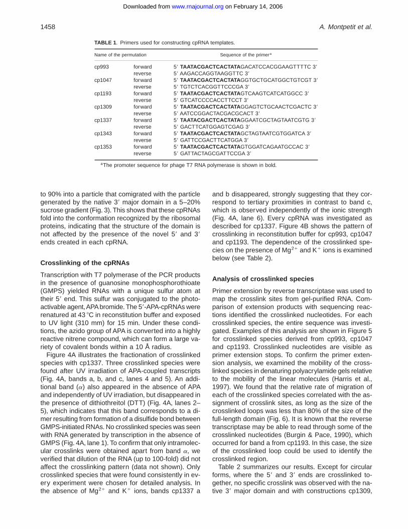
Plasmid pFD33, which contains duplicate copies of thesequence coding for the 39 major domain of E. coli 16SrRNA, was constructed by a three-fragment ligation asdescribed in Materials and Methods and Figure 1+ Poly-merase chain reaction (PCR) amplification of pFD33with appropriate primers (see Table 1) resulted in theproduction of templates coding for the circularly per-muted RNAs (cpRNAs) under control of a T7 promoter+In the corresponding transcripts, positions 921 and 1396of helix 28 are joined by a terminal GCAA tetraloop+Seven new locations for the 59 endpoint in the RNAwere investigated (Fig+ 2)+
To ascertain that the introduction of discontinuities atdifferent positions within the 39 major domain did notperturb its conformation, we verified that the differentcpRNAs were able to assemble into ribonucleoproteinparticles when incubated with a mixture of 30S ribo-somal proteins+ As mentioned in the Introduction,Samaha et al+ (1994) previously showed that the com-plete 39 domain, including the major and minor por-tions, could fold independently of the rest of the RNAmolecule and assemble with specific ribosomal pro-teins into a ribonucleoprotein particle, when incubatedwith a mixture of 30S ribosomal proteins in reconstitu-tion buffer (20 mM Mg21, 300 mM K1)+We first showedthat the native 39 major domain alone behaved exactlyas the complete 39 domain and interacted with the samesubset of proteins (data not shown)+ We next system-atically assessed the capacity of the different cpRNAsto assemble into ribonucleoprotein particles, and foundthat all the cpRNAs analyzed behaved as the native 39major domain, assembling with an efficiency superior
1456 A. Montpetit et al.
on February 14, 2006 www.rnajournal.orgDownloaded from
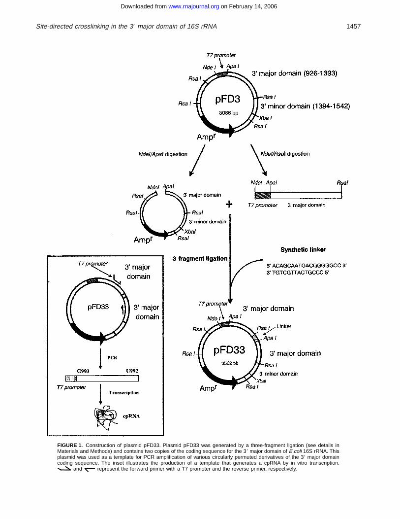
FIGURE 1. Construction of plasmid pFD33+ Plasmid pFD33 was generated by a three-fragment ligation (see details inMaterials and Methods) and contains two copies of the coding sequence for the 39 major domain of E.coli 16S rRNA+ Thisplasmid was used as a template for PCR amplification of various circularly permuted derivatives of the 39 major domaincoding sequence+ The inset illustrates the production of a template that generates a cpRNA by in vitro transcription+
and represent the forward primer with a T7 promoter and the reverse primer, respectively+
Site-directed crosslinking in the 39 major domain of 16S rRNA 1457
on February 14, 2006 www.rnajournal.orgDownloaded from
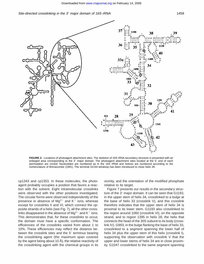
to 90% into a particle that comigrated with the particlegenerated by the native 39 major domain in a 5–20%sucrose gradient (Fig+ 3)+ This shows that these cpRNAsfold into the conformation recognized by the ribosomalproteins, indicating that the structure of the domain isnot affected by the presence of the novel 59 and 39ends created in each cpRNA+
Crosslinking of the cpRNAs
Transcription with T7 polymerase of the PCR productsin the presence of guanosine monophosphorothioate(GMPS) yielded RNAs with a unique sulfur atom attheir 59 end+ This sulfur was conjugated to the photo-activable agent,APAbromide+The 59-APA-cpRNAs wererenatured at 43 8C in reconstitution buffer and exposedto UV light (310 mm) for 15 min+ Under these condi-tions, the azido group of APA is converted into a highlyreactive nitrene compound, which can form a large va-riety of covalent bonds within a 10 Å radius+
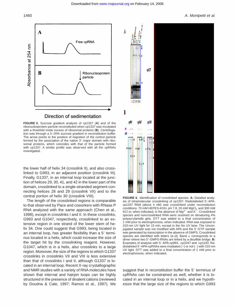
Figure 4A illustrates the fractionation of crosslinkedspecies with cp1337+ Three crosslinked species werefound after UV irradiation of APA-coupled transcripts(Fig+ 4A, bands a, b, and c, lanes 4 and 5)+ An addi-tional band (a) also appeared in the absence of APAand independently of UV irradiation, but disappeared inthe presence of dithiothreitol (DTT) (Fig+ 4A, lanes 2–5), which indicates that this band corresponds to a di-mer resulting from formation of a disulfide bond betweenGMPS-initiated RNAs+No crosslinked species was seenwith RNA generated by transcription in the absence ofGMPS (Fig+ 4A, lane 1)+ To confirm that only intramolec-ular crosslinks were obtained apart from band a, weverified that dilution of the RNA (up to 100-fold) did notaffect the crosslinking pattern (data not shown)+ Onlycrosslinked species that were found consistently in ev-ery experiment were chosen for detailed analysis+ Inthe absence of Mg21 and K1 ions, bands cp1337 a
and b disappeared, strongly suggesting that they cor-respond to tertiary proximities in contrast to band c,which is observed independently of the ionic strength(Fig+ 4A, lane 6)+ Every cpRNA was investigated asdescribed for cp1337+ Figure 4B shows the pattern ofcrosslinking in reconstitution buffer for cp993, cp1047and cp1193+ The dependence of the crosslinked spe-cies on the presence of Mg21 and K1 ions is examinedbelow (see Table 2)+
Analysis of crosslinked species
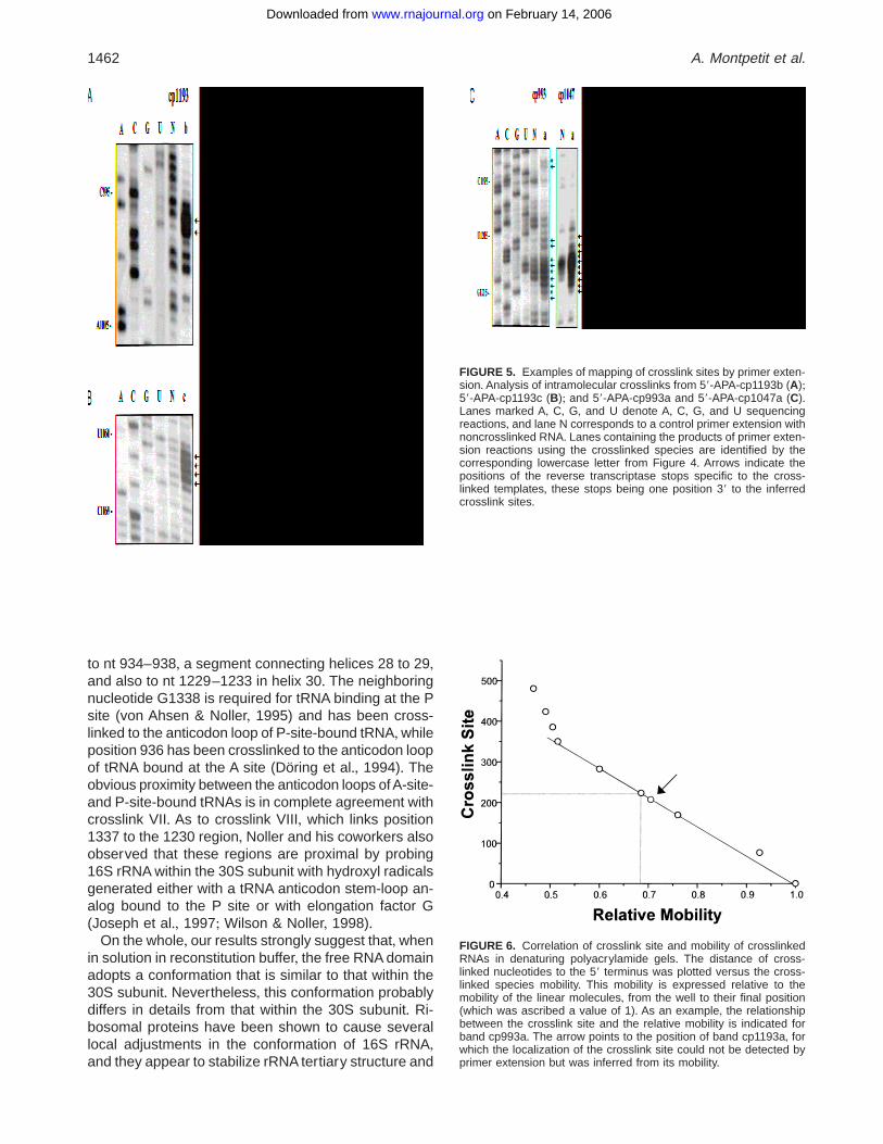
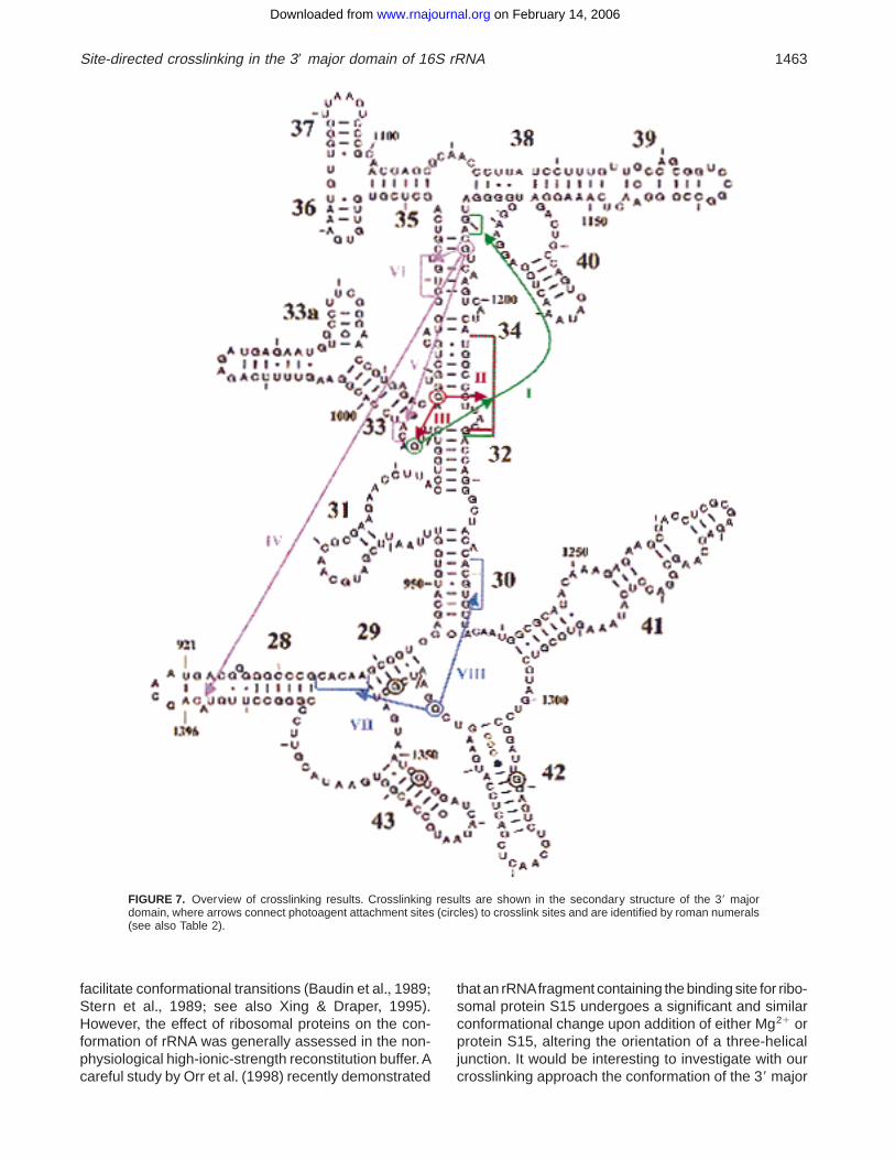
Primer extension by reverse transcriptase was used tomap the crosslink sites from gel-purified RNA+ Com-parison of extension products with sequencing reac-tions identified the crosslinked nucleotides+ For eachcrosslinked species, the entire sequence was investi-gated+ Examples of this analysis are shown in Figure 5for crosslinked species derived from cp993, cp1047and cp1193+ Crosslinked nucleotides are visible asprimer extension stops+ To confirm the primer exten-sion analysis, we examined the mobility of the cross-linked species in denaturing polyacrylamide gels relativeto the mobility of the linear molecules (Harris et al+,1997)+ We found that the relative rate of migration ofeach of the crosslinked species correlated with the as-signment of crosslink sites, as long as the size of thecrosslinked loops was less than 80% of the size of thefull-length domain (Fig+ 6)+ It is known that the reversetranscriptase may be able to read through some of thecrosslinked nucleotides (Burgin & Pace, 1990), whichoccurred for band a from cp1193+ In this case, the sizeof the crosslinked loop could be used to identify thecrosslinked region+
Table 2 summarizes our results+ Except for circularforms, where the 59 and 39 ends are crosslinked to-gether, no specific crosslink was observed with the na-tive 39 major domain and with constructions cp1309,
TABLE 1 + Primers used for constructing cpRNA templates+
Name of the permutation Sequence of the primera
cp993 forward 59 TAATACGACTCAC TATAGACATCCACGGAAGTTTTC 39reverse 59 AAGACCAGGTAAGGTTC 39
cp1047 forward 59 TAATACGACTCAC TATAGGTGCTGCATGGCTGTCGT 39reverse 59 TGTCTCACGGTTCCCGA 39
cp1193 forward 59 TAATACGACTCAC TATAGTCAAGTCATCATGGCC 39reverse 59 GTCATCCCCACCTTCCT 39
cp1309 forward 59 TAATACGACTCAC TATAGGAGTCTGCAACTCGACTC 39reverse 59 AATCCGGACTACGACGCACT 39
cp1337 forward 59 TAATACGACTCAC TATAGGAATCGCTAGTAATCGTG 39reverse 59 GACTTCATGGAGTCGAG 39
cp1343 forward 59 TAATACGACTCAC TATAGCTAGTAATCGTGGATCA 39reverse 59 GATTCCGACTTCATGGA 39
cp1353 forward 59 TAATACGACTCAC TATAGTGGATCAGAATGCCAC 39reverse 59 GATTACTAGCGATTCCGA 39
aThe promoter sequence for phage T7 RNA polymerase is shown in bold+
1458 A. Montpetit et al.
on February 14, 2006 www.rnajournal.orgDownloaded from
cp1343 and cp1353+ In these molecules, the photo-agent probably occupies a position that favors a reac-tion with the solvent+ Eight intramolecular crosslinkswere observed with the other positions investigated+The circular forms were observed independently of thepresence or absence of Mg21 and K1 ions, whereasexcept for crosslinks II and VI, which connect the op-posite strands of a helix (see Fig+ 7), all the other cross-links disappeared in the absence of Mg21 and K1 ions+This demonstrates that, for these crosslinks to occur,the domain must have a specific conformation+ Theefficiencies of the crosslinks varied from about 1 to10%+ These efficiencies may reflect the distance be-tween the crosslink sites and the 59 terminus bearingthe crosslinking agent (the maximum span coveredby the agent being about 10 Å), the relative reactivity ofthe crosslinking agent with the chemical groups in its
vicinity, and the orientation of the modified phosphaterelative to its target+
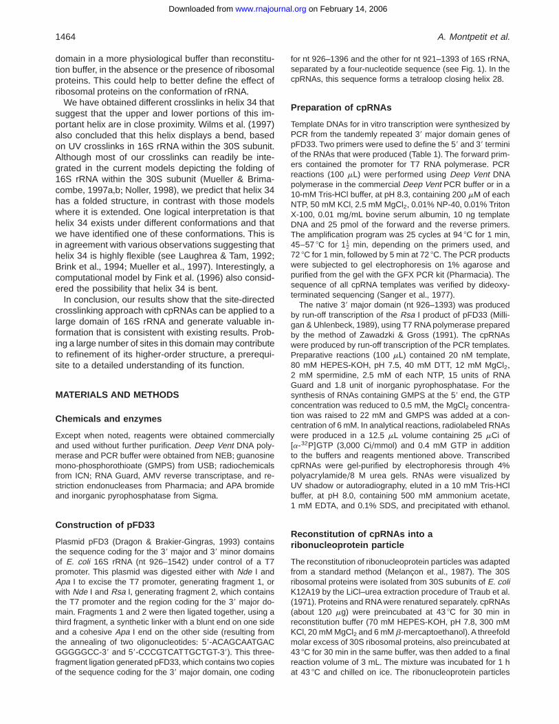
Figure 7 presents our results in the secondary struc-ture of the 39 major domain+ It can be seen that G1193,in the upper stem of helix 34, crosslinked to a bulge atthe base of helix 33 (crosslink V), and this crosslinktherefore indicates that the upper stem of helix 34 isproximal to its lower stem+ G1193 also crosslinked tothe region around 1060 (crosslink VI), on the oppositestrand, and to region 1395 in helix 28, the helix thatconnects the head of the 30S subunit to its body (cross-link IV)+G993, in the bulge flanking the base of helix 33,crosslinked to a segment spanning the lower half ofhelix 34 plus the upper stem of this helix (crosslink I),supporting the observation with crosslink V that theupper and lower stems of helix 34 are in close proxim-ity+ G1047 crosslinked to the same segment spanning
FIGURE 2. Locations of photoagent attachment sites+ The skeleton of 16S rRNA secondary structure is presented with anenlarged area corresponding to the 39 major domain+ The photoagent attachment sites located at the 59 end of eachpermutation are circled+ Nucleotides are numbered as in the 16S rRNA and helices are numbered according to thenomenclature of Brimacombe (1991)+ The terminal GCAA tetraloop has been introduced to close helix 28+
Site-directed crosslinking in the 39 major domain of 16S rRNA 1459
on February 14, 2006 www.rnajournal.orgDownloaded from
the lower half of helix 34 (crosslink II), and also cross-linked to G993, in an adjacent position (crosslink III)+Finally, G1337, in an internal loop located at the junc-tion of helices 29, 30, 41, and 42 in the lower part of thedomain, crosslinked to a single-stranded segment con-necting helices 28 and 29 (crosslink VII) and to thecentral portion of helix 30 (crosslink VIII)+
The length of the crosslinked regions is comparableto that observed by Pace and coworkers with RNase PRNA analyzed with the same approach (Chen et al+,1998), except in crosslinks I and II+ In these crosslinks,G993 and G1047, respectively, crosslinked to an ex-tensive region in one strand of the lower stem of he-lix 34+ One could suggest that G993, being located inan internal loop, has greater flexibility than a 59 termi-nus located in a helix, which could increase the size ofthe target hit by the crosslinking reagent+ However,G1047, which is in a helix, also crosslinks to a largeregion+Moreover, the size of the regions to which G1337crosslinks in crosslinks VII and VIII is less extensivethan that of crosslinks I and II, although G1337 is lo-cated in an internal loop+ Recent X-ray crystallographicand NMR studies with a variety of RNA molecules haveshown that internal and hairpin loops can be highlystructured in the presence of divalent cations (reviewedby Doudna & Cate, 1997; Ramos et al+, 1997)+ We
suggest that in reconstitution buffer the 59 terminus ofcpRNAs can be constrained as well, whether it is lo-cated in an internal loop or in a helix, and we hypoth-esize that the large size of the regions to which G993
FIGURE 3. Sucrose gradient analysis of cp1337 (A) and of theribonucleoprotein particle reconstituted when cp1337 was incubatedwith a threefold molar excess of ribosomal proteins (B)+ Centrifuga-tion was through a 5–20% sucrose gradient in reconstitution buffer+The arrow points to the position of migration of the control particleformed by the association of the native 39 major domain with ribo-somal proteins, which coincides with that of the particle formedwith cp1337+ A similar profile was observed with all the cpRNAsinvestigated+
FIGURE 4. Identification of crosslinked species+ A: Detailed analy-sis of intramolecular crosslinking of cp1337+ Radiolabeled 59-APA-cp1337 RNA (about 4 nM) was crosslinked under reconstitutionconditions: 70 mM HEPES-KOH, pH 7+8, 20 mM MgCl2, and 300 mMKCl or, when indicated, in the absence of Mg21 and K1+ Crosslinkedspecies and noncrosslinked RNA were resolved on denaturing 4%polyacrylamide gels+ DTT was added to a final concentration of1 mM prior to electrophoresis, when indicated+ RNA was exposed to310-nm UV light for 15 min, except in the No UV lane+ The Uncon-jugated sample was not modified with APA and the 59-GTP samplewas generated by transcription in the absence of GMPS+ Crosslinkedspecies are identified with letters (a–d)+ Band a corresponds to adimer where two 59-GMPS-RNAs are linked by a disulfide bridge+ B:Examples of analysis with 59-APA-cp993, -cp1047 and -cp1193+ Ra-diolabeled 59-APA-cpRNAs were irradiated (1) or not (2) with 310-nmUV light+ DTT was added to a final concentration of 1 mM prior toelectrophoresis, when indicated+
1460 A. Montpetit et al.
on February 14, 2006 www.rnajournal.orgDownloaded from
and G1047 crosslink reflects some unusual flexibility inthe lower stem of helix 34+
DISCUSSION
We have undertaken a study of the conformation of the39 major domain of 16S rRNA, in the absence of pro-teins, using a site-directed crosslinking method+ Thecrosslinks were generated in a high-ionic strength buffer,where this domain adopts a conformation recognizedby the ribosomal proteins+ The importance of rRNA war-rants a detailed investigation of its higher-order struc-ture+ rRNA may have functioned at a time without theaid of proteins (Noller, 1993), and a large body of evi-dence supports the direct role of rRNA in protein syn-thesis (Green & Noller, 1997)+ A report by Nitta et al+(1998a,b) recently suggested that in vitro-transcribednaked 23S rRNA could have peptidyl transferase ac-tivity+ Previously, Purohit & Stern (1994) had shownthat a fragment of 16S rRNA corresponding to the de-coding center of 16S rRNA had the capacity to interactwith mRNA, tRNA anticodon stem-loop, and aminogly-
coside antibiotics such as neomycin in a similar way as16S rRNA within 30S subunit, indicating that this frag-ment is able to perform some of the functions of theribosome+
Several of the crosslinks we observed in the free 39major domain are in agreement with proximity relation-ships that have been observed within the 30S subunit+Crosslink I, which joins position 993 to a segment en-compassing nt 1205–1215, is consistent with a pre-vious UV crosslink joining positions 991–1212+ Thiscrosslink was identified in 16S rRNA within the 30Ssubunit first by Stiege et al+ (1988) and, later on, with ahigher resolution, by Wilms et al+ (1997)+ Also, cross-link IV, which links the upper stem of helix 34 to helix 28in the free domain, agrees with a proximity relationshipdemonstrated between positions 1196 and 1395 in 16SrRNA within the 30S subunit, using crosslinking withmRNA analogs (Rinke-Appel et al+, 1993; Juzumieneet al+, 1995; Sergiev et al+, 1997) or cleavage with mRNAmodified with phenanthroline (Bucklin et al+, 1997)+
Crosslinks VII and VIII also indicate interesting ter-tiary interactions showing that position 1337 is linked
TABLE 2 + Analysis of crosslinked species+
Crosslinkedspecies
Dependence onMg21 and K1 a Efficiencyb Crosslinked nucleotides
Crosslinknumber
nativenative a 2 7+0 Circular formc
cp993993 a 1 7+0 G1190-A1191;U1205-
G1206; C1209-G1215I
993 b 2 2+4 Circular formc
cp10471047 a 2 8+4 U1205-C1214 II1047 b 1 7+2 G993 III1047 c 2 7+2 Circular formc III
cp11931193 a 1 0+9 1395 regiond IV1193 b 1 1+6 C995-A996 V1193 c 2 3+9 U1060-C1063 VI1193 d 2 5+4 Circular formc
cp13091309 a 2 5+7 Circular formc
cp13371337 a 1 10+2 C934-A938 VII1337 b 1 4+7 A1229-G1233 VIII1337 c 2 2+1 Circular formc
cp13431343 a 2 3+9 Circular formc
cp13531353 a 2 6+2 Circular formc
a2 indicates that the crosslinked species is detected independently of the presence of Mg21
and K1, and 1 indicates that the crosslinked species disappears in the absence of Mg21 and K1+bEfficiency indicates percent conversion to crosslinked species+cIntramolecular crosslink between the 59 and 39 ends+dThe position of this crosslink could not be identified by primer extension and was assessed
from the size of the lariat (see Fig+ 6)+
Site-directed crosslinking in the 39 major domain of 16S rRNA 1461
on February 14, 2006 www.rnajournal.orgDownloaded from
to nt 934–938, a segment connecting helices 28 to 29,and also to nt 1229–1233 in helix 30+ The neighboringnucleotide G1338 is required for tRNA binding at the Psite (von Ahsen & Noller, 1995) and has been cross-linked to the anticodon loop of P-site-bound tRNA,whileposition 936 has been crosslinked to the anticodon loopof tRNA bound at the A site (Döring et al+, 1994)+ Theobvious proximity between the anticodon loops of A-site-and P-site-bound tRNAs is in complete agreement withcrosslink VII+ As to crosslink VIII, which links position1337 to the 1230 region, Noller and his coworkers alsoobserved that these regions are proximal by probing16S rRNA within the 30S subunit with hydroxyl radicalsgenerated either with a tRNA anticodon stem-loop an-alog bound to the P site or with elongation factor G(Joseph et al+, 1997; Wilson & Noller, 1998)+
On the whole, our results strongly suggest that, whenin solution in reconstitution buffer, the free RNA domainadopts a conformation that is similar to that within the30S subunit+ Nevertheless, this conformation probablydiffers in details from that within the 30S subunit+ Ri-bosomal proteins have been shown to cause severallocal adjustments in the conformation of 16S rRNA,and they appear to stabilize rRNA tertiary structure and
FIGURE 5. Examples of mapping of crosslink sites by primer exten-sion+ Analysis of intramolecular crosslinks from 59-APA-cp1193b (A);59-APA-cp1193c (B); and 59-APA-cp993a and 59-APA-cp1047a (C)+Lanes marked A, C, G, and U denote A, C, G, and U sequencingreactions, and lane N corresponds to a control primer extension withnoncrosslinked RNA+ Lanes containing the products of primer exten-sion reactions using the crosslinked species are identified by thecorresponding lowercase letter from Figure 4+ Arrows indicate thepositions of the reverse transcriptase stops specific to the cross-linked templates, these stops being one position 39 to the inferredcrosslink sites+
FIGURE 6. Correlation of crosslink site and mobility of crosslinkedRNAs in denaturing polyacrylamide gels+ The distance of cross-linked nucleotides to the 59 terminus was plotted versus the cross-linked species mobility+ This mobility is expressed relative to themobility of the linear molecules, from the well to their final position(which was ascribed a value of 1)+ As an example, the relationshipbetween the crosslink site and the relative mobility is indicated forband cp993a+ The arrow points to the position of band cp1193a, forwhich the localization of the crosslink site could not be detected byprimer extension but was inferred from its mobility+
1462 A. Montpetit et al.
on February 14, 2006 www.rnajournal.orgDownloaded from
facilitate conformational transitions (Baudin et al+, 1989;Stern et al+, 1989; see also Xing & Draper, 1995)+However, the effect of ribosomal proteins on the con-formation of rRNA was generally assessed in the non-physiological high-ionic-strength reconstitution buffer+Acareful study by Orr et al+ (1998) recently demonstrated
that an rRNAfragment containing the binding site for ribo-somal protein S15 undergoes a significant and similarconformational change upon addition of either Mg21 orprotein S15, altering the orientation of a three-helicaljunction+ It would be interesting to investigate with ourcrosslinking approach the conformation of the 39 major
FIGURE 7. Overview of crosslinking results+ Crosslinking results are shown in the secondary structure of the 39 majordomain, where arrows connect photoagent attachment sites (circles) to crosslink sites and are identified by roman numerals(see also Table 2)+
Site-directed crosslinking in the 39 major domain of 16S rRNA 1463
on February 14, 2006 www.rnajournal.orgDownloaded from
domain in a more physiological buffer than reconstitu-tion buffer, in the absence or the presence of ribosomalproteins+ This could help to better define the effect ofribosomal proteins on the conformation of rRNA+
We have obtained different crosslinks in helix 34 thatsuggest that the upper and lower portions of this im-portant helix are in close proximity+ Wilms et al+ (1997)also concluded that this helix displays a bend, basedon UV crosslinks in 16S rRNA within the 30S subunit+Although most of our crosslinks can readily be inte-grated in the current models depicting the folding of16S rRNA within the 30S subunit (Mueller & Brima-combe, 1997a,b; Noller, 1998), we predict that helix 34has a folded structure, in contrast with those modelswhere it is extended+ One logical interpretation is thathelix 34 exists under different conformations and thatwe have identified one of these conformations+ This isin agreement with various observations suggesting thathelix 34 is highly flexible (see Laughrea & Tam, 1992;Brink et al+, 1994; Mueller et al+, 1997)+ Interestingly, acomputational model by Fink et al+ (1996) also consid-ered the possibility that helix 34 is bent+
In conclusion, our results show that the site-directedcrosslinking approach with cpRNAs can be applied to alarge domain of 16S rRNA and generate valuable in-formation that is consistent with existing results+ Prob-ing a large number of sites in this domain may contributeto refinement of its higher-order structure, a prerequi-site to a detailed understanding of its function+
MATERIALS AND METHODS
Chemicals and enzymes
Except when noted, reagents were obtained commerciallyand used without further purification+ Deep Vent DNA poly-merase and PCR buffer were obtained from NEB; guanosinemono-phosphorothioate (GMPS) from USB; radiochemicalsfrom ICN; RNA Guard, AMV reverse transcriptase, and re-striction endonucleases from Pharmacia; and APA bromideand inorganic pyrophosphatase from Sigma+
Construction of pFD33
Plasmid pFD3 (Dragon & Brakier-Gingras, 1993) containsthe sequence coding for the 39 major and 39 minor domainsof E. coli 16S rRNA (nt 926–1542) under control of a T7promoter+ This plasmid was digested either with Nde I andApa I to excise the T7 promoter, generating fragment 1, orwith Nde I and Rsa I, generating fragment 2, which containsthe T7 promoter and the region coding for the 39 major do-main+ Fragments 1 and 2 were then ligated together, using athird fragment, a synthetic linker with a blunt end on one sideand a cohesive Apa I end on the other side (resulting fromthe annealing of two oligonucleotides: 59-ACAGCAATGACGGGGGCC-39 and 59-CCCGTCATTGCTGT-39)+ This three-fragment ligation generated pFD33,which contains two copiesof the sequence coding for the 39 major domain, one coding
for nt 926–1396 and the other for nt 921–1393 of 16S rRNA,separated by a four-nucleotide sequence (see Fig+ 1)+ In thecpRNAs, this sequence forms a tetraloop closing helix 28+
Preparation of cpRNAs
Template DNAs for in vitro transcription were synthesized byPCR from the tandemly repeated 39 major domain genes ofpFD33+ Two primers were used to define the 59 and 39 terminiof the RNAs that were produced (Table 1)+ The forward prim-ers contained the promoter for T7 RNA polymerase+ PCRreactions (100 mL) were performed using Deep Vent DNApolymerase in the commercial Deep Vent PCR buffer or in a10-mM Tris-HCl buffer, at pH 8+3, containing 200 mM of eachNTP, 50 mM KCl, 2+5 mM MgCl2, 0+01% NP-40, 0+01% TritonX-100, 0+01 mg/mL bovine serum albumin, 10 ng templateDNA and 25 pmol of the forward and the reverse primers+The amplification program was 25 cycles at 94 8C for 1 min,45–57 8C for 11
2_ min, depending on the primers used, and
72 8C for 1 min, followed by 5 min at 72 8C+ The PCR productswere subjected to gel electrophoresis on 1% agarose andpurified from the gel with the GFX PCR kit (Pharmacia)+ Thesequence of all cpRNA templates was verified by dideoxy-terminated sequencing (Sanger et al+, 1977)+
The native 39 major domain (nt 926–1393) was producedby run-off transcription of the Rsa I product of pFD33 (Milli-gan & Uhlenbeck, 1989), using T7 RNA polymerase preparedby the method of Zawadzki & Gross (1991)+ The cpRNAswere produced by run-off transcription of the PCR templates+Preparative reactions (100 mL) contained 20 nM template,80 mM HEPES-KOH, pH 7+5, 40 mM DTT, 12 mM MgCl2,2 mM spermidine, 2+5 mM of each NTP, 15 units of RNAGuard and 1+8 unit of inorganic pyrophosphatase+ For thesynthesis of RNAs containing GMPS at the 59 end, the GTPconcentration was reduced to 0+5 mM, the MgCl2 concentra-tion was raised to 22 mM and GMPS was added at a con-centration of 6 mM+ In analytical reactions, radiolabeled RNAswere produced in a 12+5 mL volume containing 25 mCi of[a-32P]GTP (3,000 Ci/mmol) and 0+4 mM GTP in additionto the buffers and reagents mentioned above+ TranscribedcpRNAs were gel-purified by electrophoresis through 4%polyacrylamide/8 M urea gels+ RNAs were visualized byUV shadow or autoradiography, eluted in a 10 mM Tris-HClbuffer, at pH 8+0, containing 500 mM ammonium acetate,1 mM EDTA, and 0+1% SDS, and precipitated with ethanol+
Reconstitution of cpRNAs into aribonucleoprotein particle
The reconstitution of ribonucleoprotein particles was adaptedfrom a standard method (Melançon et al+, 1987)+ The 30Sribosomal proteins were isolated from 30S subunits of E. coliK12A19 by the LiCl–urea extraction procedure of Traub et al+(1971)+ Proteins and RNA were renatured separately+ cpRNAs(about 120 mg) were preincubated at 43 8C for 30 min inreconstitution buffer (70 mM HEPES-KOH, pH 7+8, 300 mMKCl, 20 mM MgCl2 and 6 mM b-mercaptoethanol)+A threefoldmolar excess of 30S ribosomal proteins, also preincubated at43 8C for 30 min in the same buffer, was then added to a finalreaction volume of 3 mL+ The mixture was incubated for 1 hat 43 8C and chilled on ice+ The ribonucleoprotein particles
1464 A. Montpetit et al.
on February 14, 2006 www.rnajournal.orgDownloaded from
were analyzed on a 5–20% sucrose gradient (26,000 rpm,22 h, SW28 rotor)+
Crosslinking procedure and mappingof crosslink sites
Crosslinking experiments were performed essentially as de-scribed by Burgin & Pace (1990)+ APA bromide was conju-gated to the unique sulfur at the 59-terminal phosphate ofGMPS RNA+ Analytical reactions were carried out to detectthe crosslinked species and to assess the efficiency of thecrosslinks+ In an analytical crosslinking reaction (100 mL),radiolabeled 59-APA-RNA (about 4 nM) was incubated in re-constitution buffer without b-mercaptoethanol for 30 min at43 8C and then chilled on ice for 5 min+ The solution wasexposed for 15 min at 4 8C to 310-nm UV light (Foto/PrepI,Fotodyne) at a distance of 5 cm+ It was screened by a poly-styrene filter (Petri dish, Fisher) to absorb wavelengths under300 nm+ In control experiments, the crosslinking reactionswere performed in the absence of Mg21 and K1 ions+ Thecrosslinked species were resolved on 4% polyacrylamide/8 M urea gels, visualized by autoradiography, and the effi-ciency of the crosslinks was evaluated by measuring the in-tensity of the bands in percent of the total input, using AlphaImager software (Alpha Innotech Corp+)+ The migration of thecrosslinked species was assessed in the denaturing gel rel-ative to the linear molecules+ Preparative crosslinking reac-tions were carried out to isolate crosslinked species+ In apreparative crosslinking reaction (100 mL), 59-APA-RNA (atabout 400 nM) was incubated as described for the analyticalreactions+ The crosslinked species, as well as the noncross-linked RNA, were resolved by electrophoresis through 4%polyacrylamide/8 M urea gels, visualized by staining withethidium bromide, eluted from the gel as described above,and used to map the crosslink sites by primer extension+Primers used were the reverse primers complementary to16S rRNA described in Table 1+ They were 59-end-labeledwith [g-32P]ATP (3,000 Ci/mmol) and annealed to cross-linked or noncrosslinked RNA in a 50 mM Tris-HCl buffer atpH 8+5 containing 100 mM KCl by heating for 2 min at85 8C, followed by slow cooling to room temperature+ Theoligonucleotides were then extended with AMV reverse tran-scriptase+ The products of extension were resolved by elec-trophoresis on 8% polyacrylamide/8 M urea gels+
ACKNOWLEDGMENTS
We thank Drs+ L+ DesGroseillers, F+ Dragon, S+ Michnick, andM+ O’Connor for stimulating comments and critical reading ofthis manuscript+ We also thank F+ Bélanger for his contribu-tion to some crosslinking experiments+ This study was sup-ported by a grant of the Medical Research Council of Canadato L+B+-G+A+M+ held a studentship from the National Sciencesand Engineering Research Council of Canada+
Received June 22, 1998; returned for revision July 24,1998; revised manuscript received August 11, 1998
REFERENCES
Agalarov SC, Zheleznyakova EN, Selivanova OM, Zheleznaya LA,Matvienko NI, Vasiliev VD, Spirin AS+ 1998+ In vitro assembly of a
ribonucleoprotein particle corresponding to the platform domainof the 30S ribosomal subunit+ Proc Natl Acad Sci USA 95:999–1003+
Alexander RW, Cooperman BS+ 1998+ Ribosomal proteins neighbor-ing 23S rRNA nucleotides 803–811 within the 50S subunit+ Bio-chemistry 37:1714–1721+
Arkov AL, Freistroffer DV, Ehrenberg M,Murgola EJ+ 1998+Mutationsin RNAs of both ribosomal subunits cause defects in translation+EMBO J 17:1507–1514+
Ban N, Freeborn B, Nissen P, Penczek P, Grassucci RA, Sweet R,Frank J, Moore PB, Steitz TA+ 1998+ A 9 Å resolution X-ray crys-tallographic map of the large ribosomal subunit+ Cell 93:1105–1115+
Baranov PV, Gurvich OL, Bogdanov AA, Brimacombe R, DontsovaOA+ 1998a+ New features of 23S ribosomal RNA folding: The longhelix 41–42 makes a “U-turn” inside the ribosome+ RNA 4:658–668+
Baranov PV, Sergiev PV, Dontsova OA, Bogdanov AA, BrimacombeR+ 1998b+ The database of ribosomal cross-links (DRC)+ NucleicAcids Res 26:187–189+
Baudin F, Mougel M, Romby P, Eyermann F, Ebel JP, Ehresmann B,Ehresmann C+ 1989+ Probing the phosphates of the Escherichiacoli ribosomal 16S RNA in its naked form, in the 30S subunit, andin the 70S ribosome+ Biochemistry 28:5847–5855+
Bilgin N, Richter AA, Ehrenberg M, Dahlberg AE, Kurland CG+ 1990+Ribosomal RNA and protein mutants resistant to spectinomycin+EMBO J 9:735–739+
Brimacombe R+ 1991+ RNA-protein interactions in the Escherichiacoli ribosome+ Biochimie 73:927–936+
Brink MF, Brink G, Verbeet MP, de Boer H+ 1994+ Spectinomycininteracts specifically with the residues G1064 and C1192 in 16SrRNA, thereby potentially freezing this molecule into an inactiveconformation+ Nucleic Acids Res 22:325–331+
Bucklin DJ, Vanwaes MA, Bullard JM, Hill WE+ 1997+ Cleavage of16S rRNA within the ribosome by mRNA modified in the A-sitecodon with phenantroline-Cu(II). Biochemistry 36:7951–7957+
Burgin AB, Pace NR+ 1990+ Mapping the active site of ribonucleaseP RNA using a substrate containing a photoaffinity agent+ EMBOJ 9:4111–4118+
Cate JH, Gooding AR, Podell E, Zhou K, Golden BL, Kundrot CE,Cech TR, Doudna JA+ 1996+ Crystal structure of a group I intronribozyme domain: Principles of RNA packing+ Science 273:1678–1685+
Chen JL, Nolan JM, Harris ME, Pace NR+ 1998+ Comparative photo-crosslinking analysis of the tertiary structure of Escherichia coliand Bacillus subtilis RNase P RNAs+ EMBO J 17:1515–1525+
Correll CC, Freeborn B, Moore PB, Steitz TA+ 1997+ Metals, motifs,and recognition in the crystal structure of a 5S rRNA domain+ Cell91:705–712+
Dallas A, Moore PB+ 1997+ The loop E–loop D region of Escherichiacoli 5S RNA: The solution structure reveals an unusual loop thatmay be important for binding ribosomal proteins+Structure 5:1639–1653+
Döring T,Mitchell P,Osswald M, Bochkariov D, Brimacombe R+ 1994+The decoding region of 16S RNA; a cross-linking study of theribosomal A, P and E sites using tRNA derivatized at position 32in the anticodon loop+ EMBO J 13:2677–2685+
Doudna JA, Cate JH+ 1997+ RNA structure: Crystal clear? Curr OpinStruct Biol 7:310–316+
Dragon F, Brakier-Gingras L+ 1993+ Interaction of Escherichia coliribosomal protein S7 with 16S rRNA+ Nucleic Acids Res 21:1199–1203+
Fink DL, Chen RO, Noller HF,Altman RB+ 1996+ Computational meth-ods for defining the allowed conformational space of 16S rRNAbased on chemical footprinting data+ RNA 2:851–866+
Fourmy D, Recht MI, Blanchard SC, Puglisi JD+ 1996+ Structure ofthe A site of Escherichia coli 16S ribosomal RNA complexed withan aminoglycoside antibiotic+ Science 274:1367–1371+
Green R, Noller HF+ 1997+ Ribosomes and translation+ Annu RevBiochem 66:679–716+
Harris ME, Kazantsev AV, Chen JL, Pace NR+ 1997+ Analysis of thetertiary structure of the ribonuclease P ribozyme-substrate com-plex by site-specific photoaffinity crosslinking+ RNA 3:561–576+
Harris ME, Nolan JM, Malhotra A, Brown JW, Harvey SC, Pace NR+1994+ Use of photoaffinity crosslinking and molecular modeling to
Site-directed crosslinking in the 39 major domain of 16S rRNA 1465
on February 14, 2006 www.rnajournal.orgDownloaded from
analyse the global architecture of ribonuclease P RNA+ EMBO J13:3953–3963+
Herr W, Chapman NM, Noller HF+ 1979+ Mechanism of ribosomalsubunit association: Discrimination of specific sites in 16S RNAessential for association activity. J Mol Biol 130:433–449+
Joseph S, Weiser B, Noller HF+ 1997+ Mapping the inside of theribosome with an RNA helical ruler+ Science 278:1093–1098+
Juzumiene DI, Shapkina TG,Wollenzien P+ 1995+Distribution of cross-links between mRNA analogues and 16S rRNA in Escherichiacoli 70S ribosomes made under equilibrium conditions and theirresponse to tRNA binding+ J Biol Chem 270:12794–12800+
Laughrea M, Tam J+ 1992+ In vivo chemical footprinting of the Esch-erichia coli ribosome+ Biochemistry 31:12035–12041+
Makosky PC, Dahlberg AE+ 1987+ Spectinomycin resistance at site1192 in 16S ribosomal RNA of E. coli: An analysis of three mu-tants+ Biochimie 69:885–889+
Melançon P, Gravel M, Boileau G, Brakier-Gingras L+ 1987+ Reas-sembly of active 30S ribosomal subunits with unmethylated invitro transcribed 16S rRNA+ Biochem Cell Biol 65:1022–1030+
Milligan JF, Uhlenbeck OC+ 1989+ Synthesis of small RNAs using T7RNA polymerase+ Methods Enzymol 180:51–62+
Moazed D, Noller HF+ 1990+ Binding of tRNA to the ribosomal Aand P sites protects two distinct sets of nucleotides in 16S rRNA+J Mol Biol 211:135–145+
Moine H, Dahlberg AE+ 1994+Mutations in helix 34 of Escherichia coli16 S ribosomal RNA have multiple effects on ribosome functionand synthesis+ J Mol Biol 243:402–412+
Mueller F, Brimacombe R+ 1997a+ A new model for the three-dimensional folding of Escherichia coli 16S ribosomal RNA+ I+Fitting the RNA to a 3D electron microscopic map at 20 Å+ J MolBiol 271:524–544+
Mueller F, Brimacombe R+ 1997b+ A new model for the three-dimensional folding of Escherichia coli 16S ribosomal RNA+ II+The RNA-protein interaction data+ J Mol Biol 271:545–565+
Mueller F, Stark H, van Heel M, Rinke-Appel J, Brimacombe R+ 1997+A new model for the three-dimensional folding of Escherichia coli16S ribosomal RNA+ III+ The topography of the functional centre+J Mol Biol 271:566–587+
Murgola EJ+ 1996+ Ribosomal RNA in peptide chain termination+ In:Zimmermann RA, Dahlberg AE, eds+ Ribosomal RNA: Structure,evolution, processing and function in protein biosynthesis+ NewYork/London: CRC Press+ pp 357–369+
Nitta I, Kamada Y, Noda H, Ueda T, Watanabe K+ 1998a+ Reconsti-tution of peptide bond formation with Escherichia coli 23S ribo-somal RNA domains+ Science 281:666–669+
Nitta I, Ueda T, Watanabe K+ 1998b+ Possible involvement of Esch-erichia coli 23S ribosomal RNA in peptide bond formation+ RNA4:257–267+
Nolan JM, Burke DH, Pace NR 1993+ Circularly permuted tRNAs asspecific photoaffinity probes of ribonuclease P RNA structure+Science 261:762–765+
Noller HF+ 1993+ On the origin of the ribosome: Coevolution of sub-domain of tRNA and rRNA+ In: Gesteland RF, Atkins JF, eds+ TheRNA World+ Cold Spring Harbor, New York: Cold Spring HarborLaboratory Press+ pp 137–156+
Noller HF+ 1998+ Ribosomal RNA+ In: Simons RW, Grunberg-ManagoM, eds+ RNA Structure and function+ Cold Spring Harbor, NewYork: Cold Spring Harbor Laboratory Press+ pp 253–278+
O’Connor M, Thomas CL, Zimmermann RA, Dahlberg AE+ 1997+Decoding fidelity at the ribosomal A and P sites: Influence ofmutations in three different regions of the decoding domain in16S rRNA+ Nucleic Acids Res 25:1185–1193+
Oehler R, Polacek N, Steiner G, Barta A+ 1997+ Interaction of tetra-cycline with RNA–photoincorporation into ribosomal RNA of Esch-erichia coli+ Nucleic Acids Res 25:1219–1224+
Orr JW, Hagerman PJ, Williamson JR+ 1998+ Protein and Mg21-
induced conformational changes in the S15 binding site of 16Sribosomal RNA+ J Mol Biol 275:453–464+
Purohit P, Stern S+ 1994+ Interactions of a small RNA with antibioticand RNA ligands of the 30S subunit+ Nature 370:659–662+
Ramos A, Gubser CC, Varani G+ 1997+ Recent solution structures ofRNA and its complexes with drugs, peptides and proteins+ CurrOpin Struct Biol 7:317–323+
Rinke-Appel J, Jünke N, Brimacombe R, Lavrik I, Dokudovskaya S,Dontsova O, Bogdanov A+ 1993+ Site-directed cross-linking ofmRNA analogues to 16S ribosomal RNA; a complete scan ofcross-links from all positions between ‘11’ and ‘116’ on the mRNA,downstream from the decoding site+ Nucleic Acids Res 21:2853–2859+
Samaha RR, O’Brien B, O’Brien TW, Noller HF+ 1994+ Independent invitro assembly of a ribonucleoparticle containing the 39 domain of16S rRNA+ Proc Natl Acad Sci USA 91:7884–7888+
Sanger F, Nicklen S, Coulson AR+ 1977+ DNA sequencing with chain-terminating inhibitors+ Proc Natl Acad Sci USA 74:5463–5467+
Sergiev PV, Lavrik IN,Wlasoff VA, Dokudovskaya SS, Dontsova OA,Bogdanov AA, Brimacombe R+ 1997+ The path of mRNA throughthe bacterial ribosome: A site-directed crosslinking study usingnew photoreactive derivatives of guanosine and uridine+ RNA3:464–475+
Stiege W, Kosack M, Stade K, Brimacombe R+ 1988+ Intra-RNA cross-linking in Escherichia coli 30S ribosomal subunits: Selective iso-lation of cross-linked products by hybridization to specific cDNAfragments+ Nucleic Acids Res 16:4315–4329+
Stern S, Powers T, Changchien LM, Noller HF+ 1989+ RNA-proteininteractions in 30S ribosomal subunits: Folding and function of16S rRNA+ Science 244:783–790+
Szewczak AA, Moore PB+ 1995+ The sarcin ricin loop, a modularRNA+ J Mol Biol 247:81–98+
Thygesen J, Weinstein S, Franceschi F, Yonath A+ 1996+ The suit-ability of multi-metal clusters for phasing in crystallography oflarge macromolecular assemblies+ Structure 4:513–518+
Traub P, Mizushima S, Lowary CV, Nomura M+ 1971+ Reconstitutionof ribosomes from subribosomal components+ Methods Enzymol20:391–407+
Viani-Puglisi E, Green R, Noller HF, Puglisi JD+ 1997+ Structure of aconserved RNA component of the peptidyl transferase centre+Nature Struct Biol 4:775–778+
von Ahsen U, Noller HF+ 1995+ Identification of bases in 16S rRNAessential for tRNA binding at the 30S ribosomal P site+ Science267:234–237+
Wakao H, Romby P, Ebel JP, Grunberg-Manago M, Ehresmann C,Ehresmann B+ 1991+ Topography of the Escherichia coli ribo-somal 30S subunit-initiation factor 2 complex+ Biochimie 73:991–1000+
Weitzmann CJ, Cunningham PR, Nurse K,Ofengand J+ 1993+ Chem-ical evidence for domain assembly of the Escherichia coli 30Sribosome+ FASEB J 7:177–180+
Wilms C, Noah JW, Zhong D, Wollenzien P+ 1997+ Exact determina-tion of UV-induced crosslinks in 16S ribosomal RNA in 30S ribo-somal subunits+ RNA 3:602–612+
Wilson KS, Noller HF+ 1998+ Mapping the position of translationalelongation factor EF-G in the ribosome by directed hydroxyl rad-ical probing+ Cell 92:131–139+
Xing Y, Draper DE+ 1995+ Stabilization of a ribosomal RNA tertiarystructure by ribosomal protein L11+ J Mol Biol 249:319–331+
Zawadzki V, Gross HJ+ 1991+ Rapid and simple purification of T7RNA polymerase+ Nucleic Acids Res 19:1948+
Zimmermann RA+ 1996+ The decoding domain+ In: Zimmermann RA,Dahlberg AE, eds+Ribosomal RNA: Structure, evolution, process-ing and function in protein biosynthesis+ New York/London: CRCPress+ pp 277–309+
1466 A. Montpetit et al.
on February 14, 2006 www.rnajournal.orgDownloaded from