Embed Size (px)
Citation preview
CROSSFIRE:
Debates in
Electrodiagnostic
Medicine
2004 AAEM COURSE FAAEM 51st Annual Scientific Meeting
Savannah, Georgia
Ernest W. Johnson, MDLawrence R. Robinson, MD
Jun Kimura, MDMorris A. Fisher, MDAsa J. Wilbourn, MD
Richard J. Lederman, MD, PhD
American Association ofElectrodiagnostic Medicine
2004 COURSE FAAEM 51st Annual Scientific Meeting
Savannah, Georgia
Copyright © November 2004American Association of Electrodiagnostic Medicine
421 First Avenue SW, Suite 300 EastRochester, MN 55902
PRINTED BY JOHNSON PRINTING COMPANY, INC.
Ernest W. Johnson, MDLawrence R. Robinson, MD
Jun Kimura, MDMorris A. Fisher, MDAsa J. Wilbourn, MD
Richard J. Lederman, MD, PhD
CROSSFIRE: Debates in
Electrodiagnostic Medicine
CROSSFIRE: Debates in Electrodiagnostic Medicine
Faculty
ii
Ernest W. Johnson, MD
Professor Emeritus
Department of Physical Medicine and Rehabilitation
Ohio State University
Columbus, Ohio
Dr. Johnson received his medical degree from Ohio State University in
Columbus, Ohio, interned at Philadelphia General Hospital, and com-
pleted his residency in physical medicine and rehabilitation at Ohio State
University under the sponsorship of the National Foundation of Infantile
Paralysis. He has edited the textbook Practical EMG, and authored over
130 peer-reviewed articles. He established the Super EMG continuing
medical education course in 1978, and is still involved in planning and
teaching this course. Currently, Dr. Johnson is an emeritus professor at
Ohio State University. He is a past-president of the AAEM, AAPM&R,
AAP, former chair of the American Board of Electrodiagnostic Medicine,
and has been editor of the American Journal of Physical Medicine and
Rehabilitation.
Lawrence R. Robinson, MD
Associate Professor
Department of Rehabilitation Medicine
University of Washington
Seattle, Washington
Dr. Robinson attended Baylor College of Medicine and completed his res-
idency training in rehabilitation medicine at the Rehabilitation Institute of
Chicago. He was on the faculty at the University of Pittsburgh, during
which time his interests included, among other things, phrenic nerve
lesions after cardiac surgery. He now serves as professor and chair of the
Department of Rehabilitation Medicine at the University of Washington
and is the director of the Harborview Medical Center Electrodiagnostic
Laboratory. His current clinical interests include the statistical interpreta-
tion of electrophysiologic data, laryngeal electromyography, and the study
of traumatic neuropathies. He recently received the Distinguished
Academician Award from the Association of Academic Physiatrists.
Jun Kimura, MD
Professor Emeritus
Kyoto University
Kyoto, Japan
Professor
Department of Neurology
University of Iowa Health Center
Iowa City, Iowa
Dr. Kimura received his Bachelor of Technology in 1957 and MD in 1967
from Kyoto University in Japan. He came to the United States as a
Fulbright scholar in 1962 for his neurology residency and electrophysiol-
ogy fellowships at the University of Iowa. He has served as the AAEM’s sec-
retary-treasurer, president of the AAEM, and editor of Muscle & Nerve. Dr.
Kimura received the AAEM’s Distinguished Researcher Award in 1995
and Lifetime Achievement Award in 1999. He published the 3rd edition
of Electrodiagnosis in Diseases of Nerve and Muscle in 2001. His current pro-
fessional titles include Professor Emeritus at Kyoto University, Professor of
Neurology at the University of Iowa, and president for the World
Federation of Neurology.
Morris A. Fisher, MD
Professor
Department of Neurology
Loyola University Stritch School of Medicine
Maywood, Illinois
Dr. Fisher is a graduate of Harvard Medical School. He performed his res-
idency in neurology at the Massachusetts General Hospital where he also
served as a fellow in clincal neurophysiology. He is currently a professor of
neurology at the Loyola University Stritch School of Medicine, Maywood,
Illinois. Dr. Fisher is the director of the neuromuscular program at Loyola
University Medical Center and the director of the Clincal Diagnostic
Neurophysiology Laboratories at the Hines VA Hospital. He has long been
active in research in clinical neurophysiology and has had a special interest
in F waves. His recent interests have included the electrodiagnostic evalu-
ation of patients with immunologically mediated neuropathies. Dr. Fisher
has long been active in the AAEM and has served on the Board of
Directors. He is also a past-president of the American Academy of Clinical
Neurophysiology.
Course Chair: Jeffrey A. Strakowski, MD
The ideas and opinions expressed in this publication are solely those of the specific authors and do not necessarily represent those of the AAEM.
Asa J. Wilbourn, MD
Director, EMG Laboratory
The Cleveland Clinic
Clinical Professor of Neurology
Case Western Reserve University School of Medicine
Cleveland, Ohio
Dr. Wilbourn received his neurology training at Yale University. He also re-
ceived a year of electroencephalography training and a year of electromyo-
graphy training at the Mayo Clinic in Rochester, Minnesota. His major
interests include performing EMG examinations on patients with (1)
brachial plexopathies of all types, especially thoracic outlet syndrome; (2)
footdrop of all etiologies, especially peroneal neuropathies; (3) radicu-
lopathies; and (4) iatrogenic nerve lesions, especially injection injuries. He
has served as a member of both the AAEM Education and Training
Program Committees and chair of both the Membership and Program
committees. Dr. Wilbourn has also served on the AAEM Board of
Directors and the Quality Assurance Committee.
Richard J. Lederman, MD, PhD
Professor of Medicine
Cleveland Clinic Lerner College of Medicine of Case Western ReserveUniversity
Cleveland, Ohio
Dr. Lederman received his medical and doctoral degrees from the State
University of New York, Buffalo. Subsequent training included 2 years in
medicine at Bronx Municipal Hospital Center, 2 years at the National
Institutes of Health, and 3 years of neurology residency at the
Massachusetts General Hospital. Since 1973 he has been on the staff of the
Department of Neurology at the Cleveland Clinic Foundation. Dr.
Lederman has been chair of the AAEM Constitution, Regional Workshop,
Program, and Liason Committees, and a member of the Board of
Directors. He has a special interest in the neuromuscular problems of mu-
sicians.
AAEM Course CROSSFIRE: Debates in Electrodiagnostic Medicine iii
Authors had nothing to disclose.
Please be aware that some of the medical devices or pharmaceuticals discussed in this handout may not be cleared by the FDA or cleared by the FDA for the spe-cific use described by the authors and are “off-label” (i.e., a use not described on the product’s label). “Off-label” devices or pharmaceuticals may be used if, in thejudgement of the treating physician, such use is medically indicated to treat a patient’s condition. Information regarding the FDA clearance status of a particulardevice or pharmaceutical may be obtained by reading the product’s package labeling, by contacting a sales representative or legal counsel of the manufacturer of thedevice or pharmaceutical, or by contacting the FDA at 1-800-638-2041.

iv AAEM Course
v
CROSSFIRE: Debates in Electrodiagnostic Medicine
Contents
Faculty ii
Objectives v
Course Committee vi
Controversies in Carpal Tunnel Syndrome 1Ernest W. Johnson, MD
Controversies in Carpal Tunnel Syndrome 5Lawrence R. Robinson, MD
The Clinical Utility of Late Responses in Entrapment Neuropathies 13Jun Kimura, MD
The Clinical Utility of Late Responses in Entrapment Neuropathies 17Morris A. Fisher, MD
Controversies in Evaluating Brachial Plexopathies and Thoracic Outlet Syndrome 23Asa J. Wilbourn, MD
Controversies in Evaluating Brachial Plexopathies and Thoracic Outlet Syndrome 29Richard J. Lederman, MD, PhD
CME Self-Assessment Test 35
Evaluation 39
Future Meeting Recommendations 41
O B J E C T I V E S —After attending this course, the participant will (1) understand appropriate approaches for diagnosing carpal tunnel syn-
drome (CTS), (2) understand the pros and cons of palmar stimulation in CTS, (3) learn the utility of F waves in entrapment neuropathies,
(4) understand the benefit of incremental stimulation in focal entrapment neuropathies, and (5) become familiar with appropriate tech-
niques in the evaluation of brachial plexopathies and thoracic outlet syndrome.
P R E R E Q U I S I T E —This course is designed as an educational opportunity for residents, fellows, and practicing clinical EDX consultants
at an early point in their career, or for more senior EDX practitioners who are seeking a pragmatic review of basic clinical and EDX prin-
ciples. It is open only to persons with an MD, DO, DVM, DDS, or foreign equivalent degree.
AC C R E D I TAT I O N S TAT E M E N T —The AAEM is accredited by the Accreditation Council for Continuing Medical Education to
provide continuing medical education (CME) for physicians.
CME C R E D I T —The AAEM designates attendance at this course for a maximum of 3.5 hours in category 1 credit towards the AMA
Physician’s Recognition Award. This educational event is approved as an Accredited Group Learning Activity under Section 1 of the
Framework of Continuing Professional Development (CPD) options for the Maintenance of Certification Program of the Royal College
of Physicians and Surgeons of Canada. Each physician should claim only those hours of credit he/she actually spent in the activity. The
American Medical Association has determined that non-US licensed physicians who participate in this CME activity are eligible for AMA
PMR category 1 credit.
vi
Thomas Hyatt Brannagan, III, MDNew York, New York
Kimberly S. Kenton, MDMaywood, Illinois
Dale J. Lange, MDNew York, New York
Andrew Mazur, MDPortsmouth, Rhode Island
Christopher J. Standaert, MDSeattle, Washington
T. Darrell Thomas, MDKnoxville, Tennessee
Bryan Tsao, MDShaker Heights, Ohio
2003-2004 AAEM PRESIDENT
Lois Margaret Nora, MD, JDRootstown, Ohio
2003-2004 AAEM COURSE COMMITTEE
Kathleen D. Kennelly, MD, PhDJacksonville, Florida
Controversies in Carpal Tunnel Syndrome
Ernest W. Johnson, MD
Professor Emeritus Department of Physical Medicine and Rehabilitation
Ohio State UniversityColumbus, Ohio
INTRODUCTION
Carpal tunnel syndrome (CTS) is a syndrome of pain, numb-
ness, and weakness in the hand due to compromise of the
median nerve within the carpal tunnel. Therefore, one must
have the history and physical findings as well as the electrodiag-
nostic (EDX) evidence of compromise of the median nerve
within the carpal tunnel prior to making a diagnosis.
The sensitivity and specificity of six signs of CTS have been
studied by Kuhlman and Hennessey. They found that a square-
shaped wrist (Johnson’s wrist ratio) was the most sensitive sign of
CTS (69%) and that abductor pollici brevis (APB) weakness was
the second most sensitive sign (66%).
IS THERE A SIMPLE, YET RELIABLE SCREEN FOR CARPALTUNNEL SYNDROME?
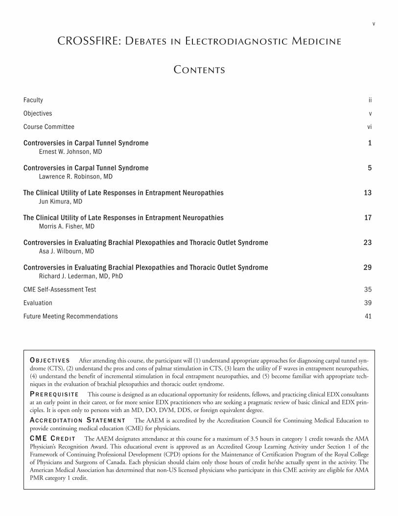
This author believes that comparing the median sensory nerve
latency to the radial nerve latency to the thumb is the best and
most reliable screen for CTS (Figure 1). A positive screen is in-
dicated with a difference of greater than .3 ms assuming that the
sensory nerve action potential (SNAP) is within normal limits—
35 µV for the median nerve and 10-15 µV for the radial nerve.
If latencies are within normal limits but amplitudes are small,
then one must consider a brachial plexus compromise or a lesion
of C6 nerve root distal to dorsal ganglion.
Figure 1 CALIBR: 1 ms; 20 µV; Top trace: stimulate radial nerveat the wrist: record digit 1; Middle trace: both median and radialnerves were stimulated at the wrist; With CTS, separation of twopeaks will be greater producing the “Bactrian Sign;” Bottom trace:stimulate median nerve at the wrist: record digit 1
WHAT IS AN “ADEQUATE” ELECTRODIAGNOSTICEXAMINATION?
Both the motor and sensory nerve fibers must be evaluated prox-
imal and distal to the carpal tunnel. This concept is controver-
sial because of the difficulties in stimulating and recording with
this technique. This should include recording the SNAP over
digit 3 with stimulation at the wrist (14 cm) and comparing it
to the SNAP with stimulation at mid-palm (7 cm).
The physician should examine the amplitude, duration, and
latency of the two segments. Usually the wrist to midpalm
segment (14-7 cm) will have slightly more than half of the total
latency. This is because of the smaller diameter of the distal
portion of the nerve and because the finger is cooler than the
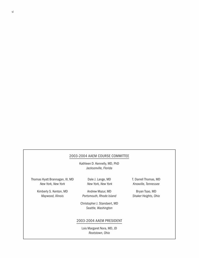
palm. For the latter reason, SNAP amplitude at the palmar will
normally be greater than 30% larger than the SNAP amplitude
when stimulating at the wrist (Figure 2). If the hand is cool, all
three parameters will be amplified—amplitude, latency, and du-
ration of SNAP.
The median compound muscle action potential (CMAP) is
evaluated by stimulating the median nerve fibers at the wrist (8
cm) as well as distal to the carpal ligament at the recurrent
branch of the median nerve and recording over the APB.
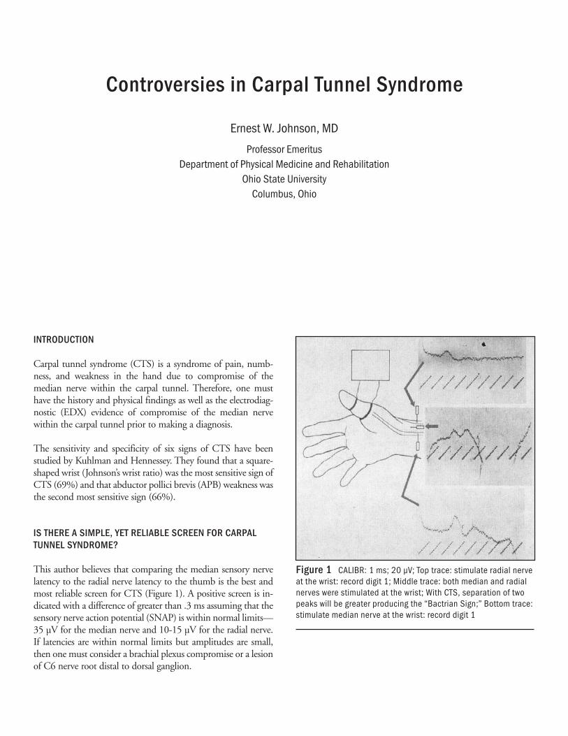
This mid-palmar stimulation is technically difficult because the
ulnar nerve could unintentionally be stimulated and the portion
of the thenar muscles innervated by the ulnar nerve (usually
deep head of flexor pollicis brevis) can contribute to the ampli-
tude of the CMAP recorded (Figure 3).
To ensure that the ulnar nerve is NOT accidentally stimulated,
note the shape and duration of the compound muscle action po-
tential (CMAP). The duration of the negative component of the
CMAP of a single thenar muscle will be less than 5 ms.
The shape must be the same at both wrist and palmar stimula-
tion. If the ulnar nerve is unintentionally stimulated in the palm,
the amplitude will be increased, but CMAP shape will differ and
the negative component of the CMAP will have increased in du-
ration.
While it is not especially difficult to stimulate only the median
nerve at the palmar site with surface bipolar electrodes, the
cathode must be tucked into the thenar muscle and the anode
rotated minimally. The CMAP shape and duration should be
noted.
It is easy to restrict the stimulus to the recurrent branch of the
median nerve using a monopolar needle electrode for stimula-
tion. The anode can be a large-surface electrode on the medial
dorsal hand, or this author has used a ring electrode on base of
digit 5.
2 Controversies in Carpal Tunnel Syndrome AAEM Course
Figure 2 CALIBR: 1 ms; 20 µV : top: digit 3 SNAP stimulatemedian nerve wrist (14 cm), Bottom: digit 3 SNAP stimulatemedian nerve at midpalm (7 cm). Top trace: stimulate mediannerve at wrist; record digit 3 (7 cm); Bottom trace: stimulatemedian nerve at wrist (14 cm); Note the increase in amplitude,latency, and duration. CALIBR: each slanted line = 1 ms; 20 µV
Figure 3 CALIBR: on figure
If the CMAP is larger (greater than 10%) with distal stimula-
tion, it is likely that there is a neurapraxic block of some of the
median motor fibers within the carpal tunnel. There are persua-
sive studies in the literature showing this phenomenon.
Unfortunately, the palm to wrist (8 cm) transcarpal mixed nerve
study which compares median and ulnar nerve latencies con-
taines both motor and sensory fibers of both nerves. This tech-
nique therefore would not differentiate CTS which
compromises only motor or only sensory axons of the nerve.
Kimura suggests that as many as 10% of CTS patients have only
motor fibers involved.
This author and colleagues have reported “acute” CTS with
largely or solely motor fibers compromised. These have usually
been individuals who have repetitive, vigorous, or continuous
grasping for 6-8 hours per day and several days in a row. Two ex-
amples are a high school hockey player who played 6-8 hours a
day for 5 days consecutively, and several auto painters who
squeezed a paint gun in a new job for 3-4 weeks in a row.
WHAT ABNORMALITIES CONFIRM CARPAL TUNNELSYNDROME?
Absolute latencies are not reliably diagnostic and are never prog-
nostic! To be more effective, one should compare latencies of an
uninvolved nerve. One should also compare latencies from the
wrist to the finger to latencies of the midpalm to the finger to re-
liably show slowing at the carpal tunnel. Evaluation of ampli-
tudes are best for diagnosis and prognosis. This should be
performed by comparing stimulation both proximal and distal
to the carpal tunnel. Severity and prognosis is best determined
by amplitude when stimuation is distal to the carpal tunnel.
CAN CARPAL TUNNEL SYNDROME BE DIAGNOSED IN THEPRESENCE OF DIABETIC POLYNEUROPATHY?
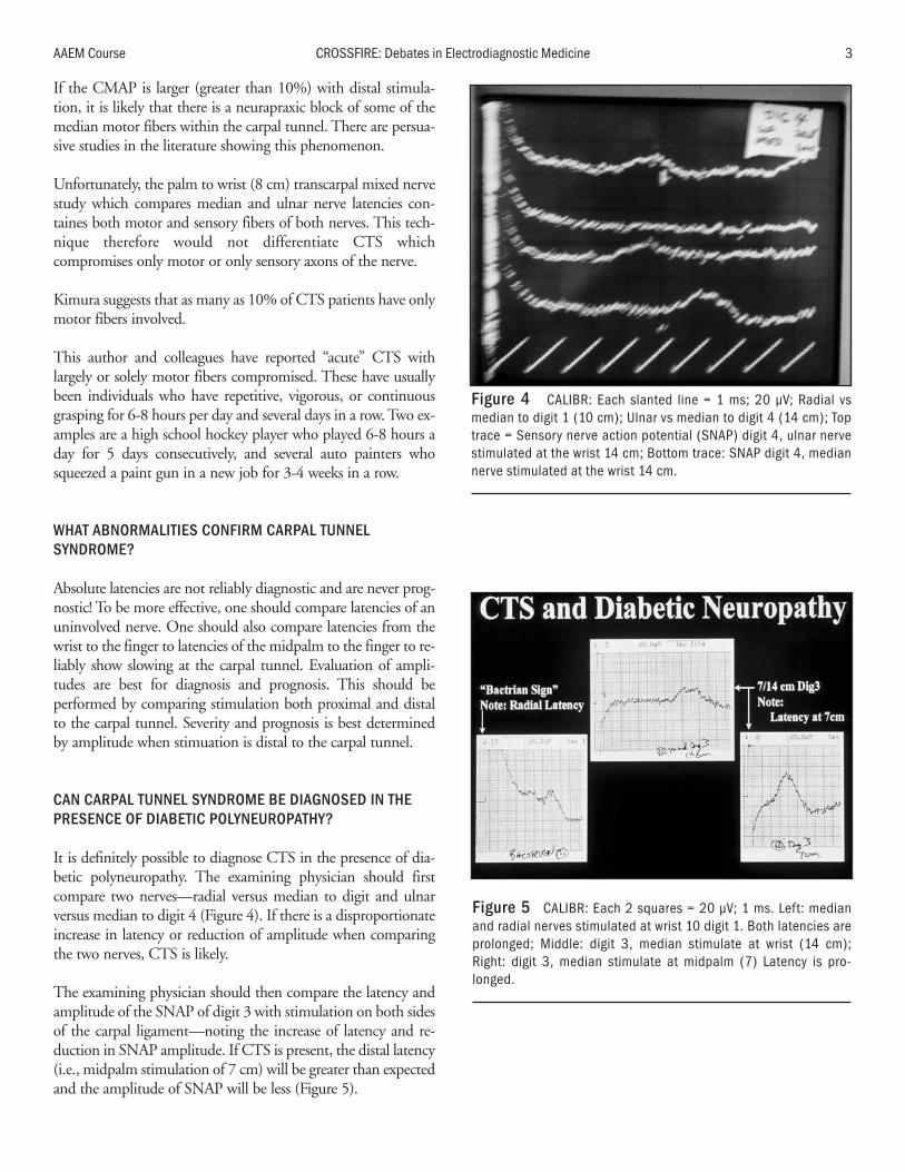
It is definitely possible to diagnose CTS in the presence of dia-
betic polyneuropathy. The examining physician should first
compare two nerves—radial versus median to digit and ulnar
versus median to digit 4 (Figure 4). If there is a disproportionate
increase in latency or reduction of amplitude when comparing
the two nerves, CTS is likely.
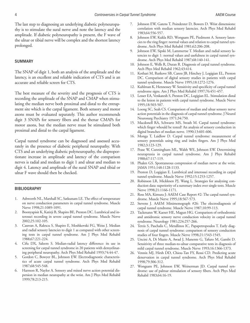
The examining physician should then compare the latency and
amplitude of the SNAP of digit 3 with stimulation on both sides
of the carpal ligament—noting the increase of latency and re-
duction in SNAP amplitude. If CTS is present, the distal latency
(i.e., midpalm stimulation of 7 cm) will be greater than expected
and the amplitude of SNAP will be less (Figure 5).
AAEM Course CROSSFIRE: Debates in Electrodiagnostic Medicine 3
Figure 4 CALIBR: Each slanted line = 1 ms; 20 µV; Radial vsmedian to digit 1 (10 cm); Ulnar vs median to digit 4 (14 cm); Toptrace = Sensory nerve action potential (SNAP) digit 4, ulnar nervestimulated at the wrist 14 cm; Bottom trace: SNAP digit 4, mediannerve stimulated at the wrist 14 cm.
Figure 5 CALIBR: Each 2 squares = 20 µV; 1 ms. Left: medianand radial nerves stimulated at wrist 10 digit 1. Both latencies areprolonged; Middle: digit 3, median stimulate at wrist (14 cm);Right: digit 3, median stimulate at midpalm (7) Latency is pro-longed.
The last step to diagnosing an underlying diabetic polyneuropa-
thy is to stimulate the sural nerve and note the latency and the
amplitude. If diabetic polyneuropathy is present, the F wave of
the ulnar or tibial nerve will be complex and the shortest latency
prolonged.
SUMMARY
The SNAP of digit 1, both an analysis of the amplitude and the
latency, is an excellent and reliable indication of CTS and is an
accurate and reliable screen for CTS.
The best measure of the severity and the prognosis of CTS is
recording the amplitude of the SNAP and CMAP when stimu-
lating the median nerve both proximal and distal to the entrap-
ment site which is the carpal ligament. Both sensory and motor
axons must be evaluated separately. This author recommends
digit 3 SNAPs for sensory fibers and the thenar CMAPs for
motor axons, but the median nerve must be stimulated both
proximal and distal to the carpal ligament.
Carpal tunnel syndrome can be diagnosed and assessed accu-
rately in the presence of diabetic peripheral neuropathy. With
CTS and an underlying diabetic polyneuropathy, the dispropor-
tionate increase in amplitude and latency of the comparison
nerves is radial and median to digit 1 and ulnar and median to
digit 4. Latency and amplitude of the sural SNAP and tibial or
ulnar F wave should then be checked.
BIBLIOGRAPHY
1. Ashworth NL, Marshall SC, Satkunam LE. The effect of temperatureon nerve conduction parameters in carpal tunnel syndrome. MuscleNerve 1998;21:1089-1091.
2. Boonyapisit K, Katirji B, Shapiro BE, Preston DC. Lumbrical and in-terossei recording in severe carpal tunnel syndrome. Muscle Nerve2002;25:102-105.
3. Cassvan A, Ralescu S, Shapiro E, Moshkovski FG, Weiss J. Medianand radial sensory latencies to digit 1 as compared with other screen-ing tests in carpal tunnel syndrome. Am J Phys Med Rehabil1988;67:221-224.
4. Cifu DX, Saleem S. Median-radial latency difference: its use inscreening for carpal tunnel syndrome in 20 patients with demyelinat-ing peripheral neuropathy. Arch Phys Med Rehabil 1993;74:44-47.
5. Gordon C, Bowyer BL, Johnson EW. Electrodiagnostic characteris-tics of acute carpal tunnel syndrome. Arch Phys Med Rehabil1987;68:545-548.
6. Harmon R, Naylor A. Sensory and mixed nerve action potential dis-persion in median neuropathy at the wrist. Am J Phys Med Rehabil1999;78:213-215.
7. Johnson EW, Gatens T, Poindexter D, Bowers D. Wrist dimensions:correlation with median sensory latencies. Arch Phys Med Rehabil1983;64:556-557.
8. Johnson EW, Kukla RD, Wongsam PE, Piedmont A. Sensory laten-cies to the ring finger: normal values and relation to carpal tunnel syn-drome. Arch Phys Med Rehabil 1981;62:206-208.
9. Johnson EW, Sipski M, Lammertse T. Median and radial sensory la-tencies to digit 1: normal values and usefulness in carpal tunnel syn-drome. Arch Phys Med Rehabil 1987;68:140-141.
10. Johnson E, Wells R, Duran R. Diagnosis of carpal tunnel syndrome.Arch Phys Med Rehabil 1962;43:414.
11. Kothari M, Rutkove SB, Caress JB, Hinchey J, Logigian EL, PrestonDC. Comparison of digital sensory studies in patients with carpaltunnel syndrome. Muscle Nerve 1995;18:1272-1276.
12. Kuhlman K, Hennessey W. Sensitivity and specificity of carpal tunnelsyndrome signs. Am J Phys Med Rehabil 1997;76:451-457.
13. Lesser EA, Venkatesh S, Preston DC, Logigian EL. Stimulation distalto the lesion in patients with carpal tunnel syndrome. Muscle Nerve1995;18:503-507.
14. Loong SC, Seah CS. Comparison of median and ulnar sensory nerveaction potentials in the diagnosis of carpal tunnel syndrome. J NeurolNeurosurg Psychiatry 1971;34:750.
15. Macdonell RA, Schwartz MS, Swash M. Carpal tunnel syndrome:which finger whould be tested? An analysis of sensory conduction indigital branches of median nerve. 1990;13:601-606.
16. Monga T, Laidlow D. Carpal tunnel syndrome: measurement ofsensory potentials using ring and index fingers. Am J Phys Med1982;3:123-129.
17. Pease W, Cunningham ML, Walsh WE, Johnson EW. Determiningneurapraxia in carpal tunnel syndrome. Am J Phys Rehabil1988;67:117-119.
18. Phalen GS. Spontaneous compression of median nerve at the wrist.JAMA 1951;148:1128-1133.
19. Preston D, Logigian E. Lumbrical and interossei recording in carpaltunnel syndrome. Muscle Nerve 1992;15:1253-1257.
20. Robinson LR, Micklesen PJ, Wang L. Strategies for analyzing con-duction data: superiority of a summary index over single tests. MuscleNerve 1998;21:1166-1171.
21. Ross MA, Kimura J: AAEM Case Report #2: The carpal tunnel syn-drome. Muscle Nerve 1995;18:567-573.
22. Stevens J. AAEM Minimonograph #26: The electrodiagnosis ofcarpal tunnel syndrome. Muscle Nerve 1987;10:99-113.
23. Tackmann W, Kaeser HE, Magun HG. Comparison of orthodromicand antidromic sensory nerve conduction velocity in carpal tunnelsyndrome. Neurology 1981;224:257-266.
24. Terzis S, Paschalis C, Metallinos IC, Papapetropoulos T. Early diag-nosis of carpal tunnel syndrome: comparison of sensory conductionstudies of four fingers. Muscle Nerve 1998;21:1543-1545.
25. Uncini A, Di Muzio A, Awad J, Manente G, Tafuro M, Gambi D.Sensitivity of three median-to-ulnar comparative tests in diagnosis ofmild carpal tunnel syndrome. Muscle Nerve 1993;16:1366-1373.
26. Vennix MJ, Hirsh DD, Chiou-Tan FY, Rossi CD. Predicting acutedenervation in carpal tunnel syndrome. Arch Phys Med Rehabil1998;79:306-312.
27. Wongsam PE, Johnson EW, Weinerman JD. Carpal tunnel syn-drome: use of palmar stimulation of sensory fibers. Arch Phys MedRehabil 1983;64:16-19.
4 Controversies in Carpal Tunnel Syndrome AAEM Course
Controversies in Carpal Tunnel Syndrome
Lawrence R. Robinson, MD
Professor and ChairDepartment of Rehabilitation Medicine
University of Washington School of MedicineSeattle, Washington
INTRODUCTION
Although carpal tunnel syndrome (CTS) is the most frequently
seen entrapment neuropathy in the electrodiagnostic (EDX) lab-
oratory, there are multiple and varied approaches that have been
described for diagnosing this condition. The lack of uniformity
in approach suggests that there are likely many unanswered
questions on how to best diagnose patients with this condition.
To determine the best approach, it is helpful to first list the cri-
teria for the best testing strategy. The best EDX testing approach
should be (in descending order of priority):
a. Specific (few false positives)
b. Sensitive (few false negatives)
c. Reliable (the same results on repeat testing)
d. Resistant to temperature effects
e. Efficient
As will be outlined, the best way to address these needs for diag-
nosing CTS is by using the combined sensory index (CSI), the
modified CSI, median motor nerve conduction studies (NCSs)
(don’t stimulate in the palm!), and occasionally needle elec-
tromyography (EMG).
WHAT IS THE COMBINED SENSORY INDEX?
The CSI is a combination of three sensory conduction compar-
isons.6 These include: median-radial sensory latency difference
to the thumb at 10 cm (thumbdiff), median-ulnar sensory
latency difference to the ring finger at 14 cm (ringdiff), and
median-ulnar sensory latency difference across the palm at 8 cm
(palmdiff). The CSI is calculated by simply adding the three
latency differences (median minus ulnar, or median minus
radial):
CSI = palmdiff + ringdiff + thumbdiff
WHAT IS THE SPECIFICITY OF INDIVIDUAL NERVECONDUCTION STUDIES AND THE COMBINED SENSORYINDEX IN THE DIAGNOSIS OF CARPAL TUNNEL SYNDROME?
Although sensitivity of various tests for CTS is often discussed,1,2
specificity is just as important or probably more important. It is
at least as important to avoid operating on a patient without
CTS, as it is to miss operating on someone with mild disease. In
5
order to measure and obtain high specificity, it is critical in any
study to have a healthy control group either studied concurrently
or studied in the same laboratory by the same investigators.
Ideally, the control group should be similar in age and other de-
mographic variables to the patient with the disease.
Usually, when collecting reference values from a control popula-
tion, one should include approximately 97.5% of the healthy
control subjects within the reference range. This is usually ac-
complished by measuring the mean value, standard deviation,
and taking mean plus or minus two standard deviations as the
reference values. This technique is appropriate if the latencies are
distributed in a Gaussian fashion. Unfortunately, a Gaussian dis-
tribution often is not present and other techniques such as per-
centile estimation or transformation are required to establish
reliable control values.7 Even after reference values are obtained,
subsequent studies of the specificity of each technique should be
measured using a control group—it cannot be assumed to be
95% simply because the mean ± 2 standard deviations from a
reference group was used. For this reason, it is important to
study a concurrent control group and measure the actual speci-
ficity, i.e., how many of the control subjects came out normal
(true negatives) or abnormal (false positives).
The specificity and sensitivity of a technique is, of course, a func-
tion of where reference (normal) values are set. When these are
set closer to the mean of the control group, sensitivity goes up
and specificity goes down; the converse is true as the reference
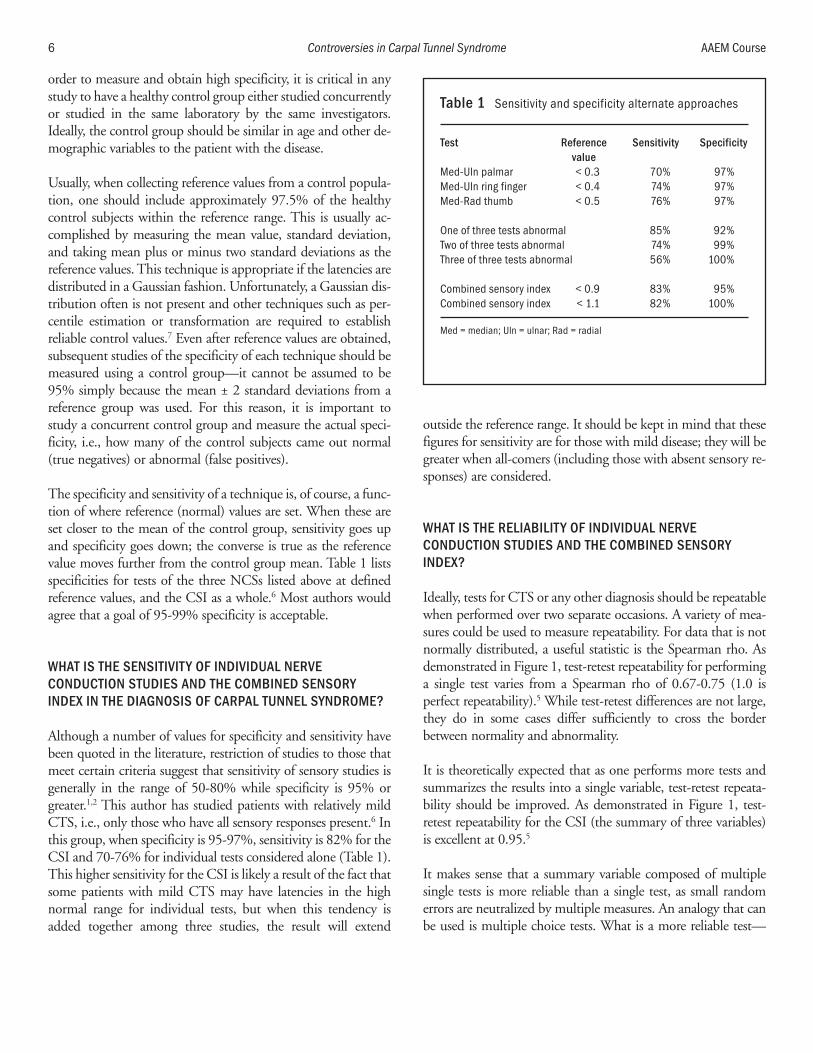
value moves further from the control group mean. Table 1 lists
specificities for tests of the three NCSs listed above at defined
reference values, and the CSI as a whole.6 Most authors would
agree that a goal of 95-99% specificity is acceptable.
WHAT IS THE SENSITIVITY OF INDIVIDUAL NERVECONDUCTION STUDIES AND THE COMBINED SENSORYINDEX IN THE DIAGNOSIS OF CARPAL TUNNEL SYNDROME?
Although a number of values for specificity and sensitivity have
been quoted in the literature, restriction of studies to those that
meet certain criteria suggest that sensitivity of sensory studies is
generally in the range of 50-80% while specificity is 95% or
greater.1,2 This author has studied patients with relatively mild
CTS, i.e., only those who have all sensory responses present.6 In
this group, when specificity is 95-97%, sensitivity is 82% for the
CSI and 70-76% for individual tests considered alone (Table 1).
This higher sensitivity for the CSI is likely a result of the fact that
some patients with mild CTS may have latencies in the high
normal range for individual tests, but when this tendency is
added together among three studies, the result will extend
outside the reference range. It should be kept in mind that these
figures for sensitivity are for those with mild disease; they will be
greater when all-comers (including those with absent sensory re-
sponses) are considered.
WHAT IS THE RELIABILITY OF INDIVIDUAL NERVECONDUCTION STUDIES AND THE COMBINED SENSORYINDEX?
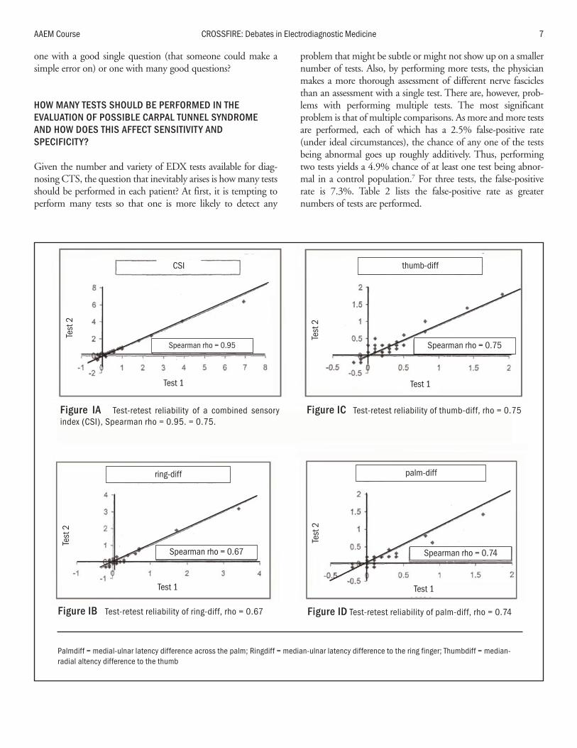
Ideally, tests for CTS or any other diagnosis should be repeatable
when performed over two separate occasions. A variety of mea-
sures could be used to measure repeatability. For data that is not
normally distributed, a useful statistic is the Spearman rho. As
demonstrated in Figure 1, test-retest repeatability for performing
a single test varies from a Spearman rho of 0.67-0.75 (1.0 is
perfect repeatability).5 While test-retest differences are not large,
they do in some cases differ sufficiently to cross the border
between normality and abnormality.
It is theoretically expected that as one performs more tests and
summarizes the results into a single variable, test-retest repeata-
bility should be improved. As demonstrated in Figure 1, test-
retest repeatability for the CSI (the summary of three variables)
is excellent at 0.95.5
It makes sense that a summary variable composed of multiple
single tests is more reliable than a single test, as small random
errors are neutralized by multiple measures. An analogy that can
be used is multiple choice tests. What is a more reliable test—
6 Controversies in Carpal Tunnel Syndrome AAEM Course
Table 1 Sensitivity and specificity alternate approaches
Test Reference Sensitivity Specificityvalue
Med-Uln palmar < 0.3 70% 97%Med-Uln ring finger < 0.4 74% 97%Med-Rad thumb < 0.5 76% 97%
One of three tests abnormal 85% 92%Two of three tests abnormal 74% 99%Three of three tests abnormal 56% 100%
Combined sensory index < 0.9 83% 95%Combined sensory index < 1.1 82% 100%
Med = median; Uln = ulnar; Rad = radial
one with a good single question (that someone could make a
simple error on) or one with many good questions?
HOW MANY TESTS SHOULD BE PERFORMED IN THEEVALUATION OF POSSIBLE CARPAL TUNNEL SYNDROMEAND HOW DOES THIS AFFECT SENSITIVITY ANDSPECIFICITY?
Given the number and variety of EDX tests available for diag-
nosing CTS, the question that inevitably arises is how many tests
should be performed in each patient? At first, it is tempting to
perform many tests so that one is more likely to detect any
problem that might be subtle or might not show up on a smaller
number of tests. Also, by performing more tests, the physician
makes a more thorough assessment of different nerve fascicles
than an assessment with a single test. There are, however, prob-
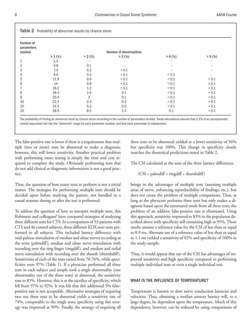
lems with performing multiple tests. The most significant
problem is that of multiple comparisons. As more and more tests
are performed, each of which has a 2.5% false-positive rate
(under ideal circumstances), the chance of any one of the tests
being abnormal goes up roughly additively. Thus, performing
two tests yields a 4.9% chance of at least one test being abnor-
mal in a control population.7 For three tests, the false-positive
rate is 7.3%. Table 2 lists the false-positive rate as greater
numbers of tests are performed.
AAEM Course CROSSFIRE: Debates in Electrodiagnostic Medicine 7
Figure IB Test-retest reliability of ring-diff, rho = 0.67 Figure ID Test-retest reliability of palm-diff, rho = 0.74
CSI
Test 1
Test 1
Test 1
Test 1
Test
2Te
st 2
Test
2Te
st 2
Spearman rho = 0.95
Spearman rho = 0.67
Spearman rho = 0.75
Spearman rho = 0.74
thumb-diff
ring-diff palm-diff
Figure IA Test-retest reliability of a combined sensoryindex (CSI), Spearman rho = 0.95. = 0.75.
Figure IC Test-retest reliability of thumb-diff, rho = 0.75
Palmdiff = medial-ulnar latency difference across the palm; Ringdiff = median-ulnar latency difference to the ring finger; Thumbdiff = median-radial altency difference to the thumb
The false-positive rate is lower if there is a requirement that mul-
tiple (two or more) tests be abnormal to make a diagnosis,
however, this will lower sensitivity. Another practical problem
with performing more testing is simply the time and cost re-
quired to complete the study. Obviously performing tests that
do not add clinical or diagnostic information is not a good prac-
tice.
Thus, the question of how many tests to perform is not a trivial
matter. The strategies for performing multiple tests should be
decided upon before studying the patient, not handled in a
casual manner during or after the test is performed.
To address the question of how to interpret multiple tests, this
Robinson and colleagues6 have compared strategies of analyzing
three different tests for CTS. In a comparison of 53 patients with
CTS and 46 control subjects, three different EDX tests were per-
formed in all subjects. This included latency difference with
mid-palmar stimulation of median and ulnar nerves recording at
the wrist (palmdiff), median and ulnar nerve stimulation with
recording over the ring finger (ringdiff), and median and radial
nerve stimulation with recording over the thumb (thumbdiff).
Sensitivities of each of the tests varied from 70-76%, while speci-
ficities were 97% (Table 1). If a physician performed all three
tests in each subject and simply took a single abnormality (one
abnormality out of the three tests) as abnormal, the sensitivity
rose to 85%. However, this is at the sacrifice of specificity, which
fell from 97% to 92%. It was felt that this additional 5% false-
positive rate is not acceptable. Alternative strategies of requiring
two out three tests to be abnormal yields a sensitivity rate of
74%, comparable to the single tests; specificity using this strat-
egy was improved at 99%. Finally, the strategy of requiring all
three tests to be abnormal yielded at a lower sensitivity of 56%
but specificity was 100%. This change in specificity closely
matches the theoretical predictions noted in Table 2.
The CSI calculated as the sum of the three latency differences
(CSI = palmdiff + ringdiff + thumbdiff)
brings in the advantages of multiple tests (assessing multiple
areas of nerve, enhancing reproducibility of findings, etc.), but
does not create the problem of multiple comparisons. Thus, as
long as the physician performs three tests but only makes a di-
agnosis based upon the summated result from all three tests, the
problem of an additive false-positive rate is eliminated. Using
this approach, sensitivity improved to 83% in the population de-
scribed above with specificity still remaining high at 95%. These
results assume a reference value for the CSI of less than or equal
to 0.9 ms. Alternate use of a reference value of less than or equal
to 1.1 ms yielded a sensitivity of 82% and specificity of 100% in
the study sample.
Thus, it would appear that use of the CSI has advantages of im-
proved sensitivity and high specificity compared to performing
multiple individual tests or even a single individual test.
WHAT IS THE INFLUENCE OF TEMPERATURE?
Temperature is known to slow nerve conduction latencies and
velocities. Thus, obtaining a median sensory latency will, to a
large degree, be dependent upon the temperature. Much of this
dependency, however, can be reduced by using comparisons of
8 Controversies in Carpal Tunnel Syndrome AAEM Course
Table 2 Probablity of abnormal results by chance alone
Number of parametersstudied Number of abnormalities
> 1 (%) > 2 (%) > 3 (%) > 4 (%) > 5 (%)1 2.5 - - - -2 4.9 0.1 - - -3 7.3 0.2 < 0.1 - -4 9.6 0.4 < 0.1 < 0.1 -5 11.9 0.6 < 0.1 < 0.1 < 0.16 14 0.9 < 0.1 < 0.1 < 0.17 16.2 1.2 < 0.1 < 0.1 < 0.18 18.3 1.6 0.1 < 0.1 < 0.19 20.4 2 0.1 < 0.1 < 0.110 22.4 2.5 0.2 < 0.1 < 0.115 31.5 5.2 0.5 < 0.1 < 0.120 39.5 8.5 1.3 0.1 < 0.1
The probability of finding an abnormal result by chance alone according to the number of parameters studied. These calculations assume that 2.5% of an asymptomaticcontrol population fall into the “abnormal” range for each parameter studied, and that each parameter is independent.
two nerves within the same limb. As noted in Table 3, tempera-
ture dependency of individual median nerve latencies is signifi-
cant.5 Comparisons of the median nerve with another nerve in
the same hand are not influenced by temperature. In addition,
the CSI (made up of comparisons between median and other
nerves in the same hand) is also not influenced significantly by
temperature. Thus, to avoid influences of temperature, as well as
age, height, and other influential variables, it is preferable to use
comparisons of median nerve latency to other nerves within the
same hand.
IS THE ENTIRE COMBINED SENSORY INDEX NEEDED IN ALLPATIENTS?
While the CSI represents an improvement over single tests, it has
been noted that it might not be necessary to perform all three
tests for the CSI when one or more are extreme values.
Specifically, if the median latency is slow compared to the ulnar
or radial nerve latency, additional testing might not help the di-
agnostic classification. Similarly, when results are “normal” addi-
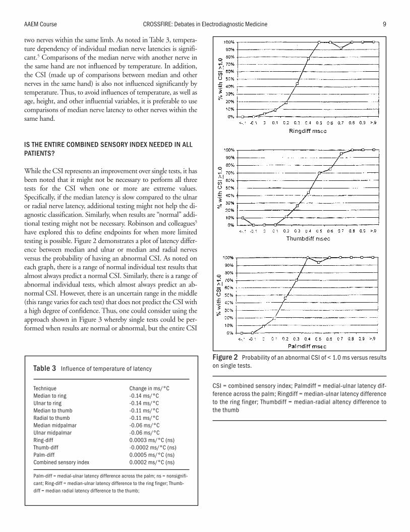
tional testing might not be necessary. Robinson and colleagues5
have explored this to define endpoints for when more limited
testing is possible. Figure 2 demonstrates a plot of latency differ-
ence between median and ulnar or median and radial nerves
versus the probability of having an abnormal CSI. As noted on
each graph, there is a range of normal individual test results that
almost always predict a normal CSI. Similarly, there is a range of
abnormal individual tests, which almost always predict an ab-
normal CSI. However, there is an uncertain range in the middle
(this range varies for each test) that does not predict the CSI with
a high degree of confidence. Thus, one could consider using the
approach shown in Figure 3 whereby single tests could be per-
formed when results are normal or abnormal, but the entire CSI
AAEM Course CROSSFIRE: Debates in Electrodiagnostic Medicine 9
Table 3 Influence of temperature of latency
Technique Change in ms/°CMedian to ring -0.14 ms/°CUlnar to ring -0.14 ms/°CMedian to thumb -0.11 ms/°CRadial to thumb -0.11 ms/°CMedian midpalmar -0.06 ms/°CUlnar midpalmar -0.06 ms/°CRing-diff 0.0003 ms/°C (ns)Thumb-diff -0.0002 ms/°C (ns)Palm-diff 0.0005 ms/°C (ns)Combined sensory index 0.0002 ms/°C (ns)
Palm-diff = medial-ulnar latency difference across the palm; ns = nonsignifi-cant; Ring-diff = median-ulnar latency difference to the ring finger; Thumb-diff = median radial latency difference to the thumb;
Figure 2 Probability of an abnormal CSI of < 1.0 ms versus resultson single tests.
CSI = combined sensory index; Palmdiff = medial-ulnar latency dif-ference across the palm; Ringdiff = median-ulnar latency differenceto the ring finger; Thumbdiff = median-radial altency difference tothe thumb
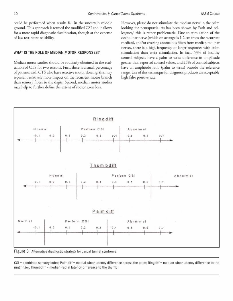
could be performed when results fall in the uncertain middle
ground. This approach is termed the modified CSI and it allows
for a more rapid diagnostic classification, though at the expense
of less test-retest reliability.
WHAT IS THE ROLE OF MEDIAN MOTOR RESPONSES?
Median motor studies should be routinely obtained in the eval-
uation of CTS for two reasons. First, there is a small percentage
of patients with CTS who have selective motor slowing; this may
represent relatively more impact on the recurrent motor branch
than sensory fibers to the digits. Second, median motor studies
may help to further define the extent of motor axon loss.
However, please do not stimulate the median nerve in the palm
looking for neurapraxia. As has been shown by Park and col-
leagues,4 this is rather problematic. Due to stimulation of the
deep ulnar nerve (which on average is 1.2 cm from the recurrent
median), and/or crossing anomalous fibers from median to ulnar
nerves, there is a high frequency of larger responses with palm
stimulation than wrist stimulation. In fact, 53% of healthy
control subjects have a palm to wrist difference in amplitude
greater than reported control values, and 25% of control subjects
have an amplitude ratio (palm to wrist) outside the reference
range. Use of this technique for diagnosis produces an acceptably
high false positive rate.
10 Controversies in Carpal Tunnel Syndrome AAEM Course
Figure 3 Alternative diagnostic strategy for carpal tunnel syndrome
CSI = combined sensory index; Palmdiff = medial-ulnar latency difference across the palm; Ringdiff = median-ulnar latency difference to thering finger; Thumbdiff = median-radial latency difference to the thumb
WHEN SHOULD NEEDLE ELECTROMYOGRAPHY OF THETHENAR MUSCLES BE PERFORMED?
There is considerable debate as to when needle EMG should be
performed in the evaluation of possible CTS. This test is quite
uncomfortable and it is also uncertain how the test results should
influence treatment or assessment of prognosis.
It is this author’s practice to perform thenar muscle needle EMG
in those patients who have abnormal median motor responses,
either in latency or amplitude, or those with a history of limb
trauma. This approach is designed to include predominantly
those patients who will have a higher likelihood of abnormali-
ties, and spare those patients from the procedure who would
have a low yield on the test. A cervical root screen is also per-
formed on those presenting with a history and/or physical ex-
amination suggestive of possible cervical radiculopathy.
RECOMMENDED APPROACH
The approach to diagnosing CTS should be well thought out
and strategized before the patient is referred to the EDX labora-
tory for testing. Specifically, the EDX consultant should know
which tests are to be performed, how many will be performed,
and how they will be interpreted to arrive at a diagnosis. Having
a strategy worked out ahead of time allows the examiner to have
a higher degree of confidence in the diagnosis.
This author recommends two alternative strategies. First, if the
question is simply diagnostic classification (abnormal or
normal), and repeat testing over time is not a likely considera-
tion, then the strategy outlined in Figure 3 is a reasonable ap-
proach. Specifically, an EDX consultant could perform studies
of the ring finger first and if results do not fall within the uncer-
tain range, then the testing is completed. If, however, the results
fall within the range of uncertainty, then the entire CSI should
be performed.
When test-retest reliability is a consideration and the examiner
wants to have a reliable result for the testing to be repeated, then
the CSI is preferable to perform on all patients. This is the ap-
proach used in this author’s laboratory because of the possibility
of repeat testing, should initial treatment prove unsuccessful or
should the patient ever present with symptoms after treatment.
Median motor conduction studies should also be performed in
all cases, but do not stimulate in the palm. Needle EMG should
be performed in cases in which the median motor response is ab-
normal in latency or amplitude, or if there is a history of trauma.
REFERENCES
1. American Association of Electrodiagnostic Medicine. Literaturereview of the usefulness of nerve conduction studies and needle elec-tromyography for the evaluation of patients with carpal tunnel syn-drome. Muscle Nerve 1999;22:S145-S167.
2. American Association of Electrodiagnostic Medicine, AmericanAcademy of Neurology, American Academy of Physical Medicineand Rehabilitation. Practice parameter for electrodiagnostic studies incarpal tunnel syndrome: summary statement. Muscle Nerve2002;25:918-922.
3. Lew H, Wang L, Robinson LR. Test-retest reliability of combinedsensory index: implications for diagnosing carpal tunnel syndrome.Muscle Nerve 2000;23:1261-1264.
4. Park TA, Welshofer JA, Dzwierzynski WW, Erickson SJ, Del ToroDR. Median “pseudoneurapraxia” at the wrist: reassessment ofpalmar stimulation of the recurrent median nerve. Arch Phys MedRehabil 2001;82:190-197.
5. Robinson LR, Micklesen PJ, Wang L. Optimizing number of tests forcarpal tunnel syndrome. Muscle Nerve 2000;23:1880-1882.
6. Robinson LR, Micklesen PJ, Wang L. Strategies for analyzing nerveconduction data: superiority of a summary index over single tests.Muscle Nerve 1998;21:1166-1171.
7. Robinson LR, Temkin NR, Fujimoto WY, Stolov WC. Impact of sta-tistical methodology on normal limits in nerve conduction studies.Muscle Nerve 1991;14:1084-1090.
AAEM Course CROSSFIRE: Debates in Electrodiagnostic Medicine 11

12 AAEM Course
13
The Clinical Utility of Late Responses inEntrapment Neuropathies
Jun Kimura, MD
Professor EmeritusKyoto University
Kyoto, JapanProfessor
Department of NeurologyUniversity of Iowa Health Center
Iowa City, Iowa
INTRODUCTION
With steady improvement and standardization of methods,11
nerve conduction studies (NCSs) have become a reliable means
of testing peripheral nerve function. They supplement clinical
observation by precisely localizing the lesion and characterizing
conduction abnormalities. Delineating the extent and distribu-
tion of the neural lesion by this means also helps quantitate the
degree of involvement. Optimal results can be expected only
with the proper choice of techniques, which in turn depends on
the type of lesions under consideration. Thus, the studies must
be conducted as an extension of the clinical examination, rather
than as a laboratory test.
This debate concerns the possible use of F waves in the evalua-
tion of a radiculopathy, which represents a focal, in contrast to
diffuse, involvement of the peripheral nerve. This manuscript
will first describe the general principle of NCSs specifically
dealing with the length of nerve segment under study. As a
natural consequence of this argument, one must conclude that
F-wave latencies cannot serve as a useful measure of radiculopa-
thy simply because of its focal nature. Other negative aspects of
F-wave study when used to diagnose radiculopathy, as well as a
more general discussion on proper application of F-wave mea-
surement for the study of diffuse neuropathic process will be dis-
cussed.
PRINCIPLES OF NERVE CONDUCTION STUDIES FOR FOCALLESIONS
A question often posed but rarely tested relates to the length of
the nerve segment under study to increase the yields of NCSs.7
Should one study a shorter or longer nerve segment for better
results?
Ordinary NCSs suffice to approximate the site of involvement
in a focal lesion. When more precise localization is required,
inching the stimulus in short increments in the range of 1 to
several centimeters along the course of the nerve can further
isolate the site of involvement within the affected segment.7 In
the evaluation of a focal lesion, studies of a longer segment lower
the sensitivity of the test because the inclusion of the unaffected
segments in the calculation dilutes the effect of slowing at the site
of the lesion. In contrast, studying a shorter segment helps isolate
a localized abnormality and provides better resolution of re-
stricted lesions that may otherwise escape detection. Assume a
nerve impulse conducting at a rate of 0.2 ms/cm (50 m/s) except
for a 1-cm segment where demyelination has doubled the con-
duction time to 0.4 ms/cm. In a 10-cm segment, normally
covered in 2 ms, a 0.2 ms increase would constitute a 10%
change, or approximately 1 standard deviation, well within the
normal range of variability. The same 0.2-ms increase, however,
represents a 100% change in latency if measured over a 1-cm
14 The Clinical Utility of Late Responses in Entrapment Neuropathies AAEM Course
segment, signaling a clear abnormality. The large per unit in-
crease in latency more than compensates for the inherent mea-
surement error associated with multiple stimulation in short
increments.4
An excessive latency increase may result from inaccurate ad-
vances of the stimulating electrodes or inadvertent spread of
stimulus current, activating a less affected, and consequently
more excitable, neighboring segment. Thus, an abrupt change in
the waveform of the recorded response provides an additional
(and perhaps more convincing) finding that nearly always ac-
companies a latency increase across the site of compression. In
fact, waveform analysis often localizes a focal lesion unequivo-
cally even in the absence of an abnormal latency prolongation.
This technique helps not only in assessing a possible compressive
lesion such as carpal tunnel syndrome at the wrist,5,8,13 ulnar
neuropathy at the elbow,1 and peroneal nerve entrapment at the
knee,4 but also in characterizing the focal nature of some wide-
spread abnormalities such as multifocal motor neuropathies.3 It
does not help in evaluating proximal lesions as might be seen in
radiculopathy.
LIMITATION OF F-WAVE STUDIES IN RADICULOPATHIES
In radiculopathy, the site of involvement lies too proximal to
assess the lesion by short incremental stimulation as described
above. The latency of an F wave elicited after stimulation of the
nerve proximally close to the lesion can isolate a relatively short
central loop that contains the site of involvement. With proxi-
mal stimulation, however, the F wave usually overlaps with the
M response, requiring a collision method to separate the two
components for latency determination.9 In addition, such a
central segment is not short enough to detect a focal radicular
lesion because a normally conducting unaffected segment dilutes
the abnormality.
F waves fall short of providing clinically useful information in
radiculopathy for a number of other reasons: (1) the minimum
F-wave latency will measure a surviving normal neuron in an in-
complete lesion, thus failing to test the affected neuron by sam-
pling error; (2) when recording from the intrinsic hand and foot
muscles, as customarily done, the study can target only C8 - T1
and S1 - S2 roots, precluding all the other levels from evaluation;
(3) F waves tested in nerves innervated by an unaffected root ob-
viously have no clinical relevance; and (4) F-wave abnormalities,
if detected in a patient with radiculopathy, have no localizing
value as the slowing may occur with lesions located distally or
proximally. Under these situations, the study, if abnormal, has
only limited clinical value in localizing the lesion and confirm-
ing the diagnosis of a radiculopathy.
SENSITIVITY OF F-WAVE STUDIES IN RADICULOPATHY
For all the reasons previously discussed, F-wave studies must, on
theoretical grounds, detect only small percentages of clinically
suspected cases of radiculopathy. In fact, although the F wave is
commonly used in the evaluation of suspected radiculopathies,
the yields have been disappointingly low. In one study,12 sensi-
tivity of the F wave in cervical radiculopathy ranged from 10-
20%. In this series, as in others, the needle examination was
abnormal more often than the F wave. F wave was abnormal in
10% of the 2093 patients who had clinical symptoms of cervi-
cal radiculopathy. This compares to 3% of the 1005 patients
who had normal clinical examinations. In some patients with
good clinical symptoms and abnormal needle study, 7% had ab-
normal F waves. Thus, the F wave was abnormal twice as often
in patients with clinical symptoms consistent with a radiculopa-
thy. In patients with an abnormal needle examination, indicat-
ing a C8 radiculopathy, the likelihood of finding an abnormal F
wave approached 20%. Patients with radiculopathies may show
statistically significant F-wave changes compared to control sub-
jects. A group difference does not suffice as a clinical diagnostic
measure that requires assessment of individual patients. F-wave
studies provide no additional information if a needle elec-
tromyography examination shows abnormalities consistent with
a radiculopathy. F-wave abnormalities found in patients with
normal needle studies also have limited clinical use because of
the lack of localizing value.
USEFULNESS OF F-WAVE LATENCIES FOR DIFFUSENEUROPATHIC CONDITIONS
In contrast to the usefulness of segmental studies for a focal
lesion, evaluation of a longer segment provides a better result in
assessing a more diffuse or multi-segmental process such as
polyneuropathies. A longer path has an advantage in accumulat-
ing all the segmental abnormalities, which individually might
not show a clear deviation from the normal range. Assume a
nerve impulse conducting at a rate of 0.2 ms/cm (50 m/s). A
20% delay for a 10 cm segment is only 0.4 ms, whereas the same
change for a 100 cm segment amounts to 4 ms, an obvious in-
crease for easy detection. Thus, in general, the longer the
segment studied, the more evident the conduction delay is for a
diffuse process. Evaluating a longer segment also improves the
overall accuracy because the same absolute measurement error
constitutes a smaller percentage of change in latency and dis-
tance.
In a study testing reproducibility of conduction studies,10 the
measures showing the range of relative intertrial variation (RIV)
within ± 10% included F-wave latency and F-wave conduction
velocity of both median and tibial nerves and sensory conduc-
tion velocity of the median nerve. In general, amplitudes showed
a greater RIV than latencies or nerve conduction velocities.
Similarly, intraclass correlation coefficiency (ICC) exceeded 0.9
for F-wave latency of the median and tibial nerves in both the
healthy subjects and the patients. In some measures, a large
among-subject variance of the amplitudes led to a high ICC
despite a considerable intertrial variability. These included the
amplitude of the median nerve sensory nerve potential and
median and tibial nerve compound muscle action potentials.
CONCLUSION
This author’s data indicate that the length of the nerve segment
under study dictates the accuracy and sensitivity of measure-
ment. Although studies of shorter or longer segments pose tech-
nical merits and demerits, the choice seems to depend entirely
on the pattern of the conduction abnormalities. As evidenced by
inching technique, a short segmental study uncovers focal
lesions involving a restricted zone better than the evaluation of a
longer distance, which tends to obscure the abnormality. In con-
trast, studies of a longer segment detect diffuse or multisegmen-
tal abnormalities better, increasing sensitivity and decreasing
measurement errors, which, in percentage, diminish in propor-
tion to the overall latency and surface distance. The increased ac-
curacy of the techniques in turn improves the reproducibility of
the results. Radiculopathies constitute a sharply focal lesion, but
its proximal location precludes the application of segmental
studies. Consequently, no currently available NCSs provide
useful information in this condition. This and other characteris-
tics make the use of F wave untenable as a measure of radicu-
lopathies.
When using NCSs, short distances magnify focal conduction
abnormalities despite increased measurement error; long dis-
tances, though insensitive to focal lesions, provide better yields
and reliability for a diffuse or multisegmental process. These
findings also underscore the importance of choosing nerve stim-
ulation techniques appropriate for the lesion identified clinically.
Thus, electrophysiologic studies are most useful when con-
ducted as an extension of the history and physical examination,
which provide the overall orientation for the subsequent physio-
logic evaluation.
REFERENCES
1. Campbell WW, Pridgeon RM, Sahni KS: Short segment incrementalstudies in the evaluation of ulnar neuropathy at the elbow. MuscleNerve 1992;15:1050-1054.
2. Geiringer SR. The value of inching techniques in the diagnosis offocal nerve lesions. Inching techniques are of limited use. MuscleNerve 1998;21:1557-1559.
3. Kaji R, Oka N, Tsuji S, Mezak T, Nishio T, Akiguchi I, Kimura J.Pathological findings at the site of conduction block in multifocalmotor neuropathy. Ann Neurol 1993;33:152-158.
4. Kanakamedala RV, Hong CZ. Peroneal nerve entrapment at the kneelocalized by short segment stimulation. Am J Phys Med Rehabil1989;68:116-122.
5. Kimura J. Facts, fallacies, and fancies of nerve conduction studies:twenty-first annual Edward H. Lambert Lecture. Muscle Nerve1997;20:777-787.
6. Kimura J. Kugelberg lecture: principles and pitfalls of nerve conduc-tion studies. Electroencephologr Clin Neurophysiol 1998;106:470-476.
7. Kimura J. Long and short of nerve conduction measures: repro-ducibility for sequential assessments. J Neurol Neurosurg Psychiatry2001;71:427-430.
8. Kimura J. The carpal tunnel syndrome: Localization of conductionabnormalities within the distal segment of the median nerve. Brain1979;102:619-635.
9. Kimura J, Butzer JF. F-wave conduction velocity in Guillain-Barrésyndrome: assessment of nerve segment between axilla and spinalcord. Arch Neurol 1975;32:524-529.
10. Kohara N, Kimura J, Kaji R, Goto Y, Ishii J. Multicenter analysis onintertrial variability of nerve conduction studies: healthy subjects andpatients with diabetic polyneuropathy. In: Kimura J, Shibasaki H,editors. Recent advances in clinical neurophysiology. Oxford:Elsevier; 1996. p 809-815.
11. Lambert EH. Diagnostic value of electrical stimulation of motornerves. Electroenceph Clin Neurophysiol 1962;22:S9-S16.
12. Rivner MH. The contemporary role of F-wave studies. F-wavestudies: limitations. Muscle Nerve 1998;21:1101-1104.
13. Seror P. Orthodromic inching test in mild carpal tunnel syndrome.Muscle Nerve 1998;21:1206-1208.
AAEM Course CROSSFIRE: Debates in Electrodiagnostic Medicine 15

16 AAEM Course
The Clinical Utility of Late Responses inEntrapment Neuropathies
Morris A. Fisher, MD
ProfessorDepartment of Neurology
Loyola University Stritch School of MedicineHines, Ilinois
INTRODUCTION
There is little work supporting the use of F waves in entrapment
neuropathies. It is the purpose of this manuscript to indicate that
there is convincing evidence that F waves should be helpful in
entrapment neuropathies.
F waves were originally described in the small muscles of the
foot20 and hence their name. The interpretation of F waves re-
quires an understanding of the electrogenesis of the F wave,
those factors that modify the F wave, and techniques for elicit-
ing the F wave.
THE PHYSIOLOGY OF THE F WAVE
It is now generally accepted that F waves are produced by an-
tidromic activation of motoneurons (MNs). In healthy subjects
the probability that any one motor unit (MU) will generate an
F wave is small. Some stimuli in a train may not be followed by
any F wave. Where F waves do follow the direct response, their
shape and size changes from stimulus to stimulus (Figure 1)
because motor unit action potentials (MUAPs) which generate
the F wave change with each successive stimulus.
Physiological Factors Influencing the Probability of an FWave
Electrical stimulation of peripheral nerves produces antidromic
activitation of motor nerve fibers as well as orthodromic im-
pulses in sensory fibers. Both might influence the excitability of
MNs and thereby the chance of an F wave. Ultimately, an F
wave is dependent on an orthodromic response following the an-
tidromic activation. This requires the orthodromic response to
traverse an MN initial segment that has previously been depo-
larized by the initial antidromic volley. As such, any factors
tending to speed up the recovery of the initial segment or which
delay or increase the magnitude of any antidromically induced
depolarization of the somadendritic membrane might well in-
crease the chance of an F wave. F waves may therefore be influ-
enced by differences in “central” excitability. For this reason, the
frequency of F-wave discharge following a series of stimuli (per-
sistence) is characteristically about 80-90% in antigravity
muscles such as the abductor pollicis brevis, calf, and abductor
hallucis, but only about 30-40% in the antigravity antagonists
such as the forearm extensors, tibialis anterior, and extensor dig-
itorum muscles.7,15
The question of a selective bias in the selection of MUs in F
waves is important and has been controversial. All studies ques-
tioning a selective activation of larger MUs in F waves have been
performed at submaximal stimulation in comparison to the
supramaximal stimulation used in the clinical situation. The di-
ameters of the largest nerve fibers in the ventral roots are roughly
twice those of the smallest ventral root fibers.5 Ranges of con-
ductions at least twice the 15-20% that are normally observed in
F waves have been noted in human motor fibers in individual
subjects.3 Changes in F-wave latency are similar to changes in
maximal evoked response latencies when the sites of stimulation
are changed. These observations would be consistent with a
17
selective bias of larger MUs in F waves. This is supported by
findings based on studies of single MUs in F waves as well as
analysis of F-wave conduction velocities.13 Fortunately, whatever
the situation, the observed variability in F-wave latencies is not
so random or great as to preclude their clinical use.
TECHNIQUES FOR STUDYING THE F WAVE
F waves are usually recorded using surface electrodes arranged in
a belly-tendon fashion with the active electrode positioned over
the innervation zone of the target muscle. F waves recorded from
distal muscles in the limbs are usually clearly separated from the
direct motor response (M wave). The more proximal the site of
stimulation, however, the greater the chance that F waves may be
obscured by the preceding direct M wave. This feature of F
waves provides a practical limitation of recording F waves from
proximal muscles.
Supramaximal stimulation should be used for recording F waves.
The size and persistence of F waves increase as the stimulus in-
tensity increases, and supramaximal stimulation provides a phys-
iologically consistent environment for analyzing other F-wave
parameters.
F waves may be affected by a previous conditioning stimulus.21
As such, F waves should be recorded at rates no faster than 0.5
Hz.
METHODS FOR ANALYZING F WAVES
Individual F waves are generated by the recurrent discharges of
anywhere from one to at the most a few MUs whose associated
MUAPs and latencies differ. This accounts for the small size of
the F wave relative to the size of the direct M potential. The
moment-to-moment changes in which MUs generate F waves
accounts for the inherently variable size, latency, and shape of the
F wave from stimulus to stimulus (Figure 1). This inherent vari-
ability also means that the analysis of F waves requires a series of
F waves and the evaluation of a number of parameters.
The most common way of assessing F waves has been to measure
the shortest latency F wave following a series of 10 or more
stimuli. Measuring the shortest F-wave latency, however, may be
difficult. In addition, individual F waves may overlap and be
confused with axon reflexes or A waves.29 A more reliable
method of comparing latencies in F waves is to calculate the
mean latency. The latter does not depend on correctly identify-
ing and measuring the latency of the shortest latency F wave, re-
flects the range of F latencies, and is more reproducible than
minimal latencies. For these reasons, mean latencies have been
recommended by a number of studies.4,8,11,26,30,33,39 Recent com-
puter-assisted analysis of F waves has used median latency values.
F-wave latencies will vary with limb length and, to a lesser
degree, age. Predicted F-wave latencies need to be adjusted for
height or limb length. This is done best by using regression equa-
tions that include height or limb length and age.
Chronodispersion refers to the difference between the minimal
and maximal latencies25 and thereby reflects the range of laten-
cies in a series of F waves. Persistence indicates the percentage of
discernible responses, (generally exceeding 20 mV), following a
series of stimuli. Identical responses in a series of F waves are
called repeater waves. Because of the variability of F waves, the
amplitudes of F wave are best measured as mean values and
related to the amplitude of the maximum M potential, i.e., the
mean F/M ratio. Normal values may be found in recent refer-
ences.9,28
18 The Clinical Utility of Late Responses in Entrapment Neuropathies AAEM Course
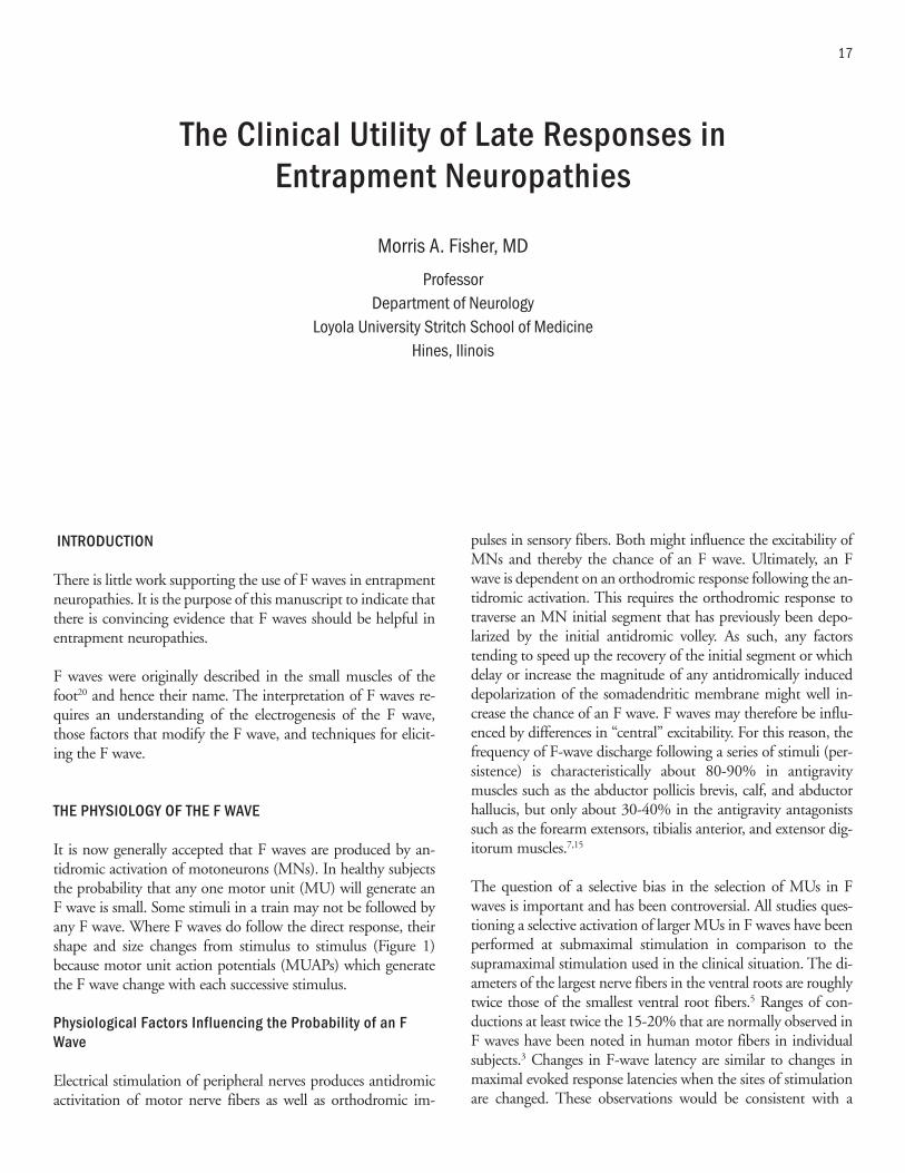
Figure 1 F waves (right) with the associated M waves (left) (A).The variability of F waves is emphasized with the responses super-imposed (B). Chronodispersion is the difference between theshortest and longest latencies. The persistence is 90%, i.e., 1 ofthe 10 responses is absent. The two largest F waves are repeaterwaves. Calibration per division: 5 mV for M waves, 500 µV for Fwaves; 5 ms.
Because of the inherent variability of F waves, sufficient F waves
must be collected to provide representative data. Ten stimuli
yielding anywhere from 7-10 F waves may suffice for most
studies of persistence and latencies. However, 20 or more stimuli
providing anywhere from 16-20 F waves may be needed for ac-
curate measurements.11,13,26,30 This can be important when com-
paring relatively small latency differences between sides, as may
be necessary in radiculopathies. A series of 20 F waves is also ad-
equate for the measuring mean F/M amplitude ratios and the
percentage of repeater waves. As few as two F waves may estab-
lish an abnormal chronodispersion if the separation in latency
between these two responses is greater than normal. Accurate
measurement of chronodispersion requires more than 20
stimuli, and may possibly require as many as 50-60 stimuli.11,23,25
At least 100 stimuli are required to determine the number of in-
dividual repeater waves. These data are based on recordings from
antigravity muscles. Additional stimuli will be needed if record-
ing from antigravity antagonists with their associated lower per-
sistences.
F WAVES AND ENTRAPMENT NEUROPATHIES
There is evidence supporting the value of F waves in entrapment
neuropathies. F-wave latencies may be prolonged in neu-
ropathies and may be abnormal even when peripheral motor
conduction studies are normal. F waves may also be more sensi-
tive than conventional motor conduction studies in axonal neu-
ropathies.12 Prolonged F-wave latencies exceeding 150% of the
upper limit of normal have been considered suggestive of de-
myelination as has the absence of F-waves in the presence of rel-
atively preserved maximum M potentials.12 F-wave latencies
have been reported to be the most stable and reliable measure-
ment for sequential nerve conduction studies in the same sub-
jects.17
F-wave parameters other than latency are important for defining
nerve dysfunction. Abnormal F waves have a high sensitivity in
acquired demyelinating neuropathies.12,16 This high sensitivity is
consistent with the effects of focal proximal demyelination.
Increased chronodispersion and decreased persistence may occur
in up to 50% of the nerves in patients with acquired demyeli-
nating polyneuropathies and may be the only abnormality in
those nerves.16 Repeater waves and mean F/M ratios are in-
creased in patients with axonal injury. The former is consistent
with a decreased MN pool available for activation, the latter with
increased motor unit size but decreased M-wave amplitudes.
Thoracic Outlet Syndrome
The potential value of F waves in entrapment neuropathies can
be surmised from one of the earliest reports dealing with this
issue.38 This study included methodological features required for
meaningful F-wave studies; namely, an adequate number of
stimuli and the evaluation of parameters other than latency. F
waves were recorded from hypothenar muscles in five patients
with true neurogenic thoracic outlet syndromes following 60
stimuli. F waves in the affected arms were prolonged by at least
2 ms in comparison to the unaffected side. The F waves in the
affected arms were also prolonged in comparison to data from
control subjects with the data related to arm length. F-wave per-
sistence was also decreased in the involved arms in comparison
to both the uninvolved arms as well as in control subjects. In one
subject, the F-wave latency prolongation resolved following
removal of the causative cervical band.
Carpal Tunnel Syndrome
Macleod19 compared F waves recorded from the abductor polli-
cis brevis (APB) in 52 healthy nerves and 147 nerves from pa-
tients with symptomatic carpal tunnel syndrome (CTS). All of
the patients had prolonged median sensory conductions across
the wrists. Analyses were based on responses following 100
supramaximal stimuli. The F-wave persistence was significantly
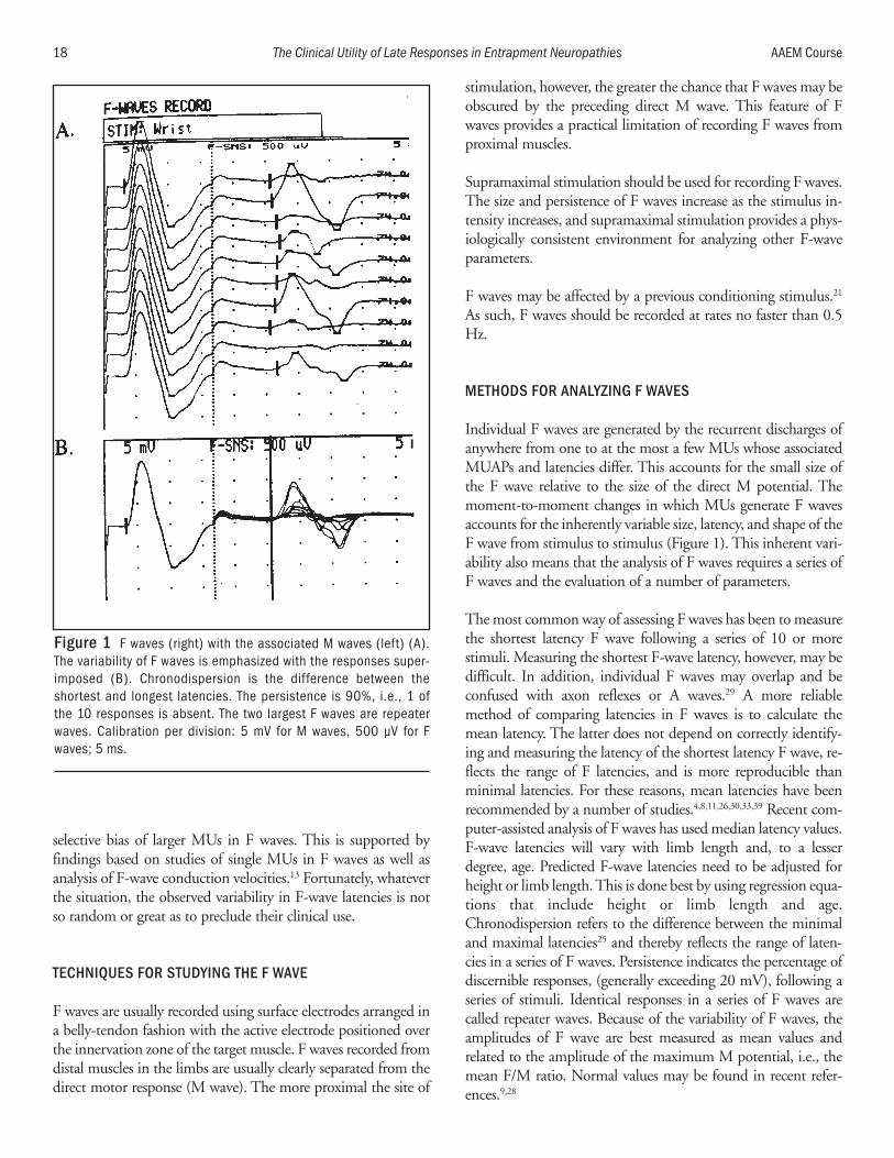
decreased (p<0.001) in the symptomatic nerves. The mean per-
sistence in the symptomatic nerves was 60% compared to about
83% in the normal nerves (Figure 2). Due to the wide range of
normal values, however, persistence was considered an insensi-
tive measure of CTS. Percent repeater waves (%RF) was also cal-
culated where %RF was the number of recurring identical F
waves divided by the total number of discernible F waves. In the
control group, the mean %RF value was about 15% while in
those with CTS, the mean %RF was about 60%. In 84.4% of
the studies in nerves with CTS, %RF was above the reference
90% upper limit of normal. In comparison, only 35% of
median distal motor latencies in the patients were prolonged.
The author concluded that in injured nerves there is a reduction
in the size of the alpha MN pool capable of generating F waves
and an enhanced backfiring capacity in that MN pool. Since
Macleod19 used 100 supramaximal stimuli, the clinical utility of
this method would be limited. Nevertheless, the study indicates
that F waves may be abnormal in CTS and could be clinically
helpful if one were interested in a sensitive measure of the inci-
dence of injury to motor fibers in patients with CTS.
Median F-wave latencies are usually 1-2 ms shorter than ulnar
F-wave latencies when recorded from hand muscles. “Inversion”
of this latency is abnormal and is consistent with CTS.22 This
inversion may be due to lesions proximal to the wrist but, in the
appropriate context, may readily provide evidence for injury to
motor fibers in CTS not otherwise available.
Carpal tunnel syndrome has provided a laboratory for defining
the potential effect of focal nerve injury on F waves. F-wave per-
sistence may be decreased where there is evidence of axonal
injury defined either by decreased M-wave amplitudes24 (60
supramaximal stimuli) or abnormal spontaneous activity10 (20
supramaximal stimuli). Chronodispersion may be significantly
AAEM Course CROSSFIRE: Debates in Electrodiagnostic Medicine 19
increased (p<0.025) in those patients with prominently pro-
longed median distal motor latencies (DML) (i.e., >5.7 ms in
comparison to an upper limit of normal of 4.2 ms) in compari-
son to those with lower DMLs. Repeater waves may be signifi-
cantly higher (p<0.001) in those with APB abnormal
spontaneous activity. F-wave amplitudes (mF/M ratios) are
almost always larger (p<0.001) in the hands with the more pro-
longed DMLs in those with bilateral CTS.10
F waves can also be used to define the pathophysiology of nerve
injury in CTS. Anastasapoulos and Chroni1 recorded F waves
following 40 supramaximal stimuli from both the APB and the
pronator quadratus (PQ). Needle electrodes were used for PQ
recordings. Median motor conduction velocities were signifi-
cantly slowed (p<0.0002) in the CTS patients in comparison to
the control subjects. Although minimal median F-wave latencies
were significantly prolonged in the CTS patients (p<0.001) in
comparison to the control subjects following elbow stimulation,
this was not true when recording from the PQ. The median
motor conduction velocity slowing proximal to the wrist in the
CTS patients was therefore believed to be due to injury to
median nerve fibers at the wrist. This methodology could be
helpful when there is a question of proximal nerve injury in pa-
tients with CTS.
The use of F waves to help define proximal injury in CTS was
also the basis of one of the earliest reports of F waves in entrap-
ment neuropathies.6 Regression lines with 99% confidence
limits were determined in normal subjects for M latency versus
minimal F-wave latency stimulating at the elbow and recording
from the APB. In 3 of 30 patients with CTS, the F latencies were
outside the 99% confidence limits consistent with an associated
proximal lesion.
Leffler and colleagues18 evaluated a large series of hands (203) in
patients with possible median nerve injury at the wrist. Based on
a multivariate logistic regression model using clinical, DML, and
F-wave latency information, the median F-wave latency has
been found to be an independent predictor of CTS study. The
“gold standard” for a diagnosis of CTS was the physician’s diag-
nosis after formal clinical and electrodiagnostic studies. These
data—both DML and F waves—were acquired using a stan-
dardized computer-based stimulation and recording system.
These types of systems and analyses—automated recordings and
multivariate regressions—may soon become routine and F
waves will be part of these evaluations.
Hemifacial Spasm
Hemifacial spasm (HFS) can be considered an entrapment neu-
ropathy due to vascular compression of the facial nerve in the
brainstem. F waves have been recorded in patients with HFS (32
supramaximal stimuli) recording from the mentalis muscle stim-
ulating the marginal mandibular branch of the facial nerve.14 In
this study, the low cutoff filter was set at 100 Hz in order to min-
imize interference from the associated M wave. In 14 patients, F
waves were suppressed during inhalation of anesthesia on the
normal side, but in only 4 of the 14 nerves with HFS. F-wave
suppression occurred following surgical decompression at the
same time as there was disappearance of the aberrant motor re-
sponses characteristic of HFS. In addition, F-wave studies of the
involved facial nerves revealed decreased persistences, F/M am-
plitude ratios, and response durations as well as increased laten-
cies after surgery in comparison to studies prior to surgery. These
studies, therefore, not only support increased facial nucleus ex-
citability in HFS and indicate a clinical use of F waves, they also
reinforce the discernible changes in F-wave parameters that may
occur with focal nerve entrapment.
20 The Clinical Utility of Late Responses in Entrapment Neuropathies AAEM Course
Figure 2 Median F waves recorded from the abductor pollicisbrevis. (A) A healthy subject with 2 repeater waves (%RF=12%). (Band C) F waves in patients with carpal tunnel syndrome with de-creased persistence and high frequency repeater waves.
A B C
Radiculopathies
Nerve roots are susceptible to focal injury due to limited colla-
gen supporting tissue and relatively fixed anatomical constraints
limiting the ability of nerve roots to move away from a site of
compression. As such, symptomatic root injury can be consid-
ered to fall under the rubric of entrapment neuropathies.
The role of F waves in radiculopathies has been controversial.
Theoretical arguments against the use of F waves have included
the long course of F waves and subsequent “dilution” of any
change in latency, as well as the multiple root innervation of
most muscles. The former argument is not supported by obser-
vations in patients with other types of focal proximal nerve
injury. The latter argument is particularly irrelevant since F-wave
parameters such as chronodispersion are arguably of unique
value in circumstances where some axons may be injured and
others not. Studies that have questioned the use of F waves in
radiculopathies could be criticized today for their methodology.
For example, in one commonly cited study, F waves were
recorded following only 10 stimuli from a muscle with a charac-
teristically low persistence (the extensor digitorum brevis) such
that analyses were made on only 3-4 F waves.
Recent studies examining F-wave parameters in addition to
latency have supported the value of F waves in the electrophysi-
ological evaluation of radiculopathies. In a study of 96 patients
with L5/S1 radiculopathies,2 over 40% of these patients had
absent or prolonged F-wave latencies and 76% had abnormal
chronodispersion. In a similar series of patients with L5/S1
injury and using similar F-wave parameters, needle electromyo-
graphy (EMG) studies were abnormal in 70% while F-wave ab-
normalities were present in 69%.31 F-wave abnormalities were
found in 13 of the 23 patients where the only evidence for
ongoing root injury was needle EMG abnormalities in the
paraspinal muscles thereby providing unique evidence of injury
to anterior primary rami. In another series of 95 patients with L5
or S1 root lesions, F waves were abnormal in 70% of the patients
and needle EMG was abnormal in 77%.35 Abnormal F-wave pa-
rameters for this study included chronodispersion. Using similar
criteria for normal versus abnormal F waves, improvement in F-
wave parameters has been correlated at a statistically significant
level with recovery in strength following surgery.34 In 20 patients
with surgically verified L4, L5, or S1 radiculopathies, needle
EMG was abnormal in 12. F-wave abnormalities, including per-
sistence, were present in eight patients. In three of these eight
patients, the needle examinations were unrevealing.36 Using in-
creased minimal latencies or chronodispersion, 69% of the tibial
or peroneal nerves studied in 10 patients with spinal stenosis had
abnormal F waves while this was true in only 24% of the nerves
in 11 patients with L5/S1 root compression syndromes.32 In all
of these patients, 3 minutes of standing produced increased F-
wave abnormalities, most noticeably up to an 8 ms increase in
chronodispersion. This study supports the idea that the fre-
quency of F-wave abnormalities may be higher when multiple
roots are involved such as in spinal stenosis. This study also sup-
ports the idea of “provocative” F-wave studies, namely definable
changes in F waves associated with maneuvers that reproduce
patients’ symptoms. A composite nerve conduction measure-
ment using multivariate logistic regression analysis including
tibial and peroneal F-wave latencies (40 stimuli) recorded from
foot muscles is reported to have diagnostic specificity of 84.3%
and a sensitivity of 83.3% for L5, S1 root injury.37 The electro-
physiological data was recorded using a computer-based auto-
matic nerve conduction testing instrument. Current reports
therefore indicate that F waves recorded and analyzed appropri-
ately may be a sensitive and valuable means of studying L5/S1
radiculopathies. These radiculopathies are of course common.
The value of F waves in cervical radiculopathies is more prob-
lematic. Most cervical radiculopathies involve the C5, C6, or C7
roots—roots that do not readily lend themselves to F-wave
studies due to their relatively proximal innnervation. A recent
report, however, describes radial F waves recorded from the ex-
tensor indicis.27 These F-wave latencies were reported prolonged
in patients with C7 radiculopathies.
CONCLUSION
F waves are a valuable tool for the evaluation of peripheral
nerves. This should be true for entrapment neuropathies, but
will require a knowledgeable use of F waves and analysis of F-
wave parameters other than latency. The potential availability of
automated analysis of F waves should enhance this process.
REFERENCES
1. Anastasopoulos D, Chroni E. Effect of carpal tunnel syndrome onmedian nerveproximal conduction estimated by F-waves. J ClinNeurophysiol 1997;14:63-67.
2. Berger AR, Sharma K, Lipton RB. Comparison of motor conductionabnormalities in lumbosacral radiculopathy and axonal polyneu-roathy. Muscle Nerve 1999;22:1053-1057.
3. Borg J, Grimby L, Hannerz S. Axonal conduction velocity and vol-untary discharge properties of individual short toe extensor motorunits in man. J Physiol 1978; 277:143-152.
4. Chroni E, Taub N, Panayiotopoulos CP. The importance of samplesize for the estimation of F wave latency parameters in the peronealnerve. Electroencephalogr Clin Neurophysiol 1996;101:375-378.
5. Dyck PJ, Jedrzejowska H, Karnes J, Kawamura Y, Low PA, O’BrienPC, Offord K, Ohnishi A, Ohta M, Pollock M, Stevens JC.Reconstruction of motor, sensory and autonomic neurons based onmorphometric study of sampled levels. Muscle Nerve 1979;2:399-405.
6. Eisen A, Schomer D, Melmed C. The application of F-wave mea-surements in the differentiation of proximal and distal upper limb en-trapments. Neurology 1977;27:662-668.
7. Fisher MA. Electrophysiological appraisal of relative segmental mo-toneurone pool excitability in flexor and extensor muscles. J NeurolNeurosurg Psychiatry 1978;41;624-629.
AAEM Course CROSSFIRE: Debates in Electrodiagnostic Medicine 21
8. Fisher MA. F-wave latency determination. Muscle Nerve1982;5:730-734.
9. Fisher MA. F-waves. In: Brown WF, Bolton CF, Aminoff MJ, editors.Neuromuscular function and disease. Philadelphia, WB Saunders;2002. p 473-481.
10. Fisher MA, Hoffen B. F-wave analysis in patients with carpal tunnelsyndrome. Electromyogr Clin Neurophysiol 1997;37:27-31.
11. Fisher MA, Hoffen B, Hultman C. Normative F-wave values and thenumber of recorded F-waves. Muscle Nerve 1994;17:1185-1189.
12. Fraser JL, Olney RK. The relative diagnostic sensitivity of different F-wave parameters in various polyneuropathies. Muscle Nerve1992;15:912-918.
13. Guiloff RJ, Modarres-Sadeghi H. Preferential generation of recurrentresponses by groups of motor neurons in man. Conventional andsingle unit F-wave studies. Brain 1991;114:1771-1801.
14. Ishikawa M, Ohira T, Namiki J, Ishihara M, Takase M, Toya S. F-wave in patients with hemifacial spasm: observations during mi-crovascular decompression operations. Neurol Res 1996;18:2-8.
15. Jusic A, Baraba R, Bogunovic A. H-reflex and F-wave potentials in legand arm muscles. Electromyogr Clin Neurophysiol 1995;35:471-478.
16. Kiers L, Clouston P, Zuniga G, Cros D. Quantitative studies of F-waves in Guillain-Barré syndrome and chronic inflammatory de-myelinating polyneuropathy. Electroencephalogr Clin Neurophysiol1994;93:255-264.
17. Kohara N, Kimura J, Kaji R, Goto Y, Ishii J. Multicenter analysis onintertribal variability of nerve conduction studies: healthy subjectsand patients with diabetic neuropathies. In: Kimura J, Shibishaski H,editors. Recent advances in clinical neurophysiology. Amsterdam:Elsevier; 1996. p 809-815.
18. Leffler CT, Gozani S, Cros D. Median neuropathy at the wrist: diag-nostic utility of clinical findings and an automated elecrodiagnosticdevice. J Occup Environ Med 2000;42:398-409.
19. Macleod WN. Repeater F-waves: a comparison of sensitivity withsensory antidromic wrist-to-palm latency and distal motor latency inthe diagnosis of carpal tunnel syndrome. Neurology 1987;86:773-778.
20. Magladery JW, McDougall DB. Electrohysiological studies of nerveand reflex in normal man. Identification of certain reflexes in the elec-tromyogram and the conduction velocity of peripheral nerves. BullJohns Hopkins Hospital 1950;86:265-290.
21. Mastaglia FL, Carroll WM: The effects of conditioning stimuli on theF-response. J Neurol Neurosurg Psychiatry 1985;48:182-184.
22. Menkes DL, Hood DC, Bush AC. Inversion of the F-waves inmedian neuropathy at the wrist (carpal tunnel syndrome): an ad-junctive electrodiagnostic method. Contemp Neurol 1997;2:2-8.
23. Nobrega JA, Manzano GM, Novo NF, Monteagudo PT. Sample sizeand the number of F-waves. Muscle Nerve 1999;22:1275-1278.
24. Ozge A, Cömelekoglu U, Tataroglu C, Yalçinkaya D, Akayatan.Subtypes of carpal tunnel syndrome: median F-wave parameters. ClinNeurol Neurosurg 2002;104:322-327.
25. Panayiotopoulos CP. F chronodispersion: a new electrophysiologicmethod. Muscle Nerve 1979;2:68-72.
26. Panayiotopoulos CP, Chroni E. F-waves in clinical neurophysiology:a review, methodological issues and overall value in peripheral neu-ropathies. Electroencephalogr Clin Neurophysiol 1996;101:365-374.
27. Papathanasiou ES, Zamba E, Papacostas SS. Radial F-waves: norma-tive values with surface recording from the exrensor indicis muscle.Clin Neurophysiol 2001;112:145-152.
28. Puksa L, Stälberg E, Falck B. Occurrence of A-waves in F-wavestudies of healthy nerves. Muscle Nerve 2003;28:626-629.
29. Puksa L, Stälberg E, Falck B. Reference values of F-wave parametersin healthy subjects. Clin Neurophysiol 2003;114:1079-1090.
30. Raudino F. F-wave: sample size and normative values. ElectromyogrClin Neurophysiol 1997;37:107-109.
31. Scelsa SN, Herskovitz S, Berger AR. The diagnostic utility of F-wavesin L5/S1 radiculopathy. Muscle Nerve 1995;18:1496-1497.
32. Tang LM, Schwartz, Swash M. Postural effects on F-wave parametersin lumbosacral root compression and canal stenosis. Brain1980;111:207-213.
33. Taniguchi MH, Hayes J, Rodriguez AA. Reliability determination ofF mean response latency. Arch Phys Med Rehabil 1993;74:1139-1143.
34. Toyokura M, Ishida A, Murakami K. Follow-up study on F-wave inpatients with lumbosacral radiculopathy. Comparison between beforeand after surgery. Electromyogr Clin Neurophysiol 1996;36:207-214.
35. Toyokura M, Murakami K. F-wave study in patients with lum-bosacral radiculopathies. Electromyogr Clin Neurophysiol1997;37:19-26.
36. Tullberg T, Svanborg E, Isaccsson J, Grane P. A preoperative and post-operative study of the accuracy and value of electrodiagnosis in pa-tients with lumbosacral disc herniation. Spine 1993;7:837-842.
37. Wells MD, Meyer AP, Emley M, Kong X, Sanchez R, Gozani S.Detection of lumbosacral nerve root compression with a novel com-posite nerve conduction measurement. Spine 2002;27:2811-2819.
38. Wulff CH, Gilliatt RW. F-waves in patients with hand wasting causedby a cervical rib and band. Muscle Nerve 1979;2:452-457.
39. Zappia M, Valentino P, Marchello LP, Paniccia M, Montagna P. F-wave normative studies in different nerves of healthy subjects.Electroencephalogr Clin Neurophysiol 1993;89:67-72.
22 The Clinical Utility of Late Responses in Entrapment Neuropathies AAEM Course
INTRODUCTION
The term thoracic outlet syndrome (TOS) is applied to a
number of conditions attributed to compromise of peripheral
nerve fibers (components of the brachial plexus), blood vessels
(subclavian/axillary artery or vein), or both, at one or more
points between the base of the neck and the axilla.17
Thoracic outlet syndrome is a confusing topic for several
reasons, the most basic being that typically the term is used in
the singular sense, when it is actually an umbrella title for a
number of very distinct disorders that vary in their validity, and
which have little in common beyond their known or presumed
site of occurrence.17 (It should be stressed at this point that, of
the various types of TOS, only one is controversial and it alone
is the principal subject of this manuscript.)
There have been at least two classifications of TOS: The original
one, proposed by Peet and colleagues in 1956, and one advanced
by Wilbourn some 3 decades later, in 1984; the latter has un-
dergone various modifications.2,5,14,17 Peet and colleagues’ classi-
fication consisted of the following: (1) cervical rib syndrome; (2)
scalenus anticus syndrome; (3) subcorcoid-pectoralis minor syn-
drome (i.e., the hyperabduction syndrome); (4) costoclavicular
syndrome; and (5) first thoracic rib syndrome.2 This original
TOS classification was based principally on what was assumed at
that time to be the source of symptoms and not on the structures
affected. It has little current relevance, because most of the enti-
ties included under it have been either abandoned or signifi-
cantly modified with the passage of time.
In contrast to the original classification, the current one subdi-
vides TOS by the particular neurovascular structure(s) compro-
mised: plexus, artery, or vein. This current classification consists
of the following: (1) vascular TOS, subdivided into arterial TOS
and venous TOS; (2) neurologic TOS, subdivided into true and
disputed TOS; and (3) vascular plus neurologic TOS, subdi-
vided into traumatic TOS and sometimes disputed TOS.2 This
classification appears to be a reasonable approach to the topic
since, with the exception of traumatic TOS, seldom is more than
one of these structures affected simultaneously.14
All of the TOS subgroups, with the glaring exception of disputed
neurologic TOS (N-TOS), share several features in common. All
of them are rare entities, are unilateral disorders, have an uncon-
tested cause, have a characteristic clinical presentation, manifest
objective clinical findings, produce generally acknowledged ab-
normalities on various objective confirmatory tests, and are uni-
23
Controversies in Evaluating BrachialPlexopathies and Thoracic Outlet
Syndrome
Asa J. Wilbourn, MD
DirectorEMG Laboratory, Neurology Department
The Cleveland Clinic FoundationCleveland, Ohio
versally acknowledged disorders. Disputed N-TOS is highly
controversial, in large part because it lacks all of the previously
noted characteristics.13,15,17
VASCULAR THORACIC OUTLET SYNDROME
Both the arterial and the venous types of vascular TOS are rare
lesions. The arterial type, also called the arterial cervical rib syn-
drome, is due to a congenital bony anomaly, generally a full-
formed cervical rib or an abnormal first thoracic rib. This
abnormal structure compresses the subclavian artery as the latter
passes from the thorax to the upper limb. Distal to the com-
pression site, post-stenotic dilatation develops because of turbu-
lent blood flow. Thrombi form on the wall of the dilated
segment and then embolize peripherally. Depending upon the
site, severity, and persistence of arterial occlusion, symptoms
range from intermittent bouts of strictly unilateral Raynaud’s
syndrome to sudden, severe, tissue threatening ischemia of the
more distal portion of the limb. This disorder is diagnosed by
routine chest radiograph (because nearly always a bony anomaly
is present at the base of the neck), and by various vascular pro-
cedures, particularly transarterial arteriography. Prompt surgical
treatment is mandatory. In fact, this entity sometimes is consid-
ered a surgical emergency by many surgeons.4,17
The venous type of vascular TOS also is known as effort throm-
bosis, Paget-von Schroetter disease, and idiopathic, spontaneous
occlusion of the subclavian-axillary vein. The cause of this disor-
der is somewhat uncertain because typically there is no bony
anomaly present in the thoracic outlet, as there is with the arte-
rial type. Most investigators believe that it is caused by congeni-
tal narrowing of the space that the subclavian vein passes
through to reach the innominate vein. The symptoms of venous
vascular TOS often develop rather abruptly, with the entire limb
becoming swollen, cyanotic, and somewhat painful. Dilated
veins, serving as collateral channels, appear over the lateral chest.
Dynamic venography is reported to be the most reliable diag-
nostic test. Although intuitively one would assume that the ap-
propriate treatment is surgical thrombectomy, this is seldom
successful alone because the thrombosis often reforms. The most
efficacious treatment appears to be thrombolytic therapy, often
followed by elective removal of the first thoracic rib (the latter is
performed on the assumption that the vein is being compressed
in a congenitally tight costoclavicular space). Sometimes balloon
dilation of a stenotic segment of vein, or vein patch angioplasty,
is required.7,17
Neither the arterial nor the venous types of vascular TOS pri-
marily affect the peripheral nervous system. Although neurolog-
ical symptoms such as pain, paresthesias, fatigue, and weakness
can occur with vascular TOS (particularly the arterial type),
these are secondary in nature, caused by the initial compromise
of the vascular system, and are not the result of simultaneous
brachial plexus damage. The electrodiagnostic (EDX) examina-
tions with both types of vascular TOS are normal.
TRAUMATIC NEUROVASCULAR THORACIC OUTLETSYNDROME
Traumatic neurovascular TOS is an uncommon disorder that
most often results from mid-clavicular fractures. It almost exclu-
sively affects adults. (The clavicle is the most commonly frac-
tured bone in the human body.) Either at the time of the injury,
or up to several months later as a result of nonunion or the for-
mation of a large callus, one or more of the neurovascular struc-
tures situated between the clavicle and the first thoracic rib are
compromised. The peripheral nervous system structures injured
are typically the proximal portions of one or more cords of the
brachial plexus. These may be damaged either directly (e.g., by a
blunt trauma, by bony fragments, or by a compressive callus) or
indirectly (e.g., by hematoma formation or pseudoaneurysm re-
sulting from primary vascular injury). Invariably, plain radi-
ographs of the clavicle reveal abnormalities. With neurological
damage, the EDX examination shows abnormalities whereas
with vascular damage, arteriograms, venograms, and other diag-
nostic vascular procedures reveal abnormalities. Many of these
patients require operative treatment, often with several sur-
geons—neurologic, orthopedic, and vascular—all involved.2
TRUE NEUROLOGIC THORACIC OUTLET SYNDROME
True N-TOS also called neurogenic cervical rib syndrome and
cervical rib and band syndrome, was first described by Thomas
and Cushing in 1903. Refinements in its clinical presentation
(i.e., that the muscles composing the lateral thenar eminence are
the most severely affected) were provided by Thorburn in 1905.
Unfortunately, almost immediately after it was described, true
N-TOS was confused with the then unknown carpal tunnel syn-
drome (CTS). For this and other reasons, it was soon plunged
into a sea of medical confusion, from which it was not extracted
until 1970, when Gilliatt and co-workers described its charac-
teristic clinical, radiographic, and electrophysiologic presenta-
tion.3,13,14,17 Patients diagnosed with true N-TOS generally are
young to middle-aged, and far more often women than men. It
is caused by two linked congenital anomalies: (1) a rudimentary
cervical rib, or an elongated C7 transverse process, from the tip
of which, (2) a radiolucent taut band extends to the first thoracic
rib. The proximal lower trunk of the brachial plexus, or more
often, the distal T1 (and to a lesser extent, the distal C8) ante-
rior primary ramus that will form the lower trunk is stretched
and angulated around this band. Characteristically, motor
changes overshadow sensory changes, to the point that Gilliatt
himself referred to this disorder as a “motor syndrome.”17
Although many patients report experiencing intermittent aching
along the medial forearm and hand, (sometimes for many years)
24 Controversies in Evaluating Brachial Plexopathies and Thoracic Outlet Syndrome AAEM Course
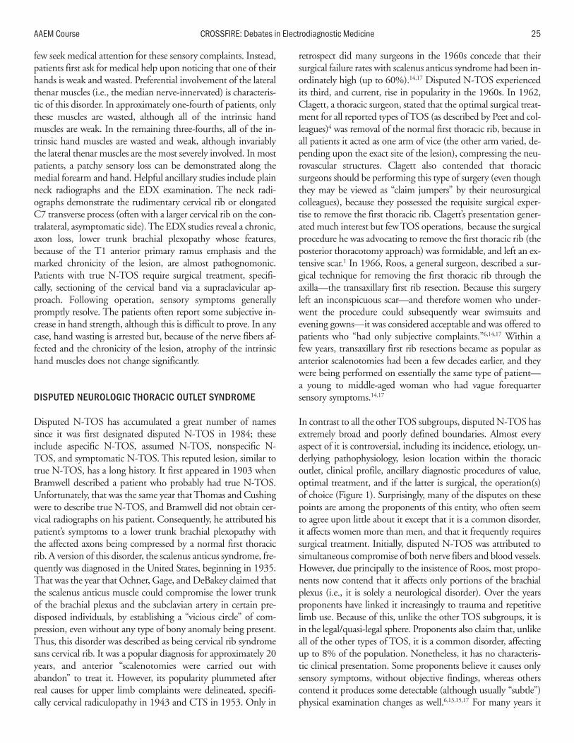
few seek medical attention for these sensory complaints. Instead,
patients first ask for medical help upon noticing that one of their
hands is weak and wasted. Preferential involvement of the lateral
thenar muscles (i.e., the median nerve-innervated) is characteris-
tic of this disorder. In approximately one-fourth of patients, only
these muscles are wasted, although all of the intrinsic hand
muscles are weak. In the remaining three-fourths, all of the in-
trinsic hand muscles are wasted and weak, although invariably
the lateral thenar muscles are the most severely involved. In most
patients, a patchy sensory loss can be demonstrated along the
medial forearm and hand. Helpful ancillary studies include plain
neck radiographs and the EDX examination. The neck radi-
ographs demonstrate the rudimentary cervical rib or elongated
C7 transverse process (often with a larger cervical rib on the con-
tralateral, asymptomatic side). The EDX studies reveal a chronic,
axon loss, lower trunk brachial plexopathy whose features,
because of the T1 anterior primary ramus emphasis and the
marked chronicity of the lesion, are almost pathognomonic.
Patients with true N-TOS require surgical treatment, specifi-
cally, sectioning of the cervical band via a supraclavicular ap-
proach. Following operation, sensory symptoms generally
promptly resolve. The patients often report some subjective in-
crease in hand strength, although this is difficult to prove. In any
case, hand wasting is arrested but, because of the nerve fibers af-
fected and the chronicity of the lesion, atrophy of the intrinsic
hand muscles does not change significantly.
DISPUTED NEUROLOGIC THORACIC OUTLET SYNDROME
Disputed N-TOS has accumulated a great number of names
since it was first designated disputed N-TOS in 1984; these
include aspecific N-TOS, assumed N-TOS, nonspecific N-
TOS, and symptomatic N-TOS. This reputed lesion, similar to
true N-TOS, has a long history. It first appeared in 1903 when
Bramwell described a patient who probably had true N-TOS.
Unfortunately, that was the same year that Thomas and Cushing
were to describe true N-TOS, and Bramwell did not obtain cer-
vical radiographs on his patient. Consequently, he attributed his
patient’s symptoms to a lower trunk brachial plexopathy with
the affected axons being compressed by a normal first thoracic
rib. A version of this disorder, the scalenus anticus syndrome, fre-
quently was diagnosed in the United States, beginning in 1935.
That was the year that Ochner, Gage, and DeBakey claimed that
the scalenus anticus muscle could compromise the lower trunk
of the brachial plexus and the subclavian artery in certain pre-
disposed individuals, by establishing a “vicious circle” of com-
pression, even without any type of bony anomaly being present.
Thus, this disorder was described as being cervical rib syndrome
sans cervical rib. It was a popular diagnosis for approximately 20
years, and anterior “scalenotomies were carried out with
abandon” to treat it. However, its popularity plummeted after
real causes for upper limb complaints were delineated, specifi-
cally cervical radiculopathy in 1943 and CTS in 1953. Only in
retrospect did many surgeons in the 1960s concede that their
surgical failure rates with scalenus anticus syndrome had been in-
ordinately high (up to 60%).14,17 Disputed N-TOS experienced
its third, and current, rise in popularity in the 1960s. In 1962,
Clagett, a thoracic surgeon, stated that the optimal surgical treat-
ment for all reported types of TOS (as described by Peet and col-
leagues)4 was removal of the normal first thoracic rib, because in
all patients it acted as one arm of vice (the other arm varied, de-
pending upon the exact site of the lesion), compressing the neu-
rovascular structures. Clagett also contended that thoracic
surgeons should be performing this type of surgery (even though
they may be viewed as “claim jumpers” by their neurosurgical
colleagues), because they possessed the requisite surgical exper-
tise to remove the first thoracic rib. Clagett’s presentation gener-
ated much interest but few TOS operations, because the surgical
procedure he was advocating to remove the first thoracic rib (the
posterior thoracotomy approach) was formidable, and left an ex-
tensive scar.1 In 1966, Roos, a general surgeon, described a sur-
gical technique for removing the first thoracic rib through the
axilla—the transaxillary first rib resection. Because this surgery
left an inconspicuous scar—and therefore women who under-
went the procedure could subsequently wear swimsuits and
evening gowns—it was considered acceptable and was offered to
patients who “had only subjective complaints.”6,14,17 Within a
few years, transaxillary first rib resections became as popular as
anterior scalenotomies had been a few decades earlier, and they
were being performed on essentially the same type of patient—
a young to middle-aged woman who had vague forequarter
sensory symptoms.14,17
In contrast to all the other TOS subgroups, disputed N-TOS has
extremely broad and poorly defined boundaries. Almost every
aspect of it is controversial, including its incidence, etiology, un-
derlying pathophysiology, lesion location within the thoracic
outlet, clinical profile, ancillary diagnostic procedures of value,
optimal treatment, and if the latter is surgical, the operation(s)
of choice (Figure 1). Surprisingly, many of the disputes on these
points are among the proponents of this entity, who often seem
to agree upon little about it except that it is a common disorder,
it affects women more than men, and that it frequently requires
surgical treatment. Initially, disputed N-TOS was attributed to
simultaneous compromise of both nerve fibers and blood vessels.
However, due principally to the insistence of Roos, most propo-
nents now contend that it affects only portions of the brachial
plexus (i.e., it is solely a neurological disorder). Over the years
proponents have linked it increasingly to trauma and repetitive
limb use. Because of this, unlike the other TOS subgroups, it is
in the legal/quasi-legal sphere. Proponents also claim that, unlike
all of the other types of TOS, it is a common disorder, affecting
up to 8% of the population. Nonetheless, it has no characteris-
tic clinical presentation. Some proponents believe it causes only
sensory symptoms, without objective findings, whereas others
contend it produces some detectable (although usually “subtle”)
physical examination changes as well.6,13,15,17 For many years it
AAEM Course CROSSFIRE: Debates in Electrodiagnostic Medicine 25
was reported to cause symptoms solely in a C8 distribution, the
so-called “lower plexus presentation.” However, an upper plexus
presentation was described in the early 1980s, in which symp-
toms reputedly are experienced principally around the upper
arm, shoulder girdle, and posterior neck and head. Typically, the
diagnosis is made whenever the patient has a “characteristic clin-
ical presentation,” whatever that may be, and some ancillary di-
agnostic procedure is positive. However, there is absolutely no
agreement among the various proponents concerning which of
the many available ancillary procedures are valuable in this
regard. At least 20 have been proposed, including physical ex-
amination, electrophysiologic examination, neuroimaging, and
miscellaneous procedures. None is universally accepted, al-
though the most widely employed is the elevated arm stress test
(EAST procedure), first described by Roos6 in 1966. Yet, this is
a non-specific test, being positive in great numbers of normal
persons and particularly in those with CTS. Moreover, it is a
claudication test (i.e., its end point is arterial compromise), a fact
that is at variance with the concept that disputed N-TOS in-
volves the brachial plexus and not the blood vessels.
Concerning the utility of the EDX examination with this disor-
der, its proponents hold three different views: (1) an upper limb
motor nerve conduction study can definitely diagnose the entity.
Urschel, a thoracic surgeon, is the most vocal advocate of this ap-
proach; unfortunately, he appears to have a poor understanding
of both motor nerve conduction studies and peripheral nervous
system anatomy; (2) although an EDX examination cannot di-
agnose this entity, it is nonetheless useful, because it can detect
other peripheral nervous system lesions, such as CTS and cervi-
cal radiculopathies, with which disputed N-TOS can be con-
fused; and (3) it is of no value at all in the assessment of patients
26 Controversies in Evaluating Brachial Plexopathies and Thoracic Outlet Syndrome AAEM Course
Figure 1 Illustrated is the thoracic outlet syndrome region. The site of injury of the brachial plexus fibers with the various types of thoracicoutlet syndrome are well-known, except for the disputed neurologic type; with the latter, the lesion has been reported by various proponentsto occur supraclavicularly (A—between the scalene muscles), retroclavicularly (B—between the clavicle and first thoracic rib), and infraclav-icularly (C—in the axilla).
with this suspected disorder. Roos, considered by many to be the
father of disputed N-TOS, has stated this opinion repeat-
edly.2,6,9,12-17
Disputed N-TOS is treated both conservatively (with physical
therapy, postural exercises, etc.) and surgically. Operations are
performed on a considerable number of patients who are said to
have intractable pain. There is considerable disagreement among
the various proponents regarding which operation is optimal.
Initially, almost all TOS surgery after 1966 consisted of transax-
illary first rib resections to treat the lower plexus presentation.
However, a few surgeons had never abandoned operation on the
scalene muscles, although the procedure had been expanded
from an anterior scalenectomy to anterior and middle scalenec-
tomies. Roos contends that the latter operation should be re-
served for patients with the upper plexus presentation.
Nonetheless, some surgeons perform either operation, regardless
of presentation (i.e., upper plexus or lower plexus type), whereas
others perform one and, if symptoms are not relieved, they
perform the other, regardless of presentation. Still others perform
both procedures as two portions of the same operation.
Moreover, other approaches for first rib removal, including the
supraclavicular and posterior thoracotomy approach, are some-
times employed (the latter particularly for reoperation). In addi-
tion, some surgeons perform epineurectomies of various
portions of the brachial plexus.6,10,11,15
The results of TOS surgery are vigorously debated. For many
years high operative success rates, typically approaching 90%,
were claimed, but these almost always were reported by the sur-
geons who had performed the operations after short follow-up
periods. Over the past 2 decades, far more pessimistic results
have been published, with much lower operative success rates
(35-65%). In one report, based on a 5-year survey of the opera-
tive results on Workman’s Compensation patients in the state of
Washington, the surgical success rate was described as
“dismal.”13,15,17
The many skeptics of disputed N-TOS have several problems
with the diagnosis. First, it is often being made by physicians
who have had little if any training or experience in diagnosing
neurologic disease, particularly brachial plexopathies, or in for-
mulating the differential diagnosis for them. Also, many propo-
nents appear remarkably naive regarding functional symptoms
and secondary gain factors in patients who have personal injury
litigation or Workman’s Compensation claims pending. In addi-
tion, some proponents now claim that this type of TOS can pre-
cipitate a great variety of additional peripheral nervous system
and musculotendinous disorders, including cervical radicu-
lopathies, various entrapment neuropathies, etc., many of which
may require additional operative treatment. Finally, most skep-
tics have serious concerns regarding the number of what they
consider to be completely unnecessary surgical procedures being
performed on patients who are labeled with this diagnosis,
because of the potential for these operative procedures to cause
grave harm to the patients, such as infection, severe permanent
brachial plexus injuries, and even death by exsanguination.12-17
An important point is that the skeptics of disputed N-TOS do
not hold identical opinions regarding it. Many consider it to be
a nonexistent disorder, one formulated primarily because an op-
erative technique had been devised to remove the normal first
thoracic rib while leaving an inconspicuous scar. Others believe
that it probably exists, although its incidence undoubtedly is far
less than its proponents proclaim, but contend that its diagnosis
is markedly difficult and relies principally on the “gut feeling” of
the clinician making the diagnosis. Unfortunately, many such
clinicians appear to experience such an “abdominal experience”
whenever they encounter any patient with vague four-quarter
symptoms.
CONCLUSIONS
The term TOS encompasses several different disorders, only
some of which involve peripheral nervous system structures and
only one of which is highly controversial. However, the contro-
versial one is also the only one that occurs with any frequency.
REFERENCES
1. Clagett OT. Research and prosearch. J Urol Nephrol 1962;44:153-166.
2. Dawson DM, Hallett M, Wilbourn AJ, editors. Thoracic outlet syn-dromes. In: Entrapment neuropathies, 3rd edition. Baltimore:Lippincott Williams and Wilkins; 1999. p 227-250.
3. Gilliatt RW, Le Quesne PM, Logue V, Sumner AJ. Wasting of thehand associated with a cervical rib or band. J Neurol NeurosurgPsychiatry 1970;33:615-624.
4. Patton GM. Arterial thoracic outlet syndrome. Hand Clinics2004;20:107-111.
5. Peet PM, Henriksen JD, Anderson TP, Martin GM. Thoracic outletsyndrome: evaluation of a therapeutic exercise program. Mayo ClinProc 1956;31:281-287.
6. Roos DB. The place of scalenectomy and first-rib resection in tho-racic outlet syndrome. Surgery 1982;92:1077-1085.
7. Sanders RJ, Hammond SL. Venous thoracic outlet syndrome. HandClinics 2004;20:113-118.
8. Urschel HC, Razzuk MA. Diagnosing upper plexus thoracic outletsyndrome with median nerve conduction studies. Ann Thoracic Surg1999;67:291-292.
9. Urschel HC, Razzuk MA. Upper plexus thoracic outlet syndrome:optimal therapy. Ann Thorac Surg 1997;63:935-939.
10. Wehbe MA, Leinberry CF. Current trends in treatment of thoracicoutlet syndrome. Hand Clinics 2004;20:119-121.
11. Wehbe MA, Whitaker ML. Epineurectomy for thoracic outlet syn-drome. Hand Clinics 2004;20:83-86.
AAEM Course CROSSFIRE: Debates in Electrodiagnostic Medicine 27
12. Whitenack SH, Hunter JM, Jaeger H, Read R. Thoracic outlet syn-drome: a brachial plexopathy. In: Hunter JM, Mackin EJ, CallahanAD, editors. Rehabilitation of the hand: surgery and therapy, 4thedition, volume 2. St. Louis: Mosby; 1995. p 859-884.
13. Wilbourn AJ. Thoracic outlet syndrome: a neurologist’s perspective.Chest Surg Clin N Am 1999;9:821-839.
14. Wilbourn AJ. Brachial plexus disorders. In: Dyck PJ, Thomas PK,editors. Peripheral neuropathy, 3rd edition. Philadelphia: WBSaunders; 1993. p 991-950.
15. Wilbourn AJ. Thoracic outlet syndromes: a plea for conservatism.Neurosurg Clin N Am 1991;2:235-245.
16. Wilbourn AJ, Cherington M. Diagnosing upper plexus thoracicoutlet syndrome with median nerve conduction studies. Ann ThoracIn Surg 1999;67:290-291.
17. Wilbourn AJ, Porter JM. Thoracic outlet syndromes Spine: State ofthe Art Reviews 1988;2:597-626.
28 Controversies in Evaluating Brachial Plexopathies and Thoracic Outlet Syndrome AAEM Course
INTRODUCTION
The subject of thoracic outlet syndrome (TOS) remains one of
the most contentious in all of medicine. Virtually every aspect of
this disorder, including its existence, name, clinical features,
methods of diagnosis, pathophysiology, and treatment all evoke
varying degrees of controversy and debate. Indeed, mention of
the diagnosis of TOS, particularly among neurologists and
physiatrists, will often elicit obvious expressions of disbelief,
snickers, and scornful looks. Why, then, would a neurologist
agree to debate in favor of this concept, particularly against one
of its most vigorous and knowledgeable opponents? The main
reason is that is author happens to believe that this is a useful di-
agnosis, one that can be made by careful clinical assessment and
can lead to an effective treatment program in the majority of pa-
tients so diagnosed.
This manuscript will focus on the so-called “nonspecific neuro-
genic” TOS (NSN-TOS) or “symptomatic type,” more prefer-
able terms then the more pejorative (although accurate)
“disputed” TOS favored by others. There is not much to say in
favor of the usefulness of electrodiagnostic (EDX) techniques to
investigate this disorder. A variety of such procedures have been
proposed and they can be helpful in excluding alternative, or co-
existing, conditions. Similarly, this manuscript will not be pro-
moting the surgical treatment of NSN-TOS, although it can be
successfully utilized for patients who remain symptomatic
despite aggressive and skillfully applied non-surgical techniques.
This author’s experience with TOS is primarily among instru-
mental musicians, a group that seems to be particularly prone to
develop symptoms. This diagnosis as been made by this author
in 75 instrumentalists out of the total of 1480 evaluated over the
past 25 years. No patient in this series, or in any other involving
musicians, to this authors knowledge, has been diagnosed with
the true neurogenic form. Lascelles and colleagues16 reported 31
patients with different forms of TOS, including 5 with what he
called the “pain/paresthesia” type. One young woman in this
group played the flute. Charness5 identified TOS in 59 limbs in
40 of 117 instrumental musicians (34%) with nerve entrapment
syndromes. In a series from the Massachusetts General Hospital
Musical Medicine Clinic,13 TOS was identified in 7% of the
1000 instrumentalists seen (included in the 32% with a neuro-
logic diagnosis). Newmark23 discussed three musicians with
symptoms suggesting NSN-TOS in his 1996 report. In a series
from the United Kingdom, Wynn Parry37 included 14 patients
diagnosed with TOS among 257 musicians with a “clear diag-
nosis” of upper limb problems out of the 617 seen at that clinic.
29
Controversies in Evaluating BrachialPlexopathies and Thoracic Outlet
Syndrome
Richard J. Lederman, MD, PhD
Professor of MedicineCleveland Clinic Lerner College of Medicine of Case Western Reserve University
Cleveland, Ohio
Winspur,36 also from the United Kingdom, suggested a diagno-
sis of TOS in 9 of 137 musicians with “specific” orthopedic/
rheumatologic diagnoses out of the 323 musicians evaluated.
MECHANISMS OF SYMPTOM PRODUCTION
Most authors have assumed that compression of the “neurovas-
cular” structures in the thoracic outlet is largely responsible for
the production of symptoms. Three potential sites of compres-
sion have been identified, including the interscalene triangle, the
costoclavicular space, and the retro- or subpectoralis minor
space.2 While it is generally agreed that anatomical factors may
predispose to the development of TOS, including the presence
of cervical ribs or possibly fibrous bands, some other process also
seems necessary. Specifically, some form of trauma, either an
acute event or, as in the musician population, repetitive stress
and postural factors, is hypothesized. Sanders and Hammond28
have suggested that scarring of the scalene muscles following
trauma might lead to compression of the brachial plexus and
offer as evidence a single histologic study in support of this
concept. Pascarelli and Hsu,25 reviewing work-related upper ex-
tremity symptoms in 485 patients (including 134 musicians),
conclude that postural misalignment is a major factor in pro-
ducing the symptoms of NSN-TOS in the 339 persons so-diag-
nosed in their series. Lindgren and colleagues20 have identified a
“functional” mechanism for production of symptoms of TOS,
based on the demonstration of subluxation of the first rib at the
costotransverse joint associated with work-related upper extrem-
ity stress, leading to tension in the scalene muscles and subse-
quent brachial plexus compression.
Brantigan and Roos4 have emphasized the role of anatomic ab-
normalities, especially fibrous bands, in the etiology of symp-
toms in neurogenic TOS. They have suggested that the major
difference between NSN-TOS and true neurogenic TOS is time
and advocate treatment before the clinical (and electrical) indi-
cators of nerve and muscle injury have appeared. Wilbourn and
Porter35 have argued strongly, and effectively, against this idea. I
have identified five patients in my series with NSN-TOS who
have had symptoms for 8-12 years. If there were an evolution
from NSN-TOS to the true neurogenic form based primarily on
time, it would seem likely that these patients would have at least
some evidence of impairment clinically or electrically. They, in
fact, did not.
Stretching of the brachial plexus has also been suggested as a
mechanism for symptom production. Clein7 first suggested the
term “droopy shoulder syndrome” for painful shoulders associ-
ated with presumed stretching of the brachial plexus. Swift and
Nichols29 reported 10 such patients, with pain or numbness in
one or both upper extremities associated with this configuration,
and suggested that brachial plexus stretch accounted for most of
the cases of NSN-TOS in their experience. Approximately half
of this author’s patients with TOS have the long neck, sloping
shoulders, and horizontal clavicles characteristic of this disorder.
Ide and colleagues15 studied 150 patients with TOS, injecting
contrast material into the supraclavicular plexus under fluoro-
scopic guidance with and without provocative maneuvers. They
found that 18% had only compression of the plexus, 8% showed
evidence of stretching only, and 74% demonstrated a combina-
tion of the two mechanisms. Eleven of the 12 patients showing
stretch only had the droopy shoulder configuration.
CLINICAL FEATURES OF NONSPECIFIC NEUROGENICTHORACIC OUTLET SYNDROME
One of the most disturbing aspects of NSN-TOS disorder is the
lack of consistent and validated clinical diagnostic criteria. In a
series published by this author, the following five features were
utilized. At least four features were required to be satisfied for the
diagnosis to be made.17,18
1. The patients have arm pain, most commonly distal and
usually maximal along the ulnar aspect of the forearm and
hand. Less commonly, there may be pain in the upper arm
and ipsilateral upper trunk.
2. Sensory symptoms, including numbness, tingling, burning,
or a swollen feeling are described, again most commonly
along the ulnar forearm and hand.
3. The symptoms are associated with or aggravated by specific
positions, such as holding the arm at or above shoulder
level.
4. Symptoms can typically be provoked by specific maneuvers,
including hyperabduction of the arm, abduction and exten-
sion of the arm, and downward traction on the arm, often
with internal rotation at the shoulder.
5. Despite these symptoms, the neurologic examination is vir-
tually always normal, with no demonstrable weakness,
sensory loss, or reflex change. Extreme care must be taken to
exclude apparent weakness related to pain or poor effort and
subjective, often non-anatomic sensory impairment.
Some patients will have tenderness in the supraclavicular fossa
overlying the scalene muscles or over the pectoralis minor inser-
tion and others may have a Tinel sign with percussion in these
areas. However, these signs are not sufficiently helpful to use as
additional diagnostic criteria.
A variety of provocative maneuvers have been advocated over the
years for helping to identify patients who have NSN-TOS:1,3 un-
fortunately all of these, including the extended arm stress test
30 Controversies in Evaluating Brachial Plexopathies and Thoracic Outlet Syndrome AAEM Course
(EAST) as described by Roos,3 can be criticized as being non-
specific9 or irrelevant.
Using the criteria outlined above, this author identified NSN-
TOS in 75 instrumental musicians, including 56 women and 19
men. These ranged in age from 15-47 years at the time of diag-
nosis (mean = 25 years). The disorder was found in all instru-
ment groups, including 32 bowed string players (25 violinists or
violists), 19 keyboard instrumentalists, and 13 woodwind
players (9 flutists). The only group showing a significant lateral-
ization were the violinists/violists, with 19 having left arm symp-
toms and 8 right arm symptoms (2 bilateral).
DIFFERENTIATION OF THORACIC OUTLET SYNDROME FROMOTHER DISORDERS
A number of identifiable syndromes can be considered in the
differential diagnosis. These would include cervical radiculopa-
thy, neuralgic amyotrophy (before the appearance of obvious
weakness and atrophy), carpal tunnel syndrome, ulnar neuropa-
thy, and other nerve entrapments. The clinical features of these
entities, with characteristic muscle weakness, variable sensory
loss, and reflex changes are well known and need not be elabo-
rated further here. The major problem confronting the clinician
is to distinguish patients with NSN-TOS from the large and het-
erogeneous group presenting with regional cervicobrachial pain
syndromes, especially those resulting from trauma. This can be
accomplished in many cases by relying heavily on adherence to
the criteria outlined earlier. The large majority of patients with
TOS have unilateral or clearly asymmetrical symptoms which, at
least early in the course, are position-dependent. The ability to
reproduce the symptoms specifically and repeatedly with
provocative maneuvers is also a dependable test, particularly in
the group with droopy shoulder configuration, where depression
of the shoulder with internal rotation for 1 minute or less pro-
duces symptoms on the affected side, with immediate relief on
releasing the downward traction on the arm.
Lindgren and colleagues20 have proposed that rotating the neck
away from the side of symptoms, followed by maximally flexing
the cervical spine (cervical rotation lateral flexion test) can be
used to identify the patients with subluxing first rib/costotrans-
verse joints. In this author’s experience this is positive in some,
but by no means all, patients with NSN-TOS, but have not
searched for evidence of the subluxation by utilizing three-di-
mensional computerized tomography (CT) scanning, as was
performed in their cases.
Howard and colleagues14 attempted to increase the sensitivity of
clinical testing with provocative maneuvers by utilizing quanti-
tative touch threshold and grip-strength devices at rest and in
provocative positions in patients with TOS. They reported a sen-
sitivity of 82% and a specificity of 100% for “clinically severe”
brachial plexus compression (as judged by the EAST and Tinel
signs). They further indicated that, in 16 of 17 patients tested
before and after surgery, the response pattern reverted to normal.
A variety of ancillary procedures have been advocated for aiding
in the diagnosis of NSN-TOS.1,3 Cervical spine radiograph can
identify the elongated C7 transverse process or cervical rib but,
in the absence of the clinical and electrical findings of true neu-
rogenic TOS, these abnormalities are not helpful in diagnosis.
Nannapaneni and Marks22 reported 51 patients with “neuro-
genic TOS” identified and surgically treated in a 13-year period.
All had radiographic abnormalities, including 5 with complete
and 34 with incomplete cervical ribs and 20 with enlarged trans-
verse processes. It is not possible to determine whether any (or
all) of these had true neurogenic TOS, since they found sensory
abnormalities in 71%, motor weakness in 59%, and atrophy in
25%. They also found EDX testing to be negative, except when
there was muscle wasting and then characterized it as “nonspe-
cific.” Gillard12 and colleagues utilized ultrasound and helical
CT in 48 patients, presumably with NSN-TOS. These were ef-
fective in identifying vascular compression, but they questioned
whether these studies are useful in establishing a diagnosis of
TOS.
Electrodiagnosis of NSN-TOS has almost as long and equally
controversial history as does the clinical diagnosis. Most experi-
enced EDX physicians would agree that electrophysiologic con-
firmation of this disorder is not possible. They would also agree
that an EDX study is particularly useful in excluding a number
of alternative diagnoses. Recording of the ulnar motor conduc-
tion velocity across the “thoracic outlet,” as promoted by Urschel
and colleagues,31 has been effectively shown to be of no value.34
A number of alternative EDX studies, including F-wave analy-
sis, cervical root stimulation, and somatosensory evoked poten-
tials, which at least theoretically should be helpful in studying
this area, have proved disappointing.11 Magnetic stimulation
may overcome some of the shortcomings of proximal electrical
stimulation in this situation. Misawa and colleagues21 compared
symptomatic and asymptomatic limbs in patients with TOS as
well as a healthy control group and found a significant prolon-
gation of compound muscle action potential latency, recording
both abductor pollicis brevis and abductor digiti minimi, in the
symptomatic limbs.
TREATMENT OF NONSPECIFIC NEUROGENIC THORACICOUTLET SYNDROME
The debate regarding treatment of NSN-TOS has proven every
bit as vigorous and acrimonious as the various aspects discussed
above. While even the most aggressive among the surgical spe-
cialists treating these patients would agree that nonsurgical
options should be tried first, the implication is that all such
methods have been exhausted by the time surgery is carried out.
AAEM Course CROSSFIRE: Debates in Electrodiagnostic Medicine 31
A variety of surgical approaches have been advocated,1,30 often
depending on the distribution of symptoms and the purported
site of compression or stretching, and a wide range of results
have also been described.19,27,32 Much of this variability can be at-
tributed to failure to differentiate the true from the non-specific
neurogenic types in some series as well as differences in duration
of follow-up and rigor in clinical assessment. The potential
dangers in surgical management have also been widely
quoted,6,33 although some authors would obviously dispute the
likelihood of injury to the brachial plexus.27
The vast majority of patients with NSN-TOS can and should be
managed non-surgically, with a combination of postural modifi-
cation, exercise, and pain control. A number of therapeutic reg-
imens have been suggested over the years, beginning with the
exercise program described by Peet and colleagues26 Most such
regimens attempt to normalize the neck and upper trunk rela-
tionships, thereby minimizing the potential compression or
stretch at the interscalene, costoclavicular, or sub-pectoralis
minor sites.9,10,24 Lindgren and colleagues20 have proposed a
series of exercises to reduce the reported subluxation of the first
rib in patients with this demonstrated abnormality. The addition
of a comprehensive pain management program, with all the
tools currently available, may be necessary in those patients who
progress to a chronic regional pain syndrome. The combination
of medication, psychotherapy, nerve blocks, and even electrical
stimulation or infusion techniques may be required to provide
symptomatic relief, particularly in those patients who decline or
are not offered surgical treatment.
Of the 75 instrumental musicians diagnosed with TOS in the
authors study, only 2 have undergone surgical decompression,
both successfully. Approximately 75% have responded favorably
to conservative treatment, including 60% who have either no
symptoms or mild and nondisabling complaints, and another
15% who are limited in their ability to play but continue in their
careers or avocation. Most of the remaining 25% have been lost
to follow-up and their current status cannot be documented. A
few who did not improve have been offered surgical treatment,
but have declined.
CONCLUSION
Nonspecific neurogenic TOS represents an identifiable entity
that commonly produces pain and sensory symptoms in the
upper limb, particularly among certain occupational groups, as
well as those suffering acute trauma. The combination of upper
limb pain and positional paresthesias suggests that intermittent
compression, or stretching, of the brachial plexus is responsible
for the production of symptoms. Nonspecific neurogenic TOS
remains a clinical diagnosis since reliable diagnostic studies that
can confirm it are not yet available. The majority of such patients
can be helped by nonoperative treatment; only a small minority
require a surgical approach. Lacking definitive clinical, electro-
physiologic, radiographic, or other criteria, NSN-TOS is likely
to remain a source of controversy, potentially to the detriment of
the afflicted patient.
REFERENCES
1. Atasoy E. Thoracic outlet compression syndrome. Orthop ClinNorth Am 1996;27:265-303.
2. Atasoy E. Thoracic outlet syndrome: anatomy. Hand Clin2004;20:7-14.
3. Brantigan CO, Roos DB. Diagnosing thoracic outlet syndrome.Hand Clin 2004;20:27-36.
4. Brantigan CO, Roos DB. Etiology of neurogenic thoracic outlet syn-drome. Hand Clin 2004;20:17-22.
5. Charness ME. Unique upper extremity disorders of musicians. In:Millender LH, Louis DS, Simmons BP, editors. Occupational disor-ders of the upper extremity. New York: Churchill-Livingstone; 1992.p 227-252.
6. Cherington M, Happer I, Machanic B, Parry L. Surgery for thoracicoutlet syndrome may be hazardous to your health. Muscle Nerve1986;9:632-634.
7. Clein LJ. The droopy shoulder syndrome. Can Med Assoc J1976;114:343-344.
8. Costigan DA, Wilbourn AJ. The elevated arm stress test: specificityin the diagnosis of the thoracic outlet syndrome. Neurology1985;35:S74-S75.
9. Crosby CA, Wehbé MA. Conservative treatment for thoracic outletsyndrome. Hand Clin 2004;20:43-49.
10. Dobrusin R. An osteopathic approach to conservative managementof thoracic outlet syndromes. J Am Osteopath Assoc 1989;89:1046-1057.
11. Dumitru D, Amato AA, Zwarts M. Electrodiagnostic medicine, 2ndedition. Philadelphia: Hanley & Belfus; 2002. p 819.
12. Gillard J, Perez-Cousin M, Hachulla E, Remy J, Hurtevent JF,Vinckier L, Thévenon A, Duquesnoy B. Diagnosing thoracic outletsyndrome: contribution of provocative tests, ultrasonography, elec-trophysiology, and helical computed tomography in 48 patients. JointBone Spine 2001;68:416-424.
13. Hochberg FH, Lederman RJ. Upper extremity difficulties of musi-cians. In: Hunter JM, Mackin EJ, Callahan AD, editors.Rehabilitation of the hand: surgery and therapy, 4th edition. St.Louis: Mosby; 1995. p 1795-1808.
14. Howard M, Lee C, Dellon AL. Documentation of brachial plexuscompression (in the thoracic inlet) utilizing provocative neurosensoryand muscular testing. J Reconstr Microsurg 2003;19:303-312.
15. Ide J, Kataoka Y, Yamaga M, Kitamura T, Takagi K. Compression andstretching of the brachial plexus in thoracic outlet syndrome: correla-tion between neuroradiographic findings and symptoms and signsproduced by provocation manoeuvres. J Hand Surg Br 2003;28:218-223.
16. Lascelles RG, Mohr PD, Neary D, Bloor K. The thoracic outlet syn-drome. Brain 1977;100:601-612.
17. Lederman RJ. Thoracic outlet syndrome: review of the controversiesand a report of 17 instrumental musicians. Med Probl Perform Art1987;2:87-91.
18. Lederman RJ. Neuromuscular and musculoskeletal problems in in-strumental musicians. Muscle Nerve 2003;27:549-561.
19. Lindgren KA. Reasons for failures in the surgical treatment of tho-racic outlet syndrome. Muscle Nerve 1995;18:1484-1486.
32 Controversies in Evaluating Brachial Plexopathies and Thoracic Outlet Syndrome AAEM Course
20. Lindgren K-A, Manninen H, Rytkönen H. Thoracic outlet syn-drome—a functional disturbance of the thoracic upper aperture?Muscle Nerve 1995;18:526-530.
21. Misawa T, Kiyono Y, Nakatsuchi Y, Shindo M, Takaoka K. Diagnosisof thoracic outlet syndrome by magnetic stimulation of the brachialplexus. J. Orthop Sci 2002;7:167-171.
22. Nannapaneni R, Marks SM. Neurogenic thoracic outlet syndrome.Br J Neurosurg 2003;17:144-148.
23. Newmark J. Thoracic outlet syndromes: a non-surgeon’s perspectivefor those caring for musicians. Work 1996;7:95-107.
24. Novak CB, Mackinnon SE. Thoracic outlet syndrome. Orthop ClinN Am 1996;27:747-762.
25. Pascarelli EF, Hsu Y-P. Understanding work-related upper extremitydisorders: clinical findings in 485 computer users, musicians, andothers. J Occup Rehabil 2001;11:1-21.
26. Peet RM, Henriksen JD, Anderson TP, Martin GM. Thoracic-outletsyndrome: evaluation of a therapeutic exercise program. Mayo ClinProc 1956;31:281-287.
27. Roos DB. Thoracic outlet syndrome is underdiagnosed. MuscleNerve 1999;22:126-129.
28. Sanders RJ, Hammond SL. Etiology and pathology. Hand Clin2004;20:23-26.
29. Swift TR, Nichols FT. The droopy shoulder syndrome. Neurology1984:34:212-215.
30. Urschel HC, Patel A. Thoracic outlet syndromes. Curr Treat OptionsCardiovasc Med 2003;5:163-168.
31. Urschel HC, Razzuk MA. Management of the thoracic outlet syn-drome. N Engl J Med 1972;286:1140-1143.
32. Wehbé MA, Leinberry CF. Current trends in treatment of thoracicoutlet syndrome. Hand Clin 2004;20:119-121.
33. Wilbourn AJ. Thoracic outlet syndrome surgery causing severebrachial plexopathy. Muscle Nerve 1988;11:66-74.
34. Wilbourn AJ, Lederman RJ. Evidence for conduction delay in tho-racic outlet syndrome is challenged. N Engl J Med 1984;310:1052-1053.
35. Wilbourn AJ, Porter JM. Thoracic outlet syndromes. In: Winer N,editor. Spine: state of the art reviews. Philadelphia: Hanley & Belfus;1988. p 597-626.
36. Winspur I. Nerve compression syndromes. In: Winspur I, WynnParry CB, editors. The musician’s hand: a clinical guide. London:Martin Dunitz; 1998. p 85-99.
37. Wynn Parry CB. The musician’s hand and arm pain. In: Winspur I,Wynn Parry CB, editors. The musician’s hand: a clinical guide.London: Martin Dunitz; 1998. p 5-12.
AAEM Course CROSSFIRE: Debates in Electrodiagnostic Medicine 33

34 AAEM Course
AAEM Course 35
CROSSFIRE: Debates in Electrodiagnostic Medicine
CME SELF-ASSESSMENT TEST
Select the ONE best answer for each question.
1. The best screening test for carpal tunnel syndrome (CTS) is:
A. Median motor latency to lumbrical.
B. Median sensory latency to digit 2.
C. Median/ulnar motor latency to interosseus.
D. Sensory nerve action potential (SNAP) amplitude to
digit 1.
E. Median/radial sensory latency to digit 1.
2. Which is the most specific and sensitive physical finding in
CTS?
A. Phalen sign.
B. Wrist ratio.
C. Reduced 2-point discrimination.
D. Tinel’s sign over median nerve at wrist.
E. Tethering sign.
3. In order to estimate the normal compound muscle action
potential (CMAP) amplitude of bilateral CTS, which is the
best method?
A. Kimura’s values in his text (3rd edition).
B. 10 mV +/- 2.
C. Use CMAP of abductor digit 5.
D. Add 30% to CMAP of non-dominant hand.
E. Stimulate distal to carpal ligament and measure.
4. In order to minimize the phase cancellation phenomenon in
sensory conduction what should the “inter-electrode” sepa-
ration be when recording?
A. 2 cm.
B. 10 mm.
C. 6 cm.
D. 4 cm.
E. It really doesn’t matter.
5. What happens if both e-1 and e-2 are both placed over the
thenar muscle?
A. Latency shortens.
B. Latency lengthens.
C. CMAP gets larger.
D. CMAP gets smaller.
E. Nothing happens.
6. What happens if one unintentionally stimulates both
median and radial nerves simultaneously while recording the
SNAP on digit 1?
A. Diagnosis of CTS will be missed.
B. False positive diagnosis of CTS.
C. If CTS is present the Bactrian sign will occur.
D. Only median SNAP will appear.
E. Only radial SNAP will occur.
7. Specificity of a test for CTS should be:
A. 100%.
B. 95-99%.
C. 90-95%.
D. 80-90%.
E. 70-80%.
8. When reference values are set to give similar high specificity,
which test is most sensitive?
A. Mid-palmar studies.
B Ring finger comparisons.
C. Thumb comparisons.
D. Median motor latency.
E. Combined sensory index.
9. Which nerve conduction result has highest test-retest relia-
bility?
A. Mid-palmar studies.
B. Ring finger comparisons.
C. Thumb comparisons.
D. Median motor latency.
E. Combined sensory index.
10. Which nerve conduction measurement will be LEAST af-
fected by cold?
A. Median sensory latency.
B. Median sensory amplitude.
C. Median motor latency.
D. Median motor conduction velocity.
E. Median-ulnar sensory latency differences.
11. With median nerve stimulation at the wrist, a 7 mV response
is recorded from abductor pollicis brevis (APB). Palm stimu-
lation produces a 10 mV response from APB. This most
likely indicates:
A. Carpal tunnel syndrome.
B. Martin-Gruber anastamosis.
C. Neurapraxia.
D. Axonotmesis.
E. Nothing.
12. F-wave persistence characteristically can vary considerably in
different muscles.
A. True.
B. False.
13. Which of the following F-wave parameters can be abnormal
in CTS?
1. Repeater waves.
2. Persistence.
3. Chronodispersion.
A. 1 and 2 are correct.
B. 1 and 3 are correct.
C. 2 and 3 are correct.
D. All are correct.
E. None are correct.
14. Which of the following are true about F waves?
1. They are inherently variable in latency, amplitude,
and configuration.
2. Chronodispersion reflects a range of conductions in
motor axons.
3. Latency abnormalities cannot be detected in
radiculopathies.
A. 1 and 2 are correct.
B. 1 and 3 are correct.
C. 2 and 3 are correct.
D. All are correct.
E. None are correct.
15. Which of the following are true about minimal F-wave la-
tencies?
1. Accurate values are always obtained after 10 stimuli.
2. They are the most reliable measure of F-wave laten-
cies.
3. They are not prolonged with focal nerve injury.
A. 1 and 2 are correct.
B. 1 and 3 are correct.
C. 2 and 3 are correct.
D. All are correct.
E. None are correct.
16. F waves have been reported abnormal with focal injury to pe-
ripheral nerves, plexi, and roots.
A. True.
B. False.
17. According to the proponents of disputed (symptomatic)
neurologic thoracic outlet syndrome (TOS), electrodiagnos-
tic studies:
A. Are of no benefit in assessment.
B. Can help in diagnosis by identifying other disorders.
C. Permit objective confirmation.
D. All of the above.
E. None of the above.
18. In practical terms, automobile accidents could be responsible
for only two types of TOS:
A. Traumatic and disputed neurologic TOS.
B. True neurologic and disputed neurologic TOS.
C. Arterial vascular and traumatic TOS.
D. Venous vascular and true neurologic TOS.
E. Disputed neurologic and venous vascular TOS.
19. The most common symptoms attributed to disputed (sym-
patomatic) neurologic TOS are:
A. Paresthesias and weakness.
B. Weakness and pain.
C. Limb coolness and discoloration.
D. Paresthesias and pain.
E. Weakness and paresthesias.
20. Very low incidence, typical clinical presentation, and objec-
tive laboratory findings are some of the features common to
all types of TOS EXCEPT:
A. True neurologic TOS.
B. Disputed neurologic TOS.
C. Traumatic TOS.
D. Venous vascular TOS.
E. Arterial vascular TOS.
21. Congenital anomalies of various types characteristically are
present with:
A. True neurological and arterial vascular TOS.
B. Arterial vascular and venous vascular TOS.
C. Disputed neurologic and arterial vascular TOS.
D. Traumatic and true neurologic TOS.
E. Disputed neurologic and traumatic TOS.
36 CME Self-Assessment Test AAEM Course

AAEM Course CME Self-Assessment Test 37
22. The diagnosis of non-specific (symptomatic) thoracic outlet
syndrome (NS-TOS) is best reserved for those with:
A. Unilateral intrinsic hand muscle atrophy.
B. Position-dependent arm pain and paresthesias.
C. Radial pulse obliteration produced by arm hyperabduc-
tion.
D. Sensory loss along the ulnar forearm and hand.
E. Reduced amplitude of the ulnar compound muscle
action potential.
23. The “droopy shoulder syndrome” is characterized by:
A. A long, thin neck and sloping shoulders.
B. Unilateral atrophy of the trapezius muscle.
C. Hypotrophy of the clavicles bilaterally.
D. Bilateral winging of the scapulae.
E. Unilateral laxity of the acromiohumeral joint.
24. Electrodiagnostic testing in NS-TOS:
A. Serves no useful role.
B. Often demonstrates slowing of ulnar motor conduction
velocity.
C. Will usually demonstrate prolonged ulnar F-wave
latency.
D. Can help to exclude alternative diagnoses.
E. Usually reveals reduced ulnar sensory response ampli-
tude.
25. Successful treatment of NS-TOS:
A. Can hardly ever be accomplished.
B. Can usually be achieved with postural correction and
therapeutic exercise.
C. Usually requires psychotherapy.
D. Usually requires first rib resection.
E. Usually requires scalenotomy plus pectoralis minor
tenotomy.
26. NS-TOS among violinists and violists most commonly
affects:
A. Middle-aged men.
B. The right (bowing) arm.
C. Young women in their third decade.
D. Beginning students.
E. Those who are left-handed.

38 AAEM Course
CROSSFIRE: Debates in Electrodiagnostic Medicine
EVALUATION
Select ANY of the answers that indicate your opinions.
Your input is needed to critique our courses and to ensure that we use the best faculty instructors and provide the best course options
in future years. Please use the computer form to answer the following questions. For the purpose of tabulating evaluations, please enter
the last 4 digits of your telephone number in the ID NUMBER box beginning with the left column and fill in the appropriate ovals
below each number. Make additional comments or list suggested topics or faculty for future courses on the comment form provided
at the end.
AAEM Course 39
27. How would you rate the quality of instruction received
during Dr. Johnson’s presentation?
A. Best possible.
B. Good.
C. Average.
D. Fair.
E. Worst possible.
28. Select any item(s), that, if changed, would have appreciably
improved Dr. Johnson’s presentation:
A. Quality of slides.
B. Quality of handout.
C. Amount of clinically relevant information in the presen-
tation.
D. Amount of scientific content in the presentation.
E. Other: please explain on the comment form at the back
of this handout.
29. How would you rate the quality of instruction received
during Dr. Robinson’s presentation?
A. Best possible.
B. Good.
C. Average.
D. Fair.
E. Worst possible.
30. Select any item(s), that, if changed, would have appreciably
improved Dr. Robinson’s presentation:
A. Quality of slides.
B. Quality of handout.
C. Amount of clinically relevant information in the presen-
tation.
D. Amount of scientific content in the presentation.
E. Other: please explain on the comment form at the back
of this handout.
31. How would you rate the quality of instruction received
during Dr. Kimura’s presentation?
A. Best possible.
B. Good.
C. Average.
D. Fair.
E. Worst possible.
32. Select any item(s), that, if changed, would have appreciably
improved Dr. Kimura’s presentation:
A. Quality of slides.
B. Quality of handout.
C. Amount of clinically relevant information in the presen-
tation.
D. Amount of scientific content in the presentation.
E. Other: please explain on the comment form at the back
of this handout.
33. How would you rate the quality of instruction received
during Dr. Fisher’s presentation?
A. Best possible.
B. Good.
C. Average.
D. Fair.
E. Worst possible.
34. Select any item(s), that, if changed, would have appreciably
improved Dr. Fisher’s presentation:
A. Quality of slides.
B. Quality of handout.
C. Amount of clinically relevant information in the presen-
tation.
D. Amount of scientific content in the presentation.
E. Other: please explain on the comment form at the back
of this handout.
35. How would you rate the quality of instruction received
during Dr. Wilbourn’s presentation?
A. Best possible.
B. Good.
C. Average.
D. Fair.
E. Worst possible.
36. Select any item(s), that, if changed, would have appreciably
improved Dr. Wilbourn’s presentation:
A. Quality of slides.
B. Quality of handout.
C. Amount of clinically relevant information in the presen-
tation.
D. Amount of scientific content in the presentation.
E. Other: please explain on the comment form at the back
of this handout.
37. How would you rate the quality of instruction received
during Dr. Lederman’s presentation?
A. Best possible.
B. Good.
C. Average.
D. Fair.
E. Worst possible.
38. Select any item(s), that, if changed, would have appreciably
improved Dr. Lederman’s presentation:
A. Quality of slides.
B. Quality of handout.
C. Amount of clinically relevant information in the presen-
tation.
D. Amount of scientific content in the presentation.
E. Other: please explain on the comment form at the back
of this handout.
39. As a result of your attendance at this course, did you learn
anything that will improve the care of your patients?
A. Yes, substantially.
B. Yes, somewhat.
C. Not sure.
D. Probably not.
E. This course was not applicable to my patients.
40. Select ALL items where improvement was needed.
A. The accuracy of advance descriptions of this course.
B. The specific topics selected for presentation.
C. The number of speakers in this course.
D. The amount of time allotted for discussion in this
course.
E. Other: please add other areas and outline specific rec-
ommendations for areas needing improvement on the
comment form at the back of this handout.
41. Should this topic be presented in the future by a different
method of presentation.
A. No, the topic of presentation should remain as a course.
B. Yes, the topic should be presented as a dinner seminar.
C. Yes, the topic should be incorporated into the plenary
session.
D. Yes, the topic should be discussed during a breakfast
session.
E. Yes, the topic should be organized as a special interest
group.
40 Evaluation AAEM Course
FUTURE MEETING RECOMMENDATIONS
Select ANY of the answers that indicate your opinions.
The following questions are included with all dinner seminar, course, and plenary session evaluations. It is only necessary to answer
these questions once during the course of the entire meeting.
AAEM Course 41
42. Please indicate below your specialty:
A. Neurologist.
B. Physiatrist.
C. PhD.
D. Other.
43. How often do you attend AAEM meetings?
A. Annually.
B. Every 2-3 years.
C. Every 4 or more years.
D. This is the first AAEM meeting I have attended.
44. With regard to this years meeting, which of the small group
sessions did you attend? (mark all that apply)
A. Experts’ roundtables.
B. Workshops.
C. Dinner seminars.
D. None of the above.
45. If you answered none of the above to the previous question,
please answer the following. The reason I did not attend the
small group sessions was due to:
A. The timing of the event.
B. The cost of the event.
C. My lack of interest in the topics offered.
D. The session was full.
46. Did this meeting provide information that will enhance care
of your patients?
A. Extremely.
B. Somewhat.
C. Very little.
D. Not at all.
47. With regard to the social event:
A. I am signed up to attend the social event.
B. I did not sign up because of the cost of the event.
C. I did not sign up because of the day the event was
offered.
D. I did not sign up because I am not interested in attend-
ing this type of function.
48. How would you rate this meeting?
A. Poor.
B. Fair.
C. Good.
D. Very good.
E. Excellent.
49. Did this meeting meet your expectations?
A. Not at all.
B. Somewhat.
C. As expected.
D. Exceeded expectations.
E. Best ever.
50. Was the printed program clear and easy to follow?
A. Yes.
B. No.
51. With regard to the meeting hotels:
A. I stayed at one of the meeting hotels.
B. I did not stay at one of the meeting hotels.
52. If you answered B to the question above, please explain why
you did not stay at one of the meeting hotels (please make
your comments under Comments, page 43).
53. How did you first learn about the meeting? (choose the
method where you first learned about the meeting)
A. Preliminary brochure mailing.
B. Registration brochure mailing.
C. The internet.
D. Email message.
E. From a friend.
54. Did you perceive any commercial bias in any of the educa-
tional sessions offered by the AAEM at this meeting?
A. Yes.
B. No.

55. Did you attend any of the Industry Forums provided this
year?
A. Yes.
B. No.
56. If you answered yes to question 55 and you attended the
Pfizer Industry Forum, how would you rate the quality of the
session?
A. Best possible.
B. Good.
C. Average.
D. Fair.
E. Worst possible.
On page 43 under comments, please provide any other
comments you have about your attendance at the Pfizer
Industry Forum.
57. If you answered yes to question 55 and you attended the
Allergan Industry Forum, how would you rate the quality of
the session?
A. Best possible.
B. Good.
C. Average.
D. Fair.
E. Worst possible.
On page 43 under comments, please provide any other
comments you have about your attendance at the
Allergan Industry Forum.
58. How do you prefer to learn new information?
A. Lecture only.
B. Lecture in conjunction with questions and answers.
C. Small group hands-on.
D. Small group discussion.
59. I plan to attend the 2005 AAEM meeting in Monterey,
California, September 21-24.
A. Yes, definitely.
B. No, definitely.
C. Will wait to see the program content.
D. Will wait to see if budget allows my attendance.
60. I would be more likely to attend the 2005 AAEM meeting if
(please make your comments under Comments, page 43):
42 Future Meeting Recommendations AAEM Course
COMMENTS
Given time and budget constraints, is there something we could do in terms of altering the format of the meeting that would
significantly increase the likelihood of your attendance at future AAEM meetings? Explain:
Write out any additional comments about specific courses or the plenary session (please indicate which), and list suggestions
for topics and speakers for future meetings. Leave at the AAEM Registration and Information Center or mail to the AAEM
Executive Office at 421 First Avenue SW, Suite 300 East, Rochester, MN 55902.

AAEM421 First Avenue SW, Suite 300 East
Rochester, MN 55902(507) 288-0100 / Fax: (507) 288-1225