Embed Size (px)
Citation preview
eCommons@AKUCentre for Regenerative Medicine & Stem CellResearch Centres of Excellence
January 2012
Cross-talk between NFkB and the PI3-Kinase/AKT pathway can be targeted in primary effusionlymphoma (PEL) cell lines for efficient apoptosisAzhar HussainAga Khan University, [email protected]
Saeeda O. AhmedKing Faisal Specialist Hospital and Research Center, Riyadh, Saudi Arabia
Maqbool AhmedKing Faisal Specialist Hospital and Research Center, Riyadh, Saudi Arabia
Omar S. KhanKing Faisal Specialist Hospital and Research Center, Riyadh, Saudi Arabia
Sally Al Abdul MohsenKing Faisal Specialist Hospital and Research Center, Riyadh, Saudi Arabia
See next page for additional authors
Follow this and additional works at: http://ecommons.aku.edu/crm
Part of the Medical Sciences Commons
Recommended CitationHussain, A., Ahmed, S., Ahmed, M., Khan, O., Abdul Mohsen, S., Platanias, L., Al-Kuraya, K., Shahab Uddin, . (2012). Cross-talkbetween NFkB and the PI3-Kinase/AKT pathway can be targeted in primary effusion lymphoma (PEL) cell lines for efficientapoptosis. PLoS ONE, 7(6), e39945-e39945.Available at: http://ecommons.aku.edu/crm/9

AuthorsAzhar Hussain, Saeeda O. Ahmed, Maqbool Ahmed, Omar S. Khan, Sally Al Abdul Mohsen, Leonidas C.Platanias, Khawla S. Al-Kuraya, and Shahab Uddin
This article is available at eCommons@AKU: http://ecommons.aku.edu/crm/9
Cross-Talk between NFkB and the PI3-Kinase/AKTPathway Can Be Targeted in Primary Effusion Lymphoma(PEL) Cell Lines for Efficient ApoptosisAzhar R. Hussain1, Saeeda O. Ahmed1, Maqbool Ahmed1, Omar S. Khan1, Sally Al AbdulMohsen1,
Leonidas C. Platanias2, Khawla S. Al-Kuraya1, Shahab Uddin1*
1 Human Cancer Genomic Research, Research Center, King Faisal Specialist Hospital and Research Center, Riyadh, Saudi Arabia, 2 Feinberg School of Medicine, Robert H.
Lurie Comprehensive Cancer Center, Northwestern University, Chicago, Illinois, United States of America
Abstract
Background: A number of constitutively activated signaling pathways play critical roles in the survival and growth ofprimary effusion lymphoma cells (PELs) including NFkB and PI3/AKT kinase cascades. NFkBis constitutively activated in anumber of malignancies, including multiple myeloma, Burkitt’s lymphoma and diffuse large cell B-cell lymphoma. However,its role in primary effusion lymphoma has not been fully explored.
Methodology/Principal Findings: We used pharmacological inhibition and gene silencing to define the role of NFkB ingrowth and survival of PEL cells. Inhibition of NFkB activity by Bay11-7085 resulted in decreased expression of p65 in thenuclear compartment as detected by EMSA assays. In addition, Bay11-7085 treatment caused de-phosphorylation of AKTand its downstream targets suggesting a cross-talk between NFkB and the PI3-kinase/AKT pathway. Importantly, treatmentof PEL cells with Bay11-7085 led to inhibition of cell viability and induced apoptosis in a dose dependent manner. Similarapoptotic effects were found when p65 was knocked down using specific small interference RNA. Finally, co-treatment ofPEL cells with suboptimal doses of Bay11-7085 and LY294002 led to synergistic apoptotic responses in PEL cells.
Conclusion/Significance: These data support a strong biological-link between NFkB and the PI3-kinase/AKT pathway in themodulation of anti-apoptotic effects in PEL cells. Synergistic targeting of these pathways using NFKB- and PI3-kinase/AKT-inhibitors may have a therapeutic potential for the treatment of PEL and possibly other malignancies with constitutiveactivation of these pathways.
Citation: Hussain AR, Ahmed SO, Ahmed M, Khan OS, Al AbdulMohsen S, et al. (2012) Cross-Talk between NFkB and the PI3-Kinase/AKT Pathway Can Be Targetedin Primary Effusion Lymphoma (PEL) Cell Lines for Efficient Apoptosis. PLoS ONE 7(6): e39945. doi:10.1371/journal.pone.0039945
Editor: Ferenc Gallyas, University of Pecs Medical School, Hungary
Received March 17, 2012; Accepted May 29, 2012; Published June 29, 2012
Copyright: � 2012 Hussain et al. This is an open-access article distributed under the terms of the Creative Commons Attribution License, which permitsunrestricted use, distribution, and reproduction in any medium, provided the original author and source are credited.
Funding: The authors have no support or funding to report.
Competing Interests: The authors have declared that no competing interests exist.
* E-mail: [email protected]
Introduction
Human infection by KSHV/HHV-8 is associated with the
development of at least three proliferative disorders: Kaposi’s
sarcoma (KS), primary effusion lymphoma (PEL) and a subset of
multicentric Castleman’s disease (MCD) [1]. Primary effusion
lymphoma (PEL) is a variant of non-Hodgkin’s lymphoma that is
mainly infected by Kaposi sarcoma associated herpesvirus (KSHV)
and sometimes also co-infected with Epstein - Barr virus (EBV) [2].
There are reports demonstrating that PEL can occasionally occur
in HIV-negative patients, especially in organ transplant recipients
and in patients with chronic hepatitis B [3,4,5,6]. Morphologically,
PEL shares features of large-cell immunoblastic and anaplastic
large-cell lymphoma [3,7]. Pleural and abdominal effusions from
patients with PEL contain a number of cytokines, which serve as
autocrine growth factors [8]. For example, IL-10 has been
reported to serve as autocrine growth factor for AIDS-related B-
cell lymphoma [9], while it has also been shown that PEL cells use
viral IL-6 and IL-10 in an autocrine fashion for their survival and
proliferation [8,9].
A number of constitutively activated signaling pathways play
critical roles in the survival and growth of PEL cells [10]. These
include NFkB, PI3-kinase/AKT and JAK/STAT survival path-
ways [11,12,13]. NFkB is now widely recognized as a key positive
regulator of cancer cell proliferation and survival via its ability to
transcriptionally activate many pro-survival and anti-apoptotic
genes such as XIAP, Bcl-2, Bcl-Xl, IkB-a, cIAP1, cIAP-2 and
survivin [14]. NFkB is a family of 5 transcriptional factors
including p50, p52, p65 (Rel-A), RelB and c-Rel, all of which
contain a REL homology domain (RHD) at the N-terminus which
mediates their dimerization, nuclear localization and DNA
binding [15].
A number of dysregulated survival pathways have the ability to
cross-talk with other survival pathways thereby increasing the
aggressiveness of various cancers [16,17]. Such cross-talking allows
cancer cells to escape death in response to different pro-apoptotic
signals, ultimately resulting in unregulated proliferation and and
the emergence of more aggressive and drug-resistant phenotypes
[17]. The NFkB survival pathway also has the ability to cross-talk
with other survival pathways including PI3-kinase/AKT [18,19]
PLoS ONE | www.plosone.org 1 June 2012 | Volume 7 | Issue 6 | e39945
in various cancers. Therefore, targeting the NFkB pathway alone
may not be sufficient to induce apoptosis of malignant cells and
combinations of various inhibitors maybe required to achieve the
desired effect.
Apoptosis is required for the normal homeostasis of normal cells
[20] and is associated with specific cellular features, such as
shrinkage, nuclear blebbing, chromatin condensation and frag-
mentation of DNA [21]. There are two major pathways by which
apoptosis can be initiated; extrinsic or death receptor pathway or
intrinsic or mitochondrial pathway [22]. Even though, the two
pathways may act independently of each other, they converge at
the level ofcaspase-3. Both the apoptotic pathways also have the
ability to cross-talk at the level of caspase-8. Although both these
apoptotic pathways can induce apoptosis alone for efficient
apoptosis to occur, both pathways need to be activated simulta-
neously.
In this study, we first examined the constitutive activity of NFkB
in PEL cell lines followed by inhibition of NFkB activity by the
specific inhibitor, Bay11-7085, in order to define the functional
relevance of this pathway in PEL cells. Our data provide evidence
that NFkB survival pathway is constitutively activated in PEL cells
and that inhibition of this pathway negatively regulates down-
stream anti-apoptotic and pro-survival targets of p65 subunit of
NFkB. We also determined whether inhibition of NFkB activity
would induce apoptosis in PEL cell lines. Finally, we determined
whether there is a cross-talk between the NFkB pathway and the
PI3-kinase/AKT pathway in PEL cells and whether combined
targeting of these pathways with sub-optimal doses of inhibitors
would induce a more potent apoptosis in PEL cells.
Materials and Methods
Cell Culture and TreatmentThe human PEL cell lines BC-1, BC-3, BCBL-1 and HBL-6
were obtained from American Type Culture Collection (Rockville
MD, USA) and cultured in RPMI 1640 medium supplemented
with 10% (v/v) Fetal bovine serum (FBS), 100 U/ml penicillin,
100 U/ml streptomycin at 37uC in an humidified atmosphere
containing 5% CO2. BC1 and HBL-6 cell lines are co-infected
with EBV and HHV-8 while BC3 and BCBL1 cell lines are HHV-
8 positive only. The cells were treated with various doses of Bay11-
7085 dissolved in Dimethyl Sulfoxide (DMSO) for different time
periods in RPMI culture media supplemented with 5% (v/v) FBS.
The control cells were incubated with maximum used amount of
DMSO only. Each experiment was performed at least three times
to confirm the results.
Reagents and AntibodiesBay11-7085 was purchased from Tocris (Ellisville, MI). Bax
6A7 monoclonal antibody and DMSO were purchased from
Sigma Chemical Co (St. Louis, MO). zVAD-fmk and
LY294002 were purchased from Calbiochem (San Diego, CA).
Antibodies against p-AKT (Ser473), p-GSK3a/b (Ser21/9), p-
Foxo1(Thr24/32), p-Bad (ser136), XIAP (cat:2042), Bcl-Xl
(cat:2762), caspase-3 (cat:9665), caspase-9 (cat:9508), PARP-1
(cat:9542), cleaved caspase-3 (cat:9661) and BID (cat:2006)
antibodies were purchased from Cell Signaling Technologies
(Beverly, MA). p-IkBa (Ser32), p65 (, IkBa (C-21), cytochrome
c (A-8), SKP2 (H-435), p27Kip1 (C-19), and Bax (N-20)
antibodies were purchased from Santa Cruz Biotechnology,
Inc. (Santa Cruz, CA). cIAP1 (AF-8171), cIAP2 (AF-8181),
survivin (AF-886), and caspase-8 (clone 84131) antibodies were
purchased from R&D (Minneapolis MN). Bcl-2 (clone 124)
antibody was purchased from Dako (Carpinteria, CA). Annexin-
FITC was purchased from Molecular Probes (Eugene OR).
Apoptotic DNA-ladder kit was obtained from Roche (Penzberg,
Germany). LightShift Chemiluminescent EMSA kit was pur-
chased from Thermo Scientific (Rockford IL).
Assessment of Cell Viability by 3-(4,5-Dimethylthiazol-2-yl)-2,5-Diphenyltetrazolium Bromide (MTT) Assays
Initially, PEL cells were seeded at the concentration of 104 cells
in triplicates in a 96 well format. Cells were then treated with
various doses of Bay11-7085 for 48 hours in a final volume of
0.2 ml for 48 hours. Cell viability was measured by MTT cell
viability assay, as previously described [23,24]. 6 wells for each
dosage including vehicle control were analyzed for each experi-
ment. * denotes statistical significance.
Live Dead AssayTo measure apoptosis, Live-Dead assay (Invitrogen, Eugene,
OR) was used as described previously [25]. Briefly, 16106 PEL
cells were treated with various doses of Bay11-7085 for 24 hours.
Following incubation, cells were re-suspended in 1 ml of PBS
containing 50 mM calcein AM and 8 mM ethidium homodimer
and cells were incubated in the dark for 20 minutes. 50 ml of
suspension was placed on slides and visualized under an Olympus
fluorescent microscope using a longpass filter.
Cell Cycle Analysis, Annexin V Staining, and DNALaddering
PEL cell lines were treated with different concentrations of
Bay11-7085 as described in the legends. For cell cycle analysis,
cells were washed once with PBS and re-suspended in 500 ml
hypotonic staining buffer and analyzed by flow cytometry as
described previously [26]. For detection of apoptosis, cells were
harvested and percentage apoptosis was measured by flow
cytometry after staining with fluorescein–conjugated annexin-V
and propidium iodide (PI) (Molecular probes, Eugene, OR) and
DNA laddering using a 1.5% agarose gel as described previously
[27].
Cell Lysis and ImmunoblottingCells were treated with Bay11-7085 as described in the legends
and lysed as previously described [28]. Briefly, and supernatant
was collected. Briefly, Following treatment, cell pellets were
resuspended in phosphorylation lysis buffer (0.5–1.0% Triton X-
100, 150 mM NaCl, 1 mM EDTA, 200 mM sodium orthovana-
date, 10 mM sodium pyrophosphate, 100 mM sodium fluoride,
1.5 mM magnesium chloride) along with protease inhibitor coctail
tablet (cat 1169748001, Roche, Indianapolis IN) and incubated on
ice for 1 hour, spun for 15 minutes at 14000RPM at 4uC and
supernatant was collected.Protein concentrations were assessed by
Bradford assay before loading the samples. 10–15 mg of proteins
were separated by SDS-PAGE and transferred to polyvinylidene
difluoride membrane (PVDF) (Immobilion, Millipore). Proteins
were immunoblotted with different antibodies and visualized by
the enhanced chemiluminescence (Amersham, Piscataway, NJ)
method.
Preparation of Nuclear Extracts for NF-kBNuclear extracts were prepared according to earlier study [27].
Briefly, 56106 cells were washed with cold PBS and suspended in
0.4 mL hypotonic lysis buffer containing protease inhibitors for 30
minutes. The cells were then lysed with 10% Nonidet P-40. The
homogenate was centrifuged, and supernatant containing the
cytoplasmic extracts was stored frozen at 280uC. The nuclear
Bay11-7085-Induced Apoptosis in PELs
PLoS ONE | www.plosone.org 2 June 2012 | Volume 7 | Issue 6 | e39945
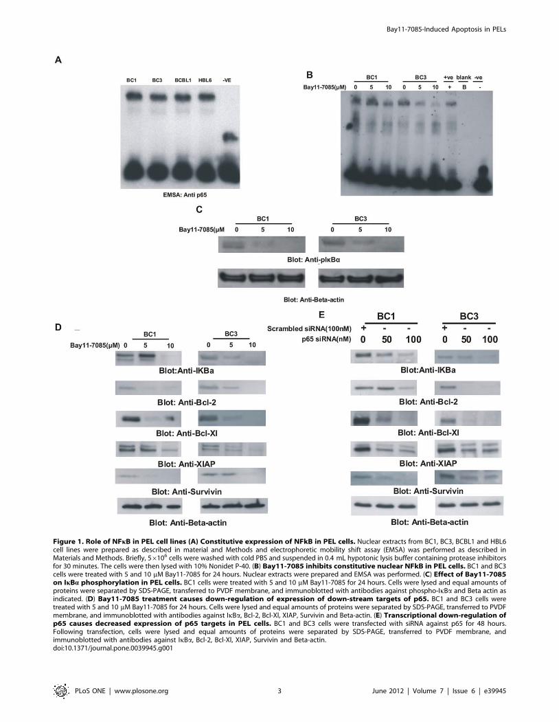
Figure 1. Role of NFkB in PEL cell lines (A) Constitutive expression of NFkB in PEL cells. Nuclear extracts from BC1, BC3, BCBL1 and HBL6cell lines were prepared as described in material and Methods and electrophoretic mobility shift assay (EMSA) was performed as described inMaterials and Methods. Briefly, 56106 cells were washed with cold PBS and suspended in 0.4 mL hypotonic lysis buffer containing protease inhibitorsfor 30 minutes. The cells were then lysed with 10% Nonidet P-40. (B) Bay11-7085 inhibits constitutive nuclear NFkB in PEL cells. BC1 and BC3cells were treated with 5 and 10 mM Bay11-7085 for 24 hours. Nuclear extracts were prepared and EMSA was performed. (C) Effect of Bay11-7085on IkBa phosphorylation in PEL cells. BC1 cells were treated with 5 and 10 mM Bay11-7085 for 24 hours. Cells were lysed and equal amounts ofproteins were separated by SDS-PAGE, transferred to PVDF membrane, and immunoblotted with antibodies against phospho-IkBa and Beta actin asindicated. (D) Bay11-7085 treatment causes down-regulation of expression of down-stream targets of p65. BC1 and BC3 cells weretreated with 5 and 10 mM Bay11-7085 for 24 hours. Cells were lysed and equal amounts of proteins were separated by SDS-PAGE, transferred to PVDFmembrane, and immunoblotted with antibodies against IkBa, Bcl-2, Bcl-Xl, XIAP, Survivin and Beta-actin. (E) Transcriptional down-regulation ofp65 causes decreased expression of p65 targets in PEL cells. BC1 and BC3 cells were transfected with siRNA against p65 for 48 hours.Following transfection, cells were lysed and equal amounts of proteins were separated by SDS-PAGE, transferred to PVDF membrane, andimmunoblotted with antibodies against IkBa, Bcl-2, Bcl-Xl, XIAP, Survivin and Beta-actin.doi:10.1371/journal.pone.0039945.g001
Bay11-7085-Induced Apoptosis in PELs
PLoS ONE | www.plosone.org 3 June 2012 | Volume 7 | Issue 6 | e39945
pellet was re-suspended in 25 mL ice-cold nuclear extraction
buffer. After 30 minutes of intermittent mixing, the extract was
centrifuged, and supernatants containing nuclear extracts were
secured. The protein content was measured by the Bradford
method. If the nuclear extracts were not used immediately, they
were stored at 280uC.
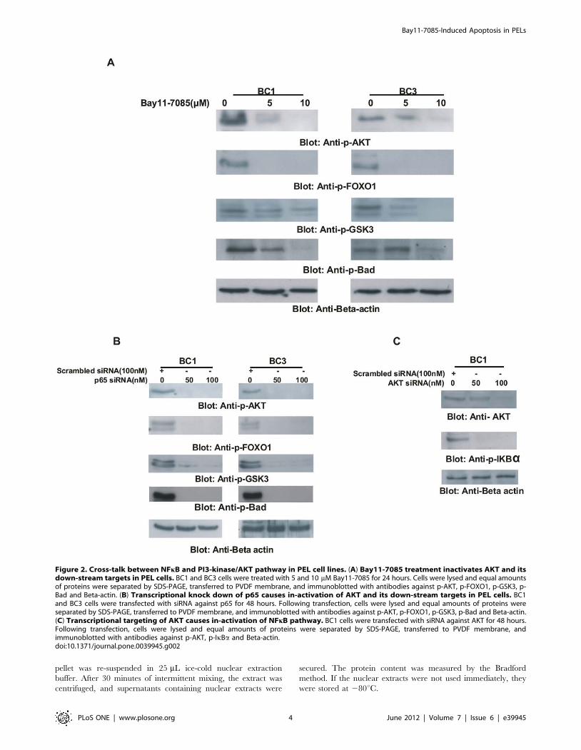
Figure 2. Cross-talk between NFkB and PI3-kinase/AKT pathway in PEL cell lines. (A) Bay11-7085 treatment inactivates AKT and itsdown-stream targets in PEL cells. BC1 and BC3 cells were treated with 5 and 10 mM Bay11-7085 for 24 hours. Cells were lysed and equal amountsof proteins were separated by SDS-PAGE, transferred to PVDF membrane, and immunoblotted with antibodies against p-AKT, p-FOXO1, p-GSK3, p-Bad and Beta-actin. (B) Transcriptional knock down of p65 causes in-activation of AKT and its down-stream targets in PEL cells. BC1and BC3 cells were transfected with siRNA against p65 for 48 hours. Following transfection, cells were lysed and equal amounts of proteins wereseparated by SDS-PAGE, transferred to PVDF membrane, and immunoblotted with antibodies against p-AKT, p-FOXO1, p-GSK3, p-Bad and Beta-actin.(C) Transcriptional targeting of AKT causes in-activation of NFkB pathway. BC1 cells were transfected with siRNA against AKT for 48 hours.Following transfection, cells were lysed and equal amounts of proteins were separated by SDS-PAGE, transferred to PVDF membrane, andimmunoblotted with antibodies against p-AKT, p-IkBa and Beta-actin.doi:10.1371/journal.pone.0039945.g002
Bay11-7085-Induced Apoptosis in PELs
PLoS ONE | www.plosone.org 4 June 2012 | Volume 7 | Issue 6 | e39945
Electrophoretic Mobility Shift Assay for NF-kBThe single-stranded 39-end biotin-labeled probe containing the
NFkBconsensus site 59-AGTTGAGGGGACTTTCCCAGGC-39,
and 39-TCAACTCCCCTGAAAG GGTCCG59 were purchased
from Metabion (Martinsried, Germany). The biotinylated oligo-
nucleotides were annealed by denaturing at 90uC for 1 minute and
cooled to room temperature for 1 hour. The EMSA binding
reactions were performed by utilizing a LightShift chemilumines-
cent EMSA kit (Pierce, Rockford, IL) as described previously [27].
Specifically, 3 mg nuclear extract was incubated in 1Xbinding
buffer containing 2.5% glycerol, 0.05% NP-40, 50 mM KCl,
5 mM MgCl2, 50 ng Poly (dI–dC) and biotinylated probe with or
without protein extract for 30 min at room temperature. The
complexes were separated on a 6% polyacrylamide–0.5XTris-
borate-EDTA gel and transferred to a positive charge nylon
membrane. After the transfer was completed, the membrane was
cross-linked and biotin-labeled DNA was detected by using a
chemiluminescent detection kit (Pierce, Rockford, IL).
Measurement of Mitochondrial Potential andCytochrome C Release
After treatment of PEL cell lines with two doses of Bay11-7085
for 48 hours, mitochondrial membrane potential was measured
using JC1 dye and release of cytochrome c was analyzed using
immunoblotting on cytosolic protein fractions as described
previously [26].
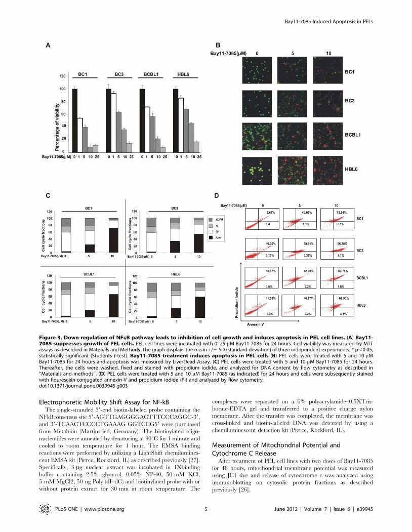
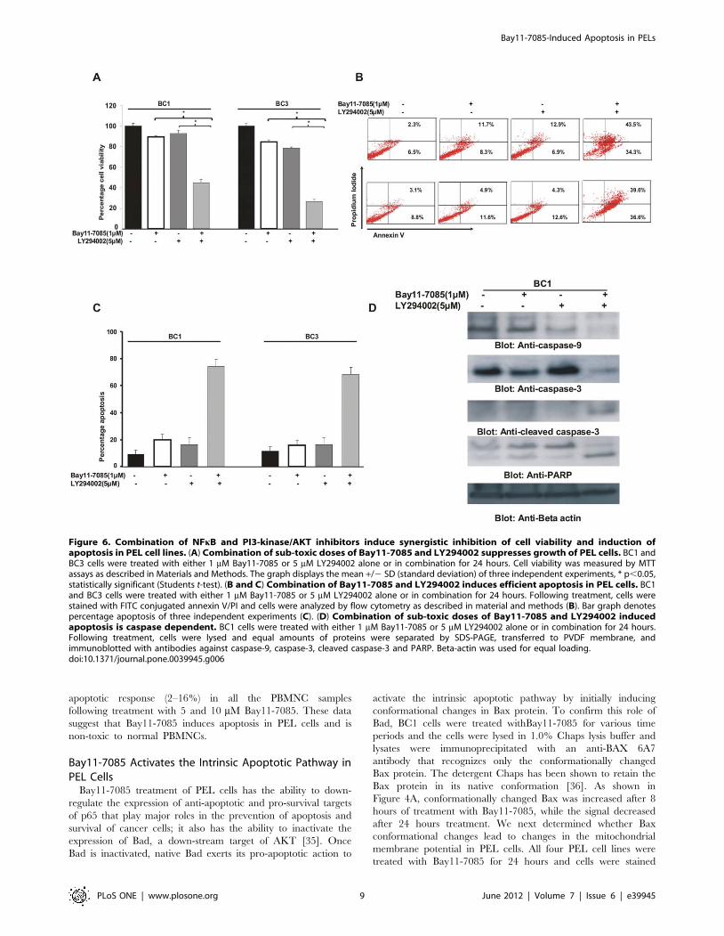
Figure 3. Down-regulation of NFkB pathway leads to inhibition of cell growth and induces apoptosis in PEL cell lines. (A) Bay11-7085 suppresses growth of PEL cells. PEL cell lines were incubated with 0–25 mM Bay11-7085 for 24 hours. Cell viability was measured by MTTassays as described in Materials and Methods. The graph displays the mean +/2 SD (standard deviation) of three independent experiments, * p,0.05,statistically significant (Students t-test). Bay11-7085 treatment induces apoptosis in PEL cells (B) PEL cells were treated with 5 and 10 mMBay11-7085 for 24 hours and apoptosis was measured by Live/Dead Assay. (C) PEL cells were treated with 5 and 10 mM Bay11-7085 for 24 hours.Thereafter, the cells were washed, fixed and stained with propidium iodide, and analyzed for DNA content by flow cytometry as described in‘‘Materials and methods’’. (D) PEL cells were treated with 5 and 10 mM Bay11-7085 (as indicated) for 24 hours and cells were subsequently stainedwith flourescein-conjugated annexin-V and propidium iodide (PI) and analyzed by flow cytometry.doi:10.1371/journal.pone.0039945.g003
Bay11-7085-Induced Apoptosis in PELs
PLoS ONE | www.plosone.org 5 June 2012 | Volume 7 | Issue 6 | e39945
Detection of Bax Conformational ChangesCells were treated with 10 mM Bay11-7085 for various time
periods and lysed with Chaps lysis buffer (10 mM HEPES
(ph 7.4), 150 mM NaCl, 1% Chaps) and immuno-precipitated
with anti-Bax-6A7 monoclonal antibody and Bax conformation
was detected as described earlier [27].
Gene Silencing Using siRNAp65 siRNA was purchased from Santa Cruz Biotechnology, Inc.
(Santa Cruz, CA, USA) while AKT siRNA and scrambled control
siRNA were purchased from Qiagen (Valencia, CA, USA). Cells
were transfected with above mentioned siRNA using Lipofecta-
mine 2000 (Invitrogen, Carlsbad, CA) for 6 hours following which
the lipid and siRNA complex was removed and fresh growth
medium was added. Cells were either lysed 48 hours after
transfection and specific protein levels were determined by
Western Blot analysis with specific antibodies.
Results
NFkB is Constitutively Activated in PEL Cell LinesThe NFkB survival pathway has been found to be
constitutively activated in various cancer cells [29,30,31], but
its role in PEL cells is unknown. We examined the activation of
status of NFkB in different PEL cell lines. Constitutive
activation of NFkB is determined by the presence of p65 sub
unit of NFkB in the nuclear compartment of cells where it
exerts its transcriptional activity [32]. We found that all four
PEL cell lines studied exhibited constitutive activation of NFkB
detected by EMSA assays (Figure 1A). We next sought to
determine whether treatment of PEL cells with a specific
inhibitor of NFkB; Bay11-7085 could inhibit translocation of
p65 into nucleus. BC1 and BC3 cell lines were treated at 5 or
10 mM final concentrations of Bay11-7085 for 24 hours and
nuclear extracts were prepared and EMSA was performed. As
shown in Figure 1B, Bay11-7085 treatment caused decreased
expression of p65 in the nuclear compartment suggesting that
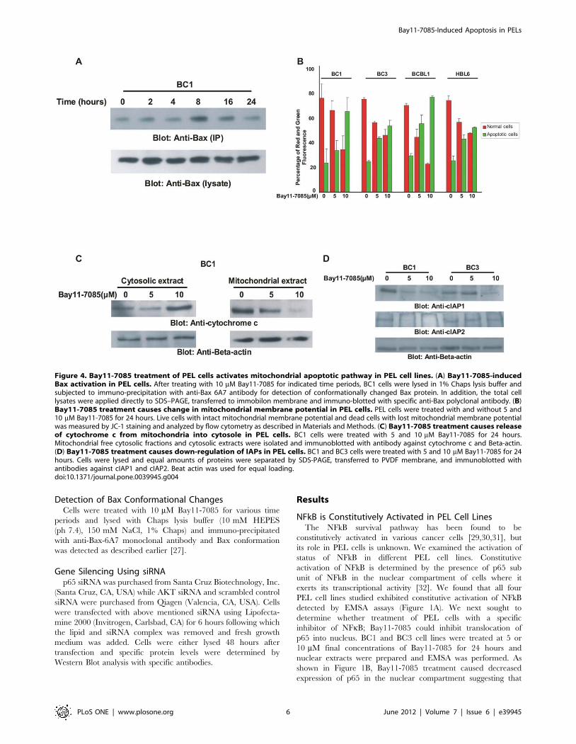
Figure 4. Bay11-7085 treatment of PEL cells activates mitochondrial apoptotic pathway in PEL cell lines. (A) Bay11-7085-inducedBax activation in PEL cells. After treating with 10 mM Bay11-7085 for indicated time periods, BC1 cells were lysed in 1% Chaps lysis buffer andsubjected to immuno-precipitation with anti-Bax 6A7 antibody for detection of conformationally changed Bax protein. In addition, the total celllysates were applied directly to SDS–PAGE, transferred to immobilon membrane and immuno-blotted with specific anti-Bax polyclonal antibody. (B)Bay11-7085 treatment causes change in mitochondrial membrane potential in PEL cells. PEL cells were treated with and without 5 and10 mM Bay11-7085 for 24 hours. Live cells with intact mitochondrial membrane potential and dead cells with lost mitochondrial membrane potentialwas measured by JC-1 staining and analyzed by flow cytometry as described in Materials and Methods. (C) Bay11-7085 treatment causes releaseof cytochrome c from mitochondria into cytosole in PEL cells. BC1 cells were treated with 5 and 10 mM Bay11-7085 for 24 hours.Mitochondrial free cytosolic fractions and cytosolic extracts were isolated and immunoblotted with antibody against cytochrome c and Beta-actin.(D) Bay11-7085 treatment causes down-regulation of IAPs in PEL cells. BC1 and BC3 cells were treated with 5 and 10 mM Bay11-7085 for 24hours. Cells were lysed and equal amounts of proteins were separated by SDS-PAGE, transferred to PVDF membrane, and immunoblotted withantibodies against cIAP1 and cIAP2. Beat actin was used for equal loading.doi:10.1371/journal.pone.0039945.g004
Bay11-7085-Induced Apoptosis in PELs
PLoS ONE | www.plosone.org 6 June 2012 | Volume 7 | Issue 6 | e39945
activation of NFkB pathway can be blocked in PEL cells
following treatment with Bay11-7085.
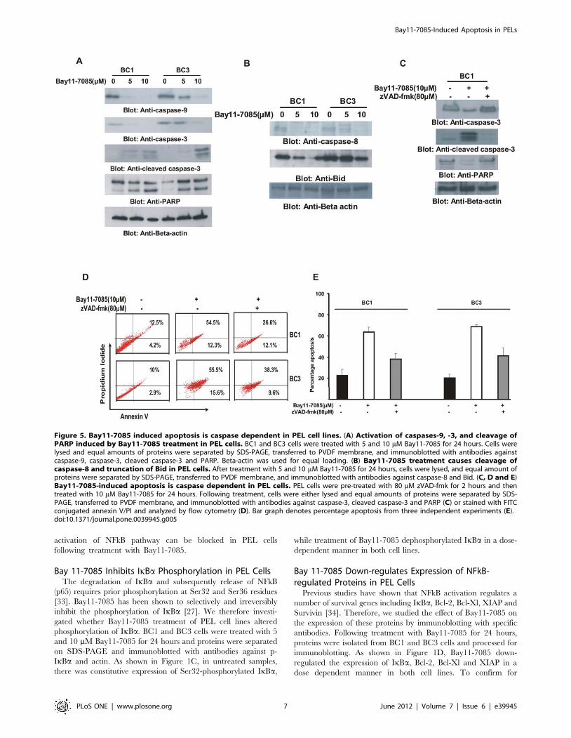
Bay 11-7085 Inhibits IkBa Phosphorylation in PEL CellsThe degradation of IkBa and subsequently release of NFkB
(p65) requires prior phosphorylation at Ser32 and Ser36 residues
[33]. Bay11-7085 has been shown to selectively and irreversibly
inhibit the phosphorylation of IkBa [27]. We therefore investi-
gated whether Bay11-7085 treatment of PEL cell lines altered
phosphorylation of IkBa. BC1 and BC3 cells were treated with 5
and 10 mM Bay11-7085 for 24 hours and proteins were separated
on SDS-PAGE and immunoblotted with antibodies against p-
IkBa and actin. As shown in Figure 1C, in untreated samples,
there was constitutive expression of Ser32-phosphorylated IkBa,
while treatment of Bay11-7085 dephosphorylated IkBa in a dose-
dependent manner in both cell lines.
Bay 11-7085 Down-regulates Expression of NFkB-regulated Proteins in PEL Cells
Previous studies have shown that NFkB activation regulates a
number of survival genes including IkBa, Bcl-2, Bcl-Xl, XIAP and
Survivin [34]. Therefore, we studied the effect of Bay11-7085 on
the expression of these proteins by immunoblotting with specific
antibodies. Following treatment with Bay11-7085 for 24 hours,
proteins were isolated from BC1 and BC3 cells and processed for
immunoblotting. As shown in Figure 1D, Bay11-7085 down-
regulated the expression of IkBa, Bcl-2, Bcl-Xl and XIAP in a
dose dependent manner in both cell lines. To confirm for
Figure 5. Bay11-7085 induced apoptosis is caspase dependent in PEL cell lines. (A) Activation of caspases-9, -3, and cleavage ofPARP induced by Bay11-7085 treatment in PEL cells. BC1 and BC3 cells were treated with 5 and 10 mM Bay11-7085 for 24 hours. Cells werelysed and equal amounts of proteins were separated by SDS-PAGE, transferred to PVDF membrane, and immunoblotted with antibodies againstcaspase-9, caspase-3, cleaved caspase-3 and PARP. Beta-actin was used for equal loading. (B) Bay11-7085 treatment causes cleavage ofcaspase-8 and truncation of Bid in PEL cells. After treatment with 5 and 10 mM Bay11-7085 for 24 hours, cells were lysed, and equal amount ofproteins were separated by SDS-PAGE, transferred to PVDF membrane, and immunoblotted with antibodies against caspase-8 and Bid. (C, D and E)Bay11-7085-induced apoptosis is caspase dependent in PEL cells. PEL cells were pre-treated with 80 mM zVAD-fmk for 2 hours and thentreated with 10 mM Bay11-7085 for 24 hours. Following treatment, cells were either lysed and equal amounts of proteins were separated by SDS-PAGE, transferred to PVDF membrane, and immunoblotted with antibodies against caspase-3, cleaved caspase-3 and PARP (C) or stained with FITCconjugated annexin V/PI and analyzed by flow cytometry (D). Bar graph denotes percentage apoptosis from three independent experiments (E).doi:10.1371/journal.pone.0039945.g005
Bay11-7085-Induced Apoptosis in PELs
PLoS ONE | www.plosone.org 7 June 2012 | Volume 7 | Issue 6 | e39945
specificity of the findings using Bay11-7085, we transfected either
p65 specific siRNA or scrambled non-specific siRNA in BC1 and
BC3 cells. After 48 hours of transfection, proteins were extracted
from the cells and immunoblotted with antibodies against IkBa,
Bcl-2, Bcl-xL and XIAP. There was concordance in the data
generated following treatment with Bay11-7085 and siRNA
against p65. As shown in Figure 1E, siRNA knockdown of p65
led to down-regulation of IkBa, Bcl-2, Bcl-xL and XIAP in both
cell lines. These data clearly suggest that inhibition of NFkB leads
to suppression of expression of genes that are involved in growth
and survival of PEL cells.
Inhibition of NFkB Inactivates AKT and its Down-streamTargets in PEL Cells
It has been previously shown that NFkB survival pathway is also
linked to other survival pathways including PI3-kinase/AKT
pathway in various cancers [18,19]. However, cross-talk between
these two pathways has not been elucidated in PEL cells.
Therefore, we determined whether there are any interactions
between NFkB and the PI3-kinase/AKT pathway. For this reason,
BC1 and BC3 cells were treated with Bay11-7085 and the
extracted proteins were immunoblotted with antibodies against p-
AKT, p-Foxo1, p-GSK3 and p-Bad. As shown in Figure 2A,
Bay11-7085 treatment of PEL cells inactivated AKT and caused
de-phosphorylation of its downstream targets Foxo1, GSK3 and
Bad. To confirm the Bay11-7085 findings, we examined the effects
of p65 knockdown in PEL cells. For that purpose, we treated PEL
cells with siRNA targeting p65. siRNA knockdown of p65 led to
inactivation of AKT and its downstream targets, FOXO1, GSK3
and Bad (Figure 2B). In addition, we found that in PEL cells in
which AKT expression had been knocked down by specific siRNA
targeted against AKT, there was inactivation of IkBa suggesting a
cross-talk between the NFkB and PI3-kinase/AKT pathway
(Figure 2C).
Bay11-7085 Inhibits Cell Viability and Induces Apoptosisof PEL Cells
Since we have shown that Bay11-7085 treatment of PEL cells
causes down-regulation of NFkB and PI3-kinase/AKT pathway,
we sought to determine whether these effects cause inhibition of
cell viability and induce apoptosis in PEL cells. BC1, BC3,
BCBL, and HBL6 cells were cultured in the presence or
absence of 0, 1, 10 and 25 mM Bay11-7085 for 24 hours and
cell viability was subsequently assessed. As shown in Figure 3A,
Bay11-7085 treatment caused inhibition of cell viability in a
dose dependent manner in all the PEL cell lines. We next
sought to determine whether inhibition of cell viability occurred
due to PEL cells undergoing cell death. For this reason, we
treated PEL cell lines with Bay11-7085 for 24 hours and after
staining them with calcein and ethidium homodimer, plasma
membrane integrity was assessed. As shown in Figure 3B,
untreated cells were stained green depicting alive cells with
plasma membrane integrity intact while cells treated with
Bay11-7085 showed an increase in red cells suggesting
disruption of plasma membrane integrity, i.e., dead cells. We
further analyzed PEL cells for cell cycle fractions after treatment
with Bay11-7085 for 24 hours and found that Bay11-7085
increased cells in subG1/Apo fraction in all the cell lines tested
(Figure 3C). This data suggested that Bay11-7085 was inducing
apoptosis in PEL cell lines. To further confirm that this increase
in the sub-G1 population indeed reflected apoptosis, PEL cells
were treated with 5 and 10 mM Bay11-7085 as indicated and
apoptotic cells were analyzed by annexin V dual staining. As
shown in Figure 3D, Bay11-7085 treatment resulted in
apoptosis in a dose dependent manner in all PEL cell lines.
We also confirmed apoptosis by DNA fragmentation in BC1
and BC3 cells treated with Bay11-7085 for 24hours (data not
shown).
Finally, we also treated five normal peripheral blood
mononuclear cells (PBMNC) from healthy donors with 5 and
10 mM Bay11-7085 to assess the effect of Bay11-7085 on
normal cells. As shown in Figure S1, there was minimal
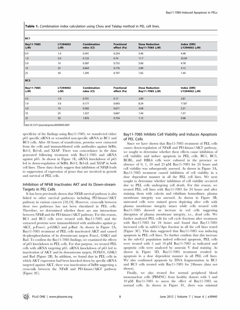
Table 1. Combination index calculation using Chou and Talalay method in PEL cell lines.
BC1
Bay11-7085(mM)
LY294002(mM)
Combinationndex (CI)
Fractionaleffect (Fa)
Dose ReductionBay11-7085 (mM)
Index (DRI)LY294002 (mM)
0.5 1.0 0.391 0.254 5.96 4.48
1.0 5.0 0.125 0.74 11.7 25.09
5.0 10 0.387 0.732 5.60 4.78
10 25 0.664 0.776 2.89 3.13
25 50 1.295 0.797 1.65 1.44
BC3
Bay11-7085(mM)
LY294002(mM)
Combinationndex (CI)
Fractionaleffect (Fa)
Dose ReductionBay11-7085 (mM)
Index (DRI)LY294002 (mM)
0.5 1.0 0.701 0.17 2.89 2.81
1.0 5.0 0.177 0.693 8.24 17.87
5.0 10 0.582 0.671 3.68 3.21
10 25 1.327 0.667 1.44 1.57
25 50 1.876 0.754 1.15 0.98
doi:10.1371/journal.pone.0039945.t001
Bay11-7085-Induced Apoptosis in PELs
PLoS ONE | www.plosone.org 8 June 2012 | Volume 7 | Issue 6 | e39945
apoptotic response (2–16%) in all the PBMNC samples
following treatment with 5 and 10 mM Bay11-7085. These data
suggest that Bay11-7085 induces apoptosis in PEL cells and is
non-toxic to normal PBMNCs.
Bay11-7085 Activates the Intrinsic Apoptotic Pathway inPEL Cells
Bay11-7085 treatment of PEL cells has the ability to down-
regulate the expression of anti-apoptotic and pro-survival targets
of p65 that play major roles in the prevention of apoptosis and
survival of cancer cells; it also has the ability to inactivate the
expression of Bad, a down-stream target of AKT [35]. Once
Bad is inactivated, native Bad exerts its pro-apoptotic action to
activate the intrinsic apoptotic pathway by initially inducing
conformational changes in Bax protein. To confirm this role of
Bad, BC1 cells were treated withBay11-7085 for various time
periods and the cells were lysed in 1.0% Chaps lysis buffer and
lysates were immunoprecipitated with an anti-BAX 6A7
antibody that recognizes only the conformationally changed
Bax protein. The detergent Chaps has been shown to retain the
Bax protein in its native conformation [36]. As shown in
Figure 4A, conformationally changed Bax was increased after 8
hours of treatment with Bay11-7085, while the signal decreased
after 24 hours treatment. We next determined whether Bax
conformational changes lead to changes in the mitochondrial
membrane potential in PEL cells. All four PEL cell lines were
treated with Bay11-7085 for 24 hours and cells were stained
Figure 6. Combination of NFkB and PI3-kinase/AKT inhibitors induce synergistic inhibition of cell viability and induction ofapoptosis in PEL cell lines. (A) Combination of sub-toxic doses of Bay11-7085 and LY294002 suppresses growth of PEL cells. BC1 andBC3 cells were treated with either 1 mM Bay11-7085 or 5 mM LY294002 alone or in combination for 24 hours. Cell viability was measured by MTTassays as described in Materials and Methods. The graph displays the mean +/2 SD (standard deviation) of three independent experiments, * p,0.05,statistically significant (Students t-test). (B and C) Combination of Bay11-7085 and LY294002 induces efficient apoptosis in PEL cells. BC1and BC3 cells were treated with either 1 mM Bay11-7085 or 5 mM LY294002 alone or in combination for 24 hours. Following treatment, cells werestained with FITC conjugated annexin V/PI and cells were analyzed by flow cytometry as described in material and methods (B). Bar graph denotespercentage apoptosis of three independent experiments (C). (D) Combination of sub-toxic doses of Bay11-7085 and LY294002 inducedapoptosis is caspase dependent. BC1 cells were treated with either 1 mM Bay11-7085 or 5 mM LY294002 alone or in combination for 24 hours.Following treatment, cells were lysed and equal amounts of proteins were separated by SDS-PAGE, transferred to PVDF membrane, andimmunoblotted with antibodies against caspase-9, caspase-3, cleaved caspase-3 and PARP. Beta-actin was used for equal loading.doi:10.1371/journal.pone.0039945.g006
Bay11-7085-Induced Apoptosis in PELs
PLoS ONE | www.plosone.org 9 June 2012 | Volume 7 | Issue 6 | e39945
with JC1, a surrogate marker for detection of changes in
mitochondrial membrane potential, followed by flow cytometry
analysis. If the mitochondria are intact, JC1 dye enters them,
remains in the monomeric form and stains the cell red,
however, if the mitochondria are damaged, JC1 cannot be
retained in the mitochondria and is expelled in the cytoplasm in
the polymeric form and stains the cell green. As shown in
Figure 4B, Bay11-7085 treatment resulted in loss of mitochon-
drial membrane potential in all the PEL cell lines as measured
by increase in JC1 stained green florescence depicting apoptotic
cells. We then studied the release of cytochrome c from
mitochondria into cytosol. As shown in Figure 4C, higher levels
of cytochrome c were detactable in cytosolic and lower levels in
the mitochondrial fraction in the BC1 cell line after Bay11-7085
treatment. These findings suggest that Bay11-7085 treatment of
PEL cell lines causes dephosphorylation of Bad leading to
activation of the mitochondrial apoptotic pathway.
Down-regulation of Inhibitor of Apoptosis Proteins (IAPs)following Treatment of PEL Cells with Bay11-7085
IAPs play an important role in inhibition of apoptosis by
disrupting the recruitment of caspases-9 and -3 for efficient
apoptosis to occur. We found that in addition to down-regulation
of XIAP and Survivin, Bay11-7085 treatment also inhibited other
IAPs including cIAP1 and cIAP2 (Figure 4D).
Bay11-7085 Treatment Induces Activation of Caspases inPEL Cells
Since caspases are important mediators of apoptosis in
response to various apoptotic stimuli [13,26,37], we investigated
whether Bay11-7085 treatment also causes their activation and
cleavage in PEL cells. BC1 and BC3 cells were treated with
Bay11-7085 for 24 hours and cell lysates were immunoblotted
with antibodies against caspase-9, caspase-3, cleaved caspase-3
and PARP. As shown in Figure 5A, Bay11-7085 treatment of
PEL cells induced activation of caspase-9 and caspase-3
cleavage in both cell lines. PARP-1, a downstream target of
caspase-3 was also cleaved in bothcell lines, a hallmark of cells
undergoing apoptosis. It has been shown by us and others that
caspase-3 has the ability to activate caspase-8 and cleave Bid,
downstream of the mitochondrial pathway to potentate the
apoptotic signal [38]. Therefore, we sought to determine the
expression of caspase-8 and Bid following treatment with Bay11-
7085. As shown in Figure 5B, Bay11-7085 treatment resulted in
reduction in the intensity of the full-length band of pro-caspase-
8 indicating activation of caspase-8 in both cell lines. Bid is a
BH3-proapoptotic protein that can be cleaved directly by
caspases-8 during apoptosis. The cleaved or truncated Bid also
plays a role in the induction of Bax conformational change and
subsequent translocation to mitochondria [39,40]. Therefore, we
examined if Bay11-7085 treatment of PEL cells could truncate
the Bid protein. As shown in Figure 5B, there was decreased
expression of Bid protein following treatment of BC1 and BC3
cell lines with Bay11-7085 suggesting that caspase-8 and Bid are
also activated following inhibition of NFkB survival pathway.
Furthermore, pre-treatment of PEL cells with 80 mM z-VAD-
fmk a universal inhibitor of caspases markedly decreased
caspase-3 activation (Figure 5C), and prevented apoptosis
(Figure 5D and E) induced by Bay11-7085, clearly indicating
that caspases play a critical role in Bay11-7085-induced
apoptosis in PEL cells. Finally, to confirm whether the intrinsic
apoptotic pathway is actually activated in Bay11-7085 induced
apoptosis, we pre-treated BC1 cells with either 80 mM caspase-8
inhibitor (IETD-CHO) or 80 mM caspase-9 inhibitor (LEHD-
CHO) for 3 hours followed by treatment with 10 mM Bay11-
7085 for 24 hours. Following treatment, cells were lysed and
proteins were extracted, immunoblotted and probed with
different antibodies. As shown in Figure S2A, caspase-8
inhibitor blocked cleavage of caspase-8 and Bid but failed to
block cleavage of caspase-3. Alternately, caspase-9 inhibitor
blocked cleavage of caspase-9, caspase-3, caspase-8 and Bid in
BC1 cells (Figure S2B). These set of data clearly indicates that
Bay11-7085 treatment induces apoptosis via intrinsic apoptotic
pathway.
Combination Treatment of PEL Cells with PI3-kinase/AKTInhibitor and Bay11-7085 Induce Synergistically PotentApoptosis
Cross-talk between NFkB pathway and PI3-kinase/AKT
pathway has been shown in various cancers [18,19], however,
the relationship between these two pathways have not been fully
explored in PEL cells. We found that inhibition of NFkB pathway
by Bay11-7085 not only inactivates the NFkB pathway, it also
inactivates AKT as well as down-stream targets of AKT such as
FOXO1, GSK3 and Bad. Utilizing this information, we aimed at
targeting PEL cell lines with a combination of NFkB and PI3-
kinase/AKT inhibitors at sub-optimal doses to determine the
synergistic therapeutic potential of such a combination. Multiple
experiments were conducted to define optimal doses for a
synergistic apoptotic response of the combination of Bay11-7085
and LY294002, a specific PI3-kinase inhibitor. As shown in
Table 1 and Figure S3, using Chou and Talalay method [41], we
found that 1 mM Bay11-7085 and 5 mM LY294002 exerted the
maximum synergistic apoptotic response in BC1 cells (combina-
tion index 0.125) and BC3 cells (combination index 0.177), with
both the values being less than 1.0 suggesting a strong synergistic
response [41]. Using these doses, we first assessed the cell viability
of cells following treatment with combination of Bay11-7085 with
LY294002. As shown in Figure 6A, neither Bay11-7085 at a
concentration of 1 mM nor LY294002 at a concentration of 5 mM
could inhibit cell viability in any of the PEL cell lines. However
when both the drugs were added together to the cultures as a
combination, there was efficient inhibition of cell viability in PEL
cells. Next, we examined the apoptotic response of PEL cells
following combination treatment with Bay11-7085 and
LY294002. As shown in Figure 6B and C, Bay11-7085 or
LY294002 alone failed to induce apoptosis at sub-optimal doses,
however, when both drugs were used in combination there was a
synergistic apoptotic response as detected by annexin V/PI dual
staining. Finally, we found that combination of Bay11-7085 and
LY294002 induced apoptosis via the activation of the caspase
cascade and cleavage of PARP (Figure 6D).
Discussion
Primary effusion lymphoma (PEL) is a very aggressive and fatal
human malignancy, which frequently and rapidly develops
resistance to conventional chemotherapeutic agents [3]. It is now
postulated that the mechanisms of lymphomagenesis involve
deregulation of several signaling pathways that may interact with
each other to escape programmed cell death [18,19]. We have
previously shown that the PI3-kinase/AKT pathway is activated in
PEL cells and is one of the driving forces behind the aggressive
phenotype of these cells [12]. In addition to the PI3-kinase/AKT
pathway, PEL cells are also dependent on other activated signaling
pathways including NFkB [42,43]. In this study, we have
investigated the molecular mechanism of NFkB mediated anti-
Bay11-7085-Induced Apoptosis in PELs
PLoS ONE | www.plosone.org 10 June 2012 | Volume 7 | Issue 6 | e39945
apoptotic role in PEL cell lines using Bay11-7085, a specific
inhibitor of NFkB pathway as well as siRNA knockdown targeting
the p65 subunit. Our data suggest that inhibition of NFkB causes
inability of the p65 subunit to enter the nucleus resulting in down-
regulation of anti-apoptotic targets of p65 such as IkBa, XIAP,
Bcl-Xl and Survivin. In addition, Bay11-7085 treatment also led to
inactivation of PI3-kinase/AKT pathway. Genetic knockdown of
p65 with siRNA also resulted in inactivation of PI3-kinase/AKT
signaling suggesting a cross-talk between these two survival
pathways in PEL cells. Cross-talk between these two pathways
has been shown in other cancers. Reciprocally, siRNA knockdown
of AKT also led to dephosphorylation of IkBa confirming the
presence of a cross-talk between the survival pathways.
Apoptosis is a multi step process and an increasing number of
genes have been identified that are involved in the control or
execution of apoptosis [44]. Inhibition of apoptosis is a key
mechanism used by cancer cells to proliferate and grow unabated.
Activation of NFkB pathway also plays a major role in inhibiting
apoptosis by causing up-regulation of key anti-apoptotic proteins
such as Bcl-2, Bcl-Xl, XIAP and Survivin [14]. In order for
efficient apoptosis to occur, these anti-apoptotic proteins need to
be down-regulated so that the apoptotic pathway is activated and
the cells die. Our studies established that Bay-11-7085 treatment
down-regulates the expression of Bcl-2 and Bcl-Xl protein and
inactivated Bad protein that in turn allowed Bax conformational
changes and subsequent translocation to mitochondria [45]
leading to the formation of mitochondrial pores. These events
result in loss of mitochondrial potential and release of cytochrome
c from mitochondria to cytoplasm [46]. Cytochrome c release
from the mitochondria has been proposed as the most critical
event for cells to initiate the apoptotic cascade [47].
Our data also established that Bay11-7085 treatment causes
down-regulation of IAPS; XIAP and Survivin that interfere with
the activation and cleavage of caspases [48]. Once these IAPs were
down-regulated along with release of cytochrome c following
inhibition of NFkB activity by Bay11-7085, there was sequential
activation of caspase-9, caspase-3 and cleavage PARP in PEL cell
lines. Furthermore, pre-treatment of PEL cells with a broad-
spectrum caspase inhibitor markedly blocked Bay11-7085-induced
apoptosis. These data clearly suggests that activation of caspase-
cascades is more prominently involved in Bay11-7085-induced
apoptosis.
In this manuscript, we have hypothesized that a functional link
might exist between AKT and NFkB pathways in the pathogenesis
of PEL and activation of these survival pathways may sustain
survival and proliferation of these malignant cells. Since our
findings suggested that NFkB and AKT pathways are activated in
PEL cells, we proposed that simultaneous targeting of these
pathways may synergistically induce apoptosis of PEL cells. We
found that when PEL cell lines treated with sub-toxic doses of
Bay11-7085 and LY294002, a specific inhibitor of PI3-kinase/
AKT, neither Bay11-7085 at a dose of 1 mM nor LY294002 at
5 mM could inhibit cell viability in any of the PEL cell lines.
However when both the drugs were given together as a
combination, there was efficient inhibition of cell viability of
PEL cells via induction of caspase-mediated apoptosis. These data
clearly indicate the importance of targeting multiple survival
pathways simultaneously using sub-toxic doses of specific inhibi-
tors thereby decreasing the chances of toxicity and increasing the
response to therapy.
Despite advances in therapeutic regimes for the treatment of
aggressive NHL over the last decade, PELs are still refractory to
conventional systemic chemotherapy with a mean overall survival
of 3 months [49]. The suggested benefit of high-dose Methotrexate
in association with CHOP (Cyclophosphamide, Doxorubicin,
Prednisolone and Vincristine)-like regimens is negatively balanced
by the hampered toxicity of Methotrexate in the presence of serous
effusions [3]. Therefore, newer therapeutic agents such as Bay11-
7085 and LY294002 may play important roles in the management
of these aggressive lymphomas in combination with conventional
chemotherapy to improve survival and decrease toxicity.
In conclusion, our results demonstrate that down-regulation of
the NFkB pathway leads to activation of the mitochondrial
apoptotic pathway. This process occurs via down-regulation of
downstream targets of p65; Bcl-2, Bcl-Xl, XIAP and Survivin,
ultimately resulting in activation of caspase-dependent apoptosis.
Simultaneously, we also investigated the cross-talk between NFkB
and PI3-kinase/AKT pathway and found that targeting of these
survival pathways simultaneously significantly increases the
apoptotic stimuli in PEL cells thereby decreasing the chances of
toxicity. Taken altogether our data suggests that Bay11-7085
possesses the chemopreventive/therapeutic potentials against these
aggressive lymphomas either alone or in combination with other
inhibitors.
Supporting Information
Figure S1 Bay11-7085 treatment is non-toxic to normalperipheral blood mononuclear cells (PBMNC). (A)
PBMNC from 5 normal healthy donors were isolated and treated
with 5 and 10 mM Bay11-7085 for 24 hours. Following treatment,
cells were harvested and stained with fluorescein conjugated
annexin V/PI and cells were analyzed by flow cytometry. Bar
graph denotes an average of 3 independent experiments.
(TIF)
Figure S2 Bay11-7085-induced apoptosis is via intrinsicapoptotic pathway in PEL cells. BC1 cells were pre-treated
with either 80 mM IETD-CHO (caspase-8 inhibitor) (A) or 80 mM
LEHD-CHO (caspase-9 inhibitor) (B) for 3 hours followed by
treatment with 10 mM Bay11-7085 for 24 hours. Following
treatment, proteins were extracted, immunoblotted and probed
with antibodies against caspase-8, caspase-9, caspase-3 and Bid.
Beta-actin was used to insure equal loading.
(TIF)
Figure S3 Synergistic apoptotic response of Bay11-7085and TRAIL in PEL cells. BC1 and BC3 cells were treated with
various combinations of Bay11-7085 and TRAIL for 24 hours and
dose effect (A and C) and Fractional effect (B and D) graphs were
generated using Calcusyn software. Apoptotic response analysis
was measured as mean 6 SD values normalized to control.
Combination indices were calculated using Chou and Talalay
methodology.
(TIF)
Author Contributions
Conceived and designed the experiments: ARH KSAK SU. Performed the
experiments: ARH SOA MA OSK SAA. Analyzed the data: ARH LCP
SU. Contributed reagents/materials/analysis tools: ARH MA KSAK.
Wrote the paper: ARH LCP SU.
Bay11-7085-Induced Apoptosis in PELs
PLoS ONE | www.plosone.org 11 June 2012 | Volume 7 | Issue 6 | e39945
References
1. Ganem D (2007) KSHV-induced oncogenesis.
2. Arora A, Chiao E, Tyring SK (2007) AIDS malignancies. Cancer Treat Res 133:21–67.
3. Carbone A, Gloghini A (2008) KSHV/HHV8-associated lymphomas.Br J Haematol 140: 13–24.
4. Boulanger E, Afonso PV, Yahiaoui Y, Adle-Biassette H, Gabarre J, et al. (2008)
Human herpesvirus-8 (HHV-8)-associated primary effusion lymphoma in tworenal transplant recipients receiving rapamycin. Am J Transplant 8: 707–710.
5. Melo NC, Sales MM, Santana AN, Costalonga EC, Pedreira AB, et al. (2008)Pleural primary effusion lymphoma in a renal transplant recipient.
Am J Transplant 8: 906–907.
6. Wu SJ, Hung CC, Chen CH, Tien HF (2009) Primary effusion lymphoma inthree patients with chronic hepatitis B infection. J Clin Virol 44: 81–83.
7. Brimo F, Michel RP, Khetani K, Auger M (2007) Primary effusion lymphoma: aseries of 4 cases and review of the literature with emphasis on cytomorphologic
and immunocytochemical differential diagnosis. Cancer 111: 224–233.8. Aoki Y, Yarchoan R, Wyvill K, Okamoto S, Little RF, et al. (2001) Detection of
viral interleukin-6 in Kaposi sarcoma-associated herpesvirus-linked disorders.
Blood 97: 2173–2176.9. Jones KD, Aoki Y, Chang Y, Moore PS, Yarchoan R, et al. (1999) Involvement
of interleukin-10 (IL-10) and viral IL-6 in the spontaneous growth of Kaposi’ssarcoma herpesvirus-associated infected primary effusion lymphoma cells. Blood
94: 2871–2879.
10. Bais C, Santomasso B, Coso O, Arvanitakis L, Raaka EG, et al. (1998) G-protein-coupled receptor of Kaposi’s sarcoma-associated herpesvirus is a viral
oncogene and angiogenesis activator. Nature 391: 86–89.11. An J, Sun Y, Fisher M, Rettig MB (2004) Antitumor effects of bortezomib (PS-
341) on primary effusion lymphomas. Leukemia 18: 1699–1704.12. Uddin S, Hussain AR, Al-Hussein KA, Manogaran PS, Wickrema A, et al.
(2005) Inhibition of phosphatidylinositol 39-kinase/AKT signaling promotes
apoptosis of primary effusion lymphoma cells. Clin Cancer Res 11: 3102–3108.13. Uddin S, Hussain AR, Manogaran PS, Al-Hussein K, Platanias LC, et al. (2005)
Curcumin suppresses growth and induces apoptosis in primary effusionlymphoma. Oncogene 24: 7022–7030.
14. Sethi G, Ahn KS, Aggarwal BB (2008) Targeting nuclear factor-kappa B
activation pathway by thymoquinone: role in suppression of antiapoptotic geneproducts and enhancement of apoptosis. Mol Cancer Res 6: 1059–1070.
15. Dolcet X, Llobet D, Pallares J, Matias-Guiu X (2005) NF-kB in developmentand progression of human cancer. Virchows Arch 446: 475–482.
16. Scheid MP, Woodgett JR (2000) Protein kinases: six degrees of separation? CurrBiol 10: R191–194.
17. Rahman MA, Amin AR, Shin DM (2010) Chemopreventive potential of natural
compounds in head and neck cancer. Nutr Cancer 62: 973–987.18. Han SS, Yun H, Son DJ, Tompkins VS, Peng L, et al. (2010) NF-kappaB/
STAT3/PI3K signaling crosstalk in iMyc E mu B lymphoma. Mol Cancer 9: 97.19. Ghosh-Choudhury N, Mandal CC, Ghosh Choudhury G (2010) Simvastatin
induces derepression of PTEN expression via NFkappaB to inhibit breast cancer
cell growth. Cell Signal 22: 749–758.20. Rossi D, Gaidano G (2003) Messengers of cell death: apoptotic signaling in
health and disease. Haematologica 88: 212–218.21. Elmore S (2007) Apoptosis: a review of programmed cell death. Toxicol Pathol
35: 495–516.22. Ghobrial IM, Witzig TE, Adjei AA (2005) Targeting apoptosis pathways in
cancer therapy. CA Cancer J Clin 55: 178–194.
23. Uddin S, Hussain AR, Ahmed M, Abubaker J, Al-Sanea N, et al. (2009) Highprevalence of fatty acid synthase expression in colorectal cancers in Middle
Eastern patients and its potential role as a therapeutic target. Am J Gastroenterol104: 1790–1801.
24. Fisher RI, Shah P (2003) Current trends in large cell lymphoma. Leukemia 17:
1948–1960.25. Hussain AR, Uddin S, Bu R, Khan OS, Ahmed SO, et al. (2011) Resveratrol
suppresses constitutive activation of AKT via generation of ROS and inducesapoptosis in diffuse large B cell lymphoma cell lines. PLoS One 6: e24703.
26. Hussain AR, Al-Rasheed M, Manogaran PS, Al-Hussein KA, Platanias LC, et
al. (2006) Curcumin induces apoptosis via inhibition of PI39-kinase/AKTpathway in acute T cell leukemias. Apoptosis 11: 245–254.
27. Hussain AR, Ahmed M, Al-Jomah NA, Khan AS, Manogaran P, et al. (2008)Curcumin suppresses constitutive activation of nuclear factor-kappa B and
requires functional Bax to induce apoptosis in Burkitt’s lymphoma cell lines. MolCancer Ther 7: 3318–3329.
28. Uddin S, Ah-Kang J, Ulaszek J, Mahmud D, Wickrema A (2004) Differentiation
stage-specific activation of p38 mitogen-activated protein kinase isoforms in
primary human erythroid cells. Proc Natl Acad Sci U S A 101: 147–152.
29. Shishodia S, Amin HM, Lai R, Aggarwal BB (2005) Curcumin (diferuloyl-methane) inhibits constitutive NF-kappaB activation, induces G1/S arrest,
suppresses proliferation, and induces apoptosis in mantle cell lymphoma.Biochem Pharmacol 70: 700–713.
30. Kim SW, Oleksyn DW, Rossi RM, Jordan CT, Sanz I, et al. (2008) Protein
kinase C-associated kinase is required for NF-kappaB signaling and survival in
diffuse large B-cell lymphoma cells. Blood 111: 1644–1653.
31. Cras A, Politis B, Balitrand N, Darsin-Bettinger D, Boelle PY, et al. (2011)Bexarotene via CBP/p300 induces suppression of NF-kappaB-dependent cell
growth and invasion in thyroid cancer. Clin Cancer Res.
32. Raskatov JA, Meier JL, Puckett JW, Yang F, Ramakrishnan P, et al. (2011)
Modulation of NF-kappaB-dependent gene transcription using programmableDNA minor groove binders. Proc Natl Acad Sci U S A.
33. Chen ZJ, Parent L, Maniatis T (1996) Site-specific phosphorylation of
IkappaBalpha by a novel ubiquitination-dependent protein kinase activity. Cell
84: 853–862.
34. Bharti AC, Donato N, Singh S, Aggarwal BB (2003) Curcumin (diferuloyl-methane) down-regulates the constitutive activation of nuclear factor-kappa B
and IkappaBalpha kinase in human multiple myeloma cells, leading tosuppression of proliferation and induction of apoptosis. Blood 101: 1053–1062.
35. Chen JH, Hsiao G, Lee AR, Wu CC, Yen MH (2004) Andrographolidesuppresses endothelial cell apoptosis via activation of phosphatidyl inositol-3-
kinase/Akt pathway. Biochem Pharmacol 67: 1337–1345.
36. Antonsson B, Montessuit S, Sanchez B, Martinou JC (2001) Bax is present as ahigh molecular weight oligomer/complex in the mitochondrial membrane of
apoptotic cells. J Biol Chem 276: 11615–11623.
37. Hussain AR, Al-Jomah NA, Siraj AK, Manogaran P, Al-Hussein K, et al. (2007)
Sanguinarine-dependent induction of apoptosis in primary effusion lymphomacells. Cancer Res 67: 3888–3897.
38. Uddin S, Siraj AK, Al-Rasheed M, Ahmed M, Bu R, et al. (2008) Fatty acid
synthase and AKT pathway signaling in a subset of papillary thyroid cancers.
J Clin Endocrinol Metab 93: 4088–4097.
39. Uddin S, Ahmed M, Bavi P, El-Sayed R, Al-Sanea N, et al. (2008) Bortezomib(Velcade) induces p27Kip1 expression through S-phase kinase protein 2
degradation in colorectal cancer. Cancer Res 68: 3379–3388.
40. Hussain AR, Ahmed M, Ahmed SO, Al-Thari S, Khan AS, et al. (2009)
Proteasome inhibitor MG-132 mediated expression of p27Kip1 via S-phasekinase protein 2 degradation induces cell cycle coupled apoptosis in primary
effusion lymphoma cells. Leuk Lymphoma 50: 1204–1213.
41. Chou TC, Talalay P (1984) Quantitative analysis of dose-effect relationships: thecombined effects of multiple drugs or enzyme inhibitors. Adv Enzyme Regul 22:
27–55.
42. Keller SA, Schattner EJ, Cesarman E (2000) Inhibition of NF-kappaB induces
apoptosis of KSHV-infected primary effusion lymphoma cells. Blood 96: 2537–2542.
43. Grossmann C, Ganem D (2008) Effects of NFkappaB activation on KSHV
latency and lytic reactivation are complex and context-dependent. Virology 375:
94–102.
44. Joseph B, Marchetti P, Formstecher P, Kroemer G, Lewensohn R, et al. (2002)Mitochondrial dysfunction is an essential step for killing of non-small cell lung
carcinomas resistant to conventional treatment. Oncogene 21: 65–77.
45. Eskes R, Desagher S, Antonsson B, Martinou JC (2000) Bid induces the
oligomerization and insertion of Bax into the outer mitochondrial membrane.Mol Cell Biol 20: 929–935.
46. Gogvadze V, Robertson JD, Zhivotovsky B, Orrenius S (2001) Cytochrome c
release occurs via Ca2+-dependent and Ca2+-independent mechanisms that areregulated by Bax. J Biol Chem 276: 19066–19071.
47. Zou H, Li Y, Liu X, Wang X (1999) An APAF-1.cytochrome c multimericcomplex is a functional apoptosome that activates procaspase-9. J Biol Chem
274: 11549–11556.
48. Smolewski P, Robak T (2011) Inhibitors of apoptosis proteins (IAPs) as potentialmolecular targets for therapy of hematological malignancies. Curr Mol Med 11:
633–649.
49. Ansari MQ, Dawson DB, Nador R, Rutherford C, Schneider NR, et al. (1996)
Primary body cavity-based AIDS-related lymphomas. Am J Clin Pathol 105:221–229.
Bay11-7085-Induced Apoptosis in PELs
PLoS ONE | www.plosone.org 12 June 2012 | Volume 7 | Issue 6 | e39945