Embed Size (px)
Citation preview

University of Tennessee, Knoxville University of Tennessee, Knoxville
TRACE: Tennessee Research and Creative TRACE: Tennessee Research and Creative
Exchange Exchange
Masters Theses Graduate School
8-2019
Cross-resistance to Phage Infection in Listeria monocytogenes Cross-resistance to Phage Infection in Listeria monocytogenes
Serotype 1/2a Mutants and Preliminary Analysis of their Wall Serotype 1/2a Mutants and Preliminary Analysis of their Wall
Teichoic Acids Teichoic Acids
Danielle Marie Trudelle University of Tennessee, [email protected]
Follow this and additional works at: https://trace.tennessee.edu/utk_gradthes
Recommended Citation Recommended Citation Trudelle, Danielle Marie, "Cross-resistance to Phage Infection in Listeria monocytogenes Serotype 1/2a Mutants and Preliminary Analysis of their Wall Teichoic Acids. " Master's Thesis, University of Tennessee, 2019. https://trace.tennessee.edu/utk_gradthes/5512
This Thesis is brought to you for free and open access by the Graduate School at TRACE: Tennessee Research and Creative Exchange. It has been accepted for inclusion in Masters Theses by an authorized administrator of TRACE: Tennessee Research and Creative Exchange. For more information, please contact [email protected].
To the Graduate Council:
I am submitting herewith a thesis written by Danielle Marie Trudelle entitled "Cross-resistance to
Phage Infection in Listeria monocytogenes Serotype 1/2a Mutants and Preliminary Analysis of
their Wall Teichoic Acids." I have examined the final electronic copy of this thesis for form and
content and recommend that it be accepted in partial fulfillment of the requirements for the
degree of Master of Science, with a major in Food Science.
Thomas G. Denes, Major Professor
We have read this thesis and recommend its acceptance:
John Peter Munafo Jr., Doris D'Souza
Accepted for the Council:
Dixie L. Thompson
Vice Provost and Dean of the Graduate School
(Original signatures are on file with official student records.)
Cross-resistance to Phage Infection in Listeria monocytogenes
Serotype 1/2a Mutants and Preliminary Analysis of their Wall
Teichoic Acids
A Thesis Presented for the
Master of Science
Degree
The University of Tennessee, Knoxville
Danielle Marie Trudelle
August 2019

ii
Acknowledgements
First, I would like to express my thanks to my major professor, Dr. Thomas Denes, who gave me
this opportunity to earn my master’s degree and has helped me to accomplish all the
achievements I have made here at the University of Tennessee. Additionally, I would like to
express my gratitude to my other committee members, Dr. John Munafo and Dr. Doris D’Souza
for providing me with instruction that has greatly expanded my knowledge in the field of food
science and enabled me to complete my work. I would also like to thank my lab mates, friends,
and family who have supported me and helped to me to get through the rigors of earning a
Master of Science degree.

iii
Abstract
Listeriosis, a foodborne illness that may lead to serious infections and/or death in
immunocompromised individuals, is caused by the Gram-positive bacterial pathogen Listeria
monocytogenes. Gram-positive bacteria contain in their cell walls a thick layer of peptidoglycan,
which attaches surface glycopolymers known as wall teichoic acids (WTA). WTA are vital for
many functions in the cell, but the primary interest within these studies concerns their role as
bacteriophage receptors. Bacteriophages, viruses that exclusively infect bacteria, have been used
for over a decade as antimicrobial agents to control L. monocytogenes in ready-to-eat foods and
food processing facilities. However, an ever present concern is the possibility of resistance to
phage developing after use of such products. Phage-resistance arises from bacterial mutations
affecting biosynthesis or glycosylation of WTA. The objective of the first study was to assess the
cross-resistance of mutant strains of L. monocytogenes to a diverse collection of bacteriophages.
The objectives of the second study were to develop a method for preliminary analysis of purified
L. monocytogenes WTA using silylation and gas chromatography with flame ionization detection
(GC-FID); to apply the same methods for silylation and GC-FID for analysis of sugar standards;
and to determine if this methodology could be streamlined to assess cruder samples. The first
study found that the mutant strain lacking rhamnose on its WTA (FSL D4-0119) was the most
resistant to phages, with only one phage able to infect it. These results can be applied in the
formulation of Listeria phage biocontrol products better able to prevent phage-resistance. The
second study obtained chromatograms for standards which included N-acetylglucosamine,
glucosamine, galactose, glucose, rhamnose, and ribitol. All L. monocytogenes strains analyzed
contained ribitol, and only FSL D4-0119 did not contain a peak for rhamnose. It was found that
the methods used can be streamlined to analyze cells that have not been processed further than

iv
autoclaving. However, to confirm the presence of the predicted WTA monomer units, standards
should be synthesized and run using the same silylation and GC-FID methods. Results obtained
using these methods with synthesized standards and the phage-resistance study will contribute to
better understanding the mechanisms behind phage-resistance.
v
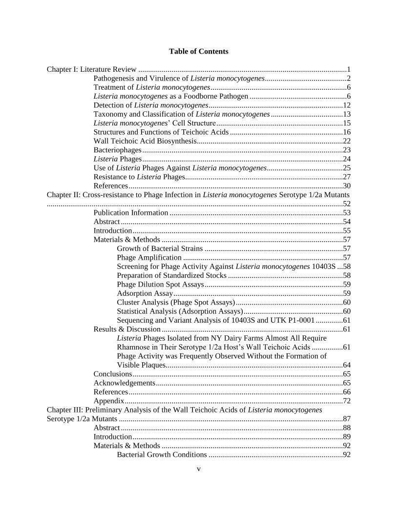
Table of Contents
Chapter I: Literature Review ...........................................................................................................1
Pathogenesis and Virulence of Listeria monocytogenes ..........................................2
Treatment of Listeria monocytogenes ......................................................................6
Listeria monocytogenes as a Foodborne Pathogen ..................................................6
Detection of Listeria monocytogenes .....................................................................12
Taxonomy and Classification of Listeria monocytogenes .....................................13
Listeria monocytogenes’ Cell Structure .................................................................15
Structures and Functions of Teichoic Acids ..........................................................16
Wall Teichoic Acid Biosynthesis...........................................................................22
Bacteriophages .......................................................................................................23
Listeria Phages .......................................................................................................24
Use of Listeria Phages Against Listeria monocytogenes .......................................25
Resistance to Listeria Phages.................................................................................27
References ..............................................................................................................30
Chapter II: Cross-resistance to Phage Infection in Listeria monocytogenes Serotype 1/2a Mutants
........................................................................................................................................................52
Publication Information .........................................................................................53
Abstract ..................................................................................................................54
Introduction ............................................................................................................55
Materials & Methods .............................................................................................57
Growth of Bacterial Strains .......................................................................57
Phage Amplification ..................................................................................57
Screening for Phage Activity Against Listeria monocytogenes 10403S ...58
Preparation of Standardized Stocks ...........................................................58
Phage Dilution Spot Assays .......................................................................59
Adsorption Assay .......................................................................................59
Cluster Analysis (Phage Spot Assays) .......................................................60
Statistical Analysis (Adsorption Assays) ...................................................60
Sequencing and Variant Analysis of 10403S and UTK P1-0001 ..............61
Results & Discussion .............................................................................................61
Listeria Phages Isolated from NY Dairy Farms Almost All Require
Rhamnose in Their Serotype 1/2a Host’s Wall Teichoic Acids ................61
Phage Activity was Frequently Observed Without the Formation of
Visible Plaques...........................................................................................64
Conclusions ............................................................................................................65
Acknowledgements ................................................................................................65
References ..............................................................................................................66
Appendix ................................................................................................................72
Chapter III: Preliminary Analysis of the Wall Teichoic Acids of Listeria monocytogenes
Serotype 1/2a Mutants ...................................................................................................................87
Abstract ..................................................................................................................88
Introduction ............................................................................................................89
Materials & Methods .............................................................................................92
Bacterial Growth Conditions .....................................................................92

vi
Cell Lysis ...................................................................................................93
Cell Wall Treatments .................................................................................93
Extraction of Wall Teichoic Acids ............................................................94
Purification of Wall Teichoic Acids ..........................................................95
Establishment of the Phosphate Standard Curve .......................................96
Determination of Wall Teichoic Acid Containing Fractions .....................96
Hydrofluoric Acid Hydrolysis ...................................................................97
Wall Teichoic Acid Analysis by Chromatography ....................................97
Liquid Chromatography with Mass Spectrometry (LC-MS) .........97
Sample Derivatization for Gas Chromatography (GC) .................98
Gas Chromatography – Flame Ionization Detection (GC-FID) ....99
Gas Chromatography – Mass Spectrometry (GC-MS) ..................99
Results & Discussion ...........................................................................................100
Analysis of Standards and Preliminary Analysis of the Wall Teichoic Acid
Monomers of L. monocytogenes Serotype 1/2a Mutants using GC-FID .100
Analysis of Wall Teichoic Acid Monomers of L. monocytogenes 10403S
using MS ..................................................................................................107
Streamlining the Analysis of Wall Teichoic Acid Monomers .................111
Conclusion ...........................................................................................................114
References ............................................................................................................115
Appendix ..............................................................................................................119
Chapter IV: Conclusion ...............................................................................................................129
Vita ...............................................................................................................................................131

vii
List of Tables
Table 2.1: Listeria monocytogenes Strains Used in this Study ......................................................73
Table 2.2: Listeria Phages Used in this Study and Their Propagation Strains ..............................74
Table 3.1: Listeria monocytogenes Strains Used in this Study ....................................................120
Table 3.2: Preparation of the Phosphate Standard Curve ............................................................121

viii
List of Figures
Figure 2.1: Average Efficiencies of Plaquing Heatmap ................................................................78
Figure 2.2: Average Relative Phage Activities Heatmap ..............................................................80
Figure 2.3: Efficiencies of Plaquing Heatmap ...............................................................................82
Figure 2.4: Relative Phage Activities Heatmap .............................................................................84
Figure 2.5: Phage Binding of LP-048 (a) and LP-125 (b) to 10403S and Mutant Strains ............86
Figure 3.1: GC-FID Chromatogram of Ribitol Standard .............................................................101
Figure 3.2: GC-FID Chromatogram of Rhamnose Standard .......................................................102
Figure 3.3: GC-FID Chromatogram of N-acetyl glucosamine Standard .....................................102
Figure 3.4: GC-FID Chromatogram of Glucosamine Standard ...................................................103
Figure 3.5: GC-FID Chromatogram of Glucose Standard ...........................................................103
Figure 3.6: GC-FID Chromatogram of Galactose Standard ........................................................104
Figure 3.7: GC-FID Chromatogram of Purified WTA Monomer of Listeria monocytogenes
10403S .........................................................................................................................................104
Figure 3.8: GC-FID Chromatogram of Purified WTA Monomer of Listeria monocytogenes FSL
0014..............................................................................................................................................105
Figure 3.9: GC-FID Chromatogram of Purified WTA Monomer of Listeria monocytogenes FSL
D4-0119 .......................................................................................................................................105
Figure 3.10: GC-FID Chromatogram of Purified WTA Monomer of Listeria monocytogenes
UTK P1-0001 ...............................................................................................................................106
Figure 3.11: LC Mass Spectrum of Purified WTA Monomer of Listeria monocytogenes 10403S
......................................................................................................................................................108
Figure 3.12: GC Mass Spectrum of Rhamnose Peak ...................................................................108
Figure 3.13: GC Mass Spectrum of Ribitol Peak ........................................................................109
Figure 3.14: GC Mass Spectrum of Ribitol-Rhamnose Peak ......................................................109
Figure 3.15: GC Mass Spectrum of Putative Ribitol-GlcNAc Peak ............................................110
Figure 3.16: GC-FID Chromatogram of Crude WTA Monomer of Listeria monocytogenes
10403S .........................................................................................................................................112
Figure 3.17: GC-FID Chromatogram of Carbohydrate Fraction from Treated Cells of Listeria
monocytogenes 10403S ................................................................................................................112
Figure 3.18: GC-FID Chromatogram of Autoclaved Cell Pellet of Listeria monocytogenes
10403S .........................................................................................................................................113
Figure 3.19: Phosphate Standard Curve.......................................................................................122
Figure 3.20: Illustration of Trimethyl Silane Molecule ...............................................................123
Figure 3.21: Illustration of Ribitol Molecule and Silylated Ribitol Molecule .............................123
Figure 3.22: Illustration of Rhamnose Molecule and Silylated Rhamnose Molecule .................124
Figure 3.23: Illustration of N-acetylglucosamine Molecule and Silylated N-acetylglucosamine
Molecule ......................................................................................................................................124
Figure 3.24: Illustration of Glucosamine Molecule and Silylated Glucosamine Molecule .........125
Figure 3.25: Illustration of Glucose or Galactose Molecule and Silylated Glucose or Galactose
Molecule ......................................................................................................................................125
Figure 3.26: Illustration of Listeria monocytogenes Serotype 1/2a WTA Monomer and Silylated
Listeria monocytogenes Serotype 1/2a WTA Monomer .............................................................126

ix
Figure 3.27: Illustration of Listeria monocytogenes FSL D4-0014 WTA Monomer and Silylated
Listeria monocytogenes FSL D4-0014 WTA Monomer .............................................................127
Figure 3.28: Illustration of Listeria monocytogenes FSL D4-0119 WTA Monomer and Silylated
Listeria monocytogenes FSL D4-0119 WTA Monomer .............................................................128

1
Chapter I: Literature Review
2
Pathogenesis & Virulence of Listeria monocytogenes
Listeria monocytogenes is a Gram-positive, non-spore forming, facultative anaerobic bacteria
that is most well-known for its role as an opportunistic foodborne pathogen. As such, illness
caused by L. monocytogenes, called listeriosis, is most dangerous for elderly, pregnant, and
immunocompromised individuals. In healthy individuals it will result in non-invasive listeriosis,
which includes typical symptoms of a foodborne infection, such as gastroenteritis and fever.
However, in susceptible individuals, much more severe symptoms may present as invasive
listeriosis. These symptoms include septicemia, bacteremia, meningitis, meningoencephalitis, or
even death in people ≥ 65 years old, children, the immunocompromised, or perinates. In pregnant
women, listeriosis is not typically dangerous for the mother as symptoms will present as a mild
flu; however, this generally precedes an abortion of the fetus (152). Additionally, pregnant
women have a 12-fold increased risk of contracting listeriosis after eating contaminated foods as
compared to healthy, non-pregnant individuals (71).
The amount of time between the ingestion of contaminated food and the emergence of symptoms
(incubation period) varies depending on the severity of the infection. In non-susceptible
individuals, symptoms of non-invasive listeriosis usually occur within 24 hours (110).
Conversely, symptoms of invasive listeriosis typically commence after three weeks, although
this period can differ significantly (5). Infection from listeriosis begins approximately 20 h after
ingestion of food contaminated with L. monocytogenes. However, the dose required for infection
is variable due to differences in host immunity and the bacterial strain (152). In susceptible
groups, doses of around 102-104 bacterial cells per gram of contaminated food can lead to serious
illness. For healthy, non-pregnant individuals, less serious illness is caused from food
3
contaminated with closer to 106-109 bacterial cells per gram of food (117, 152). The high doses
necessary for illness in such individuals is at least partially due to the acid environment of the
stomach. Studies have found that individuals taking antacids or H2-blocking agents have an
increased susceptibility to listeriosis (70, 134). Additionally, studies have found differences in
the LD50 for mice with L. monocytogenes administered either parenterally (105 - 106 cells) or
orally (109 cells). The Listeria cells that avoid succumbing to the low pH of gastric acids move
on to initiate pathogenesis in the host (152), which is orchestrated by many different virulence
factors.
Regulation of virulence factors mainly occurs at transcriptional or post-translational levels.
Transcriptional regulation is controlled by either positive regulatory factor A (PrfA), PrfA with
Sigma B, or VirR regulons. PrfA regulates virulence factors including the actin assembly
inducing protein (ActA), phospholipase A (PlcA), and Listeria adhesion protein B (LapB); PrfA
with Sigma B together regulate internalin A (InlA), internalin B (InlB), listeriolysin O (LLO),
phospholipase B (PlcB), and p60; and VirR regulates the multiple peptide resistance factor
(MprF) as well as the Dlt operon. Post-translational regulation is controlled by the enzyme
sortase A (SrtA) or the accessory secretion system SecA2. SrtA regulates internalin J (InlJ), and
has influence over the regulation of InlA and LapB, while SecA2 regulates Listeria adhesion
protein (LAP) and fibronectin-binding protein A (FbpA), with influence over the regulation of
p60 (20). Regulation of flagellar expression is also important for virulence. L. monocytogenes
expresses flagella at temperatures below 30°C; this is due to the inhibition of a transcriptional
repressor, MogR, by the anti-repressor GmaR. GmaR is activated by a response regulator known
as DegU. However, at temperatures ≥ 37°C, MogR is not inhibited and the gene coding for
4
flagellin (flaA) is down-regulated in order to avoid detection by the host innate immune system
(65, 83).
Adhesion of the bacteria to host cells is the crucial first step required for pathogenesis to occur
(143). Adhesion is made possible by many different factors present on the cell. One of the most
important is LAP, which binds preferentially to intestinal cells, both in pathogenic and non-
pathogenic Listeria species (79). LapB is another Listeria adhesion protein, but is specific to
pathogenic species (120). FbpA is novel in that it acts to both adhere to cells via binding to
human fibronectin, and acts as a chaperone for other virulence factors LLO and InlB (42). InlJ
binds specifically to MUC2 in intestinal mucus (95). The gene coding for InlJ is only found in L.
monocytogenes, making it a unique marker for identification (124). Ami is an autolytic enzyme
anchored to the cell wall that also aids in adherence to host cells (103).
Invasion is the next stage of pathogenesis. L. monocytogenes is either engulfed by phagocytes or
enters into non-phagocytic cells (117). Non-phagocytic entry into M cells of the intestine can
occur without any known virulence factors (34). However, it is more common that Listeria
initiates a zipper mechanism of endocytosis through receptor-mediated binding (35). Receptor-
mediated entry of intestinal cells occurs through interaction between bacterial InlA and E-
cadherin of epithelial goblet cells (109, 117). To access endothelium cells and hepatocytes, L.
monocytogenes utilizes InlB to bind their c-Met receptors (36). The pathogen also employs
different virulence factors to resist host defenses during invasion. MprF is a protein that adds
lysine to the membrane phospholipid diphosphatidylglycerol, giving it a positive charge. This
helps the membrane to resist cationic antimicrobial peptides (CAMPs) produced by the host

5
(146). Similarly, the prolipoprotein Lgt is a diacylglyceryl transferase which lipidates
prelipoproteins, also making L. monocytogenes more resistant to CAMPs (100).
Once L. monocytogenes has successfully invaded a cell, it must escape the phagosome created by
its endocytic entry. It does so by using LLO (encoded by the gene hly) to degrade the phagosome
(132). Two other important elements in vacuole escape are the phospholipases PI-PLC and PC-
PLC. PI-PLC is also known as PlcA, a phosphatidylinositol-specific phospholipase C; PC-PLC is
also known as PlcB, a broad-range phospholipase C (20).
ActA is a protein that enables L. monocytogenes to spread from one host cell to another by actin-
based propulsion (7, 152). It is necessary for the pathogen to polymerize actin from within the
cytoplasm of the host cell for motility (43), as flagella are not expressed at physiological
temperatures (138). The protein p60 is also necessary for L. monocytogenes to utilize the
polymerized actin (115). The propelled bacterium eventually contacts the host cell membrane,
forming a protrusion that is taken up by an adjacent host cell (71). The protein internalin C (InlC)
was found to play a role in the formation of this protrusion (119). This process enables L.
monocytogenes to spread throughout the body. The majority of the bacterial load ends up in the
liver and multiplies there (7, 152). The next stages of infection depend upon the responsiveness
of the host immune system. In healthy individuals, the immune system contains the pathogen
within the liver and proceeds to eliminate it from the body. Conversely, in susceptible
individuals, the diminished or altered immune response leads to bacteremia, enabling the
pathogen to infect the central nervous system, multiple organs (septicemia) or, in pregnant
women, the placenta and/or fetus (152).

6
Treatment of Listeria monocytogenes
Treatment of listeriosis infections is accomplished by administration of antibiotics, which may
include amikacin, amoxycillin, ampicillin, azlocillin, ciprofloxacin, chloramphenicol,
clindamycin, coumermycin, doxycycline, enoxacin, erythromycin, gentamicin, imipen,
netilmicin, penicillin, rifampin, trimethoprim or vancomycin (162), though the most commonly
used are amoxycillin, penicillin, or ampicillin, or one of these in combination with gentamicin (3,
72). In individuals sensitive to these antibiotics, trimethoprim-sulfamethoxazole can be
administered to kill the pathogen (123). Cephalosporins, fosfomycin, and first-generation
quinolones are not effective on L. monocytogenes (3, 144). L. monocytogenes is considered Beta-
lactam tolerant, hence the use of gentamicin in combination with such antibiotics for treatment in
humans (73). Antibiotic resistance though is primarily an issue in animals, such as those used for
human food sources (144). Patients with invasive listeriosis are much more likely to survive
when treated promptly with suitable antibiotics; however, it is not a guarantee that a susceptible
individual will recover from the illness without complications, or at all. Moreover, patients may
not be diagnosed in time or correctly to receive proper treatment, in which case they are at an
even greater risk of suffering irreversible neurological conditions or death (147). The possibility
of such severe health consequences is the reason that foodborne outbreaks of listeriosis are such
a serious threat.
Listeria monocytogenes as a Foodborne Pathogen
One of the major factors leading to listeriosis outbreaks is the organism’s ability to survive in the
same conditions that many food processing facilities maintain. L. monocytogenes is a
psychrotroph, meaning it can survive in cold temperatures such as those used for refrigeration of
7
food products (126). In fact, a procedure known as cold-selection in which bacteria are grown in
non-selective media at refrigeration temperature (4°C) can be used to select for Listeria (162).
The temperature range for growth of L. monocytogenes is from -0.1 to 45°C, with optimal
growth from 30 to 37°C (154, 162). Aside from its ability to thrive in cold temperatures, L.
monocytogenes can survive in a wide variety of environmental conditions, such as alkaline, acid,
and high salt (98, 162). The optimal pH range for L. monocytogenes’ survival and growth is
between 6 and 9, but it has been shown experimentally to survive at a pH as low as 4.19 (32,
162). L. monocytogenes typically has a minimum water activity (aw) of 0.92, meaning foods that
have a aw of ≥ 0.92 will promote its growth (48). However, a study demonstrated that L.
monocytogenes was able to grow in an environment with an aw of 0.91, created using a solution
of 13-14% NaCl (48). It has also been shown to survive in concentrations of NaCl as high as
18% (w/v) (32). However, growth and survival in these extremes is dependent on the other
conditions of the environment and the strain of L. monocytogenes (12). Additionally, it was
demonstrated in another study that at the lowest aw tested (0.80), L. monocytogenes still survived
approximately 8 days (102). This is relevant to recent recalls involving L. monocytogenes in nut
butters (55, 56). Although an aw of 0.70 for peanut butter (54) is lower than what is required for
pathogen growth, there is potential for it to survive long enough to cause illness. Additionally,
the bacterium can develop resistance to heat under food processing conditions that first expose it
to low levels of heat, or to solutes like salt or sugar (41). L. monocytogenes is also capable of
persisting in food processing environments for long periods of time (i.e. years) within biofilms
(105, 111).
8
In 2011, data regarding cases of foodborne illness from 2000 to 2008 was compiled to develop a
picture of the prevalence and severity of 31 major foodborne pathogens, including L.
monocytogenes. It was found to be the third leading cause of foodborne illness, contributing to
19% of deaths. All cases of listeriosis reported (average of 1,455 annually) resulted in
hospitalization; of which an average of 255 resulted in death. Listeriosis is also almost entirely
(99%) contracted through eating food contaminated with L. monocytogenes (128). More recent
data regarding the occurrence of cases of listeriosis in the U.S. was collected from 2009 to 2011.
Within this time frame, 1,651 invasive listeriosis cases were reported, which included 292
deaths/fetal losses (21% mortality rate) and 93% of patients having been hospitalized. The
majority of the cases (58%) involved adults aged 65 or older, while 14% of cases occurred in
pregnant women. Among those affected that were not in the ≥ 65 years or pregnant groups, a
high prevalence (74%) of patients with an underlying medical condition was reported (140). The
most current data regarding the 41 most prevalent foodborne pathogens, chemicals and toxins
involved in U.S. outbreaks consists of information gathered from 2009 to 2015. L.
monocytogenes contributed to over half of the deaths (52%) within these parameters,
demonstrating the continuing severity of such outbreaks (40). In addition to the physical and
emotional trauma caused by the pathogen, there is a significant financial burden that must be
considered as well. Even though the number of listeriosis cases is relatively low compared to the
other 14 major foodborne pathogens considered, it ranks as the second most costly per case. This
is attributed to its high rates of hospitalization (~94%) and death (~16%) (74). For these reasons,
many efforts have been made in an attempt to reduce the occurrence of listeriosis outbreaks, or at
least lessen their impact as much as possible.

9
Government organizations such as state health departments, the CDC (Centers for Disease
Control and Prevention), the FDA (Food and Drug Administration) and the USDA-FSIS (United
States Department of Agriculture – Food Safety and Inspection Service) all work together under
the Listeria Initiative, a program started in 2004 to process outbreak data more efficiently and
determine its source as quickly as possible (27). These organizations have also effected
legislation in an effort to decrease or eliminate the occurrence of L. monocytogenes in food. The
CDC declared listeriosis a nationally notifiable disease in 2000, meaning that health officials are
required to report any patients diagnosed with the illness to their local public health department
(28). In 2003, the USDA-FSIS with support from the FDA issued the Listeria Rule, which
maintains a zero-tolerance policy for the presence of Listeria on ready-to-eat (RTE) products
(57). This policy remains in effect, although temporarily it was not applied to foods that should
not support the growth of the bacteria. However, the decision was made to reverse these changes,
probably due to the increasing presence of L. monocytogenes on novel foods items (6).
The history of L. monocytogenes as a pathogenic organism dates back to 1924, when E.G.D.
Murray first isolated it from rabbits. The first recorded outbreak of listeriosis in humans occurred
in 1949 in Germany. It affected 85 infants, who were either stillborn or died shortly after birth
(71). However, the earliest known foodborne outbreak of listeriosis was not for another 30 years.
In 1979, raw vegetables were presumed to be the source of a listeriosis outbreak in Boston, MA,
although this was not able to be confirmed (70). In 1981, an outbreak of listeriosis in Nova
Scotia, Canada identified cabbage as the confirmed source of L. monocytogenes, and solidified
the link between consumption of foods contaminated by L. monocytogenes and contraction of
listeriosis (130). It has been well-established that L. monocytogenes is ubiquitous in nature, in

10
which it functions as a saprophyte living off decaying plant matter. Soil is a common source
from which L. monocytogenes can be isolated, indicating the possibility of it being present on
any type of produce grown in the soil (158, 160). Therefore, it is not surprising that it was the
causative agent in these outbreaks.
Subsequent outbreaks of L. monocytogenes have linked the pathogen to other foods such as RTE
meats and dairy products (22). The infamous 1985 outbreak in Los Angeles, California, was due
to contamination of Mexican-style cheese. This outbreak caused 142 people to become ill; of
these, 93 cases were pregnant women or perinates. There was a total of 48 deaths, of which
nearly all cases were in perinates or immunocompromised individuals (96). Of the 17 most
noteworthy listeriosis outbreaks in the U.S. from 1979 through 2008, seven were caused by RTE
meat products, and seven were caused by dairy products (155). However, a closer look at the
latter end of this timeline reveals a shift from RTE meats to other RTE foods. Additionally, a
summary of the 24 confirmed outbreaks of listeriosis in the U.S. between 1998 and 2008 shows
that nine out of the ten outbreaks from RTE meats occurred in the early part of the timeline,
while different types of RTE items (nacho/taco salad, sprouts) appeared later (22).
A recent review of U.S. outbreaks has revealed produce specifically as a reoccurring commodity
linked to outbreaks of listeriosis (19). These outbreaks were linked to produce items including
celery in 2010 (62), romaine lettuce in 2011 (165), sprouts in 2014 (30), stone fruits in 2014
(76), caramel apples in 2014-2015 (4), and bagged lettuce in 2015-2016 (137). An outbreak
involving frozen vegetables in four states in the U.S. also occurred recently, with cases identified
from 2013 to 2016 (25). The most notorious U.S. listeriosis outbreak involving produce (to date)

11
was in 2011, when cantaloupe grown at a farm in Colorado was identified as the commodity
responsible for 147 cases of listeriosis in 28 states. Of these cases, at least 143 were hospitalized,
and 33 died (101).
Although foods such as RTE red meats and produce have had a shifting prevalence as sources of
outbreaks, dairy associated outbreaks have remained fairly constant (19, 140). Between the years
of 1998 and 2011, 90 outbreaks of L. monocytogenes occurred in the U.S. which were linked to
dairy products. Approximately half of these outbreaks were from products made with pasteurized
milk (64). From 2010 to 2015, an outbreak associated with ice cream products from a single
company caused illness in 10 individuals in four states (116). Listeriosis outbreaks associated
specifically with soft cheeses have even increased since the mid-2000’s. Most of these were from
cheese products that had undergone pasteurization, indicating that the products were
contaminated at some point after this process (78). However, one of the most recent U.S.
listeriosis outbreaks was confirmed to be from an unpasteurized soft raw milk cheese product,
causing 2 deaths out of the 8 people affected. This outbreak spanned from September 2016 to
March 2017 and caused illnesses in four states (26).
A review of outbreaks occurring in Europe, the U.S. and Canada from 2005 to 2008 has
demonstrated that although the incidences of outbreaks in food items like deli meats have
declined, they have not disappeared (148). In the U.S., an outbreak occurring in late 2018 due to
contaminated RTE pork led to the hospitalization of four individuals in four states (29). Outside
of the U.S., the largest outbreak to date occurred in South Africa by contamination of RTE meat.
Over 1,000 cases and over 200 deaths were reported from 2017 to 2018 due to the presence of

12
Listeria within a facility producing a RTE meat known as polony (2, 125). Although many
efforts have been made to reduce the occurrence of outbreaks from L. monocytogenes, it remains
to be a significant public health threat and can have devastating effects if not properly controlled
for.
Detection of Listeria monocytogenes
To verify that L. monocytogenes is the cause of illness in suspected cases of invasive listeriosis,
patient samples are collected from blood or cerebrospinal fluid. In patients with gastroenteritis,
stool samples can be used, however these must be first selectively enriched for Listeria spp. due
to competing bacteria (3). An epidemiologic investigation of a listeriosis outbreak makes use of
whole genome sequencing (WGS) to identify clusters of listeriosis cases with information from
patient samples. This method of identification was employed in 2013 as a major upgrade to the
previous standard known as pulse-field gel electrophoresis or PFGE, which differentiates
fragments of DNA based on molecular weight (27, 61, 77). WGS is able to analyze DNA
sequences in more detail to account for differences and similarities not evident with PFGE (27,
77). Once clusters have been established, information collected from patients (i.e. food
consumption history) is applied to determine a common food source that could be causing the
outbreak (27). However, the prolonged incubation period of L. monocytogenes often creates
difficulties for food recall in affected individuals (59). Suspected food products and
environmental samples from their production facilities are tested for the pathogen using the same
methods as clinical isolates (77).

13
The zero-tolerance policy for L. monocytogenes has motivated many RTE food production
facilities to routinely test their products and production areas for the presence of Listeria. Often,
these samples are sent to third-party laboratories for analysis. To test food samples for L.
monocytogenes (or Listeria spp.), they are first enriched in selective media (69). Most selective
medias for L. monocytogenes contain (in addition to required nutrients) high salt concentrations,
nalidixic acid, acriflavine, esculin and ferric ions (68, 107). Salt, nalidixic acid, and acriflavine
inhibit growth of most other bacteria besides Listeria spp. (68). Esculin is used to differentiate
colonies as Listeria spp. is able to hydrolyze it, producing 6,7-dihydroxycoumarin which reacts
with ferric ions to produce a dark color. These colonies can then be used for further testing. For
routine analyses, L. monocytogenes is detected using automated immunoassays including
enzyme-linked immunosorbent assays (ELISA) or enzyme-linked fluorescent assays (ELFA).
Both of these methods make use of antibodies that bind specifically to virulence proteins. If
binding occurs, a fluorescent marker will be activated that can then be measured by the
instrument. A more precise measurement used in the case of a presumptive positive sample is the
polymerase chain reaction (PCR). PCR employs primers that target a specific genetic sequence,
which if present will be amplified to high enough levels that it can be detected (80).
Taxonomy and Classification of Listeria monocytogenes
The genus Listeria is currently comprised of at least 17 recognized species, including Listeria
monocytogenes, Listeria grayi, Listeria innocua, Listeria welshimeri, Listeria seeligeri, Listeria
ivanovii, Listeria marthii, Listeria rocourtiae, Listeria fleischmannii, Listeria
weihenstephanensis, Listeria floridensis, Listeria aquatica, Listeria cornellensis, Listeria
riparia, Listeria grandensis, Listeria booriae, and Listeria newyorkensis (117, 159). Of these,
14
only L. monocytogenes and L. ivanovii are pathogenic. However, illness caused by L. ivanovii is
rare, mainly occurring in ruminant animals. These species have been defined and refined using
progressively advancing approaches including numerical taxonomy, biochemical analyses, DNA
composition, and 16S rRNA gene sequencing. The same techniques have been used to determine
similarities to other organisms within the Listeriaceae family. The most closely related genus to
Listeria is Brochothrix (162). Staphylococcus and Bacillus have also demonstrated high
similarity to Listeria based on the comparison of their 23S rRNA sequences (127). One major
commonality between these closely related genera is the low G+C (guanine and cytosine)
content of their DNA (127, 162). A paramount study in 2001 reported the full genome sequences
of L. monocytogenes and L. innocua. Both have circular genomes consisting of 2,944,528 base
pairs (bp) and 3,011,209 bp, respectively, with G+C contents of 39% and 37%, respectively.
These two species share very similar genomes; most of the differences that do exist between
them are known or suspected to be related to virulence (63).
L. monocytogenes has been mainly organized by two types of classification; lineages and
serogroups/serotypes (ST). Currently, four lineages are used to group isolates of the species by
phenotypic and genotypic similarities. Lineage I consists of different strains belonging to ST
1/2b, 3b, 3c, and 4b; Lineage II consists of strains belonging to 1/2a, 1/2c, and 3a ST; and
Lineages III and IV consist of strains belonging to ST 4a, 4b, and 4c. Isolates from lineage I and
II are responsible for nearly all outbreaks and sporadic cases of listeriosis in humans (112). Prior
to the introduction of lineages, serotyping was used as the primary method for classification
(113). L. monocytogenes currently consists of at least thirteen ST to include 1/2a, 1/2b, 1/2c, 3a,
3b, 3c, 4a, 4ab, 4b, 4c, 4d, 4e and 7. Previously, L. monocytogenes was also included in
15
serogroups 5 and 6, but the strains associated with them have subsequently been reassigned as L.
ivanovii and L. welshimeri, L. seeligeri or L. innocua, respectively (49, 112, 136, 162). L.
monocytogenes serogroups 1/2, 3, and 4 are further organized according to somatic or flagellar
antigens. Serogroups 1/2 and 3 are subdivided into ST based on flagellar agglutination, and
serogroup 4 is subdivided into ST based on somatic agglutination (133, 162). ST 1/2a, 1/2b, and
1/2c are the most commonly isolated from food products, and ST 1/2a and 1/2b are most often
implicated in outbreaks of gastrointestinal listeriosis. Conversely, ST 4b is extremely frequent in
outbreaks of invasive listeriosis (144). Additionally, outbreaks occurring in Northern Europe are
often attributed to ST 1/2a, while in the U.S. the 4b ST (within lineage I) is most common (112).
However, some recent outbreaks suggest that these geographical associations are becoming less
distinct (99).
Listeria monocytogenes’ Cell Structure
L. monocytogenes’ cells normally consist of parallel, short rods with blunt ends that are
approximately 1-2 μm long and 0.4-0.5 μm wide. When grown at temperatures between 20 and
30°C, they develop 2-6 peritrichous flagella which aid in motility. Grown below 20°C, flagella
still develop but are involved with adhesion rather than motility (162). L. monocytogenes is a
Gram-positive bacterium, therefore its cell wall consists of a thick layer of peptidoglycan (PG).
The PG comprises about 30-40% of Listeria cell walls, and helps to protect the cell from changes
in osmotic pressure. It is made up of glycan chains of N-acetylglucosamine (GlcNAc) and N-
acetylmuramic acid (MurNAc) disaccharide units, which are cross-linked by pentapeptide (L-
alanine-D-glutamic acid-meso-diaminopimelic acid-D-alanine-D-alanine) stems. Listeria PG is
the A1-γ type, which is characterized by cross-linking at the meso-diaminopimelic acid; in this
16
case, to the penultimate alanine of another stem (39, 50, 51). Modifications to the PG are
important for bacterial survival in the host. It was found that O-acetylation (catalyzed by OatA)
of PG MurNAc and N-deacetylation of PG GlcNAc is essential for L. monocytogenes’ resistance
to lysozyme, a cell wall hydrolyzing enzyme produced by the host (66, 82, 118). N-deacetylation
of the PG MurNAc also aids in Listeria’s escape from the hosts’ innate immune system (14).
Structures and Functions of Teichoic Acids
All Gram-positive bacteria possess within their PG glycopolymer structures that extend to the
cell surface. The most abundant and well-studied of these structures are the teichoic acids.
Teichoic acids consist of both lipoteichoic acids (LTA) and wall teichoic acids (WTA) (157).
Both are important for cell survival; it was shown that B. subtilis mutants without either LTA or
WTA are not viable (129). LTA are not attached to the PG but anchored to the membrane of the
cell via a glycolipid. LTAs are amphiphilic molecules; L. monocytogenes possesses type I LTAs
which consist of an unbranched polyglycerol-phosphate chain that may have subunits substituted
with D-alanine and galactose residues. Mutations which affect LTA synthesis can lead to cells
with increased sensitivity to lysozyme and antibiotics. Additionally, LTA deficient cells are
impaired in cell division and form smaller colonies, and have decreased abilities for biofilm
formation, cation homeostasis, and virulence (1, 121, 156). Similar effects have been observed in
cells lacking WTA (18, 121, 145).
Although WTA and LTA share many functions, they also possess some important distinctions.
WTA polymers of Listeria do not contain a hydrophobic portion; they are made of 20-30
repeating units of a ribitol molecule that usually includes glycosyl substituents such as GlcNAc,

17
glucose, galactose, rhamnose, or hexose (81, 139). Additionally, LTAs have not been shown to
demonstrate much variation (23, 50, 157), but WTA are highly diverse, as L. monocytogenes
utilizes different combinations and positions of the aforementioned glycosyl substituents to
modify their structure. WTA are in fact the O-antigens of the Listeria cell and are essential for
differentiating between different ST. There are two types of WTA structures in L.
monocytogenes. Type I WTA consist of a ribitol with glycosyl substitutions bound to C2 and/or
C4, and are connected to each other through phosphodiester bonds between C5 and C1. Type II
WTA also have a ribitol backbone, but C2 or C4 is bound to a GlcNAc. This GlcNAc is linked
through a phosphodiester bond to C1 of the next repeat unit. However, it is possible that this
substitution is not acetylated, i.e., replaced by glucosamine (45, 108, 139).
The repeating unit of serogroup 1/2 strains is a type I WTA, which consists of a ribitol molecule
attached to GlcNAc at C2 and rhamnose at C4. This structure has been elucidated using a
number of different methods. After extraction of the WTA from the cell wall followed by
purification and hydrolysis, samples have been analyzed for molecular weight by gel filtration
chromatography (58, 84); for chemical composition by gas-liquid chromatography (GLC) (52,
84) coupled with flame ionization detection (FID) (161) or mass spectrometry (MS) (81), ultra-
performance liquid chromatography (UPLC) with electrospray ionization (ESI) and tandem MS
(MS/MS) (139) or ESI-MS/MS (46); for molecular connectivity within structures by Smith
degradation, acid hydrolysis, and oxidation/reduction reactions (58, 84, 150); and anomeric
configurations by nuclear magnetic resonance (NMR) spectrums (81, 139). Similar methods
were used to elucidate the WTA structures of L. monocytogenes within serogroups 3 (46, 52, 58,
139, 150), 4 (46, 52, 58, 139, 150, 161) and 7 (52, 139, 150). Serogroups 3 and 7 also elicit type
18
I WTAs. The repeating WTA unit of serogroup 3 strains is the same as that of serogroup 1/2
strains, but without the rhamnose substitution. Serogroup 7 strains have a WTA monomer
structure consisting of a ribitol that may be unsubstituted, or bound to a hexose at positions C2 or
C4 (45, 139). Serogroups 3 and 7 were in fact discovered to be mutants of the 1/2 serogroup.
Serogroup 3 strains have a single nucleotide polymorphism (SNP) mutation in at least one of the
genes necessary for WTA rhamnosylation (lmo 1080, lmo1081, lmo1082, lmo1083, or lmo1084).
Serogroup 7 strains have a SNP mutation in at least one of the genes required for WTA
rhamnosylation, and at least one of the genes required for WTA N-acetylglucosaminylation
(lmo1079, lmo2549, or lmo2550) (47).
Serogroup 4 differs from serogroups 1/2, 3 and 7 as it contains type II WTAs, and its ST are
divided by somatic antigens rather than flagellar antigens. As such, its different ST represent
much more variable WTA structures than serogroups 1/2, 3 and 7. ST 4a WTAs usually consist
of a ribitol with a GlcNAc substitution at C2. ST 4b WTAs typically have a glucose and a
galactose bound to a GlcNAc at C4. The repeating unit of ST 4c has a galactose substituent
connected to GlcNAc on C2, and ST 4d WTAs have a glucose substituent connected to GlcNAc
on C4 (45, 58, 139). The WTA structures of ST 4e and 4ab are less well-studied. However, it has
been demonstrated that similar to ST 4d, 4ab contains predominantly glucose and GlcNAc, while
studies on 4e have found it to contain GlcNAc with either galactose or glucose as the primary
glycosyl units (50, 58).
Many of the studies that analyzed L. monocytogenes ST 1/2, 3, or 7 WTAs also found glucose as
a minor component of the WTA composition. This glucose is not part of the WTA repeating
19
unit; rather it is part of the linkage unit which connects the WTA polymer to the PG. In these ST,
the unit consists of glucose-glucose-glycerol-phosphate-ManNAc-GlcNAc, in which the glucose
end is bound to the WTA polymer by a phosphodiester bond, and the GlcNAc end is bound to
the MurNAc of the PG by a phosphodiester bond. The whole unit is fairly conserved amongst the
species; however ST 4b and 4d have a glucose-GlcNAc unit instead of glucose-glucose, and ST
4a and 4c have only GlcNAc in this position (84, 88, 139). The ManNAc-GlcNAc-glycerol-
phosphate segment seems to be highly conserved amongst organisms in the Listeriaceae family,
as it is also found in B. subtilis and S. aureus (33, 84).
WTA comprise between 30 and 75% of the dry weight of the cell wall (10, 17, 51, 58, 145, 151).
It is estimated that there is one WTA polymer unit attached to approximately every tenth
MurNAc of the PG (52). In S. aureus, the placement of WTA regulates the cross-linking of the
PG (8). Additionally, placement of WTA throughout the cell wall ensures the proper spacing of
autolysins and cell-division machinery; WTA deficient cells do not divide properly and have an
altered shape (157). The presence of many phosphate groups within the structure of the WTA
makes them negatively charged. This phosphate store can subsequently provide a reservoir for
magnesium (Mg+2) as well through the formation of ionic bonds (93, 108). WTA are also
indispensable for survival in the harsh environments of the host (17). They have been found to
affect the expression of virulence factors involved in adherence (LAP) to and invasion (InlB) of
host cells. L. monocytogenes cells pre-treated with tunicamycin, an antibiotic that disrupts WTA
biosynthesis, had reduced levels of both these virulence factors (166). In addition to attachment
to host cells, WTAs also play a role in adherence to abiotic surfaces (i.e. biofilm formation) (17,
145). In a recent study, biofilm resistance to rinsing and cleaning was assessed in the EGE-e (a

20
ST 1/2a strain) wild-type strain vs. mutants lacking genes lmo2549 or lmo2550, essential for
GlcNAcylation of WTA. Biofilms established by mutant strains were found to detach more
easily with washing than wild-type biofilms (15). L. monocytogenes ST 4nonb mutants lacking
galactose decorations in their WTA were also found to have reduced actin tail lengths,
diminishing their capability for cell-to-cell spread in the host (142). Additionally, ST 1/2 mutants
deficient in WTA rhamnose have demonstrated a decrease in the functionality of the virulence
factors ami and InlB (24).
Much of the resistance to antibiotics and antimicrobials displayed by Gram-positive pathogens
can be attributed to modifications to their teichoic acids. CAMPs are molecules produced by
many organisms to defend against bacteria. By lessening the negative charge of their cell wall
pathogens are better able to resist such antimicrobials (108, 146). This is achieved through D-
alanylation of WTA and/or LTA, in which the structures are modified using D-alanine. This
process is controlled by the Dlt operon present in many Gram-positive bacteria. In L.
monocytogenes, LTA are D-alanylated which contributes to their virulence, but this modification
has not been found in their WTA (1, 23, 46, 81, 139). Resistance to lysozyme has also been
demonstrated in B. subtilis through D-alanylation of teichoic acids (66). WTA are necessary for
beta-lactam antibiotic resistance in B. subtilis and methicillin resistant S. aureus, possibly due to
their N-acetylglucosaminylation (18). In L. monocytogenes, mutants lacking Fri, a ferritin-like
protein, had decreased resistance to beta-lactam antibiotics due to an inability to upregulate
WTA and therefore control autolysin activity (90). Additionally, L. monocytogenes’ WTA
rhamnosylation has been shown to promote resistance to antimicrobial peptides. The rhamnose
helps to physically block antimicrobials from contacting the cell membrane (23).
21
WTA have also been found to play roles in transferring genetic information between cells. A
recent study explored the potential of WTA in B. subtilis to aid in transformation of genetic
material into cells. It was found that competent cells (i.e. cells able to receive genetic material)
treated with tunicamycin, an antibiotic inhibiting WTA synthesis, bound significantly less
exogenous DNA. It was also found that competent cell WTA were localized around the protein
ComGA, found only in competent cells. This suggests that WTA act as scaffolding for
exogenous DNA in preparation for transformation into the recipient cell (104). Additionally, it
was found that WTA of S. aureus and L. monocytogenes contribute to horizontal gene transfer by
enabling transduction of genetic material (163).
However, one of the most pivotal roles of WTA is ultimately to the detriment of the cell. WTA
are the receptors for bacteriophage adsorption to Gram-positive bacteria. Bacteriophages are
viruses that specifically infect bacteria in order to replicate. They are used to combat pathogenic
bacteria in both clinical and food industry settings (60). It has been demonstrated that genes
contributing to WTA glycosylation in L. monocytogenes are also associated with phages’ ability
to adsorb to the host in STs 1/2a (37) and 4b (31). WTA glycosylation is not required for all
instances of phage binding, as demonstrated in S. aureus (164). However, in L. monocytogenes,
strains lacking glycosylated WTA have been found to be resistant to phage binding (9, 37, 47,
142). This resistance comes at a cost to the cell, as resistant mutants were found to have
attenuated virulence in vivo (9, 142).
22
Wall Teichoic Acid Biosynthesis
The biosynthesis of WTA occurs within the cytoplasm of the cell. As L. monocytogenes contains
ribitol teichoic acids, the proteins involved in these processes are referred to as the Tar (teichoic
acid ribitol) group of proteins as opposed to the Tag (teichoic acid glycerol) group of proteins.
The first steps of WTA biosynthesis involve the formation of the linkage unit, initiated by the
specific protein TarO (17). TagO and TarO are targets for the antibiotic tunicamycin which used
is to inhibit WTA synthesis; when used at higher concentrations, it also inhibits PG synthesis
(104, 166). Previously, it was thought that glycosylation of all WTA polymers took place in the
cytoplasm, before the molecule was transferred outside of the cell (17). However, a recent study
proposed that N-acetylglucosaminylation of L. monocytogenes ST 1/2a strains 10403S and
EGDe takes place after the molecule has been transferred out of the cytoplasm. This was
hypothesized after the discovery that lmo1079 in L. monocytogenes, responsible for moving
GlcNAc residues from a C55-P—GlcNAc-lipid intermediate onto the WTA polymer, is an
ortholog of YfhO in B. subtilis, which glycosylates its LTA in this fashion. Another gene
involved in L. monocytogenes WTA glycosylation is lmo2550, which produces the C55-P—
GlcNAc-lipid intermediate. The protein GtcA, encoded by lmo2549 is thought to act as a
flippase, or an enzyme that flips the C55-P-GlcNAc-lipid intermediate across the cell wall. (121).
Rhamnosylation also occurs in ST 1/2 strains. The genes lmo1081, lmo1082, lmo1083 and
lmo1084 are involved with the biosynthesis of rhamnose in L. monocytogenes. Collectively they
comprise the rmlACBD locus, which is present in STs 1/2a, 1/2b, 1/2c, 3c, and 7. However, ST
3c has a mutation in rmlA, and ST 7 has a mutations in rmlB, and therefore cannot produce
rhamnose. To successfully add rhamnose to WTAs, the gene lmo1080 (also known as rmlT) is
required, as it produces a putative rhamnosyltransferase (23).

23
Bacteriophages
As mentioned, bacteriophage are viruses that exclusively infect and replicate in bacterial cells.
They were first characterized and named in 1917 by Felix D’Herelle, and were gaining
popularity in treating bacterial infections until the advent of commercial antibiotics in 1940.
Phage research continued with vigor in countries such as Poland and Georgia, but in most other
parts of the world was largely overlooked in favor of antibiotics. However, research and use of
phage has seen a resurgence in western countries over the past 3 decades, primarily in response
to antibiotic resistant bacteria (85, 92).
The ability of phages to successfully infect and replicate in their host is dependent on the very
specific structure of their organelles. The basic structure of most bacteriophages consists of a
capsid head containing genetic material, a tail, a baseplate, and long and short tail fibers.
Variation in these features is used to differentiate taxonomical groups. Generally, the long tail
fibers are used to probe the surface of a bacterial cell after the phage has contacted it. Once these
fibers bind enough receptors, the baseplate moves close to the cell surface, allowing the short tail
fibers to interact with receptors. In bacteriophages targeting Gram-positive bacteria, this
proximity also allows baseplate enzymes known as virion-associated peptidoglycan hydrolases
(VAPGH) to degrade the cell wall enough for the tail to transverse it and insert the phages’
genetic material (60, 67, 122).
Bacteriophages are categorized as either temperate or virulent depending on their replication
cycle. Temperate phages initiate a lysogenic cycle, meaning that their injected genetic material is
integrated into the bacterial genome as a prophage (85). This is accomplished by a protein known

24
as an integrase (38). When the bacterial cell is introduced to environmental stress (e.g. UV light),
this signals the prophage to transition to a lytic cycle. The lytic cycle, used by both virulent
phages and temperate phages, involves the exploitation of the bacterial cell machinery to produce
progeny bacteriophage. PG degrading enzymes known as endolysins are produced
simultaneously in order to lyse the cell and release the fully constructed virions (44, 85).
Listeria Phages
Phages that specifically infect bacteria in the Listeria genus belong to the order Caudovirales,
which includes the families Myoviridae, Podoviridae, and Siphoviridae. Listeria phages are
found only within the Myoviridae and Siphoviridae families, which consist of phages with
long, contractile tails or long, non-contractile tails, respectively. The genomes of all Listeria
phages contain dsDNA (87, 91). Listeria phages have been organized by orthoclusters, or
groups that share orthologous genes. Five different orthoclusters exist, grouping Listeria
phages by genome size, G+C content, and morphology. Orthocluster I contains Myoviridae
Listeria phages with a large genome size (~131-138 kb) and approximately 36% G+C content.
Orthoclusters II, III and IV contain Siphoviridae (B1) Listeria phages with a small genome size
(~36-43 kb) and approximately 35-40% G+C content. Orthocluster V contains Siphoviridae
(B3) Listeria phages with a medium genome size (~65-67 kb) and approximately 33% G+C
content. Phages from the genus P100virus (91) in orthocluster I have a capsid head diameter of
approximately 86 nm and tail dimensions (length by diameter) of 206 x 18 nm. Phages from
orthoclusters II, III and IV have similar head diameters ranging from 53 – 57 nm, but a much
larger size range of tail dimensions. Phages in the genus 2671 (orthocluster IV) have the
largest tail dimensions at 297 x 8 nm; phages in the genus 2389 (orthocluster III) have tail
25
dimensions of 160 x 7-10 nm, and phages in the genus P35 (orthocluster II) have tail
dimensions of 100 x 8 nm. Phages in orthocluster V have head dimensions of 123 x 44 nm and
tail dimensions of 162 x 7-8 nm (38).
Use of Listeria Phages Against Listeria monocytogenes
Bacteriophage research is primarily focused on exploiting their ability to combat pathogenic
bacteria. Phage applications include use within clinical, veterinary, agricultural, and food
microbiology sectors (92). One useful role of phage is in the detection of pathogenic bacteria. In
1989, Seeliger & Langer noted the importance of using phage typing to accurately determine L.
monocytogenes isolates by epidemiologists (135). Although more precise technologies are
currently used to identify L. monocytogenes isolates during an outbreak (27, 77), phages are still
employed to detect for the presence of the pathogen in food products. One method of using
Listeria phage for pathogen detection involves the use of their endolysins. Endolysins are
composed of a cell wall binding domain (CBD) at the C-terminal of the protein, and an
enzymatically active domain (EAD) at the N-terminal of the protein. Fusion of the highly
specific CBD to a fluorescent marker enables both detection and differentiation of Listeria cells
to the strain level. This method provides results faster and with higher specificity than those
currently employed (i.e. PCR, ELISA) (131). However, CBD that has not bound to cells must be
washed away to achieve accurate results, which can be difficult to accomplish in food matrixes.
Additionally, this method cannot determine live cells from dead ones. Fortunately, another
method of detection known as reporter phage based rapid detection can overcome these
shortcomings. In this method, recombinant reporter phage are engineered to emit
color/fluorescence upon integration of their DNA into the host cell. Naturally, only live host cells

26
will be affected by these phage, and the signal indicating a positive for the bacteria is emitted as
soon as infection occurs, with no need for washing. A reporter Listeria phage engineered to
encode luciferase could detect six different strains of L. monocytogenes (11).
The main use of Listeria phages in the food industry is to eliminate the presence of L.
monocytogenes. As such, two products have been developed using Listeria phages for
application in food processing facilities for use on RTE foods (106). They include ListShieldTM
by Intralytix, Inc. (Baltimore, MD, USA) and PhageGuard ListexTM by Micreos Food Safety
(Wageningen, Netherlands). ListShieldTM is a product consisting of a cocktail of six phages, and
was approved by the FDA in 2006 as a food additive in RTE foods for antimicrobial purposes
(16, 75, 97, 114). PhageGuard ListexTM (formerly Listex P100) is a phage product that targets
Listeria strains using just one bacteriophage with a broad host range, P100. It was found to be
effective against over 95% of the 250 isolates of Listeria tested, including strains from ST 1/2
and 4 (21, 106, 114). Additionally, P100 has been shown to be effective against biofilms of
strains representing all ST of L. monocytogenes (141). Both ListShieldTM and PhageGuard
ListexTM are currently approved by the USDA-FSIS as processing aids when applied to RTE
meat and poultry products (FSIS Directive 7120.1) (75, 114). In 2006, PhageGuard ListexTM was
approved as “generally recognized as safe” (GRAS) by the FDA. In 2014, ListShieldTM was re-
approved as GRAS (11).
However, considerations regarding the conditions to which phage are applied must be taken into
account for them to work properly. Temperature is an important variable which affects the ability
of Listeria phages to successfully adsorb and/or replicate. It has been demonstrated that different

27
Listeria phages within the P100virus and P70virus genera were unable to form plaques at 37°C.
Adsorption and plaquing at other temperatures was also varied, and additionally affected by the
L. monocytogenes strain used (91, 149). Other environmental conditions also have an effect on
phage activity. A study by Fister et al. found that P100 is stable under a pH range of 4 – 10, but
was inactivated at pH values ≤ 2 and ≥ 12. It was also found to be stable after storage in 2 M
NaCl for 24 h. Lutensol detergent (5%) did not affect phage infectivity after 24, but storage in
5% SDS (sodium dodecyl sulfate) solution after 24 h caused a significant reduction in phage
infectivity (53). Conversely, another study which used two different mixtures of 6 and 14 distinct
lytic Listeria phages found that neither was able to reduce bacterial populations on apple slices at
a pH of 4.4 (94). These results highlight that environmental tolerances differ among Listeria
phages, as well as the variables which much be considered when selecting them for food safety
applications.
Resistance to Listeria Phages
Although Listeria phages have been demonstrated as a viable alternative antimicrobial agent in
the food industry, L. monocytogenes can still become resistant to them. Resistance develops
through genetic alterations, which may be dependent on environmental conditions such as
temperature to be expressed phenotypically (86, 149). Phage-resistance may also develop as a
direct response to predation by bacteriophage. Genetic mutations which alter WTA biosynthesis
and/or glycosylation in L. monocytogenes confer resistance to phage. The phage are unable to
bind to, and therefore infect such cells. In an environment where infectious phage are present,
such mutations are necessary for survival (47). These mutations occur randomly (de novo
mutations), enabling those bacteria to survive and pass on the same genes to the next generation.

28
As such, the bacterial population will shift towards one that is resistant to the phages present.
However, bacteriophage also mutate and those that exhibit phenotypes capable of overcoming
such resistance will be able to propagate, again shifting the balance of the population to bacteria
susceptible to phage. This process is called co-evolution, and it is occurring constantly in
environments that contain bacteria and bacteriophage able to infect them (89).
In L. monocytogenes ST 1/2a strains, bacterial resistance has been observed due to specific
genetic alterations which affect WTA glycosylation with rhamnose and/or GlcNAc. In a study by
Bielmann et al., it was demonstrated that the Siphoviridae Listeria phage P35 requires both
GlcNAc and rhamnose in WTA for phage binding to occur. A temperate Listeria phage in the
same family, A118, required only rhamnose as a phage receptor in ST 1/2 L. monocytogenes
strains. Mutant strains of EGDe which lacked either rhamnose or GlcNAc were used to assess
which glycosylation units were required for phage binding (13). Denes et al. showed that the
phage LP-048 was unable to bind to a L. monocytogenes 10403S mutant strain that was lacking
rhamnose in its WTA, and the phage LP-125 was unable to bind to mutant strains lacking
rhamnose or GlcNAc in their WTA. This study demonstrated that phage-resistance occurs when
phage are unable to adsorb to their host, and not through other mechanisms (37). Similarly, it
was shown by Eugster et al. that mutations affecting WTA glycosylation in EGDe, another ST
1/2a L. monocytogenes strain, resulted in phage-resistance. Specifically, the phages A118 and
P40 could not bind to mutants deficient in rhamnose, and the phages P35 and A511 could not
bind to mutants deficient in either GlcNAc or rhamnose (47).

29
It has also been shown that ST 4a, 4b and 4c strains are more sensitive to phages than ST 1/2 or
3, however this is also highly dependent on the specific L. monocytogenes strain being tested and
not just the ST (153). Phages must be considered on an individual basis to determine their host
range and environmental limitations in the development of phage based products for use as
antimicrobial agents in food processing facilities.

30
References
1. Abachin, E., C. Poyart, E. Pellegrini, E. Milohanic, F. Fiedler, P. Berche, and P. Trieu‐
Cuot. 2002. Formation of D-alanyl-lipoteichoic acid is required for adhesion and virulence
of Listeria monocytogenes. Mol. Microbiol. 43:1–14.
2. Allam, M., N. Tau, S. L. Smouse, P. S. Mtshali, F. Mnyameni, Z. T. H. Khumalo, A.
Ismail, N. Govender, J. Thomas, and A. M. Smith. 2018. Whole-Genome Sequences of
Listeria monocytogenes Sequence Type 6 Isolates Associated with a Large Foodborne
Outbreak in South Africa, 2017 to 2018. Genome Announc. 6:e00538-18.
3. Allerberger, F., and M. Wagner. 2010. Listeriosis: a resurgent foodborne infection. Clin.
Microbiol. Infect. 16:16–23.
4. Angelo, K. M., A. R. Conrad, A. Saupe, H. Dragoo, N. West, A. Sorenson, A. Barnes, M.
Doyle, J. Beal, K. A. Jackson, S. Stroika, C. Tarr, Z. Kucerova, S. Lance, L. H. Gould, M.
Wise, and B. R. Jackson. 2017. Multistate outbreak of Listeria monocytogenes infections
linked to whole apples used in commercially produced, prepackaged caramel apples:
United States, 2014–2015. Epidemiol. Infect. 145:848–856.
5. Angelo, K. M., K. A. Jackson, K. K. Wong, R. M. Hoekstra, and B. R. Jackson. 2016.
Assessment of the Incubation Period for Invasive Listeriosis. Clin. Infect. Dis. 63:1487–
1489.
6. Archer, D. L. 2018. The evolution of FDA’s policy on Listeria monocytogenes in ready-
to-eat foods in the United States. Curr. Opin. Food Sci. 20:64–68.
7. Asano, K., H. Sashinami, A. Osanai, S. Hirose, H. K. Ono, K. Narita, D.-L. Hu, and A.
Nakane. 2016. Passive immunization with anti-ActA and anti-listeriolysin O antibodies
protects against Listeria monocytogenes infection in mice. Sci. Rep. 6:39628.

31
8. Atilano, M. L., P. M. Pereira, J. Yates, P. Reed, H. Veiga, M. G. Pinho, and S. R. Filipe.
2010. Teichoic acids are temporal and spatial regulators of peptidoglycan cross-linking in
Staphylococcus aureus. Proc. Natl. Acad. Sci. 107:18991–18996.
9. Autret, N., I. Dubail, P. Trieu-Cuot, P. Berche, and A. Charbit. 2001. Identification of
New Genes Involved in the Virulence of Listeria monocytogenes by Signature-Tagged
Transposon Mutagenesis. Infect. Immun. 69:2054–2065.
10. Baddiley, J. 1989. Bacterial cell walls and membranes. Discovery of the teichoic acids.
BioEssays 10:207–210.
11. Bai, J., Y.-T. Kim, S. Ryu, and J.-H. Lee. 2016. Biocontrol and Rapid Detection of Food-
Borne Pathogens Using Bacteriophages and Endolysins. Front. Microbiol. 7:474.
12. Bergholz, T. M., H. C. den Bakker, E. D. Fortes, K. J. Boor, and M. Wiedmann. 2010. Salt
Stress Phenotypes in Listeria monocytogenes Vary by Genetic Lineage and Temperature.
Foodborne Pathog. Dis. 7:1537–1549.
13. Bielmann, R., M. Habann, M. R. Eugster, R. Lurz, R. Calendar, J. Klumpp, and M. J.
Loessner. 2015. Receptor binding proteins of Listeria monocytogenes bacteriophages
A118 and P35 recognize serovar-specific teichoic acids. Virology 477:110–118.
14. Boneca, I. G., O. Dussurget, D. Cabanes, M.-A. Nahori, S. Sousa, M. Lecuit, E.
Psylinakis, V. Bouriotis, J.-P. Hugot, M. Giovannini, A. Coyle, J. Bertin, A. Namane, J.-
C. Rousselle, N. Cayet, M.-C. Prévost, V. Balloy, M. Chignard, D. J. Philpott, P. Cossart,
and S. E. Girardin. 2007. A critical role for peptidoglycan N-deacetylation in Listeria
evasion from the host innate immune system. Proc. Natl. Acad. Sci. 104:997–1002.
15. Brauge, T., C. Faille, I. Sadovskaya, A. Charbit, T. Benezech, Y. Shen, M. J. Loessner, J.
R. Bautista, and G. Midelet-Bourdin. 2018. The absence of N-acetylglucosamine in wall

32
teichoic acids of Listeria monocytogenes modifies biofilm architecture and tolerance to
rinsing and cleaning procedures. PLOS ONE 13:e0190879.
16. Bren, L. 2007. Bacteria-eating virus approved as food additive. FDA Consum. 41:20–22.
17. Brown, S., J. P. Santa Maria, and S. Walker. 2013. Wall Teichoic Acids of Gram-Positive
Bacteria. Annu. Rev. Microbiol. 67:313–336.
18. Brown, S., G. Xia, L. G. Luhachack, J. Campbell, T. C. Meredith, C. Chen, V. Winstel, C.
Gekeler, J. E. Irazoqui, A. Peschel, and S. Walker. 2012. Methicillin resistance in
Staphylococcus aureus requires glycosylated wall teichoic acids. Proc. Natl. Acad. Sci.
109:18909–18914.
19. Buchanan, R. L., L. G. M. Gorris, M. M. Hayman, T. C. Jackson, and R. C. Whiting.
2017. A review of Listeria monocytogenes: An update on outbreaks, virulence, dose-
response, ecology, and risk assessments. Food Control 75:1–13.
20. Camejo, A., F. Carvalho, O. Reis, E. Leitão, S. Sousa, and D. Cabanes. 2011. The arsenal
of virulence factors deployed by Listeria monocytogenes to promote its cell infection
cycle. Virulence 2:379–394.
21. Carlton, R. M., W. H. Noordman, B. Biswas, E. D. de Meester, and M. J. Loessner. 2005.
Bacteriophage P100 for control of Listeria monocytogenes in foods: Genome sequence,
bioinformatic analyses, oral toxicity study, and application. Regul. Toxicol. Pharmacol.
43:301–312.
22. Cartwright, E. J., K. A. Jackson, S. D. Johnson, L. M. Graves, B. J. Silk, and B. E. Mahon.
2013. Listeriosis Outbreaks and Associated Food Vehicles, United States, 1998–2008.
Emerg. Infect. Dis. 19:1–9.

33
23. Carvalho, F., M. L. Atilano, R. Pombinho, G. Covas, R. L. Gallo, S. R. Filipe, S. Sousa,
and D. Cabanes. 2015. L-Rhamnosylation of Listeria monocytogenes Wall Teichoic Acids
Promotes Resistance to Antimicrobial Peptides by Delaying Interaction with the
Membrane. PLOS Pathog. 11:e1004919.
24. Carvalho, F., S. Sousa, and D. Cabanes. 2018. l-Rhamnosylation of wall teichoic acids
promotes efficient surface association of Listeria monocytogenes virulence factors InlB
and Ami through interaction with GW domains. Environ. Microbiol. 20:3941–3951.
25. Centers for Disease Control and Prevention. 2016. Multistate outbreak of listeriosis linked
to frozen vegetables (final update). Retrieved from http://www.cdc.gov/.
26. Centers for Disease Control and Prevention. 2017. Multistate outbreak of listeriosis linked
to soft raw milk cheese made by Vulto Creamery (final update). Retrieved from
http://www.cdc.gov/.
27. Centers for Disease Control and Prevention. 2016. National Enteric Disease Surveillance:
The Listeria Initiative. Retrieved from http://www.cdc.gov/.
28. Centers for Disease Control and Prevention. 2018. National Notifiable Diseases
Surveillance System (NNDSS). Retrieved from http://www.cdc.gov/.
29. Centers for Disease Control and Prevention. 2018. Outbreak of listeria infections linked to
pork products (food safety alert). Retrieved from http://www.cdc.gov/.
30. Centers for Disease Control and Prevention. 2015. Wholesome Soy Products, Inc. sprouts
and investigation of human listeriosis cases (final update). Retrieved from
http://www.cdc.gov/.

34
31. Cheng, Y., N. Promadej, J.-W. Kim, and S. Kathariou. 2008. Teichoic Acid Glycosylation
Mediated by gtcA Is Required for Phage Adsorption and Susceptibility of Listeria
monocytogenes Serotype 4b. Appl. Environ. Microbiol. 74:1653–1655.
32. Cole, M. B., M. V. Jones, and C. Holyoak. 1990. The effect of pH, salt concentration and
temperature on the survival and growth of Listeria monocytogenes. J. Appl. Bacteriol.
69:63–72.
33. Coley, J., E. Tarelli, A. R. Archibald, and J. Baddiley. 1978. The linkage between teichoic
acid and peptidoglycan in bacterial cell walls. FEBS Lett. 88:1–9.
34. Corr, S., C. Hill, and C. G. M. Gahan. 2006. An in vitro cell-culture model demonstrates
internalin- and hemolysin-independent translocation of Listeria monocytogenes across M
cells. Microb. Pathog. 41:241–250.
35. Cossart, P., and A. Helenius. 2014. Endocytosis of Viruses and Bacteria. Cold Spring
Harb. Perspect. Biol. 6:a016972.
36. Cossart, P., J. Pizarro-Cerdá, and M. Lecuit. 2003. Invasion of mammalian cells by
Listeria monocytogenes: functional mimicry to subvert cellular functions. Trends Cell
Biol. 13:23–31.
37. Denes, T., H. C. den Bakker, J. I. Tokman, C. Guldimann, and M. Wiedmann. 2015.
Selection and Characterization of Phage-Resistant Mutant Strains of Listeria
monocytogenes Reveal Host Genes Linked to Phage Adsorption. Appl. Environ.
Microbiol. 81:4295–4305.
38. Denes, T., K. Vongkamjan, H.-W. Ackermann, A. I. M. Switt, M. Wiedmann, and H. C.
den Bakker. 2014. Comparative Genomic and Morphological Analyses of Listeria Phages
Isolated from Farm Environments. Appl. Environ. Microbiol. 80:4616–4625.
35
39. Desmarais, S. M., M. A. D. Pedro, F. Cava, and K. C. Huang. 2013. Peptidoglycan at its
peaks: how chromatographic analyses can reveal bacterial cell wall structure and
assembly. Mol. Microbiol. 89:1–13.
40. Dewey-Mattia, D., K. Manikonda, A. J. Hall, M. E. Wise, and S. J. Crowe. 2018.
Surveillance for Foodborne Disease Outbreaks — United States, 2009–2015. MMWR
Surveill. Summ. 67:1–11.
41. Doyle, M. E., A. S. Mazzotta, T. Wang, D. W. Wiseman, and V. N. Scott. 2001. Heat
Resistance of Listeria monocytogenes. J. Food Prot. 64:410–429.
42. Dramsi, S., F. Bourdichon, D. Cabanes, M. Lecuit, H. Fsihi, and P. Cossart. 2004. FbpA, a
novel multifunctional Listeria monocytogenes virulence factor. Mol. Microbiol. 53:639–
649.
43. Dramsi, S., and P. Cossart. 1998. Intracellular Pathogens and the Actin Cytoskeleton.
Annu. Rev. Cell Dev. Biol. 14:137–166.
44. Endersen, L., J. O’Mahony, C. Hill, R. P. Ross, O. McAuliffe, and A. Coffey. 2014. Phage
Therapy in the Food Industry. Annu. Rev. Food Sci. Technol. 5:327–349.
45. Eugster, M. R., M. C. Haug, S. G. Huwiler, and M. J. Loessner. 2011. The cell wall
binding domain of Listeria bacteriophage endolysin PlyP35 recognizes terminal GlcNAc
residues in cell wall teichoic acid. Mol. Microbiol. 81:1419–1432.
46. Eugster, M. R., and M. J. Loessner. 2011. Rapid Analysis of Listeria monocytogenes Cell
Wall Teichoic Acid Carbohydrates by ESI-MS/MS. PLOS ONE 6:e21500.
47. Eugster, M. R., L. S. Morax, V. J. Hüls, S. G. Huwiler, A. Leclercq, M. Lecuit, and M. J.
Loessner. 2015. Bacteriophage predation promotes serovar diversification in Listeria
monocytogenes. Mol. Microbiol. 97:33–46.
36
48. Farber, J. M., F. Coates, and E. Daley. 1992. Minimum water activity requirements for the
growth of Listeria monocytogenes. Lett. Appl. Microbiol. 15:103–105.
49. Farber, J. M., and P. I. Peterkin. 1991. Listeria monocytogenes, a food-borne pathogen.
Microbiol. Mol. Biol. Rev. 55:476–511.
50. Fiedler, F. 1988. Biochemistry of the cell surface of Listeria strains: A locating general
view. Infection 16:S92–S97.
51. Fiedler, F. 1987. Structure of Listeria monocytogenes cell walls. Bull. Inst. Pasteur
85:287.
52. Fiedler, F., J. Seger, A. Schrettenbrunner, and H. P. R. Seeliger. 1984. The biochemistry
of murein and cell wall teichoic acids in the genus Listeria. Syst. Appl. Microbiol. 5:360–
376.
53. Fister, S., C. Robben, A. K. Witte, D. Schoder, M. Wagner, and P. Rossmanith. 2016.
Influence of Environmental Factors on Phage–Bacteria Interaction and on the Efficacy and
Infectivity of Phage P100. Front. Microbiol. 7:1152.
54. Food and Drug Administration. 1984. Inspection Technical Guide: Water Activity (aw) in
Foods (Number 39). Retrieved from https://www.fda.gov/.
55. Food and Drug Administration. 2018. Recalls, Market Withdrawals & Safety Alerts.
Retrieved from https://www.fda.gov/.
56. Food and Drug Administration. 2019. Recalls, Market Withdrawals & Safety Alerts.
Retrieved from https://www.fda.gov/.
57. Food Safety and Inspection Service - United States Department of Agriculture. 2014. FSIS
compliance guideline: Controlling Listeria monocytogenes in post-lethality exposed
ready-to-eat meat and poultry products. 2013.

37
58. Fujii, H., K. Kamisango, M. Nagaoka, K. Uchikawa, I. Sekikawa, K. Yamamoto, and I.
Azuma. 1985. Structural Study on Teichoic Acids of Listeria monocytogenes Types 4a
and 4d. J. Biochem. (Tokyo) 97:883–891.
59. Gandhi, M., and M. L. Chikindas. 2007. Listeria: A foodborne pathogen that knows how
to survive. Int. J. Food Microbiol. 113:1–15.
60. García, P., L. Rodríguez, A. Rodríguez, and B. Martínez. 2010. Food biopreservation:
promising strategies using bacteriocins, bacteriophages and endolysins. Trends Food Sci.
Technol. 21:373–382.
61. Gardiner, Katheleen. 1991. Pulsed field gel electrophoresis. Anal. Chem. 63:658–665.
62. Gaul, L. K., N. H. Farag, T. Shim, M. A. Kingsley, B. J. Silk, and E. Hyytia-Trees. 2013.
Hospital-Acquired Listeriosis Outbreak Caused by Contaminated Diced Celery—Texas,
2010. Clin. Infect. Dis. 56:20–26.
63. Glaser, P., L. Frangeul, C. Buchrieser, C. Rusniok, A. Amend, F. Baquero, P. Berche, H.
Bloecker, P. Brandt, T. Chakraborty, A. Charbit, F. Chetouani, E. Couvé, A. de Daruvar,
P. Dehoux, E. Domann, G. Domı́nguez-Bernal, E. Duchaud, L. Durant, O. Dussurget, K.-
D. Entian, H. Fsihi, F. G.-D. Portillo, P. Garrido, L. Gautier, W. Goebel, N. Gómez-
López, T. Hain, J. Hauf, D. Jackson, L.-M. Jones, U. Kaerst, J. Kreft, M. Kuhn, F. Kunst,
G. Kurapkat, E. Madueño, A. Maitournam, J. M. Vicente, E. Ng, H. Nedjari, G. Nordsiek,
S. Novella, B. de Pablos, J.-C. Pérez-Diaz, R. Purcell, B. Remmel, M. Rose, T. Schlueter,
N. Simoes, A. Tierrez, J.-A. Vázquez-Boland, H. Voss, J. Wehland, and P. Cossart. 2001.
Comparative Genomics of Listeria Species. Science 294:849–852.

38
64. Gould, L. H., E. Mungai, and C. Barton Behravesh. 2014. Outbreaks Attributed to Cheese:
Differences Between Outbreaks Caused by Unpasteurized and Pasteurized Dairy Products,
United States, 1998–2011. Foodborne Pathog. Dis. 11:545–551.
65. Gründling, A., L. S. Burrack, H. G. A. Bouwer, and D. E. Higgins. 2004. Listeria
monocytogenes regulates flagellar motility gene expression through MogR, a
transcriptional repressor required for virulence. Proc. Natl. Acad. Sci. 101:12318–12323.
66. Guariglia-Oropeza, V., and J. D. Helmann. 2011. Bacillus subtilis σV Confers Lysozyme
Resistance by Activation of Two Cell Wall Modification Pathways, Peptidoglycan O-
Acetylation and d-Alanylation of Teichoic Acids. J. Bacteriol. 193:6223–6232.
67. Habann, M., P. G. Leiman, K. Vandersteegen, A. V. den Bossche, R. Lavigne, M. M.
Shneider, R. Bielmann, M. R. Eugster, M. J. Loessner, and J. Klumpp. 2014. Listeria
phage A511, a model for the contractile tail machineries of SPO1-related bacteriophages.
Mol. Microbiol. 92:84–99.
68. HiMedia Laboratories. Listeria Selective Enrichment Broth M1865. Author, Mumbai,
India.
69. Hitchins, A. D., K. Jinneman, and Y. Chen. 2017. BAM: Detection and Enumeration of
Listeria monocytogenes. FDA.
70. Ho, J. L., K. N. Shands, G. Friedland, P. Eckind, and D. W. Fraser. 1986. An Outbreak of
Type 4b Listeria monocytogenes Infection Involving Patients From Eight Boston
Hospitals. Arch. Intern. Med. 146:520–524.
71. Hof, H. 2003. History and epidemiology of listeriosis. FEMS Immunol. Med. Microbiol.
35:199–202.

39
72. Hof, H. 2004. An update on the medical management of listeriosis. Expert Opin.
Pharmacother. 5:1727–1735.
73. Hof, H. 2003. Therapeutic options. FEMS Immunol. Med. Microbiol. 35:203–205.
74. Hoffman, S., B. Maculloch, and M. Batz. 2015. Economic Burden of Major Foodborne
Illnesses Acquired in the United States No. 1476-2016-120935.
75. Intralytix. ListShieldTM Retrieved from http://www.intralytix.com/.
76. Jackson, B. R., M. Salter, C. Tarr, A. Conrad, E. Harvey, L. Steinbock, A. Saupe, A.
Sorenson, L. Katz, S. Stroika, K. A. Jackson, H. Carleton, Z. Kucerova, D. Melka, E.
Strain, M. Parish, and R. K. Mody. 2015. Listeriosis Associated with Stone Fruit —
United States, 2014. MMWR Morb. Mortal. Wkly. Rep. 64:282–283.
77. Jackson, B. R., C. Tarr, E. Strain, K. A. Jackson, A. Conrad, H. Carleton, L. S. Katz, S.
Stroika, L. H. Gould, R. K. Mody, B. J. Silk, J. Beal, Y. Chen, R. Timme, M. Doyle, A.
Fields, M. Wise, G. Tillman, S. Defibaugh-Chavez, Z. Kucerova, A. Sabol, K. Roache, E.
Trees, M. Simmons, J. Wasilenko, K. Kubota, H. Pouseele, W. Klimke, J. Besser, E.
Brown, M. Allard, and P. Gerner-Smidt. 2016. Implementation of Nationwide Real-time
Whole-genome Sequencing to Enhance Listeriosis Outbreak Detection and Investigation.
Clin. Infect. Dis. 63:380–386.
78. Jackson, K. A., L. H. Gould, J. C. Hunter, Z. Kucerova, and B. Jackson. 2018. Listeriosis
Outbreaks Associated with Soft Cheeses, United States, 1998–20141. Emerg. Infect. Dis.
24:1116–1118.
79. Jagadeesan, B., O. K. Koo, K.-P. Kim, K. M. Burkholder, K. K. Mishra, A. Aroonnual,
and A. K. Bhunia. 2010. LAP, an alcohol acetaldehyde dehydrogenase enzyme in Listeria,

40
promotes bacterial adhesion to enterocyte-like Caco-2 cells only in pathogenic species.
Microbiology 156:2782–2795.
80. Jantzen, M. M., J. Navas, A. Corujo, R. Moreno, V. López, and J. V. Martínez-Suárez.
2006. Review. Specific detection of Listeria monocytogenes in foods using commercial
methods: from chromogenic media to real-time PCR. Span. J. Agric. Res. 4:235–247.
81. Kamisango, K. I., H. Fujii, H. Okumura, I. Saiki, Y. Araki, Y. Yamamura, and I. Azuma.
1983. Structural and Immunochemical Studies of Teichoic Acid of Listeria
monocytogenes. J. Biochem. (Tokyo) 93:1401–1409.
82. Kamisango, K., I. Saiki, Y. Tanio, H. Okumura, Y. Araki, I. Sekikawa, I. Azuma, and Y.
Yamamura. 1982. Structures and Biological Activities of Peptidoglycans of Listeria
monocytogenes and Propionibacterium acnes. J. Biochem. (Tokyo) 92:23–33.
83. Kamp, H. D., and D. E. Higgins. 2009. Transcriptional and post-transcriptional regulation
of the GmaR antirepressor governs temperature-dependent control of flagellar motility in
Listeria monocytogenes. Mol. Microbiol. 74:421–435.
84. Kaya, S., Y. Araki, and E. Ito. 1985. Characterization of a novel linkage unit between
ribitol teichoic acid and peptidoglycan in Listeria monocytogenes cell walls. Eur. J.
Biochem. 146:517–522.
85. Keary, R., O. McAuliffe, R. P. Ross, C. Hill, J. O’Mahony, and A. Coffey. 2013.
Bacteriophages and their endolysins for control of pathogenic bacteria. Méndez-Vilas
Microb. Pathog. Strateg. Combat. Them Sci. Technol. Educ. Formatex Res. Cent. Badajoz
Spain 1028–1040.
41
86. Kim, J.-W., V. Dutta, D. Elhanafi, S. Lee, J. A. Osborne, and S. Kathariou. 2012. A Novel
Restriction-Modification System Is Responsible for Temperature-Dependent Phage
Resistance in Listeria monocytogenes ECII. Appl. Environ. Microbiol. 78:1995–2004.
87. Klumpp, J., and M. J. Loessner. 2013. Listeria phages. Bacteriophage 3:e26861.
88. Kojima, N., Y. Araki, and E. Ito. 1985. Structure of the linkage units between ribitol
teichoic acids and peptidoglycan. J. Bacteriol. 161:299–306.
89. Koskella, B., and M. A. Brockhurst. 2014. Bacteria–phage coevolution as a driver of
ecological and evolutionary processes in microbial communities. FEMS Microbiol. Rev.
38:916–931.
90. Krawczyk-Balska, A., and M. Lipiak. 2013. Critical Role of a Ferritin-Like Protein in the
Control of Listeria monocytogenes Cell Envelope Structure and Stability under β-lactam
Pressure. PLoS ONE 8:e77808.
91. Krupovic, M., B. E. Dutilh, E. M. Adriaenssens, J. Wittmann, F. K. Vogensen, M. B.
Sullivan, J. Rumnieks, D. Prangishvili, R. Lavigne, A. M. Kropinski, J. Klumpp, A. Gillis,
F. Enault, R. A. Edwards, S. Duffy, M. R. C. Clokie, J. Barylski, H.-W. Ackermann, and
J. H. Kuhn. 2016. Taxonomy of prokaryotic viruses: update from the ICTV bacterial and
archaeal viruses subcommittee. Arch. Virol. 161:1095–1099.
92. Kutter, E., D. De Vos, G. Gvasalia, Z. Alavidze, L. Gogokhia, S. Kuhl, and S. T. Abedon.
2010. Phage Therapy in Clinical Practice: Treatment of Human Infections. Curr. Pharm.
Biotechnol. 11:69–86.
93. Lambert, P. A., I. C. Hancock, and J. Baddiley. 1975. The interaction of magnesium ions
with teichoic acid. Biochem. J. 149:519–524.

42
94. Leverentz, B., W. S. Conway, M. J. Camp, W. J. Janisiewicz, T. Abuladze, M. Yang, R.
Saftner, and A. Sulakvelidze. 2003. Biocontrol of Listeria monocytogenes on Fresh-Cut
Produce by Treatment with Lytic Bacteriophages and a Bacteriocin. Appl. Environ.
Microbiol. 69:4519–4526.
95. Lindén, S. K., H. Bierne, C. Sabet, C. W. Png, T. H. Florin, M. A. McGuckin, and P.
Cossart. 2008. Listeria monocytogenes internalins bind to the human intestinal mucin
MUC2. Arch. Microbiol. 190:101–104.
96. Linnan, M. J., L. Mascola, X. D. Lou, V. Goulet, S. May, C. Salminen, D. W. Hird, M. L.
Yonekura, P. Hayes, R. Weaver, A. Audurier, B. D. Plikaytis, S. L. Fannin, A. Kleks, and
C. V. Broome. 1988. Epidemic Listeriosis Associated with Mexican-Style Cheese. N.
Engl. J. Med. 319:823–828.
97. Listeria-specific bacteriophage preparation. 2018. 21 CFR 172.785.
98. Liu, D., M. L. Lawrence, A. J. Ainsworth, and F. W. Austin. 2005. Comparative
assessment of acid, alkali and salt tolerance in Listeria monocytogenes virulent and
avirulent strains. FEMS Microbiol. Lett. 243:373–378.
99. Lomonaco, S., D. Nucera, and V. Filipello. 2015. The evolution and epidemiology of
Listeria monocytogenes in Europe and the United States. Infect. Genet. Evol. 35:172–183.
100. Machata, S., S. Tchatalbachev, W. Mohamed, L. Jänsch, T. Hain, and T. Chakraborty.
2008. Lipoproteins of Listeria monocytogenes Are Critical for Virulence and TLR2-
Mediated Immune Activation. J. Immunol. 181:2028–2035.
101. McCollum, J. T., A. B. Cronquist, B. J. Silk, K. A. Jackson, K. A. O’Connor, S. Cosgrove,
J. P. Gossack, S. S. Parachini, N. S. Jain, P. Ettestad, M. Ibraheem, V. Cantu, M. Joshi, T.
DuVernoy, N. W. Fogg, J. R. Gorny, K. M. Mogen, C. Spires, P. Teitell, L. A. Joseph, C.

43
L. Tarr, M. Imanishi, K. P. Neil, R. V. Tauxe, and B. E. Mahon. 2013. Multistate
Outbreak of Listeriosis Associated with Cantaloupe. N. Engl. J. Med. 369:944–953.
102. Miller, A. J. 1992. Combined Water Activity and Solute Effects on Growth and Survival
of Listeria monocytogenes Scott A. J. Food Prot. 55:414–418.
103. Milohanic, E., R. Jonquières, P. Cossart, P. Berche, and J.-L. Gaillard. 2001. The autolysin
Ami contributes to the adhesion of Listeria monocytogenes to eukaryotic cells via its cell
wall anchor. Mol. Microbiol. 39:1212–1224.
104. Mirouze, N., C. Ferret, C. Cornilleau, and R. Carballido-López. 2018. Antibiotic
sensitivity reveals that wall teichoic acids mediate DNA binding during competence in
Bacillus subtilis. Nat. Commun. 9:5072.
105. Møretrø, T., and S. Langsrud. 2004. Listeria monocytogenes: biofilm formation and
persistence in food-processing environments. Biofilms 1:107–121.
106. Moye, Z. D., J. Woolston, and A. Sulakvelidze. 2018. Bacteriophage Applications for
Food Production and Processing. Viruses 10:205.
107. Neogen Corporation. Demi-Fraser Broth Base (7656). Author, Lansing, MI.
108. Neuhaus, F. C., and J. Baddiley. 2003. A Continuum of Anionic Charge: Structures and
Functions of d-Alanyl-Teichoic Acids in Gram-Positive Bacteria. Microbiol. Mol. Biol.
Rev. 67:686–723.
109. Nikitas, G., C. Deschamps, O. Disson, T. Niault, P. Cossart, and M. Lecuit. 2011.
Transcytosis of Listeria monocytogenes across the intestinal barrier upon specific
targeting of goblet cell accessible E-cadherin. J. Exp. Med. 208:2263–2277.
110. Ooi, S. T., and B. Lorber. 2005. Gastroenteritis Due to Listeria monocytogenes. Clin.
Infect. Dis. 40:1327–1332.

44
111. Orsi, R. H., M. L. Borowsky, P. Lauer, S. K. Young, C. Nusbaum, J. E. Galagan, B. W.
Birren, R. A. Ivy, Q. Sun, L. M. Graves, B. Swaminathan, and M. Wiedmann. 2008.
Short-term genome evolution of Listeria monocytogenes in a non-controlled environment.
BMC Genomics 9:539.
112. Orsi, R. H., H. C. den Bakker, and M. Wiedmann. 2011. Listeria monocytogenes lineages:
Genomics, evolution, ecology, and phenotypic characteristics. Int. J. Med. Microbiol.
301:79–96.
113. Paterson, J. S. 1940. The Antigenic Structure of Organisms of the Genus Listerella. J.
Pathol. Bacteriol. 51:427–36.
114. PhageGuard. 2019. Listeria Solution. Retrieved from http://www.phageguard.com/.
115. Pilgrim, S., A. Kolb-Mäurer, I. Gentschev, W. Goebel, and M. Kuhn. 2003. Deletion of
the Gene Encoding p60 in Listeria monocytogenes Leads to Abnormal Cell Division and
Loss of Actin-Based Motility. Infect. Immun. 71:3473–3484.
116. Pouillot, R., K. C. Klontz, Y. Chen, L. S. Burall, D. Macarisin, M. Doyle, K. M. Bally, E.
Strain, A. R. Datta, T. S. Hammack, and J. M. Van Doren. 2016. Infectious Dose of
Listeria monocytogenes in Outbreak Linked to Ice Cream, United States, 2015. Emerg.
Infect. Dis. 22:2113–2119.
117. Radoshevich, L., and P. Cossart. 2018. Listeria monocytogenes: towards a complete
picture of its physiology and pathogenesis. Nat. Rev. Microbiol. 16:32–46.
118. Rae, C. S., A. Geissler, P. C. Adamson, and D. A. Portnoy. 2011. Mutations of the Listeria
monocytogenes Peptidoglycan N-Deacetylase and O-Acetylase Result in Enhanced
Lysozyme Sensitivity, Bacteriolysis, and Hyperinduction of Innate Immune Pathways.
Infect. Immun. 79:3596–3606.
45
119. Rajabian, T., B. Gavicherla, M. Heisig, S. Müller-Altrock, W. Goebel, S. D. Gray-Owen,
and K. Ireton. 2009. The bacterial virulence factor InlC perturbs apical cell junctions and
promotes cell-to-cell spread of Listeria. Nat. Cell Biol. 11:1212–1218.
120. Reis, O., S. Sousa, A. Camejo, V. Villiers, E. Gouin, P. Cossart, and D. Cabanes. 2010.
LapB, a Novel Listeria monocytogenes LPXTG Surface Adhesin, Required for Entry into
Eukaryotic Cells and Virulence. J. Infect. Dis. 202:551–562.
121. Rismondo, J., M. G. Percy, and A. Gründling. 2018. Discovery of genes required for
lipoteichoic acid glycosylation predicts two distinct mechanisms for wall teichoic acid
glycosylation. J. Biol. Chem. 293:3293–3306.
122. Rossmann, M. G., V. V. Mesyanzhinov, F. Arisaka, and P. G. Leiman. 2004. The
bacteriophage T4 DNA injection machine. Curr. Opin. Struct. Biol. 14:171–180.
123. Ryser, E. T., and E. H. Marth. 2007. Listeria: Listeriosis, and Food Safety. CRC Press.
124. Sabet, C., M. Lecuit, D. Cabanes, P. Cossart, and H. Bierne. 2005. LPXTG Protein InlJ, a
Newly Identified Internalin Involved in Listeria monocytogenes Virulence. Infect. Immun.
73:6912–6922.
125. Salama, P. J., P. K. B. Embarek, J. Bagaria, and I. S. Fall. 2018. Learning from listeria:
safer food for all. The Lancet 391:2305–2306.
126. Saldivar, J. C., M. L. Davis, M. G. Johnson, and S. C. Ricke. 2018. Chapter 13 - Listeria
monocytogenes Adaptation and Growth at Low Temperatures: Mechanisms and
Implications for Foodborne Disease, p. 227–248. In S.C. Ricke, G.G. Atungulu, C.E.
Rainwater, and S.H. Park (eds.), Food and Feed Safety Systems and Analysis. Academic
Press.

46
127. Sallen, B., A. Rajoharison, S. Desvarenne, F. Quinn, and C. Mabilat. 1996. Comparative
Analysis of 16S and 23S rRNA Sequences of Listeria Species. Int. J. Syst. Evol.
Microbiol. 46:669–674.
128. Scallan, E., R. M. Hoekstra, F. J. Angulo, R. V. Tauxe, M.-A. Widdowson, S. L. Roy, J.
L. Jones, and P. M. Griffin. 2011. Foodborne Illness Acquired in the United States—
Major Pathogens. Emerg. Infect. Dis. 17:7–15.
129. Schirner, K., J. Marles‐Wright, R. J. Lewis, and J. Errington. 2009. Distinct and essential
morphogenic functions for wall‐ and lipo‐teichoic acids in Bacillus subtilis. EMBO J.
28:830–842.
130. Schlech III, W. F., P. M. Lavigne, R. A. Bortolussi, A. C. Allen, E. V. Haldane, A. J.
Wort, and C. V. Broome. 1983. Epidemic Listeriosis — Evidence for Transmission by
Food | NEJM. N. Engl. J. Med. 308:203–206.
131. Schmelcher, M., T. Shabarova, M. R. Eugster, F. Eichenseher, V. S. Tchang, M. Banz,
and M. J. Loessner. 2010. Rapid Multiplex Detection and Differentiation of Listeria Cells
by Use of Fluorescent Phage Endolysin Cell Wall Binding Domains. Appl. Environ.
Microbiol. 76:5745–5756.
132. Schnupf, P., and D. A. Portnoy. 2007. Listeriolysin O: a phagosome-specific lysin.
Microbes Infect. 9:1176–1187.
133. Schönberg, A., E. Bannerman, A. L. Courtieu, R. Kiss, J. McLauchlin, S. Shah, and D.
Wilhelms. 1996. Serotyping of 80 strains from the WHO multicentre international typing
study of Listeria monocytogenes. Int. J. Food Microbiol. 32:279–287.
134. Schuchat, A., K. A. Deaver, J. D. Wenger, B. D. Plikaytis, L. Mascola, R. W. Pinner, A.
L. Reingold, C. V. Broome, B. Swaminathan, P. S. Hayes, L. Graves, R. Pierce, V.
47
Przybyszewski, R. Ransom, M. Reeves, R. Weaver, G. Anderson, E. Stone, K. Krauss, M.
Castillon, C. Harvey, T. Stull, D. Stephens, M. Farley, P. Archer, J. Strack, G. Istre, M.
Rados, J. Taylor, and L. Lefkowitz. 1992. Role of Foods in Sporadic Listeriosis: I. Case-
Control Study of Dietary Risk Factors. J. Am. Med. Assoc. 267:2041–2045.
135. Seeliger, H. P. R., and B. Langer. 1989. Serological analysis of the genus Listeria. Its
values and limitations. Int. J. Food Microbiol. 8:245–248.
136. Seeliger, H. P. R., J. Rocourt, A. Schrettenbrunner, P. A. D. Grimont, and D. Jones. 1985.
Listeria ivanovii sp. nov. Int. J. Syst. Bacteriol. 35:126–126.
137. Self, J. L. 2016. Notes from the Field: Outbreak of Listeriosis Associated with
Consumption of Packaged Salad — United States and Canada, 2015–2016. MMWR Morb.
Mortal. Wkly. Rep. 65:879–881.
138. Shen, A., H. D. Kamp, A. Gründling, and D. E. Higgins. 2006. A bifunctional O-GlcNAc
transferase governs flagellar motility through anti-repression. Genes Dev. 20:3283–3295.
139. Shen, Y., S. Boulos, E. Sumrall, B. Gerber, A. Julian-Rodero, M. R. Eugster, L. Fieseler,
L. Nyström, M.-O. Ebert, and M. J. Loessner. 2017. Structural and functional diversity in
Listeria cell wall teichoic acids. J. Biol. Chem. 292:17832–17844.
140. Silk, B. J., B. E. Mahon, P. M. Griffin, L. H. Gould, R. V. Tauxe, S. M. Crim, K. A.
Jackson, P. Gerner-Smidt, K. M. Herman, and O. L. Henao. 2013. Vital Signs: Listeria
Illnesses, Deaths, and Outbreaks — United States, 2009–2011. MMWR Morb. Mortal.
Wkly. Rep. 62:448–452.
141. Soni, K. A., and R. Nannapaneni. 2010. Removal of Listeria monocytogenes Biofilms
with Bacteriophage P100. J. Food Prot. 73:1519–1524.

48
142. Spears, P. A., E. A. Havell, T. S. Hamrick, J. B. Goforth, A. L. Levine, S. T. Abraham, C.
Heiss, P. Azadi, and P. E. Orndorff. 2016. Listeria monocytogenes wall teichoic acid
decoration in virulence and cell-to-cell spread. Mol. Microbiol. 101:714–730.
143. Suárez, M., B. Gonzalez-Zorn, Y. Vega, I. Chico-Calero, and J.-A. Vazquez-Boland.
2001. A role for ActA in epithelial cell invasion by Listeria monocytogenes. Cell.
Microbiol. 3:853–864.
144. Swaminathan, B., and P. Gerner-Smidt. 2007. The epidemiology of human listeriosis.
Microbes Infect. 9:1236–1243.
145. Swoboda, J. G., J. Campbell, T. C. Meredith, and S. Walker. 2010. Wall Teichoic Acid
Function, Biosynthesis, and Inhibition. ChemBioChem 11:35–45.
146. Thedieck, K., T. Hain, W. Mohamed, B. J. Tindall, M. Nimtz, T. Chakraborty, J.
Wehland, and L. Jänsch. 2006. The MprF protein is required for lysinylation of
phospholipids in listerial membranes and confers resistance to cationic antimicrobial
peptides (CAMPs) on Listeria monocytogenes. Mol. Microbiol. 62:1325–1339.
147. Thønnings, S., J. D. Knudsen, H. C. Schønheyder, M. Søgaard, M. Arpi, K. O. Gradel, C.
Østergaard, C. Østergaard, M. Arpi, K. O. Gradel, U. S. Jensen, S. Thønnings, J. D.
Knudsen, K. Koch, M. Pinholt, J. Smit, H. C. Schønheyder, and M. Søgaard. 2016.
Antibiotic treatment and mortality in patients with Listeria monocytogenes meningitis or
bacteraemia. Clin. Microbiol. Infect. 22:725–730.
148. Todd, E. C. D., and S. Notermans. 2011. Surveillance of listeriosis and its causative
pathogen, Listeria monocytogenes. Food Control 22:1484–1490.

49
149. Tokman, J. I., D. J. Kent, M. Wiedmann, and T. Denes. 2016. Temperature Significantly
Affects the Plaquing and Adsorption Efficiencies of Listeria Phages. Front. Microbiol.
7:631.
150. Uchikawa, K., I. Sekiawa, and I. Azuma. 1986. Structural Studies on Teichoic Acids in
Cell Walls of Several Serotypes of Listeria monocytogenes. J. Biochem. (Tokyo) 99:315–
327.
151. van der Es, D., F. Berni, W. F. J. Hogendorf, N. Meeuwenoord, D. Laverde, A. van
Diepen, H. S. Overkleeft, D. V. Filippov, C. H. Hokke, J. Huebner, G. A. van der Marel,
and J. D. C. Codée. 2018. Streamlined Synthesis and Evaluation of Teichoic Acid
Fragments. Chem. – Eur. J. 24:4014–4018.
152. Vázquez-Boland, J. A., M. Kuhn, P. Berche, T. Chakraborty, G. Domı́nguez-Bernal, W.
Goebel, B. González-Zorn, J. Wehland, and J. Kreft. 2001. Listeria Pathogenesis and
Molecular Virulence Determinants. Clin. Microbiol. Rev. 14:584–640.
153. Vongkamjan, K., A. M. Switt, H. C. den Bakker, E. D. Fortes, and M. Wiedmann. 2012.
Silage Collected from Dairy Farms Harbors an Abundance of Listeriaphages with
Considerable Host Range and Genome Size Diversity. Appl Env. Microbiol 78:8666–
8675.
154. Walker, S. J., P. Archer, and J. G. Banks. 1990. Growth of Listeria monocytogenes at
refrigeration temperatures. J. Appl. Bacteriol. 68:157–162.
155. Warriner, K., and A. Namvar. 2009. What is the hysteria with Listeria? Trends Food Sci.
Technol. 20:245–254.
50
156. Webb, A. J., M. Karatsa‐Dodgson, and A. Gründling. 2009. Two-enzyme systems for
glycolipid and polyglycerolphosphate lipoteichoic acid synthesis in Listeria
monocytogenes. Mol. Microbiol. 74:299–314.
157. Weidenmaier, C., and A. Peschel. 2008. Teichoic acids and related cell-wall
glycopolymers in Gram-positive physiology and host interactions. Nat. Rev. Microbiol.
6:276–287.
158. Weis, J., and H. P. R. Seeliger. 1975. Incidence of Listeria monocytogenes in Nature.
Appl. Environ. Microbiol. 30:29–32.
159. Weller, D., A. Andrus, M. Wiedmann, and H. C. den Bakker. 2015. Listeria booriae sp.
nov. and Listeria newyorkensis sp. nov., from food processing environments in the USA.
Int. J. Syst. Evol. Microbiol. 65:286–292.
160. Welshimer, H. J., and J. Donker-Voet. 1971. Listeria monocytogenes in Nature. Appl.
Environ. Microbiol. 21:516–519.
161. Wendlinger, G., M. J. Loessner, and S. Scherer. 1996. Bacteriophage receptors on Listeria
monocytogenes cells are the N-acetylglucosamine and rhamnose substituents of teichoic
acids or the peptidoglycan itself. Microbiology 142:985–992.
162. Whitman, W. B. 2015. Listeria, p. 1–29. In Bergey’s Manual of Systematics of Archaea
and Bacteria. Wiley, New York.
163. Winstel, V., C. Liang, P. Sanchez-Carballo, M. Steglich, M. Munar, B. M. Bröker, J. R.
Penadés, U. Nübel, O. Holst, T. Dandekar, A. Peschel, and G. Xia. 2013. Wall teichoic
acid structure governs horizontal gene transfer between major bacterial pathogens. Nat.
Commun. 4:2345.

51
164. Xia, G., R. M. Corrigan, V. Winstel, C. Goerke, A. Gründling, and A. Peschel. 2011. Wall
Teichoic Acid-Dependent Adsorption of Staphylococcal Siphovirus and Myovirus. J.
Bacteriol. 193:4006–4009.
165. Zhu, Q., R. Gooneratne, and M. A. Hussain. 2017. Listeria monocytogenes in Fresh
Produce: Outbreaks, Prevalence and Contamination Levels. Foods 6:21.
166. Zhu, X., D. Liu, A. K. Singh, R. Drolia, X. Bai, S. Tenguria, and A. K. Bhunia. 2018.
Tunicamycin Mediated Inhibition of Wall Teichoic Acid Affects Staphylococcus aureus
and Listeria monocytogenes Cell Morphology, Biofilm Formation and Virulence. Front.
Microbiol. 9.

52
Chapter II: Cross-resistance to Phage Infection in Listeria monocytogenes Serotype 1/2a
Mutants

53
Publication Information
Title: “Cross-resistance to Phage Infection in Listeria monocytogenes Serotype 1/2a Mutants”
Danielle M. Trudelle, Daniel W. Bryan, Lauren K. Hudson and Thomas G. Denes*
Department of Food Science, University of Tennessee-Knoxville, Knoxville, TN, USA, 37996-
4591
Keywords: bacteriophages; phages; Listeria monocytogenes; resistance; food safety; biocontrol
Recommended Citation: Trudelle, D. M., D. W. Bryan, L. K. Hudson, and T. G. Denes. 2019.
Cross-resistance to phage infection in Listeria monocytogenes serotype 1/2a mutants. Food
Microbiol. 84:103239. https://doi.org/10.1016/j.fm.2019.06.003
*Corresponding author: Phone: (865) 974-7425; Email: [email protected]
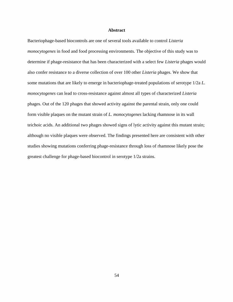
54
Abstract
Bacteriophage-based biocontrols are one of several tools available to control Listeria
monocytogenes in food and food processing environments. The objective of this study was to
determine if phage-resistance that has been characterized with a select few Listeria phages would
also confer resistance to a diverse collection of over 100 other Listeria phages. We show that
some mutations that are likely to emerge in bacteriophage-treated populations of serotype 1/2a L.
monocytogenes can lead to cross-resistance against almost all types of characterized Listeria
phages. Out of the 120 phages that showed activity against the parental strain, only one could
form visible plaques on the mutant strain of L. monocytogenes lacking rhamnose in its wall
teichoic acids. An additional two phages showed signs of lytic activity against this mutant strain;
although no visible plaques were observed. The findings presented here are consistent with other
studies showing mutations conferring phage-resistance through loss of rhamnose likely pose the
greatest challenge for phage-based biocontrol in serotype 1/2a strains.

55
Introduction
The Gram-positive foodborne pathogen Listeria monocytogenes is the causative agent of
listeriosis. Listeriosis can cause serious health problems for susceptible populations, including
septicemia, bacteremia, meningitis, and meningoencephalitis (37). Illness due to listeriosis leads
to a high rate of hospitalizations (approx. 91%) and deaths (approx. 16%). Listeriosis in humans
is contracted almost exclusively through consumption of contaminated foods (32). The largest L.
monocytogenes outbreak to-date occurred in South Africa and was caused by contamination of
polony, a ready-to-eat meat, leading to over 1,000 illnesses and 200 deaths from 2017 to 2018 (2,
30). L. monocytogenes can be particularly problematic due to its abilities to replicate at
refrigeration temperatures (31) and to persist for years in food processing facilities (26, 28). L.
monocytogenes is categorized into at least thirteen different serotypes (ST). ST 1/2a, 1/2b, and
4b are the most commonly associated with human illness (29). From 1998 to 2008, serotype 1/2a
caused 40% of the confirmed L. monocytogenes outbreaks in the U.S. (8). The largest outbreak
of L. monocytogenes in the U.S. occurred in 2011 and was caused by cantaloupe contaminated
with serotype 1/2a and 1/2b L. monocytogenes. Illnesses associated with the outbreak occurred in
28 states and were responsible for 143 hospitalizations and 33 deaths (25).
Bacteriophages (or “phages”), viruses that specifically target bacteria, have been approved for
use in the U.S. since 2006 for control of L. monocytogenes in foods and on food processing
surfaces (7). There are currently two commercial Listeria phage products approved for food
safety applications, and, more recently, products have been approved to target Salmonella
enterica, Escherichia coli, and Shigella spp. (27). Previous work has shown that phage-
resistance is consistently selected for in L. monocytogenes populations infected with Listeria
56
phages (15). A screen of L. monocytogenes strains that were isolated from European processing
facilities from 1987 to 2012 found that phage resistant L. monocytogenes only occurred after
those processing facilities had used or experimentally tested a Listeria phage product; strains that
were isolated prior to the intentional introduction of phage were all found to be sensitive (18). In
L. monocytogenes serotype 1/2a strains 10403S and EGDe, mutations conferring phage-
resistance were consistently found in genes that affect known Listeria phage binding receptors:
terminal N-acetylglucosamine (GlcNAc) and rhamnose (Rha) decorations of the wall teichoic
acids (WTA) (15, 17). Surprisingly, no other binding receptors have been conclusively shown to
be used by Listeria phages that are able to infect L. monocytogenes serotype 1/2a strains.
Here, we measure activity of a diverse collection of 120 Listeria phages, representing four of the
five known orthoclusters, against three mutant strains of L. monocytogenes 10403S (serotype
1/2a). Listeria phages have been shown to form five distinct genomic clusters, referred to as
orthoclusters. Each orthocluster has unique morphological characteristics; four contain
siphoviruses and one contains myoviruses (11, 16). None of the phages from orthocluster IV that
were available to us showed activity against our serotype 1/2a strains, so they were not included
in this study. The lysis profiles of a majority of the phages we tested were previously described
(38). The three mutant strains represent three distinct phenotypes of phage-resistance. Two of
these mutants were previously sequenced and characterized (15) and the third is characterized
here. We aimed to determine if these characterized phage-resistant mutants were broadly
resistant to Listeria phages and if any phages could overcome these common resistance types.
57
Materials & Methods
Growth of Bacterial Strains
Propagation host strains 10403S and 10403S-derived mutants (Table 2.1 in the appendix) were
prepared from stocks stored at -80°C in Brain Heart Infusion (BHI; Becton Dickinson, Sparks,
MD) broth with 15% (wt/vol) glycerol and incubated at 30°C on 1.5% (wt/vol) BHI agar plates.
Overnight cultures were prepared by inoculating BHI broth with a single colony from a streak
plate, that was prepared less than two weeks prior, and then incubated for 16±2 h at 30ºC shaking
at 160 RPM. Bacterial lawns were prepared using an agar overlay method as described
previously (35) with modifications. The agar underlay used here was prepared in square 6 x 6
grid plates (Simport Scientific, Beloeil, QC, Canada). Forty µL of overnight culture was
aliquoted into 4-4.5 mL of 0.7% (wt/vol) modified lysogeny broth (LB) morpholino-propane
sulfonic acid (MOPS) overlay agar supplemented with 0.1% (wt/vol) glucose and 1 mM each
MgCl2 and CaCl2, after equilibrating to 56°C. The mixture was vortexed briefly, poured onto the
agar underlay and allowed to solidify for 20-30 min. UTK P1-0001 was isolated from a 10403S
culture infected by LP-048 under previously described conditions (15).
Phage Amplification
Phage stocks used for the experiment (Table 2.2 in the appendix) were first amplified to a titer of
at least 1 x 10⁷ pfu/mL using the plate lysate method. Plates were prepared in duplicate using an
agar overlay method as described previously (38) with modifications. Thirty µL of overnight
culture and 100 µL of the phage dilution in saline magnesium (SM) buffer with gelatin (Fisher,
Fair Lawn, NJ) was aliquoted into 3-3.5 mL of modified LB MOPS overlay agar after
equilibrating to 56°C. The mixture was vortexed briefly, poured onto the agar underlay and
58
allowed to solidify for 20-30 min. Plates were then incubated for 18-24 h at 25°C. Five mL of
sterile SM buffer with gelatin was aliquoted onto each plate with confluent lysis and allowed to
sit for 1-2 h. The overlay agar was then carefully broken into pieces and scraped to the side of
the plate using a sterile cell scraper (VWR, Radnor, PA). The phage-containing buffer was then
siphoned off the plate using a serological pipette, centrifuged at 3000 RPM for 15 min at 4°C to
remove debris, and then sterile filtered into an amber vial using a 0.20 µm-pore size surfactant-
free cellulose acetate (SFCA) syringe filter (Corning, Incorporated, Corning, NY). Phage stocks
were stored at 4°C.
Screening for Phage Activity Against Listeria monocytogenes 10403S
From the lab collection, phage stocks with a titer of at least 1 x 10⁷ pfu/mL whose propagation
host was not L. monocytogenes Mack (ST 1/2a) were tested for activity against L.
monocytogenes 10403S (ST 1/2a) as described previously (15). Five µL of undiluted phage
stocks were spotted onto bacterial lawns of 10403S in duplicate. All phages with a titer ≥ 1 x 10⁷
pfu/mL, with a propagation host of Mack or with observed activity (visible inhibition of bacterial
growth) against 10403S, were included in the study (120 total).
Preparation of Standardized Stocks
Working stocks of each phage were prepared at a theoretical concentration of 1 x 10⁷ pfu/mL
based on recent (< 1 month old) titers.

59
Phage Dilution Spot Assays
Phages were tested for activity and/or plaque formation against 10403S and the three mutant
strains relative to their propagation host as described above. Ten µL of phage, either directly
from a standardized stock or serially diluted in phosphate buffered saline (PBS) were spotted
onto bacterial lawns. All phages were spotted within 4 h of bacterial lawns being poured. Plates
were incubated at 30°C for 18-24 h. Phage titers were calculated after observation of visible
plaques. Phage activity against each strain was measured as the greatest phage dilution where
either visible plaques were observed or visible inhibition of bacterial growth was observed (if no
countable plaques were seen) as compared to the propagation host. Inhibition of bacterial growth
was confirmed by visual comparison to controls of 10 µL spots of phage-free SM buffer with
gelatin or PBS. Three replicates were conducted.
Adsorption Assay
The adsorption of L. monocytogenes strains 10403S, FSL D4-0119 and UTK P1-0001 to
bacteriophages LP-048 and LP-125 was determined as described previously (15). Working phage
stocks of LP-125 and LP-048 at a titer of 1 x 10⁹ pfu/mL were prepared using SM buffer with
gelatin as the diluent. Sterile microcentrifuge tubes (2 mL) were prepared with 912 µL BHI, 20
µL of phage (at 1 x 10⁹ pfu/ml) and 9 µL each 1 M MgCl2 and 1 M CaCl2 before the addition of
either 50 µL bacterial overnight culture (as described above) or 50 µL sterile BHI (negative
control). After addition of the overnight culture or sterile BHI, tubes were incubated for 15 min
at 30°C with shaking at 160 RPM. Samples were then centrifuged for 1 min at 17,000 x g and
4°C. The supernatant was filter sterilized (0.20 µm-pore size SFCA syringe filter; Corning,
Incorporated, Corning, NY), serially diluted in PBS, and enumerated on duplicate plates using

60
the agar overlay method (described above under ‘Phage Amplification’) to determine the number
of unbound phage present. Three replicates were conducted.
Cluster Analysis (Phage Spot Assays)
Heatmaps were generated using the heatmap hierarchical clustering tool from the HIV databases
website which can be accessed at
https://www.hiv.lanl.gov/content/sequence/HEATMAP/heatmap.html. Clustering parameters
included a complete method using Euclidean distances, which were calculated by taking the
square root of the usual sum of squared differences distances between elements of vectors X
[columns (L. monocytogenes strains)] and Y [rows (Listeria phages)]. Cluster stability was
calculated using standard bootstraps with 100 iterations.
Statistical Analysis (Adsorption Assays)
Log10 reduction was analyzed using JMP (version 13; SAS Institute Inc., Cary, NC). Linear
models were constructed with the means of three replicates using strain as a factor. Log10
reduction was used as the model response. A Fit Y by X platform was used to compare means by
one way ANOVA using Tukey’s HSD test. Values in Figure 2.5 in the appendix represent the
log10 reduction of phage in the supernatant. This was calculated by subtracting the log-
transformed concentration of phage in the supernatant by the log-transformed concentration of
phage in the negative control. An alpha of 0.05 was used to determine significant differences.

61
Sequencing and Variant Analysis of 10403S and UTK P1-0001
DNA was extracted using a QIAamp extraction kit (Qiagen, Hilden, Germany) as previously
described (15). NexteraXT Library preparation (Illumina, San Diego, CA) and sequencing was
conducted at the University of Tennessee Genomics Core. Sequencing was performed on an
Illumina MiSeq instrument using 300 bp paired-end read chemistry. Raw reads were uploaded to
the NCBI sequencing read archive (SRA IDs SRR9115406 and SRR9115405) and then trimmed
with Trimmomatic version 0.35 (6). Trimmed reads were then quality-checked with FastQC
version 0.11.7 (3). McCortex (36) was used for variant calling in the L. monocytogenes mutant
(UTK P1-0001) and control (10403S) isolates. The L. monocytogenes 10403S RefSeq assembly
(RefSeq ID 376088) was downloaded and used as the reference. The McCortex pipeline was run
with joint calling, the “vcfs” target, and a kmer size of 57 (optimum kmer size determined with
KmerGenie version 1.7048 (12)). SnpEff version 4.3t (13) was used to annotate the vcf output
files.
Results & Discussion
Listeria Phages Isolated from NY Dairy Farms Almost All Require Rhamnose in Their
Serotype 1/2a Host’s Wall Teichoic Acids
Out of 120 phages tested, only LP-018 formed visible plaques on FSL D4-0119, the mutant
strain of 10403S lacking rhamnose in its WTA (Figure 2.1 in the appendix), and only two other
phages from the collection showed any phage activity against the strain (Figure 2.2 in the
appendix). This supports previous observations that serotype 3 strains were largely resistant to a
collection of 16 Listeria phages (24). Although a mutant of a serotype 1/2a strain, FSL D4-0119
would be expected to be classified as a serotype 3 strain. Serotype 3 strains are known to possess

62
only N-acetylglucosamine decorations in their WTA (11, 16). It has also been shown that loss of
rhamnose due to phage selection in a serotype 1/2a strain converts that strain to serotype 3, and
that serotype 3 strains typically resemble serotype 1/2 strains that have accumulated a mutation
or mutations affecting rhamnosylation genes (17). As spontaneous mutations affecting
rhamnosylation of WTAs are consistently selected for in L. monocytogenes 1/2a populations
infected with bacteriophages (15), it may be common for phage-resistance to emerge under the
selective pressure of a single phage that confers cross-resistance to a majority of phages. This
suggests that it will be a challenge for phage-based biocontrol to prevent the emergence of
phage-resistant mutants in treated environments; however, the risk of L. monocytogenes
contamination causing human illness may still be considerably reduced as rhamnose has been
shown to promote virulence of serotype 1/2 strains by increasing resistance to host antimicrobial
peptides (9) and by promoting association virulence factors to the cell wall (10). This suggests
that rhamnosylation-affecting mutations may reduce the virulence of L. monocytogenes. This is
consistent with the rarity of serotype 3 strains association with outbreak or illness (8, 29, 34). If
Listeria phage applications are expected remain effective against all L. monocytogenes strains in
the same environment for an extended period of time, it may be critical to include a phage like
LP-018 that can infect mutants lacking rhamnose in their WTA. Future studies should be
conducted to characterize LP-018 and to determine its binding receptors.
Mutations conferring phage-resistance through loss of GlcNAc in WTA (15) are less problematic
for Listeria phage applications than those that confer resistance through the loss of rhamnose, as
there are many available phages that can infect the GlcNAc deficient 1/2a strain. Thirteen out of
the 120 Listeria phages could form visible plaques on FSL D4-0014, the mutant strain of 10403S
63
lacking GlcNAc in its WTA (Figure 2.1 in the appendix), and one additional phage showed
phage activity against the strain (Figure 2.2 in the appendix). Phages that can infect FSL D4-
0014 include the well-characterized LP-048, which is a myovirus from the P100virus genus (21)
that may serve as an important model phage for studies on Listeria phage applications that aim to
reduce the emergence of phage-resistance. Specifically, previous studies of LP-048 infection
kinetics showed consistent bursts of approximately 13 phage particles under laboratory
conditions (15), and plaquing efficiency was shown to increase by up to 50% at cooler
temperatures that are more relevant to food and food processing conditions (35).
L. monocytogenes strain UTK P1-0001 was included in this study as a phage-resistant mutant of
10403S because it showed a phenotype different from the mutants lacking rhamnose and
GlcNAc in their WTA, i.e., UTK P1-0001 showed resistance to phage LP-048 and susceptibility
to LP-125 (Figures 2.1 and 2.2 in the appendix). Adsorption assays show UTK P1-0001 does not
support binding of LP-048 (Figure 2.5a in the appendix), which likely uses rhamnose as its
primary receptor. However, UTK P1-0001 does support a reduced level of binding of LP-125
(Figure 2.5b in the appendix), which is unable to bind to the rhamnose deficient mutant.
Sequencing revealed that the only mutation present in UTK P1-0001 was a deletion in
LMRG_00544 (GAATA to G at nucleotide position 1,098,886; this variant was supported by
146/146 sequencing reads that covered the position) that would cause a frameshift and early
termination, leading to a truncated protein product. LMRG_00544 encodes RmlC, which is part
of the dTDP-L-Rhamnose pathway (19). It is possible that loss of function of RmlC would result
in incorporation of dTDP-6-deoxy-D-xylo-4 hexulose (the substrate of RmlC) being incorporated
into the WTA instead of L-rhamnose; this would be consistent with our observation that this
64
mutant has a distinct phage susceptibility phenotype from FSL D4-0119, which lacks L-
rhamnose in the WTA. Forty-two of the Listeria phages showed visible plaques on UTK P1-
0001, and an additional 61 phages showed activity against the mutant strain. Out of the 120
Listeria phages tested, only 17 showed no phage activity against UTK P1-0001. This specific
type of mutation is likely not a great challenge to Listeria phage applications as only a few
Listeria phages are fully affected by it; however, the mutant strain may be useful for
differentiating and better understanding receptor requirements of Listeria phages.
Phage Activity was Frequently Observed Without the Formation of Visible Plaques
Efficiency of plaquing (or “plating”) experiments are often conducted to evaluate the host range
of bacteriophages (22). Each phage-resistant mutant we used in this study showed several phages
that failed to produce visible plaques, yet showed phage activity (Figures 2.1 and 2.2 in the
appendix). These phages were capable of lysing or significantly inhibiting the growth of the
target strains; however, if we only performed efficiency of plaquing experiments, this activity
would not be observed. The formation of a visible plaque is a complex biological process and
absence of plaque formation does not necessarily indicate virion inviability (1). If evaluating
phages for potential in biocontrol applications, visible plaque formation on all target strains may
not be necessary. For example, if constructing a cocktail that is designed to limit the emergence
of phage-resistance, it may be effective to include a phage that only shows activity against a
phage-resistance type (such as rhamnose deficient WTA); those specific mutations are likely to
be rare in the target L. monocytogenes population, so as long as the cocktail exerts selective
pressure on those mutants, they are unlikely to grow to concentrations capable of causing illness.

65
Conclusions
We have identified only one bacteriophage, LP-018, capable of infecting all three of the phage-
resistant mutants of serotype 1/2a L. monocytogenes that were used in the study. We conclude
that mutations conferring phage-resistance through loss of rhamnose likely pose the greatest
challenge for phage-based biocontrol in serotype 1/2a strains, as we found that they confer
resistance to almost all of the Listeria phages (119/120) in the diverse collection tested. These
results have the potential to aid in the rational design of Listeria phage cocktails that aim to
reduce the emergence of phage-resistance to ensure long-term efficacy.
Acknowledgements
We thank Tracey Lee Peters for the isolation and DNA extraction of UTK P1-0001 and Martin
Wiedmann for the gift of the other Listeria monocytogenes strains and phages used in this study.
66
References
1. Abedon, S. T., and J. Yin. 2009. Bacteriophage Plaques: Theory and Analysis, p. 161–
174. In Bacteriophages.
2. Allam, M., N. Tau, S. L. Smouse, P. S. Mtshali, F. Mnyameni, Z. T. H. Khumalo, A.
Ismail, N. Govender, J. Thomas, and A. M. Smith. 2018. Whole-Genome Sequences of
Listeria monocytogenes Sequence Type 6 Isolates Associated with a Large Foodborne
Outbreak in South Africa, 2017 to 2018. Genome Announc. 6:e00538-18.
3. Andrews, S. FastQC.
4. Bergholz, T. M., H. C. den Bakker, E. D. Fortes, K. J. Boor, and M. Wiedmann. 2010. Salt
Stress Phenotypes in Listeria monocytogenes Vary by Genetic Lineage and Temperature.
Foodborne Pathog. Dis. 7:1537–1549.
5. Bishop, D. K., and D. J. Hinrichs. 1987. Adoptive transfer of immunity to Listeria
monocytogenes. The influence of in vitro stimulation on lymphocyte subset requirements.
J. Immunol. 139:2005–2009.
6. Bolger, A. M., M. Lohse, and B. Usadel. 2014. Trimmomatic: a flexible trimmer for
Illumina sequence data. Bioinformatics 30:2114–2120.
7. Bren, L. 2007. Bacteria-eating virus approved as food additive. FDA Consum. 41:20–22.
8. Cartwright, E. J., K. A. Jackson, S. D. Johnson, L. M. Graves, B. J. Silk, and B. E. Mahon.
2013. Listeriosis Outbreaks and Associated Food Vehicles, United States, 1998–2008.
Emerg. Infect. Dis. 19:1–9.
9. Carvalho, F., M. L. Atilano, R. Pombinho, G. Covas, R. L. Gallo, S. R. Filipe, S. Sousa,
and D. Cabanes. 2015. L-Rhamnosylation of Listeria monocytogenes Wall Teichoic Acids
67
Promotes Resistance to Antimicrobial Peptides by Delaying Interaction with the
Membrane. PLOS Pathog. 11:e1004919.
10. Carvalho, F., S. Sousa, and D. Cabanes. 2018. l-Rhamnosylation of wall teichoic acids
promotes efficient surface association of Listeria monocytogenes virulence factors InlB
and Ami through interaction with GW domains. Environ. Microbiol. 20:3941–3951.
11. Casey, A., K. Jordan, H. Neve, A. Coffey, and O. McAuliffe. 2015. A tail of two phages:
genomic and functional analysis of Listeria monocytogenes phages vB_LmoS_188 and
vB_LmoS_293 reveal the receptor-binding proteins involved in host specificity. Front.
Microbiol. 6:1107.
12. Chikhi, R., and P. Medvedev. 2014. Informed and automated k-mer size selection for
genome assembly. Bioinformatics 30:31–37.
13. Cingolani, P., A. Platts, L. L. Wang, M. Coon, T. Nguyen, L. Wang, S. J. Land, X. Lu,
and D. M. Ruden. 2012. A program for annotating and predicting the effects of single
nucleotide polymorphisms, SnpEff. Fly (Austin) 6:80–92.
14. den Bakker, H. C., B. M. Bowen, L. D. Rodriguez-Rivera, and M. Wiedmann. 2012. FSL
J1-208, a Virulent Uncommon Phylogenetic Lineage IV Listeria monocytogenes Strain
with a Small Chromosome Size and a Putative Virulence Plasmid Carrying Internalin-Like
Genes. Appl. Environ. Microbiol. 78:1876–1889.
15. Denes, T., H. C. den Bakker, J. I. Tokman, C. Guldimann, and M. Wiedmann. 2015.
Selection and Characterization of Phage-Resistant Mutant Strains of Listeria
monocytogenes Reveal Host Genes Linked to Phage Adsorption. Appl. Environ.
Microbiol. 81:4295–4305.

68
16. Denes, T., K. Vongkamjan, H.-W. Ackermann, A. I. M. Switt, M. Wiedmann, and H. C.
den Bakker. 2014. Comparative Genomic and Morphological Analyses of Listeria Phages
Isolated from Farm Environments. Appl. Environ. Microbiol. 80:4616–4625.
17. Eugster, M. R., L. S. Morax, V. J. Hüls, S. G. Huwiler, A. Leclercq, M. Lecuit, and M. J.
Loessner. 2015. Bacteriophage predation promotes serovar diversification in Listeria
monocytogenes. Mol. Microbiol. 97:33–46.
18. Fister, S., S. Fuchs, B. Stessl, D. Schoder, M. Wagner, and P. Rossmanith. 2016.
Screening and characterisation of bacteriophage P100 insensitive Listeria monocytogenes
isolates in Austrian dairy plants. Food Control 59:108–117.
19. Giraud, M.-F., G. A. Leonard, R. A. Field, C. Berlind, and J. H. Naismith. 2000. RmlC,
the third enzyme of dTDP-L-rhamnose pathway, is a new class of epimerase. Nat. Struct.
Biol. 7:398.
20. Hodgson, D. A. 2000. Generalized transduction of serotype 1/2 and serotype 4b strains of
Listeria monocytogenes. Mol. Microbiol. 35:312–323.
21. Krupovic, M., B. E. Dutilh, E. M. Adriaenssens, J. Wittmann, F. K. Vogensen, M. B.
Sullivan, J. Rumnieks, D. Prangishvili, R. Lavigne, A. M. Kropinski, J. Klumpp, A. Gillis,
F. Enault, R. A. Edwards, S. Duffy, M. R. C. Clokie, J. Barylski, H.-W. Ackermann, and
J. H. Kuhn. 2016. Taxonomy of prokaryotic viruses: update from the ICTV bacterial and
archaeal viruses subcommittee. Arch. Virol. 161:1095–1099.
22. Kutter, E. 2009. Phage Host Range and Efficiency of Plating, p. 141–149. In
Bacteriophages.
23. Linnan, M. J., L. Mascola, X. D. Lou, V. Goulet, S. May, C. Salminen, D. W. Hird, M. L.
Yonekura, P. Hayes, R. Weaver, A. Audurier, B. D. Plikaytis, S. L. Fannin, A. Kleks, and
69
C. V. Broome. 1988. Epidemic Listeriosis Associated with Mexican-Style Cheese. N.
Engl. J. Med. 319:823–828.
24. Loessner, M. J., and M. Busse. 1990. Bacteriophage typing of Listeria species. Appl.
Environ. Microbiol. 56:1912–1918.
25. McCollum, J. T., A. B. Cronquist, B. J. Silk, K. A. Jackson, K. A. O’Connor, S. Cosgrove,
J. P. Gossack, S. S. Parachini, N. S. Jain, P. Ettestad, M. Ibraheem, V. Cantu, M. Joshi, T.
DuVernoy, N. W. Fogg, J. R. Gorny, K. M. Mogen, C. Spires, P. Teitell, L. A. Joseph, C.
L. Tarr, M. Imanishi, K. P. Neil, R. V. Tauxe, and B. E. Mahon. 2013. Multistate
Outbreak of Listeriosis Associated with Cantaloupe. N. Engl. J. Med. 369:944–953.
26. Møretrø, T., and S. Langsrud. 2004. Listeria monocytogenes: biofilm formation and
persistence in food-processing environments. Biofilms 1:107–121.
27. Moye, Z. D., J. Woolston, and A. Sulakvelidze. 2018. Bacteriophage Applications for
Food Production and Processing. Viruses 10:205.
28. Orsi, R. H., M. L. Borowsky, P. Lauer, S. K. Young, C. Nusbaum, J. E. Galagan, B. W.
Birren, R. A. Ivy, Q. Sun, L. M. Graves, B. Swaminathan, and M. Wiedmann. 2008.
Short-term genome evolution of Listeria monocytogenes in a non-controlled environment.
BMC Genomics 9:539.
29. Orsi, R. H., H. C. den Bakker, and M. Wiedmann. 2011. Listeria monocytogenes lineages:
Genomics, evolution, ecology, and phenotypic characteristics. Int. J. Med. Microbiol.
301:79–96.
30. Salama, P. J., P. K. B. Embarek, J. Bagaria, and I. S. Fall. 2018. Learning from listeria:
safer food for all. The Lancet 391:2305–2306.

70
31. Saldivar, J. C., M. L. Davis, M. G. Johnson, and S. C. Ricke. 2018. Chapter 13 - Listeria
monocytogenes Adaptation and Growth at Low Temperatures: Mechanisms and
Implications for Foodborne Disease, p. 227–248. In S.C. Ricke, G.G. Atungulu, C.E.
Rainwater, and S.H. Park (eds.), Food and Feed Safety Systems and Analysis. Academic
Press.
32. Scallan, E., R. M. Hoekstra, F. J. Angulo, R. V. Tauxe, M.-A. Widdowson, S. L. Roy, J.
L. Jones, and P. M. Griffin. 2011. Foodborne Illness Acquired in the United States—
Major Pathogens. Emerg. Infect. Dis. 17:7–15.
33. Stasiewicz, M. J., M. Wiedmann, and T. M. Bergholz. 2010. The Combination of Lactate
and Diacetate Synergistically Reduces Cold Growth in Brain Heart Infusion Broth across
Listeria monocytogenes Lineages - ProQuest. J. Food Prot. 73:631–640.
34. Todd, E. C. D., and S. Notermans. 2011. Surveillance of listeriosis and its causative
pathogen, Listeria monocytogenes. Food Control 22:1484–1490.
35. Tokman, J. I., D. J. Kent, M. Wiedmann, and T. Denes. 2016. Temperature Significantly
Affects the Plaquing and Adsorption Efficiencies of Listeria Phages. Front. Microbiol.
7:631.
36. Turner, I., K. V. Garimella, Z. Iqbal, and G. McVean. 2018. Integrating long-range
connectivity information into de Bruijn graphs. Bioinformatics 34:2556–2565.
37. Vázquez-Boland, J. A., M. Kuhn, P. Berche, T. Chakraborty, G. Domı́nguez-Bernal, W.
Goebel, B. González-Zorn, J. Wehland, and J. Kreft. 2001. Listeria Pathogenesis and
Molecular Virulence Determinants. Clin. Microbiol. Rev. 14:584–640.
38. Vongkamjan, K., A. M. Switt, H. C. den Bakker, E. D. Fortes, and M. Wiedmann. 2012.
Silage Collected from Dairy Farms Harbors an Abundance of Listeriaphages with

71
Considerable Host Range and Genome Size Diversity. Appl Env. Microbiol 78:8666–
8675.
39. Wesley, I. V., and F. Ashton. 1991. Restriction enzyme analysis of Listeria
monocytogenes strains associated with food-borne epidemics. Appl. Environ. Microbiol.
57:969–975.

72
Appendix
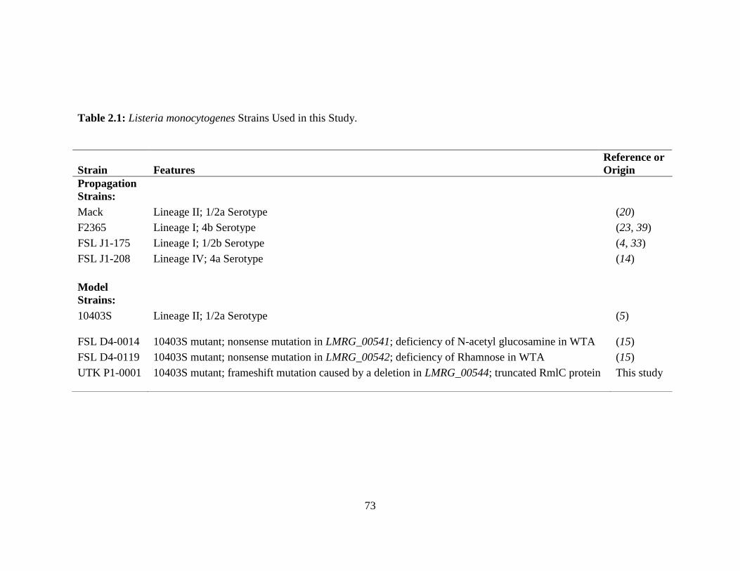
73
Table 2.1: Listeria monocytogenes Strains Used in this Study.
Strain Features
Reference or
Origin
Propagation
Strains:
Mack Lineage II; 1/2a Serotype (20)
F2365 Lineage I; 4b Serotype (23, 39)
FSL J1-175 Lineage I; 1/2b Serotype (4, 33)
FSL J1-208 Lineage IV; 4a Serotype (14)
Model
Strains:
10403S Lineage II; 1/2a Serotype (5)
FSL D4-0014
10403S mutant; nonsense mutation in LMRG_00541; deficiency of N-acetyl glucosamine in WTA (15)
FSL D4-0119 10403S mutant; nonsense mutation in LMRG_00542; deficiency of Rhamnose in WTA (15)
UTK P1-0001 10403S mutant; frameshift mutation caused by a deletion in LMRG_00544; truncated RmlC protein This study

74
Table 2.2: Listeria Phages Used in this Study and Their Propagation Strains.
Phage Propagation Host
A511 MACK
LP-018 MACK
LP-019 MACK
LP-025 MACK
LP-031 MACK
LP-038 MACK
LP-039 MACK
LP-048 MACK
LP-049 MACK
LP-064 MACK
LP-066 MACK
LP-069 MACK
LP-077 MACK
LP-083-1 MACK
LP-097 MACK
LP-098 MACK
LP-099 MACK
LP-100 MACK
LP-101 MACK
LP-102 MACK
LP-103 MACK
LP-104 MACK
LP-120 MACK
LP-123 MACK
LP-125 MACK
LP-179 MACK
LP-180 MACK
LP-095 F2365
LP-096 F2365
LP-105 F2365
LP-106 F2365
LP-116 F2365
LP-124 F2365
LP-127 F2365
LP-177 F2365
LP-178 F2365
LP-017 J1-175
LP-040 J1-175
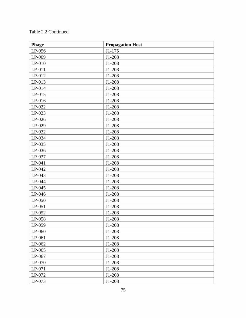
75
Table 2.2 Continued.
Phage Propagation Host
LP-056 J1-175
LP-009 J1-208
LP-010 J1-208
LP-011 J1-208
LP-012 J1-208
LP-013 J1-208
LP-014 J1-208
LP-015 J1-208
LP-016 J1-208
LP-022 J1-208
LP-023 J1-208
LP-026 J1-208
LP-029 J1-208
LP-032 J1-208
LP-034 J1-208
LP-035 J1-208
LP-036 J1-208
LP-037 J1-208
LP-041 J1-208
LP-042 J1-208
LP-043 J1-208
LP-044 J1-208
LP-045 J1-208
LP-046 J1-208
LP-050 J1-208
LP-051 J1-208
LP-052 J1-208
LP-058 J1-208
LP-059 J1-208
LP-060 J1-208
LP-061 J1-208
LP-062 J1-208
LP-065 J1-208
LP-067 J1-208
LP-070 J1-208
LP-071 J1-208
LP-072 J1-208
LP-073 J1-208

76
Table 2.2 Continued.
Phage Propagation Host
LP-078 J1-208
LP-079 J1-208
LP-080 J1-208
LP-081 J1-208
LP-082 J1-208
LP-084 J1-208
LP-086 J1-208
LP-087 J1-208
LP-089 J1-208
LP-090 J1-208
LP-091 J1-208
LP-092 J1-208
LP-108 J1-208
LP-109 J1-208
LP-110 J1-208
LP-111 J1-208
LP-112 J1-208
LP-113 J1-208
LP-114 J1-208
LP-115 J1-208
LP-118 J1-208
LP-121 J1-208
LP-122 J1-208
LP-126 J1-208
LP-128 J1-208
LP-129 J1-208
LP-130 J1-208
LP-131 J1-208
LP-132 J1-208
LP-133 J1-208
LP-134 J1-208
LP-135 J1-208
LP-136 J1-208
LP-137 J1-208
LP-138 J1-208
LP-139 J1-208
LP-141 J1-208
LP-142 J1-208
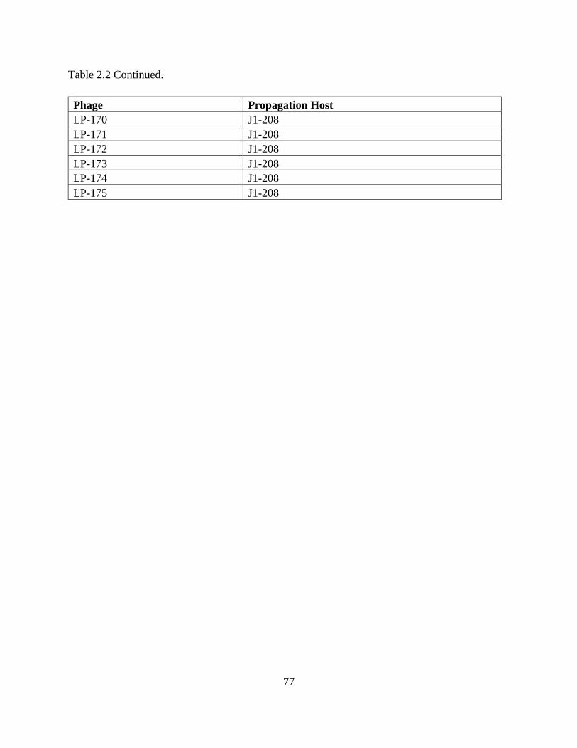
77
Table 2.2 Continued.
Phage Propagation Host
LP-170 J1-208
LP-171 J1-208
LP-172 J1-208
LP-173 J1-208
LP-174 J1-208
LP-175 J1-208
78
Figure 2.1: Average Efficiencies of Plaquing Heatmap. Values represent the titer of each
Listeria phage on each bacterial strain compared to the titer on the phage’s propagation host. “No
visible plaques” indicates that no titer was obtained, but does not necessarily indicate no activity
(Fig. 2.2). Colored dendrogram lines indicate stability of clustering. Bootstrap probabilities of ≥
70% are highlighted in red; bootstrap probabilities of ≥ 90% are highlighted in blue. Listeria
phage names are colored to indicate genome size categories. Purple indicates a large genome size
(approx. 97 to 140 kb), predicted to be within Orthocluster I; green indicates a medium genome
size (approx. 57 to 70 kb), predicted to be within Orthocluster V; orange indicates a small
genome size (approx. 31 to 43 kb), predicted to be within Orthoclusters II-IV. Bold font
indicates genome size obtained by sequencing; italic font indicates genome size obtained by
PFGE. 10403S possesses WTA with terminal GlcNAc and Rha; FSL D4-0014 possesses WTA
with terminal Rha; FSL D4-0119 possesses WTA with terminal GlcNAc. The means of three
replicates are shown (See Fig. 2.3 for values from each replicate).

79
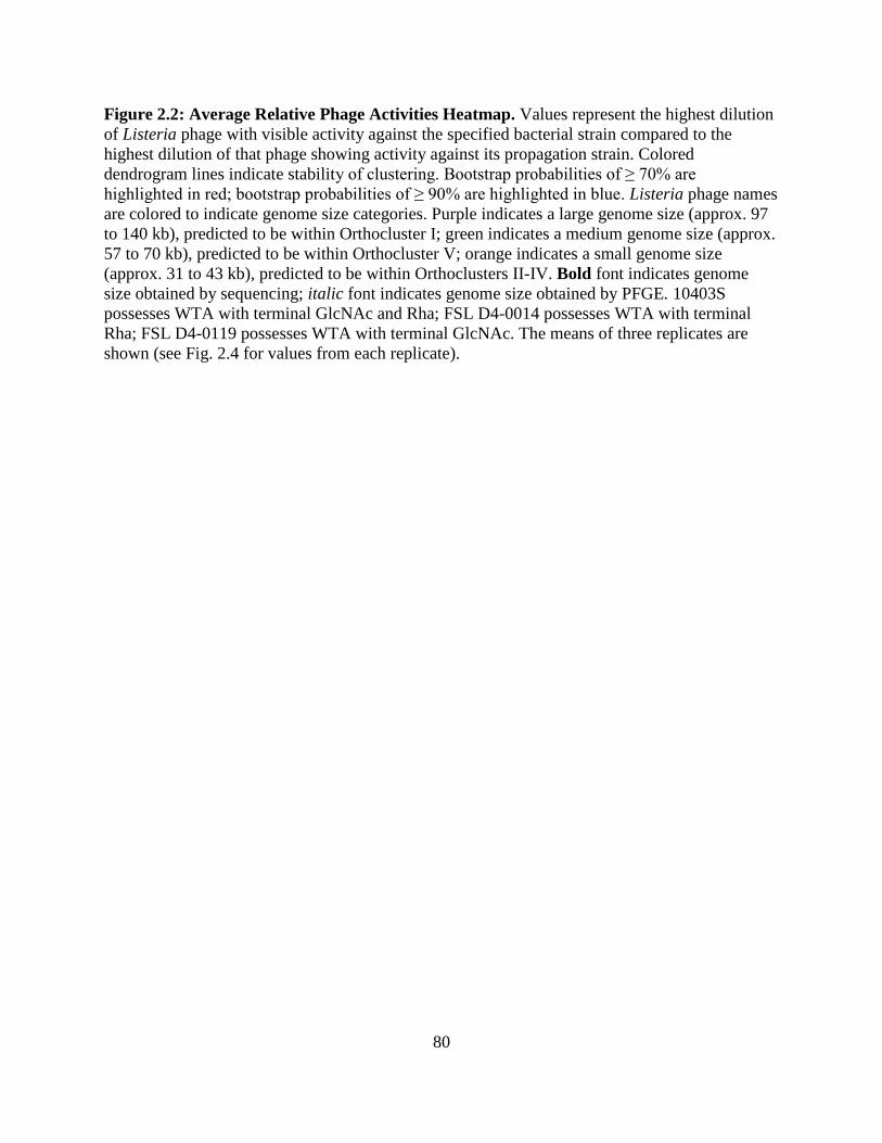
80
Figure 2.2: Average Relative Phage Activities Heatmap. Values represent the highest dilution
of Listeria phage with visible activity against the specified bacterial strain compared to the
highest dilution of that phage showing activity against its propagation strain. Colored
dendrogram lines indicate stability of clustering. Bootstrap probabilities of ≥ 70% are
highlighted in red; bootstrap probabilities of ≥ 90% are highlighted in blue. Listeria phage names
are colored to indicate genome size categories. Purple indicates a large genome size (approx. 97
to 140 kb), predicted to be within Orthocluster I; green indicates a medium genome size (approx.
57 to 70 kb), predicted to be within Orthocluster V; orange indicates a small genome size
(approx. 31 to 43 kb), predicted to be within Orthoclusters II-IV. Bold font indicates genome
size obtained by sequencing; italic font indicates genome size obtained by PFGE. 10403S
possesses WTA with terminal GlcNAc and Rha; FSL D4-0014 possesses WTA with terminal
Rha; FSL D4-0119 possesses WTA with terminal GlcNAc. The means of three replicates are
shown (see Fig. 2.4 for values from each replicate).
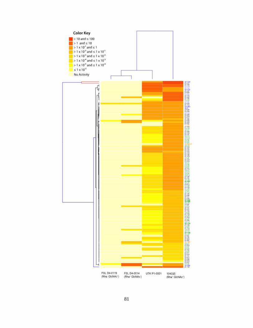
81
82
Figure 2.3: Efficiencies of Plaquing Heatmap. Values represent the titer of each Listeria phage
on each bacterial strain compared to the titer on the phage’s propagation host. “No visible
plaques” indicates that no titer was obtained, but does not necessarily indicate no activity (Fig.
2.2). Colored dendrogram lines indicate stability of clustering. Bootstrap probabilities of ≥ 70%
are highlighted in red; bootstrap probabilities of ≥ 90% are highlighted in blue. Listeria phage
names are colored to indicate genome size categories. Purple indicates a large genome size
(approx. 97 to 140 kb), predicted to be within Orthocluster I; green indicates a medium genome
size (approx. 57 to 70 kb), predicted to be within Orthocluster V; orange indicates a small
genome size (approx. 31 to 43 kb), predicted to be within Orthoclusters II-IV. Bold font
indicates genome size obtained by sequencing; italic font indicates genome size obtained by
PFGE. 10403S possesses WTA with terminal GlcNAc and Rha; FSL D4-0014 possesses WTA
with terminal Rha; FSL D4-0119 possesses WTA with terminal GlcNAc. The results of three
individual replicates are shown (see Fig. 2.1 for mean values).

83

84
Figure 2.4: Relative Phage Activities Heatmap. Values represent the highest dilution of
Listeria phage with visible activity against the specified bacterial strain compared to the highest
dilution of that phage showing activity against its propagation host. Colored dendrogram lines
indicate stability of clustering. Bootstrap probabilities of ≥ 70% are highlighted in red; bootstrap
probabilities of ≥ 90% are highlighted in blue. Listeria phage names are colored to indicate
genome size categories. Purple indicates a large genome size (approx. 97 to 140 kb), predicted to
be within Orthocluster I; green indicates a medium genome size (approx. 57 to 70 kb), predicted
to be within Orthocluster V; orange indicates a small genome size (approx. 31 to 43 kb),
predicted to be within Orthoclusters II-IV. Bold font indicates genome size obtained by
sequencing; italic font indicates genome size obtained by PFGE. 10403S possesses WTA with
terminal GlcNAc and Rha; FSL D4-0014 possesses WTA with terminal Rha; FSL D4-0119
possesses WTA with terminal GlcNAc. The results of three individual replicates are shown (see
Fig. 2.2 for mean values).

85

86
Figure 2.5: Phage Binding of LP-048 (a) and LP-125 (b) to 10403S and Mutant Strains.
Panel A shows the binding of LP-048 (requiring rhamnose for binding ST 1/2a strains) to wild-
type 10403S as compared to 10403S mutant strain FSL D4-0119, deficient in WTA rhamnose,
and 10403S mutant strain UTK P1-0001, a unique and previously uncharacterized mutant. Panel
B shows the binding of LP-125 (requiring rhamnose and GlcNAc for binding ST 1/2a strains) to
the same strains. Values represent the log10 reduction of phage in the supernatant, which
indicates phage binding. In each panel, bars that do not share letters are significantly different
from each other. Error bars represent the standard error of the mean.

87
Chapter III: Preliminary Analysis of the Wall Teichoic Acids of Listeria monocytogenes
Serotype 1/2a Mutants
88
Abstract
Listeria monocytogenes is a Gram-positive foodborne pathogen capable of causing listeriosis, an
illness that may result in serious health consequences or death. Wall teichoic acids (WTA) are
external cell wall glycopolymers present in Gram-positive bacteria and are important for many
cell functions. Additionally, they act as receptors for bacteriophages, viruses that solely infect
bacteria. Commercial products have been developed using phages to combat L. monocytogenes
in ready-to-eat products and in food processing facilities. However, an important issue that must
be considered when applying phage products is the bacteria’s ability to develop resistance
against them. Spontaneous mutations which affect WTA biosynthesis or glycosylation often
result in resistance to bacteriophages, as they can no longer successfully adsorb onto their host.
The main objective of this study was to develop alternative methods for the purification and
hydrolysis of L. monocytogenes WTA components, as well as the methods for their
derivatization and analysis using gas chromatography with flame ionization detection (GC-FID).
The methods for derivatization and GC-FID were also applied to sugar and amino sugar
standards. The chromatograms obtained with the methods used here provide peak retention times
of the compounds present in the WTA of four L. monocytogenes strains. However, this data must
be compared to synthesized standards of the presumed WTA monomer units to confirm these
findings. A secondary objective was to determine how much the methodology could be
streamlined while still providing similar results. It was demonstrated that the WTA monomer can
still be detected using GC-FID in cells that have been autoclaved without further refinement.
Once further developed, these methods can potentially be applied to rapidly assess the presence
of previously determined WTA structures, saving ample time and materials.

89
Introduction
Listeria monocytogenes is a foodborne pathogen that is known for its potential to cause
listeriosis, a disease that may lead to serious illness or death in the young, old,
immunocompromised, and pregnant (29). Major outbreaks caused by listeriosis have been
associated with various ready-to-eat (RTE) foods, including dairy products, produce, and deli
meats (18, 19, 23). Listeriosis currently ranks as one of the most deadly and costly foodborne
illnesses in the U.S. (15, 24, 26).
L. monocytogenes is a Gram-positive bacterium, indicated by the thick peptidoglycan (PG) layer
comprising approximately 30-40% of its cell wall (10). This substantial layer of PG allows for
the placement of cell wall glycopolymers (CWG), attached either to the PG itself or to the cell
membrane. In Listeria, CWG attached to the cell membrane are the lipoteichoic acids (LTA),
and CWG attached to the PG are the wall teichoic acids (WTA) (30). LTA and WTA share some
important functions in the cell, including proper cell division and morphology, biofilm
formation, ion regulation, and virulence (3, 22). Although Listeria can survive without teichoic
acids or even a cell wall as L-form bacteria, such cells require very specific growth conditions to
do so, and lose functions associated with the cell wall and CWG (5).
Unlike LTA, Listeria WTA display considerable variation within their glycosylation units, acting
as the O-antigens for the cell and are major determinants of the different serotypes (ST) (4, 7,
25). L. monocytogenes consists of at least 13 different ST, including 1/2a, 1/2b, 1/2c, 3a, 3b, 3c,
4a, 4b, 4ab, 4c, 4d, 4e and 7 (21). The ST 1/2, 3 and 7 display a type I WTA structure, consisting
of a ribitol backbone with either N-acetylglucosamine (GlcNAc), rhamnose, or a hydrogen group

90
bound to carbons 2 and/or 4. Specifically, the 1/2 ST has a GlcNAc substituent at C2 and a
rhamnose substituent at C4 (25). ST 3 WTA units are affected by a mutation in at least one of the
genes necessary for WTA rhamnosylation, and therefore are lacking rhamnose in this structure.
ST 7 strains lack GlcNAc and rhamnose due to a mutation in at least one of the genes required
for WTA rhamnosylation, as well as in at least one of the genes required for WTA N-
acetylglucosaminylation (9). Individual type I WTA units are bound at C1 and C5 of the ribitol
by a phosphodiester linkage to form polymer chains approximately 21 units long (16, 25).
A pivotal role of WTA is that they serve as the receptors for bacteriophage, viruses that
exclusively infect bacteria (13, 30). In order to replicate, bacteriophage inject genetic material
into the bacterial host, where the cellular machinery of the host is utilized to produce progeny
phage. At the end of this process, the host cell bursts and dies and then releases the progeny
phage (13). Research has demonstrated that bacterial resistance to Listeria phage occurs when
the phage are unable to adsorb to their host. This process is hypothesized to occur through
mutations in WTA glycosylation; in L. monocytogenes ST 1/2a strains, mutations that affect
WTA rhamnosylation or N-acetylglucosaminylation significantly reduce the ability of
bacteriophages to adsorb and infect (1, 6, 9).
The structures of Listeria WTA have been analyzed using many different analytical
methodologies. Usually the methods first require extraction, purification, and hydrolysis of the
WTA polymer before the sample can be analyzed. The published methods are time-consuming
and laborious. For the analysis of WTA structural components, methods including gel filtration
chromatography (12, 17), gas chromatography (11, 17) with flame ionization detection (31) or

91
mass spectrometry (MS) (16), ultra-performance liquid chromatography with electrospray
ionization (ESI) and tandem MS (MS/MS) (25), and ESI-MS/MS alone (8) have been employed.
For analysis of molecular connectivity within the WTA structure, Smith degradation as well as
general oxidation and reduction reactions have been used (12, 17, 28), and nuclear magnetic
resonance (NMR) spectrums have been produced to determine anomeric configurations within
the structure of the WTA (77, 133). Although the methods used to determine WTA structures
have progressively advanced over the past few decades, there has not been a significant
reduction in the time and materials needed to obtain and analyze pure, isolated WTA monomer
units.
The primary goal of this research was to develop methods for the preliminary analysis of WTA
monomer units of L. monocytogenes; these included methods for WTA purification and
hydrolysis, derivatization by silylation, and analysis of the products using gas chromatography
with flame ionization detection (GC-FID). Sugar and amino sugar standards normally present in
L. monocytogenes WTA were analyzed using the same silylation and GC-FID methods.
Additionally, all three methods were performed on four different strains of L. monocytogenes,
including the wild-type 10403S, and phage-resistant mutant strains demonstrated to lack
rhamnose (FSL D4-0119) or GlcNAc (FSL D4-0014) in their WTA (6), as well as a unique
phage-resistant mutant strain isolated in this laboratory (UTK P1-0001) which possesses a
truncated RmlC protein (27). Another goal of this research was to determine if the chromatogram
peaks seen in the purified sample could still be detected with progressively cruder samples. This
streamlining procedure can be applied to rapidly determine the presence or absence of a WTA
92
monomer standard without the need for a time-consuming, arduous and costly purification
process.
Materials & Methods
The methods used for bacterial growth, cell lysis, cell wall treatments, extraction and purification
of wall teichoic acids, establishment of the phosphate standard curve, determination of wall
teichoic acid containing fractions, and hydrofluoric acid hydrolysis were similar to those used by
Eugster et al., with modifications (8).
Bacterial Growth Conditions
Working stocks of Listeria monocytogenes strains (Table 3.1 in the appendix) used were stored
at -80°C in Brain Heart Infusion (BHI; Becton Dickinson, Sparks, MD) broth with 15% (wt/vol)
glycerol. Plates containing 1.5% (wt/vol) BHI agar were used for streaking out working stocks,
then incubated at 30°C for approximately 24 h. Erlenmeyer flasks (125 mL) containing 30 mL
BHI broth were used for overnight (ON) cultures and inoculated with three similar sized colonies
from a streak plate prepared less than two weeks prior. They were then incubated for 16 h at
30ºC shaking at 160 rpm. ON culture was added to Erlenmeyer flasks ¼ full with sterile BHI
broth in a 1:100 ratio (ON culture to BHI) and incubated at 30°C shaking at 160 rpm. Cells were
grown to an OD600 between approximately 0.8 and 1.0 (GENESYS 30 Visible Light
Spectrophotometer; Thermo Scientific, Waltham, MA), then autoclaved for 30 min at 121.1°C.
Flasks were cooled to approximately 4°C in an ice water bath, then the culture was aliquoted into
centrifuge bottles and centrifuged (Beckman J2-HS; Beckman Coulter Life Sciences,

93
Indianapolis, IN) at 7000 x g for 10 min at 4°C to pellet cells. The autoclaved cell pellets were
collected and frozen for storage at -20°C.
Cell Lysis
Frozen cell pellets were thawed to room temperature (RT) and resuspended in Saline Magnesium
(SM) buffer to a density of approximately 0.75 g cells/mL. Cells were lysed by at least two
passages through a French Press Pressure Cell (French Press Cell Disrupter; Thermo Electron
Corporation, Milford, MA) at 270 MPa. Lysed cells were centrifuged (Eppendorf 5804 R;
Eppendorf, Hamburg, Germany) at 1400 x g for 5 min to remove unbroken cells. The
supernatant was collected and centrifuged (Beckman J2-HS; Beckman Coulter Life Sciences,
Indianapolis, IN) at 20,000 x g for 30 min at 4°C to recover cell walls. Pellets were collected
from the supernatant until no more visible solid material remained (9 collections total per strain)
which were then washed twice with sterile ultrapure water (20,000 x g for 30 min at 4°C). Cell
wall material was pooled into Nalgene Oak Ridge tubes (Thermo Scientific, Waltham, MA),
combined and frozen for storage at -20°C.
Cell Wall Treatments
Frozen cell wall materials were thawed to RT, weighed and resuspended in 10 mM Tris-Cl (pH
7.6) for a combined volume of 36,867 µL. DNase (Alfa Aesar, Tewksbury, MA) working
solution was prepared by mixing DNase powder (lyophilized by manufacturer in 2.5 mM
calcium acetate and 2.5 mM magnesium sulfate) with sterile ultrapure water to a concentration of
10 mg/mL. RNase (Alfa Aesar, Tewksbury, MA) working solution was prepared by mixing
lyophilized RNase powder with 100 mM Tris-Cl (pH 7.6) to a concentration of 10 mg/mL.
94
Proteinase K (Fisher Scientific, Fair Lawn, NJ) working solution was prepared by mixing
lyophilized Proteinase K powder with 10 mM Tris-Cl (pH 7.6) to a concentration of 10 mg/mL.
Cell wall materials were mixed with DNase and RNase working solutions (376 µL each)
together with each enzyme at a final concentration of 100 µg /mL, then inverted 20 times and
incubated at 25°C for 3.5 h with two inversions. Following this, 380 µL of proteinase K working
solution was added for a final concentration of 100 µg /mL, inverted 20 times and incubated at
25°C for 2 h with inversions every 30 min. After enzyme treatments, cell walls were pelleted by
centrifugation at 20,000 x g for 30 min at 4°C (Beckman J2-HS; Beckman Coulter Life Sciences,
Indianapolis, IN). Supernatant was discarded and pellets were stored overnight at 4°C. Following
this, pellets were resuspended with 30 mL 4% (w/v) Sodium Dodecyl Sulfate (SDS) solution and
aliquoted into glass tubes (~10-12 mL per tube). Tubes were incubated in water for 30 min at
100°C. After cooling to RT, sample aliquots were re-combined into Nalgene Oak Ridge tubes
and SDS-insoluble material (cell pellet) was collected by centrifugation at 20,000 x g for 30 min
at 20°C (Beckman J2-HS; Beckman Coulter Life Sciences, Indianapolis, IN). Detergent was
removed after washing the pellet five times with sterile ultrapure water at 20,000 x g for 30 min
at 20°C. The resulting carbohydrate fraction was then resuspended in 5 mL sterile ultrapure
water and transferred into 50 mL centrifuge tubes, frozen on an angle at -20°C, then lyophilized
(VirTis Advantage Plus EL-85; SP Scientific, Gardiner, NY) and stored at -20°C with desiccant.
Extraction of Wall Teichoic Acids
The lyophilized carbohydrate fraction of treated cells was mixed with 25 mM glycine/HCl buffer
(pH 2.5) in Reacti-Vials (Thermo Fisher Scientific Inc., Waltham, MA) and hydrolyzed for 10
min at 100°C. After cooling to RT, samples were centrifuged (Avanti J-26 XP; Beckman Coulter

95
Life Sciences, Indianapolis, IN) at 30,000 x g for 30 min at 4°C to pellet insoluble materials. The
supernatant was collected and the pellet was resuspended in the same buffer. Hydrolysis and
centrifugation was repeated twice; all collected supernatant was pooled and dialyzed (20 mL D-
Tube Dialyzer Mega, MWCO 3.5 kDa; MilliporeSigma, Burlington, MA) at 4°C against 2 L
ultrapure water for approximately 24 h (with one change of water at 12 h) to remove buffer. The
WTA solution was then frozen on a slant at -20°C, lyophilized, and stored at -20°C with
desiccant.
Purification of Wall Teichoic Acids
Crude WTA (10 mg) was dissolved in starting buffer (750 µL of 10 mM Tris-HCl, pH 7.5) and
manually loaded onto the ÄKTA pure (GE Healthcare Bio-Sciences AB, Uppsala, Sweden)
Liquid Chromatography (LC) system. WTA purification was performed with anion-exchange
chromatography using a HiTrap DEAE FF Column (5 mL; GE Healthcare Bio-Sciences AB,
Uppsala, Sweden). The column was first equilibrated with two column volumes of the starting
buffer at a flow rate of 5 mL/min, then 20 column volumes of fractions were collected at a flow
rate of 1 mL/min into tubes by elution using a linear gradient of 0-1 M NaCl solution. Starting
and elution buffers were filtered through a 0.45 µm Nylon membrane (Whatman – GE
Healthcare, Buckinghamshire, United Kingdom) before use in the ÄKTA system. All glassware
used for buffer preparation and storage was acid washed with a 10% HCl solution and rinsed
with de-ionized (DI) and ultrapure water before use to prevent phosphate contamination.

96
Establishment of the Phosphate Standard Curve
A working stock solution of phosphate standard with a concentration of 10 mg/L PO4 was
prepared from a phosphate standard solution (Merck, Darmstadt, Germany) with a concentration
of 1,000 mg/L PO4. From the working stock solution, 5 mL phosphate standards were prepared
in concentrations ranging from 0 to 5 mg/L PO4 (Table 3.2 in the appendix). A phosphate test kit
(Spectroquant; Merck, Darmstadt, Germany) was applied to each standard as well as an ultrapure
water blank as per manufacturers’ instructions. Absorbencies of each standard were read in 10
mm cuvettes with a spectrophotometer (GENESYS 30 Visible Spectrophotometer; Thermo
Fisher Scientific, Madison, WI) at a wavelength of 690 nm. A standard curve (Figure 3.1 in the
appendix) was developed using Microsoft Excel (Version 1811) to establish a linear regression
formula (y = 0.1613x + 0.0094) for determining unknown phosphate values based on sample
absorbencies at 690 nm.
Determination of Wall Teichoic Acid Containing Fractions
Fractions obtained after WTA purification were tested for UV activity using 1 mL samples
aliquoted into acid-washed quartz cuvettes. Absorbance was read at a wavelength of 205 nm
using the NanoDrop One Spectrophotometer (Thermo Fisher Scientific Inc., Waltham, MA)
against a blank containing a 1:1 mixture of 10 mM Tris-HCl, pH 7.5 and 1 M NaCl. A 30 µL
subsample was taken from fractions showing absorbency at 205 nm, diluted to a total volume of
5 mL with ultrapure water, and treated with decomposition reagent (NANOCOLOR NanOx
Metal; Macherey-Nagel, Düren, Germany) as per manufacturer’s instructions. A blank of
ultrapure water was treated the same way. A phosphate test kit (Spectroquant; Merck, Darmstadt,
Germany) was then applied to the treated subsamples as well as the blank as per manufacturers’
97
instructions. Absorbencies were read by spectrophotometry (GENESYS 30 Visible Light
Spectrophotometer; Thermo Scientific, Waltham, MA) at a wavelength of 690 nm to calculate
phosphate concentration using the formula obtained from the phosphate standard curve (y =
0.1613x + 0.0094). Fractions calculated as having over 25 mg/L PO4 were dialyzed (20 mL D-
Tube Dialyzer Mega, MWCO 3.5 kDa; MilliporeSigma, Burlington, MA) at 4°C against 2 L
ultrapure water for 24 h (with one change of water at 12 h) to remove buffer. The WTA solution
was then frozen on a slant at -20°C, lyophilized and stored at -20°C with desiccant.
Hydrofluoric Acid Hydrolysis
Samples (2-4 mg purified WTA polymer from each strain and 10 mg each streamlining sample)
were subjected to hydrolysis using 200 µL Hydrofluoric Acid (HF) (48-51%) at 0°C for 20 h
prior to evaporation over NaOH pellets in a chamber under vacuum. Samples were subjected to
vacuum until HF evaporation was complete; the sample was then mixed with 500 µL of ultrapure
water, frozen at -20°C, lyophilized and stored at -20°C with desiccant.
Wall Teichoic Acid Analysis by Chromatography
Liquid Chromatography with Mass Spectrometry (LC-MS)
LC-MS analysis of the purified WTA monomer of 10403S was performed with an Agilent 1260
series HPLC system (Agilent Technologies Inc., Santa Clara, CA). The system was equipped
with an autosampler, a BIN Pump SL binary pump, a TCC SL thermostated column
compartment, and a DADSL diode array detector, interfaced to a 6410 triple-quadrupole LC-MS
mass selective detector equipped with an API-ESI ionization source. Prior to injection, the
sample was dissolved in methanol to a concentration of 1 mg/mL. Chromatographic separations

98
for 10 μL injection volumes were performed using a Gemini column (250 × 4.6 mm i.d.; 5.0 μm
particle size) (Phenomenex, Torrance, CA). The column temperature was set at 25°C and
operated at a 1.0 mL/minute flow rate. DI water with 0.1% formic acid (A) and methanol with
0.1% formic acid (B) were employed in the binary mobile phase with a linear gradient of 5-55%
B over 50 min; 55-90% over 5 min; elution at 90% for 5 min, followed by re-equilibration over
10 min. Data acquisition and analysis were performed using Mass Hunter Workstation Data
Acquisition, Qualitative Analysis, and Quantitative Analysis software. LC-MS analysis was
performed in both negative and positive ion mode with ionization parameters set at capillary
voltage, 3.5 kV; nebulizer pressure, 35 psi; drying gas flow, 13.0 mL/min; drying gas
temperature, 350°C; and mass scan range, m/z 300-2000. Quantitative analysis of the sample was
performed in negative ion mode with the same ionization parameters as described above.
Sample Derivatization for Gas Chromatography (GC)
Prior to analysis using gas chromatography (GC), standards and samples were derivatized
similarly to the methods used by Munafo et al. (20) with modifications. Approximately 0.5 to 1
mg of the purified WTA monomer sample for each strain and approx. 1 mg each of the
streamlining samples were derivatized in Reacti-Vials (Thermo Fisher Scientific Inc., Waltham,
MA) at approximately 70°C for 1 h using a mixture of 7 parts anhydrous pyridine and 3 parts
BSTFA (N,O-Bis(trimethylsilyl)trifluoroacetamide) with 1% TMCS (Trimethylchlorosilane) for
a total volume of 100 µL (Thermo Scientific, Bellefonte, PA). Standards of GlcNAc, rhamnose,
ribitol, galactose, glucose, and glucosamine were derivatized similarly, using 1 mg of each with a
total volume of 1 mL derivatizing reagents. Samples were then analyzed using GC coupled with
flame ionization detection (FID).

99
Gas Chromatography – Flame Ionization Detection (GC-FID)
GC-FID (6890 Series; Agilent Technologies, Santa Clara, CA) analysis was conducted similarly
to the methods used by Munafo et al. (20) with modifications. GC-FID was performed by
manual injection (Hamilton Company, Bonaduz, Switzerland) with 1 µL of the derivatized
sample and a split ratio of 3:30 (1:10). Helium was used as the carrier gas at a flow rate of 1-1.5
mL/min. The oven was set to an initial temperature of 80°C (held for one min) with ramp of
6°C/min to a maximum temperature of 240°C, which was then held 27 min for purified WTA
samples (total run time of 55 min), and 15 min for standards and streamlined samples (total run
time of 42.67 min). The column used was an HP-5 with capillary size 30.0 m x 320 µm x 0.25
µm nominal (Agilent). Data was analyzed using GC ChemStation Rev. A 10.02 [1757] software
(Agilent).
Gas Chromatography – Mass Spectrometry (GC-MS)
GC−MS was performed on an Agilent 6890 series gas chromatograph (Agilent Technologies,
Santa Clara, CA, USA) coupled to an Agilent 5973 mass spectrometer detector. The capillary
column used for chromatographic separation was a fused silica GC column HP-FFAP (30 m ×
0.25 mm × 0.25 μm; Agilent). A 1 μL split/spitless injection (1:1 split) of the derivatized purified
WTA monomer of 10403S was made by an autosampler using a 10 μL syringe. Helium was used
as a carrier gas with a constant flow of 1 mL/min. The oven temperature was initially held at
35°C for 1 min followed by an increase in temperature at a rate of 60°C/min until the oven
temperature reached 60°C. The oven was then heated at a rate of 6°C/min to reach 250°C and
held at this temperature for 5 min. The mass spectrometer detector was coupled to the GC via a

100
transfer line heated at 250°C and operated in electron ionization (EI) mode at 70 eV. The
detector scan range was m/z 50−350.
Results & Discussion
Analysis of Standards and Preliminary Analysis of the Wall Teichoic Acid Monomers of L.
monocytogenes Serotype 1/2a Mutants using GC-FID
Sugar and amino sugar standards including ribitol, rhamnose, GlcNAc, glucosamine, glucose,
and galactose were analyzed using GC-FID (Figures 3.1 - 3.6). The WTA of L. monocytogenes
wild-type strain 10403S (Rha+, GlcNAc+) (25) and mutant strains FSL D4-0014 (Rha+, GlcNAc-)
(6), FSL D4-0119 (Rha-, GlcNAc+) (6), and UTK P1-0001 (truncated RmlC protein) (27) and
were also analyzed using the same method (Figures 3.7 - 3.10). All samples contain a peak at
~18.2 min, corresponding to ribitol as demonstrated by the chromatogram of the ribitol standard
(Figure 3.1). The wild-type strain 10403S also possesses notable peaks at ~16.2 min, ~30.1 min,
and ~41.3 min (Figure 3.7). The mutant strain UTK P1-0001 possesses the same pattern of
peaks, however those at ~16.2 min and ~30.1 min are attenuated compared to 10403S (Figure
3.10). The peak at ~16.2 min corresponds to rhamnose, as confirmed by analysis of the rhamnose
standard (Figure 3.2). UTK P1-0001 has a mutation affecting genes coding for RmlC (27), an
epimerase which is involved in the biosynthesis of rhamnose (14). SnpEff version 4.3t was used
to determine that the mutation found in this strain is affecting the end of the protein (nucleotide
position at 1,098,886; end of gene at nucleotide position 1,098,895), therefore its functioning
may not be completely inhibited (27). Additionally, this strain has demonstrated binding to the
Listeria phage LP-125, which requires both GlcNAc and rhamnose for adsorption (6, 27). The

101
possibility of RmlC functioning on some level could also be indicated by the small amount of
rhamnose present in UTK P1-0001.
The peak at ~30.1 min is also present in FSL D4-0014, but not in FSL D4-0119 (Figures 3.8 –
3.9). Sequencing data and phage spot tests/adsorption assays demonstrated that FSL D4-0119 is
deficient in rhamnose (6). Therefore the peak at ~30.1 min might correspond with a WTA
monomer consisting of rhamnose and ribitol. Conversely, the peak at ~41.2 min is present in
FSL D4-0119, but not in FSL D4-0014 (Figures 3.8 – 3.9), which is deficient in GlcNAc (6).
This could indicate that this peak corresponds to a WTA monomer consisting of ribitol and
GlcNAc. To further assess these hypothesized compounds, MS analysis was performed on the
purified WTA monomer of wild-type strain 10403S.
Figure 3.1: GC-FID Chromatogram of Ribitol Standard. Peak retention time of
approximately 18.2 min.
18.2 min

102
Figure 3.2: GC-FID Chromatogram of Rhamnose Standard. Peak retention time of
approximately 16.2 min.
Figure 3.3: GC-FID Chromatogram of N-acetylglucosamine Standard. Peak retention times
of approximately 23.9, 24.6 and 25.0 min.
16.2 min
23.9 min
24.6 min
25.0 min

103
Figure 3.4: GC-FID Chromatogram of Glucosamine Standard. Peak retention time of
approximately 21.6 min.
Figure 3.5: GC-FID Chromatogram of Glucose Standard. Peak retention time of
approximately 21.2 min.
21.6 min
21.2 min

104
Figure 3.6: GC-FID Chromatogram of Galactose Standard. Peak retention times of
approximately 19.9, 20.6, and 21.4 min.
Figure 3.7: GC-FID Chromatogram of Purified WTA Monomer of Listeria monocytogenes
10403S. Notable peaks at retention times of approximately 16.2, 18.2, 30.1, and 41.3 min.
20.6 min
19.9 min → 21.4 min
41.3 min
16.2 min →
18.2 min
30.1 min

105
Figure 3.8: GC-FID Chromatogram of Purified WTA Monomer of Listeria monocytogenes
FSL D4-0014. Notable peaks at retention times of approximately 16.2, 18.2, and 30.1 min.
Figure 3.9: GC-FID Chromatogram of Purified WTA Monomer of Listeria monocytogenes
FSL D4-0119. Notable peaks at retention times of approximately 18.2 and 41.3 min.
16.2 min →
18.2 min
→
30.1 min
18.2 min
41.3 min

106
Figure 3.10: GC-FID Chromatogram of Purified WTA Monomer of Listeria monocytogenes
UTK P1-0001. Notable peaks at retention times of approximately 16.2, 18.2, 30.1, and 41.2 min.
16.2 min →
18.2 min
30.1 min
41.2 min

107
Analysis of Wall Teichoic Acid Monomers of L. monocytogenes 10403S using MS
The presence of the full WTA monomer unit of L. monocytogenes 10403S (containing ribitol,
rhamnose, and GlcNAc) was confirmed using LC-MS (Figure 3.11). The resulting spectra
indicated that the major fraction showed a base ion peak m/z 500.2 (100, [M − H]−), consistent
with a molecular weight (MW ) of 501.2. Also a peak was observed in the MS spectrum at m/z
1001.5 (18, [2M − H]−), consistent with a MW of 501.2. This was the same m/z value previously
reported to correspond with the full WTA monomer unit of a ST 1/2a strain of L. monocytogenes
(8). This supports that the methods used to isolate and hydrolyze the purified WTA monomer
unit were successful. To further assess the peaks displayed using GC-FID, GC-MS was
performed on the wild-type strain 10403S. Four major peaks were obtained at retention times of
14.4 min, 14.8 min, 25.5 min, and 30.7 min. The spectra obtained from these peaks (Figures 3.12
– 3.15) seem to correspond to the molecular weights of silyated and fractionated molecules of
rhamnose, ribitol, a ribitol-rhamnose compound, and a putative ribitol-GlcNAc compound;
however, these results must be confirmed by comparison to synthesized standards analyzed using
the same methods.
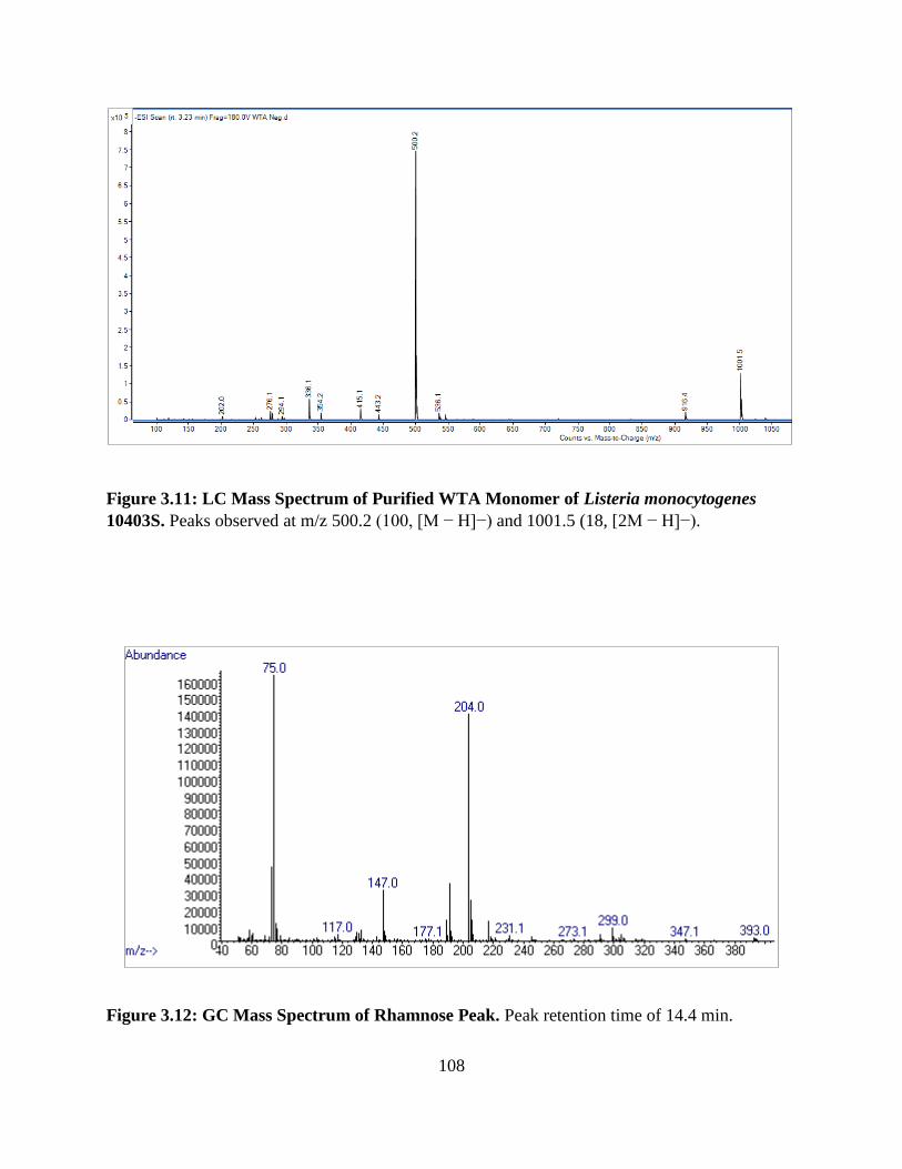
108
Figure 3.11: LC Mass Spectrum of Purified WTA Monomer of Listeria monocytogenes
10403S. Peaks observed at m/z 500.2 (100, [M − H]−) and 1001.5 (18, [2M − H]−).
Figure 3.12: GC Mass Spectrum of Rhamnose Peak. Peak retention time of 14.4 min.
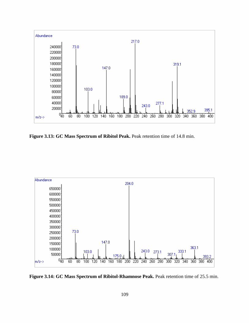
109
Figure 3.13: GC Mass Spectrum of Ribitol Peak. Peak retention time of 14.8 min.
Figure 3.14: GC Mass Spectrum of Ribitol-Rhamnose Peak. Peak retention time of 25.5 min.
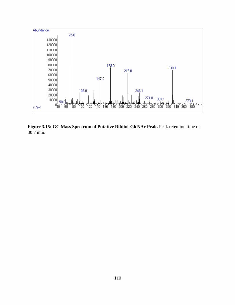
110
Figure 3.15: GC Mass Spectrum of Putative Ribitol-GlcNAc Peak. Peak retention time of
30.7 min.

111
Streamlining the Analysis of Wall Teichoic Acid Monomers
Results of the streamlining process indicate that the peaks for the compounds present in the
purified WTA sample are indeed still detectable in all samples analyzed, including cells that
were only steam-killed (Figures 3.16 – 3.18 below). However, the same amounts of
progressively cruder samples produced progressively weaker intensities of the chromatogram
peaks. Using higher concentrations of cruder cell materials could aid in obtaining stronger
chromatogram peaks for analysis. Additionally, the crudest sample (autoclaved cell pellet)
displays many more peaks earlier in the chromatogram (i.e. before ~40 min), as elements of cell
proteins, nucleic acid material, lipids, and BHI media were not removed from the sample prior to
analysis. There is also a slight shift in retention time for some of the peaks observed in the
chromatograms of the less refined samples, the most significant being the peak at ~41.3 min (as
observed in the purified WTA sample). This is a factor that should be taken into consideration
when analyzing crude cell samples, especially when assessing for the presence of compounds
which elute from the column earlier.

112
Figure 3.16: GC-FID Chromatogram of Crude WTA Monomer of Listeria monocytogenes
10403S. Notable peaks at retention times of approximately 16.2, 18.2, 30.0, and 41.0 min at a
slightly decreased intensity compared to the purified WTA monomer sample.
Figure 3.17: GC-FID Chromatogram of Carbohydrate Fraction of Treated Cells of Listeria
monocytogenes 10403S. Notable peaks at retention times of approximately 16.2, 18.2, 30.0 and
40.9 min at a decreased intensity compared to the purified WTA monomer sample.
16.2 min →
18.2 min
30.0 min
41.0 min →
40.9 min →
16.2 min →
18.2 min
30.0 min

113
Figure 3.18: GC-FID Chromatogram of Autoclaved Cell Pellet of Listeria monocytogenes
10403S. Notable peaks at retention times of approximately 18.2, 30.0, and 40.8 min at a
markedly decreased intensity compared to the purified WTA monomer sample, with many
additional peaks before ~40 min.
18.2
min 40.8 min →
30.0 min

114
Conclusion
Although it is necessary to purify samples of WTA polymers to some extent for their initial
analysis, samples can be successfully “streamlined” in order to more rapidly determine the
presence or absence of specific WTA compounds. Although peaks of interest are still detectable
using an autoclaved cell pellet that has not been further processed, the carbohydrate fraction of
treated cells produced a “cleaner” chromatogram without extensive purification methods. The
ability to analyze a sample using far less time, materials, and equipment will allow for the
analysis of more samples and enable laboratories with less resources to contribute to this work.
Although the results obtained here provide a preliminary analysis of molecules that may be
present in the WTA analyzed, further analysis using synthesized standards of WTA monomers
must be applied to confirm the presence of these predicted structures. This work has further
developed methods for purification and HF hydrolysis of WTA monomers of L. monocytogenes
that provides standards that can be used for preliminary analysis, as well as a methods for the
successful silylation and GC-FID analysis of sugar and amino sugar standards. Further analysis
using synthesized standards can provide information that further supports results obtained
through whole genome sequencing and phage adsorption assays of L. monocytogenes mutant
strains.
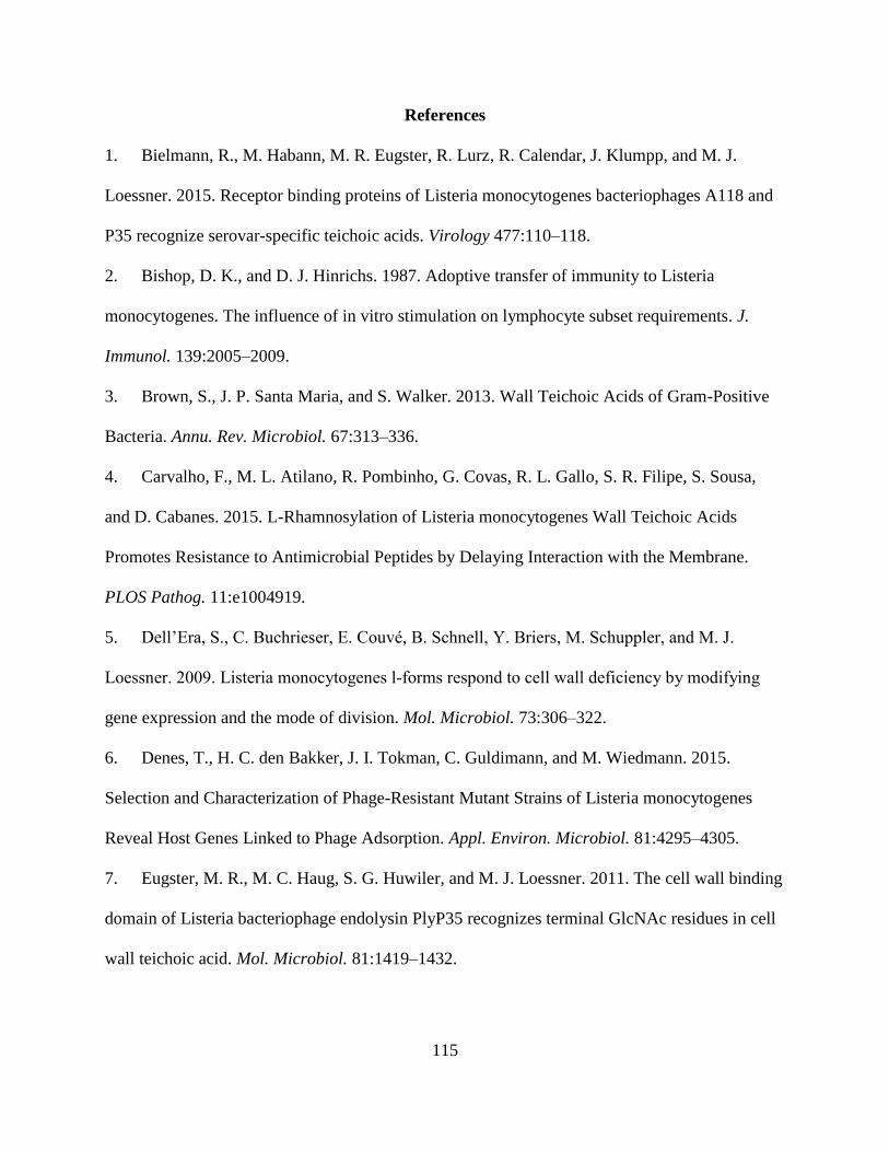
115
References
1. Bielmann, R., M. Habann, M. R. Eugster, R. Lurz, R. Calendar, J. Klumpp, and M. J.
Loessner. 2015. Receptor binding proteins of Listeria monocytogenes bacteriophages A118 and
P35 recognize serovar-specific teichoic acids. Virology 477:110–118.
2. Bishop, D. K., and D. J. Hinrichs. 1987. Adoptive transfer of immunity to Listeria
monocytogenes. The influence of in vitro stimulation on lymphocyte subset requirements. J.
Immunol. 139:2005–2009.
3. Brown, S., J. P. Santa Maria, and S. Walker. 2013. Wall Teichoic Acids of Gram-Positive
Bacteria. Annu. Rev. Microbiol. 67:313–336.
4. Carvalho, F., M. L. Atilano, R. Pombinho, G. Covas, R. L. Gallo, S. R. Filipe, S. Sousa,
and D. Cabanes. 2015. L-Rhamnosylation of Listeria monocytogenes Wall Teichoic Acids
Promotes Resistance to Antimicrobial Peptides by Delaying Interaction with the Membrane.
PLOS Pathog. 11:e1004919.
5. Dell’Era, S., C. Buchrieser, E. Couvé, B. Schnell, Y. Briers, M. Schuppler, and M. J.
Loessner. 2009. Listeria monocytogenes l‐forms respond to cell wall deficiency by modifying
gene expression and the mode of division. Mol. Microbiol. 73:306–322.
6. Denes, T., H. C. den Bakker, J. I. Tokman, C. Guldimann, and M. Wiedmann. 2015.
Selection and Characterization of Phage-Resistant Mutant Strains of Listeria monocytogenes
Reveal Host Genes Linked to Phage Adsorption. Appl. Environ. Microbiol. 81:4295–4305.
7. Eugster, M. R., M. C. Haug, S. G. Huwiler, and M. J. Loessner. 2011. The cell wall binding
domain of Listeria bacteriophage endolysin PlyP35 recognizes terminal GlcNAc residues in cell
wall teichoic acid. Mol. Microbiol. 81:1419–1432.
116
8. Eugster, M. R., and M. J. Loessner. 2011. Rapid Analysis of Listeria monocytogenes Cell
Wall Teichoic Acid Carbohydrates by ESI-MS/MS. PLOS ONE 6:e21500.
9. Eugster, M. R., L. S. Morax, V. J. Hüls, S. G. Huwiler, A. Leclercq, M. Lecuit, and M. J.
Loessner. 2015. Bacteriophage predation promotes serovar diversification in Listeria
monocytogenes. Mol. Microbiol. 97:33–46.
10. Fiedler, F. 1988. Biochemistry of the cell surface of Listeria strains: A locating general
view. Infection 16:S92–S97.
11. Fiedler, F., J. Seger, A. Schrettenbrunner, and H. P. R. Seeliger. 1984. The biochemistry of
murein and cell wall teichoic acids in the genus Listeria. Syst. Appl. Microbiol. 5:360–376.
12. Fujii, H., K. Kamisango, M. Nagaoka, K. Uchikawa, I. Sekikawa, K. Yamamoto, and I.
Azuma. 1985. Structural Study on Teichoic Acids of Listeria monocytogenes Types 4a and 4d. J.
Biochem. (Tokyo) 97:883–891.
13. García, P., L. Rodríguez, A. Rodríguez, and B. Martínez. 2010. Food biopreservation:
promising strategies using bacteriocins, bacteriophages and endolysins. Trends Food Sci.
Technol. 21:373–382.
14. Giraud, M.-F., G. A. Leonard, R. A. Field, C. Berlind, and J. H. Naismith. 2000. RmlC, the
third enzyme of dTDP-L-rhamnose pathway, is a new class of epimerase. Nat. Struct. Biol.
7:398.
15. Hoffman, S., B. Maculloch, and M. Batz. 2015. Economic Burden of Major Foodborne
Illnesses Acquired in the United States No. 1476-2016-120935.
16. Kamisango, K. I., H. Fujii, H. Okumura, I. Saiki, Y. Araki, Y. Yamamura, and I. Azuma.
1983. Structural and Immunochemical Studies of Teichoic Acid of Listeria monocytogenes. J.
Biochem. (Tokyo) 93:1401–1409.

117
17. Kaya, S., Y. Araki, and E. Ito. 1985. Characterization of a novel linkage unit between
ribitol teichoic acid and peptidoglycan in Listeria monocytogenes cell walls. Eur. J. Biochem.
146:517–522.
18. Linnan, M. J., L. Mascola, X. D. Lou, V. Goulet, S. May, C. Salminen, D. W. Hird, M. L.
Yonekura, P. Hayes, R. Weaver, A. Audurier, B. D. Plikaytis, S. L. Fannin, A. Kleks, and C. V.
Broome. 1988. Epidemic Listeriosis Associated with Mexican-Style Cheese. N. Engl. J. Med.
319:823–828.
19. McCollum, J. T., A. B. Cronquist, B. J. Silk, K. A. Jackson, K. A. O’Connor, S. Cosgrove,
J. P. Gossack, S. S. Parachini, N. S. Jain, P. Ettestad, M. Ibraheem, V. Cantu, M. Joshi, T.
DuVernoy, N. W. Fogg, J. R. Gorny, K. M. Mogen, C. Spires, P. Teitell, L. A. Joseph, C. L.
Tarr, M. Imanishi, K. P. Neil, R. V. Tauxe, and B. E. Mahon. 2013. Multistate Outbreak of
Listeriosis Associated with Cantaloupe. N. Engl. J. Med. 369:944–953.
20. Munafo, J. P., A. Ramanathan, L. S. Jimenez, and T. J. Gianfagna. 2010. Isolation and
Structural Determination of Steroidal Glycosides from the Bulbs of Easter Lily (Lilium
longiflorum Thunb.). J. Agric. Food Chem. 58:8806–8813.
21. Orsi, R. H., H. C. den Bakker, and M. Wiedmann. 2011. Listeria monocytogenes lineages:
Genomics, evolution, ecology, and phenotypic characteristics. Int. J. Med. Microbiol. 301:79–96.
22. Reichmann, N. T., and A. Gründling. 2011. Location, synthesis and function of glycolipids
and polyglycerolphosphate lipoteichoic acid in Gram-positive bacteria of the phylum Firmicutes.
FEMS Microbiol. Lett. 319:97–105.
23. Salama, P. J., P. K. B. Embarek, J. Bagaria, and I. S. Fall. 2018. Learning from listeria:
safer food for all. The Lancet 391:2305–2306.

118
24. Scallan, E., R. M. Hoekstra, F. J. Angulo, R. V. Tauxe, M.-A. Widdowson, S. L. Roy, J. L.
Jones, and P. M. Griffin. 2011. Foodborne Illness Acquired in the United States—Major
Pathogens. Emerg. Infect. Dis. 17:7–15.
25. Shen, Y., S. Boulos, E. Sumrall, B. Gerber, A. Julian-Rodero, M. R. Eugster, L. Fieseler, L.
Nyström, M.-O. Ebert, and M. J. Loessner. 2017. Structural and functional diversity in Listeria
cell wall teichoic acids. J. Biol. Chem. 292:17832–17844.
26. Silk, B. J., B. E. Mahon, P. M. Griffin, L. H. Gould, R. V. Tauxe, S. M. Crim, K. A.
Jackson, P. Gerner-Smidt, K. M. Herman, and O. L. Henao. 2013. Vital Signs: Listeria Illnesses,
Deaths, and Outbreaks — United States, 2009–2011. MMWR Morb. Mortal. Wkly. Rep. 62:448–
452.
27. Trudelle, D. M., D. W. Bryan, L. K. Hudson, and T. G. Denes. 2019. Cross-resistance to
phage infection in Listeria monocytogenes serotype 1/2a mutants. Food Microbiol. 84:103239.
28. Uchikawa, K., I. Sekiawa, and I. Azuma. 1986. Structural Studies on Teichoic Acids in Cell
Walls of Several Serotypes of Listeria monocytogenes. J. Biochem. (Tokyo) 99:315–327.
29. Vázquez-Boland, J. A., M. Kuhn, P. Berche, T. Chakraborty, G. Domı́nguez-Bernal, W.
Goebel, B. González-Zorn, J. Wehland, and J. Kreft. 2001. Listeria Pathogenesis and Molecular
Virulence Determinants. Clin. Microbiol. Rev. 14:584–640.
30. Weidenmaier, C., and A. Peschel. 2008. Teichoic acids and related cell-wall glycopolymers
in Gram-positive physiology and host interactions. Nat. Rev. Microbiol. 6:276–287.
31. Wendlinger, G., M. J. Loessner, and S. Scherer. 1996. Bacteriophage receptors on Listeria
monocytogenes cells are the N-acetylglucosamine and rhamnose substituents of teichoic acids or
the peptidoglycan itself. Microbiology 142:985–992.

119
Appendix

120
Table 3.1: Listeria monocytogenes Strains Used in this Study.
Listeria
monocytogenes Strain Features
Reference
or Origin
Wild-type Laboratory
Strain:
10403S Lineage II; 1/2a serotype (GlcNAc and rhamnose in WTA) (2)
Mutant Strains:
FSL D4-0014 10403S mutant; nonsense mutation in LMRG_00541; deficiency of GlcNAc in WTA (6)
FSL D4-0119 10403S mutant; nonsense mutation in LMRG_00542; deficiency of rhamnose in WTA (6)
UTK P1-0001
10403S mutant; frameshift mutation caused by a deletion in LMRG_00544; truncated RmlC
protein (27)

121
Table 3.2: Preparation of the Phosphate Standard Curve.
Concentration
PO4
Volume of Working
Stock (10 mg/L PO4)
Volume of Ultrapure
Water
0 0 5000 µL
0.5 mg/L 250 µL 4750 µL
1.0 mg/L 500 µL 4500 µL
1.5 mg/L 750 µL 4250 µL
2.0 mg/L 1000 µL 4000 µL
2.5 mg/L 1250 µL 3750 µL
3.0 mg/L 1500 µL 3500 µL
3.5 mg/L 1750 µL 3250 µL
4.0 mg/L 2000 µL 3000 µL
4.5 mg/L 2250 µL 2750 µL
5.0 mg/L 2500 µL 2500 µL
A working stock solution of phosphate standard with a concentration of 10 mg/L PO4 was
prepared from a phosphate standard solution with a concentration of 1,000 mg/L PO4. From the
working stock solution, 5 mL phosphate standards were prepared in concentrations ranging from
0 to 5 mg/L PO4. A phosphate test was applied to each standard as well as an ultrapure water
blank as per manufacturers’ instructions. Absorbencies of each standard were read in 10 mm
cuvettes with a spectrophotometer at a wavelength of 690 nm to develop a standard curve.
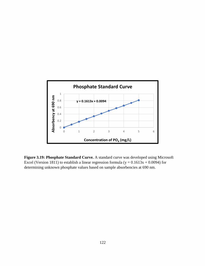
122
Figure 3.19: Phosphate Standard Curve. A standard curve was developed using Microsoft
Excel (Version 1811) to establish a linear regression formula (y = 0.1613x + 0.0094) for
determining unknown phosphate values based on sample absorbencies at 690 nm.
y = 0.1613x + 0.0094
0
0.2
0.4
0.6
0.8
1
0 1 2 3 4 5 6Ab
sorb
ency
at
69
0 n
m
Concentration of PO4 (mg/L)
Phosphate Standard Curve

123
Figure 3.20: Illustration of Trimethyl Silane Molecule.
Figure 3.21: Illustration of Ribitol Molecule (left) and Silylated Ribitol Molecule (right).
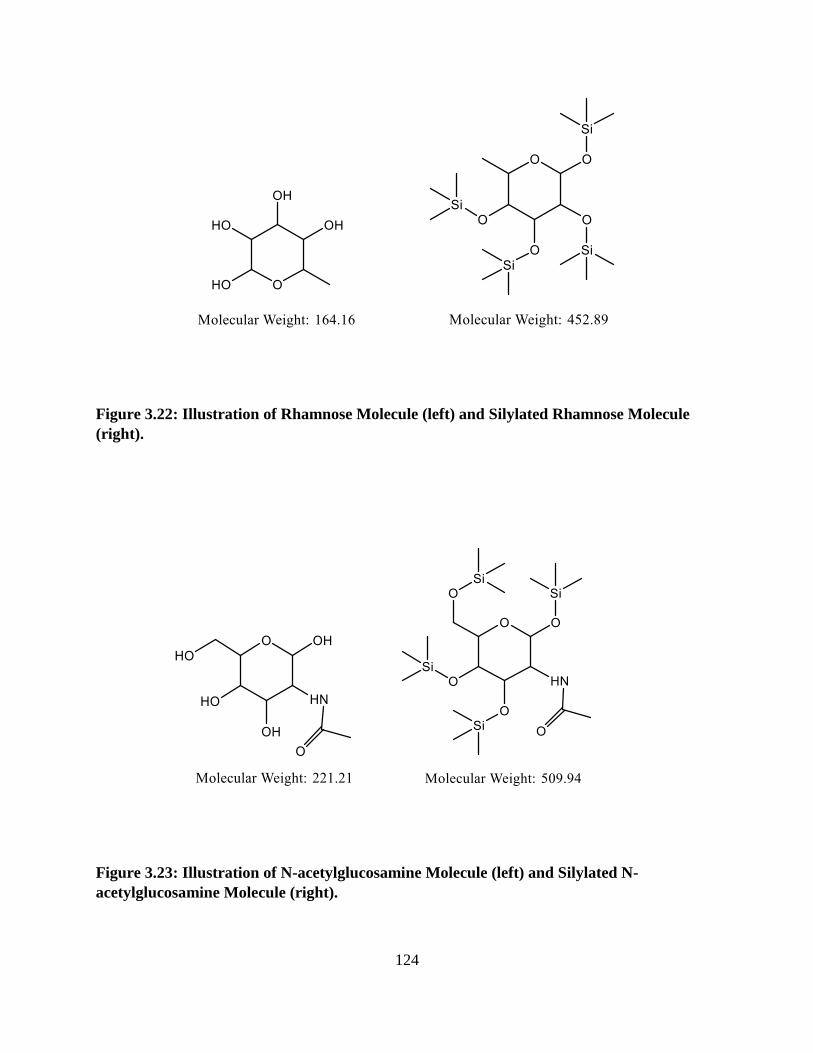
124
Figure 3.22: Illustration of Rhamnose Molecule (left) and Silylated Rhamnose Molecule
(right).
Figure 3.23: Illustration of N-acetylglucosamine Molecule (left) and Silylated N-
acetylglucosamine Molecule (right).
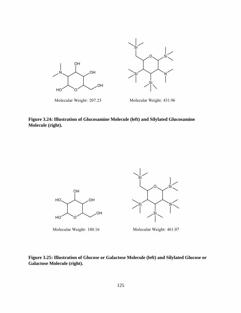
125
Figure 3.24: Illustration of Glucosamine Molecule (left) and Silylated Glucosamine
Molecule (right).
Figure 3.25: Illustration of Glucose or Galactose Molecule (left) and Silylated Glucose or
Galactose Molecule (right).
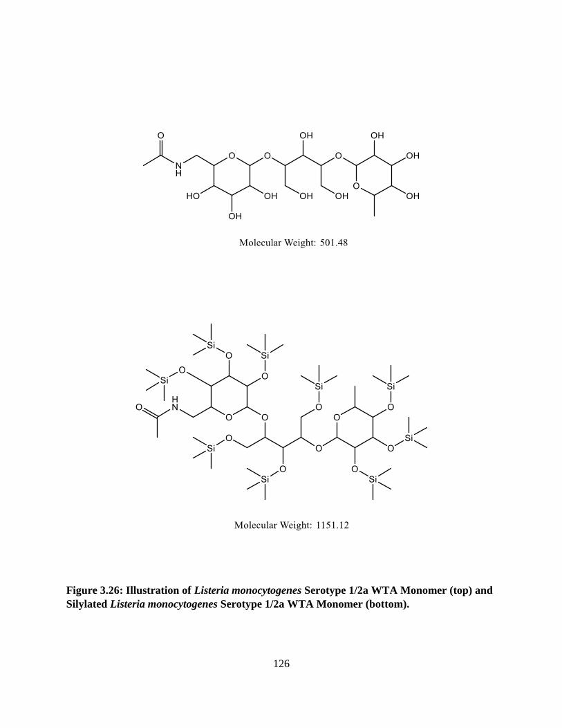
126
Figure 3.26: Illustration of Listeria monocytogenes Serotype 1/2a WTA Monomer (top) and
Silylated Listeria monocytogenes Serotype 1/2a WTA Monomer (bottom).
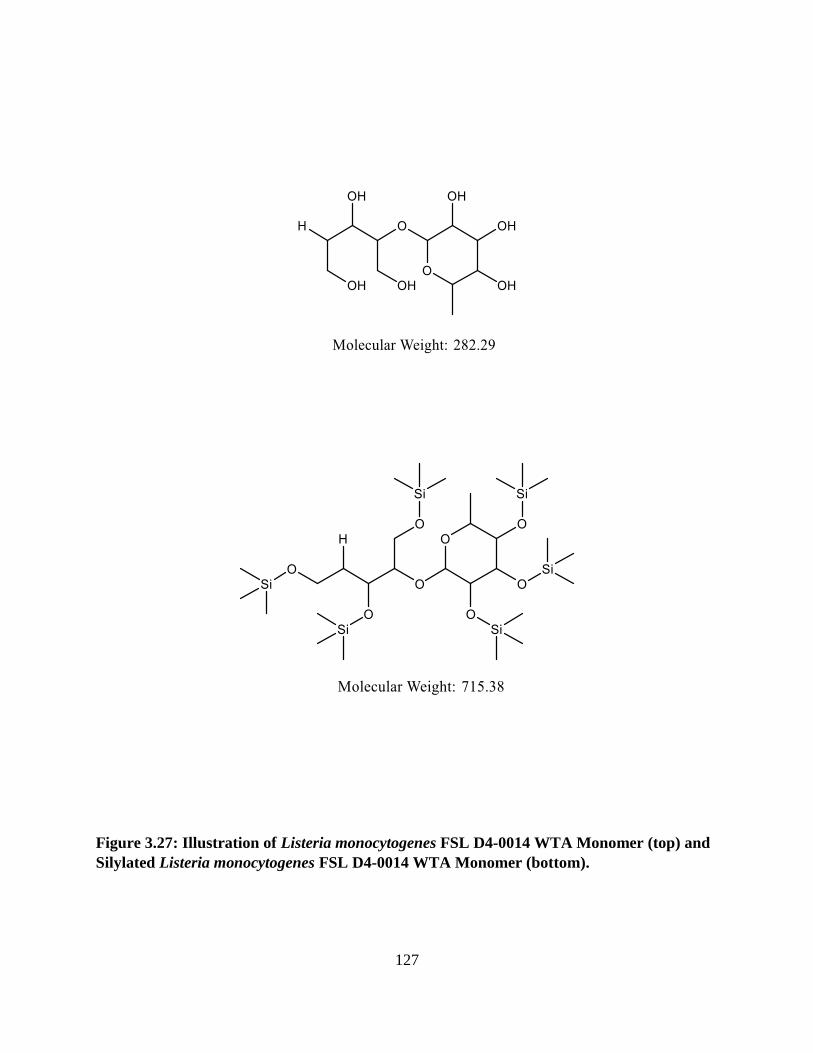
127
Figure 3.27: Illustration of Listeria monocytogenes FSL D4-0014 WTA Monomer (top) and
Silylated Listeria monocytogenes FSL D4-0014 WTA Monomer (bottom).

128
Figure 3.28: Illustration of Listeria monocytogenes FSL D4-0119 WTA Monomer (top) and
Silylated Listeria monocytogenes FSL D4-0119 WTA Monomer (bottom).

129
Chapter IV: Conclusion
The objectives of the studies presented here included determining the efficiency of plaquing
and/or activity of a collection of phages against L. monocytogenes 10403S and three mutants
derived from this strain; FSL D4-0014, FSL D4-0119, and UTK P1-0001, as well as a
preliminary analysis of the WTA structures of all these strains. One bacteriophage was identified
(LP-018) that was capable of infecting all three of the phage resistant mutants of serotype 1/2a L.
monocytogenes. It was concluded that mutations conferring phage-resistance through loss of
rhamnose likely pose the greatest challenge for phage-based biocontrol in serotype 1/2a strains,
as we found that they confer resistance to almost all of the Listeria phages (119/120) in the
diverse collection tested. Additionally, we have provided a visual comparison of the WTA
components of these L. monocytogenes strains via chromatograms obtained using GC-FID
analysis, as well as chromatograms for sugar and amino-sugar standards present in L.
monocytogenes WTA. Although the results obtained here give us a better picture of what types
of molecules may be present in these WTA, a comparison to synthesized standards must be
performed to confirm these predicted compounds. It is necessary to purify samples of WTA
polymers to some extent for their initial analysis, but subsequent unknown samples can be
successfully “streamlined” in order to more rapidly determine the presence or absence of such
previously determined WTA monomer peaks. The chromatograms of the confirmed WTA
monomers can be organized into a reference library, as the retention times of peaks should
remain nearly the same if using the same GC-FID program and column. The ability to analyze a
sample using far less time, materials, and equipment will allow for the analysis of more samples
and enable laboratories with less resources to contribute to this work. These results together
contribute to our understanding of phage-resistance in L. monocytogenes as well as Listeria

130
phage binding receptors. Additionally, they have the potential to aid in the rational design of
Listeria phage cocktails used as antimicrobial agents in food industries that aim to reduce the
emergence of phage-resistance and ensure long-term efficacy.

131
Vita
Danielle Trudelle was born on August 27th, 1986 in Glen Cove, NY. She graduated from Locust
Valley High School in Locust Valley, NY in 2004 to attend SUNY Stony Brook University in
Stony Brook, NY from August 2004 until May 2006. She then transferred to SUNY Plattsburgh
in Plattsburgh, NY and graduated with a Bachelor of Science in Food and Nutrition in May 2009.
After graduation, Danielle worked in the dietary departments of three different long term care
facilities, a laboratory rodent breeding facility, and finally a laboratory that performed analyses
on different food commodities. She then began the M.S. program in the Food Science
department at the University of Tennessee, Knoxville in August of 2017, and is expected to
graduate in August of 2019.