Embed Size (px)
Citation preview
Cronfa - Swansea University Open Access Repository
_____________________________________________________________
This is an author produced version of a paper published in :
Journal of Biological Chemistry
Cronfa URL for this paper:
http://cronfa.swan.ac.uk/Record/cronfa1512
_____________________________________________________________
Paper:
Row, P. (2006). The Ubiquitin Isopeptidase UBPY Regulates Endosomal Ubiquitin Dynamics and Is Essential for
Receptor Down-regulation. Journal of Biological Chemistry, 281(18), 12618-12624.
http://dx.doi.org/10.1074/jbc.M512615200
_____________________________________________________________ This article is brought to you by Swansea University. Any person downloading material is agreeing to abide by the
terms of the repository licence. Authors are personally responsible for adhering to publisher restrictions or conditions.
When uploading content they are required to comply with their publisher agreement and the SHERPA RoMEO
database to judge whether or not it is copyright safe to add this version of the paper to this repository.
http://www.swansea.ac.uk/iss/researchsupport/cronfa-support/
Michael J. Clague and Sylvie UrbéPaula E. Row, Ian A. Prior, John McCullough, Down-regulationand Is Essential for ReceptorRegulates Endosomal Ubiquitin Dynamics The Ubiquitin Isopeptidase UBPYand Biogenesis:Membrane Transport, Structure, Function,
doi: 10.1074/jbc.M512615200 originally published online March 6, 20062006, 281:12618-12624.J. Biol. Chem.
10.1074/jbc.M512615200Access the most updated version of this article at doi:
.JBC Affinity SitesFind articles, minireviews, Reflections and Classics on similar topics on the
Alerts:
When a correction for this article is posted•
When this article is cited•
to choose from all of JBC's e-mail alertsClick here
http://www.jbc.org/content/281/18/12618.full.html#ref-list-1
This article cites 39 references, 19 of which can be accessed free at
at SWA
NSEA
UN
IVERSITY
on September 10, 2014
http://ww
w.jbc.org/
Dow
nloaded from
at SWA
NSEA
UN
IVERSITY
on September 10, 2014
http://ww
w.jbc.org/
Dow
nloaded from
The Ubiquitin Isopeptidase UBPY Regulates EndosomalUbiquitin Dynamics and Is Essential for ReceptorDown-regulation*
Received for publication, November 28, 2005, and in revised form, February 22, 2006 Published, JBC Papers in Press, March 6, 2006, DOI 10.1074/jbc.M512615200
Paula E. Row, Ian A. Prior1, John McCullough, Michael J. Clague, and Sylvie Urbe2
From the Physiological Laboratory, University of Liverpool, Crown Street, L69 3BX Liverpool, United Kingdom
UBPY is a ubiquitin-specific protease that can deubiquitinatemonoubiquitinated receptor tyrosine kinases, as well as processLys-48- and Lys-63-linked polyubiquitin to lower denominationforms in vitro. Catalytically inactive UBPY localizes to endosomes,which accumulate ubiquitinated proteins. We have explored thesequelae of short interfering RNA-mediated knockdown of UBPY.Global levels of ubiquitinated protein increase and ubiquitin accu-mulates on endosomes, although free ubiquitin levels are unchanged.UBPY-depleted cells have more and larger multivesicular endoso-mal structures that are frequently associated through extended con-tact areas, characterized by regularly spaced, electron-dense, bridg-ing profiles. Degradation of acutely stimulated receptor tyrosinekinases, epidermal growth factor receptor and Met, is stronglyinhibited in UBPY knockdown cells suggesting that UBPY functionis essential for growth factor receptor down-regulation. In contrast,stability of the UBPY binding partner STAM is dramatically com-promised in UBPY knockdown cells. The cellular functions ofUBPY are complex but clearly distinct from those of the Lys-63-ubiquitin-specific protease, AMSH, with which it shares a bindingsite on the SH3 domain of STAM.
Activated receptor tyrosine kinases (RTK)3 generally enter the endo-somal system through incorporation into clathrin-coated vesicles anddelivery to a tubulo-vesicular compartment known as the early or sort-ing endosome. From here receptors may recycle to the plasma mem-brane or be selected for lysosomal sorting by incorporation into smallvesicles that bud away from the limiting membrane into the vacuolarlumen to generatemultivesicular bodies (MVBs) (1–3). Activated RTKssuch as epidermal growth factor (EGF) receptor (EGFR), platelet-de-rived growth factor receptor, and Met are multimonoubiquitinatedthrough the action of an E3 ubiquitin ligase, the proto-oncogene c-Cbl(4–7). Ubiquitination provides a sorting signal that is proposed toengage with the MVB sorting machinery through an initial interactionwith the UIM (ubiquitin interacting motif) domain of Hrs (hepatocytegrowth factor receptor tyrosine kinase substrate) (8–10).Classical studies elucidated polyubiquitin chains linked through an
internal lysine (Lys-48) as a proteasomal degradation signal (11–13).
However, there is an increasing appreciation that alternative polyubiq-uitin chain structures play crucial roles in cellular physiology (14, 15)and that ubiquitination is a dynamic post-translational modification,which may come to rival phosphorylation in its scope and complexity.The reversibility of ubiquitination can be attributed to the action ofdeubiquitinating enzymes (DUBs) of which there are an estimated 79encoded in the human genome (16, 17).Two DUBs, associated molecule with the SH3-domain of STAM
(AMSH) and ubiquitin-specific processing protease Y (UBPY) are knownto interact directly with signal transducing adapter molecule (STAM), aprotein that is constitutively associatedwith the endosomal sorting adapterHrs (18, 19). In fact, they share a binding site on the SH3 domain of STAMby virtue of a conserved bindingmotif, PXV/ID/NRXXKP (19). AMSH is amember of the JAMM/MPN! family of metalloproteases (20, 21), whichwe have shown to negatively regulate EGFR sorting to the lysosome asevidencedbyenhancedreceptordegradation followingacute stimulationofAMSHknockdown cells (22). UBPY is a cysteine protease of the ubiquitin-specific processing protease (UBP/USP) family, also known as USP8, andhas been proposed to regulate cellular ubiquitin levels and entry into Sphase (23). The yeast orthologue of UBPY is thought to be Doa4, whichassociateswith late componentsof theMVBsortingmachinery (24, 25) andhas been proposed to recycle ubiquitin from committed receptors (26, 27).In this paper we show that knockdown of UBPY by RNA interference hasmultiple cellular effects that include the accumulationofubiquitinatedpro-teins on endosomes, an increase in both number and size ofmultivesicularendosomes, and a block in RTK degradation.
EXPERIMENTAL PROCEDURES
Plasmids—HumanUBPY cDNAwas obtained from theKazusaDNAResearch Institute, Japan (cloneKIAA0055) and subcloned into pEGFP-C1. The C786S mutation was introduced by QuikChange site-directedmutagenesis (Stratagene) and subcloned into pEGFP-UBPY. A shortinterfering (si)RNA-resistant UBPY construct (UBPY*) was generatedby introducing five degenerate point mutations (underlined below)into UBPY within the region targeted by UBPY-specific siRNA duplex1(forward primer sequence GCCTATGTACTATATATGAAGTAC-GTCACGGTGTACAATCTTATC) and subcloning into pEGFPC1-UBPY. GST-UBPY, HA-STAM, and Flag-ubiquitin constructs weregenerous gifts from Giulio Draetta (Milan, Italy), Naomi Kitamura(Yokohama, pMIW-HA-Hbp), and John O’Bryan (Chicago, IL).
Antibodies and Other Reagents—Mouse monoclonal anti-HA anti-bodywas fromCovance, and anti-ubiquitin antibodies were fromSigma(U5379), Covance (P4G7), and Affiniti-Biomol (FK1, FK2). Anti-GFPwas a gift of Francis Barr (Martinsried, Germany). Mouse monoclonalanti-EGFR R1 and goat polyclonal anti-EGFR 1005 were from SantaCruz. Met antibodies were obtained from Cell Signaling. Anti-lyso-bisphosphatidic acid was a generous gift of Jean Gruenberg (Geneva,Switzerland). Rabbit polyclonal AMSH, Hrs, and STAM antibodies
* This work was supported by a Wellcome Trust Career Development Award (to S. U.).The costs of publication of this article were defrayed in part by the payment of pagecharges. This article must therefore be hereby marked “advertisement” in accordancewith 18 U.S.C. Section 1734 solely to indicate this fact.
1 A Royal Society University Fellow.2 To whom correspondence should be addressed. Tel.: 44-151-794-5432; Fax: 44-151-
794-4434; E-mail: [email protected] The abbreviations used are: RTK, receptor tyrosine kinase; MVB, multivesicular bodies;
EGF, epidermal growth factor; EGFR, EGF receptor; DUB, deubiquitinating enzyme;UBPY, ubiquitin-specific processing protease Y; STAM, signal transducing adaptermolecule; siRNA, short interfering RNA; HA, hemagglutinin; GFP, green fluorescentprotein; BES, 2-[bis(2-hydroxyethyl)amino]ethanesulfonic acid; SH, Src homology; E3,ubiquitin-protein isopeptide ligase.
THE JOURNAL OF BIOLOGICAL CHEMISTRY VOL. 281, NO. 18, pp. 12618 –12624, May 5, 2006© 2006 by The American Society for Biochemistry and Molecular Biology, Inc. Printed in the U.S.A.
12618 JOURNAL OF BIOLOGICAL CHEMISTRY VOLUME 281 • NUMBER 18 • MAY 5, 2006
at SWA
NSEA
UN
IVERSITY
on September 10, 2014
http://ww
w.jbc.org/
Dow
nloaded from
have been previously described (7, 22, 28, 29) Secondary antibodieswerefromMolecular Probes and Sigma. Protein A- andG-agarose were fromSigma.
Bacterial Expression and Purification of Recombinant Proteins—GST-UBPY was expressed in Rosetta (DE3) pLysS cells (Novagen) and batch-purified with glutathione-Sepharose (Pharmacia) according to manufac-turers’ instructions.PurifiedproteinwasdialyzedagainstDUB-assaybuffer.
Deubiquitination Assays—Lys-48-linked tetraubiquitin (250 ng) orLys-63-linked tetraubiquitin chains (250 ng) (Boston Biochem) wereincubated for 2 h at 37 °C in 20!l of DUB-assay buffer (50mMTris/HCl,25 mM KCl, 5 mM MgCl2, 1 mM dithiothreitol), pH 8.3, with 1 !MGST-UBPY. Proteins were resolved on 4–12% NuPAGE gels (Invitro-gen) according to manufacturer’s instructions. Resolved proteins weretransferred to nitrocellulose (0.2 !m), the membrane was boiled for 30min in deionized water, blocked in 0.5% fish skin gelatin, 0.1% Tween 20in phosphate-buffered saline, and probedwith a rabbit antibody to ubiq-uitin (Sigma) followed by ECL-based detection.
Cell Culture and Transfection—HeLa cells were cultured in 5% CO2in Dulbecco’s modified Eagle’s medium supplemented with 10% fetalcalf serum and 1% non-essential amino acids. All tissue culture reagentswere purchased from Invitrogen. HeLa cells were transfected withGeneJuice transfection reagent (Merck Biosciences) and lysed or fixed24 h post-transfection.
Cell Lysis and Immunoprecipitation—Cells were washed twice in ice-cold phosphate-buffered saline and lysed inNonidet P-40 lysis buffer (0.5%Nonidet P-40, 25 mMTris/Cl, pH 7.5, 100 mMNaCl, 50 mMNaF). Lysateswere cleared by centrifugation and incubated with antibodies and ProteinA-orProteinG-agarose for 2hat 4 °C.Alternatively, cellswere lysed in “hotSDS” lysis buffer (1% SDS, 50mMNaF, 1mM EDTA at 110 °C), and lysateswere heated at 110 °C for 10minwith intermittent vortexing, precleared bycentrifugation at 14,000 rpm for 5 min, and diluted with four volumes ofTX100-dilutionbuffer (1.25%TX100, 25mMTris, pH7.5, 125mMNaCl, 50mMNaF) prior to immunoprecipitation.
Detection of Ubiquitinated EGFR—siRNA-treated cells were starvedin serum-free medium for 16 h, stimulated with 100 ng/ml EGF forvarious times, placed on ice, and lysed in radioimmune precipitationassay buffer (10 mM Tris/HCl, pH 7.5, 100 mM NaCl, 1% (w/v) NonidetP-40, 0.1% (w/v) SDS, 1% (w/v) sodium deoxycholate, and 50 mMNaF),supplemented with protease inhibitors, phosphatase inhibitors, and 10mM N-ethylmaleimide. EGFR was immunoprecipitated overnight andimmunoprecipitates were washed in 20 mM Tris/HCl, 150 mM NaCl,and 0.1% Nonidet P-40 and resuspended in sample buffer. Three-quar-ters of each sample was analyzed by immunoblotting with anti-ubiq-uitin, and the remainder was probed in parallel for EGFR, to control foreven loading.
Depletion of Cellular UBPY by siRNA—HeLa cells were treated twiceover 96 h with either control siRNA duplex (nonspecific control VII)or UBPY-specific siRNA duplex1 (sense UGAAAUACGUGACUGUU-UAUU, antisense 5"-PUAAACAGUCACGUAUUUCAUU) or UBPY-specific siRNAduplex 2 (senseGGACAGGACAGUAUAGAUAUU, anti-sense 5"-PUAUCUAUACUGUCCUGUCCUU, Dharmacon, Lafayette,CO) andAMSH-specific siRNAduplex (senseUUACAAAUCUGCUGU-CAUUUU, antisense 5"-PAAUGACAGCAGAUUUGUAAUU, Dharma-con) at 40.8 nM concentration using Oligofectamine in the absence ofserum (Invitrogen). Four hours post-transfection, fetal bovine serum wasadded toa final concentrationof 10%.Alternatively,HeLacellswere treatedtwice over 96 h with siRNA at a concentration of 47.2 nM using a calciumphosphateprotocol. Briefly,mediumwas replacedwith10mlofDulbecco’smodifiedEagle’smedium!10% fetal bovine serum, 500!l of 0.25MCaCl2,and 0.52 nmol of siRNA were premixed and added gradually with gentle
vortexing to 500 !l of BBS (50 mM BES, pH 6.95, 280 mM NaCl, 1.5 mMNa2HP04), incubated for 20min and added dropwise to the cells. The cellswere then incubated at 3% CO2 overnight. The efficiency of UBPY knock-downwas routinely assessedbyWesternblottinganddeterminedmanuallyby counting on a fluorescence microscope the percent of cells showing anaccumulation of ubiquitin on endosomes as shown in Fig. 5E. For thesiRNA-rescue experiment, HeLa cells were treated twice over 96 h witheither control siRNA duplex or UBPY-specific siRNA duplex1. Sixty-sixhours before harvesting, the cells were transfected with pEGFPC1 or pEG-FPC1-siRNAi resistant UBPY (UBPY*). The percent of GFP-labeled cells,which retained EGFR after 4 h of EGF treatmentwas determinedmanuallyby counting on a fluorescence microscope.
Immunofluorescence—Cells were processed 24 h post-transfectionfor immunofluorescence as previously described (8). Secondary anti-bodies used were labeled with Alexa-Fluor 594 or 488. Dual-stainedconfocal images were taken with a Leica confocal SP2 AOBS (HCX PLAPO CS 63.0 # 1.40 oil objective).
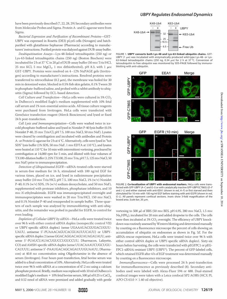
FIGURE 1. UBPY converts both Lys-48 and Lys-63-linked ubiquitin chains. GST-UBPY (1 !M) was incubated with enzymatically produced wild-type Lys-48- or Lys-63-linked tetraubiquitin chains (250 ng, 0.34 !M) for 2 h at 37 °C. Conversion oftetraubiquitin to free ubiquitin was monitored by SDS-PAGE followed by immuno-blotting with anti-ubiquitin.
FIGURE 2. Co-localization of UBPY with endosomal markers. HeLa cells were trans-fected with GFP-UBPY (A–C and G–I) or with catalytically inactive GFP-UBPY(C786S) (D–Fand J–L) and either stained with anti-EEA1 (shown in red, A–F) or first starved and thenstimulated for 10 min with 100 ng/ml EGF before staining with anti-EGFR (shown in red,G–L). All panels represent confocal sections. Insets show 3-fold magnification of theboxed area. Scale bar, 20 !m.
UBPY Regulates Endosomal Dynamics
MAY 5, 2006 • VOLUME 281 • NUMBER 18 JOURNAL OF BIOLOGICAL CHEMISTRY 12619
at SWA
NSEA
UN
IVERSITY
on September 10, 2014
http://ww
w.jbc.org/
Dow
nloaded from
Electron Microscopy—Control and UBPY-depleted HeLa cells wereincubated in media containing 10 mg/ml horseradish peroxidase (Sigma)for 30 min before fixing with 2% glutaraldehyde, 2% paraformaldehyde inphosphatebuffer, pH7.4. Followingcross-linkingofhorseradishperoxidasewith 0.075% of 3,3"diaminobenzidine tetra hydrochloride (Sigma), 0.023%H2O2, cellswerepost-fixedwith2.5%glutaraldehyde, osmicated, stainedenblocwith uranyl acetate and processed for Epon embedding according tostandard procedures. Ultrathin (70 nm) sections were cut and stainedwith lead citrate before viewing in a Tecnai Spirit electron microscope.MVBs were identified morphologically by the presence of internal ves-icles, and their maximal chord lengths were determined using AnalySIS(SIS, GmbH). Gross changes in MVB abundance were measured bycountingMVBs in cells with a coincident nuclear profile and normaliz-ing to the total cell area measured with AnalySIS (scrambled: cellularprofiles counted n$ 22, total area of cells examined$ 3353!m2,MVBscounted n $ 156; UBPY: cellular profiles counted n $ 23, total area ofcells examined area $ 3382 !m2, MVBs counted n $ 230).
RESULTS
Substrate Specificity of UBPY—We have adapted an in vitro assay forDUB activity that monitors the processing of polyubiquitin chains tolower denomination forms (22, 30). In our original analysis, AMSHshowed specificity for Lys-63-linked, whereas UBPY showed specificityfor Lys-48-linked polyubiquitin (22). This observation came with thecaveat that the Lys-63-linked chains available to us at that time werederived frommutant ubiquitin inwhich all lysines, with the exception ofLys-63, had been converted to arginines. We have now repeated theseexperiments with polyubiquitin chains derived from wild-type ubiq-uitin and find that in contrast to AMSH, UBPY shows little discrimina-tion between Lys-48 and Lys-63-linked chains (Fig. 1).
Subcellular Distribution of UBPY—We next analyzed the subcellularlocalization of UBPY and a catalytically inactive mutant UBPY (C786S)(23) in HeLa cells. We analyzed cells expressing low levels of GFP-tagged UBPY and UBPY (C786S) because our UBPY antibody is notsensitive enough to pick up the endogenous protein. In untreated cells,both wild-type and inactive UBPY are localized to the cytosol andplasma membrane (Fig. 2). UBPY (C786S) shows an additional stainingof punctate structures, identified as early endosomes by co-localizationwith anti-EEA1 (Fig. 2, D–F) and with internalized EGFR (J–L). Inter-estingly, wild-type UBPY appears to be recruited to EGFR-containingearly endosomes upon stimulation (Fig. 2, G–I).
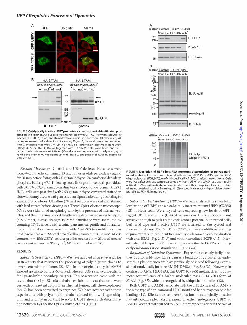
Regulation of Ubiquitin Dynamics—Expression of catalytically inac-tive, but not wild-type, UBPY causes a build up of ubiquitin on endo-somes; a phenomenon we have previously observed following expres-sion of catalytically inactive AMSH (D348A) (Fig. 3A) (22). However, incontrast to AMSH (D348A), this UBPY (C786S) mutant does not pro-mote accumulation of a higher molecular mass (!14 kDa) form ofSTAM (Fig. 3B), which is recognized by ubiquitin antibodies (22).Both UBPY and AMSH associate with the SH3 domain of STAM via
the same type of non-canonical PXXPmotif and hencemay compete forbinding (19). Effects due to overexpression of catalytically inactivemutants could reflect displacement of either endogenous UBPY orAMSH.We therefore turned to RNA interference to address the role of
FIGURE 3. Catalytically inactive UBPY promotes accumulation of ubiquitinated pro-teins on endosomes. A, HeLa cells were transfected with GFP-UBPY or with catalyticallyinactive GFP-UBPY(C786S) and stained with anti-ubiquitin antibodies (shown in red). Allpanels represent confocal sections. Scale bars, 20 !m. B, HeLa cells were co-transfectedwith GFP-tagged wild-type (wt) UBPY or AMSH or catalytically inactive mutant (mut)UBPY(C786S) or AMSH(D348A) together with HA-STAM. Cells were lysed and GFP-tagged proteins immunoprecipitated (IP) and analyzed in parallel with the lysates (right-hand panels) by immunoblotting (IB) with anti-HA antibodies followed by reprobingwith anti-GFP.
FIGURE 4. Depletion of UBPY by siRNA promotes accumulation of polyubiquiti-nated proteins. HeLa cells were treated with control siRNA (Scr), UBPY-specific siRNAoligonucleotides (UO1, UO2), or AMSH-specific siRNA (AO2) or left untreated (None). Cellswere lysed after 96 h, and samples analyzed with anti-UBPY, anti-AMSH, and anti-tubulinantibodies (A), or with anti-ubiquitin antibodies that either recognize all species of ubiq-uitinated proteins including free ubiquitin (B) or specifically react with polyubiquitinatedproteins (C, FK1). IB, immunoblot.
UBPY Regulates Endosomal Dynamics
12620 JOURNAL OF BIOLOGICAL CHEMISTRY VOLUME 281 • NUMBER 18 • MAY 5, 2006
at SWA
NSEA
UN
IVERSITY
on September 10, 2014
http://ww
w.jbc.org/
Dow
nloaded from
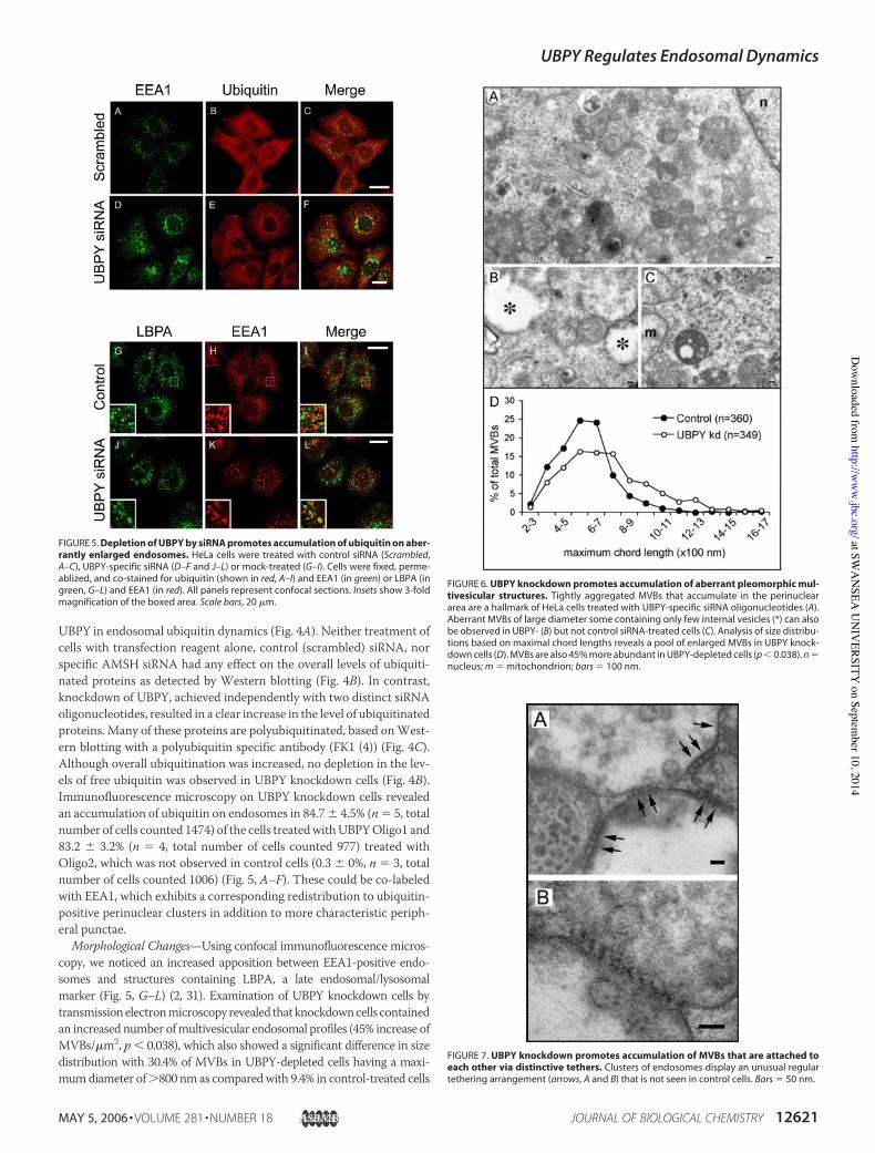
UBPY in endosomal ubiquitin dynamics (Fig. 4A). Neither treatment ofcells with transfection reagent alone, control (scrambled) siRNA, norspecific AMSH siRNA had any effect on the overall levels of ubiquiti-nated proteins as detected by Western blotting (Fig. 4B). In contrast,knockdown of UBPY, achieved independently with two distinct siRNAoligonucleotides, resulted in a clear increase in the level of ubiquitinatedproteins. Many of these proteins are polyubiquitinated, based onWest-ern blotting with a polyubiquitin specific antibody (FK1 (4)) (Fig. 4C).Although overall ubiquitination was increased, no depletion in the lev-els of free ubiquitin was observed in UBPY knockdown cells (Fig. 4B).Immunofluorescence microscopy on UBPY knockdown cells revealedan accumulation of ubiquitin on endosomes in 84.7% 4.5% (n$ 5, totalnumber of cells counted 1474) of the cells treatedwithUBPYOligo1 and83.2 % 3.2% (n $ 4, total number of cells counted 977) treated withOligo2, which was not observed in control cells (0.3 % 0%, n $ 3, totalnumber of cells counted 1006) (Fig. 5, A–F). These could be co-labeledwith EEA1, which exhibits a corresponding redistribution to ubiquitin-positive perinuclear clusters in addition to more characteristic periph-eral punctae.
Morphological Changes—Using confocal immunofluorescence micros-copy, we noticed an increased apposition between EEA1-positive endo-somes and structures containing LBPA, a late endosomal/lysosomalmarker (Fig. 5, G–L) (2, 31). Examination of UBPY knockdown cells bytransmissionelectronmicroscopyrevealed thatknockdowncells containedan increased number ofmultivesicular endosomal profiles (45% increase ofMVBs/!m2, p & 0.038), which also showed a significant difference in sizedistribution with 30.4% of MVBs in UBPY-depleted cells having a maxi-mumdiameter of'800 nm as comparedwith 9.4% in control-treated cells
FIGURE 5. Depletion of UBPY by siRNA promotes accumulation of ubiquitin on aber-rantly enlarged endosomes. HeLa cells were treated with control siRNA (Scrambled,A–C), UBPY-specific siRNA (D–F and J–L) or mock-treated (G–I). Cells were fixed, perme-ablized, and co-stained for ubiquitin (shown in red, A–I) and EEA1 (in green) or LBPA (ingreen, G–L) and EEA1 (in red). All panels represent confocal sections. Insets show 3-foldmagnification of the boxed area. Scale bars, 20 !m.
FIGURE 6. UBPY knockdown promotes accumulation of aberrant pleomorphic mul-tivesicular structures. Tightly aggregated MVBs that accumulate in the perinucleararea are a hallmark of HeLa cells treated with UBPY-specific siRNA oligonucleotides (A).Aberrant MVBs of large diameter some containing only few internal vesicles (*) can alsobe observed in UBPY- (B) but not control siRNA-treated cells (C). Analysis of size distribu-tions based on maximal chord lengths reveals a pool of enlarged MVBs in UBPY knock-down cells (D). MVBs are also 45% more abundant in UBPY-depleted cells (p & 0.038). n $nucleus; m $ mitochondrion; bars $ 100 nm.
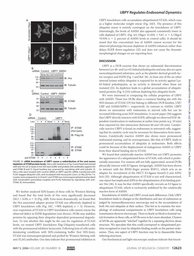
FIGURE 7. UBPY knockdown promotes accumulation of MVBs that are attached toeach other via distinctive tethers. Clusters of endosomes display an unusual regulartethering arrangement (arrows, A and B) that is not seen in control cells. Bars $ 50 nm.
UBPY Regulates Endosomal Dynamics
MAY 5, 2006 • VOLUME 281 • NUMBER 18 JOURNAL OF BIOLOGICAL CHEMISTRY 12621
at SWA
NSEA
UN
IVERSITY
on September 10, 2014
http://ww
w.jbc.org/
Dow
nloaded from
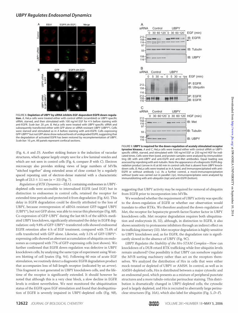
(Fig. 6, A and D). Another striking feature is the induction of vacuolarstructures, which appear largely empty save for a few luminal vesicles andwhich are not seen in control cells (Fig. 6, compare B with C). Electronmicroscopy also provides striking views of large numbers of MVBs“stitched together” along extended areas of close contact by a regularlyspaced repeating unit of electron-dense material with a characteristiclength of 23.3 % 3.1 nm (n $ 33) (Fig. 7).
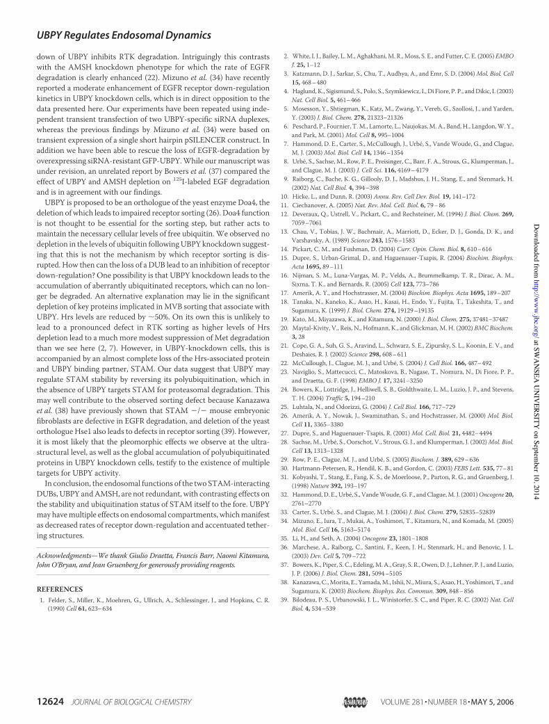
Regulation of RTK Dynamics—EEA1 containing endosomes in UBPY-depleted cells were accessible to internalized EGFR (and EGF) but indistinction to endosomes in control cells, retained the receptor forextended time periods and protected it from degradation (Fig. 8A). Thisdelay in EGFR degradation could be directly attributed to the loss ofUBPY, because overexpression of siRNA-resistant GFP-tagged UBPY(UBPY*), but not GFP alone, was able to rescue this phenotype (Fig. 8B).Co-expression of GFP-UBPY* during the last 66 h of the siRNA-medi-ated UBPY knockdown, significantly attenuated the delay in EGFR deg-radation: only 9.8% of GFP-UBPY*-transfected cells showed endosomalEGFR retention after 4 h of EGF treatment, compared with 75.4% ofcells transfected with GFP alone. Likewise, only 3.1% of GFP-UBPY*-expressing cells showed an aberrant accumulation of ubiquitin on endo-somes as compared with 77% of GFP-expressing cells (not shown). Wefurther confirmed that EGFR down-regulation was defective in UBPYknockdown cells, by analyzing the same type of experiment usingWest-ern blotting of cell lysates (Fig. 9A). Following 60 min of acute EGFstimulation, we routinely detect a diagnostic EGFRdegradation productthat accompanies loss of full-length receptor (indicated by an arrow).This fragment is not generated in UBPY knockdown cells, and the life-time of the receptor is significantly extended. It should however benoted that although this is a very clear block, a slow decline in EGFRlevels is evident nevertheless. We next monitored the ubiquitinationstatus of the EGFR upon EGF stimulation and found that deubiquitina-tion of EGFR is severely impaired in UBPY-depleted cells (Fig. 9B),
suggesting that UBPY activity may be required for removal of ubiquitinfrom EGFR prior to incorporation into MVBs.Wewonderedwhether the requirement of UBPY activity was specific
to the down-regulation of EGFR or whether our observation wouldtranslate to other RTKs. We therefore analyzed the down-regulation ofMet, the receptor for hepatocyte growth factor/Scatter factor in UBPYknockdown cells. Met receptor degradation requires both ubiquitina-tion and endocytosis (6, 32), although, in distinction to EGFR, it alsoshows sensitivity to proteasome inhibitors through an indirect effect onits trafficking itinerary (33).Met receptor degradation is highly sensitiveto UBPY knockdown and, as for EGFR, the degradation rate is signifi-cantly slowed in the absence of UBPY (Fig. 9C).
UBPY Regulates the Stability of the Hrs-STAM Complex—How canknockdown of a DUB retard RTK trafficking while free ubiquitin levelsremain unaltered? One possibility is that UBPY can somehow regulatethe MVB sorting machinery rather than act on the receptors them-selves. We analyzed the distribution of Hrs in cells that were eithermock-treated or depleted of UBPY or AMSH. In control, as well as inAMSH-depleted cells, Hrs is distributed between a major cytosolic andan endosomal pool, which presents as a mixture of peripheral punctatestructures and a more tubulo-reticular perinuclear staining. This distri-bution is dramatically changed in UBPY-depleted cells; the cytosolicpool is largely depleted, and Hrs is recruited to aberrantly large perinu-clear structures (Fig. 10A), which also label for ubiquitin (Fig. 5, D–F).
FIGURE 8. Depletion of UBPY by siRNA inhibits EGF-dependent EGFR down-regula-tion. A, HeLa cells were treated either with control siRNA (scrambled) or UBPY-specificsiRNA, starved, and then stimulated with 100 ng/ml EGF for 4 h before staining withanti-EGFR. Scale bar: 20 !m. B, HeLa cells were treated with UBPY-specific siRNA andsubsequently transfected either with GFP alone or siRNA-resistant UBPY (UBPY*). Cellswere starved and stimulated as in A before staining with anti-EGFR. Cells expressingGFP-UBPY* but not GFP alone show reduced levels of undegraded EGFR, suggesting thatthe degradation of activated EGFR has been restored by recomplementation of UBPY.Scale bar: 10 !m. All panels represent confocal sections.
FIGURE 9. UBPY is required for the down-regulation of acutely stimulated receptortyrosine kinases. A and C, HeLa cells were treated either with control siRNA or UBPY-specific siRNA, starved, and stimulated with 100 ng/ml EGF or 250 ng/ml HGF for indi-cated times. Cells were then lysed, and protein samples were analyzed by immunoblot-ting (IB) with anti-UBPY and anti-EGFR and anti-Met antibodies. Equal loading wasassessed by reprobing with anti-tubulin. Note the appearance of a diagnostic EGFR deg-radation product (arrow in A) at 60 min in control cells that is absent from UBPY knock-down cells. B, HeLa cells were treated as in A, lysed, and immunoprecipitated with anti-EGFR or without antibody (-"). As a further control, a mock-immunoprecipitationwithout lysate was carried out in parallel (-lys). Immunoprecipitates were analyzed byimmunoblotting with anti-ubiquitin (top) and anti-EGFR (bottom).
UBPY Regulates Endosomal Dynamics
12622 JOURNAL OF BIOLOGICAL CHEMISTRY VOLUME 281 • NUMBER 18 • MAY 5, 2006
at SWA
NSEA
UN
IVERSITY
on September 10, 2014
http://ww
w.jbc.org/
Dow
nloaded from
We further analyzed SDS-lysates of these cells by Western blottingand found that the total levels of Hrs were significantly decreased(50.2 % 6.0%, n $ 3) (Fig. 10B). Even more dramatically, we found thatthe Hrs-associated adapter protein STAM was effectively depleted inUBPY knockdown cells (Fig. 10C, '90% depleted, n $ 3). However,over-expression of STAM in UBPY knockdown cells cannot rescue theobserved defect in EGFR degradation (not shown). DUBs may stabilizeproteins by opposing their ubiquitin-dependent proteasomal degrada-tion. To test whether this might be the case for regulation of STAMlevels, we treated UBPY knockdown Flag-Ubiquitin-transfected cellswith the proteasomal inhibitor lactacystin. Following lysis of cells underdenaturing conditions with SDS-containing buffer (hot SDS-lysis),STAM was immunoprecipitated and probed by Western blotting withanti-FLAG antibodies. Our data indicate that proteasomal inhibition in
UBPY knockdown cells accumulates ubiquitinated STAM, which runsas a higher molecular weight smear (Fig. 10D). The presence of thisubiquitin smear is entirely contingent on the knockdown of UBPY.Interestingly, the levels of AMSH also appeared consistently lower incells depleted of UBPY, (Fig. 4A; Oligo1 51.03% % 9.67, n $ 3; Oligo270.93% n $ 2, percent of AMSH levels in control cells). It should benoted that this concomitant loss of AMSH cannot account for theobserved phenotype because depletion of AMSH enhances rather thandelays EGFR down-regulation (22) and does not cause the dramaticmorphological changes we are reporting here.
DISCUSSION
UBPY is a DUB enzyme that shows no substantial discriminationbetweenLys-48- andLys-63-linked polyubiquitin andmay also act uponmonoubiquitinated substrates, such as the platelet-derived growth fac-tor receptor and EGFR (Fig. 1 and Ref. 34). At least one of the six otherinternal lysines within ubiquitin is required for its activity against Lys-63-linked polyubiquitin, as no activity is detected when these aremutated (22). Its depletion leads to a global accumulation of ubiquiti-nated proteins (Fig. 4 (23)) without depleting free ubiquitin levels.We were interested in comparing the cellular properties of UBPY
with AMSH. These two DUBs share a common binding site with theSH3 domain of STAM (19) but belong to different DUB families, USP/UBP and JAMM/MPN!, respectively. In contrast to AMSH, UBPYshows no association with endosomes in starved cells but can berecruited following acute EGF stimulation. A recent paper (34) suggeststhat UBPY directly interacts with EGFR, althoughwe observed EGF-de-pendent translocation to endosomes at earlier time points (e.g. 10 min)than reported for this interaction (between 60 and 120 min). Catalyti-cally inactive UBPY is found on endosomes in untreated cells, suggest-ing that its catalytic cycle may be necessary for dissociation frommem-branes. Catalytically inactive AMSH also shows more pronouncedendosomal staining, and its overexpression, like that of UBPY, leads topronounced accumulation of ubiquitin at endosomes. Both effectscould be because of the displacement of endogenous AMSH or UBPYfrom their shared binding site to STAM.We found that catalytically inactive AMSH but not UBPY promotes
the appearance of a ubiquitinated form of STAM, with which it prefer-entially associates. For reasons still not fully appreciated, several DUBsphysically interact with E3 ligases. Intriguingly, AMSH has been shownto interact with the RING-finger protein RNF11, which acts as anadapter for recruitment of the HECT E3-ligases Smurf1/2 and AIP4/Itch (35). Although ubiquitination of STAM is not well characterized,one report has implicated AIP4 in the ubiquitination of its binding part-ner Hrs (36). It may be that AMSH specifically recruits an E3 ligase toubiquitinate STAM, which is exclusively stabilized by the catalyticallyinactive form of AMSH.Knockdowns of AMSH and UBPY reveal stark differences. Only UBPY
knockdown leads to changes in the distribution and size of endosomes asjudged by immunofluorescence microscopy and to the accumulation ofboth Hrs and ubiquitin at their surface. This led us to examine the mor-phology of endosomal compartments in UBPY knockdown cells usingtransmission electronmicroscopy. There is clearly no block to luminal ves-icle formation in these cells, asMVBswere in factmore abundant. Clustersof MVBs are apparently “stitched” together by a repeating electron densestructure. We speculate that this could correspond to ubiquitinated pro-teins recognized in trans by ubiquitin binding motifs on the partner endo-somes. Thus, one aspect of UBPY function may be to disassemble thesetethering structures.Our biochemical and light microscopic analyses indicate that knock-
FIGURE 10. siRNA knockdown of UBPY causes a redistribution of Hrs and severedepletion of STAM protein levels. HeLa cells treated as in Fig. 4 were fixed and stainedwith anti-Hrs (A) or lysed in hot SDS-lysis buffer and probed with antibodies against Hrsand STAM (B and C). Equal loading was assessed by reprobing with anti-tubulin. D,HeLa cells were treated with control siRNA or UBPY-specific siRNA, transfected withFLAG-tagged ubiquitin (Ub), and incubated with lactacystin (Lact.) or Me2SO for 7 h.Lysates were prepared as in B and C and STAM was immunoprecipitated and probedwith horseradish peroxidase-coupled anti-FLAG followed by reprobing with anti-STAM. IB, immunoblot.
UBPY Regulates Endosomal Dynamics
MAY 5, 2006 • VOLUME 281 • NUMBER 18 JOURNAL OF BIOLOGICAL CHEMISTRY 12623
at SWA
NSEA
UN
IVERSITY
on September 10, 2014
http://ww
w.jbc.org/
Dow
nloaded from
down of UBPY inhibits RTK degradation. Intriguingly this contrastswith the AMSH knockdown phenotype for which the rate of EGFRdegradation is clearly enhanced (22). Mizuno et al. (34) have recentlyreported a moderate enhancement of EGFR receptor down-regulationkinetics in UBPY knockdown cells, which is in direct opposition to thedata presented here. Our experiments have been repeated using inde-pendent transient transfection of two UBPY-specific siRNA duplexes,whereas the previous findings by Mizuno et al. (34) were based ontransient expression of a single short hairpin pSILENCER construct. Inaddition we have been able to rescue the loss of EGFR-degradation byoverexpressing siRNA-resistant GFP-UBPY.While ourmanuscript wasunder revision, an unrelated report by Bowers et al. (37) compared theeffect of UBPY and AMSH depletion on 125I-labeled EGF degradationand is in agreement with our findings.UBPY is proposed to be an orthologue of the yeast enzyme Doa4, the
deletion of which leads to impaired receptor sorting (26). Doa4 functionis not thought to be essential for the sorting step, but rather acts tomaintain the necessary cellular levels of free ubiquitin. We observed nodepletion in the levels of ubiquitin followingUBPY knockdown suggest-ing that this is not the mechanism by which receptor sorting is dis-rupted. How then can the loss of a DUB lead to an inhibition of receptordown-regulation? One possibility is that UBPY knockdown leads to theaccumulation of aberrantly ubiquitinated receptors, which can no lon-ger be degraded. An alternative explanation may lie in the significantdepletion of key proteins implicated inMVB sorting that associate withUBPY. Hrs levels are reduced by (50%. On its own this is unlikely tolead to a pronounced defect in RTK sorting as higher levels of Hrsdepletion lead to a much more modest suppression of Met degradationthan we see here (2, 7). However, in UBPY-knockdown cells, this isaccompanied by an almost complete loss of the Hrs-associated proteinand UBPY binding partner, STAM. Our data suggest that UBPY mayregulate STAM stability by reversing its polyubiquitination, which inthe absence of UBPY targets STAM for proteasomal degradation. Thismay well contribute to the observed sorting defect because Kanazawaet al. (38) have previously shown that STAM )/) mouse embryonicfibroblasts are defective in EGFR degradation, and deletion of the yeastorthologue Hse1 also leads to defects in receptor sorting (39). However,it is most likely that the pleomorphic effects we observe at the ultra-structural level, as well as the global accumulation of polyubiquitinatedproteins in UBPY knockdown cells, testify to the existence of multipletargets for UBPY activity.In conclusion, the endosomal functions of the two STAM-interacting
DUBs, UBPY andAMSH, are not redundant, with contrasting effects onthe stability and ubiquitination status of STAM itself to the fore. UBPYmay havemultiple effects on endosomal compartments, whichmanifestas decreased rates of receptor down-regulation and accentuated tether-ing structures.
Acknowledgments—We thank Giulio Draetta, Francis Barr, Naomi Kitamura,John O’Bryan, and Jean Gruenberg for generously providing reagents.
REFERENCES1. Felder, S., Miller, K., Moehren, G., Ullrich, A., Schlessinger, J., and Hopkins, C. R.
(1990) Cell 61, 623–634
2. White, I. J., Bailey, L.M., Aghakhani,M. R.,Moss, S. E., and Futter, C. E. (2005) EMBOJ. 25, 1–12
3. Katzmann, D. J., Sarkar, S., Chu, T., Audhya, A., and Emr, S. D. (2004)Mol. Biol. Cell15, 468–480
4. Haglund, K., Sigismund, S., Polo, S., Szymkiewicz, I., Di Fiore, P. P., andDikic, I. (2003)Nat. Cell Biol. 5, 461–466
5. Mosesson, Y., Shtiegman, K., Katz, M., Zwang, Y., Vereb, G., Szollosi, J., and Yarden,Y. (2003) J. Biol. Chem. 278, 21323–21326
6. Peschard, P., Fournier, T.M., Lamorte, L., Naujokas,M. A., Band, H., Langdon,W. Y.,and Park, M. (2001)Mol. Cell 8, 995–1004
7. Hammond, D. E., Carter, S., McCullough, J., Urbe, S., VandeWoude, G., and Clague,M. J. (2003)Mol. Biol. Cell 14, 1346–1354
8. Urbe, S., Sachse, M., Row, P. E., Preisinger, C., Barr, F. A., Strous, G., Klumperman, J.,and Clague, M. J. (2003) J. Cell Sci. 116, 4169–4179
9. Raiborg, C., Bache, K. G., Gillooly, D. J., Madshus, I. H., Stang, E., and Stenmark, H.(2002) Nat. Cell Biol. 4, 394–398
10. Hicke, L., and Dunn, R. (2003) Annu. Rev. Cell Dev. Biol. 19, 141–17211. Ciechanover, A. (2005) Nat. Rev. Mol. Cell. Biol. 6, 79–8612. Deveraux, Q., Ustrell, V., Pickart, C., and Rechsteiner, M. (1994) J. Biol. Chem. 269,
7059–706113. Chau, V., Tobias, J. W., Bachmair, A., Marriott, D., Ecker, D. J., Gonda, D. K., and
Varshavsky, A. (1989) Science 243, 1576–158314. Pickart, C. M., and Fushman, D. (2004) Curr. Opin. Chem. Biol. 8, 610–61615. Dupre, S., Urban-Grimal, D., and Haguenauer-Tsapis, R. (2004) Biochim. Biophys.
Acta 1695, 89–11116. Nijman, S. M., Luna-Vargas, M. P., Velds, A., Brummelkamp, T. R., Dirac, A. M.,
Sixma, T. K., and Bernards, R. (2005) Cell 123, 773–78617. Amerik, A. Y., and Hochstrasser, M. (2004) Biochim. Biophys. Acta 1695, 189–20718. Tanaka, N., Kaneko, K., Asao, H., Kasai, H., Endo, Y., Fujita, T., Takeshita, T., and
Sugamura, K. (1999) J. Biol. Chem. 274, 19129–1913519. Kato, M., Miyazawa, K., and Kitamura, N. (2000) J. Biol. Chem. 275, 37481–3748720. Maytal-Kivity, V., Reis, N., Hofmann, K., and Glickman, M. H. (2002) BMC Biochem.
3, 2821. Cope, G. A., Suh, G. S., Aravind, L., Schwarz, S. E., Zipursky, S. L., Koonin, E. V., and
Deshaies, R. J. (2002) Science 298, 608–61122. McCullough, J., Clague, M. J., and Urbe, S. (2004) J. Cell Biol. 166, 487–49223. Naviglio, S., Mattecucci, C., Matoskova, B., Nagase, T., Nomura, N., Di Fiore, P. P.,
and Draetta, G. F. (1998) EMBO J. 17, 3241–325024. Bowers, K., Lottridge, J., Helliwell, S. B., Goldthwaite, L. M., Luzio, J. P., and Stevens,
T. H. (2004) Traffic 5, 194–21025. Luhtala, N., and Odorizzi, G. (2004) J. Cell Biol. 166, 717–72926. Amerik, A. Y., Nowak, J., Swaminathan, S., and Hochstrasser, M. (2000) Mol. Biol.
Cell 11, 3365–338027. Dupre, S., and Haguenauer-Tsapis, R. (2001)Mol. Cell. Biol. 21, 4482–449428. Sachse, M., Urbe, S., Oorschot, V., Strous, G. J., and Klumperman, J. (2002)Mol. Biol.
Cell 13, 1313–132829. Row, P. E., Clague, M. J., and Urbe, S. (2005) Biochem. J. 389, 629–63630. Hartmann-Petersen, R., Hendil, K. B., and Gordon, C. (2003) FEBS Lett. 535, 77–8131. Kobyashi, T., Stang, E., Fang, K. S., de Moerloose, P., Parton, R. G., and Gruenberg, J.
(1998) Nature 392, 193–19732. Hammond,D. E., Urbe, S., VandeWoude,G. F., andClague,M. J. (2001)Oncogene 20,
2761–277033. Carter, S., Urbe, S., and Clague, M. J. (2004) J. Biol. Chem. 279, 52835–5283934. Mizuno, E., Iura, T., Mukai, A., Yoshimori, T., Kitamura, N., and Komada, M. (2005)
Mol. Biol. Cell 16, 5163–517435. Li, H., and Seth, A. (2004) Oncogene 23, 1801–180836. Marchese, A., Raiborg, C., Santini, F., Keen, J. H., Stenmark, H., and Benovic, J. L.
(2003) Dev. Cell 5, 709–72237. Bowers, K., Piper, S. C., Edeling,M.A., Gray, S. R., Owen,D. J., Lehner, P. J., and Luzio,
J. P. (2006) J. Biol. Chem. 281, 5094–510538. Kanazawa,C.,Morita, E., Yamada,M., Ishii, N.,Miura, S., Asao,H., Yoshimori, T., and
Sugamura, K. (2003) Biochem. Biophys. Res. Commun. 309, 848–85639. Bilodeau, P. S., Urbanowski, J. L., Winistorfer, S. C., and Piper, R. C. (2002) Nat. Cell
Biol. 4, 534–539
UBPY Regulates Endosomal Dynamics
12624 JOURNAL OF BIOLOGICAL CHEMISTRY VOLUME 281 • NUMBER 18 • MAY 5, 2006
at SWA
NSEA
UN
IVERSITY
on September 10, 2014
http://ww
w.jbc.org/
Dow
nloaded from