Embed Size (px)
Citation preview
CROHN’S DISEASE (Regional Enteritis)
Alexander Mathew, M.D.
Colon & Rectal Surgery
CROHN’S DISEASE
• Disease of unknown etiology characterized by transmural inflammation of gastrointestinal tract (primarily ileal and cecum) – Transmural inflammation (full thickness)
– Any portion of gastrointestinal tract (from mouth to anus)
– Discontinous involvement (Skip lesions)
– Intermittent disease activity
– Variety of phenotypes • Inflammatory
• Stricturing
• Fistulizing/penetrating
CROHN’S DISEASE
• Initially described by Lesniowski in 1904
• Subsequently described by Crohn, Ginzburg, and Oppenheimer in 1932
EPIDEMIOLOGY
• Affects 1-10 people per 100,000 population in North America and Northern Europe (400 000-600 000 people)
• Peak age of onset at 15-30 years with a lesser peak at 55-80 years
• Female slightly > male • Significant morbidity in pts with CD compared to
general population (up to 15% rendered incapable of working after 10-20 yrs of diagnosis)
ETIOLOGY
• Interactions between environmental, microbial, and immunologic factors in genetically susceptible hosts
• Smoking—increased relative risk (2 times)
• Oral contraception—increased relative risk
• Infection
– Mycobacterium avium paratuberculosis
– Measles virus
ETIOLOGY
• Genetic – Mutations within NOD2/CARD15 gene of
chromosome 16 (expressed on monocytes; role in apoptosis and activation to bacterial LPS) • Younger age at disease diagnosis
• Ileal disease location
• Ileocecal resections
• Higher risk of postoperative recurrence and reoperation
PATHOGENESIS
• Normally, gut suppresses inflammatory immune response to stream of microbial antigens
• In IBD, this suppression is lost
• Increased mucosal permeability (? Primary or secondary)
• Cell-mediated response predominant (excessive activation of CD4 T cells)
• Cytokine release (IL-1, IL-2, IL-6, TNF)
• Inability to induce T cell suppressor function
• Imbalance between proinflammatory and anti-inflammatory cytokines
DISTRIBUTION
• Ileocecum (50%)
• Ileum (30%)
• Colon (20%)
• Rectum and anus
• Esophagus, stomach, and duodenum
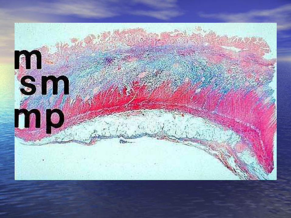
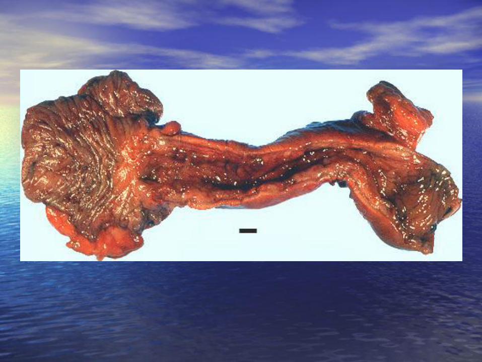
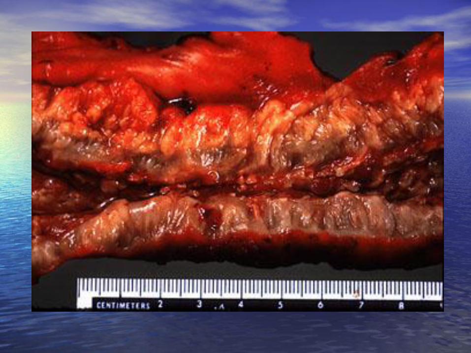
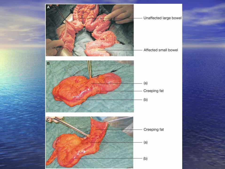
PATHOLOGY • Deep tissue noncaseating granulomas • Intralymphatic granulomas • Granulomatous vasculitis • Stiff thick-walled segment of bowel with “creeping fat”
(wrapping of mesenteric fat circumferentially) • Deep narrow linear ulcers with intervening islands of
edematous mucosa (mucosal cobblestoning) • Aphthous ulcers (developing on surface of submucosal
lymphoid nodules; earliest macroscopic lesion) • Inflammatory pseudopolyps • Enlarged mesenteric lymph nodes (non-caseating, non-
matted) • Single or multiple colonic or enteric strictures • Corkscrew appearance of serosal vessels
PATHOLOGY
• Sometimes difficult to differentiate between Crohn’s colitis and ulcerative colitis
• Indeterminate colitis in 5-10% of pts with colonic involvement alone
• Subsequent disease behavior
• Consideration of macroscopic, microscopic, radiologic, and endoscopic features together with history and clinical picture
CLINICAL COURSE
• Location of inflammation an important determinant of the type of complications
– Stricturing disease more likely in pts with small bowel involvement
– Fistulizing disease more likely in pts with ileal or perianal involvement
SYMPTOMS
• Diarrhea (70-90%) • Abdominal pain (45-65%) • Rectal bleeding (30%) • Perianal disease • Tenesmus • Fecal frequency • Weight loss • Extraintestinal manifestations (ocular,
hepatobiliary, skin, joints)
CLINICAL COURSE
• Unpredictable phases of disease activity and quiescence – 75% chronic intermittent disease
– 10% chronically active disease
– 15% remain asymptomatic
• 50% of patients will develop complications in the form of stricturing (stenosis) or perforating (fistula, abscess) disease
DIAGNOSIS
• Barium enema/small bowel follow through/ CT scan
• Endoscopy with mucosal biopsy (sigmoidoscopy, colonoscopy)
• Serologic markers (ASCA, anti-CBir1, anti-OmpC)
• MRI/Endoanal ultrasound for evaluation of perianal disease
MEDICAL VS SURGICAL THERAPY
• Complementary therapies. Careful use of medical therapy appropriately combined with surgical therapy
MEDICAL THERAPY
• Probiotics (Lactobacillus)
• Aminosalicylates (sulfasalazine, mesalamine)
• Corticosteroids (prednisone, budesonide)
• Antibiotics (metronidazole, ciprofloxacin, rifaximin)
• Immunomodulators – Azathioprine/6-Mercaptopurine
– Methotrexate
– Cyclosporine
• Biologic therapy – Infliximab (Remicade)—chimeric monoclonal anti-TNF antibody
– Adalimumab (Humira)—recombinant human monoclonal anti-TNF antibody
PRINICIPLES OF SURGICAL THERAPY
• Gut-wide disease • Ensure maximal medical therapy prior to surgical
intervention • Maximize nutritional state • Control local sepsis • Resection of least amount of bowel to re-establish
satisfactory intestinal function • Microscopic disease at resection margin does not
influence recurrence • No evidence that anastomotic technique affects
recurrence
SURGICAL THERAPY Indications
• Side effects of medical therapy
• Failure of medical therapy or steroid dependency
• Obstruction
• Symptomatic fistulas
• Abscess formation
• Hemorrhage
• Growth retardation
• Carcinoma or dysplasia
• Extraintestinal manifestations
OPERATIONS
• Resection with or without anastomosis – Ileocecal resection
– Small bowel resection
– Segmental colonic resection
– Total abdominal colectomy with ileorectal anastomosis
– Proctocolectomy with end ileostomy
– Restorative proctocolectomy (IPAA) in carefully selected patients
• Strictureplasty
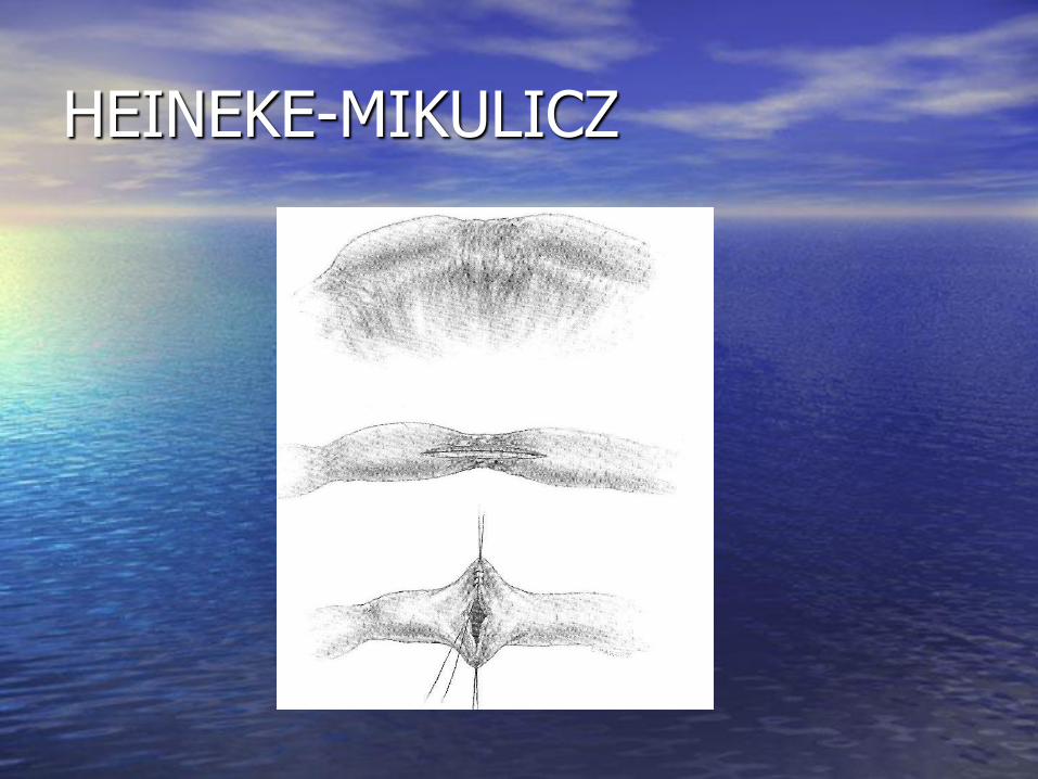
HEINEKE-MIKULICZ
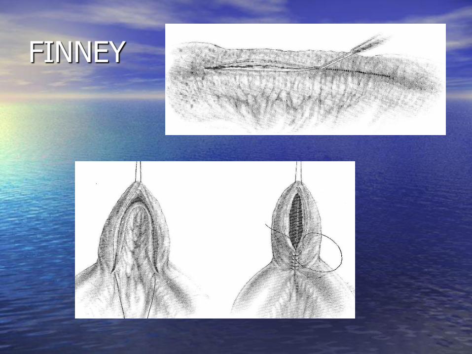
FINNEY

END