Embed Size (px)
Citation preview
471
REVIEWS
Anaesthesiology Intensive Therapy2015, vol. 47, no 5, 471–481
ISSN 0209–1712 10.5603/AIT.a2015.0072
www.ait.viamedica.pl
Critical care ultrasound in cardiac arrest. Technological requirements for performing the SESAME-protocol — a holistic approach
Daniel Lichtenstein1, Manu L.N.G. Malbrain2
1Service de Réanimation Médicale Hôpital Ambroise-Paré, Paris-West University, France 2Intensive Care Unit and High Care Burn Unit, Ziekenhuisnetwerk Antwerpen, ZNA Stuivenberg,
Antwerp, Belgium
Abstract
The use of ultrasound has gained its place in critical care as part of our day-to-day monitoring tools. A better under-standing of ultrasound techniques and recent publications including protocols for the lungs, the abdomen and the blood vessels has introduced ultrasound to the bedside of our ICU patients. However, we will prove in this paper that early machines, dating back more than 25 years, were perfectly able to do the job as compared to modern laptop machines with more features but few additional advantages. Ultrasound is not only a diagnostic tool, but should also be seen as an extension of the traditional physical examination. This paper will focus on the use of the SESAME-protocol in cardiac arrest. The SESAME-protocol suggests starting with a lung scan to rule out possible causes leading to cardiac arrest. Firstly, pneumothorax needs to be ruled out. Secondly, a partial diagnosis of pulmonary embolism is done following the BLUE-protocol. Thirdly, fluid therapy can be guided, following the FALLS-protocol. The SESAME-protocol continues by scanning the lower femoral veins to check for signs of deep venous thrombosis, followed by (or before, in case of trauma) the abdomen to detect massive bleeding. Next comes the pericardium, to exclude pericardial tamponade. Finally, a transthoracic cardiac ultrasound is performed to check for other (cardiac) causes leading to cardiac arrest. The emphasis is on a holistic approach, where ultrasound can be seen as the modern stethoscope needed by clinicians to complete the full physiological examination of their critically ill unstable patients.
Key words: cardiac arrest; ultrasound; SESAME-protocolAnaesthesiology Intensive Therapy 2015, vol. 47, no 5, 471–481
The use of ultrasound has gained its place in critical care as part of our day-to-day monitoring tools [1]. A better understanding of ultrasound techniques and recent publica-tions including protocols for the lungs, the abdomen and the blood vessels has introduced ultrasound to the bedside of our intensive care unit (ICU) patients [2−7]. Although ultrasound is not only a diagnostic tool, it should also be seen as an extension to the traditional physical examination [8]. This paper will deal with the most “critical” application of critical care ultrasound, namely patients in cardiac arrest. The task of any ICU or emergency physician is to recognize reversible causes as fast as possible, since time equals life. To achieve this goal, physical examination is too limited and the final diagnosis is often made only at autopsy. Although
there is no time at all for any traditional test (X-rays, CT scan, laboratory evaluation etc.), ultrasound is readily available [9]. Thus, to expedite the diagnosis of reversible causes of cardiac arrest, (shockable rhythms excluded), is the full domain of critical care ultrasound [2, 10].
Who will find most interest in this article? Firstly, col-leagues in performant ICUs, who will appreciate having two ultrasound machines available: one comprehensive echo-cardiographic-Doppler equipment, with transesophageal echocardiography (TEE) and all facilities for hemodynamic assessment, and one very simple, elementary machine for the rest (the “rest” includes the BLUE-protocol, the FALLS-protocol, and the present SESAME-protocol, among others). Secondly, colleagues who do not possess an ultrasound ma-
472
Anaesthesiol Intensive Ther 2015, vol. 47, no 5, 471–481
chine at all (like most doctors in the world) but still believe that costly laptop machines are mandatory, will see that these may not be fast nor small enough. Thirdly, educated colleagues who know the power of holistic ultrasound, i.e., a half-technical, half-philosophical approach, focusing on the lung first in order to obtain useful information that can normally only be acquired by expert echocardiography.
The emphasis is on a holistic approach, where ultra-sound can be seen as the modern stethoscope needed by clinicians to complete the full physiological examination of their critically ill unstable patients.
THE ROLE OF ULTRASOUND IN CARDIAC ARRESTThe SESAME-protocol is an abbreviation of the mne-
motechnic SESAMOOSSIC, which stands for “Sequential Echographic Scanning Assessing Mechanism Or Origin of Severe Shock of Indistinct Cause”. This indicates that the clinician following this protocol takes into account both the mechanism and cause, according to which comes first in the sequential SESAME screening. As an example, the pres-ence of A-profile on a lung ultrasound or a hypercontractile heart suggests a mechanism for hypovolemia, whereas free abdominal fluid may suggest abdominal bleeding as the cause of hypovolemic cardiac arrest. Although the SESAME-protocol was initially designed for patients with extremely severe shock or imminent cardiac arrest, it was rapidly ex-tended to include the situation of established cardiac arrest.
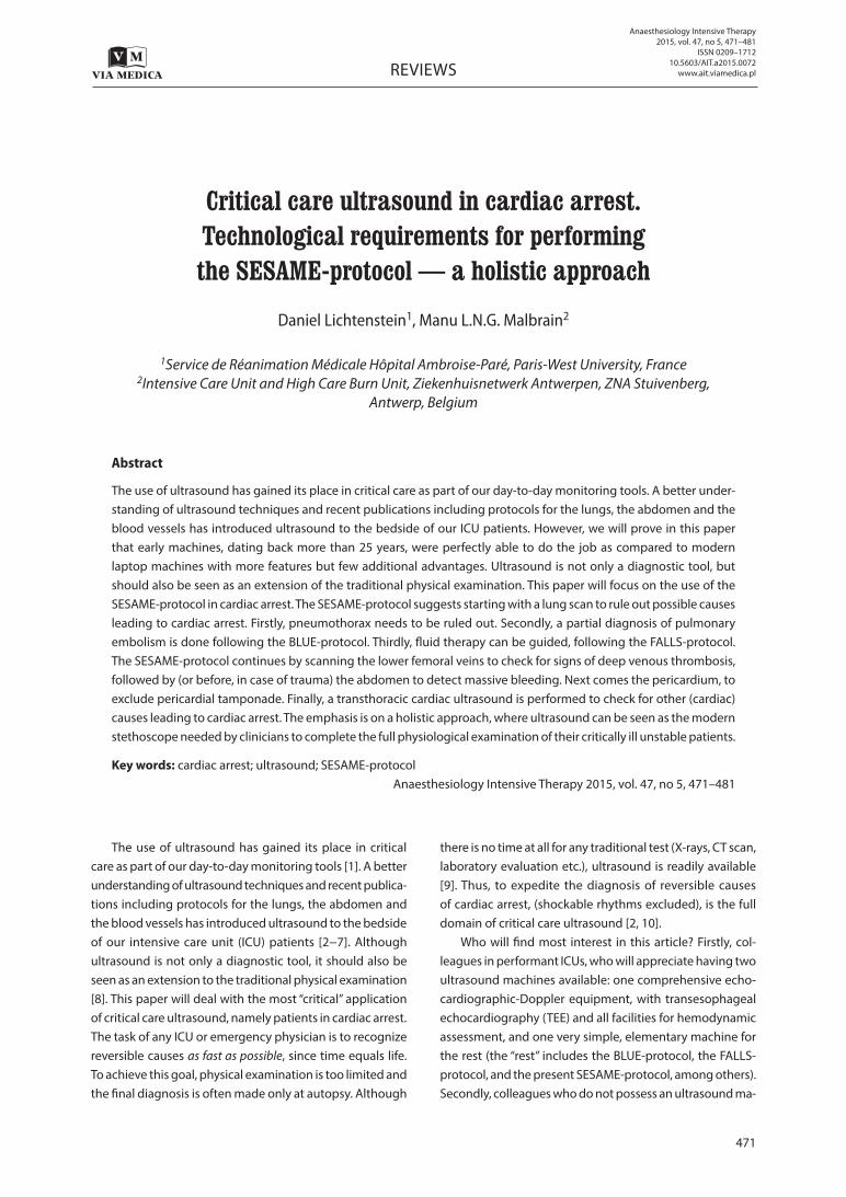
In order to make this article easy to read, we will explain the philosophy of the SESAME-protocol, step-by-step. The reader should imagine the critical situation of cardiac ar-rest in slow motion, as events happen too fast in real time when a cardiac arrest occurs. For simplicity, we will focus on intra-hospital use (in the ER the OR, the ICU or the ward), when the intensivist deals with a cardiac arrest situation at the bedside (Fig. 1).
As we understand the interest of the younger generation of doctors in “fancy” laptop machines with three probes, we advise them to get the best out of this article for their personal practice. It should also be understood that in situ-ations where the mechanisms and causes leading to the cardiac arrest, e.g. pneumothorax, are clinically obvious, the use of the SESAME-protocol is not mandatory and one should proceed directly to appropriate treatment to reverse cardiac arrest (e.g. insertion of a chest tube).
IDEAL ULTRASOUND EQUIPMENT SPECIFICATIONSIn order to perform the SESAME-protocol in a timely
manner, the operator should benefit from using a suitable ultrasound device [2].
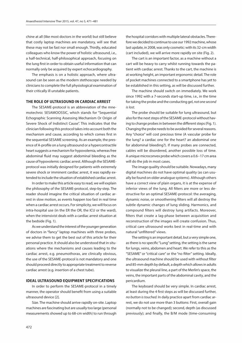
Size. The machine should arrive rapidly on-site. Laptop machines are fascinating but are usually too large (personal measurements showed up to 68-cm width) to run through
the hospital corridors with multiple lateral obstacles. There-fore we decided to continue to use our 1992 machine, whose last update, in 2008, was only cosmetic: with its 32-cm width (cart included), we will arrive more rapidly on site (Fig. 2).
The cart is an important factor, as a machine without a cart will be heavy to carry whilst running towards the pa-tient with cardiac arrest. Thanks to the cart, the machine is at working height, an important ergonomic detail. The role of pocket machines connected to a smartphone has yet to be established in this setting, as will be discussed further.
The machine should switch on immediately. We work since 1992 with a 7-seconds start-up time, i.e., in the time for taking the probe and the conducting gel, not one second is lost.
The probe should be suitable for lung ultrasound, but also for the next steps of the SESAME-protocol without hav-ing to change probes in between the different steps (Fig. 1). Changing the probe needs to be avoided for several reasons. Any “choice” will cost precious time (A vascular probe for the lung? a cardiac one for the heart? an abdominal one for abdominal bleeding?). If many probes are connected, cables will be disordered, another possible loss of time. A unique microconvex probe which covers a 0.6−17 cm area will do the job in most cases.
The image quality should be suitable. Nowadays, many digital machines do not have optimal quality (as can usu-ally be found on older analogue systems). Although others have a correct view of plain organs, it is at the expense of inferior views of the lung. All filters are more or less de-structive for an optimal SESAME-protocol: the averaging, dynamic noise, or smoothening filters will all destroy the subtle dynamic changes of lung sliding. Harmonics, and compound filters will destroy lung artifacts. Moreover, filters that create a lag-phase between acquisition and reconstruction of the images will create confusion. Thus, critical care ultrasound works best in real-time and with natural “unfiltered” views.
The setting is an important detail, but a very simple one, as there is no specific “Lung” setting, the setting is the same for lungs, veins, abdomen and heart. We refer to this as the “SESAME” or “critical care” or the “no filter” setting. Ideally, the ultrasound machine should be used with without filter and 85-mm depth by default, a depth which allows in adults to visualize the pleural line, a part of the Merlin’s space, the veins, the important parts of the abdominal cavity, and the pericardium.
The keyboard should be very simple. In cardiac arrest, at least during the 4 first steps as will be discussed further, no button is touched. In daily practice apart from cardiac ar-rest, we do not use more than 3 buttons: First, overall gain (normally not to be changed); second, depth (as discussed previously); and finally, the B/M mode (time-consuming
473
Daniel Lichtenstein, Manu L.N.G. Malbrain, SESAME-protocol in cardiac arrest
and thus not indicated in a cardiac arrest situation). Each additional button increases the risk for confusion.
A cost-effective machine has one major advantage, namely its availability. Nowadays, although it is common practice to see many ultrasound machines in the hospi-tal, in the early years when ultrasound was introduced
into the ICU, machines were lacking mainly because of cost-related issues. However, if doctors had used holistic ultrasound as soon as it was technically accessible, i.e. 1982, they would have found cost-effective machines at a time where cardiac machines were really expensive and, therefore, unavailable.
Figure 1. The SESAME-protocol
This apparently complex figure just shows, from left to right, simple features. On the far left, the five areas of investigation are shown. Next the type of probe used is listed, i.e., only one probe. Then the depth used, i.e., a standard distance (85 mm) in most steps. Then the timing for ruling out, sequentially, tension pneumothorax, lower femoral DVT, free abdominal fluids (or massive GI tract fluid), followed by pericardial tamponade. When the heart comes under analysis, most reversible cases have already been assessed. Adapted with permission from Lichtenstein [15]
ARDS — acute respiratory distress syndrome; DVT — deep venous thrombosis
474
Anaesthesiol Intensive Ther 2015, vol. 47, no 5, 471–481
We will now explain the philosophy of the SESAME-protocol in 5 simple steps that can be used when one is confronted with a cardiac arrest patient.
STEP 1. RULING OUT PNEUMOTHORAXLUNG ULTRASOUND
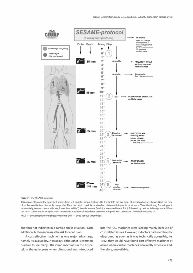
The SESAME-protocol is a sequential protocol which first scans the lungs, mainly for ruling out pneumothorax [4, 11−13]. Although this is probably not the most frequent cause, missing it would be unforgivable. Why the lungs first? To begin with, because lung ultrasound exists and is able to identify a specific pathology at the bedside, in spite of what others still believe [14]. Secondly, because the accurate window is obtained in less than two seconds (the bat sign, immediately indicating the lung surface or the seashore sign) (Fig. 3). Thirdly, as pneumothorax with cardiac arrest is usually large that it can be detected regardless of where the probe is applied on the anterior thorax (Fig. 4). Fourthly, as only less than two seconds are needed for detecting the characteristics of lung sliding and/or B-lines [4]. Fifthly, because the diagnosis is particularly easy as the patient is in quiet breathing via manual bag ventilation, i.e., enough dynamics and no interference due to dyspnea, i.e., the best
Figure 2. Two concepts
To the left, our 1992 (updated 2008) unit. To the right, a standard laptop machine. Note, among several points, that both machines have wheels, i.e., portability. Note that laptop machines are never separated from their cart in the real-life hospital setting. Note mainly that the 1992 machine is slimmer than the laptop, among other advantages (including immediate start up time). Please bear in mind that in a hospital, space is usually lacking in the lateral dimension, not in the vertical one (while ceilings are high enough)
Figure 3. Ultrasound Scan of the anterior intercostal space: bat sign and seashore sign
Panel A. The ribs (vertical arrows) with shadows are visualized. The pleural line (upper, horizontal arrows), is a horizontal hyperechogenic line, half a centimetre below the rib line in adults. The association of ribs and pleural line make a solid landmark called the bat sign. The pleural line indicates the parietal pleura. The horizontal repetition artifact of the pleural line is called the A-line (lower, small horizontal arrows). The A-line indicates that air is the main component visible below the pleural line.
Panel B. M-mode reveals the seashore sign, which indicates that the lung moves at the level of the chest wall. The seashore sign therefore indicates that the pleural line also is the visceral pleura. Above the pleural line, the motionless chest wall displays a stratified pattern. Below the pleural line, the dynamics of lung sliding show a sandy pattern. Note that both images are strictly aligned, which is of importance in critical settings. Both images, i.e., lung sliding plus A-lines define the A-profile (when found at the anterior chest wall). Adapted from “Lung ultrasound in the critically ill” with permission [3]
conditions. Finally, because the detection of the A-profile (as illustrated in Figure 5) with the BLUE-protocol will be an argument for fluid therapy, if one subsequently follows the logic of the FALLS-protocol [4, 5].
TECHNICAL CONSIDERATIONSThe patient has been intubated. The probe is applied
at the anterior chest wall, roughly at the lower BLUE-point, while the hands of the physician performing CPR are prop-erly positioned. As fast and as far as possible, the lungs are scanned, after which the cardiac compressions are contin-ued. If CPR is started before ultrasound, a rib fracture can occur, preventing differentiation of whether the pneumo-thorax was cause or consequence of cardiac arrest. If com-pressions are done before ultrasound, they must be briefly interrupted for the lung scanning, which is far from perfect with regard to hemodynamic stabilisation and coronary perfusion pressure.
The A’-profile of the BLUE-protocol strongly suggests pneumothorax (Fig. 6). Although specialists can search for the pathognomonic lung point sign, this may be time con-suming with few additional therapeutic implications and thus should be avoided in the setting of cardiac arrest (Fig. 7). The CEURF has made suggestions to solve this issue [15]. The “Australian variant” (an idea that came to mind whilst travelling in Sydney) indicates that the A’-profile on
475
Daniel Lichtenstein, Manu L.N.G. Malbrain, SESAME-protocol in cardiac arrest
an ultrasound in combination with the slightest clinical signs suggestive for pneumothorax makes the diagnosis almost certain [15]. In a cardiac arrest patient, the A’-profile in combination with specific findings upon auscultation of the thorax (e.g. slightest homolateral tympanism or decrease in breathing sounds) confirms the diagnosis. Using these tools before ultrasound would be more risky, because they
are not easy to interpret when used in isolation, and valu-able time may be lost in case there is no pneumothorax. When the Australian variant is positive, there is ample time for finding a large bore needle (on-site in the crash-cart), to perform a life saving procedure. In summary, if, after step 1, pneumothorax is excluded then one can move to step 2 of the SESAME-protocol.
STEP 2. SEARCHING FOR PULMONARY EMBOLISMA VENOUS APPROACH
Pulmonary embolism as a cause of cardiac arrest is more frequent than tension pneumothorax. The use of echocardi-ography as an initial step raises some concerns. Firstly, the user must master the technique – and this expertise may take years. Secondly, a good cardiac window must be avail-able, and sometimes this is technically impossible. If time is lost whilst trying to find a good ultrasound window, the car-diac compressions are delayed. To circumvent these issues, some may use transesophageal echocardiography (TEE). This could be an option if TEE is immediately available, and without the drawbacks as stated above (start-on time, size of machine, skill, costs etc. The SESAME-protocol proposes an approach already validated in the BLUE-protocol, where the combination of lung plus venous analysis provides 99% specificity (adding the echocardiographic data would likely improve this rate). Although the BLUE-protocol is a fast protocol that can be performed within three minutes, or less, during cardiac arrest, we count rather in seconds. The SESAME-protocol hence focuses on the lower femoral vein, an area very accessible using the microconvex probe, called
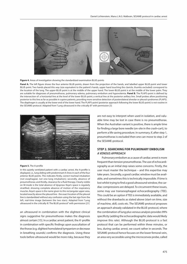
Figure 4. Areas of investigation showing the standardized examination BLUE-points
Panel A. The left figure shows the four anterior BLUE-points, drawn from the projection of the hands, and labelled upper BLUE-point and lower BLUE-point. Two hands placed this way (size equivalent to the patient’s hands, upper hand touching the clavicle, thumbs excluded) correspond to the location of the lung. The upper-BLUE-point is at the middle of the upper hand. The lower-BLUE-point is at the middle of the lower palm. They are suitable for diagnoses of pneumothorax, pulmonary edema, pulmonary embolism and hypovolemia. Panel B. The PLAPS-point is defined by the intersection of: a horizontal line at the level of the lower BLUE-point; a vertical line at the posterior axillary line. Small probes allow positioning posterior to this line as far as possible in supine patients, providing more sensitive detection of posterolateral alveolar or pleural syndromes (PLAPS). The diaphragm is usually at the lower end of the lower hand. The PLAPS-point (posterior approach following the lower BLUE-point) is not routine in the SESAME-protocol. Adapted from “Lung ultrasound in the critically ill” with permission [3]
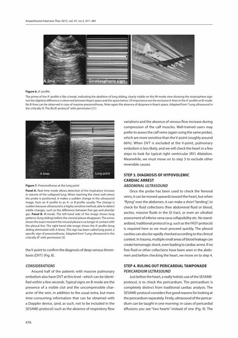
Figure 5. The A-profile
In this quietly ventilated patient with a cardiac arrest, the A-profile is displayed, i.e., lung sliding with predominant A-lines in each of the four anterior BLUE-points. This indicates firstly, correct tracheal intubation (not esophageal, not one-lung intubation); secondly, absence of pneumothorax; and thirdly, clearance for a fluid therapy. Clearly visible on M-mode is the total absence of dyspnea: Keye’s space is regularly stratified, showing complete absence of motion of the respiratory muscles. Keye’s space is the name given to this rectangular upper area, located exactly above the pleural line - the exact location of the pleural line is standardized without any confusion using the bat sign from the left, real-time image (between the two stars). Adapted from “Lung ultrasound in the critically ill. The BLUE-protocol” with permission [21]
476
Anaesthesiol Intensive Ther 2015, vol. 47, no 5, 471–481
the V-point to confirm the diagnosis of deep venous throm-bosis (DVT) (Fig. 8).
CONSIDERATIONSAround half of the patients with massive pulmonary
embolism also have DVT at this level - which can be identi-fied within a few seconds. Typical signs on B-mode are the presence of a visible clot and the uncompressible char-acter of the vein, in addition to the usual extra, but more time-consuming information that can be obtained with a Doppler device, (and, as such, not to be included in the SESAME-protocol) such as the absence of respiratory flow
variations and the absence of venous flow increase during compression of the calf muscles. Well-trained users may prefer to assess the calf veins (again using the same probe), which are more sensitive than the V-point (roughly around 66%). When DVT is excluded at the V-point, pulmonary embolism is less likely, and we will check the heart in a few steps to look for typical right ventricular (RV) dilatation. Meanwhile, we must move on to step 3 to exclude other reversible causes.
STEP 3. DIAGNOSIS OF HYPOVOLEMIC CARDIAC ARRESTABDOMINAL ULTRASOUND
Once the probe has been used to check the femoral veins, it can be moved upwards toward the heart, but when “flying” over the abdomen, it can make a short “landing”, to check for fluid collections (free abdominal fluid or blood, ascites, massive fluids in the GI tract, or even an ultrafast assessment of inferior vena cava collapsibility etc. No stand-ardized, traditional protocol (e.g. such as the FAST-protocol) is required here as we must proceed quickly. The pleural cavities can also be rapidly checked according to the clinical context. In trauma, multiple small areas of blood leakage can create hemorragic shock, even leading to cardiac arrest. If no free fluid or other collections have been seen in the abdo-men and before checking the heart, we move on to step 4.
STEP 4. RULING OUT PERICARDIAL TAMPONADEPERICARDIUM ULTRASOUND
Just before the heart, a really holistic use of the SESAME-protocol, is to check the pericardium. The pericardium is completely distinct from traditional cardiac analysis. The SESAME-protocol considers five good reasons for looking at the pericardium separately. Firstly, ultrasound of the pericar-dium can be taught in one morning: in cases of pericardial effusions you see “two hearts” instead of one (Fig. 9). The
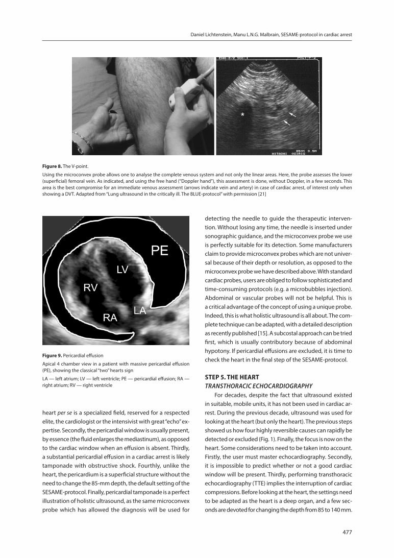
Figure 6. A’-profile
The prime of the A’-profile is like a break, indicating the abolition of lung sliding, clearly visible on this M-mode view showing the stratosphere sign: not the slightest difference is observed between Keye’s space and the space below. Of importance are the exclusive A-lines in the A’-profile on B-mode. No B-lines can be observed in case of massive pneumothorax. Note again the absence of dyspnea in Keye’s space. Adapted from “Lung ultrasound in the critically ill. The BLUE-protocol” with permission [21]
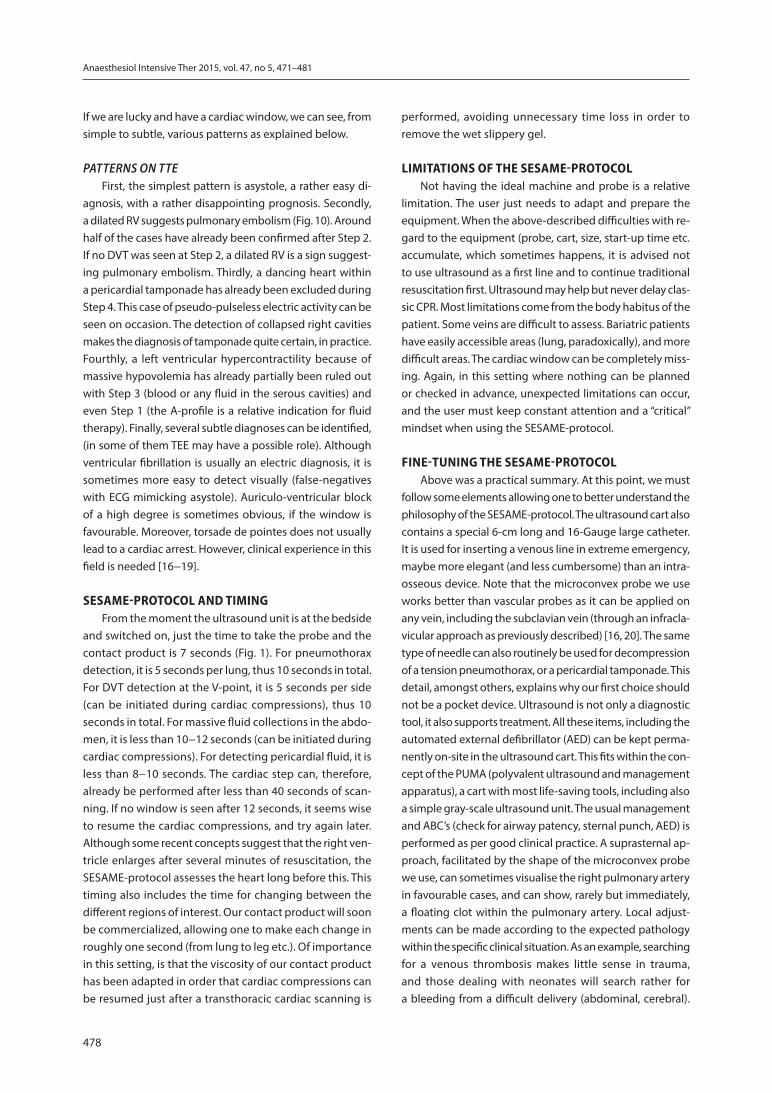
Figure 7. Pneumothorax at the lung point
Panel A. Real-time mode allows detection of the inspiratory increase in volume of the collapsed lung. When reaching the chest wall where the probe is positioned, it makes a sudden change in the ultrasound image, from an A’-profile to an A- or B-profile usually. The change is sudden because ultrasound is a highly sensitive method, able to detect subtle changes, such as the difference between free gas and alveolar gas. Panel B. M-mode. The left-hand side of the image shows lung patterns (lung sliding) before the visceral pleura disappears. The arrow shows the exact moment the visceral pleura is no longer in contact with the pleural line. The right-hand side image shows the A’-profile (lung sliding eliminated with A-lines). This sign has been called lung point, a specific sign of pneumothorax. Adapted from “Lung ultrasound in the critically ill” with permission [3]
477
Daniel Lichtenstein, Manu L.N.G. Malbrain, SESAME-protocol in cardiac arrest
heart per se is a specialized field, reserved for a respected elite, the cardiologist or the intensivist with great “echo” ex-pertise. Secondly, the pericardial window is usually present, by essence (the fluid enlarges the mediastinum), as opposed to the cardiac window when an effusion is absent. Thirdly, a substantial pericardial effusion in a cardiac arrest is likely tamponade with obstructive shock. Fourthly, unlike the heart, the pericardium is a superficial structure without the need to change the 85-mm depth, the default setting of the SESAME-protocol. Finally, pericardial tamponade is a perfect illustration of holistic ultrasound, as the same microconvex probe which has allowed the diagnosis will be used for
detecting the needle to guide the therapeutic interven-tion. Without losing any time, the needle is inserted under sonographic guidance, and the microconvex probe we use is perfectly suitable for its detection. Some manufacturers claim to provide microconvex probes which are not univer-sal because of their depth or resolution, as opposed to the microconvex probe we have described above. With standard cardiac probes, users are obliged to follow sophisticated and time-consuming protocols (e.g. a microbubbles injection). Abdominal or vascular probes will not be helpful. This is a critical advantage of the concept of using a unique probe. Indeed, this is what holistic ultrasound is all about. The com-plete technique can be adapted, with a detailed description as recently published [15]. A subcostal approach can be tried first, which is usually contributory because of abdominal hypotony. If pericardial effusions are excluded, it is time to check the heart in the final step of the SESAME-protocol.
STEP 5. THE HEARTTRANSTHORACIC ECHOCARDIOGRAPHY
For decades, despite the fact that ultrasound existed in suitable, mobile units, it has not been used in cardiac ar-rest. During the previous decade, ultrasound was used for looking at the heart (but only the heart). The previous steps showed us how four highly reversible causes can rapidly be detected or excluded (Fig. 1). Finally, the focus is now on the heart. Some considerations need to be taken into account. Firstly, the user must master echocardiography. Secondly, it is impossible to predict whether or not a good cardiac window will be present. Thirdly, performing transthoracic echocardiography (TTE) implies the interruption of cardiac compressions. Before looking at the heart, the settings need to be adapted as the heart is a deep organ, and a few sec-onds are devoted for changing the depth from 85 to 140 mm.
Figure 8. The V-point.
Using the microconvex probe allows one to analyse the complete venous system and not only the linear areas. Here, the probe assesses the lower (superficial) femoral vein. As indicated, and using the free hand (“Doppler hand”), this assessment is done, without Doppler, in a few seconds. This area is the best compromise for an immediate venous assessment (arrows indicate vein and artery) in case of cardiac arrest, of interest only when showing a DVT. Adapted from “Lung ultrasound in the critically ill. The BLUE-protocol” with permission [21]
Figure 9. Pericardial effusion
Apical 4 chamber view in a patient with massive pericardial effusion (PE), showing the classical “two” hearts sign
LA — left atrium; LV — left ventricle; PE — pericardial effusion; RA — right atrium; RV — right ventricle
478
Anaesthesiol Intensive Ther 2015, vol. 47, no 5, 471–481
If we are lucky and have a cardiac window, we can see, from simple to subtle, various patterns as explained below.
PATTERNS ON TTEFirst, the simplest pattern is asystole, a rather easy di-
agnosis, with a rather disappointing prognosis. Secondly, a dilated RV suggests pulmonary embolism (Fig. 10). Around half of the cases have already been confirmed after Step 2. If no DVT was seen at Step 2, a dilated RV is a sign suggest-ing pulmonary embolism. Thirdly, a dancing heart within a pericardial tamponade has already been excluded during Step 4. This case of pseudo-pulseless electric activity can be seen on occasion. The detection of collapsed right cavities makes the diagnosis of tamponade quite certain, in practice. Fourthly, a left ventricular hypercontractility because of massive hypovolemia has already partially been ruled out with Step 3 (blood or any fluid in the serous cavities) and even Step 1 (the A-profile is a relative indication for fluid therapy). Finally, several subtle diagnoses can be identified, (in some of them TEE may have a possible role). Although ventricular fibrillation is usually an electric diagnosis, it is sometimes more easy to detect visually (false-negatives with ECG mimicking asystole). Auriculo-ventricular block of a high degree is sometimes obvious, if the window is favourable. Moreover, torsade de pointes does not usually lead to a cardiac arrest. However, clinical experience in this field is needed [16−19].
SESAME-PROTOCOL AND TIMINGFrom the moment the ultrasound unit is at the bedside
and switched on, just the time to take the probe and the contact product is 7 seconds (Fig. 1). For pneumothorax detection, it is 5 seconds per lung, thus 10 seconds in total. For DVT detection at the V-point, it is 5 seconds per side (can be initiated during cardiac compressions), thus 10 seconds in total. For massive fluid collections in the abdo-men, it is less than 10−12 seconds (can be initiated during cardiac compressions). For detecting pericardial fluid, it is less than 8−10 seconds. The cardiac step can, therefore, already be performed after less than 40 seconds of scan-ning. If no window is seen after 12 seconds, it seems wise to resume the cardiac compressions, and try again later. Although some recent concepts suggest that the right ven-tricle enlarges after several minutes of resuscitation, the SESAME-protocol assesses the heart long before this. This timing also includes the time for changing between the different regions of interest. Our contact product will soon be commercialized, allowing one to make each change in roughly one second (from lung to leg etc.). Of importance in this setting, is that the viscosity of our contact product has been adapted in order that cardiac compressions can be resumed just after a transthoracic cardiac scanning is
performed, avoiding unnecessary time loss in order to remove the wet slippery gel.
LIMITATIONS OF THE SESAME-PROTOCOLNot having the ideal machine and probe is a relative
limitation. The user just needs to adapt and prepare the equipment. When the above-described difficulties with re-gard to the equipment (probe, cart, size, start-up time etc. accumulate, which sometimes happens, it is advised not to use ultrasound as a first line and to continue traditional resuscitation first. Ultrasound may help but never delay clas-sic CPR. Most limitations come from the body habitus of the patient. Some veins are difficult to assess. Bariatric patients have easily accessible areas (lung, paradoxically), and more difficult areas. The cardiac window can be completely miss-ing. Again, in this setting where nothing can be planned or checked in advance, unexpected limitations can occur, and the user must keep constant attention and a “critical” mindset when using the SESAME-protocol.
FINE-TUNING THE SESAME-PROTOCOLAbove was a practical summary. At this point, we must
follow some elements allowing one to better understand the philosophy of the SESAME-protocol. The ultrasound cart also contains a special 6-cm long and 16-Gauge large catheter. It is used for inserting a venous line in extreme emergency, maybe more elegant (and less cumbersome) than an intra-osseous device. Note that the microconvex probe we use works better than vascular probes as it can be applied on any vein, including the subclavian vein (through an infracla-vicular approach as previously described) [16, 20]. The same type of needle can also routinely be used for decompression of a tension pneumothorax, or a pericardial tamponade. This detail, amongst others, explains why our first choice should not be a pocket device. Ultrasound is not only a diagnostic tool, it also supports treatment. All these items, including the automated external defibrillator (AED) can be kept perma-nently on-site in the ultrasound cart. This fits within the con-cept of the PUMA (polyvalent ultrasound and management apparatus), a cart with most life-saving tools, including also a simple gray-scale ultrasound unit. The usual management and ABC’s (check for airway patency, sternal punch, AED) is performed as per good clinical practice. A suprasternal ap-proach, facilitated by the shape of the microconvex probe we use, can sometimes visualise the right pulmonary artery in favourable cases, and can show, rarely but immediately, a floating clot within the pulmonary artery. Local adjust-ments can be made according to the expected pathology within the specific clinical situation. As an example, searching for a venous thrombosis makes little sense in trauma, and those dealing with neonates will search rather for a bleeding from a difficult delivery (abdominal, cerebral).
479
Daniel Lichtenstein, Manu L.N.G. Malbrain, SESAME-protocol in cardiac arrest
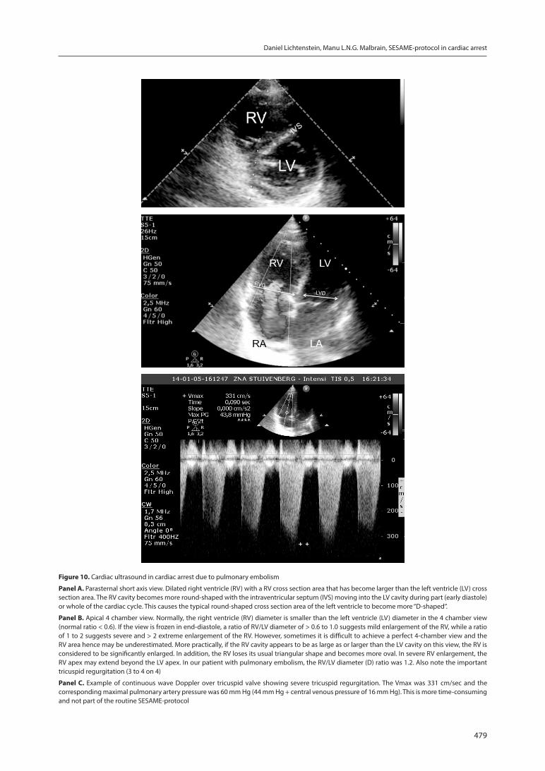
Figure 10. Cardiac ultrasound in cardiac arrest due to pulmonary embolism
Panel A. Parasternal short axis view. Dilated right ventricle (RV) with a RV cross section area that has become larger than the left ventricle (LV) cross section area. The RV cavity becomes more round-shaped with the intraventricular septum (IVS) moving into the LV cavity during part (early diastole) or whole of the cardiac cycle. This causes the typical round-shaped cross section area of the left ventricle to become more “D-shaped”.
Panel B. Apical 4 chamber view. Normally, the right ventricle (RV) diameter is smaller than the left ventricle (LV) diameter in the 4 chamber view (normal ratio < 0.6). If the view is frozen in end-diastole, a ratio of RV/LV diameter of > 0.6 to 1.0 suggests mild enlargement of the RV, while a ratio of 1 to 2 suggests severe and > 2 extreme enlargement of the RV. However, sometimes it is difficult to achieve a perfect 4-chamber view and the RV area hence may be underestimated. More practically, if the RV cavity appears to be as large as or larger than the LV cavity on this view, the RV is considered to be significantly enlarged. In addition, the RV loses its usual triangular shape and becomes more oval. In severe RV enlargement, the RV apex may extend beyond the LV apex. In our patient with pulmonary embolism, the RV/LV diameter (D) ratio was 1.2. Also note the important tricuspid regurgitation (3 to 4 on 4)
Panel C. Example of continuous wave Doppler over tricuspid valve showing severe tricuspid regurgitation. The Vmax was 331 cm/sec and the corresponding maximal pulmonary artery pressure was 60 mm Hg (44 mm Hg + central venous pressure of 16 mm Hg). This is more time-consuming and not part of the routine SESAME-protocol
480
Anaesthesiol Intensive Ther 2015, vol. 47, no 5, 471–481
Likewise, technical details can be adapted accordingly [15]. Those accustomed to working with the “4H’s-4T’s” (the 4 H’s refer to Hypoxia, Hypovolemia, Hypo- or hyperkalemia, and Hypothermia, where the 4 T’s refer to Tension pneumotho-rax, Thrombosis (DVT or PE), Tamponade, and Toxic causes) in the differential diagnosis of cardiac arrest with pulseless electrical activity (PEA) can easily adapt it. They just need to replace tension pneumothorax by Step 1, thrombosis of pulmonary artery by Step 2, hypovolemia by Step 3 and tamponade by Step 4. Other causes (hyperkalemia, toxic causes etc) are diagnosed with other traditional diagnostic tools. While the SESAME-protocol has not yet been vali-dated in the clinical setting, it uses validated applications in a specific manner, where each detail has been worked out in order to facilitate smoothness and to optimize speed. Al-though in the future pocket devices may have a role, for the time being we are used to working at bedside, with both our hands when scanning critically ill patients. Pocket devices can be useful for patients lying on the ground or in really tiny spaces (airborne medical evacuation). Some gray-scale pocket machines have technical features more compatible with the practice of the SESAME-protocol than more sophisticated de-vices. The SESAME-protocol may have a psychological impact for the caregiver as the visualisation helps one to understand the situation and may help one to cope with the sometimes poor outcomes seen after cardiac arrest, in the understanding that reversible causes were not missed.
INTEGRATION OF THE SESAME-PROTOCOL WITHIN THE CONCEPT OF HOLISTIC ULTRASOUND
The physician who has understood the philosophy, reasoning and the technical requirements of the SESAME-protocol will also master other fields beyond cardiac arrest. For instance, the simple detection of A-lines, at the first step of the SESAME-protocol, suggesting “room” for fluid therapy (as a very rough indicator, to be refined with the FALLS-protocol) can be used as a first step in many areas of medicine, critical care and emergency care. The technique, probe, and signs used to exclude pneumothorax are exactly the same in many other situations, such as the critically ill after a thoracic procedure (pleurocenthesis, insertion of deep venous catheter), in emergency medicine for limiting radiation in the management of spontaneous pneumo-thorax, in internal medicine after thoracentesis, in multiple trauma as routine care, etc. The same approach to confirm the diagnosis of deep venous thrombosis can be done more elaborately and more comprehensively (common femoral vein, calf veins, upper extremities), while this technique can be of interest in several disciplines, including geriatrics, obstetrics, emergency medicine etc. Although searching for free abdominal fluid is a standard issue in trauma patients, it can be used in many other settings as well. The same can be
said for pericardial effusions. Regarding echocardiography and considering holistic ultrasound (i.e., mainly, the integra-tion of the lung), we can describe an alternative, the simple emergency cardiac sonography. This label indicates that a partial view of the heart analysis can be sufficient provided the lung surrounds this approach. As the simplest example, if left ventricle function is difficult to assess, the detection of an A-profile indicates the absence of pulmonary edema, even at a silent, early, interstitial step, probably indicating a normal left heart function.
CONCLUSIONSThe SESAME-protocol is a very fast protocol, preferably
performed using simple equipment which is not always currently present. Many enthusiastic colleagues use the term “disruptive” when speaking about the revolution of bedside ultrasound. Although this is, indeed an unprec-edented evolution, from our perspective, it is rather a victory of the laptop machines, with their standard three probes and complex functions. In the light of the SESAME-protocol, simple machines using one distinct, universal probe should be used in order to achieve a really disruptive radical change. The present technical note on the SESAME-protocol was the opportunity to show some of the advantages of holistic ultrasound, where simple concepts do have a place in critical care ultrasound. By analyzing the illustrative case of cardiac arrest, we just described what we do already without any difference in daily clinical practice in the ICU, but here only more slowly. Last, but not least, it is noteworthy that this article could have been written in 1982, although a time where the technology and mobility of some units was less advanced than modern machines, they were perfectly suit-able for use in cardiac arrest just because of their simplic-ity and more convenient width. While modern machines may have advantages regarding better resolution for plain organs, such as the heart (in a sophisticated Doppler ap-proach), apart from this and some other modern features (like the presence of a USB port) they have no major advan-tages over the machines that were constructed more than 35 years ago. Thus, with this statement our aim aim is to give the reader some further food for thought.
ACKNOWLEDGEMENTS 1. Our deep thanks to François Jardin, who made our re-
search possible. The authors are deeply moved by the recent terrorist attacks in Paris, France and want to ex-tend their sincerest condolences and sympathy for the families, relatives and friends of all the victims. They believe that the SESAME-protocol can be helpful as point-of-care diagnostic tool in severely injured patients with balistic wounds using hand-held devices on-site.
2. The authors declare no financial disclosure.

481
Daniel Lichtenstein, Manu L.N.G. Malbrain, SESAME-protocol in cardiac arrest
3. Daniel Lichtenstein has no conflicts of interest to declare with regard to the contents of this paper. Manu L.N.G. Malbrain is member of the medical advisory board of Pulsion Medical Systems (Maquet Getinge group).
References:1. Jardin F, Farcot JC, Boisante L, Curien N, Margairaz A, Bourdarias JP:
Influence of positive end-expiratory pressure on left ventricular per-formance. N Engl J Med. 1981; 304: 387−392.
2. Lichtenstein D, van Hooland S, Elbers P, Malbrain MLNG: Ten good reasons to practice ultrasound in critical care. Anaesthesiol Intensive Ther 2014; 46: 323−335. doi: 10.5603/AIT.2014.0056.
3. Lichtenstein DA: Lung ultrasound in the critically ill. Ann Intensive Care 2014; 4: 1. doi: 10.1186/2110-5820-4-1.
4. Lichtenstein DA, Meziere GA: Relevance of lung ultrasound in the dia-gnosis of acute respiratory failure: the BLUE protocol. Chest 2008; 134: 117−125. doi: 10.1378/chest.07-2800.
5. Lichtenstein D: Fluid administration limited by lung sonography: the place of lung ultrasound in assessment of acute circulatory failure (the FALLS-protocol). Expert Rev Respir Med 2012; 6: 155−162. doi: 10.1586/ers.12.13.
6. Kirkpatrick AW, Sirois M, Laupland KB et al.: Hand-held thoracic sono-graphy for detecting post−traumatic pneumothoraces: the Extended Focused Assessment with Sonography for Trauma (EFAST). J Trauma 2004; 57: 288−295.
7. Volpicelli G, Elbarbary M, Blaivas M et al.: International evidence-based recommendations for point-of-care lung ultrasound. Intensive Care Med 2012; 38: 577−591. doi: 10.1007/s00134-012-2513-4.
8. Gillman LM, Kirkpatrick AW: Portable bedside ultrasound: the visual stethoscope of the 21st century. Scand J Trauma Resusc Emerg Med. 2012; 20: 18. doi: 10.1186/1757-7241-20-18.
9. Agricola E, Arbelot C, Blaivas M et al.: Ultrasound performs better than radiographs. Thorax. 2011; 66(9): 828−829; author reply 9. doi: 10.1136/thx.2010.153239.
10. Lichtenstein DA: How can the use of lung ultrasound in cardiac arrest make ultrasound a holistic discipline. The example of the SESAME--protocol. Medical ultrasonography. 2014; 16: 252−255.
11. Lichtenstein D: Lung ultrasound in the critically ill. Curr Opin Crit Care 2014; 20: 315−322. doi: 10.1097/MCC.0000000000000096.
12. Lichtenstein D, Meziere G, Biderman P, Gepner A: The “lung point”: an ul-trasound sign specific to pneumothorax. Intensive Care Med 2000; 26: 1434−1440.
13. Lichtenstein DA, Menu Y: A bedside ultrasound sign ruling out pneu-mothorax in the critically ill. Lung sliding. Chest 1995; 108: 1345−1348.
14. Fuhlbrigge AL, Choi AMK: Diagnostic procedures in respiratory disease. In: Kasper DL, Fauci AS, Longo DL, Hauser SL, J.L. J, Loscalzo J (ed.): Har-rison’s Principles of internal medicine. 19th ed. New York: McGraw-Hill; 2015: 1663.
15. Lichtenstein D: Lung ultrasound as the first step of management of a cardiac arrest: the SESAME-Protocol. In: Lichtenstein D (ed.): Lung ultrasound in the critically ill: The BLUE-protocol. Heidelberg: Springer--Verlag; 2015: 261−274.
16. Terkawi AS, Karakitsos D, Elbarbary M, Blaivas M, Durieux ME: Ultrasound for the anesthesiologists: present and future. ScientificWorldJournal. 2013; 2013: 683685. doi: 10.1155/2013/683685.
17. Blaivas M, Fox JC: Outcome in cardiac arrest patients found to have car-diac standstill on the bedside emergency department echocardiogram. Acad Emerg Med 2001; 8: 616−621.
18. Breitkreutz R, Walcher F, Seeger FH: Focused echocardiographic eva-luation in resuscitation management: concept of an advanced life support-conformed algorithm. Crit Care Med 2007; 35 (5 Suppl): S150−161.
19. Via G, Hussain A, Wells M et al. International evidence-based recommen-dations for focused cardiac ultrasound. J Am Soc Echocardiogr 2014; 27: 683 e1− e33. doi: 10.1016/j.echo.2014.05.001.
20. Kumar A, Chuan A: Ultrasound guided vascular access: efficacy and safety. Best Pract Res Clin Anaesthesiol. 2009; 23: 299−311.
21. Lichtenstein D: Lung ultrasound in the critically ill. The BLUE Protocol. Heidelberg: Springer-Verlag 2015.
Corresponding author:Manu L.N.G. Malbrain, MD, PhDICU and High Care Burn Unit DirectorZNA StuivenbergLange Beeldekensstraat 267B-2060 Antwerpen 6, Belgiume-mail: [email protected]
Received: 11.09.2015 Accepted: 2.11.2015