Embed Size (px)
Citation preview
This Provisional PDF corresponds to the article as it appeared upon acceptance. Copyedited andfully formatted PDF and full text (HTML) versions will be made available soon.
Extracorporeal life support following out-of-hospital refractory cardiac arrest
Critical Care 2011, 15:R29 doi:10.1186/cc9976
Morgan Le Guen ([email protected])Armelle Nicolas-Robin ([email protected])
Serge Carreira ([email protected])Mathieu Raux ([email protected])
Pascal Leprince ([email protected])Bruno Riou ([email protected])
Olivier Langeron ([email protected])
ISSN 1364-8535
Article type Research
Submission date 26 October 2010
Acceptance date 18 January 2011
Publication date 18 January 2011
Article URL http://ccforum.com/content/15/1/R29
This peer-reviewed article was published immediately upon acceptance. It can be downloaded,printed and distributed freely for any purposes (see copyright notice below).
Articles in Critical Care are listed in PubMed and archived at PubMed Central.
For information about publishing your research in Critical Care go to
http://ccforum.com/info/instructions/
Critical Care
© 2011 Le Guen et al. ; licensee BioMed Central Ltd.This is an open access article distributed under the terms of the Creative Commons Attribution License (http://creativecommons.org/licenses/by/2.0),
which permits unrestricted use, distribution, and reproduction in any medium, provided the original work is properly cited.

Extracorporeal life support following out-of-hospital
refractory cardiac arrest
Morgan Le Guen1, Armelle Nicolas-Robin
1, Serge Carreira
1, Mathieu Raux
1,
Pascal Leprince2, Bruno Riou
3 and Olivier Langeron
1
1 Department of Anesthesiology and Critical Care, Centre hospitalo-universitaire (CHU)
Pitié-Salpêtrière, Assistance Publique-Hôpitaux de Paris (APHP), Université Pierre et Marie
Curie-Paris 6, 47-83 Boulevard de l’Hôpital, 76651 Paris Cedex 13, France
2 Department of Cardio-thoracic Surgery, CHU Pitié-Salpêtrière, APHP, Université Pierre et
Marie Curie-Paris 6, 47-83 Boulevard de l’Hôpital, 76651 Paris Cedex 13, France
3 Department of Emergency Medicine and Surgery, CHU Pitié-Salpêtrière, APHP, Université
Pierre et Marie Curie-Paris 6, 47-83 Boulevard de l’Hôpital, 76651 Paris Cedex 13, France
Correspondence: Bruno Riou, E-mail: [email protected]
Abstract
Introduction Extracorporeal life support (ECLS) has recently shown encouraging results in
resuscitation of in-hospital (IH) refractory cardiac arrest. We assessed the use of ECLS
following out-hospital (OH) refractory cardiac arrest.
Methods We evaluated 51 consecutive patients who experienced witnessed OH refractory
cardiac arrest and received automated chest compression and ECLS upon arrival in the
hospital. Patients with pre-existing severe hypothermia and who experienced IH cardiac arrest
were excluded. A femoro-femoral ECLS was set up on admission to hospital by a mobile
cardiothoracic surgical team.
Results 51 patients were included, mean age 42 + 15 years. The mean delays from arrest to
cardiopulmonary resuscitation and ECLS were respectively 3 [25-75 interquartile 1-7] and
120 [102-149] min. Initial rhythm was ventricular fibrillation in 32 (63%) patients, asystole in
15 (29%) patients, and pulseless rhythm in 4 (8%). ECLS failed in 9 (18%) patients. Only two
(4%, 95% confidence interval 1-13%) patients were alive at day 28 with a favourable
neurological outcome. There was a significant correlation (R=0.36, p=0.01) between blood
lactate and delay between cardiac arrest and onset of ECLS, but not with arterial pH or blood
potassium. Deaths were the consequence of multi-organ failure (n=43, 47%), brain death
(n=10, 20%) and refractory hemorrhagic shock (n=7, 14%), and most patients (n=46, 90%)
died within 48 hours.
Conclusions This poor outcome suggests that the use of ECLS should be more restricted
following in OH refractory cardiac arrest.

Introduction
Out-of-hospital (OH) cardiac arrest remains an important cause of unexpected death in
developed countries. It still has a low survival rate despite access to improved emergency
medical care, spread of automatic defibrillation [1], and regularly updated international
guidelines [2]. Recent studies have indicated to unchanged or slightly better survival rates
after OH cardiac arrest over the past decades [3,4]. Initial rhythm and cardiac origin are
independent predictors of successful cardiopulmonary resuscitation (CPR) with better
outcome related to a shockable rhythm such as ventricular fibrillation than asystole [5,6].
Survival rate rapidly decreases with time and a refractory cardiac arrest, defined as
persistence of circulatory arrest despite more than 30 min of appropriate CPR, is usually
considered as a condition associated with no survival [7], except in some particular conditions
such as hypothermia [8].
Extracorporeal life support (ECLS) has been suggested as a therapeutic option in
refractory cardiac arrest since 1976 [9]. However, the use of this technique has remained
limited to hypothermic cardiac arrest and those occurring during the perioperative period of
cardiothoracic surgery, mainly because the results of the initial trials were disappointing
[10,11]. The ease of use of more recent miniaturized ECLS devices has permitted a wider use
of the technique. Encouraging results have been published recently by several teams in
France, Taiwan, Japan, and United States [12-16]. In these studies, most cardiac arrests
occurred in the hospital, and survival with good neurological outcome has been observed in
up to 20 to 30 % of cases [12-16]. Therefore, ECLS has been assigned a low-grade
recommendation in recent guidelines for in-hospital (IH) cardiac arrest [17].
However, the good results obtained in IH cardiac arrests should not be extrapolated to
OH cardiac arrests, mainly because there may be a longer delay in ECLS initiation [18]. Our
primary aim was to review use of ECLS for OH refractory cardiac arrest.
Materials and methods
This prospective observational study received approval from our Institutional Review
Board (CPP Pitié-Salpêtrière 2008/0701 Paris, France). Informed consent was waived due to
life threatening emergencies and the absence of any therapeutic alternative. Information to the
relatives of the patient (or to the patient in case of survival) was delivered after inclusion as
appropriate in a life-threatening context.
Patients
Over a 32-month period (from January 2008 to August 2010), all patients who were
referred to our ICU for OH refractory cardiac arrest were eligible for enrollment into this
study. They were included prospectively and consecutively if the following criteria were met:
1) witnessed OH cardiac arrest; 2) refractory cardiac arrest, defined as the absence of return of
spontaneous circulation (ROSC) after 30 min of CPR; 3) CPR pursued until arrival at our
ICU; 4) mobile cardiothoracic surgery team available; 5) lack of known severe co-morbidities
that should have preclude admission into an ICU. Patients who experienced IH cardiac arrest
were excluded as well as patients who were severely hypothermic (body temperature <32°C)
before CPR. Young children (<30 kg) were not included because our institution exclusively
takes care of adults and because specific sizes of pediatric cannulae were not available.
Conversely, patients older than 70 years were considered not eligible due to the poor expected
neurological recovery.
Protocol
Prehospital emergency medical service (EMS) team performed CPR according to the
American Heart Association guidelines [2]. In case of refractory cardiac arrest, CPR was
pursued in the prehospital phase using an automated device (Autopulse®, Zoll Inc,
Chemlsford, MA) [19]. In the Paris area, all prehospital physician-staffed emergency units
have been equipped with an automated chest compression device because France has
developed a nationwide program of organ harvesting in non-heart beating donors. As soon as
the EMS team considered that initial CPR had failed, they immediately alerted our ICU
through the emergency unit regulating center to organize the patient admission and to ensure
of the availability of the mobile cardiothoracic surgery team. This unit works in our hospital
and includes a surgeon, a resident in surgery, and a technician, together with full equipment
required to set up emergency ECLS anywhere. This mobile cardiothoracic surgery team has
had extensive experience in our hospital and in our city [12,20,21]. During transfer to the
hospital, resuscitation was continued without stopping at any moment.
At the admission to the ICU, absence of ROSC and absence of heart beat were
checked before engaging the procedure. Then mechanical ventilation and automated chest
compression device were continued until the start of ECLS. ECLS was established surgically
with a peripheral femoro-femoral cannulation. The equipment included heparinized polyvinyl
chloride tubing, a membrane oxygenator (Quadrox Bioline, Jostra-Maquet®, Orleans, France)
and venous and arterial femoral cannulae (Biomedicus Carmed, Medtronic®, Boulogne-
Billancourt, France) inserted using a surgical approach. An oxygen/air blender (Sechrist®,
Sechrist Industries, Anaheim, CA) was used to supply the membrane oxygenator. Pump flow
was initially set at 3–4 L.min-1
and then, arterial and central venous catheters were inserted to
continuously measure arterial blood pressure and allow frequent blood sampling. To avoid
limb ischemia, an anterograde reperfusion catheter for distal limb perfusion was inserted.
Objectives to optimize organ perfusion were PaO2 > 100 mmHg, normocapnia and arterial
blood pressure > 60 mmHg with administration of fluids, blood transfusion to achieve a
hematocrit > 35%, or vasopressive drugs (norepinephrine or epinephrine). Mild hypothermia
(target body temperature 33-35 °C) was maintained during the first 24h using external cooling

(pulsed-air blanket) and neuromuscular blocking agents with sedatives were administered
[22]. Minimum lung ventilation was maintained to avoid pulmonary collapse during ECLS
with a tidal volume of 4-5 ml.kg-1
, respiratory rate 6 min-1
, and positive end-expiratory
pressure of 5 cmH2O. To avoid coagulation in the membrane oxygenator, non fractioned
heparin was intravenously administered during ECLS with repeated control to maintain the
activated clotting time ratio over 2.0, but this administration was postponed in most patients
because of major coagulation abnormalities. An inhibitor of the proton pump was
systematically given to prevent upper gastrointestinal bleeding. The possible cause of cardiac
arrest was immediately investigated with regular and repeated troponin Ic assays, trans-
thoracic or trans-oesophageal echocardiography and electrocardiogram as soon as an electric
activity was present. In this context, patients with strong indications of acute myocardial
infarction were transported to the catheter laboratory with ECLS to perform a percutaneous
coronary angiography and any appropriate invasive treatment [23]. Sedation was stopped after
a 24 hour period of mild hypothermia and then neurological (clinical examination and
eventually electroencephalogram) and infectious status were checked. Withdrawal from
ECLS required an echocardiography assessment of myocardial function (left ventricular
ejection fraction >50%) and an arterial blood PaO2/FiO2 ratio >150 mmHg. The pump flow
was progressively reduced to check for the absence of any deterioration in hemodynamic
status. The possibility of the use of a ventricular assistance device or heart transplantation was
examined if irreversible damage in myocardium function was diagnosed with an unsuccessful
weaning from ECLS despite a favourable neurological outcome. Discontinuation of ECLS
was based upon evidence of multiple organ failure (MOF), massive bleeding, or brain death.
Any ECLS-associated complications were carefully monitored.
Measurements
The following variables were recorded according to the Utstein style [24]: age, sex,
cardiovascular risk factors, delays from collapse to basic CPR, advanced CPR, installation of
automated chest compression device, arrival into ICU, and installation of ECLS, initial
cardiac rhythm, use of vasopressor and defibrillation during initial CPR, supposed cause of
cardiac arrest. End-tidal CO2 during CPR before ECLS was also recorded [25]. During CPR,
signs of life (respiratory gasps, movements) were noted.
The following biological measurements were performed before ECLS: arterial blood
gas analysis, blood lactate (normal range <1.8 mmol.L-1
), serum creatinine, blood potassium,
fibrinogen, and prothrombin activity. Troponin Ic (normal range <0.15 µg.L-1,
Stratus
autoanalyser, Dade-Behring, Paris La Défense, France) and protein S100β (normal range
<0.10 µg.L-1
, LIA-mat 300 analyzer, Byk-Sangtec France Laboratories, Le Mée sur Seine,
France) were also measured [26]. The evolution of arterial pH and blood lactate levels were
recorded after 1-2 hours of ECLS, and the following variables calculated: delta arterial pH,
blood lactate clearance expressed as percent of initial values, number of patients with blood
lactate clearance ≤-10%, as previously described [27].
The final outcome was determined at day 28 and the Glasgow coma outcome scale
was determined at 6 months. The Glasgow outcome scale defines the following categories: 1,
death; 2, persistent vegetative state; 3, severe disability (minimally conscious state, severe
motor deficit, aphasia, and need for continuous help); 4, moderate disability; 5, good
recovery.
Since the start of the study (January 2009), French guidelines of the indications for the
use of ECLS in refractory cardiac arrest have been published [28]. These guidelines
considered the following variables to determine whether or not ECLS is indicated following
out-of-hospital cardiac arrest: duration of no flow (≤ 5 min), duration of low flow (≤100 min),
end-tidal CO2 (≥10 mmHg), at least in non-hypothermic patients and in patients without life

signs during on-going CPR (Additional file 1) [28]. Thus we assessed if the main criteriae
(no-flow, low-flow, ETCO2) recommended in these guidelines were followed during our
study and if the whole algorithm was respected.
Statistical analysis
Data are expressed as means ± SD or median [25-75 interquartile] for non-Gaussian
variables (Kolmogorov test). Categorical variables are given as percentages and their 95%
confidence interval. Comparison between two groups was performed using the Student t test,
Mann-Whitney U test, or Fisher’s exact method as appropriate. Correlation between two
variables was performed using the least square linear regression. All P values were two-tailed
and a p value of less than 0.05 was considered significant. Statistical analysis was performed
using NCSS 6.0 software (Statistical Solutions Ltd, Cork, Ireland).

Results
During the study period, we performed ECLS in 59 patients who had experienced a
refractory cardiac arrest. Three patients who had suffered IH cardiac arrest were excluded as
well as 5 patients with severe hypothermia (24.5 + 1.8 °C) before cardiac arrest. Thus, 51
patients were finally included in the study. The main characteristics of our population are
shown in table 1. The supposed causes of cardiac arrest were cardiac (n=44, 86%), trauma
(n=3, 6%), drug overdose (n=2, 4%), respiratory (n=1, 2%), and electrocution (n=1, 2%).
Only one patient had signs of life during CPR before ECLS.
ECLS flow could not be established in 9 (18%) patients. These patients had a more
prolonged no flow duration (2.5 [1-6] vs 3 [0.5-6.5] min, P=0.04) and a lower ETCO2 (9 + 3
vs 12 + 2 min, P=0.046) than the remaining patients. In one case, the failure was the
consequence of an impossible cannulation of femoral artery, probably related to an aortic
dissection. The remaining failures of ECLS were related to an insufficient pump flow despite
massive fluid challenge and transfusion. In the remaining 42 patients the initial ECLS output
was 3.6 + 1.8 L.min-1 providing an arterial blood pressure of 67 + 39 mmHg. During the
ECLS procedure, 30 (59%) patients required blood transfusion (4 [2-6] packed red blood cell
units). Body temperature was 34.1 + 0.9 °C.
Twenty of the 42 patients in whom ECLS was installed (48%) patients underwent
coronary angiography because of clinical or electrical signs suggesting myocardial infarction
but significant coronary abnormalities were noted in only 10 of these patients (50%).
Angioplasty without stenting was performed in 1 patients and coronary stent following
percutaneous transluminal angioplasty was inserted in 7 patients. Coronary spasm was
diagnosed in 2 of these patients.
Arterial blood gas samples at the admission showed severe lactic acidosis (Table 1)
Initial blood lactate level was significantly correlated with the OH duration of cardiac arrest
until ECLS (p=0.02) (Figure 1). In contrast, the correlations between arterial pH (R=0.05) or
blood potassium levels (R=0.23) were non significant. After 1 hour of ECLS, blood lactate
levels slightly but significantly decreased whereas arterial pH markedly increased (Figure 2).
Seventeen of the 42 patients in whom ECLS was installed (40%) survived after 24
hours of ECLS, but only 5 (12%) after 48 hours. At day 28, only two patients were alive,
providing a global survival rate of 4% (95% confidence interval 1-13%). The cause of death
was refractory MOF (n=23, 45%), brain death (n=10, 20%), and severe hemorrhage (n=7,
14%), and failure of ECLS in the remaining patients (n=9, 18%).
In the first survivor (cardiac cause, ventricular fibrillation, no flow 1 min, low flow
132 min, protein S100 1.5 µg.L-1
), withdrawal from ECLS was possible only at day 36
because of severe and prolonged heart failure (left ventricular ejection volume estimated to
30%) and an implantable automatic defibrillator was inserted. His length of stay in the ICU
was 58 days, and 187 days in the hospital. Follow-up at six month showed only minor
cognitive dysfunction (Glasgow outcome scale of 5) but a persistent altered left ventricular
ejection fraction (35%). In the second survivor (cardiac cause, ventricular fibrillation, no flow
0 min, low flow 170 min, protein S100 4.5 µg.L-1
), withdrawal from ECLS was possible at
day 5. His length of stay in the ICU was 25 days and 77 days in the hospital, with a Glasgow
outcome score of 4 at 6 months.
Conformity of the cases considering a no-flow period ≤ 5 min was noted in 36 (71%)
patients, a low-flow period ≤ 100 min in 14 (27%) patients, and an end-tidal CO2 ≥ 10 mmHg
in 32 (63%) patients. Conformity to the whole algorithm of the French guidelines was seen in
8 (16 %) patients. Figure 3 shows the distribution of values of no-flow, low-flow, arterial pH,
blood lactate and potassium, in patients who died and survivors. Although the two survivors
fulfilled the criteria of a no-flow of less than 5 min, they did not fulfil that of a low flow of
less than 100 min (Figure 3). In an attempt to identified futile ECLS, we compared patients

who survived less than 24 hours and those who survived more than 24 hours (post hoc
comparison). Only ETCO2 (18 + 10 vs 29 + 12 mmHg, P=0.006) and blood lactate clearance
during ECLS (+11% [-24;+26] vs -22% [-26;-1], P=0.045) were significantly different
between patients who survived less or more than 24 hours.

Discussion
Our primary objective was to assess the use of ECLS following OH refractory cardiac
arrest. In a selected population, we observed a 4% survival with a good neurological outcome.
Although this survival rate is close to that observed in France in the general population who
undergo non refractory OH cardiac arrest [29], it represents a low survival rate compared to
previous studies of ECLS in IH cardiac arrest.
Refractory cardiac arrest is defined by the lack of ROSC within a period of at least 30
min of CPR in the absence of pre-existing hypothermia [1,28]. Because this condition is
associated with no survival, it is an indication for stopping CPR and declaring the patient
dead. It indicates both the absence of the likelihood of restoring cardiac activity and a poor
chance of obtaining good neurological outcome. The introduction of ECLS has created a new
paradigm since a refractory cardiac arrest might be now defined only as a function of the
possibility of obtaining a good neurological recovery since ECLS support cardiac function
[27]. Recent publications have shown very encouraging results with 17 to 30% survivors with
good neurological outcome [12-14,16,30]. Several studies and a meta-analysis have also
reported favourable outcome in children [29,31,32]. However, most of these patients
experienced IH cardiac arrests. The marked difference in prognosis between IH and OH
cardiac arrest has been well-recognized but is only partly explained by a shorter treatment
delay [18]. Kagawa et al. [15] compared IH and OH refractory cardiac arrest treated with
ECLS and reported a lower survival rate in OH cardiac arrest (10 vs 26%). Our results cannot
be extrapolated to patients with recurrent cardiac arrest [33] and those with circulatory failure
after ROSC, in whom ECLS might be a therapeutic option. ECLS has also been successfully
used in patients with cardiogenic shock before cardiac arrest, particularly in severe drug
intoxication [34].
Several factors may explain the low survival rate in OH refractory arrest treated with
ECLS. The most important one is probably the delay required to start ECLS (i.e. low-flow),
the minimum being 75 min in our study whereas ECLS was started within 50 min in 50 % of
patients in a previous study [14]. Some studies reported a relationship between the probability
of survival and the low-flow duration [14] but some other did not [32]. Part of this delay is
unavoidable but some time could probably be saved by earlier alerting of the system before
reaching the 30 min delay to diagnose a refractory cardiac arrest [28]. The no-flow duration
may also be crucial and the best candidates remain those patients who benefit from immediate
CPR (i.e., 0 no-flow). The role of the delay to initiate advanced CPR may be far less
important, was not considered in the French guidelines [28], and the essential role of
immediate CPR even without ventilation has been largely confirmed [35]. However, other
important factors should be considered, particularly the quality of CPR during ground
transportation. The limited number of persons available to perform CPR during the
prehospital phase and the difficulties associated to transportation were strong arguments to
use an automated chest compression device. However, the quality of CPR provided may not
be optimal since this device failed to improve survival in a randomized study [19] and may be
more heterogeneous between patients than standard CPR.
Victims of refractory cardiac arrest are widely considered as potential non-heart
beating organ donors [36]. From an ethical point of view, and beyond the dead organ donor
rule [37], it is essential that a clear separation exists between those patients who should be
considered for organ donation and whose death is declared and those patients who might
benefit from a therapeutic option like ECLS. The French guidelines tried to help the physician
by explaining the contra-indications for ECLS in refractory cardiac arrest [28]. Our study
suggests that the criteria of a no-flow ≤ 5 min and an ETCO2 ≥ 10 mmHg remains
appropriate, although this last criteria could be considered as too liberal taken into account the
lower ETCO2 in patients who survived less than 24 hours. In contrast, a low-flow ≤ 100 min
might be too restrictive since a survivor was observed after a no-flow of 132 min (Figure 3),
as previously reported [12]. However, because of the low global survival rate, extension of the
criteria may not be suitable and most studies performed in IH cardiac arrest only considered
patients with a low flow ≤ 100 min [13,14]. Thus, although the low-flow criteria may remain
a matter of debate, further criteria are warranted.
Prolonged CPR leads to severe lactic acidosis and hyperkalemia and we observed a
more severe decrease in pH as that observed in IH cardiac arrest [30]. Müllner et al. [38]
have demonstrated a significant correlation between total duration of cardiac arrest and
admission levels of arterial lactate concentration and observed that a lactate level >16.3
mmol.L-1
was systematically associated with impaired neurological recovery. However, this
result may not apply to patients undergoing ECLS and, although we observed a significant
correlation between duration of low-flow and blood lactates (Figure 1), no precise threshold
could be used to decide or not ECLS (Figure 3), as previously noted [12]. Arterial pH and
blood potassium are also potential biological candidates. Nevertheless, the lack of significant
relationship between low-flow duration and these biological variables is not encouraging. The
troponin Ic values is probably not useful since it reflects cardiac injury related to both CPR
and the cause of cardiac arrest. The value of protein S100 might be helpful although our
survivors have elevated values. Although most of early deaths were related to MOF and
massive hemorrhage, biological variables exploring hemostasis abnormalities also do not
seem very interesting for that purpose. We consider that a large multicenter study with an
increased number of survivors and using multivariate analysis is mandatory to improve the
decision to perform or not ECLS in these patients.
There is a growing interest in measuring lactate clearance [26,39]. We observed that
ECLS induced a rapid and marked increase in pH but a slight decrease in blood lactate during
the first hours (Figure 2). Although there was no significant difference in arterial pH change
during ECLS between patients who survived more than 24 hours and those who did not, the
blood lactate clearance was significantly greater, suggesting that blood lactate clearance may
help to decide an earlier interruption of futile ECLS.
A potential limitation of a wider use of ECLS in refractory cardiac arrest was the fear
that it may lead to the survival of patients with poor neurological recovery and the associated
use of costly resources and considerable suffering for the patient and its relatives [28]. Our
study confirms that, in non-survivors, death rapidly occurs due to irreversible MOF or
massive hemorrhage. Moreover, most patients with isolated brain injury evolved to brain
death and not to a vegetative state. As compared to previous studies [12-15,30], we observed
a higher incidence of MOF and massive hemorrhage, probably because of the longer CPR
duration, and this is reflected by the major hemostasis abnormalities observed before ECLS.
Some limitations in our study deserve consideration. First, the sample size was
relatively low. Nevertheless our study enables us to alert medical community about the risk of
futile resuscitation in most OH refractory cardiac arrest. Second, the absence of a control
population of victims of cardiac arrest was ethically justified because natural evolution of
these refractory cardiac arrests remains death. Our results may not apply to a pediatric
population since the cause of OH cardiac arrest in children markedly differs from adults.
Because the respiratory causes are predominant in children, a refractory cardiac arrest may
indicate that the heart suffered from prolonged anoxia and thus that severe brain damage has
occurred.
Conclusions
ECLS may be an appropriate therapeutic option in patients following OH refractory
cardiac arrest cardiac since survival with good neurological outcome can be observed.

However, because the survival rate remains markedly lower (4%) than that in IH refractory
cardiac arrest, indication of ECLS should be restricted to a highly selected population. Further
prospective multicenter studies are needed to define population with OH refractory cardiac
arrest that would benefit from ECLS.
Key messages
• Extracorporeal life support (ECLS) has shown encouraging results in resuscitation of
in-hospital refractory cardiac arrest.
• We assessed the use of ECLS following out-hospital refractory cardiac arrest in 51
patients and observed a low survival rate (4%).
• Further prospective multicenter studies are needed to define population with out-of-
hospital refractory cardiac arrest that would benefit from ECLS.
Abbreviations
CPR, cardiopulmonary resuscitation; ECLS, extracorporeal life support; EMS, emergency
medical service; ETCO2, end-tidal carbon dioxide; IN, in hospital; MOF, multiple organ
failure; OH, out-of-hospital; ROSC: return of spontaneous circulation.
Acknowledgements
The authors thank David Baker, DM, FRCA (Department of Anesthesiology and Critical
Care, CHU Necker-Enfants Malades, Paris) for reviewing the manuscript.
Competing interests
The authors declare that they have no competing interests.

Authors’ contributions
MLG and ANR conceived the study, performed acquisition, analysis, and interpretation of the
data. SC and MR made substantial contributions to acquisition and interpretation of the data,
and helped to draft the manuscript; PL conceived the study and was responsible for the ECLS
mobile team; BR conceived the study, performed the statistical analysis, and wrote the
manuscript; OL conceived the study and participated in its design and coordination. All
authors read and approved the final manuscript.

References
1. International Liaison Committee on Resuscitation. 2005 International Consensus on
Cardiopulmonary Resuscitation and Emergency Cardiovascular Care Science with
Treatment Recommendations. Circulation 2005;112(Suppl III): III-1-136.
2. Kitamura T, Iwami T, Kawamura T, Nagao K, Tanaka H, Hiraide A, Implementation
Working Group for the All-Japan Utstein Registry of the Fire and Disaster Management
Agency. Nationwide public-access defibrillation in Japan. N Engl J Med 2010; 362:994-
1004
3. Sasson C, Rogers MA, Dahl J, Kellermann AL. Predictors of survival from out-of-
hospital cardiac arrest: a systematic review and meta-analysis. Circ Cardiovasc Qual
Outcomes 2010; 3: 68-81
4. Hollenberg J, Herlitz J, Lindqvist J, Riva G, Bohm K, Rosenqvist M, Svensson L.
Improved survival after out-of-hospital cardiac arrest is associated with an increase in
proportion of emergency crew-witnessed cases and bystander cardiopulmonary
resuscitation. Circulation 2008; 118: 389-96
5. Cobb LA, Fahrenbruch CE, Olsufka M, Copass MK. Changing incidence of out-of-
hospital ventricular fibrillation, 1980-2000. JAMA 2002, 288:3008-13.
6. Meaney PA, Nadkarni VM, Kern KB, Indik JH, Halperin HR, Berg RA. Rythms and
outcomes of adult in-hospital cardiac arrest? Crit Care Med 2010; 38: 101-8
7. American Heart Association in collaboration with International Liaison Committee on
Resuscitation. 2005 AHA guidelines for cardiopulmonary resuscitation and emergency
cardiovascular care. Part 6: CPR techniques and devices. Circulation 2005; 112(Suppl
IV): IV-47-50.

8. Alfonzo A, Lomas A, Drummond I, McGugan E. Survival after 5-h resuscitation
attempt for hypothermic cardiac arrest using CVVH for extracorporeal rewarming.
Nephrol Dial Transplant 2009; 24: 1054-6
9. Mattox KL, Beall AC. Resuscitation of the moribund patient using a portable
cardiopulmonary bypass. Ann Thorac Surg 1976; 22: 436-42.
10. Hill JG, Bruhn PS, Cohen SE, Gallagher MW, Manart F, Moore CA, Seifert PE, Askari P,
Banchieri C. Emergent applications of cardiopulmonary support : A multiinstitutional
experience. Ann Thorac Surg 1992 ; 54 : 699-704.
11. Dembitsky WP, Moreno-Cabral RJ, Adamson RM, Daily PO. Emergency resuscitation
using portable extracorporeal membrane oxygenation. Ann Thorac Surg 1993; 55: 304-9.
12. Mégarbane B, Leprince P, Deye N, Résière D, Guerrier G, Rettab S, Théodore J, Karyo S,
Gandjbakhch I, Baud FJ. Emergency feasibility in medical intensive care unit of
extracorporeal life support for refractory cardiac arrest. Intensive Care Med 2007; 33:
758-64.
13. Masseti M, Tasle M, Le Page O, Deredec R, Babatasi G, Buklas D, Thuaudet S,
Charbonneau P, Hamon M, Grollier G, Gérard JL, Khayat A. Back from irreversibility:
extracorporeal life support for prolonged cardiac arrest. Ann Thorac Surg 2005; 79: 178-
84
14. Chen JS, Ko WJ, Yu HY, Lai LP, Lai LP, Huang SC, Chi NH, Tsai CH, Wang SS, Lin
FY, Chen YS. Analysis of the outcome for patients experiencing myocardial infarction
and cardiopulmonary resuscitation refractory to conventional therapies necessitating
extracorporeal life support rescue. Crit Care Med 2006; 34: 950-7
15. Kagawa E, Inoue I, Kawagoe T, Ishihara M, Shimatani Y, Kurisu S, Nakama Y, Dai K,
Takauki O, Ikenaga H, Morimoto Y, Ejiri K, Oda N. Assessment of outcomes and
differences between in- and out-of-hospital cardiac arrest treated with cardiopulmonary
resuscitation with extracorporeal life support. Resuscitation 2010; 81: 968-73
16. Jasbi BE, Ortiz B, Alla KR, Smith SC, Glaser D, Walsh C, Chillcott S, Stahovich M,
Adamson R, Dembitsky W. A 20-year experience with urgent percutaneous
cardiopulmonary bypass for salvage of potential survivors of refractory cardiovascular
collapse. J Thorac Cardiovasc Surg 2010; 139: 753-7
17. ECC Committee, Subcommittee and Task Force of the American Heart Association. 2005
American Heart Association Guidelines for Cardiopulmonary resuscitation and
emergency cardiovascular care. Circulation 2005; 112 (suppl IV): IV1-203.
18. Frederiksson M, Aune S, Bang A, Thoren AB, Lindqvist J, Karisson T, Herlitz J. Cardiac
arrest outside and inside hospital in a community: mechanisms behind the differences in
outcome and outcome in relation to time of arrest. Am Heart J 2010; 159: 749-56
19. Hallstrom A, Rea TD, Sayre MR, Christenson J, Anton AR, Mosesso VN, Van Ottingham
L, Olsufka M, Pennington S, White LJ, Yahn S, Husar J, Morris MF, Cobb LA. Manual
chest compression vs use of an automated chest compression device during resuscitation
following out-of-hospital cardiac arrest: a randomized trial. JAMA 2006; 295: 2661-4.
20. Combes A, Leprince P, Luyt CE, Bonnet N, Trouillet JL, Leger P, Pavie A, Chastre J.
Outcomes and long-term quality-of-life of patients supported by extracorporeal
membrane oxygenation for refractory cardiogenic shock. Crit Care Med 2008, 36:1404-
11.
21. Leprince P, Aubert S, Bonnet N, Bonnet N, Trouillet JL, Leger P, Pavie A, Chastre J.
Peripheral extracorporeal membrane oxygenation (ECMO) in patients with
posttransplant cardiac graft failure. Transplant Proc 2005, 37:2879-80
22 Hypothermia after Cardiac Arrest Study Group. Mild therapeutic hypothermia to
improve the neurologic outcome after cardiac arrest. N Engl J Med 2002; 346: 549-56
23 Spaulding CM, Joly LM, Rosenberg A, Monchi M, Weber SN, Dhainaut JF, Carli P.
Immediate coronary angiography in survivors from out-of-hospital cardiac arrest. N
Engl J Med 1997; 336: 1629-33
24. Idris AH, Berg RA, Bierens J, Bossaert L, Branche CM, Gabrielli A, Graves SA, Handley
AJ, Hoelle R, Morley PT, Papa L, Pepe PE, Quan L, Szpilman D, Wigginton JG, Modell JH.
Recommended guidelines for uniform reporting of data from drowning: the "Utstein
style". Circulation 2003; 108: 2565-74
25. Levine RL, Wayne MA, Miller CC. End-tidal carbon dioxide and outcome of out-of-
hospital cardiac arrest. N Engl J Med 1997; 337: 301-6
26. Sanchez-Peña P, Pereira AR, Sourour NA, Biondi A, Lejean L, Colonne C, Boch AL, Al
Hawari M, Abdennour L, Puybasset L. S100β as an additional prognostic marker in
subarachnoidal aneurismal hemorrhage. Crit Care Med 2008; 36: 2267-73.
27. Jones AE, Shapiro NI, Trzeciak S, Arnold RC, Claremont HA, Kline JA, Emergency
Medicine Shock Research Network (EMShockNet) Investigators. Lactate clearance vs
central venous oxygen saturation as goals of early sepsis therapy: a randomized clinical
trial. JAMA 2010; 303: 739-46
28. Riou B, Adnet F, Baud F, Cariou A, Carli P, Combes A, Devictor D, Dubois-Randé JL,
Gérard JL, Gueugniaud PY, Ricard-Hibon A, Langeron O, Leprince L, Longrois D, Pavie A,
Pouard P, Rozé JC, Trochu JN, Vincentelli A. Guidelines for indications for the use of
extracorporeal life support in refractory cardiac arrest. Ann Fr Anesth Reanim 2009; 28:
182-90.
29. Gueugniaud PY, David JS, Chanzy E, Hubert H, Dubien PY, Mauriaucourt P, Bragança
C, Billères X, Clotteau-Lambert MP, Fuster P, Thiercelin D, Debaty G, Ricard-Hibon A,
Roux P, Espesson C, Querellou E, Ducros L, Ecollan P, Halbout L, Savary D, Guillaumée F,
Maupoint R, Capelle P, Bracq C, Dreyfus P, Nouguier P, Gache A, Meurisse C, Boulanger B,
Lae C, Metzger J, Raphael V, Beruben A, Wenzel V, Guinhouya C, Vilhelm C, Marret E.
Vasopressin and epinephrine vs. epinephrine alone in cardiopulmonary resuscitation. N
Engl J Med 2008; 359: 21-30.
30. Thiagarajan RR, Brogan TV, Scheurer MA, Laussen PC, Rycus PT, Bratton SL. Bartlett
RH, Gattinoni L. Extracorporeal membrane oxygenation to support cardiopulmonary
resuscitation in adults. Ann Thorac Surg 2009; 87: 778-85
31. Huang SC, Wu ET, Chen YS, Chang CI, Chiu IS, Wang SS, Lin FY, Ko WJ.
Extracorporeal membrane oxygenation rescue for cardiopulmonary resuscitation in
pediatric patients. Crit Care Med 2008; 36: 1607-13
32. Morris MC, Wernovsky G, Nadkarni VM. Survival outcomes after extracorporeal
cardiopulmonary resuscitation instituted during active chest compressions following
refractory in-hospital pediatric cardiac arrest. Pediatr Crit Care Med 2004, 5: 440-6.
33. Tsai FC, Wang YC, Huang YK, Tseng CN, Wu MY, Chu JJ, Lin PJ. Extracorporeal life
support to terminate refractory ventricular tachycardia. Crit Care Med 2007; 35: 1673-6.
34. Daubin C, Lehoux P, Ivascau C, Tasle M, Bousta M, Lepage O, Quentin C, Masseti M,
Charbonneau P. Extracorporeal life support in severe drug intoxication : a retrospective
cohort study of seventeen cases. Crit Care 2009; 13: R138
35. Hallstrom A, Cobb L, Johnson E, Copass M. Cardiopulmonary resuscitation by chest
compression alone or with mouth-to-mouth ventilation. N Engl J Med 2000; 342: 1546-53
36. Sanchez-Fructoso AI, Marques M, Prats D, Conesa J, Calvo N, Perez-Contin MJ,
Blazquez J, Fernandez C, Corral E, Del Rio F, Nunez JR, Barrientos A. Victims of cardiac
arrest occurring outside the hospital: a source of transplantable kidneys. Ann Intern Med
2006; 145: 157-164.
37. Truog RD, Miller FG. The dead donor rule and organ transplantation. N Engl J Med
2007 ; 359 : 674-5
38. Mullner M, Sterz F, Domanovits H, Behringer W, Binder M, Laggnern AN. The
association between blood lactate concentration on admission, duration of cardiac
arrest, and functional neurological recovery in patients resuscitated from ventricular
fibrillation. Intensive Care Med 1997; 23:1138-43
39. Donnino MW, Miller J, Goyal N, Loomba M, Sankey SS, Dolcourt B, Sherwin R, Otero
R, Wira C. Effective lactate clearance is associated with improved outcome in post-
cardiac arrest patients. Resuscitation 2007; 75: 229-34
Additional files
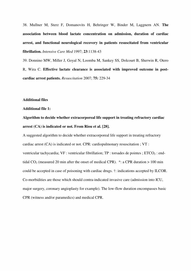
Additional file 1:
Algorithm to decide whether extracorporeal life support in treating refractory cardiac
arrest (CA) is indicated or not. From Riou et al. [28].
A suggested algorithm to decide whether extracorporeal life support in treating refractory
cardiac arrest (CA) is indicated or not. CPR: cardiopulmonary resuscitation ; VT :
ventricular tachycardia; VF : ventricular fibrillation; TP : torsades de pointes ; ETCO2 : end-
tidal CO2 (measured 20 min after the onset of medical CPR). *: a CPR duration > 100 min
could be accepted in case of poisoning with cardiac drugs. †: indications accepted by ILCOR.
Co-morbidities are those which should contra-indicated invasive care (admission into ICU,
major surgery, coronary angioplasty for example). The low-flow duration encompasses basic
CPR (witness and/or paramedics) and medical CPR.

Table 1 Main characteristics of the patients (n=51).
Variable Values Range
Age (year) 42 + 15 13-70
Men
Women
46 (90%)
5 (10%)
Comorbidity
- Hypertension
- Diabetes mellitus
- Ischemic heart disease
- Other cardiac disease
6 (12%)
3 (6%)
11 (20%)
10 (20)
Site of cardiac arrest
- home
- work
- public
- sport
19 (37%)
6 (12%)
20 (39%)
6 (12)
Initial rhythm
- ventricular fibrillation
- asystole
- pulseless rhythm
32 (63%)
15 (29%)
4 (8%)
Defibrillation
- patients receiving shock
- number of shocks
37 (72%)
4 [2;6]
1-20
Epinephrine
- patients receiving epinephrine
- dose (mg)
51 (100%)
13 [10;20]
2-100
End tidal CO2 (mmHg) 22 + 12 0-50
Delay
- fall to basic CPR (min)
- fall to advanced CPR (min)
- fall to automated CPR (min)
- fall to ICU admission (min)
- fall to ECLS onset (min)
3 [1;7]
12 [5;23]
41 [30;55]
90 [65;115]
120 [102;149]
1-22
0-40
15-110
48-175
75-195
Biological measurement
-Arterial pH
-Blood lactate (mmol.L-1
)
-Arterial bicarbonate (mmol.L-1
)
-PaO2 (mmHg)
-PaCO2 (mmHg)
-Blood potassium (mmol.L-1
) *
-Serum creatinine (µmol.L-1
)
-Prothrombin time (%)
- Fibrinogene (g.L-1
)
-Hemoglobin (g.L-1
)
-Troponin Ic (µg.L-1
)
-Protein S100 (µg.L-1
) †
6.93 ± 0.17
19.9 ± 6.7
16.5 ± 12.1
135 + 129
69 + 25
5.1 + 1.7
129 ± 30
39 ± 16
1.3 [<0.6;1.6]
109 ± 25
3.98 [0.93;85.5]
4.2 [2.4;10.4]
6.56-7.25
7.7-40.8
1.9-58.7
6-489
19-128
2.7-10.5
51-275
11-66
<0.6-3.6
59-169
0-669.0
0-36.0
Values are mean + SD, median [25;75 interquartile], or number (percentages). CPR:
cardiopulmonary resuscitation; ECLS: extracorporeal life support. *: blood potassium could
not be measured in 4 patients because of hemolysis.†: Protein S100 was measured in only 27
patients.

Legends for figures
Figure 1. Relationship between initial blood lactate level and delay between fall and onset of
extracorporeal life support (ECLS) (n=48).
Figure 2. Kinetic of arterial pH (Panel A) and arterial blood lactate (Panel B) during the first
hour following extracorporeal life support (ECLS) (n=38). Box plot represents median, 25-75
interquartile, and extreme values.
Figure 3. Distribution of the values of no-flow, low-flow, end-tidal CO2 (ETCO2) initial
arterial pH, blood lactate, and kaliemia in the studied population (n=51). The grey zones and
vertical bars indicate the threshold considered in the French guidelines for no-flow (≤ 5 min),
low flow (≤ 100 min), and ETCO2 (≥ 10 mmHg).

Figure 1

Figure 2

Figure 3

Additional files provided with this submission:
Additional file 1: AdditionalFile1.doc, 36Khttp://ccforum.com/imedia/1002590825069768/supp1.doc