Embed Size (px)
Citation preview

1
Critical Care Nurses Association of the Philippines, Inc 2013 CCNAPI Annual Convention
February 21-22, 2013 Diamond Hotel Philippines, Manila
PROGRAM OF ACTIVITIES
DAY 1: Thursday, February 21, 2013 7:00 am - 8:00 am REGISTRATION
8:00 am - 8:30 am OPENING CEREMONY Ribbon Cutting of Scientific Exhibitors HON. CARMENCITA M. ABAQUIN
Chairmam, Board of Nursing Together with the other Members of the Board of Nursing
Doxology and National Anthem MARIA RHONA T. ESTACIO, RN ICU Nurse, The Medical City
Welcome and Opening Remarks FERDINAND P. AGANON, RN, MAN
Over-all Chair, 2013 CCNAPI Annual Convention Acknowledgement of Participants BEDA B. GALICIA, RN
Secretary, Critical Care Nurses Association of the Philippines, Inc. 8:30 am - 9:30 am Introduction of the Keynote Speaker
MARIA ISABELITA C. ROGADO, RN, MAN President, Critical Care Nurses Association of the
Philippine, Inc. Message from the Keynote Speaker NOEL C. CADETE, RN, MAN
National President, Philippine Nurses Association COFFEE BREAK
9:30 am - 10:30 am PLENARY 1: “Rapid Response in Acute Brain Attack” DAIWAI M. OLSON, PhD, RN, CCRN
Associate Professor of Neurology & Neurotherapeutics
Associate Professor of Neurosurgery Staff Nurse, Neurocritical Care Unit University of Texas Southwestern
10:30 am – 11:30 am PLENARY 2: “Interpretation of Dysrhythmias in ACLS” MARIA ISABELITA C. ROGADO, RN, MAN Professor, Arellano University Graduate School of
Nursing 11:30 am -12:30 pm PLENARY 3: “Challenges in Sepsis Management”
DAIWAI M. OLSON, PhD, RN, CCRN
2
12:30 pm - 1: 00 pm LUNCH BREAK 1:00 pm -5:00 pm Beyond the Books: Self Directed Learning (Workshop)
Workshop 1 : “Assessment and Management of the Neurological Patient”
Main Preceptor : Ferdinand P. Aganon, RN, MAN Nurse Manager, Critical Care Unit- Nursing Service Dept. The Medical City
Preceptors : Louie Paul Eugenio, RN Sherlline Carillo, RN Celedonia M. Bienes
Speaker : Richmond G.O. Chang, MD, DPBA Sponsor : Biosolutions, Inc.
Workshop 2 : “Early Goal Directed Therapy in Sepsis” Main Preceptor : Beda B. Galicia, RN Assistant Nurse Manger, The Medical City
Preceptors : Charliemagne M. Marinas, RN Raizzah T. Mansoura, RN
John Christian M. Agustin, RN Sponsor : CCNAPI Workshop 3 : “Rapid Interpretation and Management of Dysrhythmias
in ACLS” Main Preceptor : Maria Isabelita C. Rogado, RN, MAN
Preceptors : Marvin S. De La Cruz, RN Mark Gil De De La Rosa, RN Rogelio E. Gayeta, Jr., RN
Sponsor : Hospira Philippines, Inc.
DAY 2: Friday, February 22, 2013 8:00 am - 9:00 am PLENARY 4: “Nuts and Bolts in Neonatal Resuscitation”
WILFREDO R. SANTOS, MD, FPPS, FPSNBMJ President, Philippine Society of Newborn Medicine Neontologist, University of Santo Tomas
9:00 am – 10:00 am PLENARY 5: “Wound Care Assessment and Management” RAMON O. RIBU, MD
Section Head, CTAMS & Wound Care, Philippine Heart Center 10:00 am – 10:15 am COFFEE BREAK 10:15 am – 11:00 am PLENARY 6: “Palliation and Spiritual Care: Phenomenology
Approach in Critical Care” RUDOLF CYMORR KIRBY P. MARTINEZ, MAN, RP-RN, CAA, LMT, CSTP, PhD(c)
Complementary & Alternative Therapy Nurse Practitioner Pain Management Clinician and Staff Nurse
Philippine Children‟s Medical Center

3
11:00 am - 12:00 nn PLENARY 7: “Empowerment of Nurses through a Career Progression Program”
MARCO ANTONIO STO. TOMAS, RN, MAN Member, Board of Nursing
Chair, Council for Nursing Advancement, Recognition & Specialization 12:00 nn - 1: 00 pm LUNCHEON SYMPOSIUM (ABBOTT Laboratories)
1:00 pm -5:00 pm Beyond the Books: Self Directed Learning (Workshop) Workshop 4 : “Dynamic of Wound Care: Assessment and Wound Bed Preparation”
Speaker: : Enrique M. Casto, III, MD Main Preceptor : Ferdinand P. Aganon, RN, MAN
Preceptors : Louie Paul Eugenio, RN Sherlline Carillo, RN Celedonia M. Bienes
Sponsor : BBraun Philippines Workshop 5 : “Resuscitation of the Newborn: The Basics”
Main Preceptor : Maria Isabelita C. Rogado, RN, MAN : Florentina Uy-TY, MD Preceptors : Marvin S. De La Cruz, RN
Mark Gil De De La Rosa, RN Rogelio E. Gayeta, Jr., RN
Sponsor : CCNAPI Workshop 6 : (BMI Computation)” Main Preceptor : Beda B. Galicia, RN
Preceptors : Charliemagne M. Marinas, RN Raizzah T. Mansoura, RN John Christian M. Agustin, RN
Speaker : Cristy Rodondo, MD Sponsor : Abbott Nutrition Laboratories
5:00 pm – 5:30 pm CLOSING CEREMONIES Closing Remarks Convention Evaluation
Raffle Draw Distribution of Certificates
4
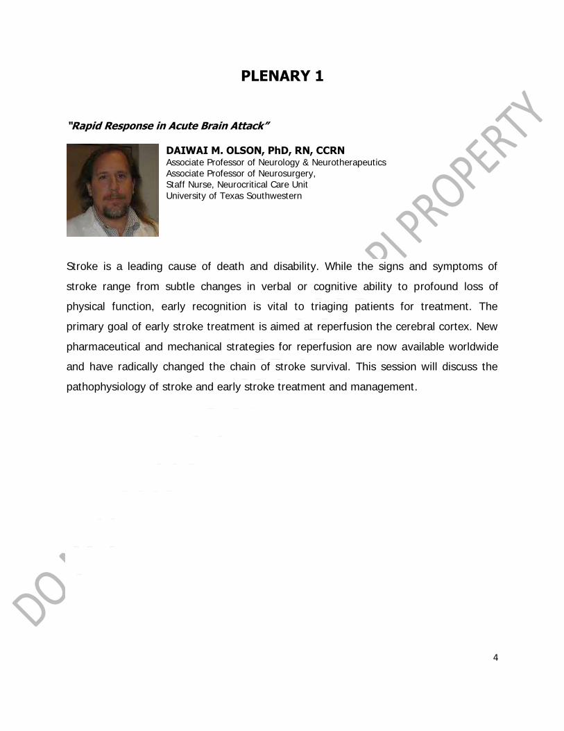
PLENARY 1
“Rapid Response in Acute Brain Attack”
DAIWAI M. OLSON, PhD, RN, CCRN Associate Professor of Neurology & Neurotherapeutics Associate Professor of Neurosurgery, Staff Nurse, Neurocritical Care Unit
University of Texas Southwestern
Stroke is a leading cause of death and disability. While the signs and symptoms of
stroke range from subtle changes in verbal or cognitive ability to profound loss of
physical function, early recognition is vital to triaging patients for treatment. The
primary goal of early stroke treatment is aimed at reperfusion the cerebral cortex. New
pharmaceutical and mechanical strategies for reperfusion are now available worldwide
and have radically changed the chain of stroke survival. This session will discuss the
pathophysiology of stroke and early stroke treatment and management.

5
Rapid Response in Acute Brain Attack
DaiWai M. Olson PhD RN CCRN Associate Professor of Neurology & Neurotherapeutics
Associate Professor of Neurosurgery University of Texas – Southwestern
Dallas, TX
Objectives: • Early recognition and identification of the signs and symptoms of stroke
• Triage and evaluate stroke patients • Strategies in preventing therapeutic delays • Updates in the Chain of Stroke Survival
• Approaches to early treatment and management
Acute Ischemic Stroke Versus Hemorrhagic Stroke
Stroke = disruption of blood flow to an area of the brain AIS = disrupts blood flow by clot ICH disrupts blood flow by diversion (secondary pressure)
SAH disrupts blood flow by diversion (secondary vasospasm) *Any Stroke = Brain Attack
Discussion: Primarily ischemic stroke
EARLY RECOGNITION AND IDENTIFICATION OF THE SIGNS AND SYMPTOMS OF
STROKE.
*To understand the signs and symptoms we need to have some understanding of the
pathophysiology.
Brain
• Brain does not store oxygen • Brain does not store glucose
• Apoptosis • Need to restore PERFUSION
How do we produce ENERGY?
Energy = ATP. ATP production is either aerobic or anaerobic.

6
Energy Production
STEP 1: Glycolysis - Glycolysis produces 2 ATP. Without oxygen, pyruvate is converted into
lactate (lactic acid)
• Does not require oxygen
• Is the first step in converting food to energy
• Occurs in the cytoplasm
• Produces pyruvate and NADH
Kreb‟s(1937) Citric Acid Cycle
*You must have ATP and oxygen to initiate glycolysis .
Apoptosis - Cells are pre-programmed to commit SUICIDE. Neurons are „sort of‟ like lemmings
How can we disrupt apoptosis?
• Think about the pathway. It is more than just restoring blood FLOW.
Perfusion
-Perfusion is more than “blood flow”. The delivery of nutrient rich blood to biological tissues.
French “perfuser” = “to pour over”
Poisseulle‟s Theorem
Flow =(π) x (r2) x (perfusion pressure)/ (8) x (blood viscosity) x (vessel length)
*Flow is determined by the size of the pipe and pressure you are using to push it
through.Divided by. How thick the fluid is and how far it has to go.
Oxygen & Hemoglobin
- HgB has four polypeptide chains. Each chain has one heme group. Each heme group has one
iron ion. Each iron ion can bind with one O2 molecule. THEREFORE – each HgB can bind with 4
O2 molecules
Early recognition and identification of the signs and symptoms of stroke.
• FAST - Face, Arms, Speech and Time
• NIHSS - more complicated but more thorough. These are TOOLS, Primarily for
Healthcare Workers. There is, however, a good rule of thumb

7
• MINOR STROKE / TIA
Triage and evaluate “ALL” stroke patients
Conclusions: Patients with TIA have
similar or worse
12-month post discharge risk of death or re-
hospitalization as compared with those with AIS.
Outcomes after TIA and AIS might be improved with
better adherence to secondary preventive guidelines.

8
Preventing Delays
1. Communication & Teamwork
2. Process 3. Organizational Culture
4. Performance Monitoring & Feedback 5. Overcoming Barriers
What do we already know ?
• Early tPA improves outcomes in acute ischemic
stroke
• EMS pre-notification is not a standard of care
• EMS pre-notification might be helpful – but
data is limited
*Disclosures*
Dr. Olson is a member of the Duke Clinical Research Institute (DCRI) at Duke University. The
DCRI serves as the data coordinating center for the American Heart Association‟ Get With The
Guidelines programs.
Funding / Support: The Get With The Guidelines-Stroke (GWTG-Stroke) program is provided by
the American Heart Association/American Stroke Association. The GWTG-Stroke program is
currently supported in part by a charitable contribution from Bristol-Myers Squib/Sanofi
Pharmaceutical Partnership and the American Heart Association Pharmaceutical Roundtable.
GWTG-Stroke has been funded in the past through support from Boeringher-Ingelheim and
Merck.
*Evidence*
• Lin C, Peterson ED, Smith EE, Saver JL, Liang L, Xian Y, Olson DM, Shah BR,
Hernandez AF, Schwamm LH, Fonarow G. (2012).
Patterns, predictors, variations and temporal trends in emergency medical service
hospital pre-notification for acute ischemic stroke.
Journal of the American Heart Association.
DOI: 10.1611/JAHA.112002345.
• Lin C, Peterson ED, Smith EE, Saver JL, Liang L, Xian Y, Olson DM, Shah BR,
Hernandez AF, Schwamm LH, Fonarow GC. (2012)
Emergency medical service hospital pre-notification is associated with improved
evaluation and treatment of acute ischemic stroke.
Circulation: Cardiovascular Quality and Outcomes.
DOI:10.1161/CIRCOUTCOMES.112.965210

9
Goal of this Study
1. Evaluate whether EMS pre-notification is associated with use of tPA among patients
arriving within either 3 hours or 4.5 hours
2. Evaluate if EMS pre-notification is associated with timeliness in acute ischemic stroke
evaluation and treatment, among those treated
Study Population
• Get With the Guidelines- Stroke
registry
• April 1, 2003 and April 2, 2011
• Final study population: 371,988
EMS-transported patients from 1,585
participating sites
Statistical Considerations
• Variable:
EMS pre-notification vs No EMS pre-
notification
• Outcomes
• Door-to-Imaging (DTI) time, DTI ≤
25min
• Door-to-Needle (DTN) time, DTN ≤
60min
• Arrival by 2h, treat by 3h
Univariate analysis: Pearson Chi-square test and Wilcoxon rank-sum test
Multivariable logistic regression analysis generalized estimating equations (GEE)
p = Probability
- Despite how improbable you may think an event is ... It may still happen!
“Chaz The Magnificent”
I was in Vegas and got to talking with the great and powerful (but homeless)
Chaz The Magnificent
Chaz wanted me to take him into the casino (he wasn‟t allowed in unless accompanied by someone who had bathed) and we would bet on roulette. C.t.M. guaranteed me that voices in his head could predict the outcome of any binomial distribution. And therefore – he wanted to play roulette…betting on red and black. I told C.t.M. that if he could demonstrate the ability to predict a random binomial event sufficiently such that he was better than chance… I would take him gambling (but only after he took a bath).

10
I opted to test C.t.M. outside the casino – using the toss of a coin.
I toss a coin (not actually random…but close enough for our example)
If C.t.M. guesses (the voices tell him) correctly - - -Then the answer is “yes”
So, What can we say about C.t.M.
after 2 tosses of the coin?
Chance – and chance alone –
Gives a 0.25 probability that the voices in
C.t.M.‟s head will be correct
on 2 tosses of the coin.
I‟m not willing to gamble - YET
What would convince you?
How many times would C.t.M. have correctly state the coin toss before you
bet YOUR LIFE SAVINGS at roulette
based on the voices in his head?
Show of hands (2,3,4,5,6,7,8,9,10,20,50, 95)
There are 32 possible outcomes for the voices.
Only 1 of the 32 is Y-Y-Y-Y-Y
P(YYYYY) = 1/sum of all probable outcomes =
1/32 = .0250 = .03
Therefore “the voices are right (p < .05)”
11
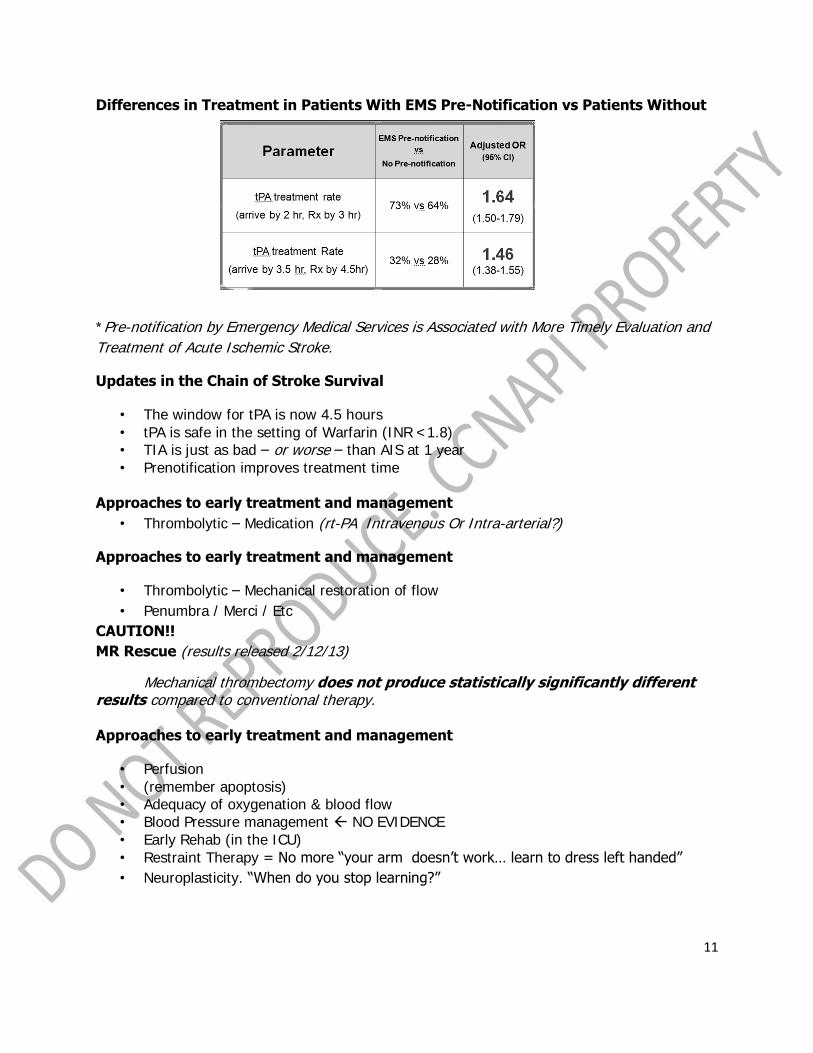
Differences in Treatment in Patients With EMS Pre-Notification vs Patients Without
*Pre-notification by Emergency Medical Services is Associated with More Timely Evaluation and
Treatment of Acute Ischemic Stroke.
Updates in the Chain of Stroke Survival
• The window for tPA is now 4.5 hours
• tPA is safe in the setting of Warfarin (INR <1.8) • TIA is just as bad – or worse – than AIS at 1 year
• Prenotification improves treatment time
Approaches to early treatment and management
• Thrombolytic – Medication (rt-PA Intravenous Or Intra-arterial?)
Approaches to early treatment and management
• Thrombolytic – Mechanical restoration of flow
• Penumbra / Merci / Etc
CAUTION!!
MR Rescue (results released 2/12/13)
Mechanical thrombectomy does not produce statistically significantly different results compared to conventional therapy.
Approaches to early treatment and management
• Perfusion
• (remember apoptosis) • Adequacy of oxygenation & blood flow • Blood Pressure management NO EVIDENCE
• Early Rehab (in the ICU)
• Restraint Therapy = No more “your arm doesn‟t work… learn to dress left handed”
• Neuroplasticity. “When do you stop learning?”

12
Secondary Prevention
AVAIL: Hospital Recruitment
• AHA – GWTG hospitals (January 2006) – Roughly 300 hospitals
• Contacted to participate based on enrollment in GWTG-Stroke
– Actively enrolling
– Committed to continued enrollment
– Research coordinator/Site
coordinator
AVAIL: 12 month persistence by medication class
Patient-Related Factors and Persistence

13
Approaches to early treatment and management
Transitions of Care
Olson DM, Prvu Bettger J, Alexander KP, Kendrick AS, Irvine JR, Wing L, Coeytaux RR, Dolor RJ, Duncan PW, Graffagnino C.
Evidence Report No. 202. (Prepared by the Duke Evidence-based Practice Center under
Contract No. 290-2007-10066-I.) AHRQ Publication No. 11(12)-E011. Rockville, MD. Agency for Healthcare Research and Quality.
October 2011. How was this done?
• 5,783 citations identified past 2000-2011
• 4605 – excluded based on title/abstract • 22 – excluded based on not original data
• 750 articles subjected for FULL READ • 668 – excluded (534-not transition, not peer, no data, no comparator, not stroke/MI) • 34 studies of 4,146* stroke patients
• 19 studies of 15,216 MI pts *Everything we know is based on less than 5,000 patients
Intervention – TYPE
1. Hospital-Initiated Support 2. Patient and Family education
1. hospital-based 2. home-based
3. Community-based Support
4. Chronic Disease management
*
In the interest of time = = = we will only look at
Hospital-Initiated Support

14
Conclusion • Early Supported Discharge may be helpful
Certainly the most promising • For stroke – no other intervention had sufficient evidence of benefit to be
recommended. *only 34 studies in the history of man in the entire world
*only 18 studies of stroke and ESD
Caregivers
• Most often Spouse/partner (in transition studies) • But can be anyone
Usual care
• Most studies used “usual care” as their comparator, but few studies actually tell us what
usual care really is.
What does this mean?
• Stroke care continues to evolve • Nursing care is an important component of stroke care • There is much more work to be done
• YOU can do an important study ! ! !

15
PLENARY II
“Interpretation of Dysrhythmias in ACLS”
MARIA ISABELITA C. ROGADO, RN, MAN Professor, Arellano University Graduate School of Nursing
In this age of the new millennium, EKG interpretation is an expected skill amongst
nurses not only in the critical care areas but in any setting where there is need for
cardiac monitoring. This skill in EKG interpretation is necessary particularly in the events
of resuscitation. It is essential that institutions have emergency policies and procedures
in place, along with a continuing competency education program and yearly refresher
programs. These programs should include validation of dysrhythmia interpretation skills
and problem solving of case studies.
This topic on Interpretation of Dysrhythmia in ACLS will tackle the basics of EKG
interpretation and how nurses can rapidly determine what they see on a scope or
rhythm so that appropriate and early management can be instituted. It will cover
dysrhythmias such as tachycardia and bradycardia in pre-arrest scenarios; shockable
and non-shockable rhythms during arrest scenarios and appreciation of ST elevation
during Acute Coronary Syndrome.

16
INTERPRETATION OF DYSRHYTHMIAS IN ACLS
MARIA ISABELITA C. ROGADO, RN, MAN Professor,
Arellano University Graduate School of Nursing
OBJECTIVES
• Relate skills of dysrhythmia interpretation to clinical assessment and selection of
appropriate dysrhythmia treatment algorithm
• Discuss how dysrhythmia interpretation, patient assessment findings, and treatment
options are evaluated and selected
INTRODUCTION: THE BASICS
Newton‟s Third Law:
“To every action, there is equal and opposite reaction”
Cardiac Output: the amount of blood ejected by the left
ventricle in one minute
• Adult: 5-8 liters of ejected blood per minute.
• With strenuous activity, CO can increase to an amazing 25 liters per minute
• Cardiac Output = Stroke Volume x Heart Rate • Stroke volume is the amount of blood ejected by
the left ventricle with each contraction.
• SV = EDV - ESV

17
CONDUCTION SYSTEM
Functions: 1. Generate electrical impulses
2. Conduct impulses to the heart muscles PQRST
P: Atrial depolarization
QRS: Ventricular depolarization
T: Ventricular Repolarization
PR: Conduction of the impulse from
SA node to the AV node
ST : End of Ventricular depolarization
and start of ventricular repolarization
INTRODUCTION: DYSRRHYTMIAS
• Clinical dysrhythmia interpretation must be made in conjunction with patient
assessment.
• Consider the effect that a particular dysrhythmia on a patient‟s well-being
• Determine the significance of the abnormal rhythm disturbance
• Decision can be made as to which therapy is indicated.
Case #1:
“The weak and dizzy elderly woman”
In the ED, a 64-year-old wife was complaining of dizziness and generalized weakness for the
last two hours.
The patient, awake and appearing weak, describes she had brief “passing out” while getting up
from the bed to go to the bathroom. She was feeling fine until about two hours ago, when she
suddenly became dizzy and had to be helped to lie down
The patient denies chest pain, shortness of breath, diarrhea, palpitations, nausea or vomiting,
blood in her bowel movements, abnormally dark stools, or prior episodes similar to today‟s
events.
Her past medical history is significant for hypertension for which she is taking a diuretic and an
ACE-inhibitor.
Her medication has not been changed recently, and she takes her medication as directed.

18
Physical Examination:
• Alert and oriented;
• BP: 84/60 mmHg; PR: 36/min.; RR: 26/min.
• SpO2: 98% saturation.
• Her skin is pale and sweaty,
• Neck veins are not distended.
• Breath sounds: clear
• Heart sounds: regular without a murmur.
• Abdominal and neurologic examinations are normal
Initial Action:
• Oxygen is administered and
• Monitoring (cardiac) is started along with
• Insertion of an intravenous intermittent infusion device.
The ECG monitor recorded the tracing
*Third Degree Heart Block
with ventricular beat at 37
bpm
• Independent atrial (P) and ventricular (QRS) activity
• Very slow regular ventricular rhythm
• QRS complexes are wide and distorted, (low ventricular escape pacemaker)
• P-R intervals have no constant value; unrelated; being paced differently
BRADYCARDIA ALGORITHM
• Symptomatic bradycardia: a heart rate less than 60 bpm that elicits signs and symptoms
• Symptomatic bradycardia exists when the following 3 criteria are present:
1.) The heart rate is slow;
2.) The patient has symptoms; and
3.) The symptoms are due to the slow heart rate
*Functional or relative bradycardia occurs when a patient may have a heart rate within normal sinus
range, but the heart rate is insufficient for the patients condition. An example would be a patient with an
heart rate of 80 bpm when they are experiencing septic shock.
Decrease Cardiac Output
• Signs: – Hypotension
– Orthostatic HpN
– Diaphoresis
– Pulmonary congestion on PE and CXR
– Congestive Heart Failure
– Pulmonary Edema
– Brady-related frequent escape PVC or
VT
• Symptoms: – Chest discomfort or pain
– Shortness of breath
– Decreased level of
consciousness
– Weakness
– Fatigue
– Light headedness
– Dizziness, syncope

19
Unstable Persistent Bradycardia
• Atropine
– First Dose: 0.5 mgs bolus
– Repeat every 3-5 minutes
– Maximum: 3 mgs
• Transcutaneous Pacing
• Dopamine Infusion
– 2-10 mcg/kg per minute
• Epinephrine Infusion
– 2- 10 mcg/minute
Sinus Bradycardia
Rhythm : Regular
Rate : less than 60 BPM P waves : present before every QRS consistent in shape
PR interval : usually normal QRS : usually normal Conduction : normal

20
FIRST DEGREE AV BLOCK
Rate : Usually within normal range, but depends on the underlying
rhythm
Rhythm : Regular
P waves : Normal in size and shape
PR interval : prolonged (>0.20 sec) but constant
QRS duration : Usually 0.10 sec or less unless an intraventrucular conduction
delay exists
FIRST DEGREE AV BLOCK
• Not a dysrhythmia itself
• Condition describing the consistent prolonged PR interval
• Impulse from SA to ventricles are delayed (not blocked) in the AV node
What Causes it?
• Ischemia
• Medications
• Rheumatic Heart Disease
• Hyperkalemia
• Acute MI (Inferior wall MI)
• Increase vagal tone
FIRST DEGREE AV BLOCK
What Do I Do About It?
• First Degree AV Block in acute MI must be monitored closely
• Symptomatic bradycardia – Treat bradycardia!
• Oxygen
• IV access
• IV Atropine
• Transcutaneous pacing

21
SECOND DEGREE AV BLOCK
Type I (Wenckebach, Mobitz Type I)
Rate : Atrial rate is greater than ventricular rate Rhythm : Atrial Regular, ventricular irregular
P waves : Normal in size and shape, some Pws are not followed by QRS PR interval : Lengthens with each cycle
QRS duration : Usually 0.10 sec or less but is periodically dropped
• Wenckebach pattern is the progressive lengthening of the PR interval followed by a P
wave with no QRS
• Conduction delay happens at the level of the AV node
What Causes it?
• Associated with RCA block (supplies AV node in 90% of the population)
• Disturbance in the balance between sympathetic and parasympathetic divisions
• Increase in parasympathetic tone – conduction in AV is slowed down
What Do I Do About It?
• Symptomatic due to meds – Stop medication!
• Slow heart rate with serious symptoms
• Atropine
• Temporary Pacing
• Observe closely for increasing AV block

22
Type II (Mobitz Type II)
Rate : Atrial rate is greater than ventricular rate, VR often slow
Rhythm : Atrial Regular, ventricular irregular
P waves : Normal in size and shape, some Pws are not followed by QRS
PR interval : Within normal limits or slightly prolonged but constant for the
conducted beats. There may be some shortening of the PRI that follows a nonconducted beat
QRS duration: Usually 0.10 sec or greater, periodically absent after Pw
• Conduction delay occurs below the AV node (Bundle of His or at the level of the Bundle
Branches
• More serious than Mobitz I
• Often progresses to complete heart block
What Cause It?
• LCA supplies the bundle branches
• Acute myocarditis
• Anterior MI
• Other types of organic disease
2:1 Conduction (2:1 AV Block)
• Two pw occur for every QRS (2:1 conduction)
• Narrowed QRS - associated with Second Degree Type 1
• Wide QRS – associated with delay in the conduction below the AV node and is usually a
type II block
23
What Do I Do About It?
• Prepare for pacing
• Atropine should be avoided
• Will not improve the block but will increase the rate of discharge of the SA node
• Triggers the situation in which fewer impulses are conducted through the
ventricles therefore VR is furthered slowed down
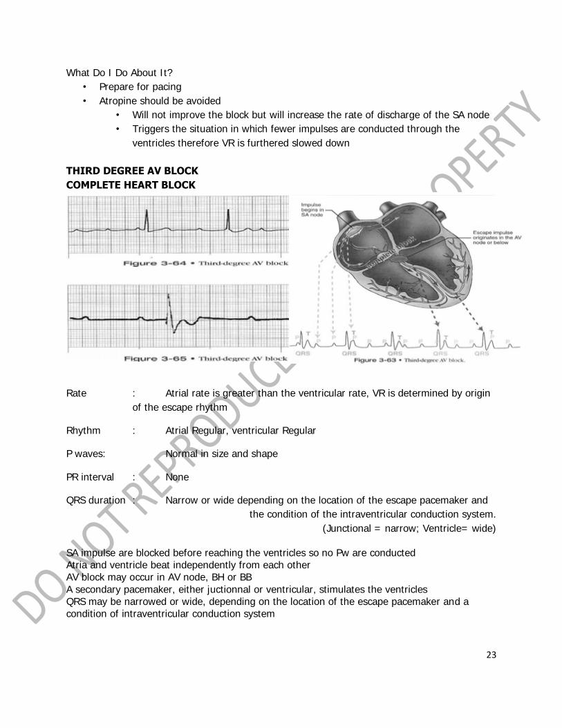
THIRD DEGREE AV BLOCK
COMPLETE HEART BLOCK
Rate : Atrial rate is greater than the ventricular rate, VR is determined by origin
of the escape rhythm
Rhythm : Atrial Regular, ventricular Regular
P waves: Normal in size and shape
PR interval : None
QRS duration : Narrow or wide depending on the location of the escape pacemaker and
the condition of the intraventricular conduction system.
(Junctional = narrow; Ventricle= wide)
SA impulse are blocked before reaching the ventricles so no Pw are conducted Atria and ventricle beat independently from each other AV block may occur in AV node, BH or BB
A secondary pacemaker, either juctionnal or ventricular, stimulates the ventricles QRS may be narrowed or wide, depending on the location of the escape pacemaker and a condition of intraventricular conduction system

24
What Caused it? • Associated with an inferior MI
• Result of a block above the Bundle of His • Resulting rhythm is usually stable
• Escape pacemaker is usually junctional (narrow QRS) • VR of more than 40 bpm
What Do I Do About It? • Narrow QRS and symptomatic due to slow rate • Wide QRS and symptomatic
• Transcutaneous Pacemaker (TCP)
Bradycardia Algorithm 2010 AHA Update: For symptomatic bradycardia or unstable bradycardia IV infusion chronotropic agents (dopamine & epinephrine) is now recommended as an equally effective alternative to external pacing when atropine is ineffective.
Case #1 Treatment Considerations
• A : alert; good airway • B : adequate breathing, clear breath sounds; RR=26 SpO2 = 98%
• C : inadequate; slow HR (37 bpm), but not in need of cardiac compressions; BP = 80/60; signs of adrenergic discharge: pallor and diaphoresis
• D: dizzy with loss of consciousness initially; Now: alert and oriented; neuro check is
normal patient‟s symptoms appear to be directly related to her slow heart rate.
Causes
• Third-degree heart block in elderly patients is usually due to gradual fibrosis of the A-V conduction system
• The patient needs to be assessed and treated as if she were experiencing an acute coronary event
Treatment Plan
• Emergency Cardiac Care Treatment Algorithm for Adult Bradycardia • Stable or Unstable (?)
• Start with either atropine or artificial pacing
Case #2:
“The case of the man who „passed out‟ during his dialysis session.” • A 70-year-old male with a history of hypertension, end stage renal disease, and insulin-
dependent diabetes mellitus arrives in the emergency department
• He experiencing a brief syncopal episode during his dialysis treatment thirty minutes before arrival.
• The patient diaphoretic and hypotensive and was administered oxygen and 500 cc of normal saline in the Dialysis Center.
• This occurred just as he was finishing his dialysis session.
• His bedside glucometer determination was 140 mg/dl.

25
• Vital signs in the ED: • BP: 130/60 mmHg
• PR: regular at 150/min. • RR: 26 bpm
• The patient is alert, and afebrile • His lung sounds are clear and there is no pedal edema. • He denies chest pain or shortness of breath but is complaining of palpitations, nausea,
and generalized weakness.
*Atrial flutter with a 2:1 A-V conduction and a rapid ventricular rate at 150/minute
• The rhythm is grossly regular with a rapid ventricular rate, • ventricular (QRS) complexes are narrow. • P waves are not visible; seen as flutter waves,
• The ST segments are difficult to evaluate due to the flutter waves. • The A-V relationship consists of 2:1 conduction,
• Ventricular rate of 150/minute is very fast for a patient with limited cardiac reserve. TACHYDYSRHYTHMIAS
• Narrow-QRS Tachycardias • Wide-QRS Tachycardias • Irregular Tachycardias
• Atrial Flutter • Atrial Fibrillation
• Supraventricular Tachycardia (SVT) • Monomorphic VT • Polymorphic VT
• Wide-complex tachycardia of uncertain type

26
Sinus Tachycardia
Rhythm : Regular
Rate : faster than 100 BPM P waves : present before every QRS
consistent in shape may be hidden in Tw PR interval : normal, difficulty to measure
QRS : usually normal Conduction : normal
Atrial Flutter

27
Atrial Fibrillation
Supraventricular Tachycardia (SVT)

28
Tachycardia Algorithm
• An unstable tachycardia exists when cardiac output is reduced to the point of causing
serious signs and symptoms. • Serious signs and symptoms commonly seen with unstable tachycardia are:
chest pain, signs of shock, HpN altered mental status,
weakness, fatigue, and syncope • The most common causes of tachycardia that should be treated outside of the ACLS
tachycardia algorithm are:
dehydration, hypoxia,
fever, and sepsis Administration of OXYGEN and NORMAL SALINE are of primary importance for the treatment of
causative factors of sinus tachycardia and should be considered prior to ACLS intervention.
There may be other contributing causes and review of the H‟s and T‟s of ACLS should take
place as needed.
• Is the patient responsive?
• A-B-C-D – Oxygen (4L/m) – Hook the patient to
the monitor – Start an IV access

29
• Stable (narrow QRS complex) → vagal maneuvers → adenosine 6 mgs (if regular)
→ beta-blocker/calcium channel blocker → get an expert
• Stable (wide/regular/monomorphic) → adenosine (regular and monomorphic) →
consider antiarrhythmic infusion (Procainamide 20- 50 mgs / min; Amiodarone 150 mgs)
→ get an expert
• Unstable tachycardia should be treated immediately with synchronized
cardioversion.
2010 ACLS GUIDELINES
Synchronized Cardioversion
Atrial Fibrillation • Biphasic – 120J • Monophasic – 200J
Atrial Flutter/SVT • Biphasic/Monophasic – 50-100J
Monomorphic VT • Biphasic/Monophasic – 100J
New Medication Protocols
• Adenosine is recommended in the initial diagnosis and treatment of stable,
undifferentiated regular, monomorphic wide-complex tachycardia
• It is important to note that adenosine should not be used for irregular wide-complex
tachycardias because it may cause degeneration of the rhythm to VF.
• Indication: • PSVT
IV Dose: 6 mg bolus followed by 12 mg in 1-2 minutes if needed *Adenosine IV Bolus
• Very short half-life • Rapid bolus • 6 mg given as a rapid intravenous bolus (administered over a 1-2 second period) • Followed by 20 cc NSS • Elevate arm / site
Case #3: “The case of the hospitalized patient recovering from an MI.”
• A 54-year-old man, was recovering in the • intensive care unit from an uncomplicated inferior wall MI that he experienced 24 hours
before. • Thrombolytic therapy opened the occluded coronary artery and his chest pain, along
with the acute ECG changes, subsided. • The nurse went to his room in response to a dysrhythmia alarm and found the patient
unresponsive, propped up in bed, not breathing, and without a pulse.
• The nurse summoned help, lowered the head of the bed, and observed the ECG rhythm

30
The ECG monitor recorded the tracing
• The rhythm consists of a rapid, disorganized ventricular tachydysrhythmia.
• Distinct QRS complexes are absent. • The ECG baseline zigzags across the tracing. • Discrete P waves are also not visible, AV relationship cannot be determined.
• The patient‟s clinical appearance confirms the interpretation
Initial Treatment Considerations
• Goal: to convert this life-threatening ECG dysrhythmia to a nonlethal rhythm.
• Initiate countershock as rapidly as possible at 360 joules
• Start CPR
• Epinephrine and Amiodarone
SHOCKABLE RHYTHMS
• Pulseless Ventricular Tachycardia (PVT)
• Ventricular Fibrillation
Pulseless VT / VF Algorithm
• Ventricular Tachycardia
– Monomorphic
– Polymorphic
– Torsades de Pointes
• Ventricular Fibrillation
– Course
– Fine

31

32

33
*Fine VF
*Course VF *The best hope for resuscitation remains early detection and early defibrillation. For persistent or recurrent ventricular fibrillation or pulseless ventricular tachycardia, consider the following anti-arrhythmic therapies: Amiodarone in a 300-mg rapid intravenous bolus. Lidocaine (1 mg/kg) intravenously. Magnesium sulfate in a 1- to 2-g dose for polymorphic forms of VT or in known
hypomagnesemic states. Procainamide IV at 30 mg/min up to 17 mg/kg (average of 1200 mg per 70-kg
patient) can be given for refractory cases, but it is not recommended because it is often not practical to administer during cardiac arrest due to the preparation and administration time
Non-Shockable Rhythms
• Pulseless Electrical Activity (PEA)
• Asystole

34
Case #4: “The case of a pulseless rhythm”
• The nurse went to his room in response to a dysrhythmia alarm and found the patient unresponsive, propped up in bed, not breathing,
and without a pulse. • The nurse summoned help, lowered the head of the bed, and
observed the ECG rhythm
Pulseless Electrical Activity
• Always check the pulse with the rhythm
• Vassopressor (IV / IO) – Epinephrine 1 mg repeat every 3-5 minutes – Vassopressin 40 units
• Administer drugs during CPR. Do not stop CPR to administer drugs. • Consider advanced airway and capnography
Asystole
• Things to check to ensure that this is true asystole: – loose leads or leads not connected to the patient;
– signal gain; – ensure that the patient is pulseless – You confirm that this is true asystole and that the patient has no pulse. You begin the
pulseless arrest algorithm. Your first step is to: – Begin CPR immediately – Give epinephrine 1 mg IV/IO

35
2010 ACLS Guidelines *New Medication Protocols
• Atropine is not recommended for routine use in the management of PEA/asystole and
has been removed from the ACLS Cardiac Arrest Algorithm
• Indications: • Symptomatic sinus bradycardia
• Second Degree Heart Block Mobitz I • IV Dose:
• .5 – 1 mg every 3-5 minutes • Max dose is .04mg/kg • Can be given down ET tube?

36
PLENARY III
“Challenges in Sepsis Management”
DAIWAI M. OLSON, PhD, RN, CCRN Associate Professor of Neurology & Neurotherapeutics
Associate Professor of Neurosurgery, Staff Nurse, Neurocritical Care Unit University of Texas Southwestern
Every critical care nurse is challenged to provide care to patients with sepsis and prevent sepsis
in patients at risk. The evidence-based practice for sepsis prevention is evolving from a robust
debate. Key to this discussion is an understanding of the pathophysiology of sepsis, including
identifying at risk populations. This session is aimed at discussing evidence for the role of the
nurse in both prevention and treatment of sepsis in the ICU setting.

37
“Challenges in Sepsis Management”
DAIWAI M. OLSON, PhD, RN, CCRN Associate Professor of Neurology & Neurotherapeutics
Associate Professor of Neurosurgery, Staff Nurse, Neurocritical Care Unit
University of Texas Southwestern
OBJECTIVES • Present factors that increase the risk of developing or preventing sepsis
• Discuss strategies / methods of prevention and early detection of Sepsis
• Discuss the current trends and advances in caring and managing sepsis
• Roles of the nurse in the prevention and management of sepsis
PATHOPHYSIOLOGY
In order to understand the body's response to sepsis, we must first review the pathophysiology.
Sepsis is a complex process; it is the body's systemic response to an infection. When the body
is unable to contain a localized infection at its source, the infecting organism leaks into the
bloodstream, causing sepsis. This is associated with inflammation, coagulopathy, and the
maldistribution of blood flow.
When the invading organism, or antigen, enters the bloodstream, it releases endotoxin, a toxic
substance usually associated with gram-negative bacteria. In response, the body's immune
system releases proinflammatory mediators, such as prostaglandins and cytokines, including
tumor necrosis factor and interleukins, into circulation.2,3,5 Cytokines are immunomodulators
released by white blood cells in response to the endotoxins, and together they are responsible
for causing vasodilatation, increased capillary permeability, and increased coagulation.5,6 In a
healthy person under normal circumstances, the body can control these processes and heal; but
in the septic patient, the endotoxins stimulate the release of too much of the
immunomodulators, causing an exaggerated, excessive response.3
Vasodilatation is the body's way of increasing blood flow to the affected area, thereby
transporting more white blood cells, such as macrophages, to control the original infection.
However, vasodilatation, without a proportionate increase in blood volume, leads to
hypotension. Increased capillary permeability allows fluid to leak out of the blood stream and
into surrounding tissue, causing edema. This further reduces blood pressure. Concurrently,
fibrinolysis is impaired leading to a decrease in clot breakdown. This is thought to be the body's
38
attempt at confining the antigen.3 However, the formation of fibrin clots leads to microthrombi,
causing hypoperfusion of tissues, tissue necrosis, and eventually organ failure.
SEPSIS IS NOT.. • Just a bad infection • An infection that has spread
• Systemic infection • Infection in the bloodstream (Septicemia)
• A viral infection • Determined by length of stay
Sepsis is a response to infection in which there is an interruption in the tissue perfusion to
vital organs
Heart Attack = interrupted perfusion to myocardium (caused by clot or plaque)
Brain Attack = interrupted perfusion to cerebral cortex (caused by clot or hemorrhage) Sepsis = interrupted perfusion to multiple vital organs (caused by infection)
Sepsis is an Emergency. ~ 35% of patients with sepsis will die. Early recognition and treatment is associated with higher recovery rate. Without treatment Sepsis will usually
progress to Septic Shock. ~50-75% of patients with Septic Shock will die.
Odds Ratio • Odds ratios are most typically used for case-control studies. • Odds ratios estimate the relative risk of a rare event being observed in some population
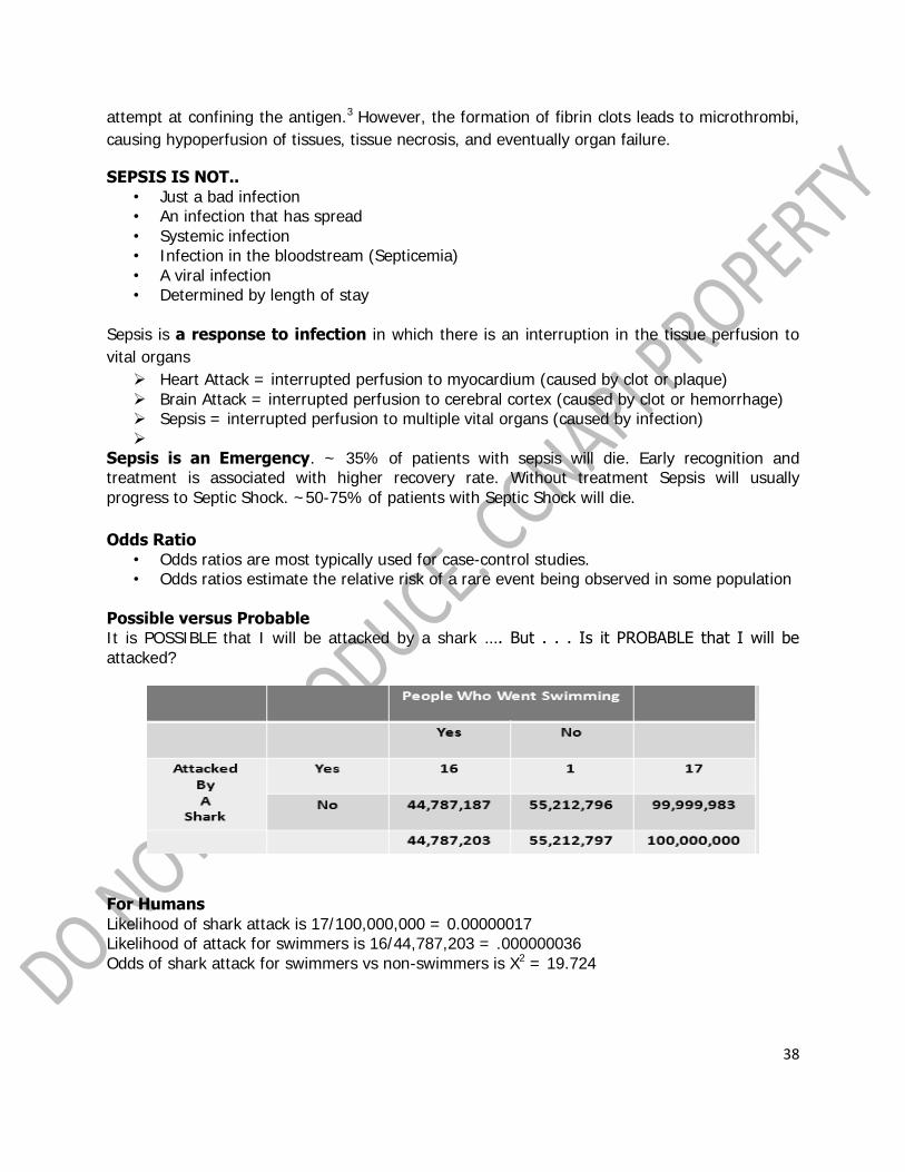
Possible versus Probable It is POSSIBLE that I will be attacked by a shark …. But . . . Is it PROBABLE that I will be
attacked?
For Humans
Likelihood of shark attack is 17/100,000,000 = 0.00000017 Likelihood of attack for swimmers is 16/44,787,203 = .000000036
Odds of shark attack for swimmers vs non-swimmers is X2 = 19.724

39
RISK of SHARK ATTACK among
• Did you go swimming at
DUSK?
• Did you get attacked by a
shark?
SO - - how do we interpret ?
Answer -- -- don‟t swim at DUSK
DUSK increases RISK.11 – thousand
times more likely If you swim and
You swim at dusk
For SWIMMERS
Likelihood of a shark attack is 15/44,787,203 = 0.00000349
Likelihood of shark attack for dusk swimmers is 15/59,795 = 000251
Odds of shark attack swimming dusk vs daytime = X2 = 11237.13

40
Factors that increase the risk of developing sepsis
• Very young - Very old • Very poor - Very rich
• Already sick – Infirm
• Post surgical / at home
– Hospitalized – Drug / Alcohol
– Cancer • Chemotherapy
• Open wounds/injuries (including trauma)
• Treatment conditions – Foley catheter – IV catheters
• Central Line – Wound drainage collection
– Immobility – Antibiotic – Hx Resistant infection (MRSA, VRE)
Dengue Fever / Dengue virus
• Dengue Fever does not CAUSE sepsis! …..BUT…. Dengue Fever may result in weak,
infirm, body in which a bacterial infection may result in sepsis.

41
Preventing sepsisfor patients WITH infection present
• Prompt recognition of the initial infection
– NURSING CARE = INSPECT !
• Good Health
• Good Nutrition
• Prevent spread of the infection
– Between organ systems
– To new tissue(s)
• Prevent new infection
– Secondary organism
Early Recognition
• Signs & Symptoms of infection
– Rubor = redness
– Tumor = swelling
– Dolar = pain
– Calor = heat
– Loss of Function
Prevention Strategies
• Bundles
– VAP, ICU, Central Line
• Handwashing campaigns
• SCIP (Surg Care Imp Prog)
• CHECKLIST MANIFESTO
• 10,000 lives campaign
• Environmental controls
– Healthier Hospitals Initiative
Specific Examples
• Pre-op check (“is this the right patient?”)
• LeapFrog
• VAP Bundle
• 100,000 lives
• Healthier Hospitals Initiative
• Closing the Gap Initiative
*Not a single initiative has resulted in more harm than good!

42
The Checklist Manifesto
Atul Gawanda
Perhaps the „best known‟ medical safety initiative
“Despite your ownpersonal awesomeness,
a checklist can help you.”

43
IHI
Institute for Healthcare Improvement
• IHI MRSA infection prevention – Screening – Preventive cross contamination
– HANDWASHING !
When does an Infection become SEPSIS?
• Disruption of perfusion to more than one organ/system
– Don‟t forget the SKIN
– Bladder
– Blood stream
– CSF
– Gut (multiple organs)
– Lymph
– Lungs
Systemic Inflammatory Response Syndrome (SIRS)
“OFTEN” preceeds SEPSIS
TWO or MORE of the below = SIRS
• Body temperature < 36C or > 38C
• Heart rate greater than 90 /min
• Hyperventilation (respiratory rate > 20 /min)
• PaCO2 < 32 mm Hg (normal 35 to 45 mm Hg)
• WBC > 12,000/mm3 or <4,000/mm3 (normal 5,000 to 10,000/mm3)
In addition to rubor, tumor, dolar, calor, loss of function
– Altered mental status
– Acute oliguria (urine output <0.5 ml/kg/h)
– Hyperglycemia in the absence of diabetes
– Hypoxemia
– Coagulopathy (INR > 1.5)
– Gastric ileus
Current trends and advances in caring and managing sepsis
- www.survivingsepsis.org

44
PRESSURE. A comparison of size and volume
PERFUSION. The delivery of nutrient rich blood to biological tissues
FLOW.
Poiseuilles‟ Theorem – This Address the flow.
Radius becomes the most
important determinant of CBF
Length is often considered a constant
Viscosity (think Hct)

45
How can you measure BP? McKay D.W.
Measuring blood pressure: a call to bare arms? Canadian Med Assn Journal. Feb 26 2008;178(5):591-593.
Key points of the article
• B/P readings are indirect measurements - accuracy depends on factors related to the patient
and the equipment.
• Automated cuffs, despite having clinical approval, may be inaccurate in some individuals for
reasons that are not well understood.
• Advances in the technology warrant a re-examination of the assumptions underlying B/P
measurement.
• Differences in patients and product may limit the generalizability.
Where can you measure BP?
Auscultation
• Arm
• Wrist
• Finger
• Thigh
• Ankle
Intra-arterial
• Radial
• Brachial
• Femoral
• Pedal
• Jugular
• Aorta
How can you measure perfusion?
• Mean arterial pressure (MAP)
• Oxygen extraction (O2ER)
• Measures of blood flow
– Velocity
– Stroke volume
• Measures of Metabolism
Why „did‟ we think MAP was a reflection of perfusion?
MAP = DBP + 1/3 SBP
MPAP = PDias + 1/3 PSys
Perfusion – Oxygenation
Supply side
• Arterial blood gas • Oxygen availability
(CaO2)
• Oxygen delivery

46
Arterial blood gas
• pH
• PaCO2
• Bicarb
• PaO2
Oxygen availability (CaO2)
– Hemoglobin
• 1.38 = the milliliters of oxygen carried per gram of hemoglobin (1.34 –
1.39)
– Dissolved oxygen / PaO2
– Arterial oxygen content
• Formula
– CaO2 = arterial oxygen content
– CaO2 = Hgb x SaO2 x 1.38 + (0.0031 x PaO2)
• Normal values
– CaO2 = 17 – 20 ml O2 / 100 ml blood
Oxygen delivery (DO2)
– Hemoglobin
• 1.38
– Cardiac output
• Stroke volume x Heart rate
– Formula
• DO2 = oxygen delivery
• DO2 = CO x Hgb x SaO2 x 1.38 x 10
– Normal values
• DO2 = 900-100 ml/min
• DO2I = 360-600 ml/min/m2
Demand Side
Venous oxygen content (CvO2)
– SvO2
• SvO2 = DO2 – VO2
• Normals= 60 – 75 mmHg
– Venous oxygen content
• Formula
– CvO2 = venous oxygen content
– CvO2 = Hgb x SvO2 x 1.38 + (0.0031 x PvO2)
• Normal values
– CvO2 = 13 – 16 ml O2 / 100 ml blood

47
Oxygen consumption (VO2)
– Formula
• VO2 = oxygen consumption
• VO2 = (CaO2 –CvO2) x CO x 10
• VO2 = CO x Hgb x (SaO2 –SvO2) x 1.38 x 10
– Normal values
• VO2 = 220-290 ml/min
• VO2I = 108-165 ml/min/m2
Oxygen balance
Oxygen extraction ratio (O2ER)
• Formula
– O2ER = oxygen extraction ratio
– O2ER = [ ( CaO2 –CvO2 ) / CaO2 ] x 100
• Normal values
– O2ER = 22% - 30%
– O2ERI = 20% - 25%
Where can you measure perfusion?
• Local –
– at the tissue/organ level. Licox and Thenar eminence
• Referred –
– measures of systemic perfusion. *Caution: Maybe – but is this referred? What is inferred?
• Inferred –
– Oxygen delivery
– blood pressure
How can you measure flow?
• Direct –
– Invasive
– NIRS
– Doppler
• Indirect
– PICCO – LiDCO – Flotrac - Cheetah
Transcranial Doppler
• assesses intra-arterial velocities of blood flow through the cerebral arteries.
• Increase in velocity = vasospasm
• Operator dependent.
Roles of the nurse in the prevention and management of sepsis
-Aseptic Technique

48
Urosepsis
• Sterile insertion ! ! !
• Clean catheter and meatus
• Drain often
– Not the same as EMPTY often
• Remove early
Up and GO
• This is a great opportunity for nursing research
• Make the patient get up and GO
– Early foley catheter removal
Decubiti as source
• Mobility / Turning
H.A.P.
• Bundle
• Oral care
• Subglottal suction
• NO SALINE LAVAGE
• Elevate HOB
Roles of the nurse in the prevention and management of sepsis
• Front Lines
– Prevent initial infection
– Recognize any infection
– Prevent spread of infection
49
PLENARY IV
“Nuts and Bolts in Neonatal Resuscitation”
WILFREDO R. SANTOS, MD, FPPS, FPSNBMJ
President,
Philippine Society of Newborn Medicine
This lecture is about the indications of neonatal resuscitation, its risk factors. It will also discuss
updates on resuscitation based on the 16th edition of Neonatal Resuscitation Program (NAP) of
the AHA and AAP.

50
NUTS & BOLTS IN NEWBORN RESUSCITATION
Why the NEED to LEARN Neonatal Resuscitation?
• Approximately 10% of newborns require some assistance to begin breathing at birth • Less than 1% require extensive resuscitative measures
Risk Factors Associated with the Need for Neonatal Resuscitation
• ANTEPARTUM FACTORS Maternal DM Post-term gestation
Pregnancy-induced HPN Multiple gestation
Fetal anemia / isoimmunization Size-dates discrepancy
Previous fetal or neonatal death Drug therapy such as Magnesium
Bleeding in 2nd or 3rd trimester sulfate, adrenergic-blocking drugs
Maternal infection Maternal substance abuse
Maternal cardiac, renal, pulmonary, Fetal malformation or anomalies
thyroid or neurologic disease Diminished fetal activity
Oligo/polyhydramnios No prenatal care
PROM Maternal age< 16 or >35 years
Fetal hydrops
• INTRAPARTUM FACTORS
Emergency CS Persistent fetal bradycardia
Forceps or vacuum-assisted delivery Non-reassuring fetal heart rate patterns

51
Breech or other abnormal presentation Use of general anesthesia
Premature labor Uterine hyperstimulation
Precipitous labor Narcotics administered to mother
Chorioamnionitis within 4 hours of delivery
PROM > 18 hours before delivery Meconium-stained amniotic fluid
Prolonged labor > 24 hours Prolapsed cord
Prolonged second stage of labor >2 hours Abruptio placenta
Macrosomia Placenta previa
Significant intrapartum bleeding
Clinical Manifestations which may need neonatal resuscitation
• Apnea • Bradycardia
• Cyanosis • Pallor • Congenital anomalies like diaphragmatic hernia, upper airway anomalies, neurologic
anomalies • Prematurity : at higher risk of resuscitation
Why are premature babies at higher risk of resuscitation?
• Surfactant deficiency • Immature brain development • Weak respiratory muscles
• Rapid heat loss which may be due to thin skin, large body surface area, decreased fat • Susceptible to hypovolemic shock due to small blood volume • Immature immune system
Rapid Assessment of the 3 Characteristics
• Term gestation? • Crying or breathing? • Good muscle tone?

52
• If the answer to all 3 of these questions is “YES” the baby does NOT need resuscitation and should not be separated from the mother (dried, skin-to-skin, initiate breastfeeding)
• Observe for Breathing, Activity and Color
• If the answer to ANY of the 3 Assessment Questions is “NO” the infant should receive one or more of the following 4 categories of action in sequence:
A. Initial Steps in stabilization (provide warmth, clear airway if necessary, dry, stimulate)
B. Ventilation
C. Chest compressions
D. Administration of epinephrine and/or volume expansion
How Do You Prepare for a Resuscitation
• At every birth, you should be prepared to resuscitate a newborn because the NEED for resuscitation can come as a complete SURPRISE!
• With careful consideration of risk factors, more than half of all newborns who will need resuscitation can be identified prior to birth.
• Recruit additional skilled personnel • Prepare the necessary equipment
Why is Apgar Score not used to guide resuscitation
• The Apgar Score is NOT used to determine the need for resuscitation, what resuscitation steps are necessary, or when to use them
Initial Steps of Stabilization
• Provide warmth • Position the baby : “sniffing position” • Clear the airway
• Dry the baby • Stimulate breathing
Provide Warmth
• Temperature control:
> prewarming the delivery room to 26‟C

53
> covering the baby in plastic wrapping
> exothermic mattress
> under radiant heat source
> swaddling
> skin-to-skin
> prewarming the linen
Clearing the airway
• When amniotic fluid is clear:
• It is recommended that suctioning immediately following birth should be reserved for
babies who have obvious obstruction to spontaneous breathing or require PPV
• When meconium is present:
• In the absence of RCT‟s there is insufficient evidence to recommend a change in the current practice of performing ET suctioning on non-vigorous babies with MSAF.
• If attempted intubation is prolonged and not successful, bag-mask ventilation should be considered, especially if with bradycardia
Assessment of Oxygen Need and Administration of Oxygen
• There is a large body of evidence that blood oxygen levels in umcompromised babies generally do not reach extrauterine values until approximately 10 minutes following birth
• The use of Pulse oximeter • Administration of supplementary oxygen
• PPV if heart rate < 100 bpm after the initial steps
Endotracheal Intubation
• Initial endotracheal suctioning of nonvigorous meconium-stained newborns • If bag-mask ventilation is prolonged or ineffective • When chest compressions are performed
• For special conditions like, congenital diaphragmatic hernia or ELBW and extremely premature babies

54
Chest Compression
• Indicated for a heart rate that is < 60 per minute despite adequate ventilation with
supplementary oxygen for 30 seconds. • Thumb technique and the two finger technique
• 3:1 ratio of compressions to ventilations with 90 compressions and 30 breaths
Medications
• Epinephrine : preferred is IV route • Recommended dose is 0.01-0.03 mg/kg/dose • Volume expansion with crystalloid solution or blood at 10 ml/kg
• Naloxone is not recommended
Post-resuscitation Care
• Electrolytes and metabolic imbalance corrected
• Induced therapeutic hypothermia • Nutritional support: carbohydrates and protein on the first 24 HOL ( protein may be
given at 3 gm/kg)
• Antibiotics if warranted
Guidelines for Withholding and Discontinuing Resuscitation
• Extreme prematurity (AOG <23 weeks or BW<400 grams) • Anencephaly
• Trisomy 13 • In conditions associated with uncertain prognosis in which survival is borderline, the
morbidity rate is high, and the anticipated burden to the child is high, parental desires
concerning initiation of resuscitation should be supported
Discontinuing Resuscitative Efforts
• In a newly born with no detectable heart rate it is appropriate to consider stopping resuscitation if the heart rate remains undetectable for 10 minutes
The Evidence
• Extensive review of evidences for the guidelines on newborn resuscitation were
presented in the 2010 International Consensus on Cardiopulmonary Resuscitation and Emergency Cardiovascular Care Science With Treatment Recommendations
55
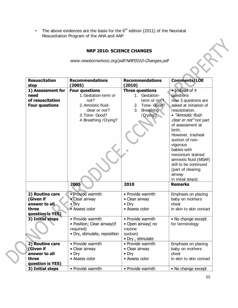
• The above evidences are the basis for the 6th edition (2011) of the Neonatal Resuscitation Program of the AHA and AAP
NRP 2010: SCIENCE CHANGES
www.newbornwhocc.org/pdf/NRP2010-Changes.pdf
Resuscitation step
Recommendations (2005)
Recommendations (2010)
Comments/LOE
1) Assessment for need of resuscitation Four questions
Four questions 1. Gestation-term or
not? 2. Amniotic fluid-
clear or not? 3. Tone- Good? 4. Breathing /Crying?
Three questions 1. Gestation-
term or not? 2. Tone- Good? 3. Breathing
/Crying?
• Instead of 4 questions now 3 questions are asked at initiation of resuscitation. • “Amniotic fluid- clear or not” not part of assessment at birth. However, tracheal suction of non-vigorous babies with meconium stained amniotic fluid (MSAF) still to be continued (part of clearing airway in initial steps)
2005 2010 Remarks
2) Routine care (Given if answer to all three question is YES)
• Provide warmth • Clear airway • Dry • Assess color
• Provide warmth • Clear airway • Dry • Assess color
Emphasis on placing baby on mothers chest in skin to skin contact
3) Initial steps • Provide warmth • Position; Clear airway(if required) • Dry, stimulate, reposition
• Provide warmth • Open airway( no routine suction) • Dry , stimulate
• No change except for terminology
2) Routine care (Given if answer to all three question is YES)
• Provide warmth • Clear airway • Dry • Assess color
• Provide warmth • Clear airway • Dry • Assess color
Emphasis on placing baby on mothers chest in skin to skin contact
3) Initial steps • Provide warmth • Provide warmth • No change except

56
• Position; Clear airway(if required) • Dry, stimulate, reposition
• Open airway( no routine suction) • Dry , stimulate
for terminology
4) Assessment (after initial steps and ongoing) 4.1) Assessment for need for progressive steps after initial Steps 4.2) Assessment of heart rate
Look for 3 signs • Hear rate • Color • Respiration Palpation of umbilical cord pulsation for 6 sec and multiply by 10
Look for 2 signs • Heart rate • Respiration( Labored, unlabored, apnea, gasping) Auscultation of heart at the precordium is the most accurate
• Color has been removed from the signs of assessment • Pre-cordial auscultation better than umbilical cord palpation for detection of heart rate (LOE2, LOE4)
5) Positive pressure ventilation (PPV) 5.1) Indication for PPV 5.2) Assessment of effectiveness of resuscitation steps once PPV is started
Indications are(any 1 out of 3)
1. Hear rate < 100/min
2. Apnea or gasping
3. Persistent central cyanosis despite free flow oxygen
Heart rate Color Respiration
Indications (1 out of 2) 1. Hear rate
< 100/min
2. Apnea or gasping
Heart rate Pulse oximetry Respiration
• Persistent central cyanosis is not mentioned in the indication for PPV; use pulse oximetry to assess oxygenation • Increase in HR most sensitive indicator of resuscitation efficacy (LOE5)
5) Positive pressure ventilation (PPV) 5.1) Indication for PPV 5.2) Assessment of effectiveness of resuscitation steps once PPV is
Indications are(any 1 out of 3)
4. Hear rate < 100/min
5. Apnea or gasping
6. Persistent central cyanosis
Indications (1 out of 2) 3. Hear rate
< 100/min
4. Apnea or gasping
Heart rate Pulse oximetry Respiration
• Persistent central cyanosis is not mentioned in the indication for PPV; use pulse oximetry to assess oxygenation • Increase in HR most

57
started despite free flow oxygen
Heart rate Color Respiration
sensitive indicator of resuscitation efficacy (LOE5)
5) Oxygenation 5.1) Assessment of oxygenation
• Based on color • Pulse oximetry recommended for only preterm < 32weeks with need for PPV
• Based on pulse oximetry for both term and preterm in case of following Situations a. Anticipated need for resuscitation b. Need for PPV for more than few breaths c. Persistent cyanosis d. Supplementary oxygen
• Attach probe to right hand or wrist (measure pre-ductal saturations) • Attach neonatal probe before connecting it to machine • Recording of tracing may take 1-2 min • Pulse oximetry should not replace clinical assessment
5.2) Target saturation (pre-ductal)
Not defined Target SpO2 ranges provided as a part of algorithm
1min- 60-65% 2 min- 65-70% 3min- 70-75% 4min- 75-80% 5min- 80-85% 10min- 85-95% (same for both term and preterm)
6) Initial oxygen concentration for resuscitation in case of PPV
Term babies(≥ 37 weeks) • Start with 100% O2 during PPV • However if room air resuscitation is started supplemental O2 up to 100% should be given if no improvement within 90 seconds following birth • In case non availability of O2- start room air resuscitation
Term babies (≥ 37 weeks) • Start with room air (21%) • No improvement in heart rate or oxygenation as assessed by pulse oximetry- use higher concentration by graded increase up to 100% to attain target saturations • Use blender for graded increased in delivered oxygen concentrations
LOE-2 • Paradigm shift from 100% to 21% O2 for resuscitation of term babies needing PPV • Supplemental oxygen started at 90 sec from birth in case of no improvement • Use of blender and pulse oximetry is recommended for term babies also
Preterm babies(<32weeks)
Preterm(<32weeks) • Initiate resuscitation
• Preterm start with O2

58
• Start with oxygen concentration somewhere between 21-100% • No specific concentration recommended • Advocates use of blender for graded increment or decrement of O2 • Pulse oximetry for targeting SPO2-85-95%
using O2 concentration between 30-90% • Titrate O2 concentration to attain SPO2 values recommended at different time points • Uses blended air oxygen mixture judiciously guided by pulse oximetry
concentration 30-90% and then increase or decrease • No evidence to give appropriate initial oxygen strategy for infants 32-37 weeks
7) Peripartum suctioning for neonates born through meconiumstained amniotic fluid
• No routine oropharyngeal and nasopharyngeal suction • Tracheal suction only in non-vigorous babies born through meconium stained amniotic fluid (MSAF) • Intrapartum suctioning for MSAF not advised
• No routine oropharyngeal and nasopharyngeal suction required • Tracheal suction of nonvigorous babies with MSAF still to be continued though evidence for the same is conflicting • Intrapartum suctioning for infants with MSAF , after delivery of head before delivery of shoulder not advised
• No evidence for or refuting tracheal suction even in non vigorous babies born through MSAF (LOE 4) • However no change suggested to existing practice • If tracheal intubation is unsuccessful or there is severe bradycardiathen proceed to PPV
8) Initial breath strategy Positive pressure ventilation (PPV)
• No specific recommendation for short or long inflation time • No specific PIP recommendation • No specific recommendation for PEEP • Guiding of PPV looking at chest rise and improvement in heart rate
• No specific recommendation for short or long inflation time as evidence is conflicting • PIP- for initial breaths 20- 25 cm H2O for preterm and 30-40 cm H2O for some term babies • PEEP likely to be beneficial for initial stabilization
• No specific recommendation for inflation time (LOE 1) • Addition of PEEP in preterm suggested (LOE 5)
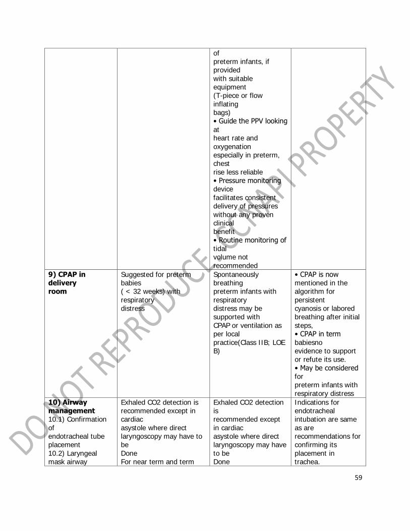
59
of preterm infants, if provided with suitable equipment (T-piece or flow inflating bags) • Guide the PPV looking at heart rate and oxygenation especially in preterm, chest rise less reliable • Pressure monitoring device facilitates consistent delivery of pressures without any proven clinical benefit • Routine monitoring of tidal volume not recommended
9) CPAP in delivery room
Suggested for preterm babies ( < 32 weeks) with respiratory distress
Spontaneously breathing preterm infants with respiratory distress may be supported with CPAP or ventilation as per local practice(Class IIB; LOE B)
• CPAP is now mentioned in the algorithm for persistent cyanosis or labored breathing after initial steps, • CPAP in term babiesno evidence to support or refute its use. • May be considered for preterm infants with respiratory distress
10) Airway management 10.1) Confirmation of endotracheal tube placement 10.2) Laryngeal mask airway
Exhaled CO2 detection is recommended except in cardiac asystole where direct laryngoscopy may have to be Done For near term and term
Exhaled CO2 detection is recommended except in cardiac asystole where direct laryngoscopy may have to be Done
Indications for endotracheal intubation are same as are recommendations for confirming its placement in trachea.
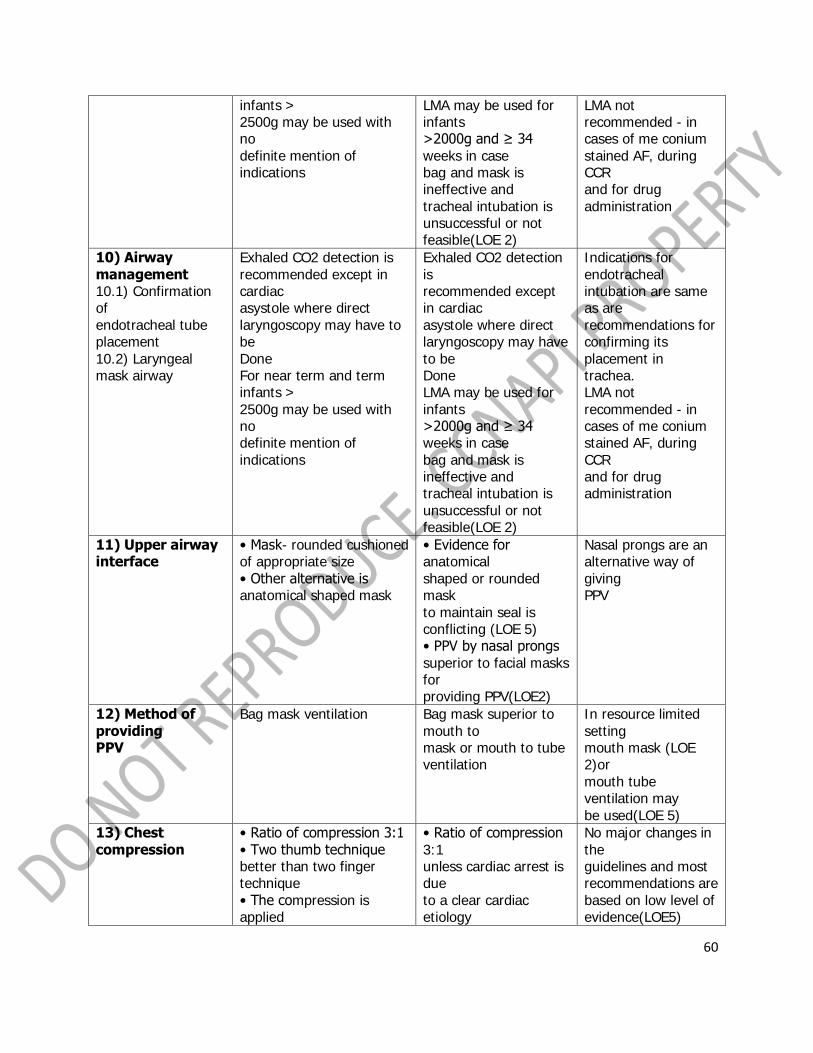
60
infants > 2500g may be used with no definite mention of indications
LMA may be used for infants >2000g and ≥ 34 weeks in case bag and mask is ineffective and tracheal intubation is unsuccessful or not feasible(LOE 2)
LMA not recommended - in cases of me conium stained AF, during CCR and for drug administration
10) Airway management 10.1) Confirmation of endotracheal tube placement 10.2) Laryngeal mask airway
Exhaled CO2 detection is recommended except in cardiac asystole where direct laryngoscopy may have to be Done For near term and term infants > 2500g may be used with no definite mention of indications
Exhaled CO2 detection is recommended except in cardiac asystole where direct laryngoscopy may have to be Done LMA may be used for infants >2000g and ≥ 34 weeks in case bag and mask is ineffective and tracheal intubation is unsuccessful or not feasible(LOE 2)
Indications for endotracheal intubation are same as are recommendations for confirming its placement in trachea. LMA not recommended - in cases of me conium stained AF, during CCR and for drug administration
11) Upper airway interface
• Mask- rounded cushioned of appropriate size • Other alternative is anatomical shaped mask
• Evidence for anatomical shaped or rounded mask to maintain seal is conflicting (LOE 5) • PPV by nasal prongs superior to facial masks for providing PPV(LOE2)
Nasal prongs are an alternative way of giving PPV
12) Method of providing PPV
Bag mask ventilation Bag mask superior to mouth to mask or mouth to tube ventilation
In resource limited setting mouth mask (LOE 2)or mouth tube ventilation may be used(LOE 5)
13) Chest compression
• Ratio of compression 3:1 • Two thumb technique better than two finger technique • The compression is applied
• Ratio of compression 3:1 unless cardiac arrest is due to a clear cardiac etiology
No major changes in the guidelines and most recommendations are based on low level of evidence(LOE5)

61
at the lower one third of sternum • The depth of compression should be one-third of the antero-posterior diameter of the chest
where ratio of 15:2 may be considered • Two thumb technique better than two finger technique • The compression is applied at the lower one third of sternum • The depth of compression should be one-third of the antero-posterior diameter of the chest
14) Drugs 14.1) Naloxone
Naloxone considered in case of infants born to mothers with history of opiod exposure within 4 hours of delivery and there is persistent respiratory depression even after restoration of heart rate and color by effective PPV
• Naloxone is not recommended as part of initial resuscitation in babies with respiratory depression. • Focus needs to be on effective ventilation
• Safety and long term effects on naloxone not established(LOE 5) • Naloxone is not indicated in delivery room.
15) Supportive care 15.1)Therapeutic Hypothermia
No sufficient evidence to recommend routine use of modest systemic or selective cerebral hypothermia after resuscitation in infants with suspected asphyxia Avoid hyperthermia in such cases
Therapeutic hypothermia (whole body or selective head cooling) recommended for infants ≥ 36weeks with moderate to severe hypoxic ischemic encephalopathy as per the protocol used in major cooling trials with provision for monitoring for side effects and
Lack of supporting evidence from resource-limited settings, need of intensive and multidisciplinary care during therapeutic hypothermia and established follow-up services after discharge limit the applicability in middle- and low-income countries
62
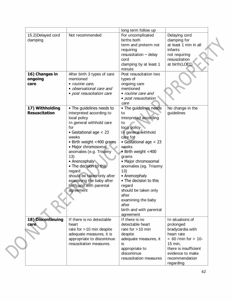
long term follow up
15.2)Delayed cord clamping
Not recommended For uncomplicated births both term and preterm not requiring resuscitation – delay cord clamping by at least 1 minute
Delaying cord clamping for at least 1 min in all infants not requiring resuscitation at birth(LOE1)
16) Changes in ongoing care
After birth 3 types of care mentioned • routine care, • observational care and • post resuscitation care
Post resuscitation two types of ongoing care mentioned • routine care and • post resuscitation care
17) Withholding Resuscitation
• The guidelines needs to interpreted according to local policy In general withhold care for • Gestational age < 23 weeks • Birth weight <400 grams • Major chromosomal anomalies (e.g. Trisomy 13) • Anencephaly • The decision to this regard should be taken only after examining the baby after birth and with parental agreement
• The guidelines needs to interpreted according to local policy In general withhold care for • Gestational age < 23 weeks • Birth weight <400 grams • Major chromosomal anomalies (eg. Trisomy 13) • Anencephaly • The decision to this regard should be taken only after examining the baby after birth and with parental agreement
No change in the guidelines
18) Discontinuing care
If there is no detectable heart rate for >10 min despite adequate measures, it is appropriate to discontinue resuscitation measures.
If there is no detectable heart rate for >10 min despite adequate measures, it is appropriate to discontinue resuscitation measures
In situations of prolonged bradycardia with heart rate < 60 /min for > 10-15 min, there is insufficient evidence to make recommendation regarding

63
continuation or discontinuation of resuscitation
19) Educational program to teach resuscitation
No mention of such a section
AHA/AAP NRP should adopt simulation, briefing-debriefing techniques in designing an educational program for acquisition and maintenance of skills necessary for effective neonatal resuscitation.
This recommendation is newly added to design NRP programme in a more effective manner.
Common Errors in Newborn Resuscitation
• Delivery room too cold for the baby • Non-preparation of equipment
• Doses of emergency drugs • No team leader is assigned
• Unprepared or non-skilled personnel • What drugs to give in what route of adminstration • Too much resuscitation/ too little resuscitation
• No need for ECG tracings to verify heart rate
Take home message
• Be prepared always • Be an NRP Provider
• Always update yourself

64
PLENARY V
“Wound Care Assessment and Management”
RAMON O. RIBU, MD
Section Head, CTMAS & Wound Care,
Philippine Heart Center
Wound healing is a complex series of events that are interlinked and dependent on one another
. Acute wounds usually follow a well-defined process. In the past this model has been applied
to chronic wounds , but it is now known that chronic wound healing is different from acute
wounds. Wound bed preparation as a concept allows the clinician to focus systematically on all
of the critical components of a chronic woundto identify the cause of the problem and
implement care program to achieve a stable wound that has healthy tissue and well-
vascularized wound bed

65
WOUND HEALING &
WOUND BED PREPARATION
Ramon O. Ribu, MD, FPCS, FPATACSI
Department of Cardiovascular Surgery & Anesthesia
Philippine Heart Center
BRIEF HISTORY
2000 B.C.
• Sumerians employed two modes of wound treatment.
Incantations
poultice
• The 1650 B.C. Edwin Smith Surgical Papyrus
describes at least 48 different types of wounds
• Ebers Papyrus, 1550 B.C.
relates the use of concoctions containing
honey (antibacterial properties)
lint (absorbent properties)
grease (barrier)
• Galen of Pergamum (120–201 A.D.)
doctor to the Roman gladiators
emphasized the importance of maintaining a moist environment
It took almost 19 centuries for this important concept to be proven scientifically
epithelialization rate increases by 50% in a moist wound environment when
compared to a dry wound environment
• Ignaz Philipp Semmelweis, a Hungarian obstetrician (1818–1865)
puerperal fever incidence
• Louis Pasteur (1822–1895)
the theory of spontaneous generation of germs
germs were always introduced into the wound from the environment
On a visit to Glasgow, Scotland, Lister noted that some areas of the city's sewer
system were less murky than the rest.
Discovered water from pipes that were dumping waste containing carbolic acid
(phenol) was clear.
In 1865, Lister began soaking his instruments in phenol and spraying the
operating rooms, reducing the mortality rates from 50 to 15%.
• 1876, Robert Wood Johnson
attended an impressive lecture by Lister in 1876 soon left the meeting and began
10 years of research
He developed antiseptic dressing in the form of cotton gauze impregnated with
iodoform.

66
Since then, several other materials have been used to impregnate cotton gauze
to achieve antisepsis.
• The 1960s and 1970s led to the development of polymeric dressings.
• These polymeric dressings can be custom made to specific parameters:
o permeability to gases (occlusive vs. semiocclusive), varying degrees of
absorbency, different physical forms.
• Due to the ability to customize, the available range of materials that aid in wound care
has grown exponentially to include an ever expanding variety.
• Currently, the practice of wound healing encompasses manipulation and/or use of,
among others,
inflammatory cytokines,
growth factors,
and bioengineered tissue.
It is the combination of all these modalities that enables optimal wound
healing.
WOUND HEALING
Wound Depth Involvement
A. Epidermis
1. Stratum Corneum 3. Stratum Spinosum
2. Stratum Lucidum 4. Stratum Basale
B. Dermis
1. Papillary Layer
2. Reticular Layer
C. Hypodermis( SuperficialFascia)
D. Deep Fascia
E. Muscle
Normal wound healing follows a predictable pattern that can be divided into overlapping
phases:
PHASE 1: Hemostasis and Inflammation
PHASE 2: Proliferation
PHASE 3: Maturation and Remodeling
The cellular, biochemical, and mechanical phases of wound healing
A. The hemostatic / inflammatory phase.
Hemostasis precedes and initiates inflammation with the ensuing release of chemotactic factors
from the wound site
Exposure of subendothelial collagen to platelets results in platelet aggregation, degranulation,
and activation of the coagulation cascade.

67
Cellular infiltration after injury follows a characteristic, predetermined sequence
PMNs are the first infiltrating cells to enter the wound site, peaking at 24 to 48h
A. The hemostatic / inflammatory phase.
Increased vascular permeability local prostaglandin release chemotactic substances such as
complement factors and interleukin-1
Latter inflammatory phases reflecting infiltration by mononuclear cells and lymphocytes.

68
B. The proliferation phase.
The second phase of wound healing and roughly spans days 4 through 12
It is during this phase that tissue continuity is re-established. Fibroblasts and endothelial cells
are the last cell populations to infiltrate the healing wound, and the strongest chemotactic
factor for fibroblasts is PDGF.
Upon entering the wound environment, recruited fibroblasts first need to proliferate, and then
become activated, to carry out their primary function of matrix synthesis remodeling.
Matrix Synthesis Remodeling
Collagen Synthesis
Collagen, the most abundant protein in the body, plays a critical role in the successful
completion of adult wound healing.
Type I collagen is the major component of extracellular matrix in skin. Type III, which is also
normally present in skin, becomes more prominent and important during the repair process.
Collagen synthesis, as well as posttranslational modifications, are highly dependent on systemic
factors such as an adequate oxygen supply; the presence of sufficient nutrients (amino acids
and carbohydrates) and cofactors (vitamins and trace metals); and the local wound
environment (vascular supply and lack of infection). Addressing these factors and reversing
nutritional deficiencies can optimize collagen synthesis and deposition.
Proteoglycan Synthesis
Glycosaminoglycans comprise a large portion of the "ground substance" that makes up
granulation tissue.
The major glycosaminoglycans present in wounds are dermatan and chondroitin sulfate.
Fibroblasts synthesize these compounds, increasing their concentration greatly during the first 3
weeks of healing.

69
C. The Maturation and Remodeling Phase
Begins during the fibroplastic phase, and is characterized by a reorganization of previously
synthesized collagen.
There is a net shift toward collagen synthesis and eventually the re-establishment of
extracellular matrix composed of a relatively acellular collagen-rich scar.
The deposition of matrix at the wound site follows a characteristic pattern:
fibronectin and collagen type III constitute the early matrix scaffolding;
glycosaminoglycans and proteoglycans represent the next significant matrix
components; and collagen type I is the final matrix.
Fibril formation and fibril cross-linking result in decreased collagen solubility, increased strength,
and increased resistance to enzymatic degradation of the collagen matrix. Scar remodeling
continues for many (6 to 12) months postinjury, gradually resulting in a mature, avascular, and
acellular scar.
The mechanical strength of the scar never achieves that of the uninjured tissue.
Epithelialization
This process is characterized primarily by proliferation and migration of epithelial cells adjacent
to the wound
The process begins within 1 day of injury and is seen as thickening of the epidermis at the
wound edge.
Marginal basal cells at the edge of the wound lose their firm attachment to the underlying
dermis, enlarge, and begin to migrate across the surface of the provisional matrix
Fixed basal cells in a zone near the cut edge undergo a series of rapid mitotic divisions,
and these cells appear to migrate by moving over one another in a leapfrog fashion until the
defect is covered.
The healing by epithelialization of superficial cutaneous wounds
Re-epithelialization is complete in less than 48 hours in the case of approximated incised
wounds, but may take substantially longer in the case of larger wounds, where there is a
significant epidermal/dermal defect.
Types of Wound Healing
1. Primary Intention
An incised wound that is clean and sutured closed is said to heal by primary intention.
2. Secondary Intention
Often, because of bacterial contamination or tissue loss, a wound will be left open to heal by
granulation tissue formation and contraction; this constitutes healing by secondary intention.
3. TERTIARY INTENTION
Delayed primary closure, or healing by tertiary intention, represents a combination of the first
two, consisting of the placement of sutures, allowing the wound to stay open for a few days,
and the subsequent closure of the sutures

70
Classification of Wounds
Wounds are classified as either:
1. Acute
2. Chronic.
Acute Wounds
Acute wounds heal in a predictable manner and time frame.
The process occurs with few, if any, complications, and the end result is a well-healed
wound.
Surgical wounds can heal in several ways.
An incised wound that is clean and sutured closed is said to heal by primary intention.
Chronic Wounds
Chronic wounds are defined as wounds that have failed to proceed through the orderly
process that produces satisfactory anatomic and functional integrity or that have
proceeded through the repair process without producing an adequate anatomic and
functional result.
The majority of wounds that have not healed in 3 months are considered chronic.
The increased production of exudates that often accompanies increased microbial
load has been associated with the degradation of growth factors and matrix
metalloproteinases (MMPs) which subsequently affect cell proliferation and wound
healing .
In general, factors that adversely affect wound healing can be remembered by using the
mnemonic device DIDN'T HEAL, as follows:
D = Diabetes:
The long-term effects of diabetes impair wound healing by diminishing sensation and arterial
inflow. In addition, even acute loss of diabetic control can affect wound healing by causing
diminished cardiac output, poor peripheral perfusion, and impaired polymorphonuclear
leukocyte phagocytosis.
I = Infection:
Infection potentiates collagen lysis. Bacterial contamination is a necessary condition but is not
sufficient for wound infection. A susceptible host and wound environment are also required.
Foreign bodies (including sutures) potentiate wound infection.
D = Drugs:
Steroids and antimetabolites impede proliferation of fibroblasts and collagen synthesis.
N = Nutritional problems:
Protein-calorie malnutrition and deficiencies of vitamins A, C, and zinc impair normal wound-
healing mechanisms.
T = Tissue necrosis
resulting from local or systemic ischemia or radiation injury, impairs wound healing.

71
H = Hypoxia:
Inadequate tissue oxygenation due to local vasoconstriction resulting from sympathetic
overactivity may occur because of blood volume deficit, unrelieved pain, or hypothermia,
especially involving the distal extent of the extremities.
E = Excessive tension on wound edges:
This leads to local tissue ischemia and necrosis.
A = Another wound:
Competition between several healing areas for the substrates required for wound healing
impairs wound healing at all sites.
L = Low temperature:
The relatively low tissue temperature in the distal aspects of the upper and lower extremities (a
reduction of 1-1.5°C [2-3°F] from normal core body temperature) is responsible for slower
healing of wounds at these sites.
Specific etiologies
1. Arterial insufficiency
2. Venous insufficiency
Patients with varicose veins or nonfunctional venous valves after deep vein thrombosis develop
ambulatory venous hypertension, that is, distal venous pressure remains elevated despite
ambulation. This constant venous hypertension seems to cause white cell and fibrin buildup,
which impairs capillary blood flow or traps growth factors. Macromolecules pass into the dermis
and eventually cause the hemosiderin deposition and brawny induration in the distal leg (gaiter
area) characteristic of chronic venous insufficiency.
3. Lymphedema
Although not typically a cause of ulceration, extremity ulcers may fail to heal because of
untreated lymphedema. Nocturnal leg elevation and elastic wraps or support hose are
appropriate adjuncts to the treatment of recalcitrant wounds in edematous extremities. For
advanced and nonresponsive lymphedema, complex decongestive physiotherapy is a useful
treatment option.
4. Neuropathy
Sensory neuropathy involving the feet may lead to unrecognized episodes of trauma caused by
ill-fitting shoes. This is compounded by motor neuropathy causing intrinsic muscle weakness
and spaying of the foot on weight bearing. The result is a convex foot with a rocker-bottom
appearance. Multiple fractures go unnoticed, until bone and joint deformities become marked.
This is termed a Charcot foot (ie, neuropathic osteoarthropathy) and is observed most
commonly in people with diabetes mellitus, affecting approximately 2% of persons with
diabetes.
5. Pressure (decubitus) ulcers
Pressure (decubitus) ulcers occur because of prolonged ischemia-producing external pressure,
usually to a soft tissue region overlying a bony prominence. Tissue ischemia results when

72
external pressure exceeds capillary closing pressure (ie, 25-32 mm Hg in healthy individuals),
the minimum pressure that causes collapse of the capillary when applied to a capillary bed.
6. Neoplasms
Neoplasms strongly suggest malignancy in any chronic nonhealing wound, particularly if the
wound appeared to occur spontaneously.
7.Radiation damage
Gamma radiation and x-ray exposure cause a zone of stasis, in which local blood supply is
impaired by coagulative necrosis due to thrombotic occlusion of smaller arteries. Gamma and x-
ray radiation also spawn ionized oxygen that adversely affects DNA. The long-term result is
inhibition of regeneration of skin cells from dividing basal cells. This may cause recalcitrant
painful skin ulcers. The surrounding skin is atrophic, with atrophy of hair follicles and a paucity
subcutaneous fat.
8. Atheroembolism syndrome
Patchy areas of ischemia involving the feet, especially in the presence of palpable pedal pulses,
suggest the possibility of atheroembolism of plaque fragments from ulcerated, although
nonocclusive, proximal atherosclerotic plaques or from thrombi lining the wall of an infrarenal
aortic aneurysm.

73
Impediments to Wound Healing
Devitalised / Necrotic tissue
Debris
Exudates
Slough
Biofilm
Wound Assessment
"Assess the whole person and not just the hole in the person" -Dr. Gary Sibbald
Wound Bed Preparation
The term «Wound bed preparation (WBP)» was first described by Falanga et al (2000)
defined as the global wound management plan to accelerate endogenous healing and
to enhance the effectiveness of advanced wound care products
The ultimate aim is to ensure formation of healthy granulation tissue resulting in
complete wound closure
The TIME framework means identifying essential elements for optimal wound bed
preparation and correcting abnormalities contributing to impaired wound healing.
The TIME framework (T= Tissue, non-viable or deficient / I= Infection or Inflammation
/ M= Moisture imbalance / E= Edge of wound, non-advancing or undermined) was
developed by Schultz et al in 2003 to provide a systematic approach to the management
of chronic wounds
Proper WBP
Reduction of bacterial bioburden in the wound bed
Removal of necrotic tissue and slough
Control of exudate
Management of cellular dysfunction and biochemical imbalance
3 Components of Local Care – DIM
Debridement
Infection and inflammation
Moisture balance
If wound bed preparation is optimised and healing is stalled, the additional
E or the edge of non-healing wounds represents the potential use of advanced active
therapies to stimulate healing.
Remember this, DIM before DIME

74
WOUND ASSESSMENT
Wound Bed
Necrotic
Wound containing dead tissue. It may appear hard, dry and black. Dead connective tissue may
appear grey. Eschars with time may soften by autolysis and bacterial liquefacation. The
presence of dead tissue in wounds delays healing.
Sloughy
Slough is formed by an accumulation of dead cells in the wound exudate. It is light yellow in
colour and must not be confused with infected tissue and pus.
Granulating
Healthy red tissue, which occurs during the proliferative phase of healing. Firbroblasts migrate
to the wound to produce collagen fibres. The tissue is well vascularised and bleeds easily.
Epithelializing
Process by which the wound surface is covered by new epithelium, this begins when the wound
has filled with granulation tissue. The tissue is pink, almost white, and only occurs on top of
healthy granulation tissue.
Wound Measurement
Wound measurement is a vital aid to examining the healing process within a wound. Chronic
wounds should be measured 4 weekly (diabetic foot ulcers – weekly).
The wound should be measured at its greatest length and breadth:
The two measurements are then multiplied to give an approximate wound area in CM2. This
method can be unreliable where different professionals are assessing the same wound and also
where the shape of the wound is quite irregular.
Exudates
Exudate is produced by all acute and chronic wounds (to a greater or lesser extent) as part of
the natural healing process but may become more viscous and malodorous in infected wounds.
It plays an essential part in the healing process in that it:
Contains nutrients, energy and growth factors for metabolising cells
Contains high quantities of white blood cells
Maintains a moist environment for wound healing
Infection
Wound infection may be defined as the presence of bacteria or other organisms, which lead to
a host reaction. A host reaction can present as any one or combination of the following
signs:
Redness (erythema) around the wound
The production of large amounts of exudate or pus
A change in exudate colour
Malodour
A raised systemic temperature

75
Localised pain
Localised heat
Lymphangitis
Delayed or abnormal wound healing
Wound breakdown
The appearance of fragile tissues which may bleed easily when touched or at the time of
a dressing change.
Wound Beds need to be assessed for:
• Granulation tissue (RED)
• Fibrin slough (YELLOW)
• Eschar (BLACK)
• Bone
• Tendon
• Other underlying structure
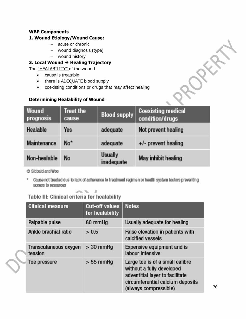
76
WBP Components
1. Wound Etiology/Wound Cause:
– acute or chronic
– wound diagnosis (type)
– wound history
3. Local Wound Healing Trajectory
The “HEALABILITY” of the wound
cause is treatable
there is ADEQUATE blood supply
coexisting conditions or drugs that may affect healing
Determining Healability of Wound

77
WOUND ASSESSMENT
What are the goals for this patient?
healing the wound
protecting the wound from further breakdown/infection
comfort
decrease in pain during dressing change
What is the patient's prognosis?
What is the patient's quality of life?
Is current pain control adequate?

78
WOUND BED PREPARATION

79

80
Types of Wound Debridement
Surgical/Sharp Debridement
Most expeditious form, but not always feasible due to: Pain Bleeding potential
Cost Lack of clinician expertise

81
Autolytic Debridement
Highly selective process involving macrophage and endogenous proteolytic
enzymes which liquefy and separate necrotic tissue and eschar from healthy tissue.
Natural process is further enhanced by the use of occlusive and semi-occlusive dressings and those which interact to create a moist environment
Enzymatic Debridement
A less common method of debridement but effective in the removal of hard necrotic eschar where
surgical debridement is not an option. Exogenous enzymes are applied to the wound bed where they combine with the
endogenous enzymes in the wound to break down the devitalised tissue. Mechanical Debridement
Biological Debridement
Larval or Maggot Therapy
Larval therapy is a quick, efficient method of removing slough and debris from a
wound
Not all patients or staff find this debridement method socially acceptable
Sterile larvae secrete powerful enzymes to break down devitalised tissue without
destroying healthy granulation tissue (Thomas et al, 1998).
Osmotic Debridement
If debridement is effective, the T of TIME is removed and wounds can progress through the
remaining phases of wound healing.
Microbes and Chronic Wounds
z All chronic wounds are contaminated by bacteria.
z Wound healing occurs in the presence of bacteria.
z Certain bacteria appear to aid wound healing.
z It is not the presence of organisms but their interaction with the patient that determines
their influence on wound healing.
z Wound contamination: the presence of non-replicating organisms in the wound.
z All chronic wounds are contaminated.
z These contaminants come from the indigenous microflora and/or the environment.
z Most contaminating organisms are not able to multiply in a wound. (Ex. Most organisms
in the soil won‟t grow in a wound).

82
Microbiology of Wounds
z The microbial flora in wounds appear to change over time.
z Acute wound; Normal skin flora predominate.
z S. aureus, and Beta-hemolytic Streptococcus soon follow. (Group B Streptococcus and S.
aureus are common organisms found in diabetic foot ulcers)
z After about 4 weeks
z Facultative anaerobic gram negative rods will colonize the wound.
z Most common ones= Proteus, E. coli, and Klebsiella.
z As the wound deteriorates deeper structures are affected. Anaerobes become more
common. Oftentimes infections are polymicrobial (4-5).
z Long-term chronic wounds oftentimes contain more anaerobes than aerobes.
z Aerobic gram-negative rods also infect wounds late in the course of chronic wound
degeneration. Usually acquired from exogenous sources; bath and foot water
z Ex. Pseudomonas, Acinetobacter, Stenotrophomonas (Xanthomonas).
z Organisms like Pseudomonas are not very invasive unless the patient is highly
compromised (ex. Ecthyma gangrenosum in neutropenic patients).
z These organisms are associated with marked wound deterioration due to endotoxin,
enzymes, and exotoxins.
z As the wounds go deeper and become more complex they can infect the underlying
muscles and bone causing osteomyelitis.
z Coliforms and anaerobes are associated with osteomyelitis in these patients. You also
see Staphylococcus aureus.
z Enterococcus and Candida are often isolated from wounds.
z Treating a patient for these organisms is only indicated if there are no other pathogens
present and the organisms are present in high concentrations (106 per gram of tissue)
z In summary: early chronic wounds contain mostly gram-positive organisms.
z Wounds of several months duration with deep structure involvement will have on
average 4-5 microbial pathogens, including anaerobes (see more gram-negative
organisms).
Dose of Bacteria
z Differs depending on the organism involved.
z Some organisms would need to be in high concentrations. (ex. Candida, Enterococcus)
z Various combinations of bacterial species result in more host damage (synergy)
z Example; Group B Streptococcus (S. agalatiae) and Staphylococcus aureus.
z Organisms that should be treated regardless of the numbers present.
z Beta-hemolytic streptococci, Mycobacteria sp., Bacillus anthracis, Yersinia pestis,
Corynebacterium diphtheriae, Erysipelothrix rhusiopathiae, Leptospira sp., Treponema
sp., Brucella sp., Clostridium sp., VZV, HSV, dimorphic fungi, Leishmaniasis.
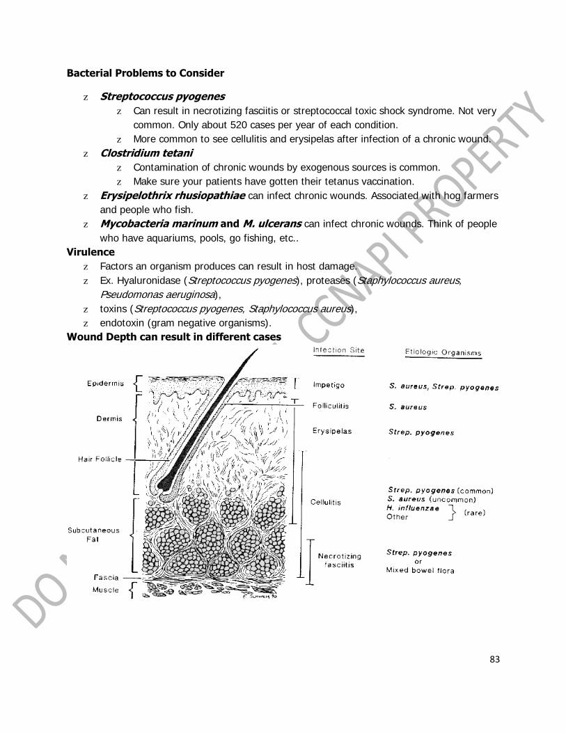
83
Bacterial Problems to Consider
z Streptococcus pyogenes
z Can result in necrotizing fasciitis or streptococcal toxic shock syndrome. Not very
common. Only about 520 cases per year of each condition.
z More common to see cellulitis and erysipelas after infection of a chronic wound.
z Clostridium tetani
z Contamination of chronic wounds by exogenous sources is common.
z Make sure your patients have gotten their tetanus vaccination.
z Erysipelothrix rhusiopathiae can infect chronic wounds. Associated with hog farmers
and people who fish.
z Mycobacteria marinum and M. ulcerans can infect chronic wounds. Think of people
who have aquariums, pools, go fishing, etc..
Virulence
z Factors an organism produces can result in host damage.
z Ex. Hyaluronidase (Streptococcus pyogenes), proteases (Staphylococcus aureus,
Pseudomonas aeruginosa),
z toxins (Streptococcus pyogenes, Staphylococcus aureus),
z endotoxin (gram negative organisms).
Wound Depth can result in different cases

84
Host Resistance
z Host resistance is the single most important determinant in wound infection.
z Local and Systemic factors both play a role in increasing the chances a wound will
become infected.

85

86

87

88
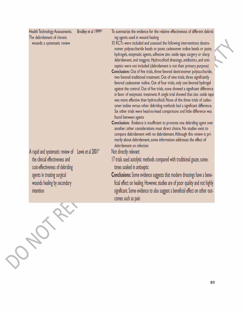
89

90

91

92

93
BIOFILMS
are complex microbial communities containing bacteria and fungi;
are relatively stable, three-dimensional communities of microbial cells encased in a
complex mixture of extracellular polymers.
are detrimental to wound healing and will hinder the process, but infection causes tissue
destruction

94
BIOFILM FORMATION
Stage 1: Reversible Surface Attachment
Free-floating, or planktonic, bacteria encounter a submerged surface and within
minutes can become attached. They begin to produce slimy extracellular polymeric
substances (EPS) and to colonize the surface. The initial attachment is reversible.
Stage 2: Permanent Surface Attachment
As the bacteria multiply, they become more firmly attached (sessile) and differentiate,
changing gene expression patterns in ways that promote survival. This is usually the
result of a type of bacterial communication known as quorum sensing.
Stage 3: Slimy Protective Matrix
Once firmly attached, the bacteria begin to secrete a surrounding matrix known as
extracellular polymeric substance (EPS). This is a protective matrix or „slime‟. Small
bacterial colonies then form an initial biofilm
Effects of Biofilms
Bacterial biofilms are known to contribute to numerous chronic inflammatory diseases and
recent evidence suggests that biofilms also play important roles in impairing healing in chronic
wounds. Biofilms have high levels of tolerance to antibodies, antibiotics, disinfectants and
phagocytic inflammatory cells.

95
HOW ARE BIOFILMS RELEVANT TO WOUNDS?
Biofilms are a major contributor to diseases that are characterised by an underlying
bacterial infection and chronic inflammation, eg. periodontal disease, cystic
fibrosis, chronic acne and osteomyelitis
biofilm cells have increased virulence and resistance to immunological defenses, and
decreased sensitivity to inhibitors such as antibiotics and antiseptics.
HOW QUICKLY DO BIOFILMS FORM?
within minutes > biofilms attach themselves
2 – 4 hours > form strongly attached microclonies
6 – 12 hours > develop initial EPS & become increasingly
tolerant to biocides, eg. Antibiotics,
Antiseptics, & Disinfectants
within 24 hours > rapidly recover from mechanical disruption
& reform mature biofilm
2 – 4 dayS > evolve into fully mature biofilm colonies that
are extremely resistant to biocides & shed
planktonic bacteria
HOW DO MATURE BIOFILMS PROTECT BATERIA?
Biofilms greatly enhance the tolerance of microorganisms embedded in the matrix to the
immune system, antimicrobials
and environmental stresses (eg. nutritional or oxygen limitation). This tolerance may approach
complete resistance to factors that would easily kill these same microbes when growing in an
unprotected, planktonic state.
THE ROLE OF WOUND CLEANSING IN WBP
WOUND CLEANSING is one of the basic principles of proper WBP
Usual irrigation solution include:
Normal Saline
Water
Antiseptics

96
Advanced wound irrigation
Thorough wound cleansing with an advanced wound irrigation solution should be the
aim of every clinician as a prerequisite for normal wound healing and as an efficient
contribution to wound bed preparation by the removal of biofilm (Seipp 1999), slough
and necrotic tissue.
Although water may be used as a wound cleanser, and has not been seen to increase
the risk of infection or delay healing evidence is emerging that the combination of PHMB
with a surfactant (betaine) has an increased
ability to penetrate difficult-to-remove coatings, lifting debris, bacteria and biofilm from
the wound.
Surface tension is a parameter to measure cleansing efficacy.
The surface tension of PHMB + Betaine solution is LOWER than the surface tension of
water. This allows better removal of biofilm than water
Efficacy of various wound irrigation solutions against biofilms. Seipp HM, Hofmann S,
Hack A, Skowronsky A, Hauri A., ZfW 2005;4(5):160-163.
Conclusion: The results showed no decrease in the original biofilm load after
exposure to normal saline solution as well as Ringer‟s solution, while the
surfactant polyhexanide solution (Prontosan® Wound Irrigation Solution)
achieved a significant reduction (p<0.001) of the biofilm by 87%.

97

98

99

100
Moisture Balance
New guidelines highlight that rapid healing of the wound is best achieved by providing a moist
environment, which accelerates the rate of wound repair by up to 40%
SELECTION OF Wound Dressings
Specific product focus for each TYPE of wound at every PHASE of wound healing.
Moist wound healing has been the evidence based /best practice approach since Dr. George
Winter first published his studies in 1962.
Providing a balance is essential for necessary healing
• Maintains optimal temperature
• Decreases infection potential
• Decreases patient pain at dressing change
• Decreases TRAUMA to the fragile granulating tissue of the wound bed
• Moist = moist like your eyeball
An ideal dressing should keep the wound bed moist, protect periwound skin from maceration,
protect from secondary infection, be free of toxic components, and not cause trauma upon
removal. To enhance healing, the ideal dressing should provide thermal insulation and should
be left intact for as long as possible.

101
Wound Dressing
Note:
It is important to remember to reassess the wound each time you change the dressing.
Documentation needs to be specific:
measure and describe the wound.
Dressing selection/treatment should also be clearly documented.
The word is GENTLY.
Minimize wound trauma during wound cleansing.
Normal Saline is the agent of choice.
No betadine, chlorohexadine, or Hydrogen
peroxide.
Choice of Dressings
It should be recognised that a wound will require treating differently at various stages of its
healing. No dressing is suitable for all wounds.

102
Following careful selection of the appropriate management plan for the patient, the wound
assessment tool should be used to monitor the progress of the wound through to its healing
stage.
Guideline statement
Criteria for Choosing a Dressing In Order of Importance (Miller & Collier, 1997)
1. Choose a dressing that maintains a moist environment at the wound/dressing interface.
(The only possible exceptions are peripheral necrosis secondary to arterial disease.
2. Choose a dressing that is able to control (remove) exudate. A moist wound environment is
good, a wet environment is not beneficial
3. Choose a dressing that does not stick to the wound and cause trauma on removal
4. Choose a dressing that protects the wound from the outside environment
5. Choose a dressing that will aid debridement if there is necrotic or sloughy tissue in the
wound (caution withischaemic lesions)
6. Choose a dressing that will keep the wound close to normal body temperature
7. Choose a dressing that is acceptable to the patient
8. Choose a dressing that is cost-effective
9. Diabetes – choose a dressing that will allow frequent inspection

103

104

105

106

107

108

109

110

111

112
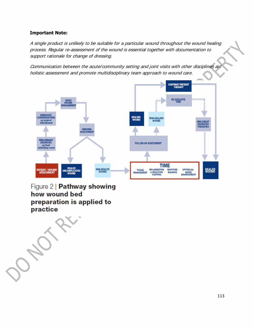
113
Important Note:
A single product is unlikely to be suitable for a particular wound throughout the wound healing
process. Regular re-assessment of the wound is essential together with documentation to
support rationale for change of dressing.
Communication between the acute/community setting and joint visits with other disciplines aid
holistic assessment and promote multidisciplinary team approach to wound care.

114
Application of Bioengineered Skin Substitutes
1. Apligraf (Organogenesis; Novartis) is a bilayered skin substitute produced by combining
bovine collagen and living cells derived from tissue-cultured human infant foreskins. One study
of diabetic foot ulcers demonstrated 12-week healing rates of 39% for patients who received
only standard wound care versus 56% for those who were treated by application of an Apligraf
after a period of standard wound care.
2. Dermagraft (Smith & Nephew, Inc) is human fibroblast-derived dermal
substitute manufactured by seeding dermal fibroblasts onto a 3-dimensional
bioabsorbable scaffold. It has been marketed for use in the treatment of diabetic foot ulcers,
venous ulcers, and pressure sores. A clinical trial showed improved healing rates in diabetic foot
ulcers.
4. Oasis (Healthpoint, Ltd), a relatively new product, is a xenogeneic acellular collagen matrix
derived from porcine small intestinal submucosa in such a way that an extracellular matrix
and natural growth factors remain intact. This provides a scaffold for inducing wound healing.50
Do not use this in patients with allergies to porcine materials.
5. Cultured epithelial autograft (Epicel; Genzyme Tissue Repair, Cambridge, Mass) is an
epidermal replacement that is grown in a tissue culture from a skin biopsy taken from the

115
recipient and is cocultured with mouse cells. Preparation of the graft requires about 2 weeks of
culture time.
Consider other topical agents
Topically applied platelet-derived growth factors have a modestly beneficial effect in promoting
wound healing.
1. Becaplermin gel 0.01% (Regranex), recombinant human platelet-derived growth
factor (PDGF) that is produced through genetic engineering, is approved by the US
Food and Drug Administration (FDA) to promote healing of diabetic foot ulcers. Regranex
is contraindicated in persons with known skin cancers at the site of application. Freeze-dried
platelet-rich plasma has shown promise in a recent animal study.
2. Collagenase : Collagen comprises a significant fraction of the necrotic soft tissues in chronic
wounds. The enzyme collagenase, which is derived form fermentation of Clostridium
histolyticum, helps remove nonviable tissue from the surface of wounds. However,
collagenase is not a substitute for an initial surgical excision of a grossly necrotic wound.
Pain
The pain associated with chronic wounds is often underestimated. In over 50% of cases, nurses
recording of patients‟ pain differed from self-reporting. In most of these cases, the nurses had
underestimated the patients‟ pain.
Pain assessment tools have many advantages:
Patient has a more active role in dealing with their pain The patient may feel that their pain is being taken seriously The tool often prompts more effective pain relieving measures, as documented
evidence exists. PERIWOUND AREA
Surrounding tissues may present as: healthy macerated
dry/flaky eczematous
blue/black discolouration oedema
erythema cellulitis
The surrounding skin should be examined carefully as part of the process of assessment and
appropriate action taken.

116
NUTRITIONAL ASPECTS OF WOUND HEALING
Nutritional status plays a critical role in the wound healing process. Neglecting the nutritional
health of the individual may totally compromise all wound management to be carried out.
(Wallace, 1994)
Optimal Nutrition Helps To Maintain Immune Competence.
THE ESSENTIAL NUTRIENTS FOR WOUND HEALING
Protein, Vitamin C, B Complex and A, Zinc, Iron and Copper are essential for wound healing.
In addition to these nutrients, it is essential that adequate energy/calories are obtained from
fats and carbohydrates to prevent tissue protein being used as a source of energy.
PROTEIN
Requirements: 1.2 – 2.0g protein/kg/24h
Protein is required for healing tissues. Without adequate protein normal protein synthesis and
wound healing are inhibited. The immune response is diminished and there is a delay in matrix
formation.
Protein Sources:-
Meat, fish, eggs, milk, cheese, yoghurt, pulses and nuts.
Nutritional sip feeds will provide important sources of protein and other nutrients if dietary
intake is inadequate.
ENERGY
Requirements: 30-40 Kcal/kg/24h
An adequate energy/calorie intake is essential in order to prevent dietary and tissue protein
being used as a source of energy rather than for wound healing.
Energy Sources:-
All foods provide energy and preserve tissue protein.
Carbohydrate sources – bread, potatoes, breakfast cereal, rice and pasta, oils, spreads, butter,
margarine, fried foods.
Fat sources – oils and fats, butter, margarine, fried foods.
VITAMINS
Vitamin C
Requirements: A minimum of 60mg vitamin C.
Vitamin supplements from 200 mg – 1g per day are sometimes recommended [Taylor, 1974],
however excessive doses may cause renal stones [Morton, 1995].
Vitamin C is required for collagen synthesis and aids iron absorption.
Vitamin C is not stored in the body with patients rapidly becoming deficient. Supplements may
be necessary.

117
Vitamin C Sources:- Citrus fruits and juices, blackcurrant juice drinks and fruit squashes fortified
with vitamin C tomato juice, all fruit and vegetables.
MINERALS
Zinc
Deficiency is associated with poor wound healing.
Zinc is required for collagen synthesis, epithelialization and cell proliferation.
Zinc supplements have been found to improve the healing of leg ulcers where zinc deficiency is
identified.
However, where there is no deficiency excess zinc can impair healing (Wells, 1994).
Zinc sources:- Liver, meat, fish, eggs, pulses including baked beans, wholegrain cereals.

118
PLENARY VI
“Palliation and Spiritual Care: Phenomenology Approach in Critical Care”
RUDOLF CYMORE KIRBY P. MARTINEZ, MAN, RP-RN, CAA, LMT,
CSTP, PhD (c)
Complementary & Alternative Therapy Nurse Practitioner,
Pain Management Clinician and Staff Nurse
Philippine Children‟s Medical Center
"OF SHADOWS AND BUTTERFLIES": SITUATING THE ROLE OF PHENOMENOLOGY IN
CRITICAL CARE THRU THE LENS OF FILIPINO NURSE'S SPIRITUALITY
This presentation situates the role of phenomenology in the critical care setting thru the lens of
the nurse's daily mundane experiences with death and dying process. It will explore how
phenomenology, both as an approach and an attitude, can provide an immense wealth of
qualitative "evidence" which will inevitably inform their practice as critical care nurses. As a
subsequent example, this presentation will provide a glimpse of the rites and rituals that nurses
associate with the death and dying process, reflected upon thru the phenomenological
approach. These rites and rituals will be viewed and appreciated from the level of being a
simple personal experience to that of being a part of a uniquely Filipino cultural consciousness.
Lastly, this presentation will explore how these insights may help nurses develop their
competence as a professional and compassion as a human being.

119
“OF THE SHADOWS AND BUTTERFLIES”
PHENOMENOLOGY IN CRITICAL CARE
Objectives:
• Familiarize with Phenomenology
• Situate Phenomenology in Critical • Care Setting
• Understand Filipino Spirituality • through Phenomenology
Phenomenology
The Study of a Phenomenon
Phenomenology:
“Bridges the gap between what is familiar in our worlds and what is unfamiliar” – Gadamer
“Prime intent is to describe and explore the meaning and essence of unconsolidated henomena
as lived experiences” – Speziale
REALITY PHENOMENON
Phenomenology is based on the assumption of the meaning of Reality
Reality is on People
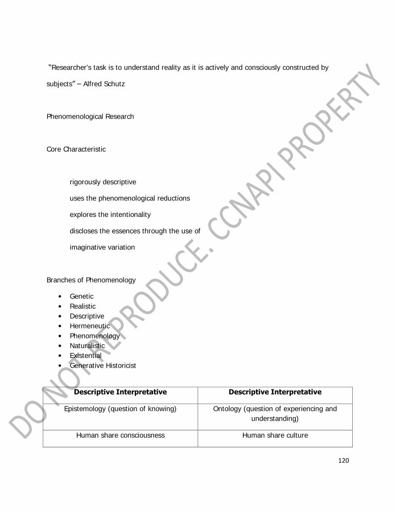
120
“Researcher's task is to understand reality as it is actively and consciously constructed by
subjects” – Alfred Schutz
Phenomenological Research
Core Characteristic
rigorously descriptive
uses the phenomenological reductions
explores the intentionality
discloses the essences through the use of
imaginative variation
Branches of Phenomenology
• Genetic
• Realistic
• Descriptive
• Hermeneutic
• Phenomenology
• Naturalistic
• Existential
• Generative Historicist
Descriptive Interpretative Descriptive Interpretative
Epistemology (question of knowing) Ontology (question of experiencing and
understanding)
Human share consciousness Human share culture

121
Data speaks for itself Interpreter participate in making
data
Bracketing defending objectivity Hermeneutic circle (co-creation,
preunderstanding)
Historicality:
“A person‟s history or background, includes what a culture gives a person from birth and is
handed down, presenting ways of understanding” – Heidegger
“To be human was to Interpret” - Heidegger
Heideggerian Assumptions - Benner (1994)
• Human are social dialogical being
• Understanding is always before us
• Interpretation presupposes a shared understanding between the researchers & co-
researchers
• Interpretation involves the interpreter
“Language is the universal medium in which understanding occurs. Understanding occurs in
interpreting” – Gadamer
“Understanding and interpretation are bound together and interpretation is always an evolving
process, thus a definitive interpretation is likely never possible” – Annells
What do we want to understand in Interpretative Phenomenology?
• The experiences of people

122
• The meanings of their experiences • The insight from those meanings
• Your journey in understanding what you • Understood
Phenomenology is both a Research Approach and an Attitude
Phenomenology Contextualize Reality
Makes Understanding Culturally Sensitive
“We cannot change anything until we accept it. Condemnation does not liberate, it oppresses.”
– Carl Jung
Critical Care Setting is Rich in Phenomenon that
could be explored though Phenomenology
Such as the MeSuch as the Meeting of Healing and Suffering
“Healing Through Suffering
Suffering Through Healing”:
The World of Pediatric
Cancer Nurses
Research Approach
Interpretative Phenomenology - Bridge the gap between what is known & unknown
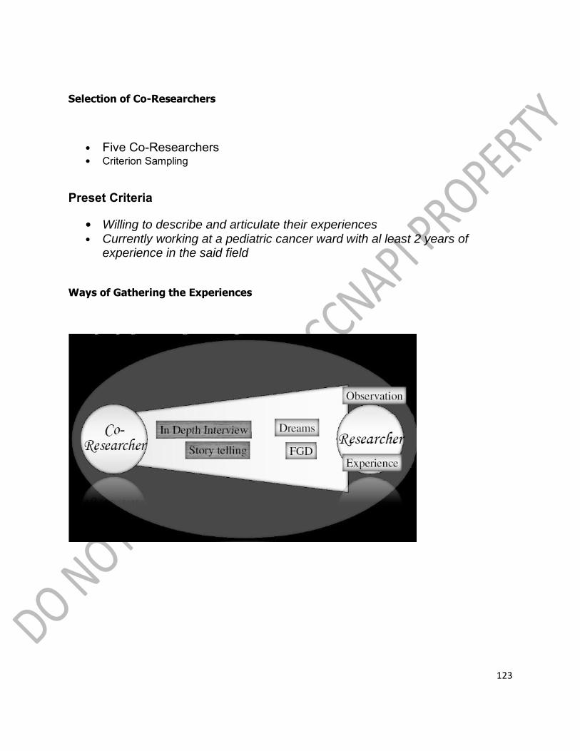
123
Selection of Co-Researchers
• Five Co-Researchers
• Criterion Sampling
Preset Criteria
• Willing to describe and articulate their experiences • Currently working at a pediatric cancer ward with al least 2 years of
experience in the said field
Ways of Gathering the Experiences

124

125

126

127
Madugo” (Bloody): The Insignia of PCN
• Source of Distinguished Identity • Blood if Life, Blood Transfusion is • Transmission of Life • Life of their Patient is at their Hands • Blood Culture Reflected in Language
“Toxic”: Poison of Overindulgence
• Encapsulate the World of a PCN • Beyond Optimum, Things will be Toxic,
i.e. Chemotherapy, Duty, Attachment
• Toxicity Consumes One‟s Being, Blur the • Boundaries of Things • Toxicity is an Interplay of Time and Space
“Paruparo” (Butterflies): On Death and Dying
• Inseparable and Imminent Reality

128
• Death Mask: Stoic, Emotionally Detached • Depersonalized Death: Dying Body, Living Spirit
• To be a PCN is to Embrace the “Paruparo” • Living Everyday as if it‟s Their Last
• Messenger, Soothsayer & Creator
Thematic Embodiment: Eidetic Insight
• Is a paradox of suffering and healing • Disparity of meanings and the seemingly polarity of things are but two sides of the coin,
opposite but in unity and can never be separated
• There can be no healing without suffering nor suffering without healing
Symbolic Representation
YinYang = Unity of Opposite, Inseparability of Healing & Suffering

129
Red = Blood, Life, Healing
Black = Toxicity, Death, Suffering
Cross = End & Beginning of Life
Critical Care Setting is Rich in Phenomenon that could be explored though Phenomenology
Such as the Experience of Death and Dying
Unarmed:
Experiences of Seeing Death and Dying Among Filipino Nurses Working with Children
“…nurses shall collaborate with other health care providers for the curative, preventive, and
rehabilitative aspects of care, restoration of health, alleviation of suffering, and when recovery
is not possible, towards a peaceful death…” - RA 9173 Sec. 28

130
Death and Dying is a Normal Phenomenon Among Hospital Nurses
Nurses Tend to Shy Away on the Topic of Death and Dying
Both Taboo and Less Explored
Shrouded in Secrecy and Mystery
What Insight maybe derived from Filipino Nurses Working with Children‟s
Experiences of Death and Dying

131
Interpretative Phenomenology –
Bridge the gap between what is known & unknown
Five Co-Researchers
Criterion Sampling
Preset Criteria

132
•Willing to describe and articulate their experiences
•Currently working at a pediatric ward with at least two years of experience in the said
field

133

134

135
“Distance”: Fear of Death
• Pangamba (Dread) • First experience of Death
• Distancing oneself from Death • Patient is not seen as someone close to our heart
“When you know they have cancer, it‟s a different feeling, you‟ll pity them… when they have
cancer you know they‟ll eventually die, its inevitable”
“Detachment”: Fear of Dying
• Pag-aagam-agam (Uneasiness)
• Masked: Emotional Detachment
• General Feeling of Awkwardness

136
• “job description” type of care
• Keeping one‟s self intact
“I‟ll see them as just my patients, I detached myself from them since I‟m on the verge of
crying… have you tried doing CPR while crying? They (children) are good people then they‟ll die
at your hand, on your duty, on your shift…”
“Depersonalized”: Fear for Oneself
• Dalamhati (Grief) • Repeated Experience of Death and Dying
• Emotional Callousness
• Accepting Death as Inevitable • Wear on or Wear out
“I thought I‟ll be closer to patient (as time goes by) but now I realized that instead, I am
becoming calloused… I feel nothing even pity even if the patient have nothing, even if he is an
only child… I feel nothing ”

137
Interwoven work of life and death
Unarmed
Paradox of Closeness
Multi Layered Spectrum of Meaning: Loosing the Child to Loosing one‟s Touch of Humanity
Symbolic Representation
The Weeping Cherub
• Lost of the child • Touch of Humanity
• Hardened Persona
• Puer • Connection

138
Context: Filipino Consciousness
Cultural Values Inform our
World View which Affect
How We Appreciate Things
KAPWA
SHARED IDENTITY (Shared Inner Self, “The other person is also yourself”)
• the core of Filipino psychology, it is
humaneness at the highest level
• implies unique moral obligation to treat
one another as equal fellow human beings
Treat the other person as you treat yourself
because the other person is also yourself
PAKIRAMDAM
(Knowing Through Feeling or Tacit Knowing; Participatory Sensitivity)
• A unique social skill inherent in Filipino personhood
• The need for openness and basic trust is a precondition for this active process of sensing
subtle cues
FILIPINO PROXEMICS
• To be alone a German needs four thick walls which shut off all light and sound from the
outside.
• A Japanese needs only thin paper screens
• A Filipino does not need a physical structure to spend time alone. All that is needed is for him
to stay in a corner and keep quiet.

139
PHENOMENOLOGY
Place Prime Importance of Human
Experience as a Source of Valuable
Insight to Inform Our Practice
Brings Back Humanity to Nursing
Research and Practice

140
PLENARY VII
“Empowerment of Nurses through a Career Progression Program”
HON. MARCO ANTONIO STO. TOMAS, RN, MAN
Member, Board of Nursing
Chair, Council for Nursing Advancement,
Recognition & Specialization
The International Scene
In 1992, the Philippines, together with the governments of Brunei, Darussalam, Republic of
Indonesia, Malaysia, Republic of Singapore, Kingdom of Thailand, and the Socialist Republic of
Vietnam were all signatories to the DECLARATION which provided that ASEAN shall move
towards a higher plane of economic cooperation to secure regional peace and prosperity and
further declared that the ASEAN Free Trade Area (AFTA) shall be established in the region.
This signaled the beginning of globalization which among others aimed to provide long-term
growth and development and enhance productivity, efficiency and global competitiveness of
professionals; promote opportunities; and to ensure delivery of better professional services.
This event in global history was geared to realize:
1. Liberalization of the Practice of various Professions, Nursing being one of these
2. Elimination of Discriminatory Practices
3. Institution of Deregulatory Policies
4. Provision of Mutual Recognition Measures
5. Harmonization and Standardization of Qualifications for Examination and Licensure as
well as Regulatory Requirements Pursuant to International Agreements
As a result of the 1992 Singapore ASEAN Agreement, ASEAN Member States pursued
further exploration of measures on border and non-border areas of cooperation to supplement
and complement the liberalization of trade pursuant to the Framework Agreement on Enhancing

141
ASEAN Economic Cooperation. Member Economies also recognized that intra-ASEAN economic
cooperation will secure a liberal trading framework for trade-in services which would strengthen
and enhance trade- in services among ASEAN Member States, and they committed to the rules
and principles of the General Agreement on Trade-in Services (GATS) which aims to eventually
permit the Liberalization of Trade-in Services between or among the parties to an economic
integration agreement. This moreover affirmed that ASEAN Member States shall extend to one
another preference in trade-in services.
There was also the Uruguay Round of Talks and GATS. The General Agreement on Trade in
Services (GATS) was negotiated in the first set of multilaterally-agreed and legally-enforceable
rules and disciplines ever negotiated to cover international trade-in services. These Agreements
covered (1) a framework of general rules and disciplines, (2) annexes addressing special
conditions relating to individual sectors, and (3) national schedules of Market Access
commitments. These schedules like tariff schedules under the GATS are an integral part of the
agreement. A Council for Trade in Services oversees the operation of this agreement.
And there was the APEC and its BOGOR Declaration. Under this declaration, the Asia-Pacific
Economic Cooperation (APEC) member economies committed themselves to achieving free and
open trade-in services by 2010/2020. APEC members should begin to develop programs on
services for incorporation into their individual action plans. In some cases, action plans dealing
with services will inevitably need to refer to investment issues, since comercial presence is an
important means of delivering services abroad. For this reason, work on trade services will
need to be coordinated with work on free and open investments being done by APEC
Investment Experts. APEC‟s work best focused on major impediments to trade services and on
removing these impediments.
Come 2015, Filipino Professionals, especially nurses, are to witness the
beginning effects of all these international commitments signed by the Government
of the Republic of the Philippines.
National Scene. How has the Philippine Nursing Sector prepared for the impact of
globalization?
The practice of a profession like Nursing in the Philippines is governed by a professional
regulatory law which was enacted by the legislative body/congress pursuant to the Police

142
Powers and Parens Patria of the State, as enshrined in the Philippine Constitution, to protect
the life, health, property, and welfare of the people via possession of the adequate levels of
knowledge, skills and competence, and of good moral character (integrity) by the
registered/licensed professional or the professional nurse. Every professional Regulatory law
(PRL) is to be administered and enforced by a Professional Regulatory Board (PRB) such as the
Board of Nursing.
In 1996, anticipating that the W.T.O.-ASEAN-AFAS-AFTA Liberalization of Trade-in Services will
take full effect by 1998 and the W.T.O.-APEC-GATS Liberalization of Trade-in Services having
adopted 2010/2020 as their target, then Board of Nursing through its Chairman Dr. Aurora
Yapchiongco, tasked a core group to carefully study the possibility of putting in place a
credentialing program for the nursing profession in pursuit of the need to prepare for global
competition, higher productivity, and better quality nursing products in order to access the
world market via APEC/GATTS. This move paved the way for the promulgation in February 18,
1999 of Board Resolution No. 14 which ruled for the adoption of a National Nursing Specialty
Certification Program. This was consequently followed by Board Resolution No. 24, series of
1999 which incorporated some amendments thereto. The first NSC Council was inducted into
office during the BoN Chairmanship of Hon. Corazon de la Pena. Nursing history will tell us that
these events happened between the legal-life of Republic Act 7164 passed in 1991 and upon its
repeal and implementation of R.A. 9173 passed in the year.
It is also worth recognizing that in 2002 the Association of Nursing Service Administrators or
ANSAP “voluntarily” set into motion its so-called Career Pathways for Nurse Practitioners as
private organizational initiative designed to give R.Ns under its organizational sphere of
influence the opportunity for career development within an acceptable framework which may
facilitate possible promotion in a clinical ladder that expected the professional nurse to assume
increasing responsibilities and rewards. This model and that of Patricia Benner‟s framework and
mechanisms are worthy of adaptation in as much as these models are very much in sync with
globally recognized and utilized mechanisms of advancement not only in theory but in practice
and the Board of Nursing sees no impediment for its legal adaption.
With the issuance of EXECUTIVE ORDER NO. 83, Series of 2012 by President Benigno S.
Aquino “Institutionalizing the Philippine Qualifications Framework (PQF)” as THE national policy
that describes the levels of educational qualifications and sets the corresponding standards for
qualification outcomes and which became effectivity last 1 October 2012, the Philippine Nursing
Sector MUST navigate effectively amidst a multitude of pressing socio-economic and political
events in Philippine Nursing history and where all the more a National Nursing Career
143
Progression Program (NNCCP) is more than relevant in achieving local upscaling of
competencies and building further our international competitive advantage.
Today we feel all the more that what our former PRC Board of Nursing started in the 90‟s as
the Nursing Specialty Certification Program is at best our “unfinished business” in the pursuit of
professional relevance, quality, and global comparability and competitiveness, and excellence.
Hence our revitalized effort contained in the National Nursing Career Progression Program
(NNCCP) comes with a more unified and harmonized direction in keeping with the State Policy
of promoting “better career prospects and a dignified existence for our Filipino
nurses” (R.A. 9173 – Statement of Policy).
Indeed the CCNAPI‟s call to STAND T.A.L.L. (Together Advance in Learning and Life) is very
timely as the agenda that all organized Philippine Nursing Groups must undertake is exactly
this, no more and no less if we wish to achieve our vision in Philippine Nursing of being the
BEST FOR THE FILIPINOS AND THE CHOICE OF THE WORLD.
Empowerment of Nurses Through a Career Progression Program
OBJECTIVES:
At the end of this talk, participants shall have gained the necessary start-up understanding and
appreciation of the policy-directions of government through the Professional Regulatory Board
of Nursing in leveling-up and keeping the Philippine Nursing Profession nationally relevant but
globally comparable and competitive.

144
This plenary talk shall focus on:
1. Present policy-framework/s which sets in motion the over-all and over-arching national
directions of setting standards in education and training, technical and professional practice,
and quality assurance measures for our Philippine Human Resources. This covers review of
relevant mandates e.g.:
1.1 E.O. No. 83 – Institutionalizing a Philippine Qualifications Framework (PQF)
and its IRR, and
1.2 BON Resolution No. 22, Series 2009 Providing for a National Nursing Career
Progression Program for Filipino Nurses which covers NOT ONLY nursing
specialties but General Nursing Practice as well.
2. Reiterate the challenge of putting in place a national machinery which pursues measures
towards professional competitiveness upscaling targeting a more prepared Philippine Nursing
Workforce in our country especially in the light of upcoming MRA negotiations in the ASEAN and
EU Communities.
Historical Brief
In 1992 – the following “economies” signed a DELCARATION which provided that ASEAN
shall move towards a higher plane of economic cooperation to secure regional peace and
rosperity and further declared that the ASEAN Free Trade Area (AFTA) shall be established
in the region.
1. Philippines 2. Brunei, Darussalam
3. Republic of Indonesia 4. Malaysia 5. Republic of Singapore
6. Kingdom of Thailand 7. Socialist Republic of Vietnam

145
This signaled the beginning of globalization which among others aimed to provide long-term
growth and development and enhance productivity, efficiency and global competitiveness of
professionals; promote opportunities; and to ensure delivery of better professional services
Historical Brief
This event in global history was geared to realize:
Liberalization of the Practice of various Professions, Nursing being one of these (others incl. Medicine, Dentistry, Accountancy, Engineering, Architecture,
Agriculture) Elimination of Discriminatory Practices
Institution of Deregulatory Policies Provision of Mutual Recognition Measures (MRAs) Harmonization and Standardization of Qualifications for Examination and Licensure as
well as Regulatory Requirements Pursuant to International Agreements
Historical Brief
As a result of the 1992 ASEAN Agreement, ASEAN Member States:
1. pursued further exploration of measures on border and non-border areas of cooperation
to supplement and complement the liberalization of trade pursuant to the Framework Agreement on Enhancing ASEAN Economic Cooperation;
2. Member Economies also recognized that intra-ASEAN economic cooperation will secure a
liberal trading framework for trade-in services which would strengthen and enhance trade-in services among ASEAN Member States; and
3. they committed to the rules and principles of the General Agreement on Trade-in Services (GATS) which aims to eventually permit the Liberalization of Trade-in Services between or among the parties to an economic integration
agreement; and 4. that this affirmed that ASEAN Member States shall extend to one another
preference in trade-in services.
There was also the Uruguay Round of Talks and GATS. This GATS was negotiated in the first
set of multilaterally-agreed and legally-enforceable rules and disciplines ever negotiated to
cover international trade-in services. These Agreements covered:
(1) a framework of general rules and disciplines,
(2) annexes addressing special conditions relating to individual sectors, and

146
(3) national schedules of Market Access commitments. These schedules like tariff
schedules under the GATS are an integral part of the agreement. A Council for Trade in
Services oversees the operation of this agreement.
And there was the APEC and its BOGOR Declaration. Under this declaration:
(1) the Asia-Pacific Economic Cooperation (APEC) member economies committed
themselves to achieving free and open trade-in services by 2010/2020;
(2) APEC members should begin to develop programs on services for incorporation into
their individual action plans;
(3) In some cases, action plans dealing with services will inevitably need to refer to
investment issues, since comercial presence is an important means of delivering services
abroad. For this reason, work on trade services will need to be coordinated with work on free
and open investments being done by APEC Investment Experts;
(4) APEC‟s work best focused on major impediments to trade services and on removing
these impediments.
BY 2015 -
The Philippines will witness the beginning effects of all the international
commitments signed by the Government of the Republic of the Philippines.
In 1996, anticipating that the W.T.O.-ASEAN-AFAS-AFTA Liberalization of Trade-in Services will take full effect by 1998;
And as the W.T.O.-APEC-GATS Liberalization of Trade-in Services adopted 2010/2020 as their target, then Board of Nursing Chairman Dr. Aurora Yapchiongco tasked a core group to carefully study the possibility of putting in place a credentialing program for the
nursing profession in pursuit of the need to prepare for global competition, higher productivity, and better quality nursing products in order to access the world market via
APEC/GATTS This move paved the way for the promulgation in February 18, 1999 of Board
Resolution No. 14 which ruled for the adoption of a National Nursing Specialty
Certification Program. This was consequently followed by Board Resolution No. 24, series of 1999 which incorporated some amendments thereto. The first NSC
Council was inducted into office during the BoN Chairmanship of Hon. Corazon de la Pena. Nursing history will tell us that these events happened between the legal-life of Republic Act 7164 passed in 1991 and upon its repeal and implementation of R.A. 9173
passed in the year 2000.
147
In 2002 the Association of Nursing Service Administrators of the Philippines took the initiative of establishing/setting into motion a Career Pathway for Nurse Practitioners Program designed to give RNs the opportunity for career development within an acceptable framework which may facilitate possible promotion within a clinical
ladder that expected the professional nurse to assume increasing responsibilities and rewards.
This model and that of Benner‟s conceptual framework and mechanisms are worthy of
adaptation in as much as these are by now globally recognized and utilized not only in theory but in practice and the current Board of Nursing sees no impediment for its legal adaption. Hence, BON Resolution No.22, series of 2009 came into being
Institutionalizing a National Nursing Career Progression Program (NNCPP) adhering to the State Policy of promoting “better career prospects and a dignified existence for our Filipino nurses” (R.A. 9173 – Statement of Policy), Like any professional sector, navigating through a multitude of pressing socio-economic and political events in our country we can rightfully say that during
this part of our Nursing History, THIS IS YET AN “UNFINISHED BUSINESS” in the pursuit of professional relevance, quality, and global comparability and
competitiveness, and excellence… Especially in the light of E.O. 83, Series 2012
EXECUTIVE ORDER NO. 83,
Series of 2012
“Institutionalization of the Philippine Qualifications Framework”
Date of Effectivity : 1 October 2012
Implementing Rules and Regulations of E.O. No. 83
Date of publication: 28 December 2012
PQF
Is the national policy that describes the levels of educational qualifications and sets the
corresponding standards for qualification outcomes
Competency-based
Labor-market driven Assessment based qualification recognition
COVERAGE
148
All sectors, levels and modes of delivery of the Philippines‟ tri-focalized education system: basic education, technical vocational education and training and higher
education; and All institutions and systems which provide trainings, specializations, skills and
competencies, professional experience or through lifelong learning
OBJECTIVES
Establishes national standards and level for OUTCOMES of education, training, specialization, skills and competencies
Provide national regulatory and QUALITY ASSURANCE arrangements for education and training
Support the development and maintenance of PATHWAYS AND EQUIVALENCES which provide access to qualifications
Supports individual LIFELONG LEARNING goals by providing the basis for individuals
to progress through education and training Aligns the PQF with international qualifications framework to support the national and
international mobility of learners and workers through increased RECOGNITION of the
value and comparability of Philippine QUALIFICATIONS

149
PQF NATIONAL COORDINATING COMMITTEE
Chairman : Secretary, Department of Education
Members : Secretary, Department of Labor and Employment
Director-General, Technical Education and Skills
Development Authority
Chairperson, Commission on Higher Education
Chairperson, Professional Regulation Commission
RESPONSIBILITIES

150
DEPED, TESDA and CHED
To make detailed descriptors for each qualification level based on learning standards in basic
education, competency standards of training regulations, and policies and standards of higher
education academic programs
PRC and CHED
To review the framework and contents of the licensure examinations of each of the professions
and align them with that of the PQF
DEVELOPMENT OF PQF
Issue: addressing job-skills mismatch
Participation of the Industry Sector
Industry sector representatives shall be consulted and tapped in detailing and application of the
PQF to ensure alignment of educational outcomes to job requirements
8-LEVEL QUALIFICATIONS DESCRIPTORS
Defined in terms of 3 domains
1. knowledge, skills and values (the kind of knowledge and skills involved)
2. application (the context in which the knowledge and skills are applied)
3. degree of independence (responsibility and accountability)

151
LEVEL 1
KNOWLEDGE, SKILLS AND VALUES Knowledge and skills that are manual or concrete or
practical and/or operational in focus.
APPLICATION Applied in activities that are set in a limited range of
highly familiar and predictable contexts; involve
straightforward, routine issues which are addressed by
following set rules, guidelines or procedures.
DEGREE OF INDEPENDENCE In conditions where there is very close support, guidance
or supervision; minimum judgment or discretion is
needed.
QUALIFICATION TYPE NATIONAL CERTIFICATE I
LEVEL 2
KNOWLEDGE, SKILLS AND VALUES Knowledge and skills that are manual, practical and/or
operational in focus with a variety of options.
APPLICATION Applied in activities that are set in a range of familiar and predictable contexts; involve routine issues which
are identified and addressed by selecting from and following a number of set rules, guidelines or
procedures.
DEGREE OF INDEPENDENCE In conditions where there is substantial support,
guidance or supervision; limited judgment or discretion
is needed.

152
QUALIFICATION TYPE NATIONAL CERTIFICATE II
LEVEL 3
KNOWLEDGE, SKILLS AND VALUES Knowledge and skills that are a balance of theoretical
and/or technical and practical.
Work involves understanding the work process,
contributing to problem solving, and making decisions to
determine the process, equipment and materials to be
used.
APPLICATION Applied in activities that are set in contexts with some unfamiliar or unpredictable aspects; involve routine and
non-routine issues which are identified and addressed by interpreting and/or applying established guidelines or
procedures with some variations.
DEGREE OF INDEPENDENCE Application at this level may involve individual
responsibility or autonomy, and/or may involve some
responsibility for others. Participation in teams including
team or group coordination may be involved.
QUALIFICATION TYPE NATIONAL CERTIFICATE III
LEVEL 4
KNOWLEDGE, SKILLS AND VALUES Knowledge and skills that are mainly theoretical and/or
abstract with significant depth in one or more areas;
contributing to technical solutions of a non-routine or
contingency nature; evaluation and analysis of current
practices and the development of new criteria and
procedures.
APPLICATION Applied in activities that are set in range of contexts,
most of which involve a number of unfamiliar and/or
unpredictable aspects; involve largely non-routine issues
which are addressed using guidelines or procedures
which require interpretation and/or adaptation.
DEGREE OF INDEPENDENCE Work involves some leadership and guidance when
organizing activities of self and others
QUALIFICATION TYPE NATIONAL CERTIFICATE IV

153
LEVEL 5
KNOWLEDGE, SKILLS AND VALUES Knowledge and skills that are mainly theoretical and/or abstract with significant depth in some areas together
with wide-ranging, specialized technical, creative and conceptual skills. Perform work activities demonstrating breadth, depth
and complexity in the planning and initiation of alternative approaches to skills and knowledge applications across a broad range of technical and/or
management requirements, evaluation and coordination.
APPLICATION Applied in activities that are supervisory, complex and non-routine which require an extensive interpretation and/or adaptation/ innovation.
DEGREE OF INDEPENDENCE In conditions where there is broad guidance and
direction, where judgment is required in planning and selecting appropriate equipment, services and
techniques for self and others. Undertake work involving participation in the development of strategic initiatives, as well as personal
responsibility and autonomy in performing complex technical operations or organizing others
QUALIFICATION TYPE DIPLOMA
LEVEL 6
KNOWLEDGE, SKILLS AND VALUES Graduates at this level will have a broad and coherent knowledge and skills in their field of study for professional work and lifelong learning
APPLICATION Application in professional work in a broad range of
discipline and/or for further study
DEGREE OF INDEPENDENCE Independent and /or in teams of related field
QUALIFICATION TYPE Baccalaureate Degree
LEVEL 7
KNOWLEDGE, SKILLS AND VALUES Graduates at this level will have advanced knowledge
and skills in a specialized or multi-disciplinary field of study for professional practice, self-directed research and/or lifelong learning
APPLICATION Applied in professional work that requires leadership and
management in a specialized or multi-disciplinary professional work and/or research and/or for further
study

154
DEGREE OF INDEPENDENCE Independent and or in teams of multidisciplinary
QUALIFICATION TYPE Post-Baccalaureate Program
LEVEL 8
KNOWLEDGE, SKILLS & VALUES Graduates at this level have highly advanced systematic
knowledge and skills in highly specialized and/or complex multidisciplinary field of learning for complex research and/or professional practice or for the
advancement of learning
APPLICATION Applied in highly specialized or complex multi-disciplinary field of professional work that requires innovation,
and/or leadership and management and/or research in a specialized or multi-disciplinary field
DEGREE OF INDEPENDENCE Independent and/or in teams of multi-disciplinary and more complex setting
QUALIFICATION TYPE Doctoral Degree and Post-Doctoral Programs
BENEFITS
For the Person
Encourages lifelong learning allowing the person to start at the level that suits him and
then build-up his qualifications as his needs and interests develop and change over time
Certificates and licenses are recognized by government
For the Employer
Assures that standards and qualifications are consistent to job requirements/demand
Provide common understanding on standards, qualifications and levels

155
For the Education and Training Providers
Ensures transparency in training provision, conformance to standards and preciseness of
accountability for learning outcomes
Provides common understanding of policies and guidelines on credit transfer,
articulation, portability, bridges pathways and recognition of prior learning
For the Authorities
Provides the standards, taxonomy and typology of qualifications as bases for granting
approvals to providers and stakeholders
Harmonizes qualifications in education and training across Philippines

156
Structure: Domains
1. Knowledge and Skills
2. Application
3. Responsibility
Dreyfus Model of Skill Acquisition
Craig McClure, MD
EOSG
University of Arizona
March 2005
Dreyfus Model of Competency Development

157
ASSUMPTION: The model may be applied in the process of engaging RNs in PROGRESSIVE
COMPETENCY enhancement programs where “trained mentors” in nursing works to facilitate
assisting RNs go through the various levels of career progression until they reach the peak of
professional advancement as provided for in the policy framework.

158

159

160

161

162

163

164
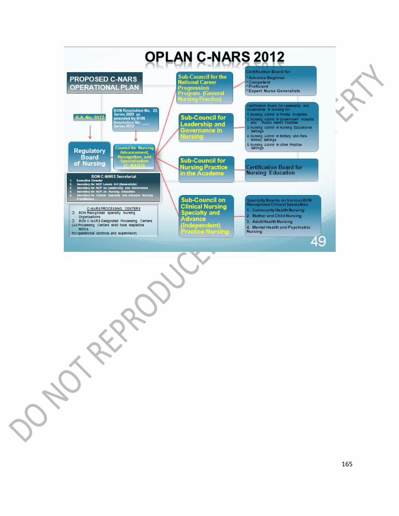
165