Embed Size (px)
Citation preview
CRISPR screen identifies the NCOR/HDAC3 complex asa major suppressor of differentiationin rhabdomyosarcomaMichael P. Phelpsa, Jenna N. Baileya, Terra Vleeshouwer-Neumanna, and Eleanor Y. Chena,1
aDepartment of Pathology, University of Washington, Seattle, WA 98195
Edited by Robert E. Kingston, Massachusetts General Hospital/Harvard Medical School, Boston, MA, and approved November 14, 2016 (received for reviewJune 23, 2016)
Dysregulated gene expression resulting from abnormal epigeneticalterations including histone acetylation and deacetylation hasbeen demonstrated to play an important role in driving tumorgrowth and progression. However, the mechanisms by whichspecific histone deacetylases (HDACs) regulate differentiation insolid tumors remains unclear. Using pediatric rhabdomyosarcoma(RMS) as a paradigm to elucidate the mechanism blocking differ-entiation in solid tumors, we identified HDAC3 as a major suppres-sor of myogenic differentiation from a high-efficiency Clusteredregularly interspaced short palindromic repeats (CRISPR)-basedphenotypic screen of class I and II HDAC genes. Detailed character-ization of the HDAC3-knockout phenotype in vitro and in vivo usinga tamoxifen-inducible CRISPR targeting strategy demonstrated thatHDAC3 deacetylase activity and the formation of a functional com-plex with nuclear receptor corepressors (NCORs) were critical inrestricting differentiation in RMS. The NCOR/HDAC3 complex spe-cifically functions by blocking myoblast determination protein 1(MYOD1)-mediated activation of myogenic differentiation. Interest-ingly, there was also a transient up-regulation of growth-promotinggenes upon initialHDAC3 targeting, revealing a unique cancer-specificresponse to the forced transition from a neoplastic state to terminaldifferentiation. Our study applied modifications of CRISPR/CRISPR-associated endonuclease 9 (Cas9) technology to interrogate thefunction of essential cancer genes and pathways and has providedinsights into cancer cell adaptation in response to altered differentia-tion status. Because current pan-HDAC inhibitors have shown disap-pointing results in clinical trials of solid tumors, therapeutic targetsspecific to HDAC3 function represent a promising option for differen-tiation therapy in malignant tumors with dysregulated HDAC3 activity.
histone deacetylase | HDAC3 | NCOR | rhabdomyosarcoma | CRISPR
Abnormal epigenetic alterations play an important role indriving tumor growth and progression (1, 2). Histone deace-
tylases (HDACs), which are major epigenetic modifiers, aredysregulated in a significant subset of cancers (3, 4). Althoughpan-HDAC inhibitors have elicited promising therapeutic re-sponses in some hematologic malignancies (1, 2, 5), limitedtherapeutic benefits have been reported in clinical trials for mostsolid tumors, including sarcomas (6). The inefficacy of HDACinhibitors in solid tumors most likely results in part from theirbroad and unknown substrate range and their pleiotropic effects.Despite these early clinical failures, HDACs remain prominenttherapeutic targets in cancers because of their ability to repro-gram gene-expression networks. Improved understanding of themolecular mechanisms underlying specific HDAC function willlead to more effective drug and therapy designs.Rhabdomyosarcoma (RMS), which consists of two major
subtypes, embryonal (ERMS) and alveolar (ARMS), is the mostcommon pediatric soft tissue malignancy. Although the twomajor subtypes are driven by distinct genetic alterations, both arecharacterized by a block in the myogenic differentiation program(7, 8). We have previously shown that treatment of RMS cellswith HDAC inhibitors results in the suppression of tumor growth
through the induction of myogenic differentiation (9). However,the mechanism by which aberrant activity of specific HDAC(s)represses differentiation and contributes to the malignant trans-formation of RMS remains unclear.Although recent advances in Clustered regularly interspaced
short palindromic repeats (CRISPR)/CRISPR-associated endo-nuclease 9 (Cas9) genome-editing technology have facilitated theidentification of essential tumor genes, detailed phenotypic andfunctional characterization of essential cancer genes with thecurrent technology is limited by the inability to expand mutanttumor clones harboring essential gene mutations and by poorCRISPR targeting efficiency in pooled cells. In this study, weused modifications of CRISPR/Cas9 genome-editing technology,including high-efficiency phenotypic screens and inducible genetargeting, to interrogate the functions of essential cancer genes.These genomic tools were used to identify the underlying HDAC-mediated epigenetic mechanisms blocking differentiation of RMStumor cells, which are essential for tumor progression.
ResultsCRISPR-Mediated Knockout of HDAC3 Induces Myogenic Differentiationin RMS. To characterize the role of specific HDACs in regulatingRMS tumor growth, we performed a CRISPR/Cas9-based phe-notypic screen of class I and class II HDAC genes using human381T ERMS cells (Fig. 1A and Fig. S1A). In contrast to singleguide RNA (gRNA) CRISPR screens, the lentiviral phenotypicscreen used dual gRNAs (DgRNA) targeted to each HDAC geneto increase overall targeting efficiency to 50–80% (Fig. 1B andTable S1). This strategy enabled direct analysis of phenotypic ef-fects of pooled tumor cells without the need for stable selection orisolation of mutant clones.CRISPR-mediated targeting of HDAC1, 2, 3, 4, and 6 signif-
icantly decreased tumor cell growth (Fig. 1C). Knockout of eitherHDAC3 or HDAC4 also resulted in distinct myogenic differentia-tion, as shown by the presence of morphologically multinucleated
Significance
Current histone deacetylase (HDAC) inhibitors have shown mixedresults in the treatment of many cancer types. Our study hasdemonstrated significant antitumor phenotypes resulting fromtargeted disruption of HDAC3 and the NCOR complexwith genomeengineering technology. Our findings provide compelling evidencethat the HDACs and their essential interacting factors remain keycancer therapeutic targets and that the next generation of selectiveHDAC inhibitors may improve survival of cancer patients.
Author contributions: M.P.P. and E.Y.C. designed research; M.P.P., J.N.B., T.V.-N., and E.Y.C.performed research; M.P.P., J.N.B., T.V.-N., and E.Y.C. analyzed data; and M.P.P. and E.Y.C.wrote the paper.
The authors declare no conflict of interest.
This article is a PNAS Direct Submission.1To whom correspondence should be addressed. Email: [email protected].
This article contains supporting information online at www.pnas.org/lookup/suppl/doi:10.1073/pnas.1610270114/-/DCSupplemental.
15090–15095 | PNAS | December 27, 2016 | vol. 113 | no. 52 www.pnas.org/cgi/doi/10.1073/pnas.1610270114
Dow
nloa
ded
by g
uest
on
Apr
il 3,
202
0
myotubes highlighted by myosin heavy chain (MF20)-positiveimmunostaining. However, the effect of HDAC4 knockout waslimited compared with the suppression of tumor cell growth(>90% reduction in growth) and the extent of differentiation (60–80% differentiated) exhibited by HDAC3 knockout (Fig. 1 D–Fand Fig. S1A). HDAC3 targeting also induced myogenic differ-entiation to varying degrees in a panel of five additional RMS cellslines (RD, SMS-CTR, Rh3, Rh5, and Rh30) derived from bothERMS and ARMS subtypes (Fig. 1G and Fig. S1B). In addition tosingle HDAC gene knockout, we targeted HDAC1 and HDAC2simultaneously because they are known to have redundant func-tions (10). Double knockout of HDAC1 and HDAC2 resulted inno evidence of myogenic differentiation (Fig. S1A), suggestingthat HDAC1 and 2 have a role in regulating ERMS proliferationbut not myogenic differentiation pathways.Immunohistochemistry (IHC) revealed that normal skeletal
muscle lacked nuclear HDAC3 expression. By contrast, 22 of 23primary ERMS samples and 20 of 24 primary ARMS samples
exhibited distinct nuclear expression of HDAC3 (Fig. 1 H–J).Proliferating human myoblasts as well as all analyzed RMS celllines also exhibited nuclear HDAC3 expression, suggesting thatHDAC3 likely has important functions in undifferentiated myoblast-like cells (Fig. S1C).The level of myogenic differentiation observed with HDAC3
targeting was substantially higher than has been previouslyreported for treatment of RMS cells with pan-HDAC inhibitors(9). Because pan-HDAC inhibitors are unable to induce large-scale differentiation in RMS, we treated RMS cells with theHDAC3-selective inhibitor RGFP966 (Selleck Chemicals LLC)to determine if direct HDAC3 inhibition can induce the extent ofmyogenic differentiation observed with HDAC3 knockout. Sur-prisingly, the treatment of RMS cells with RGFP966 resulted inonly modest growth suppression (Fig. S2 A–C) and myogenicdifferentiation (30–35%) (Fig. S2 D–F), suggesting that currentHDAC inhibitors lack the potency necessary to suppress growthand induce differentiation of RMS as a single agent.
Conditional HDAC3 Knockout Arrests Tumor Growth and InducesMyogenic Differentiation of RMS Tumors in Vivo. To investigatethe function of HDAC3 in RMS, we developed a tamoxifen-inducible Cas9-ERT2 PiggyBac transposon to control gene targetingtemporally both in vitro and in vivo (Fig. 2A). Tamoxifen-inducedHDAC3 gene knockout in ERMS cells (Fig. 2B) validated theresults from the CRISPR phenotypic screen by inducing growtharrest and concomitant myogenic differentiation in more than75% of the treated cells (Fig. 2 C and D and Fig. S3 A–F). Thishigh-efficiency inducible gene targeting of pooled RMS tumorcells significantly reduced overall HDAC3 protein levels withoutaffecting the levels of other class I HDAC proteins (Fig. 2Band Fig. S3G). HDAC3-knockout cells exhibited up-regulatedexpression of key myogenic regulatory genes (Fig. S3 H–K) withno increase in apoptosis (Fig. S3L), suggesting that the decreasein tumor cell growth was predominantly caused by terminalmyogenic differentiation.We validated the HDAC3 loss-of-function effects in vivo by
inducing HDAC3 targeting in ERMS tumor xenografts. Immu-nocompromised NOD-SCID Il2rg−/− (NSG) mice were injectedon bilateral flanks with tamoxifen-inducible 381T ERMS cellstargeting either HDAC3 or a safe-harbor region of the genome(Chr 4: 58,110,237–58,110,808; GRCH38.p2) (Fig. 2A). After tu-mor development, the mice were treated with tamoxifen to inducetargeted gene knockout in the respective tumors (Fig. 2E). Incontrast to safe-harbor control xenografts, HDAC3-targeted xe-nografts showed at least 50% reduction in tumor growth 2 weeksafter tamoxifen treatment (Fig. 2 F and G). Histological analysis ofHDAC3-knockout tumors revealed an increase in the presence ofmultinucleated cells resembling myotubes (Fig. 2 H and I). Simi-larly, HDAC3-targeted xenografts demonstrated a significant de-crease in the proliferation index (Ki67) (Fig. 2 J and K) and anincreased number of MF20+ differentiated cells (Fig. 2 L and M)but no increase in apoptosis (cleaved caspase-3) (Fig. 2 N and O).
The Nuclear Receptor Corepressor/HDAC3 Complex Blocks MyogenicDifferentiation in RMS. Using mass spectrometry and coimmuno-precipitation (co-IP) assays on 381T ERMS cells, we identifiednuclear receptor corepressors 1 (NCOR1) and 2 (NCOR2) asthe top HDAC3-interacting factors with transcription factor-bindingactivity (Fig. S4). Nuclear expression of NCOR1 was also detectedin myoblasts and all analyzed RMS cell lines (Fig. S5 A–I). Althoughindividual knockout of NCOR1 and NCOR2 resulted in limitedmyogenic differentiation, likely because of functional redundancy,double knockout of NCOR1 and NCOR2 in a panel of six RMS celllines (RD, 381T, SMS-CTR, Rh3, Rh5, and Rh30) resulted in large-scale myogenic differentiation and reduction in cell growth, similarto that seen with HDAC3 knockout (Fig. 3 A–D and Fig. S5 J–O).These results suggest that the NCOR/HDAC3 transcriptional re-pressor complex, not an alternative HDAC3-mediated process,is primarily responsible for restricting myogenic differentiationin RMS.
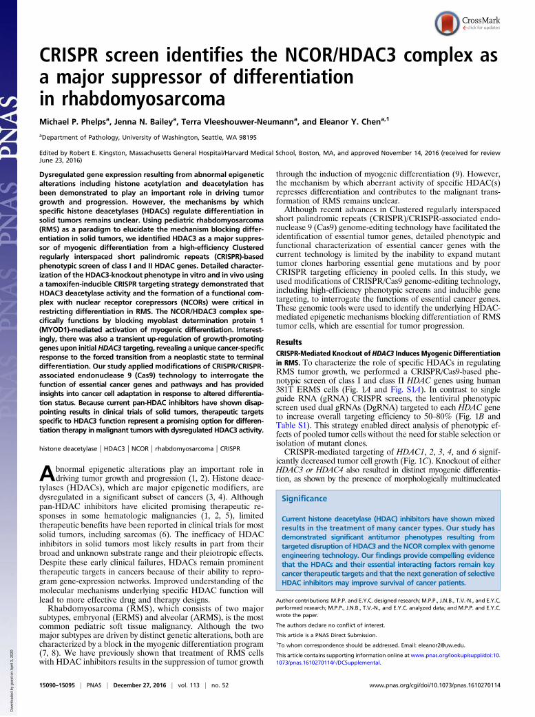
Fig. 1. CRISPR-based phenotypic screen of class I and II HDAC genes.(A) Schematics of the CRISPR phenotypic screen. (B, Upper) Depiction of the PCR-based method for detecting genomic DNA deletions between gRNAs as indi-cated by black arrows. Blue and green arrows represent primer pairs spanningeach gRNA target site. (Lower) Evidence of HDAC class I and II gene targeting byPCR amplification of gDNA deletions in HDAC targeted cells. (C) Cell growthchange by cell counts in 381T cells with CRISPR targeting of each class I and IIHDAC gene, normalized to cells with safe-harbor control targeting 10 d afterlentiviral transduction. (D and E) Immunofluorescence (IF) of MF20 in 381T cellswith control safe-harbor (D) or HDAC3 CRISPR (E) targeting. (Scale bar: 50 μm.)(F) Quantification ofMF20 IF from targeted deletion of HDAC class I and II genes.(G) Quantification of MF20 IF in ERMS (RD and SMS-CTR) and ARMS (Rh3, Rh5,and Rh30) cell lines with HDAC3 CRISPR targeting. (H and I) IHC for HDAC3 inprimary human skeletal muscle (H) and a representative RMS tumor section(I) (IHC magnification: 400×.) (Scale bar: 500 μm.) (Insets) Arrowheads point tonuclei. (Scale bar: 10 μm.) (J) Summary of HDAC3 IHC from human RMS tumorsamples. Error bars in C, F and G represent mean ± SD of three biological rep-licates. *P < 0.05, **P < 0.01, ***P < 0.001, ****P < 0.0001.
Phelps et al. PNAS | December 27, 2016 | vol. 113 | no. 52 | 15091
MED
ICALSC
IENCE
S
Dow
nloa
ded
by g
uest
on
Apr
il 3,
202
0
We next performed structure–function analysis to determinewhether the NCOR-interaction domain and/or the enzymaticactivity of HDAC3 were required for its function in RMS. Site-directed mutagenesis of HDAC3 was used to produce two mu-tant proteins, one lacking two distinct NCOR-binding sites in theN terminus (ΔB12) (11) and the second with a Y298F pointmutation in the catalytic site (Fig. 3E) (11). These mutantHDAC3 overexpression constructs were made CRISPR/Cas9resistant by introducing silent mutations in the two HDAC3gRNA target sites (Fig. 3E). Both HDAC3 mutants exhibitedloss of deacetylase activity (Fig. 3F) when overexpressed inHDAC3-knockout ERMS cells, as is consistent with previousstudies demonstrating that the NCOR/HDAC3 complex is re-quired for HDAC3 deacetylase activity (11, 12). However, onlythe ΔB12 HDAC3 mutant failed to interact with the NCORcomplex (Fig. 3G). Myogenic differentiation in HDAC3-targetedERMS cells was fully rescued by the overexpression of
CRISPR/Cas9-resistant WT HDAC3 (Fig. 3 H–K and Fig. S5P).In contrast, overexpression of either the ΔB12 or the Y298FHDAC3 mutant failed to restrict differentiation after HDAC3knockout, suggesting that both the NCOR/HDAC3 complex andHDAC3 deacetylase activity were critical for suppressing dif-ferentiation in RMS (Fig. 3 L–O and Fig. S5P). Interestingly, theY298F HDAC3 mutant was also able to induce myogenic dif-ferentiation independently in ERMS cells, likely through domi-nant-negative competition with endogenous HDAC3 (Fig. 3 Nand O and Fig. S5P).
The NCOR/HDAC3 Complex Blocks Myoblast Determination Protein1-Mediated Myogenic Differentiation in ERMS. To identify essen-tial genes and pathways regulated by the NCOR/HDAC3 complex,RNA sequencing was performed on ERMS cells 2, 4, and 10 dafter tamoxifen-induced CRISPR-mediated HDAC3 targeting.Based on Gene Ontology (GO) analysis, differentially expressedgenes immediately after HDAC3 knockout (day 4; 414 genes, P <0.05) were enriched for receptor and signal transduction functions,whereas late-stage differentially expressed genes (day 10; 199genes, P < 0.05) were enriched for cytoskeletal protein binding
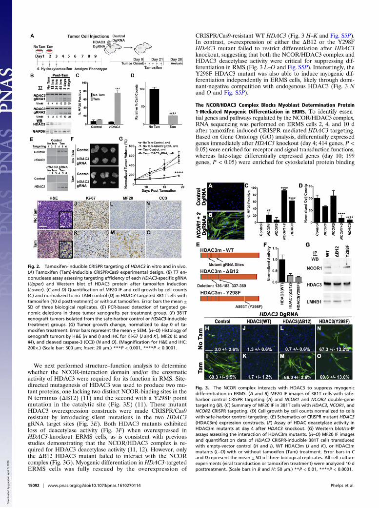
Fig. 2. Tamoxifen-inducible CRISPR targeting of HDAC3 in vitro and in vivo.(A) Tamoxifen (Tam)-inducible CRISPR/Cas9 experimental design. (B) T7 en-donuclease assay assessing targeting efficiency of each HDAC3-specific gRNA(Upper) and Western blot of HDAC3 protein after tamoxifen induction(Lower). (C and D) Quantification of MF20 IF and cell growth by cell counts(C) and normalized to no TAM control (D) in HDAC3-targeted 381T cells withtamoxifen (10 d posttreatment) or without tamoxifen. Error bars the mean ±SD of three biological replicates. (E) PCR-based detection of targeted ge-nomic deletions in three tumor xenografts per treatment group. (F) 381Txenograft tumors isolated from the safe-harbor control or HDAC3-inducibletreatment groups. (G) Tumor growth change, normalized to day 0 of ta-moxifen treatment. Error bars represent the mean ± SEM. (H–O) Histology ofxenograft tumors by H&E (H and I) and IHC for Ki-67 (J and K), MF20 (L andM), and cleaved caspase-3 (CC3) (N and O). (Magnification for H&E and IHC:200×.) (Scale bar: 500 μm; Inset: 20 μm.) ***P < 0.001, ****P < 0.0001.
Fig. 3. The NCOR complex interacts with HDAC3 to suppress myogenicdifferentiation in ERMS. (A and B) MF20 IF images of 381T cells with safe-harbor control CRISPR targeting (A) and NCOR1 and NCOR2 double-genetargeting (B). (C) Summary of MF20 IF in 381T cells with HDAC3, NCOR1, andNCOR2 CRISPR targeting. (D) Cell growth by cell counts normalized to cellswith safe-harbor control targeting. (E) Schematics of CRISPR mutant HDAC3(HDAC3m) expression constructs. (F) Assay of HDAC deacetylase activity inHDAC3m mutants at day 4 after HDAC3 knockout. (G) Western blot/co-IPassays assessing the interaction of HDAC3m mutants. (H–O) MF20 IF imagesand quantification data of HDAC3 CRISPR-inducible 381T cells transducedwith empty-vector control (H and I), WT HDAC3m (J and K), or HDAC3mmutants (L–O) with or without tamoxifen (Tam) treatment. Error bars in Cand D represent the mean ± SD of three biological replicates. All cell-cultureexperiments (viral transduction or tamoxifen treatment) were analyzed 10 dposttreatment. (Scale bars in B and H: 50 μm.) **P < 0.01, ****P < 0.0001.
15092 | www.pnas.org/cgi/doi/10.1073/pnas.1610270114 Phelps et al.
Dow
nloa
ded
by g
uest
on
Apr
il 3,
202
0
and striated muscle structural and contraction functions (Fig. 4A–C and Table S2). This global gene-expression profile inducedby HDAC3 knockout in ERMS cells highlighted the clear tran-sition from a neoplastic to fully differentiated muscle state.Previous studies have shown that repression of myoblast de-
termination protein 1 (MYOD1)-driven differentiation pathwaysresults in a characteristic arrest of RMS cells in a myoblast-likeproliferative phase of development, regardless of subtype or ini-tiating genetic events (8, 13, 14). However, even though MYOD1dysfunction was implicated in RMS more than two decades ago(15), the precise mechanism of MYOD1 transcriptional repressionremains unclear. To determine whether the NCOR/HDAC3complex is involved in the repression of MYOD1 target genes inERMS, we first analyzed the promoter motif of the top 500 dif-ferentially expressed genes 4 days afterHDAC3 gene targeting andidentified an enrichment for the E-box motif, the binding site forMYOD1, and the interacting factor T-cell factor 3 (TCF3) (alsoknown as “E2A”) [P < 9.00E-20, false discovery rate (FDR) q<1.32E-17]. By comparing the expression levels of 137 knownMYOD1 target genes (14) at 2, 4, and 10 d after HDAC3 tar-geting, we observed enrichment for select downstream MYOD1target genes (P < 0.0001) 4 d and 10 d after HDAC3 knockout(Fig. 4 D and E). Using ChIP studies, we further demonstratedbinding of HDAC3 to the E-box–containing regulatory regions ofMYOD1 target genes, but this interaction was abrogated uponMYOD1 knockout (Fig. 4F). The same E-box regulatory regionsshowed increased binding for acetylated H3K9, a histone mark fortranscriptionally active promoters, upon HDAC3 targeting (Fig.
4G). MYOD1 also was shown by co-IP to interact directly withE2A and the NCOR/HDAC3 complex in ERMS cells (Fig. S6).We next investigated the importance of MYOD1 in regulating
myogenesis in ERMS using a dual gene-targeting strategy to dis-rupt MYOD1 function in HDAC3-knockout ERMS cells. 381TERMS cells were transduced with Cas9- and MYOD1-targetingDgRNA lentivirus to establish MYOD1-knockout cells. Three dayslater, the MYOD1-targeted cells were transduced with HDAC3-targeting DgRNA lentivirus to produce dual MYOD1/HDAC3-knockout cells. MYOD1 targeting completely blocked the ability ofERMS cells to differentiate after HDAC3 knockout (Fig. 4H).Similar HDAC3 dual gene targeting of E2A and of the knownMYOD1 coactivators CREB-binding protein (CBP) and E1Abinding protein p300 (EP300) also limited the ability of HDAC3-deficient RMS cells to undergo myogenic differentiation (Fig. 4H).Taken together, these findings show that the NCOR/HDAC3complex interacts with MYOD1/E2A and associated cofactors torepress transcriptional activation of myogenic genes.
Loss of the Differentiation Block in ERMS Causes Transient Up-Regulation of Cell Growth Pathways. In addition to the enrich-ment forMYOD1 target genes, pathway analysis of gene-expressionchanges in the early stage of HDAC3 knockout (i.e., day 4)revealed a transient up-regulation of tumor growth-promotingsignaling pathways, most of which were down-regulated uponmyogenic differentiation by day 10. This cellular proliferation re-sponse was mediated by growth factors such as insulin-like growthfactor 1 (IGF1), hepatocyte growth factor (HGF), and TGFβ, aswell as epithelial–mesenchymal transition and metastasis path-ways (Fig. 5A and Table S3). We therefore performed a secondCRISPR phenotypic screen in 381T ERMS cells to determine thefunction of 42 candidate genes with signaling or transcriptionalactivity linked to HDAC3 through expression-profiling or massspectrometry studies (Fig. 5B). None of the candidate genes wasable to prevent myogenic differentiation when targeted withHDAC3 in a dual gene-targeting phenotypic screen. However, in-dependent disruption of five of the genes [ephrin type-A receptor 5(EPHA5), myogenic factor 5 (MYF5), forkhead box P2 (FOXP2),nitric oxide synthase 1 (NOS1), and AT-rich interactive domain-containing protein 1 (ARID1A)] significantly decreased tumor cellgrowth without inducing myogenic differentiation (Fig. 5C). ChIPassays demonstrated enrichment of the acetylated histone markH3K9ac in the promoters of MYF5, FOXP2, NOS1, and EPHA5(Fig. 5D), suggesting that up-regulation of these genes was likelycaused by altered histone landscapes in the regulatory regions.
DiscussionIn this study, we have identified HDAC3 as a major suppressor ofmyogenic differentiation in RMS from a high-efficiency CRISPRphenotypic screen of class I and class II HDAC genes and furthercharacterized the HDAC3 loss-of-function phenotype in vitro andin vivo using a tamoxifen-inducible CRISPR gene-targeting strategy.HDAC3 knockout in RMS cells resulted in significant suppressionof tumor growth through the activation of terminal myogenic dif-ferentiation. Interestingly, HDAC4 knockout also increased myo-genic differentiation in RMS cells to a limited extent, supporting thecurrent model that class IIa HDACs, including HDAC4, function asscaffolding molecules to recruit HDAC3 to transcriptional targetsites (16). Although HDAC1 and HDAC2 have been shown to haveredundant functions in muscle development (10), disruption ofHDAC1 and HDAC2 did not induce any significant effect on RMScell differentiation. However, targeting either gene did reduceoverall cell proliferation to a limited extent. Our findings indicatethat abnormal HDAC3 activity, rather than HDACs 1 and 2, isessential for the repression of terminal myogenic differentiation inRMS cells.The HDAC3-knockout phenotype was recapitulated by double
targeting of both NCOR1 and NCOR2, demonstrating that HDAC3blocks myogenic differentiation in RMS through the NCOR/HDAC3transcriptional repressor complex. The NCOR complex was origi-nally identified as transcriptional corepressors of nuclear receptors,
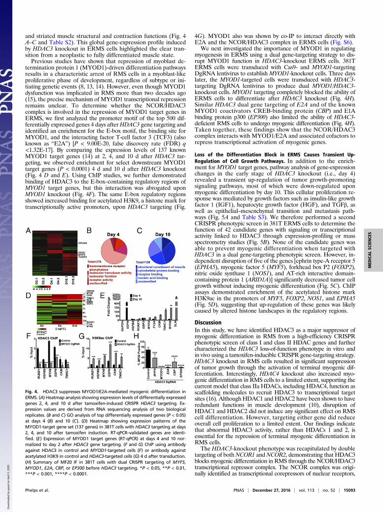
Fig. 4. HDAC3 suppresses MYOD1/E2A-mediated myogenic differentiation inERMS. (A) Heatmap analysis showing expression levels of differentially expressedgenes 2, 4, and 10 d after tamoxifen-induced CRISPR HDAC3 targeting. Ex-pression values are derived from RNA sequencing analysis of two biologicalreplicates. (B and C) GO analysis of top differentially expressed genes (P < 0.05)at days 4 (B) and 10 (C). (D) Heatmap showing expression patterns of theMYOD1-target gene set (137 genes) in 381T cells with HDAC3 targeting at days2, 4, and 10 after tamoxifen induction. RT-qPCR–validated genes are identi-fied. (E) Expression of MYOD1 target genes (RT-qPCR) at days 4 and 10 nor-malized to day 2 after HDAC3 gene targeting. (F and G) ChIP using antibodyagainst HDAC3 in control and MYOD1-targeted cells (F) or antibody againstacetylated H3K9 in control and HDAC3-targeted cells (G) 4 d after transduction.(H) Summary of MF20 IF in 381T cells with dual CRISPR targeting of MYF5,MYOD1, E2A, CBP, or EP300 before HDAC3 targeting. *P < 0.05, **P < 0.01,***P < 0.001, ****P < 0.0001.
Phelps et al. PNAS | December 27, 2016 | vol. 113 | no. 52 | 15093
MED
ICALSC
IENCE
S
Dow
nloa
ded
by g
uest
on
Apr
il 3,
202
0
e.g., thyroid hormone receptor and retinoid acid receptor (17). Theassociation of HDAC3 and the NCOR complex has been shown tobe essential for the regulation of circadian and metabolic physiol-ogy, muscle physiology, and genomic stability (3, 18, 19). HDAC3binds to the NCOR complex through the deacetylase-activatingdomain (DAD), which activates HDAC3 deacetylase activity (11,12). In agreement with these findings, we demonstrated throughstructure function analysis that the interaction of HDAC3 with theNCOR complex is required for functional deacetylase activity andNCOR/HDAC3-mediated repression of myogenic differentiation inRMS. Of note, the antitumor effects elicited by the treatment ofRMS cells with the HDAC3-selective inhibitor RGFP966 were notas robust as the loss-of-function phenotype generated by CRISPR-mediated HDAC3 knockout. Although RGFP966 inhibits histonedeacetylase activity, the substrate range and mechanism of actionunderlying RGFP966 remain unclear. Our findings suggest thatscreening drug candidates for NCOR/HDAC3 complex specificityrather than for deacetylase activity may improve the therapeuticefficacy of HDAC inhibitors for RMS and other solid tumors withdysregulated HDAC3 activity.
ERMS and ARMS share pathologic features of myogenicdifferentiation arrest characterized by defects in the MYOD1-mediated transcriptional program (8, 13–15). Previous studies inthe ERMS RD cell line have shown that competitive binding ofMYOD1 by inhibitory factors such as musculin and other alteredE-box transcription factors, as well as repression of transcriptionby the Polycomb repressive complex 2 (PRC2), can contribute toblocking the differentiation of tumor cells (20, 21). However, thecommon mechanism blocking the differentiation in RMS re-mains unknown. Here we demonstrate that both ERMS andARMS subtypes undergo extensive myogenic differentiationfrom CRISPR targeting of either HDAC3 or combined NCOR1and NCOR2 knockout. Nuclear HDAC3 is present in prolifer-ating myoblasts as well as in RMS cells but is down-regulated indifferentiated skeletal muscle, supporting a role for HDAC3in undifferentiated myogenic cells. Furthermore, the NCOR/HDAC3 complex interacts with MYOD1 and associated cofac-tors to repress the transcriptional activation of myogenic genes.Although the initiating genetic events in ERMS and ARMS areunique, our findings suggest that the driving mechanisms of bothRMS subtypes converge on the NCOR/HDAC3 complex as oneof the common mechanisms for repressing MYOD1-mediatedmyogenic differentiation, in turn promoting uncontrolled pro-liferation of myoblast-like cells (Fig. 6). Although our data suggestthat the NCOR/HDAC3 complex is critical to suppressing myo-genic differentiation in RMS, the molecular pathways or geneticalterations leading to dysregulation of NCOR/HDAC3 activity inRMS remain to be elucidated.The goal of differentiation therapy in cancer is to reactivate the
endogenous differentiation program with concomitant loss of tu-mor cell phenotypes (22). We have demonstrated by phenotypiccharacterization and expression-profiling studies that targeteddisruption of the NCOR/HDAC3 complex by CRISPR is sufficientto switch RMS cells from a neoplastic state to a terminally dif-ferentiated muscle state, indicating the feasibility and potentialefficacy of differentiation therapy in RMS. Surprisingly, targetingHDAC3 in ERMS cells also resulted in a transient but robust up-regulation of oncogenic genes and pathways including IGF, HGF,and MAPK signaling pathways before the completion of terminaldifferentiation (Fig. 6). In our CRISPR screen of 42 additionalcandidate genes linked to HDAC3 function, we have identifiedFOXP2, EPHA5, NOS1, or ARID1A as essential genes for RMStumor growth. Recurrent mutations in FOXP2 and ARID1A and adifferential methylation pattern of NOS1 have been shown in RMS(8, 23). These studies along with our loss-of-function studies indi-cate the important roles of these genes in RMS pathogenesis.
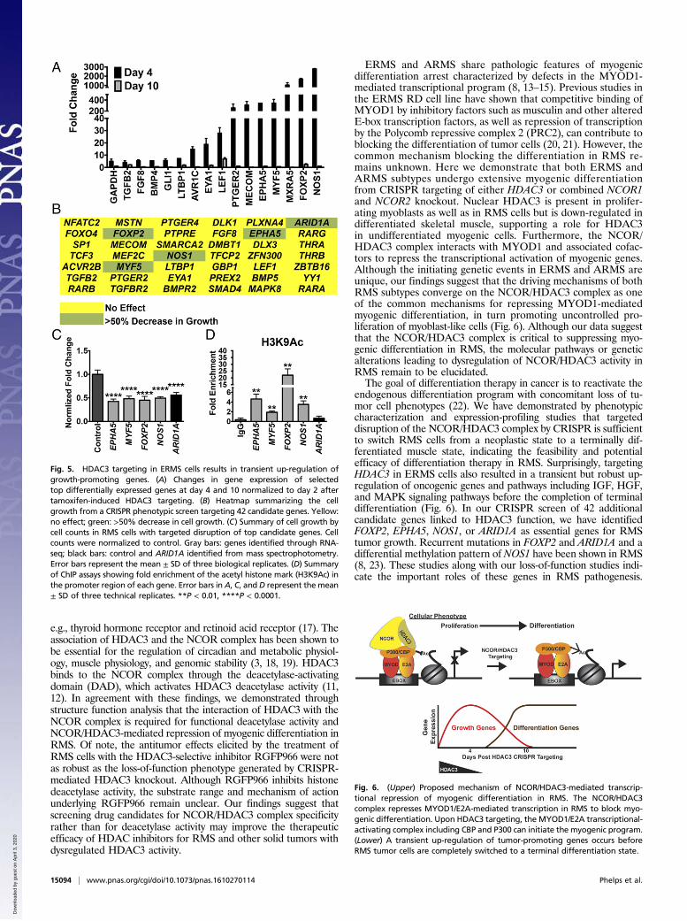
Fig. 5. HDAC3 targeting in ERMS cells results in transient up-regulation ofgrowth-promoting genes. (A) Changes in gene expression of selectedtop differentially expressed genes at day 4 and 10 normalized to day 2 aftertamoxifen-induced HDAC3 targeting. (B) Heatmap summarizing the cellgrowth from a CRISPR phenotypic screen targeting 42 candidate genes. Yellow:no effect; green: >50% decrease in cell growth. (C) Summary of cell growth bycell counts in RMS cells with targeted disruption of top candidate genes. Cellcounts were normalized to control. Gray bars: genes identified through RNA-seq; black bars: control and ARID1A identified from mass spectrophotometry.Error bars represent the mean ± SD of three biological replicates. (D) Summaryof ChIP assays showing fold enrichment of the acetyl histone mark (H3K9Ac) inthe promoter region of each gene. Error bars in A, C, and D represent the mean± SD of three technical replicates. **P < 0.01, ****P < 0.0001.
Fig. 6. (Upper) Proposed mechanism of NCOR/HDAC3-mediated transcrip-tional repression of myogenic differentiation in RMS. The NCOR/HDAC3complex represses MYOD1/E2A-mediated transcription in RMS to block myo-genic differentiation. Upon HDAC3 targeting, the MYOD1/E2A transcriptional-activating complex including CBP and P300 can initiate the myogenic program.(Lower) A transient up-regulation of tumor-promoting genes occurs beforeRMS tumor cells are completely switched to a terminal differentiation state.
15094 | www.pnas.org/cgi/doi/10.1073/pnas.1610270114 Phelps et al.
Dow
nloa
ded
by g
uest
on
Apr
il 3,
202
0
Based on the hyperacetylated status of histone H3 in the promoterregions of MYF5, FOXP2, EPHA5, and NOS1, a subset of geneswith oncogenic function likely undergoes transient activation uponHDAC3 loss of function. The transient up-regulation of tumor-promoting genes could represent either direct de-repression of thetranscription block by the HDAC3 repressor complex or a secondaryadaptive response as tumor cells transition from a neoplastic state to aterminally differentiated state. The implication of our findings fordifferentiation therapy is that incomplete induction of differentiationmay not be sufficient to counteract continued growth of tumor cellsbecause of the transient up-regulation of oncogenic genes and path-ways (Fig. 6).Our study demonstrated that genome-engineering technology
could be used to induce large-scale differentiation of cancer cellswhere direct chemical inhibitors have proven ineffective. Knockout ofthe NCOR/HDAC3 complex revealed a significant vulnerability ofRMS cells to targeted disruption of factors blocking differentiation.This effect was observed in both RMS subtypes (ARMS and ERMS),suggesting that there are common pathways restricting differentiationof tumor cells with similar tissue lineage regardless of initiating onco-genic events. Because pan-HDAC inhibitors have shown disappointingresults in clinical trials of solid tumors, the development of new tar-geted therapies with increased selectivity to specific HDACs or inter-acting repressor complexes may improve treatment of certain cancertypes in which current pan-HDAC inhibitors have previously failed.
MethodsAnimal studies were approved by the University ofWashington Subcommitteeon Research Animal Care under protocol no. 4330-01_SC_v19. Archived par-affin tissue blocks for human RMS tumor samples were obtained under anapproved human Internal Review Board protocol 14988 at Seattle Children’sHospital/University of Washington. Additional RMS tissue microarrays wereobtained from US Biomax, Inc.
CRISPR/Cas9 Gene Targeting. Rapid single and multiplex gene knockout wasaccomplished by treating RMS cells with multiple lentiviral particles containingseparate DgRNA-targeting and Cas9-expressing viruses. Lentiviral-transducedRMS cells were split into specific experiments 3 d postinfection and were an-alyzed 7 d later without antibiotic selection or isolation of clonal cells. EachDgRNA-targeting viral vector contained dual gRNAs targeting conserved exonsof each gene.All DgRNAswere cloned into the CRISPR plasmids using a one-stepGibson reaction.
CRISPR/Cas9-inducible cells were created using PiggyBac transposition.Stable cell lines were integrated with an all-in-one construct containingDgRNAs and expressing an ERT2-Cas9-ERT2 fusion protein for tamoxifen-inducible gene targeting. Inducible gene targeting in vitro was accomplished
by treating ERMS cells for 3 d with 2 μM 4-hydroxytamoxifen. For structure–function studies, HDAC3-inducible cells were transduced with constructsoverexpressing either WT or mutant CRISPR/Cas9-resistant HDAC3 linked toa T2A-GFP cassette. Cells were sorted for GFP fluorescence, and then en-dogenous HDAC3 was targeted with tamoxifen treatment.
In vivo CRISPR-inducible tumor xenografts were created by injecting NSGimmunocompromised mice with tamoxifen-inducible cells targeting either asafe-harbor region of the genome or HDAC3. After tumor development, themice were treated with five injections of 100 mg/kg tamoxifen to inducein vivo gene targeting. Targeting efficiency was validated from both in vitroand in vivo experiments using either a T7 endonuclease assay to examinetargeting efficiency at individual gRNA target sites or PCR to look for ge-nomic deletions between both gRNAs.
Cellular Assays. RMS cell growth was quantified by direct cell counting. Analysisof RMS differentiation was determined with IHC by fixing treated cells with 2%paraformaldehyde followed by MF20 immunostaining. Stained cultures wereimaged and quantified for the percent of MF20+ cells. An annexin V flowcytometry-based assay was used to analyze apoptosis. To assess HDAC3 deace-tylase activity, a nuclear extract kit (Active Motif, catalog no. 40010) was used toobtain nuclear extracts from HDAC3-knockout 381T cells overexpressing theCRISPR-resistant HDAC3 mutants at 4 d post targeting. HDAC3 (WT or mutant)was immunoprecipitated from nuclear extracts, and HDAC deacetylase activitywas quantified using a fluorescent HDAC activity assay kit (Active Motif, catalogno. 56200), which uses a short peptide substrate that contains an acetylated ly-sine residue that can be deacetylated by class I, IIB, and IV HDAC enzymes.
Co-IP. Co-IP of HDAC3 or NCOR1was performed using the nuclear complex co-IP kit (Active Motif, catalog no. 54001). Two hundred micrograms of extractand 2 μg of antibody were used. Dynabead protein G (Life Technologies,catalog no. 10003D) was used to capture the protein/antibody complex.
All quantitative RT-PCR (RT-qPCR) primer pairs and ChIP primer pairs arelisted in Table S4.
Detailed experimental procedures are described in SI Methods.
ACKNOWLEDGMENTS. We thank the Histology and Imaging Core at Universityof Washington and the Genomics Resource at Fred Hutchinson Cancer ResearchCenter, in particular Jerry Davison; Jessica Gianopulos (an undergraduate re-search assistant at University of Washington) for her valuable work validatingCRISPR targeting efficiency; and Dr. Michael Dyer (St. Jude Children’s ResearchHospital) and Dr. Myron Ignatius (Massachusetts General Hospital) for commentsand suggestions for the manuscript. The laboratory of Dr. Ray Monnat (Univer-sity of Washington) identified and characterized the safe-harbor genomic locusused for control CRISPR-targeting constructs. This work was supported by NIHGrants K08AR063165 and R01CA196882 (to E.Y.C.), a St. Baldrick’s FoundationScholar Award (to E.Y.C.), the Rally Foundation (E.Y.C.), and the Sarcoma Foun-dation of America (E.Y.C.).
1. Piekarz RL, Bates SE (2009) Epigenetic modifiers: Basic understanding and clinicaldevelopment. Clin Cancer Res 15(12):3918–3926.
2. West AC, Johnstone RW (2014) New and emerging HDAC inhibitors for cancertreatment. J Clin Invest 124(1):30–39.
3. Bhaskara S, et al. (2010) Hdac3 is essential for the maintenance of chromatin structureand genome stability. Cancer Cell 18(5):436–447.
4. Santoro F, et al. (2013) A dual role for Hdac1: Oncosuppressor in tumorigenesis, on-cogene in tumor maintenance. Blood 121(17):3459–3468.
5. Olsen EA, et al. (2007) Phase IIb multicenter trial of vorinostat in patients with per-sistent, progressive, or treatment refractory cutaneous T-cell lymphoma. J Clin Oncol25(21):3109–3115.
6. Cote GM, Choy E (2013) Role of epigenetic modulation for the treatment of sarcoma.Curr Treat Options Oncol 14(3):454–464.
7. Keller C, Guttridge DC (2013) Mechanisms of impaired differentiation in rhabdo-myosarcoma. FEBS J 280(17):4323–4334.
8. Shern JF, et al. (2014) Comprehensive genomic analysis of rhabdomyosarcoma revealsa landscape of alterations affecting a common genetic axis in fusion-positive andfusion-negative tumors. Cancer Discov 4(2):216–231.
9. Vleeshouwer-Neumann T, et al. (2015) Histone deacetylase inhibitors antagonizedistinct pathways to suppress tumorigenesis of embryonal rhabdomyosarcoma. PLoSOne 10(12):e0144320.
10. Montgomery RL, et al. (2007) Histone deacetylases 1 and 2 redundantly regulatecardiac morphogenesis, growth, and contractility. Genes Dev 21(14):1790–1802.
11. Sun Z, et al. (2013) Deacetylase-independent function of HDAC3 in transcription andmetabolism requires nuclear receptor corepressor. Mol Cell 52(6):769–782.
12. You S-H, et al. (2013) Nuclear receptor co-repressors are required for the histone-deacetylase activity of HDAC3 in vivo. Nat Struct Mol Biol 20(2):182–187.
13. Cao L, et al. (2010) Genome-wide identification of PAX3-FKHR binding sites inrhabdomyosarcoma reveals candidate target genes important for development andcancer. Cancer Res 70(16):6497–6508.
14. MacQuarrie KL, et al. (2013) Comparison of genome-wide binding of MyoD in normalhuman myogenic cells and rhabdomyosarcomas identifies regional and local sup-pression of promyogenic transcription factors. Mol Cell Biol 33(4):773–784.
15. Tapscott SJ, Thayer MJ, Weintraub H (1993) Deficiency in rhabdomyosarcomas of afactor required for MyoD activity and myogenesis. Science 259(5100):1450–1453.
16. Fischle W, et al. (2002) Enzymatic activity associated with class II HDACs is dependenton a multiprotein complex containing HDAC3 and SMRT/N-CoR. Mol Cell 9(1):45–57.
17. Baniahmad A, Tsai SY, O’Malley BW, Tsai MJ (1992) Kindred S thyroid hormone re-ceptor is an active and constitutive silencer and a repressor for thyroid hormone andretinoic acid responses. Proc Natl Acad Sci USA 89(22):10633–10637.
18. Alenghat T, et al. (2008) Nuclear receptor corepressor and histone deacetylase 3govern circadian metabolic physiology. Nature 456(7224):997–1000.
19. Yamamoto H, et al. (2011) NCoR1 is a conserved physiological modulator of musclemass and oxidative function. Cell 147(4):827–839.
20. Marchesi I, Fiorentino FP, Rizzolio F, Giordano A, Bagella L (2012) The ablation of EZH2uncovers its crucial role in rhabdomyosarcoma formation. Cell Cycle 11(20):3828–3836.
21. Yang Z, et al. (2009) MyoD and E-protein heterodimers switch rhabdomyosarcoma cellsfrom an arrested myoblast phase to a differentiated state. Genes Dev 23(6):694–707.
22. Cruz FD, Matushansky I (2012) Solid tumor differentiation therapy - is it possible?Oncotarget 3(5):559–567.
23. Chen X, et al.; St. Jude Children’s Research Hospital–Washington University PediatricCancer Genome Project (2013) Targeting oxidative stress in embryonal rhabdomyosarcoma.Cancer Cell 24(6):710–724.
Phelps et al. PNAS | December 27, 2016 | vol. 113 | no. 52 | 15095
MED
ICALSC
IENCE
S
Dow
nloa
ded
by g
uest
on
Apr
il 3,
202
0


![Detection and characterization of clustered regularly ... · interspaced short palindromic repeats (CRISPR)/cas in . V. parahaemolyticus [7]. The CRISPR-cas (CRISPR-associated proteins)](https://img.pdfslide.us/doc/110x75/5ed528faf68ca435874eb4e1/detection-and-characterization-of-clustered-regularly-interspaced-short-palindromic.jpg)