Embed Size (px)
Citation preview

CRICO Breast Care Management AlgorithmA DECISION SUPPORT TOOL
CBS 2019

Failure to diagnose breast cancer affects health care providers across a spectrum of specialties. To reduce the likelihood of such events, a task force of breast care specialists and primary care physicians, coordinated by CRICO, identified the key factors contributing to allegations of mismanaged breast care and subsequently developed the CRICO Breast Care Management Algorithm. The recommendations within the CRICO Algorithm are based on either a) broadly accepted evidence or b) conservative practices which may lack supportive evidence, but represent proven risk management strategies (and pose no risk of patient harm). Our goal is to aid primary care providers at various decision points across three domains of breast health care:
• asymptomatic women eligible for screening,• individuals seeking an assessment of their risks for developing breast cancer, and• patients who present with specific breast complaints.
The CRICO Algorithm is designed to help providers of primary breast care appropriately use available diagnostic tools. The provider is expected to gather information such as family history, atypia on previous biopsy, thoracic radiation before age 30, and reproductive risk factors to determine if changes to normal screening, or a referral to high-risk counseling, is indicated.
Even after a referral, providers of primary breast care have a responsibility for tracking and coordinating their patients’ ongoing breast care. In addition to being a tenet of good care, comprehensive provider follow up is a significant safeguard against allegations of failure to diagnose breast cancer.
The CRICO Breast Care Management Algorithm is a suggested guideline and should not be construed as a standard of care; care plans for individual patients must be based on the provider’s professional judgment. Respected experts endorse differing recommendations, especially for mammographic screening, and physicians may choose to follow alternate recommendations as their standard practice.
Improving Breast Patient Safety
CRICO BREAST CARE MANAGEMENT ALGORITHM
1
© 2019 CRICO
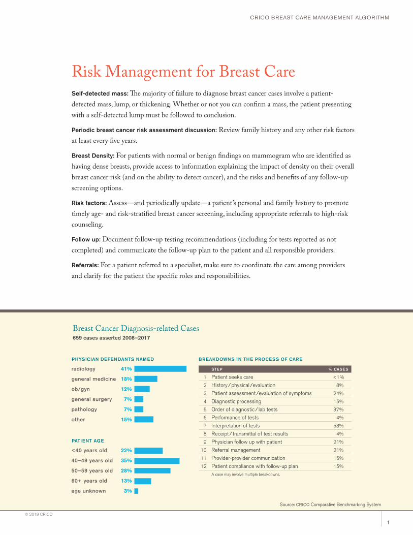
BREAKDOWNS IN THE PROCESS OF CARE
STEP % CASES
1. Patient seeks care <1%
2. History / physical /evaluation 8%
3. Patient assessment /evaluation of symptoms 24%
4. Diagnostic processing 15%
5. Order of diagnostic / lab tests 37%
6. Performance of tests 4%
7. Interpretation of tests 53%
8. Receipt / transmittal of test results 4%
9. Physician follow up with patient 21%
10. Referral management 21%
11. Provider-provider communication 15%
12. Patient compliance with follow-up plan 15%
A case may involve multiple breakdowns.
Breast Cancer Diagnosis-related Cases659 cases asserted 2008–2017
PHYSICIAN DEFENDANTS NAMED
radiology
general medicine
ob/gyn
general surgery
pathology
other
41%
18%
12%
7%
7%
15%
<40 years old
40–49 years old
50–59 years old
60+ years old
age unknown
PATIENT AGE
22%
35%
28%
13%
3%
Risk Management for Breast CareSelf-detected mass: The majority of failure to diagnose breast cancer cases involve a patient-detected mass, lump, or thickening. Whether or not you can confirm a mass, the patient presenting with a self-detected lump must be followed to conclusion.
Periodic breast cancer risk assessment discussion: Review family history and any other risk factors at least every five years.
Breast Density: For patients with normal or benign findings on mammogram who are identified as having dense breasts, provide access to information explaining the impact of density on their overall breast cancer risk (and on the ability to detect cancer), and the risks and benefits of any follow-up screening options.
Risk factors: Assess—and periodically update—a patient’s personal and family history to promote timely age- and risk-stratified breast cancer screening, including appropriate referrals to high-risk counseling.
Follow up: Document follow-up testing recommendations (including for tests reported as not completed) and communicate the follow-up plan to the patient and all responsible providers.
Referrals: For a patient referred to a specialist, make sure to coordinate the care among providers and clarify for the patient the specific roles and responsibilities.
Source: CRICO Comparative Benchmarking System
CRICO BREAST CARE MANAGEMENT ALGORITHM
2
© 2019 CRICO
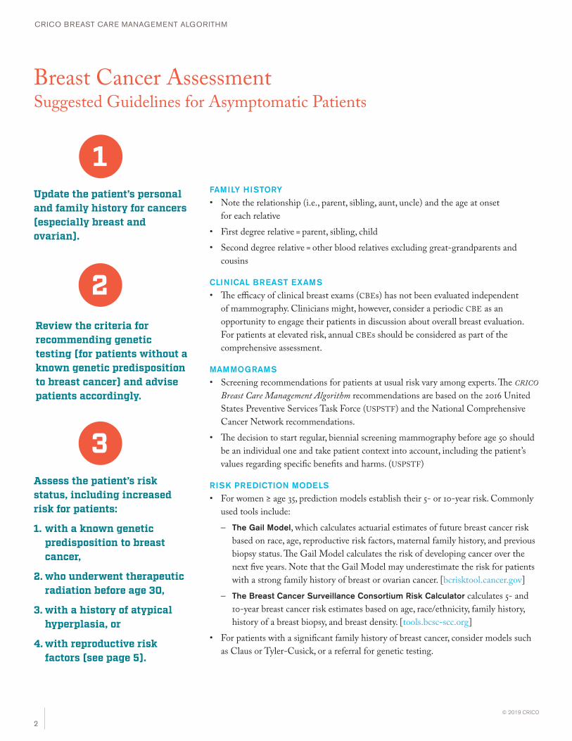
Breast Cancer Assessment Suggested Guidelines for Asymptomatic Patients
1
2
3
Update the patient’s personal and family history for cancers (especially breast and ovarian).
Review the criteria for recommending genetic testing (for patients without a known genetic predisposition to breast cancer) and advise patients accordingly.
Assess the patient’s risk status, including increased risk for patients:
1. with a known genetic predisposition to breast cancer,
2. who underwent therapeutic radiation before age 30,
3. with a history of atypical hyperplasia, or
4. with reproductive risk factors (see page 5).
FAMILY HISTORY
• Note the relationship (i.e., parent, sibling, aunt, uncle) and the age at onset for each relative
• First degree relative = parent, sibling, child • Second degree relative = other blood relatives excluding great-grandparents and
cousins
CLINICAL BREAST EXAMS
• The efficacy of clinical breast exams (CBEs) has not been evaluated independent of mammography. Clinicians might, however, consider a periodic CBE as an opportunity to engage their patients in discussion about overall breast evaluation. For patients at elevated risk, annual CBEs should be considered as part of the comprehensive assessment.
MAMMOGRAMS
• Screening recommendations for patients at usual risk vary among experts. The CRICO Breast Care Management Algorithm recommendations are based on the 2016 United States Preventive Services Task Force (USPSTF) and the National Comprehensive Cancer Network recommendations.
• The decision to start regular, biennial screening mammography before age 50 should be an individual one and take patient context into account, including the patient’s values regarding specific benefits and harms. (USPSTF)
RISK PREDICTION MODELS
• For women ≥ age 35, prediction models establish their 5- or 10-year risk. Commonly used tools include: – The Gail Model, which calculates actuarial estimates of future breast cancer risk
based on race, age, reproductive risk factors, maternal family history, and previous biopsy status. The Gail Model calculates the risk of developing cancer over the next five years. Note that the Gail Model may underestimate the risk for patients with a strong family history of breast or ovarian cancer. [bcrisktool.cancer.gov]
– The Breast Cancer Surveillance Consortium Risk Calculator calculates 5- and 10-year breast cancer risk estimates based on age, race/ethnicity, family history, history of a breast biopsy, and breast density. [tools.bcsc-scc.org]
• For patients with a significant family history of breast cancer, consider models such as Claus or Tyler-Cusick, or a referral for genetic testing.

CRICO BREAST CARE MANAGEMENT ALGORITHM
3
© 2019 CRICO
Breast Cancer Screening Considerations and Recommendations
...for patients at usual risk for breast cancer PATIENTS WHO HAVE NONE OF THE RISK FACTORS LISTED ABOVE
RECOMMENDATIONS
• Age ≥ 50: Begin bi-annual mammograms, consider annual CBE• Age 40-49: Annual breast cancer risk discussion with risk factor review and
CBE, consider bi-annual mammogram• Age < 40: Consider CBE every 1–3 years
...for patients with a known genetic predisposition to breast cancer KNOWN CARRIER OF A BRCA1 OR BRCA2 MUTATION
(HIGH PENETRANCE BREAST CANCER PREDISPOSING GENES)
• Untested individual with known close relative with BRCA1 or BRCA2 mutation
• Known carrier or untested individual with known close relative with another hereditary breast cancer syndrome gene (Li-Fraumeni syndrome, Cowden’s disease, Peutz-Jeghers syndrome, hereditary diffuse gastric cancer, other)
RECOMMENDATIONS
• Beginning at age 25, CBE at least once per year• Annual mammogram and MRI beginning at age 25 or individualized based
on earliest age onset in family. Preliminary data suggest that alternating MRI and mammography every six months may be helpful.
• If close relative, consider genetic testing
...for patients without a known genetic predisposition to breast cancerPATIENTS WHO SHOULD CONSIDER GENETIC TESTING
• Personal history of breast cancer diagnosed at age ≤ 50
• Personal history of ovarian cancer at any age• Male relative with breast cancer • 1st- or 2nd-degree relative diagnosed with breast cancer at < age 50
• 1st-degree relative, or (paternal) 2nd-degree relative diagnosed with DCIS at age ≤ 40 or ovarian cancer (any age)
• A diagnosis of breast cancer (or DCIS) and ovarian cancer in a single 1st- or 2nd-degree relative—or two close relatives in the same lineage
• Two relatives in the same lineage with early onset breast cancer• Women of Ashkenazi Jewish ancestry may be included despite fewer
affected relatives or later age onset
RECOMMENDATIONS
• For women whose genetic test results are positive, follow the recommendations above
• For women whose genetic test results are negative:• Women in a family with a known mutation who test negative are true
negative and should follow the recommendations for patients at usual risk (below).
• Women in a family without a known mutation who test negative should be referred to a genetics center. If possible, genetic testing should be performed with a genetic counselor or genetics expert.
• Consider breast MRI for patients with a lifetime risk of breast cancer > 20% as defined by BRACPRO or other models that are largely dependent on family history.
...for patients with a higher than usual risk for breast cancer THERAPEUTIC THORACIC RADIATION (E.G. HODGKINS) AGE < 30
• Risk from therapeutic radiation is much greater than risk from diagnostic radiation. The risk from infant thymus radiation, fluoroscopy for TB, or multiple X-rays for scoliosis is not well quantified.
RECOMMENDATIONS
• Annual mammogram beginning 8–10 years after radiation or at age 25
• Consider CBE at least once per year beginning at age 25
• Annual MRI in addition to annual mammogram
HISTOLOGY
• Lobular carcinoma in situ (LCIS)• History of ductal carcinoma in situ (DCIS)• History of invasive breast cancer• Atypical ductal or lobular hyperplasia (ADH or ALH): consider using the Gail
Model for risk assessment
RECOMMENDATIONS
• Annual mammogram after diagnosis• CBE at least once per year• Consider referral to high-risk counseling or risk reducing medication
REPRODUCTIVE AND OTHER RISK FACTORS
• Menarche before age 12
• Nulliparity• First birth after age 30
• Prior breast biopsy• >5 years of combined estrogen/progesterone hormone replacement
therapy
RECOMMENDATIONS
• For a patient age ≥ 35 with a constellation of these risk factors, consider assessment via the Gail Model to determine her level of risk for breast cancer.
• For patients with Gail Model five-year risk ≥ 1.67: CBE at least once per year, annual mammogram, consider high-risk counseling or risk reducing medication. (USPSTF recommends starting medication at ≥ 3.0. Patient may also be eligible for risk reducing clinical trials.)
CRICO BREAST CARE MANAGEMENT ALGORITHM
4
© 2019 CRICO
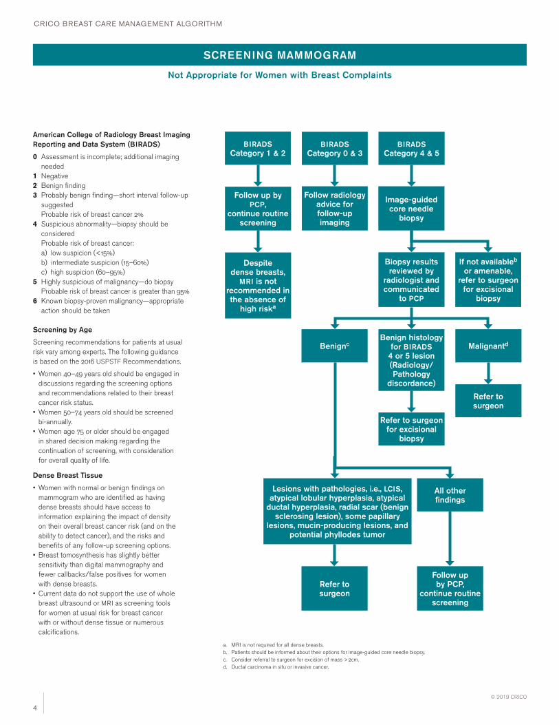
SCREENING MAMMOGRAM
BIRADS Category 1 & 2
BIRADS Category 0 & 3
BIRADS Category 4 & 5
Follow up by PCP,
continue routine screening
Follow radiology advice for follow-up imaging
Image-guided core needle
biopsy
If not availableb or amenable,
refer to surgeon for excisional
biopsy
Biopsy results reviewed by
radiologist and communicated
to PCP
Screening by Age
Screening recommendations for patients at usual risk vary among experts. The following guidance is based on the 2016 USPSTF Recommendations.
• Women 40–49 years old should be engaged in discussions regarding the screening options and recommendations related to their breast cancer risk status.
• Women 50–74 years old should be screened bi-annually.
• Women age 75 or older should be engaged in shared decision making regarding the continuation of screening, with consideration for overall quality of life.
Dense Breast Tissue
• Women with normal or benign findings on mammogram who are identified as having dense breasts should have access to information explaining the impact of density on their overall breast cancer risk (and on the ability to detect cancer), and the risks and benefits of any follow-up screening options.
• Breast tomosynthesis has slightly better sensitivity than digital mammography and fewer callbacks/false positives for women with dense breasts.
• Current data do not support the use of whole breast ultrasound or MRI as screening tools for women at usual risk for breast cancer with or without dense tissue or numerous calcifications.
American College of Radiology Breast Imaging Reporting and Data System (BIRADS)
0 Assessment is incomplete; additional imaging needed
1 Negative2 Benign finding3 Probably benign finding—short interval follow-up
suggested Probable risk of breast cancer 2%
4 Suspicious abnormality—biopsy should be considered Probable risk of breast cancer:
a) low suspicion (<15%) b) intermediate suspicion (15–60%) c) high suspicion (60–95%)5 Highly suspicious of malignancy—do biopsy
Probable risk of breast cancer is greater than 95%6 Known biopsy-proven malignancy—appropriate
action should be taken
Benign histology for BIRADS
4 or 5 lesion (Radiology/Pathology
discordance)
Refer to surgeon for excisional
biopsy
Benignc
Refer to surgeon
Malignantd
Refer to surgeon
All other findings
Lesions with pathologies, i.e., LCIS, atypical lobular hyperplasia, atypical
ductal hyperplasia, radial scar (benign sclerosing lesion), some papillary
lesions, mucin-producing lesions, and potential phyllodes tumor
Follow up by PCP,
continue routine screening
a. MRI is not required for all dense breasts. b. Patients should be informed about their options for image-guided core needle biopsy.c. Consider referral to surgeon for excision of mass > 2cm.d. Ductal carcinoma in situ or invasive cancer.
Despite dense breasts,
MRI is not recommended in the absence of
high riska
Not Appropriate for Women with Breast Complaints
CRICO BREAST CARE MANAGEMENT ALGORITHM
5
© 2019 CRICO
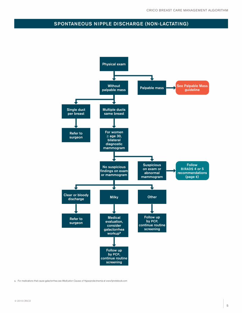
SPONTANEOUS NIPPLE DISCHARGE (NON-LACTATING)
Single duct per breast
Multiple ducts same breast
Refer to surgeon
For women ≥≥ age 30, bilateral
diagnostic mammogram
Clear or bloody discharge Milky
Refer to surgeon
Medical evaluation, consider
galactorrhea workupa
Follow up by PCP,
continue routine screening
No suspicious findings on exam or mammogram
Suspicious on exam or abnormal
mammogram
a. For medications that cause galactorrhea see Medication Causes of Hyperprolactinemia at www.fpnotebook.com
Follow BIRADS 4 or 5
recommendations (page 6)
Physical exam
Without palpable mass Palpable mass See Palpable Mass
guideline
Other
Follow up by PCP,
continue routine screening
CRICO BREAST CARE MANAGEMENT ALGORITHM
6
© 2019 CRICO
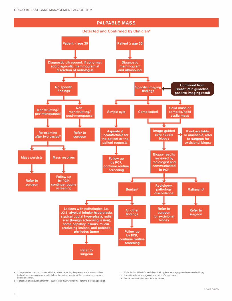
PALPABLE MASS
Patient < age 30
a. If the physician does not concur with the patient regarding the presence of a mass, confirm that routine screening is up to date. Advise the patient to return if her concern or symptoms persist or change.
b. If pregnant or not cycling monthly—but not later than two months—refer to a breast specialist.
Diagnostic mammogram
and ultrasound
Patient ≥≥ age 30
No specific findings
Menstruating/pre-menopausal
Re-examine after two cyclesb
Diagnostic ultrasound. If abnormal, add diagnostic mammogram at
discretion of radiologist
Mass persists
Refer to surgeon
Non-menstruating/
post-menopausal
Refer to surgeon
Mass resolves
Follow up by PCP,
continue routine screening
Specific imaging findings
Solid mass or complex/solid
cystic massSimple cyst Complicated
Aspirate if uncomfortable for the patient or the patient requests
Follow up by PCP,
continue routine screening
Image-guided core needle
biopsy
Radiology/ pathology
discordance
Refer to surgeon
for excisional biopsy
Benignd
Refer to surgeon
Malignante
Refer to surgeon
Continued from Breast Pain guideline, positive imaging result
c. Patients should be informed about their options for image-guided core needle biopsy.d. Consider referral to surgeon for excision of mass >2cm.e. Ductal carcinoma in situ or invasive cancer.
Biopsy results reviewed by
radiologist and communicated
to PCP
All other findings
Follow up by PCP,
continue routine screening
If not availablec or amenable, refer
to surgeon for excisional biopsy
Detected and Confirmed by Cliniciana
Lesions with pathologies, i.e., LCIS, atypical lobular hyperplasia, atypical ductal hyperplasia, radial scar (benign sclerosing lesion), some papillary lesions, mucin-
producing lesions, and potential phyllodes tumor
CRICO BREAST CARE MANAGEMENT ALGORITHM
7
© 2019 CRICO
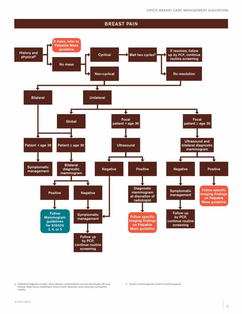
BREAST PAIN
History and physicala
If mass, refer to Palpable Mass
guideline
a. Differential diagnosis includes: chest wall pain, costochondritis, cervical radiculopathy, MI, lung disease, hiatal hernia, cholelithiasis, thoracic aortic dissection, aortic aneurysm, post partum mastitis.
No mass
Cyclical
Non-cyclical
Wait two cyclesb
No resolution
If resolves, follow up by PCP, continue routine screening
Bilateral Unilateral
Global Focalpatient < age 30
Ultrasound
Negative Positive
Diagnostic mammogram
at discretion of radiologist
Follow specific imaging findings
on Palpable Mass guideline
Patient < age 30 Patient ≥≥ age 30
Bilateral diagnostic
mammogram
Symptomatic management
Positive Negative
Follow Mammogram
guidelines for BIRADS 3, 4, or 5
Symptomatic management
Follow up by PCP,
continue routine screening
Focalpatient ≥≥ age 30
Ultrasound and bilateral diagnostic
mammogram
Negative Positive
Follow specific imaging findings
on Palpable Mass guideline
Symptomatic management
Follow up by PCP,
continue routine screening
b. Cycles if premenopausal; months if postmenopausal.

CRICO BREAST CARE MANAGEMENT ALGORITHM
8
© 2019 CRICO
Physician-Patient Discussion and Take-home Points Related to Breast Patient Safety
PATIENT-DETECTED LUMP/MASS
A self-discovered lump should be followed to resolution even if there is
provider-patient discordance on the presence of the lump. Follow every
mass to conclusion.
PATIENT UNSATISFIED WITH A NEGATIVE FINDING
Engage the patient in a discussion about her breast care management
subsequent to negative test/imaging results. Develop a clear and
effective plan, and ensure the patient’s understanding and agreement
of that plan.
Document all interactions as they occur to support future care and to
clarify any disputes that may arise later. This includes:
• in the history and physicals section of the record, include the findings
of the breast examination (note—in quotes—what the patient said, as
well as your own findings);
• for a confirmed lump or lesion, use a diagram or description to record
the exact location and size (if known); and
• for an unconfirmed mass, record—in the patient’s words—the location
and nature of the complaint.
SIGNIFICANCE OF EARLY DETECTION OF BREAST CANCER
Without reliable evidence that early detection of breast cancer can
significantly reduce the risk of mortality, health care providers cannot
guarantee a cure based on the timing of the diagnosis. Patients may
need to be educated as to the rigors and subtleties of research data,
and discrepancies in findings among various studies.
RISK OF BREAST CANCER FOR WOMEN YOUNGER THAN AGE 30
Be careful not to dismiss patients under age 30, who have an
approximately 1 in 2,000 chance of being diagnosed with breast
cancer at an early age.1 Women with multiple risk factors—especially
those that indicate a high level of risk, such as BRCA1/BRCA2 gene
mutation in a family member under age 40—should be concerned
about the possibility of early breast cancer.
PATIENTS IDENTIFIED AS HAVING DENSE BREASTS
Offer patients access to information explaining the impact of breast
density on their overall breast cancer risk (and on the ability to detect
cancer), and the risks and benefits of any follow-up screening options.
• Provide all patients the opportunity for a follow-up discussion (with
you or a designee) to ensure that they comprehend their overall
breast cancer risk, and the risks and benefits of any follow-up
screening options. For some patients, printed/online information may
be sufficient.
• Document any decisions reached regarding additional cancer
screening due to breast density.
COMMUNICATION
• Communicate all abnormal findings to the patient and document that
act.
• Avoid sending the wrong message to a patient by only telling her that
a palpable lump is probably benign. Stress that additional studies may
be needed to look for evidence of malignancy.
• Share any uncertainty on your part in a way that helps your patient
appreciate the importance of follow up.
• Confirm and document with other providers which of you will be the
clinician of record and responsible for ordering tests and following up
with the patient.
TEST RESULTS
• Explain to the patient how test results will be communicated to her
and (if appropriate) other clinicians.
• Document any telephone conversations with patients regarding the
reported results.
• To ensure notification of test results, employ a system to track ordered
tests through the receipt and communication to the patient.

CRICO BREAST CARE MANAGEMENT ALGORITHM
9
© 2019 CRICO
FOLLOW UP
• Make follow-up or test appointments before the patient leaves your
office.
• Physicians and patients share responsibility for follow up; explain
to your patients your tracking and compliance system (contacting
patients a day or two before their follow-up appointments can reduce
non-adherence).
• Track all surgical referrals to ensure that you are receiving a timely
report from the breast specialist or surgeon.
• Ask the Radiology department, breast care center, or specialist
to notify your office of patients who do not keep scheduled
appointments. Document all patient no-shows or cancellations
including for time-sensitive testing.
• If a patient refuses follow up, explain the risks of not having a
recommended diagnostic test or procedure. Note the patient’s refusal
for follow up in the record; consider using an informed refusal form
signed by the patient.
DOCUMENTATION
• Document a thorough breast examination in the history and physical
examination; enter, in quotes, the patient’s breast complaints and
what she says.
• Use a diagram or description to record the exact location and size (if
known) of all confirmed lumps or lesions.
• For an unconfirmed mass, record—in the patient’s words—the
location and nature of the complaint.
• In the event that a patient’s breast care is being managed by another
clinician, document any available information from those visits needed
to ensure that subsequent exams are performed when appropriate.
• Update any known changes to the patient’s risk factor assessment
and your recommendations for screening based on that patient’s
current risk for developing breast cancer.
• Consider using a problem list to highlight patients with a positive
family history of breast cancer.
Referencea. Siegal R, Naishadham D, Jemal A. Cancer statistics, 2012. CA: A Cancer Journal for Clinicians.
2012;62:10–29.
Reference Articles1. Berg WA et al. Reasons women at
elevated risk of breast cancer refuse breast MR imaging screening: ACRIN 6666. Radiology. 2010;254:79–87.
2. National Comprehensive Cancer Network Practice Guidelines in Oncology. Breast cancer screening and diagnosis guidelines. Version 1. 2016. Available at www.nccn.org/professionals/physician_gls/pdf/breast-screening.pdf.
3. National Comprehensive Cancer Network Practice Guidelines in Oncology. Genetic/familial high-risk assessment: breast and ovarian. Version 2. 2016. Available at www.nccn.org/professionals/physician_gls/pdf/genetics_screening.pdf.
4. U.S. Preventive Services Task Force. Screening for breast cancer: U.S. Preventive Services Task Force Recommendation Statement. Ann Intern Med. 2016;164:279–96. doi:10.7326/M15–2886.
5. Nelson HD et al. Risk factors for breast cancer for women aged 40 to 49 years: a systematic review and meta-analysis. Annals of Internal Medicine. 2012 May;156(9):635–48.
1325 Boylston Street • Boston, MA 02215617.450.5100 • www.rmf.harvard.edu
The CRICO Breast Care Management Algorithm is a decision support tool for the evaluation of breast health and the care of a patient with a breast complaint. It is intended for review by clinicians providing primary breast care and application in line with the risk assessment processes in place where they practice. Care plans for individual patients must be based on the providers’ professional judgment; this document should not be construed as conveying a universal standard of care.
CRICO BREAST CARE MANAGEMENT
ALGORITHM TASK FORCE
Judy E. Garber, MD, MPHProfessor of Medicine Director, Center for Cancer Genetics and Prevention Susan F. Smith Chair Dana Farber Cancer Institute
Jennifer Haas, MDProfessor of Medicine Peter L. Gross, MD, Chair in Primary Care Medicine Massachusetts General Hospital
Gila Kriegel, MDAssistant Professor in Medicine Beth Israel Deaconess Medical Center
Sughra Raza, MDAssociate Professor of Radiology Associate Director, Breast Imaging Director, Women’s Imaging Fellowship Program Department of Radiology Brigham and Women’s Hospital
Michelle Specht, MDAssistant Professor in Surgery Massachusetts General Hospital
Susan Troyan, MD, FACS (Chairman)Instructor Emerita Harvard Medical School
CRICO BREAST CARE MANAGEMENT
ALGORITHM REVIEW COMMITTEE
Robert Barbieri, MDKate Macy Ladd Professor of Obstetrics, Gynecology and Reproductive Biology Chief of Obstetrics/Gynecology Brigham and Women’s Hospital
Christopher Coley, MDInstructor in Medicine Assistant Chief of Medicine for Quality Assurance and Patient Safety Massachusetts General Hospital
Mehra Golshan, MDAssociate Professor of Surgery Dr. Abdul Mohsen and Sultana Al-Tuwaijri Distinguished Chair in Surgical Oncology Brigham & Women’s Hospital
Jennifer Potter, MDProfessor of Medicine Director, Women’s Health Center Beth Israel Deaconess Medical Center
Betty Rafferty, MDDirector of Breast Imaging and Medical Director for Women’s Health Imaging Andover Medical Center
Isaac Schiff, MDJoe Vincent Meigs Professor of Gynecology Massachusetts General Hospital
Nadine Tung, MDAssociate Professor of Medicine Director, Cancer Risk and Prevention Program Beth Israel Deaconess Medical Center
PROJECT SUPPORT: CRICO
Alison Anderson Jock Hoffman Carol Keohane, MS, RN
© 2019 The Risk Management Foundation of the Harvard Medical Institutions, Incorporated. All rights reserved. This material may not be reproduced, displayed, modified or distributed without the express prior written permission of the copyright holder.
For more information contact the CRICO Patient Safety Department at 617.450.5592.
photography: cover iStock Photo | GM Stock Films; p11: Richard Schultz